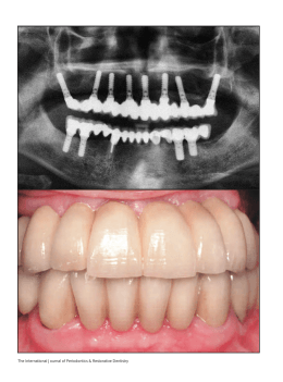
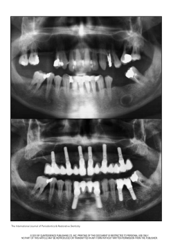
Braz J Oral Sci. October/December 2004 - Vol. 3 - Number 11
Porous surface of extraoral implants:
report of two cases rehabilitated with
a new Brazilian extraoral implant
Luciano Lauria Dib1
Joaquim Augusto Piras de Oliveira2
Renata Lazari Sandoval3
Ulf Nannmark4
1
Professor of Stomatology at UNIP, São
Paulo, Brazil.
2
Prosthodontist, São Paulo, Brazil.
3
Stomatologist, Brasília, Brazil.
4
Assistant Professor at the University of
Göteborg, Sweden.
Received for publication: June 30, 2004
Accepted: August 30, 2004
Abstract
Maxillofacial defects caused by cancer treatment are a huge problem,
affecting the quality of life of patients. Some of these deformities are
minimized using facial epistheses, which needs some additional retenion
devices, like glasses or skin adhesives. The use of extraoral fixtures as
bone anchorage was introduced many years ago and since then many
patients were rehabilitated with better results. Nevertheless, in many
cases due to poor bone conditions (i.e. irradiated bone) the success rate
of implants are not so good, causing difficulties to rehabilitate the
cases. One possible cause of fixture failure could be the poor primary
stability achieved in some cases, due to the small dimensions of the
fixtures causing a few bone contact with the surface of the implant.
Nowadays many researches are being done related to the surface of the
fixture, searching for a better primary stability and for a increase of
bone contact. Ordinary extraoral implants usually possess a machined
surface, until now there is no published report about surface
modifications in this kind of implant. This paper presents a new
porous surfaced Brazilian extraoral implant (MasterExtraâ, Conexão,
Sistema de Próteses, São Paulo, Brazil) that can provide optimal facial
rehabilitation due to enhanced bone-to-implant contact and greater
long-term stability. Two case reports are described to elucidate its use.
Key Words:
extraoral implants, maxillofacial prostheses, facial rehabilitation.
Correspondence to:
Luciano Lauria Dib
Rua Afonso Brás 525, conjunto 81.
São Paulo-SP, Brazil.
CEP: 04311-000
Phone: (11) 3842 2798
E-mail: [email protected]
633
Braz J Oral Sci. 3(11):633-638
Porous surface of extraoral implants: report of two cases rehabilitated with a new Brazilian extraoral implant
Introduction
The loss of facial structures through ablative or traumatic
activities may produce significant psychological trauma in
the patient, aggravating the actual physical loss. Although
surgical procedures, as autogenous grafting, may be able to
fill in or close off large defects, the final result may be less
than optimal in providing acceptable levels of function and
aesthetics, hence leading to a decreased quality of life to the
patient.
Prosthetic restoration of facial defects is also an option of
rehabilitation, however its success depends on satisfactory
retention and stability. The standard technique for retention
of facial prostheses has been through the use of adhesives.
Prolonged use of adhesives on the skin often leads to
inflammation of the skin and associated tissues, which
produces discomfort and an unnatural appearance of the
junction between the prosthesis and the skin, which in turn
might lead to avoidance of wearing the prosthesis. In
addition, correct positioning of the prosthesis is often
affected.
With the advent of osseointegration in dental rehabilitation1
and recent advances in surgical and laboratory techniques,
it has been possible to transfer and extend the
osseointegration principle to facial rehabilitation. Retention,
stability and aesthetics have been significantly improved
with the use of endosseous implants, resulting in more
natural appearing and functioning prostheses. In 1977, at
the Sahlgrenska Hospital, University of Göteborg, Sweden,
the first endosseous implants were installed in the temporal
bone 2 . In a later stage, a percutaneous abutment was
connected to the implant to support a bone conduction
hearing processor. In 1979, at the same institution, the first
implants were placed in the mastoid region to retain an
auricular prosthesis2. Since then, endosseous implants have
been internationally used for this, and other purposes.
To compensate for the lesser bone quantity in the temporal,
orbital and midface regions, compared to the maxilla and the
mandible, extraoral implants are shorter than intraoral
implants, 3-5 mm in length vs. 10-18 mm respectively, with a
peripheral flange. This flange will increase the implant surface
area in contact with the bone. Perforations in the flange add
additional surface area and will also provide mechanical
stability. Ordinary extraoral fixtures possess a machined
surface whereas in intraoral fixtures various surface
modifications of the titanium implant are used to increase
the mechanical interlocking, at both, microscopical and
macroscopical level, between bone and implant3. Several
different techniques i.e. particle blasting, plasma sprayed
coatings and wet chemical etching, have been used to modify
the implant surface.
In this paper we report two cases of facial rehabilitation with
the use of a new Brazilian extraoral implant with a Ti-blasted
and etched surface (MasterExtra®, Conexão, Sistema de
Próteses, São Paulo, Brazil). Until now extraoral implants were
commercially available only with a machined surface and 3
mm or 4mm long. In the present paper we describe the first
extraoral implant produced with surface modifications to
enhance implant stability. In addition, this new implant is
fabricated with more options of length, varying from 3 mm to
8 mm.
Resonance Frequency Analysis (RFA) technique was
employed to measure clinical implant stability together with
clinical examinations of function and esthetics. The resonance
frequency of a transducer attached to the implant abutment
was measured by using a frequency response analyzer, a
personal computer and dedicated software (Osstellâ,
Integration Diagnostics, Sweden). The measurements are
expressed in Implant Stability Quotients (ISQ; range 1-100).
This quotient is based on the resonance frequency of the
transducer on a scale from 3.500 Hz (0 ISQ) to 8.500 Hz (100
ISQ), and, a higher ISQ value corresponds to a higher stability.
It is considered optimal values those above 604.
Randomly selected extraoral fixtures were also examined by
scanning electron microscopy (SEM) in order to verify the
microscopical structure of the surface. These examinations
were performed in a LEO Gemini 982 analytical SEM.
Case Reports
Patient 1. A 23-year-old male was referred for evaluation of
definitive prosthetic rehabilitation with a bone-anchored
auricular prosthesis (Figure 1a). He had lost his right ear 3
years ago in a car accident. Until this clinical evaluation he
had been using a hat to hide his defect since the accident.
At the time of referral he attended a course at the university
but due to psychological difficulties of facing his colleagues
at school he stopped studying.
Using standard technique for fixture placement, 2 extraoral
implants (MasterExtra®, Conexão, Sistema de Próteses, São
Paulo, Brazil) were placed in the mastoid process (Figure
1b). The implants were left unloaded for 3 months (Figure
1c). After impression, a metallic bar was fabricated and
connected to the abutments. For the next step, magnets
were retained to the bar (Figure 1d). Magnets were sealed
into place with acrylic resin; corresponding magnets were
placed within the silicone prosthesis to permit automatic
correct positioning of the prosthesis (Figure 1e). As the
prosthesis was finalized, the patient was able to return to
school to finish his studies (Figure 1f).
At the 1-year follow-up RFA was performed. The RFA
measurements were performed at the fixture level. The ISQ
values were 76 for both fixtures indicating an optimal healing
in of the extraoral fixtures. These values correspond to values
found in this region by Heo et al.4 or actually somewhat
higher ISQ values.
There were no signs of skin irritation or ongoing infections
around the implants.
634
Braz J Oral Sci. 3(11):633-638
Porous surface of extraoral implants: report of two cases rehabilitated with a new Brazilian extraoral implant
Fig. 1a. Close-up photograph of the auricular defect.
Fig. 1d. Construction of a bar with magnets for retention of the
prosthesis.
Fig. 1b. Insertion of the fixtures in the mastoid area.
Fig. 1e. The auricular prosthesis in place.
Fig. 1c. Incision closed and the implants left unloaded.
635
Braz J Oral Sci. 3(11):633-638
Porous surface of extraoral implants: report of two cases rehabilitated with a new Brazilian extraoral implant
skin irritation or ongoing infections around the implants.
Before the rehabilitation with extraoral implants the patient
used an adhesive retained prosthesis. The use and required
daily removal of the adhesive removed her right eyebrow.
She was forced to compensate it with make-up. Today she is
very satisfied with her new implant supported prosthesis
and she has her eyebrow back.
Fig. 2a. Orbital defect.
Fig. 1f. Final aesthetic result.
Patient 2. A 28-year-old female with an orbital defect due to
tumor surgery was referred for evaluation of prosthetic
rehabilitation with a bone-anchored orbital prosthesis (Figure
2a). She had a history of retinoblastoma of the right eye,
which was treated by surgical eradication and radiotherapy
when she was 5 years old.
Using standard technique 4 extra-oral implants (MasterExtra®,
Conexão, Sistema de Próteses, São Paulo, Brazil) were placed
in the frontal bone (Figure 2b, 2c). In the orbit, usually three
implants are used to retain the prosthesis, but due to a higher
rate of implant loss in the frontal bone, and in particular in
irradiated patients, one implant was placed additionally to
compensate for potential implant loss.
After a healing-in time of 6 months the implants were exposed.
At this time, all implants were clinically well integrated but
only three implants were chosen to be exposed for retention
of the prosthesis. As in Case 1, the same type of metallic bar
with magnets was fabricated for prosthesis retention (Figure
2d, 2e).
Resonance frequency analysis, RFA, was in this case
performed 1 month after prosthesis placement. Also in this
case all measurements were performed at the fixture level.
The ISQ was 68 (Figure 2f) for the medial and lateral fixtures
and 76 for the central fixture, corresponding to well
incorporated and integrated fixtures. There were no signs of
Fig. 2b. Preparation of the holes in the orbit and implant close-up.
Fig. 2c. Insertion of 4 fixtures.
636
Braz J Oral Sci. 3(11):633-638
Porous surface of extraoral implants: report of two cases rehabilitated with a new Brazilian extraoral implant
Fig. 2d. Bar with magnets for retention of the prosthesis.
Fig. 2e. Orbital prosthesis in place.
Fig. 2f. Resonance Frequency Analysis measurement.
637
Discussion
A facial defect due to congenital or acquired deficits often
has a significant psychological impact5-6. Some methods for
facial rehabilitation have been described, but the great
majority does not restore patient’s self-confidence. Facial
rehabilitation with prostheses retained by implants has
provided a new horizon for these patients because it offers
adequate mechanical retention and accurate positioning of
the prostheses 7. The use of implants improve patient’s
quality of life, making possible participation in routine
activities and function in society with confidence that their
defect will be less noticeable. This has also been proved in
patients receiving intraoral implants for treating total
edentulousness 8.
It has been reported that the bone integration of
titanium implants is modulated by its surface
characteristics. Based on this concept, various titanium
implants with different types of surface roughness or
morphology have been developed to optimize bone
integration. The percentage of direct bone-implant
interface of a rough titanium implant is greater than
that of a smooth implant 9-10.
From the scanning electron microscopical investigations
performed in the present study, it is obvious that the surface
is greatly enhanced by the blasting and etching procedures
used (Figure 3a, 3b).
Long-term function of osseointegrated implants is
dependent on implant stability as the implant is subjected
to stresses associated with supporting, retaining and
stabilizing prosthetic restorations. The use of poroussurfaced implants in facial rehabilitation may offer an
increased implant stability and load bearing capacity.
Furthermore, it may also shorten the healing-in time of the
implants, i.e. a decreased period between fixture
installation and abutment connection (prosthetic
reconstruction).
By means of the resonance frequency analysis technique,
implant stability can be clinically measured and followed
with time. Bone loss will be detected as a decrease of the
resonance frequency, which is expressed in Implant Stability
Quotients (ISQ) varying from 1 to 100. In the present paper,
both cases revealed high ISQ values. It was measured 76 in
each fixture inserted in the auricular case. In the orbital case
the ISQ was 68 for the medial and lateral fixtures and 76 for
the central fixture. ISQ values higher than 60 indicated a
high stability at the timepoints measured4. However, RFA
measurements must be accompanied by clinical testing and
evaluation for each individual fixture in each patient.
In conclusion, the extraoral porous implant presented and
used in this study, is an excellent and promising alternative
in facial rehabilitation by means of osseointegrated implants.
New studies must be performed analyzing both experimental
and clinical long-term results.
Braz J Oral Sci. 3(11):633-638
Porous surface of extraoral implants: report of two cases rehabilitated with a new Brazilian extraoral implant
6.
7.
8.
9.
10.
Thompson A, Kent G. Adjusting to disfigurement: processes
involved in dealing with being visibly different. Clin Psychol
Rev 2001; 21: 663-82.
Arcuri MR, Rubenstein JT. Facial implants. Dent Clin North
Am 1998; 42(1): 161-75.
Trulsson U, Engstrand P, Berggren U, Nannmark U,
Branemark PI. Edentulousness and oral rehabilitation:
experiences from the patients’ perspective. Eur J Oral Sci
2002; 110: 417-24.
Trisi P, Rao W, Rebaudi A. A histometric comparison of
smooth and rough titanium implants in human low-density
jawbone. Int J Oral Maxillofac Implants 1999; 14: 689-98.
Novaes AB Jr, Souza SL, de Oliveira PT, Souza AM.
Histomorphometric analysis of the bone-implant contact
obtained with 4 different implant surface treatments placed
side by side in the dog mandible. Int J Oral Maxillofac Implants
2002; 17: 377-83.
Fig. 3a - Scanning electron micrograph of the implant surface.
Fig. 3b - Scanning electron micrograph of the implant used in the
study
Acknowledgements
The authors acknowledge the assistance of Dr. Crystianne
P. Seignemartin who was responsible for the fabrication of
the orbital prosthesis showed in this paper.
References
1.
2.
3.
4.
5.
Worthington PH, Bränemark PI. Advanced osseointegration
surgery: applications in the maxillofacial region. Chicago:
Quintessence; 1992.
Tjellström A. Osseointegrated systems and their applications
in the head and neck. Adv Otolaryngol Head Neck Surg 1989;
3: 39-70.
Suzuki K, Aoki K, Ohya K. Effects of surface roughness of
titanium implants on bone remodeling activity of femur in
rabbits. Bone 1997; 21: 507-14.
Heo SJ, Sennerby L, Odersjo M, Granstrom G, Tjellstrom A,
Meredith N. Stability measurements of craniofacial implants
by means of resonance frequency analysis. A clinical pilot
study. J Laryngol Otol 1998; 112: 537-42.
Rankin M, Borah GL. Perceived functional impact of abnormal
facial appearance. Plast Reconstr Surg 2003; 111: 2140-6.
638
Download