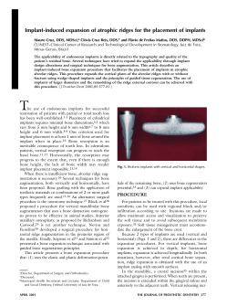
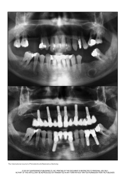
The International Journal of Periodontics & Restorative Dentistry
161
Tapered Implants:
From Indications to Advantages
Célia Coutinho Alves, DDS*
Manuel Neves, DDS**
Tapered implants have become routine for immediate implant placement after
tooth extraction. It seemed extremely advantageous to use tapered implants in
type 4 bone, where primary stability is difficult to achieve. The authors established
a surgical implant placement protocol to be followed in areas where type 4 bone
and a wide bone ridge (≥ 8 mm) are present. First, preparation of the implant alveolus is done exclusively with cylindric osteotomes, rather than with conic
osteotomes or drills. The final cylindric osteotome is the same diameter as the
final twist drill that is typically used in conventional preparation of the implant
alveolus (a technique that can be adapted for use with other tapered implants).
Because of the self-tapping property of the tapered implant used and its anatomic
design, this surgical technique was developed to optimize the bone compaction
effect in the coronal third of the implant, improving bone density and providing
better primary stability values (≥ 70 ISQ, via the Osstell Mentor device). With the
strong bond that is created between the implant surface and the surrounding
bone using this technique, immediate loading can be predictable, even in the
type 4 bone that is commonly found in the maxillary tuberosity. (Int J Periodontics
Restorative Dent 2009;29:161–167.)
*Private Practice, Porto, Portugal; Postgraduate Fellow in Periodontology, Faculty of
Dentistry, University of Porto, Portugal; Invited Assistant Master in Periodontology,
Instituto Superior de Ciéncias da Saude Norte (ISCSN), Porto, Portugal.
**Private Practice, Porto, Portugal.
Correspondence to: Célia Coutinho Alves, Clinica Medicina dentaria Dr Manuel Neves,
Rua do Amial, no. 283 r/c, 4200-060 Porto, Portugal; fax: 00351 228347769;
email: [email protected].
Several previous studies have demonstrated that individuals with a strong
susceptibility to periodontal disease
can be successfully treated with
osseointegrated implants.1–3 Implants
placed in patients with a history of periodontitis have a 5-year survival rate
similar to that observed for implants
placed in non-diseased persons.
Although the 10-year survival of 1-stage
implants is somewhat lower than that
observed in non-diseased patients,
implant placement remains a good
treatment alternative for periodontally
compromised patients.4 Immediate
implant placement into fresh extraction sites is considered to be a predictable and acceptable procedure.5–7
Advanced periodontal destruction
is often associated with extraction of
the teeth. Oral rehabilitation in these
cases may include an implantsupported reconstruction. Immediately loaded implants present an alternative treatment modality for
periodontally compromised patients
that might better meet patients’
needs.8–10 Patient desires have pushed
the clinicians toward earlier loading,
which minimizes the inconvenience of
a conventional transitional prosthesis
Volume 29, Number 2, 2009
162
a
b
c
Fig 1 Implant site preparation for TE
implant placement.
d
during the healing following extraction
and implant placement.11 Immediate
placement of dental implants at the
time of tooth extraction has yielded
favorable, predictable results; early
loading of immediately placed dental
implants has been studied and has
also been met with predictable
results.12–14 Advantages include better
bone and soft tissue preservation,
reduced postoperative pain, significant reduction of clinical chair time,
and greater patient acceptance. The
main rationale, and one of the most
important reasons for immediate
implant placement, is to preserve alveolar bone height and width. 15
Immediate placement can lead to a
favorable crown-implant ratio, better
esthetics, and a favorable maxillomandibular relationship.16,17
Implant therapy involving maxillary sinus lifting in periodontally compromised patients can be successfully done, as well as the treatment of
advanced periodontal destruction
with immediately loaded implants
and simultaneous bone augmenta-
e
tion.8,18 An implant-supported fixed
prosthesis is an acceptable and predictable treatment option for rehabilitation in patients who have lost
their teeth due to periodontal disease. This observation seems to be
valid for both edentulous and partially dentate patients.19 The literature supports immediate placement
and immediate loading in mandibles
of edentulous patients using crossarch stabilization of the implants and
a fixed passively fitting prosthesis on
multiple implants that show verifiable
primary stability upon placement.20
Immediate implant placement after
tooth extraction has been shown to
be a predictable technique.5,21–23
Several dental implant systems
have been created with tapered
implant bodies designed to simulate
the shape of the original tooth root.
Such implants are typically indicated
for situations of tooth extraction followed by immediate implant placement. For this same purpose, Institut
Straumann recently introduced the TE
(tapered effect) implant. The authors
The International Journal of Periodontics & Restorative Dentistry
have used this kind of implant in their
practice in immediate postextraction
cases and have also found them
advantageous in type 4 bone if implant
socket preparation is done with the
osteotome technique. Primary stability
seems to be a major criterion to predict success in these two special situations.24 To ensure satisfactory primary
stability, it appears that the implant
needs to be placed 3 to 5 mm beyond
the bottom of the bony alveolus.16
Some implant mobility is usually
described, even in completely healed
edentulous sites, which may be
explained by the low bone density.25
Osteotome Preparation for
TE Implant Placement in
Type 4 Bone
In addition to its primary indication for
immediate implant placement, the
authors have found that TE implants
may also be placed, with good success, in type 4 bone, as can be found
in the maxillary tuberosity, especially
163
Fig 2a (right)
Initial panoramic radiograph.
Figs 2b and 2c (below) Panoramic radiograph and lateral cephalograph, obtained after extraction and grafting, with the radiologic
guide in place.
when the bone width (at least 8 mm)
and height are sufficient. In such cases,
direct initial preparation is recommended using a 2.2-mm cylindric
osteotome without any initial mechanical drilling. This procedure compacts
the trabecular bone, and it improves
the clinician’s tactile sensation of the
presence of the posterior cortical wall
of the maxillary sinus (Fig 1).
Implant socket preparation continues following the usual osteotome
sequence, which further condenses
the bone. The final bone compaction
occurs during implant placement,
especially at the implant’s more coni-
cal coronal third. Placement of the
tapered implant is facilitated by its selftapping characteristic and minimal
thread pitch of 0.8 mm.
To illustrate the advantages of this
technique, the authors present the
case of a 60-year-old nonsmoking
male patient who needed a full-arch
maxillary rehabilitation (Fig 2a). First, all
remaining maxillary teeth were
extracted. At that time, some of the
largest bone defects and largest
extraction sockets were regenerated
with bovine mineralized bone graft
(Bio-Oss, Geistlich) and a bilayer collagen membrane (Bio-Gide, Geistlich).
Six months later, a panoramic radiograph and a lateral cephalograph
were obtained (Figs 2b and 2c) with a
radiographic guide in place. Computed
tomography was also performed; some
of the higher-density xenograft biomaterial could still be seen at the regenerated sites (Figs 2d to 2f). The computed tomography showed the
presence of a wide and high maxillary
tuberosity, with low-density bone and a
low maxillary sinus, the mesial wall of
which continued to a wide and high
alveolar region in the premaxillary
region. These anatomic characteristics
were present on both sides of the arch.
Volume 29, Number 2, 2009
164
Figs 2d to 2f Computed tomograms (from posterior to anterior) of
the right quadrant.
Without elevating a flap, eight circular incisions were made according to
a surgical guide that had been previously fabricated. Six standard
Straumann implants (4.1 mm diameter,
12 mm length; Standard Plus) and two
Straumann TE implants (4.1 mm and
4.8 mm diameter, 14 mm length) were
placed. The two TE implants were
placed in the maxillary tuberosity following the surgical technique previously described (Fig 2g). A panoramic
radiograph obtained immediately
after surgery showed the angulated
placement (about 30 degrees) of the
implants in the tuberosity, which was
done to prevent sinus perforation
(Fig 2h). Resonance frequency tests
(Fig 2i) (Osstell Mentor, Straumann)
were performed on the two implants
placed in the maxillary tuberosities.
These showed ISQ values of 71 and 73
(first and second quadrants, respectively). As is known from the literature,26 an ISQ value of ≥ 70 indicates
a good bond between the implant
The International Journal of Periodontics & Restorative Dentistry
and surrounding bone, making early
implant loading a more predicable
treatment option.
All eight implants were loaded
within 24 hours after surgery with a
provisional full-arch fixed prosthesis
(Figs 2j to 2l). The definitive implantsupported porcelain-fused-to-metal
fixed prosthesis was delivered 6
months later. None of the eight
implants placed in the maxilla were
lost, even though two of them were
placed in type 4 bone and immediately
165
Fig 2g Surgical phase showing six standard implants and two TE implants in place.
Fig 2h
Fig 2i Resonance frequency testing with
the Osstell Mentor device was performed
on the TE implants.
Figs 2j and 2k
Panoramic radiograph taken immediately after surgery.
Provisional full-arch fixed prostheses.
Fig 2l Panoramic radiograph with provisional prosthesis in place
(acrylic is radiotranslucent).
Fig 2m
Panoramic radiograph with definitive prosthesis in place.
Volume 29, Number 2, 2009
166
Fig 2n
Definitive porcelain-fused-to-metal full-arch fixed prosthesis.
Fig 2p
Periapical radiographs obtained at the 3-year follow-up.
loaded (Figs 2m to 2o). Periapical
radiographs obtained at a 3-year
follow-up appointment showed
acceptable bone levels (Fig 2p).
Conclusion
In addition to its primary indication for
postextraction placement, the authors
suggest that tapered implants can be
used successfully in type 4 bone in the
maxillary tuberosities if socket preparation is done exclusively with
osteotomes. More studies should be
done to confirm this technique.
Fig 2o
Extraoral view of definitive prosthesis.
References
1. Nevins M, Langer B. The successful use of
osseointegrated implants for the treatment
of the recalcitrant periodontal patient. J
Periodontol 1995;66:150–157.
2. Ellegaard B, Baelum V, Karring T. Implant
therapy in periodontally compromised
patients. Clin Oral Implants Res 1997;8:
180–188.
3. Wennström JL, Ekestubbe A, Gröndahl K,
Karlsson S, Lindhe J. Oral rehabilitation
with implant-supported fixed partial dentures in periodontitis-susceptible subjects.
A 5-year prospective study. J Clin
Periodontol 2004;31:713–724.
4. Baelum V, Ellegaard B. Implant survival in
periodontally compromised patients.
J Periodontol 2004 Oct;75:1404–1412.
The International Journal of Periodontics & Restorative Dentistry
5. Becker W, Dahlin C, Becker BE, et al. The
use of e-PTFE barrier membranes for bone
promotion around titanium implants
placed into extraction sockets: A prospective multicenter study. Int J Oral Maxillofac
Implants 1994;9:31–40.
6. Schwartz-Arad D, Gulayey N, Chaushu G.
Immediate versus non-immediate implantation for full-arch fixed reconstruction following extraction of all residual teeth: A
retrospective comparative study.
J Periodontol 2000;71:923–928.
7. Rosenquist B, Grenthe B. Immediate
placement of implants into extraction sockets: Implant survival. Int J Oral Maxillofac
Implants 1996;11:205–209.
167
8. Romanos GE. Treatment of advanced periodontal destruction with immediately
loaded implants and simultaneous bone
augmentation: A case report. J Periodontol
2003 Feb;74:255–261.
9. Evian CI, Emling R, Rosenberg ES, et al.
Retrospective analysis of implant survival
and the influence of periodontal disease
and immediate placement on long-term
results. Int J Oral Maxillofac Implants
2004;19:393–398.
10. Karoussis IK, Salvi GE, Heitz-Mayfield LJ,
Brägger U, Hämmerle CH, Lang NP. Longterm implant prognosis in patients with
and without a history of chronic periodontitis: A 10-year prospective cohort study of
the ITI Dental Implant System. Clin Oral
Implants Res 2003;14:329–339.
11. Maló P, Rangert B, Nobre M. “All-on-Four”
immediate-function concept with
Brånemark System implants for completely
edentulous mandibles: A retrospective
clinical study. Clin Implant Dent Relat Res
2003;5(suppl 1):2–9.
12. Ericsson I, Randow K, Nilner K, Peterson A.
Early functional loading of Brånemark dental implants: 5-year clinical follow-up study.
Clin Implant Dent Relat Res 2000;2:70–77.
13. Tarnow DP, Emtiaz S, Classi A. Immediate
loading of threaded implants at stage 1
surgery in edentulous arches: Ten consecutive case reports with 1- to 5-year data. Int
J Oral Maxillofac Implants 1997;12:
319–324.
14. Nikellis I, Levi A, Nicolopoulos C.
Immediate loading of 190 endosseous
dental implants: A prospective observational study of 40 patients with up to 2-year
data. Int J Oral Maxillofac Implants
2004;19:116–123.
15. Schwartz-Arad D, Yaniv Y, Levin L, Kaffe I.
A radiographic evaluation of cervical bone
loss associated with immediate and
delayed implants placed for fixed restorations in edentulous jaws. J Periodontol
2004;75:652–657.
17. Orenstein IH, Petrazzuolo V. A new angle
on restoring anterior teeth with root-form
implants: Clinical report. Implant Soc
1993;4:10–11,16.
18. Ellegaard B, Kølsen-Petersen J, Baelum V.
Implant therapy involving maxillary sinus lift
in periodontally compromised patients. Clin
Oral Implants Res 1997;8:305–315.
19. Yi SW, Ericsson I, Kim CK, Carlsson GE,
Nilner K. Implant-supported fixed prostheses for the rehabilitation of periodontally compromised dentitions: A 3-year
prospective clinical study. Clin Implant
Dent Relat Res 2001;3:125–134.
20. Chiapasco M. Early and immediate
restoration and loading of implants in completely edentulous patients. Int J Oral
Maxillofac Implants 2004;19(suppl):76–91.
21. Barzilay I. Immediate implants: Their current status. Int J Prosthodont 1993;
6:169–175.
22. Krump JL, Barnett BG. The immediate
implant: A treatment alternative. Int J Oral
Maxillofac Implants 1991;6:19–23.
23. Lazzara RJ. Immediate implant placement
into extraction sites: Surgical and restorative advantages. Int J Periodontics
Restorative Dent 1989;9:332–343.
24. Henry PJ, Tan AE, Leavy J, Johansson CB,
Albrektsson T. Tissue regeneration in bony
defects adjacent to immediately loaded
titanium implants placed into extraction
sockets: A study in dogs. Int J Oral
Maxillofac Implants 1997;12:758–766.
25. Lekholm U, Zarb GA. Patient selection and
preparation. In: Brånemark P-I, Zarb GA,
Albrektsson T. Tissue-Integrated Prostheses:
Osseointegration in Clinical Dentistry.
Chicago: Quintessence, 1985:199–209.
26. Kondo H, Ito Y, et al. Application of resonance frequency analysis to hydroxyapatite coated ABQ implant. Hawaii convention, March.
16. Schwartz-Arad D, Chaushu G. The ways
and wherefores of immediate placement
of implants into fresh extraction sites: A literature review. J Periodontol 1997;68:
915–923.
Volume 29, Number 2, 2009
Download