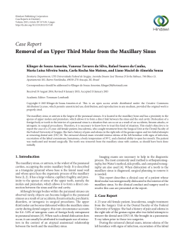
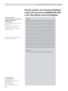
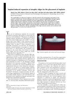
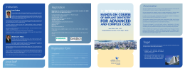
R. Periodontia - Março 2010 - Volume 20 - Número 01
SINUS FLOOR ELEVATION ASSOCIATED WITH DENTAL
IMPLANTS IN ONE OR TWO SURGICAL STAGES - A
REVIEW
Elevação de seios maxilares associadas a implantes dentários em um ou dois estágios cirúrgicos.
Revisão de Literatura
Diogo Godoy Zanicotti1, João César Zielak2, Allan Fernando Giovanini3, Cícero de Andrade Urban4, Tatiana Miranda
Deliberador5
RESUMO
The sinus floor elevation technique has been used in
an important way on partially or fully edentulous maxillas.
This procedure permits the placement of dental implants
whereas the residual bone height is insufficient. The
presence of a reabsorbed bone crest with the impossibility
of a dental implant placement requires an intervention on
the maxillary sinus, with the proposal to increase the bone
quantity for the implant placement. It is possible to do these
procedures in one or two surgical stages according to the
technique and implant placement. The objective of this
study was to review the literature about the sinus
augmentation techniques, lateral window or osteotomes,
and the placement of dental implants in one or two surgical
stages. There were obtained clinical studies and literature
review studies by internet search, MEDLINE and text books.
These studies were analyzed and placed in an analytical
review. Aspects relative to anatomy, access the maxillary
sinus, placement of the implants in one or two surgical
stages, implant survival in grafted sinus, residual bone
amount, graft materials and surgical complications were
analyzed.
UNITERMOS: bone transplantation; bone substitutes;
dental implantation, endosseous; maxillary sinus; osseous
substitutes. R Periodontia 2010; 20:22-29.
1
Mestrando em Odontologia Clínica, Universidade Positivo, Curitiba, PR, Brasil
2
Professor Doutor do Mestrado em Odontologia Clínica, Universidade Positivo, Curitiba, PR, Brasil
3
Professor Doutor do Mestrado em Odontologia Clínica, Universidade Positivo, Curitiba, PR, Brasil
4
Professor Doutor do Mestrado em Odontologia Clínica, Universidade Positivo, Curitiba, PR, Brasil
5
Professor Doutor do Mestrado em Odontologia Clínica, Universidade Positivo, Curitiba, PR, Brasil
Recebimento: 14/07/09 - Correção: 16/09/09 - Aceite: 01/12/09
INTRODUÇÃO
The maxillary sinuses impose an important
influence on partially or fully edentulous maxillary
rehabilitations with dental implants. In a rehabilitation
process with implants the space to place a dental
prosthesis over an implant may be jeopardized by
the remaining space between the dental arches.
Because of this small space, an alveolar crest onlay
bone graft is unlikable to be made. However if a bone
graft is placed between the sinus membrane and the
sinus floor, it is possible to improve the osseous
thickness of the alveolar crest without compromising
the space to place a prosthetic tooth (Sendyk &
Sendyk, 2002).
The presence of a bone crest with a reduced
bone height demands an inter vention on the
maxillary sinus with the purpose to increase the bone
amount available to implant insertion (Gulherme et
al, 2009). The surgical procedure can be executed in
one or two surgical stages when related to the
implant insertion. Since the first publication about
this kind of surgery by Boyne & James (1980) various
materials and techniques has been used for sinus lift
procedures (Wallace, 2006; Van der Bergh et al, 2000).
There are some techniques to access the maxillary
sinus, the lateral window technique, the osteotome
22
periomar2010 06-08-10.pmd
22
8/6/2010, 10:28 AM
R. Periodontia - 20(1):22-29
technique and some modifications of these techniques
(Wallace, 2006).
Different materials are described on the literature to
improve the bone volume on this region. Autogenous bone,
bovine bone, human tissue bank bone, synthetics bone
substitutes as calcium sulfate, bioactive glass, hidroxyapatite
and an association of these materials are used for (De
Leonardis & Pecora, 2000; Cordioli et al, 2001; Xu et al, 2003;
Chen & Cha, 2005; Wallace et al, 2005; Chiapasco et al, 2006;
Marin et al, 2007).
The objective of this study was to review the literature
about the sinus augmentation techniques, lateral window
or osteotomes, and the placement of dental implants in one
or two surgical stages. There were obtained clinical studies
and literature review studies by internet search, MEDLINE
and text books. These studies were analyzed and placed in
an analytical review. Aspects relative to anatomy, access the
maxillary sinus, placement of the implants in one or two
surgical stages, implant survival in grafted sinus, residual bone
amount, graft materials and surgical complications were
analyzed.
LITERATURE REVIEW
Anatomy
The maxillary sinuses are pneumatic compartments of
the paranasal sinuses group, witch includes the etmoidal,
frontal and esphenoidal sinuses. Some plausible functions
might be warm and humidify air, reduction of the skull weight,
act like a resonance space and retain mucus (Figún & Garrino,
1994).
The maxillary sinus in the adult can be compared to
a quadrangular triangle with its base at the lateral nasal
wall and its apex extending to the zygomatic process.
The volume of the maxillary sinus range from 2 and 25 cm3
with a mean size range from 8 to 12 cm3 (Figún & Garrino,
1994).
Between the structures presented in the maxillary
sinuses, the sinus septa and the Schneiderian membrane
(sinus membrane) play an important role when surgically
managing the sinus. These membrane, which is consisted
by a ciliated pseudo stratified epithelium, is delicate and needs
a carefully manipulation to avoid ruptures. The osseous septas
influences the management of this membrane, because it is
going to make harder the membrane freeing. This happens
because the septa acts like a physical barrier to the curettes
(Kim et al, 2006). In one study it was find an average of 26,5%
septas in 200 studied maxillary sinuses. These septas were
Figure 1: schematic drawing of the lateral window technique.
distributed in the anterior, medium and posterior regions
being 25,4%, 50,8% and 23,7% respectively. The same
authors identified a variation between 0 and 20,18 mm in
the sizes of the septas studied (Kim et al, 2006).
Regarding the maxillary sinus anatomy there is an
important phenomena that occurs after dental losses. The
pneumatization is the process that explains most variable
sizes of the sinus. Bone density and thickness can be explain,
in part, by the chewing forces applied. The pneumatization
process is likely an architectural response to the muscles and
chewing forces, in agreement to the “Wolff’s Law” (“law of
use and unused”) (Magini & Coura, 2006).
Disease and abnormalities of the maxillary sinuses
Beaumont et al. (2005) determined the prevalence of
disease related to maxillary sinuses in patients scheduled to
sinus lift procedures. This study showed that 40% of all
individuals had some disease and/or abnormality in yours
sinuses. The methods to identify these problems were
tomographies and endoscopies. A great diversity of disease
was founded. The most common conditions were chronic
sinusitis and sinus cysts, presented as 77% and 72% of the
patients respectively. It was concluded that after solve those
conditions, a sinus floor elevation is predictable. These
findings point the importance of a detailed medical history
and a clinical and radiographical evaluation. The thickness of
the sinus membrane is not affected by the sinus
augmentation procedure and this surgery does not lead to
a chronic or acute sinusitis (Peleg et al, 1999).
Techniques to access the maxillary sinus
The lateral window technique (figs. 1 and 2) begins with
a mucoperiosteal flap to access the lateral wall of the sinus.
A lateral window is performed with diamond burs (with
23
periomar2010 06-08-10.pmd
23
8/6/2010, 10:28 AM
R. Periodontia - 20(1):22-29
Figure 2: schematic drawing of the lateral window technique.
Figure 3: schematic drawing of the osteotome technique.
surgical drill or high speed hand piece) or piezoelectric burs.
The residual bone of the lateral window can be elevated
towards the sinus or completely removed. The sinus
membrane is elevated superiorly to the medial sinus wall.
Therefore the membrane became the superior and distal wall
of the compartment that will receive the osseous graft. After
positioning the graft, the lateral window is covered with a
biological membrane and the flap is sutured in position
(Wallace, 2006). The literature review developed by
Fugazzotto (2003) shows that the lateral window technique
is highly predictable.
Summers proposed a simpler and less invasive way to
increase the bone quantity at the sinus floor where, with
the use osteotomes (figs. 3 and 4), the inferior wall is elevated
into the maxillary sinus and an osseous graft it is or it is not
placed inside the space created (Fugazzotto, 2003). Two
studies, a retrospective and a prospective respectively, report
that the osteotome technique for sinus augmentation is
predictable, less invasive and without statistically significant
differences in relation to the lateral window technique
(Wallace, 2006; Ozyuvaci et al, 2003). In a systematic review
and meta-analisys the authors concluded that implants
placed in a previously elevated maxillar y sinus with
osteotomes had similar success rates to implants inserted
conventionally in a period of 3 years (Emmerich et al, 2005).
Sendyk & Sendyk (2002) indicates that the osteotome
technique has to be use when the sinus elevation does not
exceed 5 mm. When this amount is not sufficient, the lateral
window technique should be the professional selection.
Graft materials
The graft materials are fundamental important for the
prognosis since different materials have different grades of
osteogenesis. For this reason most surgeons prefer to use
autogenous grafts as theirs fist choice for osseous grafting
or a mixture of autogenous and biomaterial bone grafts for
sinus surgeries (Sendyk et al, 2004).
The osteoconductive capability of different particle sizes
from deproteinized bone was analyzed in an animal model
with rabbits. It was concluded that small particles with size
var ying between 300 and 500 ìm showed a better
osteoconduction than bigger particles with sizes ranging
between 850 and 1000 ìm (Xu et al, 2003). The evaluation
with computed tomography scanning performed by
Murakami et al. (1999) showed that implants placed in
maxillary sinus augmented with autogenous bone remained
stable after 12 months.
In an 8 years retrospective study, a combination of
osseous grafts were studied, which includes a demineralized
freeze-dried bone matrix, a small-particle spherically shaped
peptide-coated product (Pep-Gen P-15, Dentsply Friadent
Ceramed) and bovine-derided mineralized bone materials.
The success rate reached with the placement of implants
after 8 years was 99,27% (Chen & Cha, 2005). A histological
evaluation in a case report study, where a bovine graft was
performed, observed the presence of newly formed bone,
particles of the graft material, connective fibrous tissue and
giant multinucleated cells (promoting a graft particles
reabsoption) in the augmented maxillary sinus. These findings
did not influence the success of the implant maintenance
after 6 month of the graft procedure and after 2 years of
implant maintained in function (Marin et al, 2007).
Cordioli et al. (2001) used bioactive glass and autogenous
bone in surgeries for maxillary sinus augmentation with
simultaneous implant insertion. These authors concluded
that the use of these materials is safe and predictable in
relation to a simultaneous implant insertion. The using of
calcium sulfate as grafting material was evaluated in a
24
periomar2010 06-08-10.pmd
24
8/6/2010, 10:28 AM
R. Periodontia - 20(1):22-29
prospective study, which had a success rate of 98,5% in the
implant maintenance af ter 1 year. The histological
examination of the specimens analyzed showed 100% of
living bone without graft remnants after a period of 6 or 9
months (De Leonardis & Pecora, 2000).
In a study evolving 101 implants placed in 36 augmented
maxillary sinuses, with an association of PRP (platelet-rich
plasma) and bovine inorganic bone, Gonçalves et al. (2008)
obtained an implant success rate of 90,09%. The authors
suggest that implants placed in maxillary sinuses, augmented
with an association of PRP and bovine inorganic bone, show
a great success rate.
A literature review demonstrated that any kind of graft
material used does not influence in a significantly matter the
survival rates of placed implants in augmented maxillary sinus.
The survival rates of implants inserted in augmented sinuses
with autogenous bone, allografts, xenografts, alloplastic
materials and a mixtures of these materials were 89%, 93,4%,
95,5%, 98,4% and 93,8% respectively (Chiapasco et al, 2006).
Tepper et al. (2002) indicate the graft packing to reduce
implant displacement, independently of the technique or
graft material used.
The difference in osseous formation within the grafted
sinuses with mineralized bovine bone associated or not to
absorbable or non absorbable membranes placed over the
lateral window was studied by Wallace et al. (2005) It were
found differences in the histomorphometric analyses revealing
that the groups with membranes showed a greater newly
bone formation of 30% and in the group without membrane
14%. In according to these results the authors suggest the
placement of membranes (absorbable or non absorbable) at
the lateral window, although the success rates of the implants
were 97,8% for the membrane group (n = 129) and 100%
for the group without membrane (n = 6).
The dimensional stability of different graft materials after
sinus floor augmentation was studied with computed
tomography scans. After 6 months changes occur with a
dimensional mean loss of 26% for the grafted material
(Kirmeier et al, 2008).
Figure 4: schematic drawing of the osteotome technique.
by a lateral window approach associated with an implant
insertion, it is necessary at minimum 4 mm of residual bone
height to support the implant. If the residual bone height is
less than 4 mm the placement of implant should be done in
a second surgical approach 6 to 8 month after the grafting
procedure. Some formulas were addressed to facilitate the
clinician’s decisions. In the case of a residual bone height of
2x-2 (x represent the residual bone height coronal to the
sinus floor at the time of therapy) it is sufficient to support
implant, the sinus floor elevation with osteotomes and
immediate implant placement can be done. If 2x-2 is
insufficient to support implant but 4x-6 is adequate, the sinus
floor elevation with osteotomes can be performed and
allowed to heal. After 12 months the area can be reentered
with a new osteotome approach and simultaneous implant
placement. If 2x-2 and 4x-6 are both insufficient to retain
the implant, a lateral window surgery is carried out and the
area is reentered after 6 to 8 months for implant insertion.
In a retrospective study 1.557 implants placed in 1.100
patients were evaluated. There was no correlation between
implant failure and the method used to elevate the maxillary
sinus floor (osteotome or lateral window techniques). In the
same study the success rate for implantation was 99,27%,
where only 8 implants failed during the osseointegration fase
(Chen & Cha, 2005).
Implant placement in 1 or 2 surgical stages
Surgical and post-surgical complications
The immediate placement of implants after a graft
procedure is not indicated when the residual bone height is
less than 4 to 5 mm and in case of a poor quality of bone
(Chiapasco et al, 2006). Fugazzotto (2003) proposed a
hierarchy of treatment selection for sinus augmentations after
a critical analysis of the literature. This author concluded that
if the sinus augmentation procedure is going to be performed
Schwartz-Arad et al. (2004) verified in their study that
perforation of the Schneiderian membrane was the major
intra-operative complication during a sinus graft procedure.
In study made by Hernández-Alfaro et al. (2008) the
prevalence of membrane perforations was 25,15% with an
implant survival rate of 97,14% in the membrane perforation
25
periomar2010 06-08-10.pmd
25
8/6/2010, 10:28 AM
R. Periodontia - 20(1):22-29
group. Fugazzotto & Vlassis (2003) describes the
management of the membrane perforations using plateletrich plasma (PRP), bioabsorbable porcine membrane or
synthetic membranes. The use of bioabsorbable sutures prior to the bioabsorbable membrane placement is also
described for membrane per forations management
(Karabuda et al, 2006). The occurrence of different postoperatory complications like acute sinus inflammation,
persistent infection around the implant and the presence cyst
associated to the buccal surface of the implant can be found.
In the study where these problems occurred it was a strong
association between the membranes perforations and postoperative complications (P < 0,001). However, there were
no correlations between post-operative complications and
implant survival (Schwartz-Arad et al, 2004).
DISCUSSION
The success of maxillary sinus augmentation procedures
for the placement of implants is well documented in the
literature. However, some anatomic and surgical conditions
have to be considered. The presence of sinus septa can
obstruct the Schneiderian membrane displacement (Van der
Bergh et al, 2000). The membrane perforations are the
major intra-operative complication and it is related to
the manipulation of these membranes by techniques to
access the maxillary sinus (Hernández-Alfaro et al, 2008). The
sinus membrane is fragile and a delicate approach should
prevail, which can be achieved with a less invasive surgical
procedure.
A point to be considered in the techniques choice
and in the immediate or later insertion of the implant is the
quantity of native residual alveolar bone which will retain
the implant. At least 4 mm of residual bone is important for
implant placement (Wallace, 2006; Chiapasco et al, 2006).
On the other hand, for Mardinger et al. (2007) this value
was established by the literature by an arbitrary way. In a
study performed in humans, the authors compared two
groups. In the test group 25 patients presented 1 to 3 mm
of residual bone and in the control group 30 patients
presented more than 4 mm of native bone prior to the
surgery. The results showed 92% of success rate for the tested
group and 98,7% for the control group with statistically
significant difference (P < 0,069). The technique used was
lateral window with simultaneous implant insertion, where
the residual alveolar bone range between 1 to 3 mm. For
the authors this procedure can be performed, in a predictable
way, if some planning and performing cares, when doing
the surgical procedure, are taken (Mardinger et al, 2007).
Although, it seems that a minimum 4 mm height of native
bone is safer, because it can promote grater implant stability.
This aspect is also relevant when choosing the operatory
technique, because it has influence in the osteotome
technique when augmenting the sinus (Chiapasco et al,
2006).
The osteotome technique is used when the residual bone
height is equal or more than 4 mm and the lateral window
technique is indicated when the residual bone height is less
than 4 mm (Fugazzotto, 2003). Those factors
aforementioned, technique selection and implant placement
in one or two surgical stages seem to be correlated. This
happens because of the direct relationship among implant
insertion, osteotome technique and the need of implant
stabilization. The use of osteotomes suggest a minor
aggression to the maxillary sinus according to Ioannidou &
Dean (2000) however the lateral window seems to be more
appropriated when the residual bone is less than 4 mm
according to the reports of Fugazzotto (2003)
abovementioned.
The type of graft placed in a grafting procedure seems
not to have influence on the success of implants placed in
grafted sinuses, according to the broad literature where most
graft materials were used (Chiapasco et al, 2006). Therefore,
is important to remember that Wallace et al. (2005) suggest
the use of absorbable or non-absorbable membranes over
the lateral window access to increase the vital bone
formation. The implant survival rate aforementioned for this
study (97,8% for the membrane group and 100,0% for the
group without membrane) is similar for both groups.
However, this result should be considered with precaution
because of the great difference in the number of implants
placed in the groups (n = 6 in the group without membrane
and n = 129 in the group with membrane).
The Schneiderian membrane perforations do not
influence the survival rates of implants (Schwartz-Arad et al,
2004; Hernández-Alfaro et al, 2008; Fugazzotto & Vlassis,
2003). These results should be related to the management
of perforations with absorbable sutures and/or the placement
of absorbable or non-absorbable membranes, by this way
reestablishing the superior wall of the grafted cavity and
protecting the perforated membrane.
The implant survival rate inserted in grafted maxillary
sinuses is high ranging between 90 to 100% according to
the paper analyzed. This success is non dependent on the
surgical technique, graft material and the moment to place
the implant. These findings are correlated with those found
by Olson et al. (2000) which presented by a multicenter,
26
periomar2010 06-08-10.pmd
26
8/6/2010, 10:28 AM
R. Periodontia - 20(1):22-29
prospective and randomized study, a survival rate of 97,5%
from 120 implants inserted by many techniques, graft
materials and timing to place the implants. Gonçalves et al.
(2008) also corroborate with this findings showing an implant
success rate of 90,09% in implants placed in maxillary sinus
grafted with a biomaterial of bovine origin and platelet-rich
plasma.
As Guilherme et al. (2009) demonstrated in a studied
questioning 84 patients, after sinus lifting procedures
concluded that in despite of the traumatic feeling, the
patients present a better quality of life.
CONCLUSION
The literature shows a lack of evidence-based in
prospective, randomized and controlled studies, determining
which is the most indicate technique for the major clinical
situations presented in this paper.
Within the limits of this analytical character review, is
shown that sinus augmentation procedures, which are
represented by the lateral window technique and the
osteotome technique, are indicate for implant insertion in
one or two surgical stages. This is provided since all the
anatomical and surgical aspects that involves sinus grafting
procedures, with the objective of bone augmentation in the
maxillary sinus, are respected.
The authors thanks to Dr. Edson and Dr. Carla, both
Professors of the Professional Masters Program in Clinical
Dentistry, Positivo University for English reviewing this article.
RESUMO
As cirurgias de elevação dos seios maxilares vêm sendo
utilizadas de maneira importante na reabilitação de maxilas
edêntulas parciais ou totais com implantes dentais. A presença de um rebordo alveolar reabsorvido, que impossibilite
a colocação de um implante, exige uma intervenção no seio
maxilar com o propósito de aumentar a quantidade de osso
disponível para colocação de implantes. As cirurgias de levantamento de seio maxilar vêm sendo utilizadas com este
propósito. Dentre as técnicas de elevação do assoalho de
seio maxilar existem as de um e dois estágios cirúrgicos com
relação à colocação de implantes. O objetivo deste estudo
foi realizar uma revisão de literatura sobre a técnica de acesso lateral e a técnica com uso de osteótomos para elevação
da parede inferior do seio maxilar e a colocação de implantes em um ou dois estágios cirúrgicos. Foram obtidos estudos clínicos e revisões de literatura através de ferramentas
de busca na internet e base de dados MEDLINE; além de
livros texto. Estes trabalhos foram analisados e dispostos em
uma revisão analítica da literatura. Aspectos relativos ao acesso do seio maxilar, colocação de implantes em um e dois
estágios, estabilidade dos implantes em seios maxilares enxertados, quantidade de osso remanescente, materiais de
enxerto, complicações cirúrgicas, anatomia do seio maxilar
e complicações decorrentes do ato cirúrgico foram
analisados.
UNITERMS: transplante ósseo; substitutos ósseos; implante dentário endoósseo; seio maxilar; substitutos ósseos.
27
periomar2010 06-08-10.pmd
27
8/6/2010, 10:28 AM
R. Periodontia - 20(1):22-29
REFERÊNCIAS BIBLIOGRÁFICAS
1- Guilherme A S, Zavanelli R A, Fernandes J M A, Castro A T, Barros C A,
Souza J E A, Cozac C D, Santos V A. Implantes osseointegráveis em
áreas com levantamento do seio maxilar e enxertos ósseos. RGO
2009;57(2):157-163.
2- Sendyk W R, Sendyk C L. Reconstrução óssea por meio do levantamento
do assoalho do seio maxilar. In: Gomes L A, editor. Implantes
osseointegrados: técnica e arte. São Paulo: Livraria Santos Editora
2002:109-22.
3- Boyne P J, James R A. Grafting of the maxillary sinus floor with
autogenous marrow and bone. J Oral Surg 1980;38:613-616.
4- Wallace S S. Maxillary Sinus Augmentation: Evidence-Based Decision
Making with a Biological Surgical Approach. Compendium
2006;27(12):662-669.
5- Van der Bergh J P A, ten Bruggenkate C M, Disch F J M, Tuinzing D B.
Anatomical aspects of sinus floor elevations. Clin Oral Imp Res
11,2000;256-265.
6- Xu H, Shimizu Y, Asai S, Ooya, K. Experimental sinus grafting with the
use of deproteinized bone particles of different sizes. Clin Oral Impl
Res 14,2003:548-555.
7- Chen L, Cha J. An 8-year retrospective study: 1.100 patients receiving
1.557 implants using the minimally invasive hydraulic sinus condensing
technique. J Periodontol 2005;76:482-491.
8- Marin C, Granato R, Claus J D P, Rivero E R C, Gil J N. Histological
evaluation of inorganic bovine bone graft in maxillary sinus: a case
report. Rev Cir Traumatol Buco-Maxilo-fac 2007;7(1):37-42.
9- Cordioli G, Mazzocco C, Schepers E, Brugnolo E, Majzoub Z. Maxillary
sinus floor augmentation using bioactive glass granules and autogenous
bone with simultaneous implant placement. Clinical and histological
findings. Clin Oral Impl Res 2001;12:270-278.
15- Kim M-J, Jung U-W, Kim C-S, Kim K-D, Choi S-H, Kim C-K, Cho K-S.
Maxillary sinus septa: Prevalence, height, location, and morphology. A
reformatted computed tomography. J Periodontol 2006;77:903-908.
16- Magini R S, Coura G S. Anatomia e fisiologia do seio maxilar. In: Magini
R S, editor. Enxerto ósseo no seio maxilar. Estética e função. São Paulo:
Livraria Editora Santos 2006:17-35.
17- Beaumont C, Zafiropoulos G-G, Rohmann K, Tatakis D N. Prevalence
of maxillary sinus disease and abnormalities in patients scheduled for
sinus lift procedures. J Periodontol 2005;76:461-467.
18- Peleg M, Chaushu G, Mazor Ziv, Ardekian L, Bakoon M. Radiological
findings of the post-sinus lift maxillary sinus: Acomputerized
tomography follow-up. J Periodontol 1999;70:1564-1573.
19- Fugazzotto P A. Augmentation of the posterior maxilla: A proposed
hierarchy of treatment selection. J Periodontol 2003;74:1682-1691.
20- Ozyuvaci H, Bilgiç B, Firatli E. Radiologic and histomorphometric
evaluation of maxillary sinus grafting with alloplastic graft materials. J
Periodontol 2003;74:909-915.
21- Emmerich D, Att W, Stappert C. Sinus floor elevation using osteotomes:
A systematic review and meta-analysis. J Periodontol 2005;76:12371251.
22- Sendyk W R, Sendyk C L, Jahn R S. Enxertos ósseos para reconstrução
da maxilla posterior atrófica. In: Querido M R M, Gomes Y L F, editores.
Implantes osseointegrados: inovando soluções. São Paulo: Artes
Médicas 2004:115-37.
23- Murakami K, Itoh T, Watanabe S, Naito T, Yokota M. Periodontal and
computer tomography scanning evaluation of endosseous implants in
conjunction with sinus lift procedure. A 6 - case series. J Periodontol
1999;70:1254-1259.
10- De Leonardis D, Pecora G E. Prospective study on the augmentation
of the maxillary sinus with calcium sulfate: Histological results. J
Periodontol 2000;71:940-947.
24- Tepper G, Haas R, Zechner W, Krach W, Watzek G. Three-dimensional
finite element analysis of implant stability in the atrophic posterior
maxilla. A mathematical study of the sinus floor augmentation. Clin
Oral Impl Res 13,2002;657-665.
11- Gonçalves A R Q, Maior C M V, Gigli R E, Motta S H G. Avaliação do
sucesso de implantes osseointegráveis em enxerto de seio maxilar. RGO
2008;56(4):423-427.
25- Kirmeier R, Payer M, Wehrschuetz M, Jakse N, Platzer S, Lorenzoni M.
Evaluation of three-dimensional changes after sinus floor augmentation
with different grafting materials. Clin Oral Impl Res 19,2008;366-372.
12- Chiapasco M, Zaniboni M, Boisco M. Augmentation procedures for
the rehabilitation of deficient edentulous ridge with oral implants. Clin
Oral Impl Res 2006;17(suppl. 2):136-159.
26- Schwartz-Arad D, Herzberg R, Dolev E. The prevalence of surgical
complications of the sinus graft procedures and their impact on implant
survival. J Periodontol 2004;75:511-516.
13- Wallace S S, Froum S J, Cho S-C, Elian N, Monteiro D, Kim B S, Tamo
D P. Sinus Augmentation Utilizing Anorganic Bovine Bone (Bio-Oss)
with Absorbable and Nonabsorbable Membranes Placed over the Lateral
Window: Histomorphometric and Clinical Analyses. Int J Periodontics
Restorative Dent, 2005;25:551-559.
27- Hernández-Alfaro F, Torradeflot M M, Marti C. Prevalence and
management of Schneiderian membrane perforations during sinus-lift
procedures. Clin Oral Impl Res 19,2008;91-98.
14- Figún M E, Garrino R R. Anatomia odontológica funcional e aplicada.
São Paulo: Editora Médica Panamericana 1994:487-496.
28- Fugazzotto P A, Vlassis J. A simplified classification and repair system
for sinus membrane perforations. J Periodontol 2003;74:1534-1541.
29- Karabuda C, Arisan V, Özyuvaci H. Effects of sinus membrane
28
periomar2010 06-08-10.pmd
28
8/6/2010, 10:28 AM
R. Periodontia - 20(1):22-29
perforations on the success of dental implants placed in the augmented
sinus. J Periodontol 2006;77:1991-1997.
30- Mardinger O, Nissan J, Chaushu G. Sinus floor augmentation with
simultaneous implant placement in the severely atrophic maxilla:
Technical problems and complications. J Periodontol 2007;78:18721877.
31- Ioannidou E, Dean J W. Osteotome sinus floor elevation and
simultaneous, non-submerged implant placement: Case report and
literature review. J Periodontol 2000;71:1613-1619.
32- Olson J W, Dent C D, Morris H F, Ochi S. Long-term assessment (5 to
71 months) of endosseous dental implants placed in the augmented
maxillary sinus. J Periodontol 2000;5:152-156.
Endereço para correspondência:
Av. da República Argentina, 369 – 10º andar - conj. 1001 e 1002
CEP: 80240-210 – Curitib – Paraná - Brasil
Tel.: 55 41 9615-0137
Fax: 55 41 3343-6229
E-mail: [email protected]
29
periomar2010 06-08-10.pmd
29
8/6/2010, 10:28 AM
Baixar