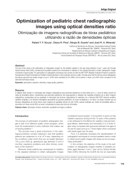
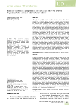
Radiopacity assessment of composite
resins through digital systems and
InSight intraoral film
Avaliação da radiopacidade de resinas compostas por meio de sistemas
digitais e filme radiográfico InSight
Mateus Ericson Flores*
Francisco Haiter Neto**
Frab Norberto Boscolo***
Gisele Rovani****
Melissa Feres Damian*****
Objective: this study aimed to assess the radiopacity of
eight composite resins recommended for class II restorations. Materials and method: hence, 2 mm thick and
4 mm in diameter test specimens were made and compared to enamel, dentin, and aluminum using four digital systems: two semi-direct by storage phosphor image
plates (SPIP) - Digora™, and DenOptix™, two charged
couple devices (CCD) - Sens-A-Ray™ and Computerized Dental Radiograph (CDR™), and radiographic film
Kodak Insight IS-21™. The radiographs were scanned
to obtain the indirect digital image, and along with
direct and semi-direct digital images the radiographic
densities were assessed in specific software. The pixel
values from the aluminum step wedge were submitted
to a linear regression from where the equivalent in millimeters for enamel, dentin, and resins were determined.
Results: density means of resins were equal or superior
to the means obtained for enamel in all digital systems
and the conventional film. SureFil™ resin presented greater numerical radiopacity in all digital systems and the
radiographic film. ALERT™ resin showed the smallest
radiopacity among the studied resins in all digital systems and radiographic film. ALERT™ and Definite™ were
statistically equivalent to enamel in the Sens-A-Ray™
system and in radiographic film. The remaining resins
(Charisma™, FillMagic™, P60™, Prodigy™, SureFil™, and
Z250™) presented higher radiopacities in comparison to
enamel. Only ALERT™ presented radiopacity similar to
enamel in other digital systems (CDR™, DenOptix™, and
Digora™). In these digital systems, Charisma™, Definite™,
FillMagic™, P60™, Prodigy™, SureFil™, and Z250™ were
more radiopaque than enamel. Conclusion: all resins
showed equal or higher radiopacities of enamel in all
assessment systems. There was a statistical correlation
between systems CDR™ and Sens-A-Ray™, and between
Digora™ and DenOptix™; Sens-A-Ray™ also showed correlation with radiographic film. The percentage of density means equivalent to aluminum millimeters for enamel and dentin were 119.6% and 101.6%, respectively.
Keywords: Composite resins. X-rays. Digital dental radiography.
http://dx.doi.org/10.5335/rfo.v18i2.2563
*
**
***
****
*****
DDS, PhD, professor, University of Passo Fundo, Passo Fundo, RS, Brazil.
DDS, PhD, professor, University of Campinas, Campinas, SP, Brazil.
DDS, PhD, chairman, University of Campinas, Campinas, SP, Brazil.
DDS, MS, professor, University of Passo Fundo, Passo Fundo, RS, Brazil.
DDS, PhD, professor, University of Pelotas, Pelotas, RS, Brazil.
RFO, Passo Fundo, v. 18, n. 2, p. 175-179, maio/ago. 2013
175
Introduction
The development of composite resins gives great aesthetic possibilities for dental restorations.
However, for ideal clinical analysis of interproximal
contour, contact point adequacy, overhangs, cavity
wall adaptation, marginal gaps, voids, and recurrent decay these materials must provide sufficient
radiopacity to be visible on radiographic examinations, in accordance with ADA standards1,2 and ISO
40493. These standards require that the radiopacity
of composite resins to be used in class 1 and class 2
restorations should be greater than an equivalent
thickness of aluminum (mm) or equivalent aluminum percentage (%Al). Prior studies found this aluminum standard to be comparable in radiopacity to
an equivalent thickness of dentin4-7.
The introduction of digital radiographic systems for dentistry in the 1980s brought benefits such
as reduced patient dosage, increased film speed,
ability to enhance images as desired, and ability
to send them via internet8,9. Research on digital
images was primarily powered with CCD and SPIP
resolution, noise formation, density scale, compression of files, and potential effects on clinical diagnosis10-12. Up to now, few studies have investigated
digital systems for the evaluation of composite resin radiopacities13,14. The aim of this study was to
certify the existence of uniformity of results among
different types of digital systems and conventional
dental x-ray film, and to determine their compliance with ISO 4049.
Materials and method
Five samples of each type of posterior composite resins were light cured through a split plexiglass mold - 4 mm in diameter and 2 mm thick
(Table 1). Each specimen was light activated for
40 seconds with a XL 1500™ halogen curing light
(3M ESPE Dental Products, St. Paul, MN, USA)
at > 650 mW/cm2. Enamel and dentin specimens
were 2.0 mm thick, prepared from recently extracted human third molars. Samples’ thicknesses
were measured with a digital micrometer with two
decimal places.
Table 1 - Composite resins and manufacturers. Piracicaba-SP (Brazil), 2001
Resins
™
ALERT
Pentron™ Technologies, LLC – Wallingford, CT, USA
Charisma™
Definite
Manufacturer
Heraeus Kulzer GmbH & Co. KG – Hanau, Germany
™
FillMagic
Degussa Hülls – Frankfurt, Germany
™
™
P60
Vigodent – Rio de Janeiro, Brazil
3M ESPE Dental Products – St. Paul, Minnesota, USA.
™
Prodigy
SureFil
Kerr Corporation – Orange, CA, USA
™
Z250™
Dentsply International – York, PA, USA
3M ESPE Dental Products – St. Paul, Minnesota, USA.
Radiographs were taken of a specimen for each
of the 8 materials, enamel, dentin, and an aluminum step wedge, using 4 digital systems (Figura
1). Two semi-direct storage phosphor image plates Digora™ (Soredex - Nilsiänkatu 10-14, PO Box 250,
00031 Helsinki, Finland) and DenOptix™ (Gendex
Division - 901 W. Oakton St. Des Plaines, IL 60018-
1884, USA), and two CCD - Sens-A-Ray™ (Dent-X
Corporation USA - 250 Clearbrook Road Elmsford
New York 10523, USA) and CDR™ (Schick Technologies, Inc. 30-00 47th Avenue - Long Island City, NY
11101, USA) were used. The features of the digital
systems are contained in Table 2.
Table 2 - Features of digital system. Piracicaba-SP (Brazil), 2001
Manufacturer
™
Sensor
Active size (mm)
Pixel size (µm)
Digora
DenOptix™
Sens-A-Ray™
Soredex (Helsinki, Finland)
Gendex (Des Plaines, IL, USA)
Dent-X Corporation (New York, NY, USA)
SPIP*
SPIP*
CCD*
30 x 40
31 x 41
23,5 X 27,6
70 x 70
85 x 85
44 x 44
CDR™
Schick Technologies (Long Island, NY, USA
CCD*
25,2 x 36,5
48 x 48
*SPIP: storage phosphor image plate system. CCD: charged couple device
176
RFO, Passo Fundo, v. 18, n. 2, p. 175-179, maio/ago. 2013
Eastman Kodak Insight IS-21™ (Eastman Kodak Co., 343 State Street - Rochester, NY 14650,
USA) radiographs of the specimens were also
taken. Films and CCD digital systems were exposed for 0.05 s and 0.40 s to SPIP digital systems.
Target film distance of 70 kV, 10 mA, and 400 mm
standardized by a wooden support with a GE 1000™
(General Electric Company, 3000 N, Grandview
Blvd, MD W-407 Waukesha, WI 53188, USA) x-ray
machine was used. Films were processed in a Gendex GXP™ film processor (Gendex Division - 901 W.
Oakton St. Des Plaines, IL 60018-1884, USA) with
Kodak™ chemicals.
Figure 1 - Aluminum step wedge, resins, lead and tooth on sensor
CCD CDR- Schick Technologies. Piracicaba-SP (Brazil),
2001
Digital images were recorded and electronically
stored. All films were scanned into a computer with
an HP 6100C/T™ scanner (Hewlett-Packard, 3000
Hanover Street Palo Alto, CA 94304-1185).
Relative radiographic densities of each module
- aluminum step wedge, enamel, dentin, lead, and
resins were separately depicted using a histogram
in each software (Figura 2). Five replications of
readings were performed in each module.
The mean density value in pixels for the seven
first steps of the density scale, as well as the ones
for each resin seen in each digital system and the
scanned InSight films were statistically analyzed
using linear regression, where the mean values in
pixels were converted into an aluminum equivalent
(mm). The results were analyzed using variance
analysis and the Tukey’s test at 5%.
Results
Table 3 shows the radiopacity data for the eight
composite resins, enamel, and dentin specimens examined. The mean density values of all resins were
equal or superior to the mean values obtained for
the enamel specimens in all digital systems and
the scanned radiographic film. SureFil™ presented
greater numerical radiopacity in all digital systems and the scanned radiographic film. ALERT™
showed the lowest radiopacity among the studied
resins in all digital systems and the scanned radiographic film. ALERT™ and Definite™ were statistically equivalent to enamel in the Sens-A-Ray™
system and the scanned radiographic film. The
remaining resins (Charisma™, FillMagic™, P60™,
Prodigy™, SureFil™, and Z250™) had higher radiopacities in comparison to enamel. Only ALERT™
presented radiopacity similar to enamel in other
digital systems (CDR™, Denoptix™ and Digora™). In
these digital systems, Charisma™, Definite™, FillMagic™, P60™, Prodigy™, SureFil™, and Z250™ were
more radiopaque than enamel.
Table 3 – Mean values equivalent in aluminum (mm) of resins, enamel, and dentin for each system, separately. Piracicaba-SP
(Brazil), 2001
ALERT
Sens-A-Ray
CDR
DenOptix
3.752 c
3.760 f
4.246 e
Digora
4.324 e
InSight Film
3.784 d
Charisma™ 5.246 ab
5.656 bc 6.196 abc 5.970 b
4.876 bc
Definite™
4.204 c
4.574 e
3.914 d
FillMagic™
4.968 b
5.218 cd 6.296 ab
5.774 bc 4.634 c
P60™
5.730 a
5.904 ab 5.584 d
5.496 bc 5.638 a
4.886 b
5.104 de 5.794 bcd 5.590 bc 4.868 bc
5.704 a
6.230 a
™
Prodigy
SureFil
™
™
5.272 d
6.736 a
4.868 d
6.526 a
5.322 ab
Z250
5.562 a
5.704abc 5.722 cd
5.320 cd 5.358 ab
Enamel
4.140 c
3.770 f
4.282 e
3.896 e
3.876 d
Dentin
2.112 d
1.764 g
2.256f
1.978 f
2.052 e
Averages followed by distinct letters differ among themselves to the significance level of 5% (p < 0.05), inside the same column.
Figure 2 - Image of the histogram analyses in SPIP - DenOptix. Piracicaba-SP (Brazil), 2001
RFO, Passo Fundo, v. 18, n. 2, p. 175-179, maio/ago. 2013
177
The radiopacity values for enamel and dentin
reported in this study were similar for all digital
systems and the radiographic film (Table 4).
Table 4 - Enamel and dentin equivalent in aluminum and its conversion for aluminum percentage in all digital systems and the
scanned radiographic film. Piracicaba-SP (Brazil), 2001
Enamel
S-A-R™
CDR™
DenOptix™
Digora™
InSight Film™
Al equivalent
4.140
3.770
4.282
3.896
3.876
Dentin
% Al
207
188
214
195
194
Al equivalent
2.112
1.764
2.256
1.978
2.052
% Al
105
88
113
99
103
Discussion
All resins evaluated in this research presented
radiopacity equal to or higher than that of the enamel specimens tested, and complied with ISO 4049.
The suggestion of increased radiopacity for composite resins was offered by other studies4,5,15-18. They
believed that it could improve the diagnosis of caries, marginal gap, and other defects. However, too
high radiopacity may have the opposite effect due to
the high radiopacity difference between the composite resin and the adhesive4,7,19.
SureFil™ showed higher numerical radiopacity in
all digital systems and the scanned radiographic InSight film. These results were similar to those reported
by Watts20, who suggested that the increased percentage of filling would be responsible for higher radiopacity.
Some researchers4,16,21-26 have evaluated restorative materials. They found that some resins are not
in accordance with ISO 4049. These authors found
dental materials with equal or higher radiopacity
than enamel; materials with equal or higher radiopacity than dentin; and materials with lower radiopacity than dentin.
The means suffered variations regarding these issues among the systems, but all of them were
similar to those obtained by Stanford et al.7, with
79% for dentin and 222% for enamel; Williams and
Billington27, with 100% for dentin and 210% for enamel; Williams and Billington28 with 100% for dentin
and 220% for enamel; El-Mowafy et al.5, with 116%
for dentin and 184% for enamel; and Bouschlicher
et al.25 with 100% for dentin and 165% for enamel.
These small variations may be due to the time of
tooth storage, and the consequent loss of radiopacity by demineralization28.
Conclusions
After detailed analysis and discussion of the results we can conclude that all composite resin evaluated by four digital systems and a radiographic
InSight film are in accordance with ISO 4049. Also,
the radiopacities of enamel and dentin in aluminum equivalent converted to percentage suffer variations according to the way of attainment, however they are within the values found in literature.
Resumo
Objetivo: este estudo teve como objetivo avaliar a radiopacidade de oito tipos de resinas compostas indicadas para restaurações de classe II. Materiais e método:
para tanto foram confeccionados corpos de prova de
2 mm de espessura e 4 mm de largura, os quais foram
comparados com esmalte, dentina e alumínio, utilizando-se quatro sistemas digitais, sendo dois semidiretos
por placas de fósforo fotoestimuladas – Digora® e DenOptix®, dois CCD – Sens-A-Ray® e CDR®, e também o
filme radiográfico Kodak Insight IS-21®. As radiografias
foram escaneadas, obtendo-se imagem digital indireta,
e, juntamente com as imagens digitais diretas e semidiretas, suas densidades radiográficas foram avaliadas em
software específico. Os valores de pixel da escala de
alumínio foram submetidos a uma regressão linear, de
onde se pôde obter o equivalente em milímetros para
o esmalte, dentina e resinas. Resultados: para todos os
sistemas digitais e para o filme convencional, as médias
das densidades das resinas foram iguais ou superiores
às médias obtidas pelo esmalte. Para todos os sistemas
digitais e para o filme radiográfico, a resina Surefil®
apresentou maior radiopacidade numérica. A resina
ALERT® apresentou a menor radiopacidade entre as resinas estudadas em todos os sistemas digitais e filme
radiográfico. ALERT® e Difinite® foram estatisticamente
equivalentes ao esmalte no sistema digital Sens-A-Ray®
e no filme radiográfico. As demais resinas (Charisma®,
FillMagic®, P60®, Prodigy®, Surefil® e Z250®) tiveram
maior radiopacidade em comparação ao esmalte. Somente ALERT® teve radiopacidade similar à do esmalte
em outros sistemas digitais (CDR®, Denoptix® e Digora®). Nesses sistemas digitais, Charisma®, FillMagic®,
P60®, Prodigy®, Surefil® e Z250® foram mais radiopacos
do que o esmalte. Conclusão: todas as resinas, em todos os sistemas de avaliação, mostraram radiopacidade
igual ou superior ao esmalte. Houve correlação estatística entre os sistemas CDR® e Sens-A-Ray® e entre Digora® e DenOptix®, tendo o Sens-A-Ray® apresentado
correlação também com o filme radiográfico. As médias de densidade equivalente em mm de alumínio em
porcentagem para esmalte e dentina foram de 119,6%
e 101,6%, respectivamente.
Palavras-chave: Resinas compostas. Raios X. Radiografia dental digital.
178
RFO, Passo Fundo, v. 18, n. 2, p. 175-179, maio/ago. 2013
References
1.
American Dental Association Council on Dental Materials,
Instruments and Equipment. The desirability of using radiopaque plastic in dentistry: a status report. J Am Dent
Assoc 1981; 102:347-9.
2.
American Dental Association Council on Dental Materials,
Instruments and Equipment. Status report on posterior
composites. J Am Dent Assoc 1983; 107:74-6.
3.
Bouschlicher MR, Cobb DS, Boyer DB. Radiopacity of compomers, flowable and conventional resin composites for posterior restorations. Oper Dent 1999; 24:20-5.
4.
Curtis-Jr PM, Von-Fraunhofer JA, Farman AG. The
radiographic density of composite restorative resins.
Oral Surg Oral Med Oral Pathol Oral Radiol Endod 1990;
70:226-30.
5.
El-Mowafy OM, Brown JW, McComb J. Radiopacity of direct
ceramic inlay restoratives. J Dent 1991; 19:366-8.
6.
Abou-Tabl ZM, Tidy DC, Combe EC. Radiopacity of composite restorative. Br Dent J 1979; 147:187-9.
7.
Stanford CM, Fan PL, Schoenfeld CM, Knoeppel R, Stanford JW. Radiopacity of light-cured posterior composite resins. J Am Dent Assoc 1987; 115:722-4.
8.
Van der Stelt P. Principles of digital imaging. Dent Clin North Am 2000; 44:237-48.
9.
Wenzel A. Digital radiography and caries diagnosis. Dentomaxillofac Radiol 1998; 27:3-11.
10. Janhom A, Van der Stelt PF, Van Ginkel FC, Geraets WGM.
Effect on the compressibility and diagnostic accuracy for
caries detection of digital bitewing radiographs. Dentomaxillofac Radiol 1999; 28:6-12.
11. M∅ystad A, Svanaes DB, Risnes S, Larhein TA, Gröndahl
HG. Detection of approximal caries with a storage phosphor
system. A comparison of enhanced digital images with dental X-ray film. Dentomaxillofac Radiol 1996; 25:202-6.
12. Welander U, McDavid WD, Sanderink GCH, Tronje G,
Mörner AC, Dove SB. Resolution as defined by line spread
and modulation transfer functions for four digital intraoral
radiographic systems. Oral Surg Oral Med Oral Pathol
Oral Radiol Endod 1994; 78:109-15.
13. Farman TT, Farman AG, Scarfe WC, Goldsmith LJ. Optical
densities of dental resin composites: a comparison of CCD,
storage phosphor, and Ektaspeed Plus radiographic film.
Gen Dent 1996; 44:532-7.
19. Akerboom HBM, Kreulen CM, Amerongen WE, Mol A. Radiopacity of posterior composite resins, composite resin luting cements, and glass ionomer lining cements. J Prosthet
Dent 1993; 70:351-5.
20. Watts DC. Radiopacity vs. composition of some barium and
strontium glass composites. J Dent 1987; 15:38-43.
21. Van Dijken JWV, Wing KR, Ruyter IE. An evaluation of the
radiopacity of composite restorative materials used in class
I and II cavities. Acta Odontol Scand 1989; 47:401-7.
22. Toyooka H, Taira M, Wakasa K, Yamaki M, Fujita M. Radiopacity of 12 visible-light-cured dental composite resins. J
Oral Rehabil 1993; 20:623-5.
23. Sidhu SK, Shah PMM, Chong BS, Ford TRP. Radiopacity of
resin-modified glass-ionomer restorative cements. Quintessence Int 1996; 27:639-43.
24. Marouf N, Sidhu SK. A study on the radiopacity of different
shades of resin-modified glass-ionomer restorative materials. Oper Dent 1998; 23:10-4.
25. Bouschlicher MR, Cobb DS, Boyer DB. Radiopacity of compomers, flowable and conventional resin composites for posterior restorations. Oper Dent 1999; 24:20-5.
26. Hara AT, Serra MC, Rodrigues-Jr AL. Radiopacity of Glass-Ionomer/Composite Resin Hybrid Materials. Braz Dent J
2001; 12:85-9.
27. Williams JA, Billington RW. A new technique for measuring
the radiopacity of natural tooth substance and restorative
materials. J Oral Rehabil 1987; 14:267-9.
28. Williams JA, Billington RW. The radiopacity of glass ionomer dental materials. J Oral Rehabil 1990; 17:245-8.
Corresponding author:
Mateus Ericson Flores
Rua Cesario Rosseto, 414
99074-210 Passo Fundo, RS, Brazil
Phone/Fax: +55 (54) 3316 8403
E-mail: [email protected]
Recebido: 13/08/2012. Aceito: 08/10/2013.
14. Gürdal P, Akdeniz BG. Comparison of two methods for radiometric evaluation of resins-based restorative materials.
Dentomaxillofac Radiol 1998; 27:236-9.
15. Cook WD. An investigation of radiopacity of composite restorative materials. Aust Dent J 1981; 26:105-12.
16. Omer OE, Wilson NHF, Watts DC. Radiopacity of posterior
composites. J Dent 1986; 14:178-9.
17. Prevost AP, Forest D, Tanguay R, DeGrandmont
P. Radiopacity of glass ionomer dental materials.
Oral Surg Oral Med Oral Pathol Oral Radiol Endod 1990;
70:231-5.
18. Goshima T, Goshima Y. Radiographic detection of recurrent carious lesions associated with composite restorations.
Oral Surg Oral Med Oral Pathol Oral Radiol Endod 1990;
70:236-9.
RFO, Passo Fundo, v. 18, n. 2, p. 175-179, maio/ago. 2013
179
Baixar