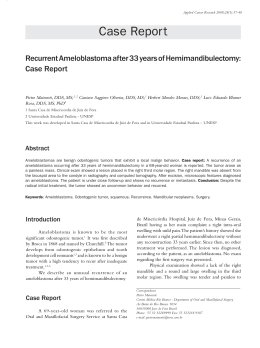
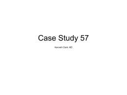
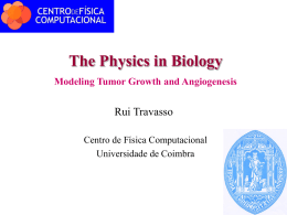
Braz Dent J (2011) 22(x): xx-xx
ISSN 0103-64401
Ameloblastic fibroma
Maxillary Ameloblastic Fibroma: A Case Report
Daniela Otero Pereira da COSTA1
Mônica Diuna CALASANS-MAIA1
Ricardo Lopes da CRUZ2
Simone de Queiroz Chaves LOURENÇO1
2Department
1Department of Pathology, UFF - Fluminense Federal University, Niterói, RJ, Brazil
of Oral and Maxillofacial Surgery, INTO - Institute of Orthopedics and Trauma, Rio de Janeiro, RJ, Brazil
Ameloblastic fibroma is a relatively rare benign odontogenic tumor in which both the epithelial and ectomesenchymal components are
neoplastic. An 8-year-old Caucasian boy was referred to the dentist for evaluation of failed eruption of the maxillary left first molar.
The panoramic radiograph showed a well-circumscribed unilocular radiolucency involving an unerupted maxillary left first permanent
molar. The lesion was enucleated and the material was sent for histopathologic examination. Microscopically, it was composed by
cords and islands of odontogenic epithelium in a myxoid cell-rich stroma that closely resemble the dental papilla with histopathological
diagnosis of ameloblastic fibroma. After 24 months of follow-up no recurrence was observed and the maxillary left first molar erupted
spontaneously through the buccal mucosa and was aligned with a fixed orthodontic appliance. This case emphasized the importance
of careful differential diagnosis of intraosseous oral lesions and reported a rarity of the lesion and its atypical location.
Key Words: ameloblastic fibroma, odontogenic tumor, maxillary tumor.
INTRODUCTION
Ameloblastic fibroma (AF) is a benign
odontogenic tumor considered a true mixed tumor
composed of neoplastic epithelium and mesenchymal
(1) This tumor is rare, representing only 2% of all
odontogenic tumors (1-5).
AF has been reported in patients aged 7 weeks
to 51 years (6,7), but the tumor is considered a tumor of
childhood and adolescence and occur almost exclusively
in the first and second decades of life (6,8-13). In the
majority of cases, the lesion arises in the mandible and
patients present as a slow-growing, painless lesion and/or
failure of the tooth eruption (8-12). However, the tooth
impaction and delayed eruption may be associated with
dental developmental defects or abnormalities such as
amelogenesis imperfecta (14). In other cases, the tumors
are asymptomatic and discovered during routine oral/
radiographic examination (12).
Radiographically, AF appears as well-defined
unilocular or multilocular radiolucencies (2,7,10). It has
been reported that asymptomatic cases present unilocular
radiolucency, whereas multilocular cases are associated
with jaw swelling (15). However, if the lesion contains
radiopaque masses in the internal structure, the diagnosis
may be calcifying epithelial odontogenic tumor (16).
There are several variants of AF. If dentin is
present, the tumor is ameloblastic fibrodentinoma.
If dentin and enamel are present, the tumor is called
ameloblastic fibro-odontoma (AFO) (15,17). Mitotic
figures may be present in AF. The presence of a large
number of cells in mitosis and atypical mitosis should
expand the differential diagnosis to include malignant
entities, like ameloblastic fibrosarcoma (1).
The most appropriate treatment method for AF
is still uncertain. A conservative approach is suggested
by several authors (1,10,11). However, tumors may
recur following surgical removal and progress to
malignancy (7,18,19). Thus, long-term follow-up of AF
is recommended (18). This report describes an interesting
case of AF that affected the maxilla of a young boy and was
associated with an unerupted maxillary left first molar.
CASE REPORT
In May 2008, an 8 year-old Caucasian boy was
Correspondence: Profa. Dra. Simone de Queiroz Chaves Lourenço, Avenida das Acácias 150, bl. 01, ap. 104, Barra da Tijuca, 22776-000 Rio de
Janeiro, RJ, Brasil. Tel: +55-21-3325-1648. Fax:+55-21-2629-9128. e-mail:[email protected]
Braz Dent J 22(x) 2011
2
D.O.P. da Costa et al.
referred to the dentist for evaluation of failed eruption
of the permanent maxillary left first molar. Physical
examination showed a generally healthy child and the
medical, surgical, family, and social histories were
unremarkable.
Intraoral examination revealed a small painless
expansion of the buccal cortical maxillary plate and the
first molar was not visualized. The overlying mucosa
was intact and normal in color and consistency.
Radiographic examination by means of a
panoramic radiograph (Fig. 1A) showed a well-defined
unilocular radiolucent lesion in the left maxilla. The
crown of the permanent first molar was apparent within
the lesion. The second molar was displaced upwards.
Computed tomography (CT) scanning confirmed the
findings of the panoramic radiograph (Fig. 1B). Based
on clinical and roentgenographic findings, a presumptive
preoperative diagnosis of dentigerous cyst was done.
Under local anesthesia, the lesion was totally
removed through an intraoral approach and protecting
the definitive first molar. The enucleated material was
sent to the Anatomic Pathology Service of the Antonio
Pedro University Hospital, Niterói, RJ, Brazil for
histological examination.
Microscopically, the lesion consisted of cords
and islands of proliferating odontogenic epithelium.
The cords often revealed a double or triple layer of
cuboidal cells, resembling the dental lamina. The
island showed peripheral rim of columnar similar to the
inner enamel epithelium. The center showed a loosely
arranged spindle-shaped epithelium identical to stellate
reticulum. The epithelial components laid in a myxoid
cell-rich stroma with little collagens closely resembling
the primitive dental papilla. No hard tooth structures
or mitotic images were noted. The histopathological
diagnosis was AF (Fig. 2).
After 24 months of follow-up, no recurrence was
observed. The first molar was subjected to traction and
was aligned with a fixed orthodontic appliance (Fig. 3A).
However, radiographic examination revealed that the
second molar was impacted to the first molar (Fig. 3B).
DISCUSSION
AF is a mixed tumor both epithelial and
mesenchymal neoplastic proliferation (1). This tumor
is usually diagnosed between the first and second
decades of life and the majority of cases of occur in the
mandible (2,10,12,15). However, few cases of the AF
in the maxilla have been reported, as the case presented
in this paper (9,10,13).
The clinical manifestations of AF are not
specific and the lesion is frequently discovered as an
incident finding in a routine radiographic examination,
such as cysts and other odontogenic tumors (1,9).
Radiographically, the differential diagnosis of the
AF must be made with entities such as dentigerous
cysts, ameloblastoma, odontogenic keratocysts (9) and
fibrosarcoma ameloblastic (11,18).
Philipsen et al. (17) managed AF by enucleation
with careful clinical and radiographic follow-up. AF
recurrence may not be true recurrence, but rather a
Figure 1. Imaging. A= Initial panoramic film showing a well-circumscribed unilocular radiolucency involving an unerupted permanent
maxillary left first molar and displacement of the maxillary left second molar (arrow). B= Computed tomography (CT) scan confirming
the findings of the panoramic radiograph.
Braz Dent J 22(x) 2011 Ameloblastic fibroma
3
residual lesion due to incomplete enucleation. Some
authors believe that an aggressive treatment is not
justified (2,15,17). However, Chen et al. (7) reported
that patients treated by radical excision experience fewer
recurrences compared to those treated conservatively;
Those authors found recurrence in 14 of 41 cases. Dallera
et al. (2), on the other hand, reported no recurrences
for 6 cases with an average follow-up of 15 years. In
the present case, the lesion was enucleated with no
recurrence at two year.
While uncommon, the possibility of malignant
transformation of AF into ameloblastic fibrosarcoma has
been reported by Chen et al. (7) and Kobayashi et al. (18).
For those authors, AF showed no signs of malignancy
until the second recurrence. Chen et al. (7) reported
that the rates of malignant transformation in 5-year and
10-year follow-up were 10.2% and 22.2%, respectively.
However, according to Kousar et al (19) rapid sarcomatous
transformation of an AF occurred within 6 months.
In the case reported here, the main feature
was the failure of eruption of a maxillary first molar.
This disturbance may or may not be associated with a
pathology, such as AF. Thus, this case emphasizes the
importance of careful differential diagnosis in dentistry,
while reporting a rare lesion and its atypical location.
Figure 2. Histological section showing ameloblastic epithelium
in cords and island lie in a myxoid cell-rich stroma resembling
the primitive dental papilla (HE, ×100 magnification).
ACKNOWLEDGEMENTS
RESUMO
Fibroma ameloblástico é um tumor odontogênico benigno
relativamente raro, em que ambos os componentes epiteliais
e ectomesenquimais são neoplásicos. Menino de oito anos de
idade, branco, foi encaminhado ao dentista para avaliar a falha na
erupção do primeiro molar maxilar do lado esquerdo. A radiografia
panorâmica revelou imagem radiolúcida, unilocular, bem
circunscrita, envolvendo o primeiro molar permanente maxilar
esquerdo incluso. A lesão foi enucleada e o material encaminhado
para avaliação histopatológica. Microscopicamente, era composta
de ilhas e cordões de epitélio odontogênico num estroma mixóide
rico em células, que se assemelhava à papila dentária, com
diagnóstico histopatológico de fibroma amelobástico. Após 24
meses de acompanhamento, nenhuma recorrência foi observada e
o primeiro molar permanente maxilar irrompeu espontaneamente
através da mucosa bucal e foi alinhado com aparelho ortodôntico
fixo. Esse caso enfatiza a importância do cuidadoso diagnóstico
diferencial das lesões orais intra-ósseas e relato de lesão rara e
sua localização atípica.
The authors would like to thank the Brazilian Federal Agency
Figure 3. Follow-up. A= Image showing tracioned and aligned the maxillary left first molar with the fixed orthodontic appliance. B=
Periapical film showing that the second molar is impacted against the first molar.
Braz Dent J 22(x) 2011
4
D.O.P. da Costa et al.
for Support and Evaluation of Graduate Education (CAPES) for
the financial support.
REFERENCES
1. Barnes L, Eveson J, Reichart P, et al. (eds). Pathology and genetics
of tumours of the head and neck: World Health Organization
classification of tumours: International Histological Classification
of Tumors. 3rd ed. Lyon: IARC Press, 2005.
2. Dallera P, Bertoni F, Warchetti C, Bacchini P, Campobassi
A. Ameloblastic fibroma: A follow-up of six cases. Int J Oral
Maxillofac Surg 1996;25:1999-2202.
3. Tawfik MA, Zyada MM. Odontogenic tumors in Dakahlia, Egypt:
analysis of 82 cases. Oral Surg Oral Med Oral Pathol Oral Radiol
Endod 2010;109: e97-e73.
4. Osterne RLV, Brito RGM, Alves APNN, Cavalcante RB, Souza
FB. Odontogenic tumors: A 5-year retrospective study in a
Brazilian population and analysis of 3406 cases reported in the
literature. Oral Surg Oral Med Oral Pathol Oral Radiol Endod.
2011;111:474-481.
5. Ebenezer V, Ramalingam B. A cross-sectional survey of prevalence
of odontogenic tumours. J Maxillofac Oral Surg 2011 [Epub ahead
of print. DOI: 10.1007/s12663-011-0170-8].
6. Mosby EL, Russell D, Noren S, Barker BF. Ameloblastic fibroma
in a 7-week-old infant: a case report and review of the literature.
J Oral Maxillofac Surg 1998;56:368-372.
7. Chen Y, Li TJ, Yi, SF. Ameloblastic fibroma and related lesions:
a clinicopathologic study with reference to their nature and
interrelationship. J Oral Pathol Med 2005;34:588-595.
8. McGuinness NJ, Faughan T, Bennani F, Connolly CE. Ameloblastic
fibroma of the anterior maxilla presenting as a complication of
tooth eruption: a case report. J Orthod 2001;28:115-117.
9. López RAG, Ortega L, Corchón MAG, Sández AB. Ameloblastic
fibroma of the mandible: report of the two cases. Med Oral
2003;8:150-153.
Braz Dent J 22(x) 2011 10. Dimitrakopoulos I, Psomaderis K, Zaramboukas T. Ameloblastic
fibroma of the mandible associated with root resorption and
unerupted teeth: a case report. Quintessence Int 2008;39:523-527.
11. Kim SG, Jang HS. Ameloblastic fibroma: report of a case. J Oral
Maxillofac Surg 2002;60:216-218.
12. Pitak-Arnnop P, Chaine A, Dhanuthai K, Bertrand JC, Bertolus C.
Extensive ameloblastic fibroma in an adolescent patient: a case
report with a follow-up of 4 years. Eur J Dent 2009;3:224-227.
13. Nelson BL, Folk GS. Ameloblastic Fibroma. Head and Neck
Pathol 2009;3:51-53.
14. Canger EM, Çelenk P, Yenisey M, Odyakmaz SZ. Amelogenesis
imperfecta, hypoplastic type associated with some dental
abnormalities: a case report. Braz Dent J 2010;21:170-174.
15. Chen Y, Wang JM, Li TJ. Ameloblastic fibroma: A review of
published studies with special reference to its nature and biological
behavior. Oral Oncol 2007; 43: 960-9694.
16. Deboni MCZ, Naclério-Homem MG, Pinto Junior DC, Traina AA,
Cavalcanti MGP. Clinical, radiological and histological features of
Calcifying epithelial odontogenic tumor: case report. Braz Dent J
2006,17:171-174
17. Philipsen HP, Reichart PA, Praetorius F. Mixed odontogenic
tumors and odontomas. Considerations on interrelationship.
Review of the literature and presentation of 134 new cases of
odontomas. Oral Oncol 1997;33:86-99.
18. Kobayashi K, Murakami R, Fuji T, Hirano A. Malignant
transformation of ameloblastic fibroma to ameloblastic
fibrosarcoma: case report and review of the literature. J
CranioMaxillofac Surg 2005;33:352-355.
19. Kousar A, Hosein MM, Ahmed Z, Minhas K. Rapid sarcomatous
transformation of an ameloblastic fibroma of the mandible: Case
report and literature review. Oral Surg Oral Med Oral Pathol Oral
Radiol Endod 2009;108;e80-e85.
Accepted March 2, 2011
Baixar