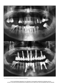
Implant-induced expansion of atrophic ridges for the placement of implants
Mauro Cruz, DDS, MDSci,a Clóvis Cruz Reis, DDS,† and Flávio de Freitas Mattos, DDS, DDPH, MDScib
CLINEST–Clinical Center of Research and Technological Development in Stomatology, Juiz de Fora,
Minas Gerais, Brazil
The applicability of endosseous implants is directly related to the topography and quality of the
patient’s residual bone. Several techniques have tried to expand the applicability through implant
design alterations and surgical techniques for bone augmentation. This article describes an
implant-induced bone expansion procedure that facilitates the placement of implants in atrophic
alveolar ridges. This procedure expands the cortical plates of the alveolar ridges with or without
fracture using wedge-shaped implants and the principles of guided tissue regeneration. The use of
implants of larger diameters and the remodeling of the ridge external contour can be achieved with
this procedure. (J Prosthet Dent 2001;85:377-81.)
T
he use of endosseous implants for successful
restoration of patients with partial or total tooth loss
has been well-established.1-3 Placement of cylindrical
implants requires minimal bone dimensions,4,5 which
vary from 5 mm height and 6 mm width6,7 to 8 mm
height and 6 mm width.8,9 One criterion used for
implant placement is at least 1 mm of bone around the
implant when in place.10 Bone resorption is an
inevitable consequence of tooth loss. In edentulous
patients, vertical resorption can progress to reach the
basal bone.11,12 Horizontally, the resorption may
progress to the extent that, even if there is enough
bone height, the lack of bone width may render
implant placement impossible.13,14
When there is insufficient bone, alveolar ridge augmentation is necessary.10 Several techniques for bone
augmentation, both vertically and horizontally, have
been proposed. Bone grafting with the application of
synthetic materials or combinations of 2 or more graft
types frequently are used.15-26 An alternative surgical
procedure is the osteotome technique.27 Block et al28
proposed a procedure for vertical mandibular bone
augmentation that uses a bone distraction osteogenesis proven to be effective in animal studies. Anterior
maxillary osteoplasty, as proposed by Richardson and
Cawood,29 is yet another technique. Nevins and
Fiorellini30 developed a surgical procedure for horizontal ridge augmentation in the premolar region of
the maxilla. Finally, Scipioni et al31 and Simion et al32
presented a bone expansion technique associated with
guided bone regeneration principles.
This article presents a bone expansion procedure
that: (1) uses the elastic and plastic deformation poten-
aDirector,
Department of Surgery and Orthodontics.
†Deceased.
bMunicipal
Health Secretariat and Lecturer, Department of Child
and Social Dentistry, Federal University of Juiz de Fora.
APRIL 2001
Fig. 1. Bioform implants with vertical and horizontal shapes.
tials of the remaining bone, (2) uses bone regeneration
potential,33 and (3) can expand implant applicability.
PROCEDURE
For patients to be treated with this procedure, local
anesthetic can be used with regional block and/or
infiltration according to site. Incisions are made to
allow maximum access and visualization to preserve
the soft tissue and to avoid subsequent membrane
exposure.34 Soft tissue management must accommodate the enlargement of the bone crest.
Because 2 types of implants are used (vertical and
horizontal) (Figs. 1 and 2), there are differences in the
expansion procedures. For vertical implants, bone
expansion is achieved in depth; for horizontal
implants, expansion is achieved longitudinally. In both
situations, however, after total cortical bone separation, ridge expansion is obtained with the use of an
implant analog with smooth surfaces.
In the mandible, a crestal incision35 within the
attached gingiva is performed. When teeth are present,
the incision is extended within the gingival sulcus and
anteriorly to the adjacent teeth. Vertical releasing inciTHE JOURNAL OF PROSTHETIC DENTISTRY 377
THE JOURNAL OF PROSTHETIC DENTISTRY
A
CRUZ, REIS, AND MATTOS
B
Fig. 2. Frontal and lateral views of Bioform implant. Lateral view shows wedged shape.
A, Implant schematic design; B, 5 × 15-mm implants.
Fig. 3. Mucoperiosteal detachment and bone crest exposure.
Fig. 4. Diagram of 3.3-mm implant in 3.0-mm crest shows
bone expansion.
sions are made at the ends of the crestal incision
approximately 7 mm away from the proposed length
of the osteotomy. The lingual flap is raised to full
thickness and continued mesiodistally and within the
gingival sulcus with the same width used in the
vestibular side. When necessary, a vertical releasing
incision of 3 to 4 mm is used to release the periosteum.
In the vestibular side, the periosteum releasing incisions start from the most apical part of the vertical
releasing incision and continue 3 to 4 mm to the center of the crestal incision.36
In the maxillary arch, a crestal incision and 2 vertical buccal releasing incisions are made following the
same principles described for the mandible. In the
palatal side, an intrasulcular incision is performed
with the same wideness used in the buccal side. The
mucoperiosteal flap is carefully detached, maintaining
periosteal integrity (Fig. 3) so that the periosteum can
be used as a natural barrier for guided bone regeneration (GBR).
After bone crest exposure, selection of implant
diameter is made. In situations of advanced resorption that result in a 3.0-mm width crest, it is possible
to place a 3.3-mm diameter or larger implant (Fig.
4), depending on the site and type of bone. The
diameter of the implants has to be compatible with the
expected degree of bone expansion.
The initial osteotomy is made with a 0.60-mm wide
disk (ISO 310 204 045 171 060, Komet, Rio de
Janeiro, Brazil), and the cortical bone is split to a disk
depth of 1.7 mm (Fig. 5). After that, the medullar
bone is cut to the depth of the desired implant using a
long cylindrical bur (ISO 310 204 682 336 012,
Komet) (Fig. 6). The horizontal extension of the bone
cuts depends on the presence of teeth, bone flexibility,
the desired degree of expansion, and the diameter of
the implant. The longer the cut, the greater the flexibility of the buccal and lingual parts of the bone.
Successive insertions of the implant analog are
made (Fig. 7); their wedged design leads to the
desired expansion. For vertical implants, after the
introduction of one third of the implant length, circular movements are made to enlarge the osteotomy.
In some situations, fracture of the buccal cortical
bone is induced (Fig. 8), either through manual pressure on the implant analog or through percussion. In
such situations, the implant analog is inserted 1 or 2 mm
short of total length of the implant, allowing the later
378
VOLUME 85 NUMBER 4
CRUZ, REIS, AND MATTOS
Fig. 5. Bone cortical split with disk.
Fig. 7. Partial introduction of implant analog through in-andout, circular movements. Smooth surface helps introduction.
Fig. 6. Medullar bone cut with bur.
Fig. 8. Total implant introduction through percussion and
fracture of buccal cortical bone.
the final movement to guarantee its initial stability.
Although fracture of the buccal cortical bone occurs
at times, the requirements for GTR are always maintained. For horizontal implants, lateral movements are
used and seating (Fig. 9).
With or without fracture, all GTR requirements are
followed and a total occlusive membrane37 (Allumina,
Alloplastic membrane for tissue isolation in GTR,
Demac) is used. Patients should be seen postoperatively
for weekly examinations until 45 days after surgery. The
sutures are removed within 5 to 10 days. When the
membrane is exposed during the healing process, it is
controlled until its removal. For that, Alvogyl (DFL,
Spécialités Septodont, Saint-Maur-des-Fossés, Cedex,
France) is applied 1 to 2 mm under the surgical borders.
The medication is applied every 3 days until the membrane is removed after at least 21 days of healing. The
membrane can be removed 30, 40, or 60 days after
surgery. In those situations in which exposure does not
occur, the membrane should be maintained until reentry surgery (6 months after the initial surgery).
APRIL 2001
THE JOURNAL OF PROSTHETIC DENTISTRY
Fig. 9. Introduction of horizontal implant.
Efforts are made to achieve primary closure of the
surgical flaps. The eversed crestal flap technique34 is
used, and the horizontal mattress suture alternated
379
THE JOURNAL OF PROSTHETIC DENTISTRY
with simple interrupted ones are conducted.36 All
patients receive before and after surgery antibiotics,
anti-inflammatories, and analgesics.
During the reentry surgery, soft tissue conditions
are evaluated and some reconstructive flaps can be
used. The goal is to obtain keratinized gingiva around
the implants.39 Rotational or advanced flaps40 are used
depending on local conditions. Visual confirmation of
bone formation should be obtained and tests applied
to evaluate the expected osseointegration.2,38 Implants
can receive individual or a multitooth cemented prosthesis. Follow-up of individual patients1,2,38 should
involve several clinical examinations during the first
year and, thereafter, at least 1 checkup annually,
including a clinical examination and radiographs.
Implant stability, peri-implant radiolucency, and the
absence of signs and symptoms such as pain, infection,
and neuropathies should be observed and controlled.
DISCUSSION
Wedge-shaped implants, whose characteristics
change from circular in the cervical area to wedged in
the apex, are the main factor that allows the use of
bone expansion and enables perfect fit between the
implants and the osteotomy. Summers27 incorporated
these characteristics in an osteotome technique.
However, the instruments for the technique had the
shape of a cylinder to facilitate initial stability within
the bone. The cylindrical form of the osteotome
requires larger amounts of bone both vertically and
horizontally. In this procedure, smaller bone dimensions are required both cervical and apically. Other
authors have proposed surgical techniques for ridge
expansion, also taking advantage of bone elastic and
plastic properties. Scipioni et al31 achieved alveolar
ridge augmentation through buccal displacement of
the buccal cortical plate, allowing for implant placement in narrow ridges. In a procedure by Simion et
al,32 the alveolar ridge was split longitudinally with
chisels before placing implants. All these techniques
are similar to the implant-induced expansion, as they
allow for immediate implant placement. However,
unlike the implant-induced expansion procedure, they
do not take advantage of the similarities between the
fracture line of the expanded ridge and the design of
the implants. With the procedure described here,
deeper bone cuts and fractures can be avoided because
of the way that the wedge-shaped implants adapt
themselves to the expansion line.
Initial stability in this procedure is created by compression between the bone cortex and the implant
itself. It could be argued that the implant-induced
expansion procedure reduces the initial stability of the
implants, particularly in those situations in which fracture is necessary. However, in such situations, the
osteotome technique would not be an alternative
380
CRUZ, REIS, AND MATTOS
because of the reduced dimensions of available
bone. Furthermore, even when fracture occurs, the
fixation and initial stability of the implants are adequate for the desired bone regeneration and
osseointegration.
Different bone types have different elastic and plastic properties.8,41 Bone crests in young patients likely
will allow for immediate expansion. However, in older
patients, bone crests are more resistant to expansion
and less resistant to fracture.42,44 There are also differences in bone quality between maxilla and mandible as
well as between different areas of the maxilla and
mandible.44-46 Therefore, it may be necessary to deepen the bone cut with a bur before the implant analog
is introduced. The initial evaluation of the patient, the
tactile sensitivity of the operator, and his/her judgment are essential to decide how much to cut and how
much to expand.
As a consequence of bone resorption, the external
contour of the alveolar bone often is lost with deleterious consequences for the final esthetic result of the
implants. The degree of bone expansion that can be
achieved with this procedure allows for the reconstruction of bone contour in both the maxilla and
mandible. The implant-induced expansion procedure
can be applied in patients whose residual maxillary or
mandibular bone would not allow for placement of
traditional cylindrical implants without any previous
bone augmentation.
SUMMARY
The implant-induced expansion procedure allows
the placement of implants in areas in which bone
resorption makes the use of traditional cylindrical
implants impossible. The procedure uses the elastic,
plastic, and regenerative properties of bone associated
with an appropriate implant design. The degree of
bone expansion obtained can remodel the alveolar
bone, an important esthetic achievement.
REFERENCES
1. Adell R, Lekholm U, Rockler B, Branemark PI. A 15-year study of
osseointegrated implants in the treatment of the edentulous jaw. Int J
Oral Surg 1981;10:387-416.
2. Albrektsson T, Zarb G, Worthington P, Eriksson AR. The long-term efficacy of currently used dental implants: a review and proposed criteria of
success. Int J Oral Maxillofac Implants 1986;1:11-25.
3. Albrektsson T, Dahl E, Enbom L, Engevall S, Engquist B, Eriksson AR,
et al. Osseointegrated oral implants. A Swedish multicenter study of
8139 consecutively inserted Nobelpharma implants. J Periodontol
1988;59:287-96.
4. Lekholm U. Surgical considerations and possible shortcomings of host
sites. J Prosthet Dent 1998;79:43-8.
5. Aguillar Meimban CO. Available bone is the foremost criterion in the
insertion of endosteal implants. J Phillipp Dent Assoc 1996;47:3-21.
6. Keller EE, Desjardins RP, Tolman DE, Laney WR, Van Roekel NB.
Reconstruction of the severely resorbed mandibular ridge using the tissue-integrated prosthesis. Int J Oral Maxillofac Implants 1986;1:101-9.
7. Worthington P. Clinical aspects of the severe mandibular atrophy. In:
VOLUME 85 NUMBER 4
CRUZ, REIS, AND MATTOS
8.
9.
10.
11.
12.
13.
14.
15.
16.
17.
18.
19.
20.
21.
22.
23.
24.
25.
26.
27.
28.
29.
30.
Worthington P, Branemark PI, editors. Advanced osseointegration
surgery. Chicago: Quintessence; 1992. p. 119-22.
Branemark PI, Zarb GA, Albrektsson T, editors. Tissue-integrated prostheses: osseointegration in clinical dentistry. Chicago: Quintessence;
1985. p. 199-209.
Shulman LB. Surgical considerations in implant dentistry. J Dent Educ
1988;52:712-20.
Bahat O. Treatment planning and placement of implants in the posterior
maxillae: report of 732 consecutive Nobelpharma implants. Int J Oral
Maxillofac Implants 1993;8:151-61.
Cruz M, Reis CC. Surgical repositioning of the inferior mandibular nerve
for the placement of implants. J Clínica Odontología 1997;13:41-50.
Carlsson GE, Haraldson T. Fundamental aspects of mandibular atrophy.
In: Worthington P, Branemark PI, editors. Advanced osseointegration
surgery. Chicago: Quintessence; 1992. p. 109-18.
Golds L. The prosthetic treatment in the presence of gross resorption of
the mandibular alveolar ridge. J Dent 1985;13:91-101.
Krekmanov L. A modified method of simultaneous bone grafting and
placement of endosseous implants in the severely atrophic maxilla. Int J
Oral Maxillofac Implants 1995;10:682-8.
Quinn PD, Kent K, MacAfee KA II. Reconstructing the atrophic mandible
with inferior border grafting and implants: a preliminary report. Int J Oral
Maxillofac Implants 1992;7:87-93.
Balshi TJ, Magid MJ. Mandibular rehabilitation: a case study using inferior cadaver graft. Int J Oral Maxillofac Implants 1995;10:589-94.
Cain JR, Mitchell DL, Markowitz NR, Wiebelt FJ. Prosthodontic restoration with dental implants and an intraoral cranial bone onlay graft: a
case report. Int J Oral Maxillofac Implants 1993;8:98-104.
Van Sickels JE, Montgomery MT. Review of surgical ridge augmentation
procedures for the atrophied mandible. J Prosthet Dent 1984;51:5-10.
Curtis TA, Ware WH. Autogenous bone graft procedures for atrophic
edentulous mandibles. J Prosthet Dent 1977;38:366-79.
Frame JW, Edmondson HD, Furniss A. Mandibular reconstruction using
split autogenous bone grafts. Br J Oral Maxillofac Surg 1987;25:1-8.
Bell WH. Current concepts of bone grafting. J Oral Surg 1968;26:118-24.
Bays RA. Current concepts in bone grafting. In: Irby WB, Shelton DW,
editors. Current advances in oral and maxillofacial surgery. St. Louis:
Mosby; 1983. p. 109-20.
Kraut RA. Composite graft for mandibular alveolar ridge augmentation:
a preliminary report. J Oral Maxillofac Surg 1985;43:856-9.
Jensen J, Sindet-Pedersen S. Autogenous mandibular bone grafts and
osseointegrated implants for reconstruction of the severely atrophied
maxilla: a preliminary report. J Oral Maxillofac Surg 1991;49:1277-87.
Donovan MG, Dickerson NC, Hanson LJ, Gustafson RB. Maxillary and
mandibular reconstruction using calvarial bone grafts and Branemark
implants: a preliminary report. J Oral Maxillofac Surg 1994;52:588-94.
Wagner JD, Moore DL. The use of cylindrical osteotomes for harvesting
cancellous bone from the ilium. J Oral Maxillofac Surg 1991;9:433-4.
Summers RB. A new concept in maxillary implant surgery: the osteotome
technique. Compendium 1994;15:152, 154-6, 158 passim; quiz 162.
Block MS, Almerico B, Crawford C, Gardiner D, Chang A. Bone
response to functioning implants in dog mandibular alveolar ridges augmented with distraction osteogenesis. Int J Oral Maxillofac Implants
1998;13:342-51.
Richardson D, Cawood JI. Anterior maxillary osteoplasty to broaden the
narrow maxillary ridge. Int J Oral Maxillofac Surg 1991;20:342-8.
Nevins M, Fiorellini JP. The placement of maxillary posterior implants. In:
Nevins M, Mellonig JT, editors. Implant therapy. Clinical approaches and
evidence of success. Volume 2. Chicago: Quintessence; 1998. p. 153-69.
APRIL 2001
THE JOURNAL OF PROSTHETIC DENTISTRY
31. Scipioni A, Bruschi GB, Calesini G. The edentulous ridge expansion technique: a five-year study. Int J Periodontics Restorative Dent
1994;14:451-9.
32. Simion M, Baldoni M, Zaffe D. Jawbone enlargement using immediate
implant placement associated with a split-crest technique and guided tissue regeneration. Int J Periodontics Restorative Dent 1992;12:462-73.
33. Buser D, Dallin C, Schenk RK. Guided bone regeneration in implant
dentistry. 1st ed. Chicago: Quintessence Publishing Co Inc; 1994.
34. Landsberg CJ. The eversed crestal flap: a surgical modification in
endosseous implant procedures. Quintessence Int 1994;25:229-32.
35. Landsberg CJ. Complete flap coverage in augmentation procedures
around dental implants using the everted crestal flap. Pract Periodontics
Aesthet Dent 1995;7:13-22; quiz 24.
36. Tinti C, Parma-Benfenati S. Vertical ridge augmentation: surgical protocol and retrospective evaluation of 48 consecutively inserted implants.
Int J Periodontics Restorative Dent 1998;18:434-43.
37. Cruz M, Reis CC, Cruz e Silva V. Alloplastic membrane for tissue isolation in guided tissue regeneration. Odont Mod 1993;20:6-13.
38. Smith DE, Zarb GA. Criteria for success of osseointegrated endosseous
implants. J Prosthet Dent 1989;62:567-72.
39. Cochran DL, Hermann JS, Schenk RK, Higginbottom FL, Buser D.
Biologic width around titanium implants. A histometric analysis of the
implanto-gingival junction around unloaded and loaded nonsubmerged
implants in the canine mandible. J Periodontol 1997;68:186-98.
40. Bahat O, Handelsman M. Periodontal reconstructive flaps—classification and surgical considerations. Int J Periodontics Restorative Dent
1991;11:480-7.
41. Brunski JB. Biomaterials and biomechanics in dental implant design. Int
J Oral Maxillofac Implants 1988;3:85-97.
42. Nigg BM, Grimston SK. Biomaterials—bone. In: Nigg BM, Herzog W,
editors. Biomechanics of the musculo-skeletal system. Chichester,
England: John Wiley; 1995. p. 47-78.
43. Bays RA. The patophysiology and anatomy of edentulous bone loss. In:
Fonseca RJ, Davis WH, editors. Reconstructive preprosthetic oral and
maxillofacial surgery. Philadelphia, PA: WB Saunders; 1986. p. 1-17.
44. Jaffin RA, Berman CL. The excessive loss of Branemark fixtures in type
IV bone: a 5-year analysis. J Periodontol 1991;62:2-4.
45. Babbush CA. Dental implants: principles and practice. Philadelphia, PA:
WB Saunders; 1991.
46. Misch C. Density of bone: effect on treatment plans, surgical approach,
healing, and progressive bone loading. Int J Oral Implantol 1990;6:23-31.
Reprint requests to:
DR MAURO CRUZ
CLINEST–CLINICAL CENTER OF RESEARCH AND TECHNOLOGICAL DEVELOPMENT
STOMATOLOGY
AV. BARÃO DO RIO BRANCO, 2288/1203
JUIZ DE FORA - MG
CEP: 36016-910
BRAZIL
FAX: (55)3215-3957
E-MAIL: [email protected]
IN
Copyright © 2001 by The Editorial Council of The Journal of Prosthetic
Dentistry.
0022-3913/2001/$35.00 + 0. 10/1/114449
381
Baixar