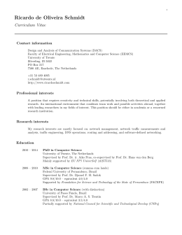
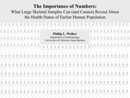
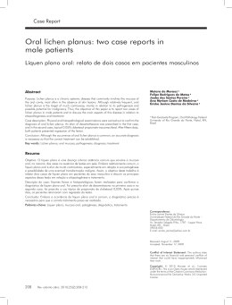
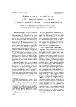
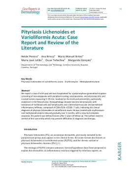
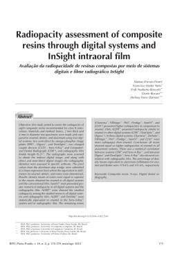
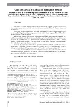
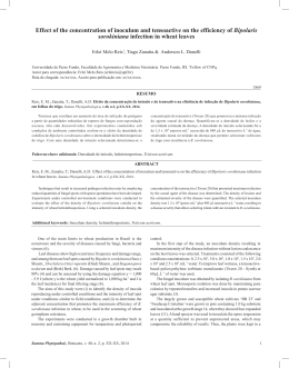
Jornal de Pediatria - Vol. 77, Nº1 , 2001 59
0021-7557/01/77-01/59
Jornal de Pediatria
Copyright
© 2001 by Sociedade Brasileira de Pediatria
CASE REPORT
Hypomelanosis of Ito - case report
Adriana S. de Almeida,1 Wânia E. Cechin,2 Jussara Ferraz,3 Rubens Rodriguez,4
Anita Moro,5 Rudah Jorge,6 Leila C. da Rosa7
Abstract
Objectives: the authors report a case of hypomelanosis of Ito (HI), a rare neurocutaneous syndrome
with neurological and chromosomal alterations associated with cutaneous involvement and recurrent
pneumonia.
Case report: a male patient, age 1 year and 11 months, was admitted with bilateral bronchopneumonia
to the São Vicente de Paulo Hospital. Examination revealed hypochromic maculas on the skin, compatible
with HI, and a delay in neuropsychomotor development. The patient was submitted to incisive biopsy of
the abdominal skin lesions, electroencephalogram, magnetic resonance, and cytogenetic evaluation.
Results: histology and immunohistochemistry evinced absence of melanin and reductio of melanocyte
in focal areas of the epidermis. The electroencephalogram revealed diffuse cortico-subcortical dysfunction.
Encephalic magnetic resonance imaging was compatible with arachnoid cyst in the temporal region.
Karyotype showed chromosome mosaicism (46, XY) and interstitial deletion of bands 22.2 to 24.4 of the
long arm of chromosome 10 (25%).
Conclusions: analysis of skin lesions is important for the etiologic definition of neuropediatric
disorders.
J Pediatr (Rio J) 2001; 77(1): 59-62: hypomelanosis of Ito, neurocutaneous syndromes, incontinentia
pigmenti achromians.
Introduction
Since 1952, when Ito described the syndrome of
incontinentia pigmenti achromiens, dermatologic, genetic,
and neurologic findings have been the object of detailed
reports.1 This name was chosen because the cutaneous
lesions of the syndrome, as seen on a negative, were similar
to those of incontinentia pigmenti of Bloch-Sulzberger. In
1973, Jelinek and colleagues suggested that the syndrome
be named hypomelanosis of Ito, which is currently the most
widely used nomenclature.2
The hypomelanosis of Ito (HI), or incontinentia pigmenti
achromiens or, still, achromic and systematized nevi is a
rare neurocutaneous syndrome possibly related to autosomal dominant inheritance and that is more frequent among
women. It is characterized by hypopigmented areas with
irregular borders, streaks, whorls, or patches and it is
commonly associated with neurological abnormalities.3,4
HI is usually present at birth and, at times, repigmentation
can occur with time. Moreover, others have described HI as
a neuroectodermal disorder that appears to be a nonspecific
manifestation of chromosome mosaicism.5 According to
Williams and Elster, there are approximately 95 cases of HI
reported in the literature until 1990.6
1. Undergraduate student, Universidade de Passo Fundo School of Medicine, RS.
2. Preceptor, Pediatrics Service Residency, São Vicente de Paulo University Hospital, Passo Fundo, RS.
3. Dermatologist.
4. Professor of Pathology, Universidade de Passo Fundo, RS.
5. Cytogeneticist, Human Cytogenetics Laboratory, São Vicente de Paulo
University Hospital, Passo Fundo, RS.
6. Preceptor, Pediatrics Service Residency, São Vicente de Paulo University Hospital, Passo Fundo, RS.
7. Undergraduate student, Universidade de Passo Fundo School of Medicine, RS.
59
60 Jornal de Pediatria - Vol. 77, Nº1, 2001
Hypomelanosis of Ito - Almeida AS et alii
Our objective is to report a case of a child with
hypomelanosis of Ito who presented with chromosomal and
neurological abnormalities associated to cutaneous involvement and recurrent pneumonia. It is also our objective to
review the current literature on HI.
Case report
Male patient with dark-brown skin color, age 1 year and
11 months (02.17.1998), born and residing in the city of
Passo Fundo, state of Rio Grande do Sul, southern Brazil.
Patient presented with suspected bilateral bronchopneumonia that was confirmed by chest X-ray. It was the third
episode of bronchopneumonia reported since 4 months of
age. Patient was born from normal delivery, term gestation,
and with congenital club foot.
At admission, patient was submitted to physical examination and the following casual findings were observed:
hypochromic, asymmetrical, bilateral maculae with irregular borders, distributed in stripes and in the form of streaks,
whorls, and patches following Blaschko’s lines and affecting mainly the trunk and limbs, but not the face, palm,
planta, and mucous membranes (figures 1 and 2). According to the mother, the patient had presented these lesions
from birth. In addition, it was also observed that patient
presented delayed development of teeth. Patient has 4
siblings and there are no references of similar lesions in
other members of the family.
We carried out incision for biopsy of the lesions on the
abdomen. The histologic findings, following FontanaMasson staining, and the immunohistochemical assay, by
means of avidin-biotin method, indicated, respectively,
absence of melanin and of melanocyte in focal areas of the
epidermis (figures 3 and 4).
Figure 1 - Hypomelanosis of Ito: hypochromic maculae located on the trunk of the patient and in linear disposition
on the arm
Figure 2 - Hypomelanosis of Ito: linear, hypodermic, and irregular lesions on the abdomen
Neurological evaluation indicated a delay in
neuropsychomotor development for age according to the
Denver Developmental Screening Test. Electroencephalogram (EEG) examination indicated diffuse cortex and subcortex dysfunction. Brain magnetic resonance imaging
(MRI) was compatible with arachnoidal cyst in left anterior
temporal region (Figure 5).
Patient presented with chromosome mosaicism and
normal karyotype (46, XY) and interstitial deletion of
bands 22.2 to 24.4 of the long arm of chromosome 10
(karyotype 46,XY/46XY, del(10)(q.22.2 - 24.2), as shown
in Figure 6.
Discussion
The hypomelanosis of Ito is a leukoderma whose pathogenesis is unknown 2 and that is characterized by
hypopigmented cutaneous lesions with linear or irregular,3
and unilateral or bilateral form. HI lesions can present
progression or regression with time. They can also be
associated with other complications, that consist of ocular,
musculoskeletal and oral alterations, hypotonia,
macrocephalia, microcephalia, congenital cardiac malformations, urological and genital malformations.7 According
to Hermida and colleagues, non-cutaneous abnormalities,
particularly of the central nervous system, eye, teeth and
skeleton, have been reported in 76-94% of patients. 8
Hypomelanosis of Ito - Almeida AS et alii
Figure 3 - Focal areas of the skin presenting hypopigmentation;
Fontana-Masson (400x)
The pigmentation of the skin depends on a series of
factors, among which the content of melanin is the most
important.9 Fontana-Mason staining is used to make the
pigmentation more evident for microscopy. Moreover, the
number of melanocytes on the skin can be either normal or
reduced. The anti-S100 autoantibodies are markers for the
S100 protein in melanocytes. In the case presented, we
observed absence of melanocytes in focal areas of the skin
with no immunomarkers for anti-S100. Also, other histologic studies have shown that hypopigmented areas have
normal melanocytes with reduction of intracellular content
of melanin.9 Conversely to incontinentia pigmenti, in HI
there are no melanphage found on the skin, and that is why
the nomenclature hypomelanosis of Ito is preferred over
incontinentia pigmenti achromiens.3
Figure 4 - Focal areas of the skin with no immunomarkers for
anti-S100; immunohistochemical assay, avidin-biotin method (400x)
Jornal de Pediatria - Vol. 77, Nº1 , 2001 61
Neurologic complications are more frequent and, also,
more severe.7 Ross postulated that brain anomalies with a
migration disorder and a disorder in cells of the neural crest,
during embryo life, would be the reason for cutaneous
hypopigmentation and, also, the gray matter heterotopias
found in the autopsy of these patients.2,3 Neither a specific
type of brain abnormality nor an associated involvement of
the central nervous system have been described for HI
patients.2 Neurologic alterations include seizures, delayed
psychomotor development, alterations of tonus, gait disorders, and so on. Out of these alterations, mental retardation
and seizures are more common and have been described in
over 50% of cases.2,7 Approximately 10% of HI patients
have presented seizures during the first year of life, and
another 10% have presented autistic behavior. 7 The only
evident neurologic alteration observed in our patient, until
the present moment, was the delay in neuropsychomotor
development.
As described by Glover and colleagues, there are no
definitive alterations at EEG for hypomelanosis of Ito.
Findings of abnormal rhythmic EEG can be an indication of
neuronal migration defects. In this sense, it is possible that
there may be a distinctive sub-group of patients with Ito’s
syndrome who present with an early onset of intractable
seizures and have poorer prognosis.10
Cranial MRI findings in patients with HI include
hemimegalencephaly, medulloblastoma, cortical malformations, dilated Virchow-Robin spaces, brain atrophy,
Figure 5 - Magnetic resonance imaging indicating arachnoidal
cyst in anterior, left temporal region
Hypomelanosis of Ito - Almeida AS et alii
62 Jornal de Pediatria - Vol. 77, Nº1, 2001
References
Figure 6 - Interstitial deletion of bands 22.2 to 24.4 of the long
arm of chromosome 10 (25%)
small discrete bilateral periventricular cysts, abnormal white
matter signal, and gray matter heterotopias and other neuronal migration defects.6,11 Our patient presented with
arachnoidal cyst at MRI, which is a relatively common
abnormality. Some cysts present asymptomatic, such as in
the case of our patient. In case symptoms are present, they
are usually secondary to the compression of adjacent structures following hydrocephalus, and to visual symptoms.12
We did not find a direct association between arachnoidal
cysts and HI in the literature; thus, the cyst may be a casual
finding and not related to HI.
HI is a clinically well-characterized syndrome in which
chromosomal instability may be a component.13,14 Chromosome abnormalities, particularly those of translocation
and mosaicism, have been reported in approximately 50%
of cases;7 which supports the hypothesis that HI is the result
of migration of two primary melanocyte clones, each with
a different pigmentation potential. According to Lenzini
and colleagues, the X-chromosome was involved in 53% of
cases with chromosomal abnormalities.15 Our patient presented alteration in the chromosome 10, which, until this
moment, had not been described in the literature.
It is important to include the hypomelanosis of Ito in
differential diagnosis of incontinentia pigmenti, depigmented
nevi, focal dermal hypoplasia, segmental vitiligo, and linear and whorled nevoid hypermelanosis.3
Finally, it was our intention to underscore the importance of skin lesions for the etiologic definition of
neuropediatric disorders.
1. Esquivel EE, Pitt MC, Boyd SG. EEG Findings in Hypomelanosis
of Ito. Neuropediatrics 1991; 22:216-19.
2. Ardinger HH, Bell WE. Hypomelanosis of Ito. Wood’s Light
and Magnetic Resonance Imaging as Diagnostic Measures. Arch
Neurol 1986; 43: 848-50.
3. Sampaio SAP, Rivitti EA. Dermatologia. 2nd ed. São Paulo:
Artes Médicas, 2000; 26:269-70.
4. Lowy G, Alonso EJF, Cestari SCP, Cestari TF, Oliveira ZNP.
Atlas de Dermatologia Pediátrica – Topográfico e Morfológico.
Rio de Janeiro: MEDSI ; 2000. p.209-10.
5. Chitayat D, Friedman JM, Johnston MM. Hypomelanosis of Ito
- a nonspecific marker of somatic mosaicism: report of case with
trisomy 18 mosaicism. Am J Med Genet 1990; 35: 422-4.
6. Williams DW, Elster AD. Cranial MR imaging in hypomelanosis
of Ito. J Comput Assist Tomogr 1990; 14: 981-3.
7. Pascual-Castroviejo I. Hypomelanosis of Ito. Neurologia 1997;
12: 300-5.
8. Hermida A, Eiris J, Alvarez-Moreno A, Alonso-Martin A, Barreiro
J, Castro-Gago M. Hypomelanosis of Ito: autism, segmental
dilatation of colon and unusual neuroimaging findings. Rev
Neurol 1997; 25: 71-4.
9. Buzas JW, Sina B, Burnett JW. Hypomelanosis of Ito. Report of
a case and review of the literature. J Am Acad Dermatol 1981; 4:
195-204.
10. Esquivel EE, Pitt MC, Boyd SG. EEG Findings in hypomelanosis
of Ito. Neuropediatrics 1991; 22: 216-19.
11. Steiner J, Adamsbaum C, Desguerres I, Lalande G, Raynaud F,
Ponsot G, et al. Hypomelanosis of Ito and brain abnormalities:
MRI findings and literature review. Pediatr Radiol 1996; 26:
763-8.
12. Fitz CR. Anomalias Congênitas do Cérebro. In: Haaga JR,
Lanzieri CF, Sartoris DJ, Zerhouni EA, eds. Tomografia
computadorizada e ressonância magnética do corpo humano. 3rd
ed. Rio de Janeiro: Guanabara Koogan; 1996. p. 108-9.
13. Ruiz-Maldonado R, Toussaint S, Tamayo L, Laterza A, del
Castillo V. Hypomelanosis of Ito: diagnostic criteria and report
of 41 cases. Pediatr Dermatol 1992; 9: 1-10.
14. Mckusick VA. Mendelian inheritance in man – A catalog of
human genes and genetic disorders. 12th ed. EUA: The Johns
Hopkins University Press, 1998; 2: 954-5.
15. Lenzini E, Bertoli P, Artifoni L, Battistella PA, Baccichetti C,
Peserico A. Hypomelanosis of Ito: involvement of chromosome
aberrations in this syndrome. Ann Genet 1991; 34: 30-2.
Correspondence:
Dr. Adriana Silveira de Almeida
Rua Alberto Borella, 441 – Cx. Postal 122
CEP 99150-000 – Marau, RS – Brazil
Phone: +55 54 342.1025
E-mail: [email protected]
Baixar