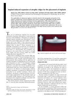
Simple bone cyst: a case report and review of the literature
Cisto ósseo simples: relato de caso e revisão da literatura
Sanyra Lopes Dias1, Leonardo de Freitas Silva2, Thiago Saldanha de Lucena Sande Vieira1, Gardênia Matos
Paraguassú3, Patrícia Leite Ribeiro Lambert4, Antônio Fernando Pereira Falcão4, Roberto Almeida de Azevedo4,5
1
Oral and Maxillofacial Surgery Residency, Federal University of Bahia, Salvador-BA, Brazil; 2Oral and Maxillofacial Surgery Residency,
Institute Dr. José Frota, Fortaleza-CE, Brasil; 3Doctor Program in Dentistry and Health, Federal University of Bahia, Salvador-BA, Brazil;
4
School of Dentistry, Federal University of Bahia, Salvador-BA, Brazil; 5Oral and Maxillofacial Surgery Residency, St. Anthony Hospital,
Salvador-BA, Brazil.
Abstract
Simple bone cysts are pseudocysts affecting long bones and, less frequently, the jaws, especially the mandible. These cysts are generally detected during routine radiography, with the frequent observation of a well-delimited radiolucent area sending projections between the roots
of the teeth involved. Simple bone cysts are mainly diagnosed during the second decade of life and have an excellent prognosis, but their
etiology is uncertain. The aim of this paper is to report the case of a simple bone cyst in a 15-year-old adolescent seen at the Stomatology
Outpatient Clinic, School of Dentistry, Federal University of Bahia. The etiology, clinical course and prognosis of this lesion are discussed
based on a systematic review of the literature.
Descriptor: Bone cysts
Resumo
Cistos ósseos simples são pseudocistos que afetam ossos longos e, menos frequentemente, os ossos maxilares, especialmente da mandíbula.
Esses cistos geralmente são detectados durante os exames imaginológicos de rotina, com a observação frequente de uma área radiolúcida
bem delimitada com projeções entre as raízes dos dentes envolvidos. Cistos ósseos simples são diagnosticados principalmente durante a
segunda década de vida e possuem um excelente prognóstico, mas a sua etiologia é incerta. O objetivo deste artigo é relatar o caso de
um cisto ósseo simples em um adolescente de 15 anos de idade, visto no Ambulatório de Estomatologia da Faculdade de Odontologia da
Universidade Federal da Bahia. A etiologia, curso clínico e prognóstico desta lesão são discutidos com base em uma revisão de literatura
sistemática.
Descritor: Cistos ósseos
Introduction
with simple bone cysts being more commonly found in
the posterior portion (area of the molars) of the mandible1-2,8. There are reports of simple bone cysts at unusual
sites such as the condylar and coronoid processes and
zygomatic arc2,12.
Simple bone cysts normally do not produce symptoms
and are discovered during radiographic exams requested
for other reasons1-3,8-10,13.
The presence of pain, edema, paresthesia, displacement and root resorption of the involved tooth, fistula
and pathological fracture has been reported on rare occasions14. Pulp vitality is unlikely to be altered even in
the case of teeth in which the roots are associated with
the lesions. However, cystic expansion may increase
the root pressure due to traumatic force, with a consequent temporary reduction in the response to the electric
pulp test13.
Periapical radiographs can be used for radiographic
assessment but these radiographs do not encompass the
whole extent of the lesion. Panoramic radiography, computed tomography and magnetic resonance are more precise imaging exams in the diagnosis of simple bone
cysts13. Radiographically, most lesions appear as a welldelimited radiolucent defect2,9. Simple bone cysts range
in diameter from 1 to 10 cm9. Margins with good definition in contrast to areas with less precise limits might be
observed in the same lesion8,12. Cysts may become radiopaque over time, an event corresponding to new bone
Since 1992, the World Health Organization (WHO)
has proposed the term “solitary bone cyst” for lesions
initially described by Lucas in 1929 and by Blum in
1932. However, hemorrhagic cyst, traumatic cyst, pseudocyst, simple bone cyst, extravasation cyst and idiopathic bone cavity are terms referring to the same type
of lesion1-5. Although the WHO recommends the term
solitary bone cyst, simple bone cyst is the term most
frequently found in the literature. This variation in terminology reflects the uncertainties regarding the etiology
and pathogenesis of traumatic bone cysts1-2,4,6-8. Among
the different etiological possibilities, the trauma-hemorrhage theory has been defended by most investigators.
This theory suggests that trauma to a bone unable to
provoke a fracture results in an intraosseous hematoma.
If the hematoma does not undergo organization and repair it may liquefy, with the consequent formation of a
cystic defect8.
Simple bone cysts have been reported in almost all
bones of the body, notably long bones such as the humerus and femur3-4,7-10. Involvement of the jaws is common and these cysts more frequently affect patients between 10 and 20 years of age3-4,6-11.The prevalence of
simple bone cysts is higher among men than women,
with a male:female ratio of about 3:28-9,12. Among the
facial bones, involvement of the maxilla is rare9,13-14,
J Health Sci Inst. 2012;30(3):295-8
295
formation. This alteration has not been observed in young
individuals since it is a late finding during the healing
process of the cyst. When the cyst involves various teeth,
the radiolucent defect frequently shows projections resembling upward bent cupolas insinuating between the
dental roots. This feature is highly suggestive of a simple
bone cyst8. Although this is not a characteristic of these
lesions, simple bone cysts may occasionally appear as a
multilocular radiotransparency associated with cortical
expansion and tumefaction of slow growth8,14.
Computed tomography is an important auxiliary tool
for the diagnosis of intraosseous lesions such as odontogenic tumors and cysts, as well as for the identification
of the precise location of important related anatomical
structures, such as the eminence of the mentuall foramen,
mandibular canal, maxillary sinus, nasal cavities and proximity to the roots of adjacent teeth15. Magnetic resonance
imaging provides a view of multiple planes with a contrast
to soft tissue and permits the analysis of the interior of
the lesions, showing the presence or absence of fluid
and thus contributing to a better distinction between simple bone cysts and other odontogenic or non-odontogenic
lesions13.
The walls of the defect might be lined with a thin
layer of vascular fibrous connective tissue or may demonstrate proliferation of a thickened myxofibromatous
matrix frequently intermingled with cellular and reactional bone trabeculae. This lining may exhibit areas
of vascularization, fibrin, erythrocytes and occasional
giant cells adjacent to the bone surface. There is no
evidence of any epithelial lining. The bone surface
close to the cavity frequently presents areas of resorption (Howship’s lacunae) indicative of past osteoclastic
activity8,12.
We report here the case of a simple bone cyst in a
15-year-old adolescent seen at the Stomatology Clinic,
School of Dentistry, at Federal University of Bahia, Brazil. A systematic literature review is presented, discussing the etiology, clinical course and prognosis of this
lesion.
Figure 1 (A and B). Preoperative radiographic aspects
For better assessment computed tomography was performed which revealed a single-cavity lesion in the right
anteroinferior region. The lesion presented a regular and
well-defined, but not completely corticalized, contour
and was associated with the roots of teeth 4.2 and 4.3
but did not promote root resorption. In addition, the
lesion caused erosion of lingual cortical bone but without
distending it. The intralesional density was inconclusive,
with values indicating the presence of a thick fluid or fibrous tissue, thus raising doubts regarding the diagnosis
of traumatic bone cyst or odontogenic keratocyst (Figure
2 – A and B).
Figure 2. Sagital (A) and coronal (B) computed tomography images
On the basis of the findings, surgical treatment was indicated. No cellularized lesion or cystic content was observed during the surgical procedure, with the observation
of a clearly visible cavity with intact bone walls which
led to the diagnosis of traumatic bone cyst (Figure 3).
Case report
Patient R.D.L, a 15-year-old white boy, a student, born
in Salvador, Bahia, was referred to the Stomatology Clinic
of Federal University of Bahia, by his orthodontist, who
noticed the presence of a radiolucent area on the right
side of the mandible during the examination of radiographs for orthodontic documentation. During anamnesis,
the patient reported that he was unaware of the lesion
which had been asymptomatic. When asked about his
medical-dental history, the patient reported that he had
had chicken pox and had been submitted to herniorrhaphy during childhood.
Clinical examination showed no swelling or other significant sign in the area. Radiographically, the lesion presented as a well-delimited unilocular radiolucent area in
the periapical region of teeth 4.2 and 4.3. The lesion produced no resorption of the root or lamina dura and the
teeth involved presented preserved pulp vitality (Figure
1 – A and B).
Dias SL, Silva LF, Vieira TSLS, Paraguassú GM, Lambert PLR, Falcão AFP et al.
Figure 3. Transurgical aspect
296
J Health Sci Inst. 2012;30(3):295-8
Material was collected for histopathological examination and bleeding induced inside the cavity showed the
lack of a capsule or membrane. Histopathological analysis
of the collected material showed the presence of small
bone spicules usually next to the hemorrhagic exudate.
This finding, together with the clinical data, permitted
the definitive diagnosis of a simple bone cyst.
Six months after the surgical intervention a new
radiograph was taken (Figure 4), which revealed a
slightly radiopaque area, a finding indicating new bone
formation.
the bone because of inadequate venous drainage, local
disturbances in bone growth, ischemic necrosis of bone
marrow and altered bone metabolism resulting in osteolysis, in addition to intraosseous vascular deformities, benign
neoplastic degenerative lesions, altered calcium metabolism, low-grade infections, venous obstruction and bone
tumors undergoing cystic degeneration1.
The radiographic features of the present case agree
with those reported by most investigators who characterize a simple bone cyst as a predominantly round or oval
well-delimited radiotransparency with regular or irregular
margins but well-defined by a delicate cortical layer. The
lesion may extend between the roots of the erupted teeth,
producing a characteristic jagged contour8,12.
Although frequently suggestive, the radiographic features of simple bone cysts are not definitive for the diagnosis8,12, with the need for a combined analysis of clinical
history, physical exam, imaging findings, surgical exploration and histopathological results13. Otherwise, simple
bone cysts might be confused with a wide variety of
odontogenic and non-odontogenic radiolucent lesions
of the jaws.
Matsumura et al.1 (1998) suggested that the size of the
radiolucent lesion and the extent of cortical bone expansion of the cysts should be monitored by radiography
and that these cysts should be treated surgically if the
findings indicate growth. First, the diagnosis of a simple
bone cyst (no growth) should be established and the lesion should be monitored radiographically. However, according to Castro and Paro12 (2002) simple bone cysts
cannot be identified positively only based on the history
of the patient and on the clinical and radiographic features. Thus, these authors do not recommend that a lesion
suspected to be a simple bone cyst be only radiographed
periodically. In the case of simple bone cysts, a differential
diagnosis is necessary to exclude the possibility of other
lesions such as a dentigerous cyst, odontogenic keratocyst, adenomatoid odontogenic tumor, ameloblastoma
and central giant cell granuloma, or even the association
with other diseases such as thrombocytopenic purpura13.
The present case agrees with Castro and Paro12 (2002)
since computed tomography revealed inconclusive intralesional density values indicating the presence of thick
fluid or fibrous tissue. In addition, the lesion presented a
regular contour in the absence of root resorption and
distension of cortical bone, raising doubts regarding the
possible diagnosis of an odontogenic keratocyst or simple
bone cyst. Thus, surgical exploration was necessary to
establish the diagnosis.
In the case of gnathic bone lesions, simple surgical exploration is normally sufficient for the establishment of the diagnosis. Although the bone walls of the cavity are frequently
smooth and bright, curettage is recommended during surgical
exploration for the collection of tissue specimens for microscopic examination in order to rule out the possibility of
more severe diseases. A lesion classified as a simple bone
cyst during surgical exploration will occasionally prove to
be a lesion with a thin lining such as an odontogenic keratocyst or ameloblastoma during microscopic examination.
Surgery was the treatment of choice in the present case, as
recommended by other investigators1,4,12-13.
Figure 4. Radiograph obtained 6 months after surgical intervention
Discussion
The etiology of simple bone cysts is still uncertain, but
some investigators have suggested an association with
trauma8,12. However, no such association was observed in
the present case. Some theories have been raised to explain
the clinical and pathological characteristics of this disease8.
The trauma-hemorrhage theory has been extensively defended, as demonstrated by the wide use of the term itself,
i.e., traumatic bone cyst. This theory suggests that trauma
to a bone unable to cause a fracture results in an intraosseous hematoma. If the hematoma does not undergo organization and repair it may liquefy, with the consequent
formation of a cystic defect. Some patients may recall an
episode of trauma to the affected region, but the significance of this information is uncertain8. Other etiological
theories include the incapacity of interstitial fluid to leave
J Health Sci Inst. 2012;30(3):295-8
297
Simple bone cyst: a case report and review of the literature
References
The treatment of simple bone cysts is based on surgical
exploration to induce bone formation2,5,13,16. Healing occurs
about 6 to 24 months after bleeding and closure of the
area, a fact justifying periodic radiographs performed at 6
and 12 months for the confirmation of bone repair, with
the case being concluded after complete healing of the
tissue8,16. In the present case, 6 months after surgical intervention a new radiograph was taken which showed a mild
radiopacity at the site of the lesion, indicating the occurrence of the healing process through new bone formation.
Although observed in some cases, recurrence or persistence of simple bone cysts is highly uncommon and
the prognosis is therefore excellent8. Follow-up at 1 or 2
years is sufficient in view of the slow growth of recurrent
lesions. In most cases, cure or recurrence is confirmed
within 3 years after surgery. Once access to the cavity is
established, the surgeon only needs to promote bleeding
of the lesion before its closure. Bone repair commonly
occurs after formation of a clot, with few chances of recurrence. Aspiration prior to surgery has been performed
to prevent accidents in cases of vascular intraosseous lesions. An antibiotic cover should be applied after surgical
intervention in view of the possible formation of an intraosseous hematoma12.
1. Matsumura S, Murakami S, Kakimoto N, Furukawa S, Kishino M,
Takeshi I et al. Histologic and radiographic findings of the simple
bone cyst. Oral Surg Oral Med Oral Pathol Oral Radiol Endod.
1998;85:619-25.
2. Sverzut CE, Gomes PP, Sverzut AT, Tozetto ALG. Cisto ósseo solitário: relato de um caso clínico. Rev Dent Press Ortodon Ortop
Maxilar. 2002;7:63-7.
3. Copete MA, Kawamata A, Langlais RP. Solitary bone cyst of the
jaws. Radiographic review of 44 cases. Oral Surg Oral Med Oral
Pathol Oral Radiol Endod. 1998;85:221-5.
4. Fregnani ER, Ramos FMM, Nadalin MR, Silva-Sousa YTC, Perez
DEC. Simple bone cyst: possible misdiagnosis in periapical pathology. Gen Dent. 2007;55:129-31.
5. Suei Y, Taguchi A, Tanimoto K. Simple bone cyst of the jaws: evaluation of treatment outcome by review of 132 cases. J Oral Maxillofac Surg. 2007;65:918-23.
6. Azevedo RA, Marques JAF, Soares SS, Carneiro Júnior B, Santana
SI. Cisto ósseo simples: relato de casos clínicos. BCI. 2002;9:139-43.
7. González HJ, Moret CY. Quiste óseo traumático bilateral asociado a tratamiento de ortodoncia. Presentación de um caso y revisión de la literatura. Acta Odontol Venez. 2001;40:116-6.
9. Puricelli E, Chaves KDB, Ligocki AF, Moresco FC, Rovani G, Rosolen MA et al. Cisto ósseo traumático em área de rizogênese:relato
de um caso. Rev Fac Odontol Porto Alegre. 1997;38:19-25.
8. Neville BW, Dam DD, Allen CM, Bouquot JE. Patologia oral e
maxilofacial. 2.ª ed. Rio de Janeiro: Guanabara Koogan; 2004.
Conclusion
On the basis of a literature review and clinical followup of a patient with a radiolucent lesion diagnosed as a
simple bone cyst, we conclude that this lesion is of uncertain etiology and that it can be considered a pseudocyst
because of the absence of epithelial lining. The final diagnosis should be established considering clinical-anamnesis, radiographic, imaging, surgical and histological findings. Surgical treatment is important for the establishment
of the final diagnosis because, radiographically, simple
bone cysts of the mandible resemble a series of other intraosseous lesions. Thus, radiographic follow-up for one
or two years after surgical treatment is necessary for the
confirmation of a possible cure or recurrence. A review
of the literature showed a low rate of recurrence and an
excellent prognosis of simple bone cysts of the mandible
not associated with other lesions.
10. Teixeira RG, Bueno CES, Miranda ME, Höfling RTB, Bussadori
SK. Cistos ósseos simples. Análise clínica e radiográfica de 22 casos
na mandíbula. Rev Gaúcha Odontol. 2003;51:243-8.
11. Ferreira Júnior FO, Damante JH, Lauris JR. Simple bone cyst
versus odontogenic keratocyst: differential diagnosis by digitized
panoramic radiography. Dentomaxillofac Radiol. 2004;33:373-8.
12. Castro AL, Paro MLC. Cisto ósseo traumático em mandíbula.
RFO UFP. 2002:39-42.
13. Lago CA, Cauás M, Pereira AMP, Portela L. Cisto ósseo traumático em mandíbula: relato de caso. Rev Cir Traumatol Buco-Maxilo-Fac. 2006;6:17-22.
14. Suei Y, Taguchi A, Kurabayashi T, Kobayashi F, Nojiri M, Tanimoto K et al. Simple bone cyst: investigation on the presence of gas
in the cavity using computed tomography. Oral Surg Oral Med Oral
Pathol Oral Radiol Endod. 1998;86:592.
15. Paiano GA, Chiarelli M, Dunker C. Tomografia computadorizada como método auxiliar no diagnóstico e tratamento de lesões
intra-ósseas: caso clínico de odontoma composto. Rev Odonto
Ciênc. 2006;21:292-6.
16. Tong AC, Ng Io, Yan BS. Variations in clinical presentations of simple
bone cyst: report of cases. J Oral Maxillofac Surg. 2003;61: 1487-91.
Endereço para correspondência:
Gardênia Matos Paraguassú
Faculdade de Odontologia da Universidade Federal da Bahia
Rua Araújo Pinho, 62 - Canela
Salvador-BA, CEP 40110-150
Brasil
E-mail: [email protected]
Recebido em 1 de dezembro de 2011
Aceito em 13 de março de 2012
Dias SL, Silva LF, Vieira TSLS, Paraguassú GM, Lambert PLR, Falcão AFP et al.
298
J Health Sci Inst. 2012;30(3):295-8
Baixar