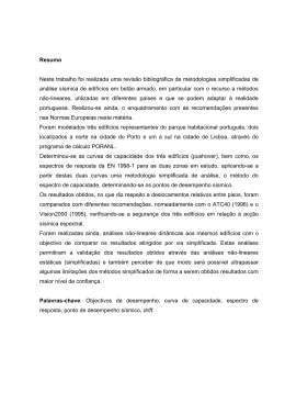
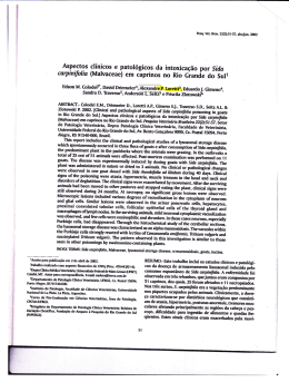
ASSOCIAÇÃO ENTRE ANTICORPOS ANTICARDIOLIPINA E PRÉECLÂMPSIA: REVISÃO SISTEMÁTICA DA LITERATURA E METANÁLISE
Aline Defaveri do Prado
Porto Alegre, 2010
PONTIFÍCIA UNIVERSIDADE CATÓLICA DO RIO GRANDE DO SUL
PROGRAMA DE PÓS-GRADUAÇÃO EM MEDICINA E CIÊNCIASDA SAÚDE
ÁREA DE CONCENTRAÇÃO: CLÍNICA MÉDICA
1
FACULDADE DE MEDICINA
ASSOCIAÇÃO ENTRE ANTICORPOS ANTICARDIOLIPINA E PRÉECLÂMPSIA: REVISÃO SISTEMÁTICA DA LITERATURA E METANÁLISE
AUTORA: ALINE DEFAVERI DO PRADO
ORIENTADOR: PROF. DR. HENRIQUE LUIZ STAUB
CO-ORIENTADOR: PROF. DR. BERNARDO LESSA HORTA
Dissertação de Mestrado apresentada ao Programa de PósGraduação em Medicina e Ciências da Saúde da Pontifícia
Universidade Católica do Rio Grande do Sul, para obtenção
do título de Mestre em Medicina
Porto Alegre, Fevereiro de 2010
2
LISTA DE ABREVIATURAS
SAF – Síndrome antifosfolipídica
aCL – anticardiolipina
LES –lúpus eritematoso sistêmico
AAF – anticorpos antifosfolípides
PE – pré-eclâmpsia
RS – revisão sistemática
OR – odds ratio
IC – intervalo de confiança
IC95% – intervalo de confiança 95%
RO – razão de odds
3
Sumário
1
2
INTRODUÇÃO ..................................................................................................... 7
1.1
Síndrome Antifosfolipídica e Anticorpos Anticardiolipina ............................. 8
1.2
Definições de Pré-Eclâmpsia ....................................................................... 10
1.3
Anticorpos Anticardiolipina e Pré Eclâmpsia ............................................... 12
1.4
Revisão Sistemática e Metanálise ................................................................ 14
1.5
Metanálise de Estudos Epidemiológicos ...................................................... 16
1.6
Interpretação do Gráfico da Metanálise........................................................ 20
1.7
Heterogeneidade e Metarregressão .............................................................. 21
1.8
Viés de Publicação ...................................................................................... 24
1.9
Objetivo ...................................................................................................... 26
1.10
Hipótese Operacional .................................................................................. 26
1.11
Hipótese Conceitual..................................................................................... 26
1.12
Referências bibliográficas............................................................................ 27
ARTIGO EM PORTUGUÊS ............................................................................... 32
2.1
ANEXOS .................................................................................................... 56
2.1.1 Anexo 1 – Lista de autores e especialistas em síndrome
antifosfolipídica contactados por email: ............................................................... 57
2.1.2
Anexo 2 – Fluxograma da busca de estudos ............................................. 59
4
Anexo 3 – Critérios Modificados de Downs and Black ........................................ 60
2.2
2.2.1
TABELAS................................................................................................... 65
Tabela 1 – Características dos estudos primários...................................... 66
2.2.2 Tabela 2 – Pressão arterial, valores de proteinúria e classificação
de gravidade utilizados nas definições de PE dos estudos primários ..................... 67
2.2.3 Tabela 3 – Metanálise em subgrupo de estudos de acordo com o
tamanho amostral ................................................................................................. 68
2.2.4 Tabela 4 – Metarregressão para as variáveis delineamento,
qualidade e tamanho do estudo............................................................................. 69
2.2.5 Tabela 5 – Metanálise do subgrupo de estudos com delineamento
caso-controle ....................................................................................................... 69
2.2.6 Tabela 6 – Metanálise do subgrupo de estudos com delineamento
coorte 70
2.3
3
GRÁFICOS ................................................................................................. 71
2.3.1
Gráfico 1– Todos os estudos: qualquer gravidade de PE .......................... 72
2.3.2
Gráfico 2 – Estudos com PE leve ............................................................. 73
2.3.3
Gráfico 3 – Estudos com PE grave ........................................................... 73
2.3.4
Gráfico 4 – Funnel Plot para todos os estudos .......................................... 74
VERSÕES EM LÍNGUA INGLESA ................................................................... 75
3.1
ARTIGO TRADUZIDO PARA A LÍNGUA INGLESA .............................. 76
3.2
ARTIGO SUBMETIDO PARA PUBLICAÇÃO ......................................... 98
3.3
ATTACHMENTS ..................................................................................... 119
3.3.1 Attachment 1 – Authors and antiphospholipid syndrome experts
contacted by email: ............................................................................................ 120
3.3.2
Attachment 2 – Search strategy flow chart ............................................. 122
3.3.3
Attachment 3 – Modified Downs and Black Criteria .............................. 123
3.4
3.4.1
TABLES ................................................................................................... 126
Table 1 – Primary studies characteristics................................................ 127
5
3.4.2 Table 2 – Blood arterial pressure, proteinuria and severity
according to primary studies PE definitions ....................................................... 128
3.4.3
Table 3 – Subgroup metanalysis according to study size ........................ 129
3.4.4 Table 4 – Variables study design, study quality and study size
metaregression analysis...................................................................................... 130
3.4.5
Table 5 – Case-control studies metanalysis ............................................ 130
3.4.6
Table 6 –Cohort studies metanalysis ...................................................... 130
3.5
FIGURES .................................................................................................. 131
3.5.1
Figure 1 – All studies: any severity of PE .............................................. 132
3.5.2
Figure 2 – Mild PE ................................................................................ 133
3.5.3
Figure 3 – Severe PE ............................................................................. 133
3.5.4
Figure 4 – Funnel Plot – all studies ........................................................ 134
6
1
INTRODUÇÃO
7
1.1
Síndrome Antifosfolipídica e Anticorpos Anticardiolipina
A síndrome antifosfolipídica (SAF) caracteriza-se por eventos tromboembólicos
arteriais e venosos associados a anticorpos anticardiolipina (aCL), anticoagulante lúpico
ou anticorpos anti-beta2-glicoproteína I. A SAF é uma doença auto-imune descrita na
década de 80, primária ou associada ao lúpus eritematoso sistêmico (LES) 1, cujo
diagnóstico é baseado em um conjunto de critérios clínicos e laboratoriais recentemente
revisados 2. Um dos critérios clínicos diagnósticos atuais desta entidade é a ocorrência
de nascimento prematuro de neonato morfologicamente normal devido à eclâmpsia, préeclâmpsia grave ou insuficiência placentária (2).
Os anticorpos antifosfolípides (AAF) compreendem um grupo heterogêneo e
complexo
de
autoanticorpos
cujos
alvos
são
os
fosfolípides
(cardiolipina,
fosfatidilserina, fosfatidilinositol) da cascata da coagulação e de membranas celulares,
principalmente as de plaquetas e células endoteliais 3. A cardiolipina é um fosfolipídeo
de carga negativa que está presente com pouca expressão na membrana celular, mas que
se encontra preferencialmente na membrana interna mitocondrial 4. Por ser facilmente
obtida do coração bovino, a cardiolipina foi padronizada como fosfolipídeo em ensaios
de fase sólida 3, 5, 6.
Apesar de o ensaio imunoenzimático ELISA para detecção de anticorpos aCL
IgG e IgM ter sido padronizado desde o primeiro simpósio sobre AAF em 1987, os
ensaios laboratoriais para detecção desses anticorpos nas publicações nem sempre
seguem tal protocolo . Frente a essa variabilidade – e impossibilidade de comparação,
portanto, dos resultados entre os estudos – desenvolveu-se recomendações para
uniformização dos métodos de testagem e de expressão dos resultados. Atualmente, a
8
investigação de anticorpos aCL deve ser feita através de ELISA e os valores devem ser
expressos em unidades GPL ou MPL
5, 7, 8
. Não se recomenda expressar os resultados
em média e desvio padrão, pois os valores de anticorpos aCL seguem uma distribuição
assimétrica, e não Gausseana. Assim, testes não-paramétricos são os apropriados para a
definição dos valores de referência 9, 10.
Acredita-se que os fenômenos vaso oclusivos na SAF sejam provenientes da
indução de trombose pelos aCL/AAF. O mecanismo exato pelo qual os aCL/AAF
estimulam a trombogênese, no entanto, não é completamente conhecido. Atualmente,
se aceita que a proteína plasmática de ligação fosfolipídica beta2-glicoproteína I seja o
principal alvo antigênico para os aCL/AAF, desempenhando papel central na
fisiopatogenia desta doença
11-14
.
Apesar de haver consenso sobre o potencial
patogênico dos aCL/AAF, é sabido que esses anticorpos surgem em hiper estimulação
imunológica no contexto de infecções e neoplasias
15
e podem estar presentes em
indivíduos normais, sendo descritos em baixas freqüências e titulação, incluindo
gestantes e idosos 16-18.
9
1.2
Definições de Pré-Eclâmpsia
Pré-eclâmpsia (PE) é uma síndrome de hipertensão e proteinúria que inicia após
as primeiras 20 semanas de gestação. Especificamente, considera-se diagnóstico de PE
para a gestante previamente normotensa que apresentar pressão arterial sistólica (PAS)
igual ou acima de 140 mmHg e/ou pressão arterial diastólica (PAD) igual ou acima de
90 mmHg após 20 semanas de idade gestacional, acompanhada de proteinúria de, no
mínimo, 300 mg em coleta de urina de 24 horas 19, 20.
A classificação de PE entre as patologias hipertensivas da gestação, no entanto,
não é homogênea. Diversos grupos de especialistas publicaram consensos para, entre
outros objetivos, estabelecer a nomenclatura e a diferenciação entre hipertensão crônica,
hipertensão gestacional, pré-eclâmpsia/eclâmpsia e pré-eclâmpsia sobreposta em
hipertensão crônica
19-24
. As classificações propostas pelos grupos Canadian
Hypertensive Society (CHS)
21
, National High Blood Pressure Education Program
Working Group (NHBPEPWG)
19
, Organização Mundial da Saúde (OMS)
24
,
Australasian Society for the Study of Hypertension (ASSH) 22, International Society for
the Study of Hypertension (ISSH)
23
e American College of Obstetrics and
Gynecologists (ACOG) 20 estão descritas na tabela 1, que foi reproduzida de Comparsi e
colaboradores 25.
Como não há padronização universalmente aceita na definição das doenças
hipertensivas gestacionais, observam-se publicações onde os autores não utilizam
nenhuma das propostas descritas acima para definição da síndrome hipertensiva de
interesse.
Dessa forma, conceitos outros de PE - que provavelmente refletem as
convicções pessoais dos autores - são amplamente usados nas publicações científicas e
incluem variados pontos de corte para a pressão arterial sistólica, diastólica e para a
10
quantificação da proteinúria. A divergência conceitual e classificatória sobre as
síndromes gestacionais hipertensivas e, em especial, sobre a definição de PE, implica
em diferenças nas populações em estudo dos artigos científicos 25, 26.
A PE é um dos principais motivos de morbimortalidade materna e fetal e sua
prevalência é estimada em aproximadamente 8% de todas as gestações. Nos Estados
Unidos, é a causa mortis materna em 16,1% de todos os óbitos na gestação, enquanto,
na América Latina, é responsável por 25,7% dos casos
27
. Sendo uma patologia
frequente e grave, esforços têm sido feitos para identificar testes laboratoriais ou de
imagem que sirvam como rastreamento para PE. Duas recentes revisões sistemáticas
com metanálise encontraram pouca evidência de testes adequados para predição de PE
28, 29
. Os estudos identificados demonstraram testes de baixa sensibilidade e, aqueles
testes que atingiram sensibilidade acima de 90% - dosagem de alfafetoproteína,
fibronectina, índice de massa corporal e ecografia Doppler de artérias uterinas apresentavam problemas metodológicos e avaliações pobres de qualidade 28.
11
1.3
Anticorpos Anticardiolipina e Pré Eclâmpsia
Fatores de risco clássicos para PE – nuliparidade, resistência à insulina, história
prévia de PE, história familiar de PE, hiperhomocistenemia, diabetes pré-gestacional,
hipertensão crônica, obesidade, extremos de idade reprodutiva, gestação múltipla,
infecções maternas, anomalias congênitas e cromossômicas, mola hidatiforme, doenças
renais, trombofilias e doenças reumatológicas auto-imunes, como lúpus eritematoso
sistêmico e síndrome antifosfolipídica – são conhecidos, aceitos e descritos 30, 31. Novos
fatores de risco, agora relacionados à paternidade e inseminação artificial também têm
sido identificados 32-34.
O papel dos anticorpos aCL como fatores de risco para PE, de modo isolado e
fora do contexto de doença autoimune é alvo de diversas pesquisas nas últimas duas
décadas. Estudos clínicos – com delineamentos caso-controle ou coortes prospectivos,
em sua maioria – foram publicados a fim de investigar essa relação
35-54
. Essas
pesquisas, no entanto, mostram resultados controversos, ora a favor, ora contra a
associação entre aCL e PE.
Uma revisão sistemática publicada sobre testes de rastreamento para PE na
população geral incluiu a pesquisa de anticorpos aCL 55. Tal estudo buscou publicações
com delineamentos de coorte ou transversais que analisassem qualquer teste (não
especificamente os anticorpos aCL), laboratorial ou de imagem, realizado durante a
gravidez com o objetivo de predizer PE. Concluiu-se que não havia nenhum teste
disponível até 2004 cuja capacidade de predição de PE fosse adequada, sendo
necessário o desenvolvimento de estudos longitudinais específicos. Outra revisão
sistemática, desta vez delineada para buscar associação entre diversas variáveis
maternas (idade, número de gestações, história familiar, doenças crônicas, índice de
12
massa corporal, entre outras) e PE incluiu a busca de anticorpos aCL e anticoagulante
lúpico. Cinco estudos foram analisados, demonstrando-se risco relativo de 9.72 (IC 4.34
– 21.75) na metanálise de dois estudos com delineamento de coorte
56
. Todavia,
considerando que as revisões não foram organizadas de modo específico para estudo
dos anticorpos aCL, não há menção sobre o ponto de corte para positividade dos
anticorpos aCL, bem como o método laboratorial usado para sua detecção na inclusão
dos artigos. Dessa forma, a real associação entre as variáveis aCL e PE permanece
indefinida.
13
1.4
Revisão Sistemática e Metanálise
Revisão sistemática (RS) é um tipo de estudo onde os sujeitos da pesquisa são os
estudos primários. Considera-se estudo primário os ensaios clínicos randomizados,
estudos de coorte, caso-controle e transversais. A RS, juntamente com as pesquisas de
análise econômica (custo-minimização, custo-efetividade, custo-benefício e custoutilidade) caracteriza-se por ser um estudo secundário, um “estudo de estudos” 57.
De acordo com Egger e Smith, as revisões sistemáticas devem ser entendidas
como estudos observacionais da evidência
58
, cuja metodologia deve ser
minuciosamente documentada, seguindo um protocolo de identificação dos estudos que
seja sistemático e reprodutível com o objetivo de minimizar vieses 59. A RS é um tipo
de pesquisa delineada para responder a uma pergunta claramente formulada. Utilizando
métodos explícitos e rigorosos, a RS busca identificar, selecionar, avaliar criticamente,
coletar e analisar dados de estudos primários relevantes
60
. O desenvolvimento da RS
envolve a estruturação de um projeto de pesquisa, com definição da questão de
pesquisa, elaboração de critérios de elegibilidade dos estudos, definição da estratégia de
busca dos artigos nas bases de dados (que deve ser a mais ampla possível, para evitar
que estudos sejam perdidos), seleção dos estudos de acordo com os critérios de
elegibilidade, avaliação da qualidade dos artigos selecionados, extração dos dados,
análise e interpretação dos resultados 57, 60.
As revisões sistemáticas são relevantes porque conseguem sumarizar um grande
volume de dados provenientes de estudos individuais. Servem como instrumentos para
transformar informação em conhecimento
57
e permitem extrapolar achados de estudos
independentes, avaliar a consistência entre os estudos e explicar as possíveis
inconsistências e divergências 61. Quando desenvolvidas, compreendidas e utilizadas de
14
modo adequado, as revisões sistemáticas configuram um importante alicerce para a
prática da medicina baseada em evidências, organização de diretrizes para a prática
clínica e formulação de políticas de saúde. Além de poder identificar o status corrente
do conhecimento sobre um tema específico, a RS tem o poder de sinalizar o que ainda
não se conhece e apontar caminhos necessários para responder à questão de pesquisa.
Após a conclusão da RS, deve ser realizada uma metanálise, quando apropriado.
A metanálise é a aplicação de um método estatístico para combinar os resultados dos
estudos primários.
15
1.5
Metanálise de Estudos Epidemiológicos
Por excelência, metanálises são utilizadas para sumarizar o efeito de tratamentos
oriundos de ensaios clínicos randomizados. No entanto, também é possível utilizar a
técnica da RS e metanálise para sumarizar evidência sobre testes diagnósticos, testes de
rastreamento, prognóstico e etiologia de doenças, custo-efetividade
60
. Revisões
sistemáticas de ensaios clínicos nível 1, coortes prospectivas nível 1 e coortes de fase
clínica inicial nível 1 são os estudos de maior grau de evidência científica, ou seja,
evidência nível 1A 62.
A análise de dados de estudos observacionais tem seu papel, por exemplo, na
resposta a questões de pesquisa envolvendo investigação de causalidade, etiologia ou
associações entre variável e desfecho. Em situações em que a magnitude de risco em
estudos primários é pequena ou quando os resultados de estudos individuais são
discordantes, a metanálise de estudos observacionais pode trazer uma resposta precisa
sobre uma associação 63, além de explicitar as razões para as diferenças nos resultados
dos estudos primários. No entanto, as peculiaridades da metanálise de estudos não
experimentais devem ser consideradas, em especial no que concerne o erro sistemático.
É importante diferenciar metanálises de ensaios clínicos de metanálises de
estudos observacionais. Como os experimentos são protegidos pela randomização, o
potencial de viés metodológico se torna infinitamente menor em comparação aos
estudos de caso-controle, coortes ou transversais. A metanálise de ensaios clínicos
baseia-se na suposição de que cada trial contenha uma estimativa livre de viés do efeito
de um determinado tratamento. Os estudos observacionais, por outro lado, produzem
estimativas de associação que podem se desviar da realidade devido à presença de
fatores confundidores e de erros
63
. Nesses casos, mesmo após o ajuste para fatores de
16
confusão ter sido realizado na análise dos dados, ainda existe a possibilidade de haver
confundimento residual 64. Além disso, os estudos observacionais são suscetíveis a viés
de seleção e de informação e que muitas vezes são importante fonte de heterogeneidade
entre os estudos
63
. Dessa forma, recomenda-se que a simples combinação estatística
dos resultados individuais não seja o componente proeminente na metanálise de estudos
observacionais. Deve-se procurar na RS as diferenças e similaridades entre os
resultados, as populações estudadas e os cenários clínicos dos estudos primários. Na
existência de heterogeneidade entre os estudos, se deve tentar identificar as principais
fontes, contribuindo assim para o delineamento de próximos estudos 63. As metanálises
de estudos observacionais já produziram importantes evidências para a tomada de
decisão em políticas públicas de saúde, principalmente para exposições onde a
realização de estudos randomizados seria extremamente difícil, como no caso da
amamentação65.
17
Definição de Metanálise, Testes Estatísticos e Interpretação
O agrupamento de resultados em uma metanálise não é adequado como tarefa
final para todas as revisões sistemáticas,se os estudos selecionados ao final da RS forem
extremamente heterogêneos no que diz respeito aos participantes do estudo, grupocontrole, definição do desfecho e exposição ou intervenção 57.
Para a realização da metanálise, é necessário, inicialmente, obter medidas
individuas dos estudos primários. Para cada um é necessário, no caso de estudos
observacionais, obter a medida de associação (odds ratio ou risco relativo) e o erro
padrão dessa medida. Para desfechos clínicos dicotômicos (como é o caso do
desenvolvimento ou não de PE na presença dos anticorpos aCL), é necessário obter o
número de eventos e o tamanho da amostra de cada grupo. Os pesos são calculados para
refletir a quantidade de informação que cada estudo contém. Na prática, o peso é o
inverso da variância, que se correlaciona com o tamanho da amostra
66
.
Conseqüentemente, os estudos com maior tamanho da amostra terão maior peso na
obtenção da estimativa combinada.
O resultado final da metanálise será uma média ponderada dos valores das
medidas de associação de cada estudo. Seguindo o exemplo da revisão sistemática
proposta nesta dissertação, obtêm-se as estatísticas sumárias (odds ratio, erro padrão) e
os pesos de cada estudo através da combinação das variáveis abaixo em fórmulas
matemáticas bem estabelecidas 66.
1. Número de pacientes com aCL positivas e PE
2. Número de pacientes sem aCL positivas e PE
18
3. Número de pacientes com aCL positivas e sem PE
4. Número de pacientes sem aCL positivas e sem PE
5. Número total de cada grupo de pacientes (aCl positivas, aCL negativas, com PE,
sem PE).
Após a obtenção das estatísticas sumárias de cada estudo, aplica-se um método
estatístico para combinar os resultados individuais. Existem vários métodos para a
obtenção da estimava combinada (Método do Inverso da Variância, Métodos de
Mantel-Haenszel, Método de Odds Ratio de Peto, Modelo de Efeito Randômico de
DerSimonian e Laird, Método Bayesiano)
66
. Essas técnicas estatísticas podem ser
classificadas resumidamente em dois modelos – efeito fixo e efeito randômico –
sendo a diferença central a maneira como a variabilidade entre os estudos é
abordada. O modelo de efeito fixo considera que a variabilidade entre os estudos é
exclusivamente devida ao acaso, ou seja, existe um “efeito verdadeiro” e os estudos
se distribuem aleatoriamente ao redor deste efeito O modelo de efeito randômico
assume que há fontes adicionais de variabilidade além do acaso, considerando a
variância de cada estudo primário. Neste caso não existe um “único efeito
verdadeiro”, mas n efeitos verdadeiros 58, 66. Diferenças substanciais na metanálise calculada através de modelos de efeitos fixo e randômico - somente serão vistas se
houver grande heterogeneidade entre os estudos primários 58.
As estatísticas sumárias bem como a metanálise e o gráfico completo podem ser
geradas através de programas estatísticos como STATA e RevMan.
19
1.6
Interpretação do Gráfico da Metanálise
A combinação das medidas de efeito individuais dos estudos primários é
representada graficamente através do gráfico da floresta, ou forest plot. Neste gráfico, as
linhas horizontais representam os intervalos de confiança de cada estudo primário. Se a
linha horizontal ultrapassar a linha vertical central do gráfico, isto significa que não há
diferença estatística na intervenção ou associação avaliada no estudo em questão 57, 59.
O ponto central de cada linha horizontal representa o odds ratio (OR) do estudo.
Se o ponto central estiver à esquerda da linha central do gráfico, significa que o efeito
medido é de proteção. Se estiver à direita da linha central do gráfico, significa que o
efeito é de aumento de risco. O tamanho do ponto central indica o peso relativo de cada
estudo no resultado final 57, 59.
O losango localizado na parte inferior do gráfico indica o resultado final da
combinação dos efeitos dos estudos, ou seja, representa a metanálise. O ponto central do
losango representa o OR e seu tamanho representa o intervalo de confiança (IC) 57, 59.
20
1.7
Heterogeneidade e Metarregressão
O resultado da medida de efeito observado em cada estudo primário é uma
estimativa do efeito verdadeiro mensurada com certo grau de imprecisão. A
heterogeneidade estatística engloba o conceito de que o efeito verdadeiro de uma
intervenção ou associação não é idêntico entre os estudos. A heterogeneidade clínica e
metodológica entre os estudos incluídos na metanálise necessariamente leva à
heterogeneidade estatística 67. Para o amplo entendimento do resultado da metanálise, é
essencial analisar a presença de heterogeneidade entre os estudos 68.
De acordo com Higgins 69 revisões sistemáticas de estudos sobre um mesmo
tema irão fatalmente unir informações que contém elementos de diversidade, pois os
estudos primários poderão diferir no desenho, condução, características dos
participantes, intervenções, exposições e desfechos avaliados. Tal diversidade é
comumente referida como heterogeneidade metodológica, podendo ocasionar diferenças
nos resultados dos estudos. Estatisticamente, detecta-se heterogeneidade quando a
variação entre os resultados dos estudos primários encontra-se acima da variação
explicada pelo acaso 69. Considerando-se a imensa possibilidade de cenários em que os
estudos são planejados e executados (heterogeneidade clínica e metodológica), a
presença de heterogeneidade estatística é esperada nas metanálises 70.
Um teste para heterogeneidade testa a hipótese nula de que todos os estudos
estão avaliando o mesmo efeito, ou seja, que existe homogeneidade. Dois métodos para
investigação de heterogeneidade são comumente usados nas metanálise: o teste Q (teste
X2 de Cochran) 71 e o teste I268.
O teste Q identifica a presença de heterogeneidade estatisticamente significante
ou sua ausência através do valor de P. Possui pouco poder para detectar
21
heterogeneidade verdadeira quando há poucos estudos na metanálise. Apresenta, ainda,
poder excessivo quando há uma grande quantidade de estudos com tamanho amostral
expressivo na metanálise, detectando frequentemente heterogeneidade - claramente sem
significado - nessa situação 68, 69.
O teste I2, desenvolvido por Higgins e colaboradores
68
, tem a habilidade de
medir a consistência entre os estudos incluídos na metanálise, avaliando em que
extensão a heterogeneidade afeta a conclusão da metanálise. O teste I2 descreve um
valor percentual da variação entre os estudos que ocorre devido à heterogeneidade e não
em razão do acaso. Valores de 0% indicam ausência de heterogeneidade. Valores acima
de zero indicam valores crescentes de heterogeneidade. A interpretação de, por
exemplo, um teste I2 = 55% em uma metanálise é a de que 55% da variabilidade entre os
estudos primários ocorre devido à heterogeneidade e não ao acaso. Os autores do teste
consideram valores de 25% como baixa heterogeneidade, 50% como moderada
heterogeneidade e 75% como alta heterogeneidade. O teste I2 pode ser usado para
análise de subgrupos, para investigação de causas de heterogeneidade e para
comparação entre metanálises diferentes.
A fim de investigar as causas para a heterogeneidade quantificada com os testes
descritos acima, é possível aplicar a técnica de metarregressão, baseada em análise de
regressão ponderada, onde se objetiva relacionar o tamanho do efeito encontrado na
metanálise a uma ou mais variáveis dos estudos primários
67
. Este método permite
investigar o quanto uma variável ou característica particular de um estudo primário
influencia o resultado final da metanálise, como também, qual o percentual da
heterogeneidade que é devido a esta variável.
22
Os métodos estatísticos utilizados na metarregressão levam em conta o peso de
cada estudo, a variância intraestudo, a variância entre estudos e a presença de
heterogeneidade residual. Como a variância entre os estudos é considerada na técnica
estatística, utiliza-se análise de efeito randômico, mas à medida que a heterogeneidade é
reduzida, o componente da variância entre os estudos é reduzido. Nos casos em que toda
a heterogeneidade é explicada pelas covariáveis incluídas no modelo, este se transforma
em modelo fixo. A estatística τ2 estima a variância entre os estudos. Recomenda-se que
o valor de τ2 seja calculado através da estimativa de máxima likelihood restrita (REML
– restricted maximum likelihood estimate). O software STATA disponibiliza o
programa estatístico para a análise de metarregressão 72.
É importante ressaltar que a metarregressão pode trazer conclusões falso
positivas. Isso ocorre especialmente porque a metarregressão é uma análise post hoc, ou
seja, feita após a observação de padrões nos resultados dos estudos primários. Também,
porque múltiplas análises podem ser realizadas (múltiplas variáveis dos estudos podem
ser usadas) nos poucos estudos incluídos na metanálise
67
. Recomenda-se limitar o
número de variáveis utilizadas e, idealmente, estipular na concepção da revisão
sistemática e metanálise, quais características dos estudos primários serão testadas na
metarregressão 67.
23
1.8
Viés de Publicação
Sabe-se que uma proporção substancial dos projetos de pesquisa não alcança a
publicação em revistas indexadas
73
. Infelizmente, a publicação não é um processo
randômico, sendo influenciada pela natureza e direção dos resultados
74
. Estudos com
resultados positivos têm maior probabilidade de ser publicados, de ser publicados
rapidamente, de ser publicados em inglês e de ser citados por outros autores 74. Portanto,
apresentam, também, maior probabilidade de ser encontrados na busca eletrônica
durante a RS.
O viés de publicação na RS pode se tornar evidente através da associação entre o
tamanho do efeito e o tamanho de cada estudo primário, e pode ser examinado gráfica e
estatisticamente.
O gráfico em funil, ou funnel plot, é a representação gráfica do viés de
publicação. No funnel plot, o efeito do tratamento (ou da associação) - o odds ratio - de
cada estudo que compõe a metanálise é representado no eixo horizontal, e o erro padrão
do estudo, no eixo vertical 75. A escala logarítmica é utilizada para garantir que efeitos
de mesma magnitude, mas de direções diferentes ( OR de 0.5 e 2, por exemplo) fiquem
eqüidistantes de 176. Os estudos com tamanhos amostrais pequenos ficam dispostos na
parte inferior do gráfico, pois a precisão em estimar uma medida de associação, ou o
efeito de um tratamento (erro padrão), é proporcional ao número de participantes 75. Na
ausência de viés, espera-se que os estudos primários fiquem dispostos no gráfico de
maneira simétrica, assemelhando-se a um funil invertido 75. Assim, deve haver estudos
demonstrando resultados positivos e negativos, de tamanhos pequenos e grandes, em
proporção similar.
24
Apesar de a assimetria no funnel plot ser caracteristicamente associada ao viés
de publicação, é importante ressaltar que existem outras razões para explicá-la.
Heterogeneidade, baixa qualidade de estudos pequenos, análise inadequada, escolha
errônea da medida de associação, fraude e, ainda, o acaso, são fatores que podem
explicar a assimetria gráfica 76.
Existem métodos bem estabelecidos que traduzem, do ponto de vista estatístico,
a informação do funnel plot através da associação entre a medida de efeito e o tamanho
do estudo: o modelo de Begg e Mazumdar 77 e o teste de Egger 78. O primeiro consiste
em um teste de correlação entre a estimativa de efeito e o erro padrão. O teste de Egger
usa regressão logística linear, baseando-se no desvio padrão e na precisão dos estudos, e
parece ser mais sensível do que o modelo de correlação
76, 78
. Quando estatisticamente
significantes, os testes indicam a presença de viés de publicação. Ambos os testes estão
disponíveis no programa STATA.
25
1.9 Objetivo
Revisar sistematicamente as evidências sobre a associação entre anticorpos aCL
e pré-eclâmpsia.
1.10 Hipótese Operacional
Não há associação entre presença de anticorpos aCL e pré-eclâmpsia
demonstrável através de revisão sistemática e metanálise.
1.11 Hipótese Conceitual
Há associação entre presença de anticorpos aCL pré-eclâmpsia demonstrável
através da revisão sistemática e metanálise.
26
1.12 Referências bibliográficas
1.
2.
3.
4.
5.
6.
7.
8.
9.
10.
11.
12.
13.
14.
15.
HUGHES GR, HARRIS NN, GHARAVI AE. The anticardiolipin syndrome. J
Rheumatol 1986;13:486-9.
MIYAKIS S, LOCKSHIN MD, ATSUMI T, et al. International consensus statement
on an update of the classification criteria for definite antiphospholipid syndrome
(APS). J Thromb Haemost 2006;4:295-306.
LOVE PE SA. Antiphospholipid antibodies: anticardiolipin and the lupus
anticoagulant in systemic lupus erythematosus (SLE) and in non-SLE disorders.
Ann Intern Med 1990;112:682-98.
BRANCH DW, ROTE NS, DOSTAL DA, SCOTT JR. Association of lupus
anticoagulant with antibody against phosphatidylserine. Clin Immunol
Immunopathol 1987;42:63-75.
E. N. HARRIS MLB, C. G. MACKWORTH-YOUNG, A. E. GHARAVI, B. M. PATEL, S.
LOIZOU, G. R. V. HUGHES. Anticardiolipin antibodies: detection by
radioimmunoassay and association with thrombosis in systemic lupus
erythematosus. Lancet 1983;322:1211-4
GHARAVI AE HE, ASHERSON RA, ET AL. Anticardiolipin antibodies: isotype
distribution and phospholipid specificity. Ann Rheum Dis 1987:1-6.
TINCANI A, ALLEGRI F, SANMARCO M, et al. Anticardiolipin antibody assay: a
methodological analysis for a better consensus in routine determinations--a
cooperative project of the European Antiphospholipid Forum. Thromb Haemost
2001;86:575-83.
HARRIS EN, HUGHES GR. Standardising the anti-cardiolipin antibody test.
Lancet 1987;1:277.
PETRI M. Epidemiology of the antiphospholipid antibody syndrome. J
Autoimmun 2000;15:145-51.
PETRI M, RHEINSCHMIDT M, WHITING-O'KEEFE Q, HELLMANN D, CORASH L.
The frequency of lupus anticoagulant in systemic lupus erythematosus. A study
of sixty consecutive patients by activated partial thromboplastin time, Russell
viper venom time, and anticardiolipin antibody level. Ann Intern Med
1987;106:524-31.
MATSUURA E, IGARASHI Y, FUJIMOTO M, ICHIKAWA K, KOIKE T.
Anticardiolipin cofactor(s) and differential diagnosis of autoimmune disease.
Lancet 1990;336:177-8.
MCNEIL HP, SIMPSON RJ, CHESTERMAN CN, KRILIS SA. Anti-phospholipid
antibodies are directed against a complex antigen that includes a lipid-binding
inhibitor of coagulation: beta 2-glycoprotein I (apolipoprotein H). Proc Natl
Acad Sci U S A 1990;87:4120-4.
MATSUURA E, IGARASHI Y, FUJIMOTO M, et al. Heterogeneity of anticardiolipin
antibodies defined by the anticardiolipin cofactor. J Immunol 1992;148:3885-91.
MATSUURA E, IGARASHI Y, YASUDA T, TRIPLETT DA, KOIKE T. Anticardiolipin
antibodies recognize beta 2-glycoprotein I structure altered by interacting with
an oxygen modified solid phase surface. J Exp Med 1994;179:457-62.
VAARALA O, PALOSUO T, KLEEMOLA M, AHO K. Anticardiolipin response in
acute infections. Clin Immunol Immunopathol 1986;41:8-15.
27
16.
17.
18.
19.
20.
21.
22.
23.
24.
25.
26.
27.
28.
29.
30.
31.
32.
33.
34.
35.
EL-ROEIY
A, GLEICHER N. Definition of normal autoantibody levels in an
apparently healthy population. Obstet Gynecol 1988;72:596-602.
FIELDS RA, TOUBBEH H, SEARLES RP, BANKHURST AD. The prevalence of
anticardiolipin antibodies in a healthy elderly population and its association with
antinuclear antibodies. J Rheumatol 1989;16:623-5.
LOCKWOOD CJ, ROMERO R, FEINBERG RF, CLYNE LP, COSTER B, HOBBINS JC.
The prevalence and biologic significance of lupus anticoagulant and
anticardiolipin antibodies in a general obstetric population. Am J Obstet
Gynecol 1989;161:369-73.
Report of the National High Blood Pressure Education Program Working Group
on High Blood Pressure in Pregnancy. Am J Obstet Gynecol 2000;183:S1-S22.
ACOG practice bulletin. Diagnosis and management of preeclampsia and
eclampsia. Number 33, January 2002. Obstet Gynecol 2002;99:159-67.
HELEWA ME, BURROWS RF, SMITH J, WILLIAMS K, BRAIN P, RABKIN SW.
Report of the Canadian Hypertension Society Consensus Conference: 1.
Definitions, evaluation and classification of hypertensive disorders in
pregnancy. Cmaj 1997;157:715-25.
BROWN MA, HAGUE WM, HIGGINS J, et al. The detection, investigation and
management of hypertension in pregnancy: full consensus statement. Aust N Z J
Obstet Gynaecol 2000;40:139-55.
DAVEY DA, MACGILLIVRAY I. The classification and definition of the
hypertensive disorders of pregnancy. Am J Obstet Gynecol 1988;158:892-8.
AKINKUGBE A GN, KINKAID-SMITH P ET AL. The hipertensive disorders of
pregnancy; Report of a WHO Study Group. World Health Organization 1987.
COMPARSI AB PDCB, POLI DE FIGUEIREDO CE, PAULA LG. Pré-eclâmpsia:
diagnóstico e tratamento. ACTA Médica 2001;22:293-309.
HARLOW FH, BROWN MA. The diversity of diagnoses of preeclampsia.
Hypertens Pregnancy 2001;20:57-67.
KHAN KS, WOJDYLA D, SAY L, GULMEZOGLU AM, VAN LOOK PF. WHO
analysis of causes of maternal death: a systematic review. Lancet
2006;367:1066-74.
CNOSSEN JS, TER RIET G, MOL BW, et al. Are tests for predicting pre-eclampsia
good enough to make screening viable? A review of reviews and critical
appraisal. Acta Obstet Gynecol Scand 2009;88:758-65.
MEADS CA, CNOSSEN JS, MEHER S, et al. Methods of prediction and prevention
of pre-eclampsia: systematic reviews of accuracy and effectiveness literature
with economic modelling. Health Technol Assess 2008;12:iii-iv, 1-270.
ODEGARD RA, VATTEN LJ, NILSEN ST, SALVESEN KA, AUSTGULEN R. Risk
factors and clinical manifestations of pre-eclampsia. Bjog 2000;107:1410-6.
SIBAI B, DEKKER G, KUPFERMINC M. Pre-eclampsia. Lancet 2005;365:785-99.
SAFTLAS AF, LEVINE RJ, KLEBANOFF MA, et al. Abortion, changed paternity,
and risk of preeclampsia in nulliparous women. Am J Epidemiol
2003;157:1108-14.
EINARSSON JI, SANGI-HAGHPEYKAR H, GARDNER MO. Sperm exposure and
development of preeclampsia. Am J Obstet Gynecol 2003;188:1241-3.
DEKKER G, ROBILLARD PY. The birth interval hypothesis-does it really indicate
the end of the primipaternity hypothesis. J Reprod Immunol 2003;59:245-51.
DEKKER GA, DE VRIES JI, DOELITZSCH PM, et al. Underlying disorders
associated with severe early-onset preeclampsia. Am J Obstet Gynecol
1995;173:1042-8.
28
36.
37.
38.
39.
40.
41.
42.
43.
44.
45.
46.
47.
48.
49.
50.
51.
52.
BRANCH DW, ANDRES R, DIGRE KB, ROTE NS, SCOTT JR. The association of
antiphospholipid antibodies with severe preeclampsia. Obstet Gynecol
1989;73:541-5.
GANZEVOORT W, REP A, DE VRIES JI, BONSEL GJ, WOLF H, PETRAINVESTIGATORS FT. Relationship between thrombophilic disorders and type of
severe early-onset hypertensive disorder of pregnancy. Hypertens Pregnancy
2007;26:433-45.
YASUDA M, TAKAKUWA K, TOKUNAGA A, TANAKA K. Prospective studies of the
association between anticardiolipin antibody and outcome of pregnancy. Obstet
Gynecol 1995;86:555-9.
BRIONES-GARDUNO JC, DIAZ DE LEON-PONCE M, BARRIOS-PRIETO E, SALAZAREXAIRE JD. [IgM antiphospholipical antibodies in preeclampsia-eclampsia]. Cir
Cir 2003;71:449-54.
LEE RM, BROWN MA, BRANCH DW, WARD K, SILVER RM. Anticardiolipin and
anti-beta2-glycoprotein-I antibodies in preeclampsia. Obstet Gynecol
2003;102:294-300.
BRANCH DW, PORTER TF, RITTENHOUSE L, et al. Antiphospholipid antibodies in
women at risk for preeclampsia. Am J Obstet Gynecol 2001;184:825-32;
discussion 832-4.
VALDES-MACHO E, CABIEDES J, VILLA AR, CABRAL AR, ALARCON-SEGOVIA D.
Anticardiolipin and anti-beta2-glycoprotein-I antibodies in hypertensive
disorders of pregnancy. Arch Med Res 2002;33:460-5.
VON TEMPELHOFF GF, HEILMANN L, SPANUTH E, KUNZMANN E, HOMMEL G.
Incidence of the factor V Leiden-mutation, coagulation inhibitor deficiency, and
elevated antiphospholipid-antibodies in patients with preeclampsia or HELLPsyndrome. Hemolysis, elevated liver-enzymes, low platelets. Thromb Res
2000;100:363-5.
HEILMANN L, SCHNEIDER DM, VON TEMPELHOFF GF, KUSE S.
Antiphospholipid-antibodies and other thrombophilic defects in patients with a
history of early onset severe preeclampsia or HELLP-syndrome. Geburtshilfe
Und Frauenheilkunde 2000;60:95-100.
DREYFUS M, HEDELIN G, KUTNAHORSKY R, et al. Antiphospholipid antibodies
and preeclampsia: a case-control study. Obstet Gynecol 2001;97:29-34.
NESTOROWICZ B, OSTANEK L, RONIN-WALKNOWSKA E, et al. [Antiphospholipid
antibodies in high-risk pregnancy]. Ginekol Pol 2000;71:500-8.
MARTINEZ-ABUNDIS E, GONZALEZ-ORTIZ M, CORTES-LLAMAS V, SALAZARPARAMO M. Anticardiolipin antibodies and the severity of preeclampsiaeclampsia. Gynecol Obstet Invest 1999;48:168-71.
FIALOVA L, KALOUSOVA M, SOUKUPOVA J, et al. Markers of inflammation in
preeclampsia. Prague Med Rep 2004;105:301-10.
KURKI T, AILUS K, PALOSUO T, YLIKORKALA O. Antibodies to oxidized lowdensity lipoprotein, cardiolipin, and phosphatidyl serine fail, to predict the risk
of preeclampsia. Hypertension in Pregnancy 1996;15:251-256.
TAYLOR PV, SKERROW SM, REDMAN CW. Pre-eclampsia and anti-phospholipid
antibody. Br J Obstet Gynaecol 1991;98:604-6.
D'ANNA R, SCILIPOTI A, LEONARDI J, SCUDERI M, JASONNI VM, LEONARDI R.
Anticardiolipin antibodies in pre-eclampsia and intrauterine growth retardation.
Clin Exp Obstet Gynecol 1997;24:135-7.
ALLEN JY, TAPIA-SANTIAGO C, KUTTEH WH. Antiphospholipid antibodies in
patients with preeclampsia. Am J Reprod Immunol 1996;36:81-5.
29
53.
54.
55.
56.
57.
58.
59.
60.
61.
62.
63.
64.
65.
66.
67.
68.
69.
70.
71.
72.
73.
FACCHINETTI F, MAROZIO L, FRUSCA T, et al. Maternal thrombophilia and the
risk of recurrence of preeclampsia. Am J Obstet Gynecol 2009;200:46 e1-5.
MELLO G, PARRETTI E, MAROZIO L, et al. Thrombophilia is significantly
associated with severe preeclampsia: results of a large-scale, case-controlled
study. Hypertension 2005;46:1270-4.
CONDE-AGUDELO A, VILLAR J, LINDHEIMER M. World Health Organization
systematic review of screening tests for preeclampsia. Obstet Gynecol
2004;104:1367-91.
DUCKITT K, HARRINGTON D. Risk factors for pre-eclampsia at antenatal
booking: systematic review of controlled studies. Bmj 2005;330:565.
CASTRO AA SH, GUIDUGLI F, CLARK OAC. Curso de revisão sistemática e
metanálise [Online]. São Paulo: LED-DIS/UNIFESP; 2002. Disponível em:
URL: http://www.virtual.epm.br/cursos/metanalise., 2002.
EGGER M, SMITH GD, PHILLIPS AN. Meta-analysis: principles and procedures.
Bmj 1997;315:1533-7.
EGGER M, SMITH GD. Meta-Analysis. Potentials and promise. Bmj
1997;315:1371-4.
HAYNES B. Conducting Systematic Reviews. Clinical Epidemiology: How to Do
Clinical Practice Research: Lippincott Williams & Wilkins, 2006 (vol 1).
MULROW CD. Rationale for systematic reviews. Bmj 1994;309:597-9.
BOB PHILLIPS CB, DAVE SACKETT, DOUG BADENOCH, SHARON STRAUS, BRIAN
HAYNES, MARTIN DAWES. Levels of Evidence (March 2009): University of
Oxford, 2009.
EGGER M, SCHNEIDER M, DAVEY SMITH G. Spurious precision? Meta-analysis
of observational studies. Bmj 1998;316:140-4.
SMITH GD, PHILLIPS AN. Confounding in epidemiological studies: why
"independent" effects may not be all they seem. Bmj 1992;305:757-9.
HORTA BL, BAHL R, MARTINES JC, VICTORA CG. Evidence on the long-term
effect of breastfeeding. Sistematic Review and Meta-analyses. In: Organization
WH, ed. Maternal and Child Health. Geneva: World Health Organization, 2007.
DEEKS JJ AG, BRADBURN MJ. Statistical methods for examining heterogeneity
and combining results from several studies in meta-analysis. In: Egger M SG,
Altman GD, ed. Systematic Reviews in Health Care. Meta-analysis in context:
BMJ books, 2001 (vol 1).
THOMPSON SG, HIGGINS JP. How should meta-regression analyses be
undertaken and interpreted? Stat Med 2002;21:1559-73.
HIGGINS JP, THOMPSON SG, DEEKS JJ, ALTMAN DG. Measuring inconsistency in
meta-analyses. Bmj 2003;327:557-60.
HIGGINS JP, THOMPSON SG. Quantifying heterogeneity in a meta-analysis. Stat
Med 2002;21:1539-58.
HIGGINS JP. Commentary: Heterogeneity in meta-analysis should be expected
and appropriately quantified. Int J Epidemiol 2008;37:1158-60.
WHITEHEAD A, WHITEHEAD J. A general parametric approach to the metaanalysis of randomized clinical trials. Stat Med 1991;10:1665-77.
THOMPSON S. Why and how sources of heterogeneity should be investigated. In:
Matthias Egger, George Davey Smith, Douglas Altman, eds. Systematic
Reviews in Health Care. Meta-analysis in context. London: BMJ Books, 2001.
DICKERSIN K. The existence of publication bias and risk factors for its
occurrence. Jama 1990;263:1385-9.
30
74.
75.
76.
77.
78.
EGGER M, DICKERSIN K, SMITH GD. Problems and limitations in conducting
systematic reviews. In: Egger M, Smith GD, Altman D, eds. Systematic
Reviews in Health Care. Meta-analysis in context. London: BMJ Books, 2001.
STERNE JA, EGGER M, SMITH GD. Systematic reviews in health care:
Investigating and dealing with publication and other biases in meta-analysis.
Bmj 2001;323:101-5.
STERNE JAC, M.; E, SMITH GD. Investigating and dealing with publication and
other biases. In: Egger M, Smith GD, Altman D, eds. Systematic Reviews in
Health Care. Meta-analysis in context. London: BMJ Books, 2001.
BEGG CB, MAZUMDAR M. Operating characteristics of a rank correlation test for
publication bias. Biometrics 1994;50:1088-101.
EGGER M, DAVEY SMITH G, SCHNEIDER M, MINDER C. Bias in meta-analysis
detected by a simple, graphical test. Bmj 1997;315:629-34.
31
2
ARTIGO EM PORTUGUÊS
32
ASSOCIAÇÃO ENTRE ANTICORPOS ANTICARDIOLIPINA E PRÉECLÂMPSIA: REVISÃO SISTEMÁTICA E METANÁLISE
Association of anticardiolipin antibodies with preeclampsia: systematic review
and metanalysis
Aline Defaveri do Prado, Bernardo Lessa Horta, Deise Marcela Piovesan,
Henrique Luiz Staub
Serviço de Reumatologia do Hospital São Lucas da Pontifícia Universidade
Católica do Rio Grande do Sul, Centro de Pesquisas Epidemiológicas da
Universidade Federal de Pelotas
Endereço para correspondência:
Aline Defaveri do Prado
Serviço de Reumatologia
Hospital São Lucas da PUCRS
Av. Ipiranga 6690, sala 220
CEP 90610-000 – Porto Alegre – Brasil
e-mail: [email protected]
33
Resumo
Objetivo: O presente estudo teve por objetivo revisar sistematicamente as
evidências da literatura acerca da associação entre anticorpos aCL e PE isolada.
Métodos: Os autores procederam busca computadorizada nos bancos de dados
PUBMED e LILACS até junho de 2009, busca por citações dos artigos
selecionados através da base de dados ISI Web of Science, revisão de livros-texto
especializados, revisão da lista de referências dos artigos e contato com autores
especializados em síndrome antifosfolipídica. Foram incluídos estudos que
apresentassem delineamento caso-controle, coorte ou transversal controlado;
grupo-controle de gestantes saudáveis; casos e controles sem doença autoimune
concomitante; dosagem de anticorpos aCL por ensaio imunoenzimático com
ponto de corte≥20 unidades para IgG e/ou IgM (níveis moderados ou altos);
desfecho pré-eclâmpsia sem restrição de definição e gravidade; dados suficientes
para cálculo de risco relativo ou odds ratio (OR). Resultados: Foram
identificados 68528 publicações e 64 artigos foram selecionados para a análise
final. Doze estudos foram incluídos na metanálise. O pooled OR para a
associação entre anticorpos aCL e PE foi de 2,86 (IC95% 1,37-5,98). Houve
forte associação entre anticorpos aCL e PE grave (pooled OR 11,15 IC95%
2,66-46,75) com evidência de moderada heterogeneidade (I2 70,2%). O funnel
plot identificou um discreto viés de publicação, com a falta de estudos negativos
com tamanho amostral pequeno; no entanto o teste de Egger foi não significante
(P = 0,359). Entre estudos com tamanho amostral inferior a 200 pacientes, o
pooled OR foi 1,99 (IC 95% 0,58-6,82). Entre estudos com 201 ou mais
pacientes, o pooled OR foi 3,86 (IC95% 1,36-10,93). A metarregressão
identificou as variáveis delineamento e tamanho do estudo como associadas à
heterogeneidade; a proporção da variância entre os estudos explicada por essas
variáveis foi de 59,83% e 38,83%, respectivamente. Entre as coortes, houve
associação mais significativa entre anticorpos aCL e PE (pooled OR 10,18
IC95% 2,42-42,80), com baixa heterogeneidade (I2 39,9% e teste Q não
significativo), do que nos estudos de caso-controle (pooled OR 1,68 (IC95%
0,92-3,08), também com baixa heterogeneidade (I2 44% e teste Q não
significativo). Conclusão: os dados desta revisão sistemática com metanálise
indicam que níveis moderados ou altos de anticorpos aCL se associam com a
ocorrência de PE isolada.
Palavras chave: anticorpos anticardiolipina, pré-eclâmpsia, metanálise
.
34
INTRODUÇÃO
A síndrome antifosfolipídica (SAF), descrita na década de 80 1, é uma diátese
trombótica típica do adulto jovem. Caracteriza-se por eventos tromboembólicos arteriais
e venosos associados a anticorpos antifosfolípides (AAF) “clássicos”, nomeadamente
anticorpos anticardiolipina (aCL), anti-beta2-glicoproteína I e anticoagulante lúpico 2. A
pré-eclâmpsia (PE) compreende uma das morbidades gestacionais vistas na SAF 2. Os
fenômenos isquêmicos observados na SAF, incluindo trombose placentária, são
atribuídos à ação dos AAF; os mecanismos intrínsecos que desencadeiam esta
trombofilia auto-imune ainda não são claros, entretanto
3,
4
. Sabe-se que
aproximadamente 50% das gestantes com diagnóstico de SAF desenvolvem PE 5.
Os AAF são um grupo heterogêneo e complexo de autoanticorpos. Os alvos são
fosfolípides (cardiolipina, fosfatidilserina, fosfatidilinositol) ou cofatores fosfolipídicos
(beta2-glicoproteína I) da cascata da coagulação e de membranas celulares,
principalmente as de plaquetas e células endoteliais
6
. Embora o ensaio
imunoenzimático para detecção de anticorpos IgG e IgM aCL esteja padronizado desde
1987 7, nem todos os estudos obedecem estas orientações metodológicas. No intuito de
se evitar heterogeneidade na detecção de autoanticorpos aCL, atualmente se recomenda
a expressão dos resultados em unidades GPL ou MPL
8-10
.
A pré-eclâmpsia (PE) é uma das principais causas de morbimortalidade maternofetal, e sua prevalência aproximada é de 8% do total de gestações. Nos Estados Unidos,
é responsável por 16,1% de todos os óbitos maternos, enquanto na América Latina é
responsável por 25,7% dos casos 11. Sendo uma afecção freqüente e grave, esforços têm
35
sido efetuados para identificar testes laboratoriais ou de imagem que sirvam como
rastreamento para PE.
A classificação da doença hipertensiva da gestação não é matéria de aceitação
uniforme. Diversas tentativas de consensos visando estabelecer a diferenciação entre
hipertensão crônica, hipertensão gestacional, PE/eclâmpsia e PE sobreposta à
hipertensão crônica foram publicadas
12-17
. Estes dados foram recentemente agrupados
por Comparsi et al 18.
Nas últimas duas décadas, o papel dos autoanticorpos aCL como fatores de risco
para PE isolada foi averiguado em diversos estudos, principalmente coortes e de casocontrole
19-38
.
Uma eventual associação entre positividade para anticorpos aCL e
ocorrência de PE isolada permanece controversa. O presente estudo teve por objetivo
revisar sistematicamente as evidências sobre a associação entre anticorpos aCL e PE
isolada.
36
MATERIAL E MÉTODOS
Estratégias de Busca
As seguintes estratégias de busca de artigos foram utilizadas na revisão
sistemática da literatura: busca computadorizada nos banco de dados PUBMED e
LILACS, busca por citações dos artigos selecionados na base de dados ISI Web of
Science, revisão de livros-texto especializados, revisão da lista de referências dos
artigos, contato com autores e especialistas em SAF (anexo1). Detalhes de cada
estratégia de busca serão explicitados a seguir e no fluxograma de busca de artigos
(anexo 2).
A busca de artigos foi realizada nas bases de dados PUBMED e LILACS, para
artigos publicados até junho de 2009, utilizando os seguintes
unitermos (1)
antiphospholipid syndrome, (2) Hughes’ syndrome, (3) anticardiolipin antibodies, (4)
antiphospholipid antibodies, (5) anti-cardiolipin, (6) preeclampsia, (7) pre-eclampsia.
Inicialmente, os resumos foram revisados pela primeira autora (do Prado, AD) e
selecionados para leitura do texto completo se preenchessem os seguintes critérios:
delineamento de coorte, caso-controle ou transversal controlado;
grupo-controle com
gestantes saudáveis; casos e controles sem diagnóstico de doença autoimune sistêmica
concomitante; dosagem de anticorpos aCL IgG e/ou IgM através do método ELISA
com ponto de corte ≥ 20 unidades GPL ou MPL 2; desfecho PE sem restrição de
definição ou estratificação de gravidade. Informações acerca de definição e de
classificação da gravidade de PE foram coletadas de cada artigo, quando presentes.
Caso a leitura do resumo não fornecesse dados suficientes para a aplicação dos critérios
acima, a referência era selecionada para leitura do texto completo. Os motivos de não37
seleção dos artigos, além do preenchimento dos critérios de inclusão, foram: temas nãorelacionados ao binômio anticorpos aCL-PE; artigos de revisões narrativas ou diretrizes;
relatos de caso ou séries de casos; experimentos em pesquisa básica. Após o término da
busca de todos os unitermos nas bases de dados, procedeu-se à pesquisa de citações na
base de dados ISI Web of Knowledge para todos os artigos identificados inicialmente,
inclusive os artigos de revisões e diretrizes. Finalmente, quarenta autoridades na área de
SAF foram contatados via email e solicitados a informar sobre estudos relevantes nãopublicados e em andamento (anexo 1).
A seguir, o texto completo das publicações selecionadas ao final da busca foi
revisado de maneira independente por duas autoras (do Prado AD e Piovesan DM), e
incluídos na metanálise se continuassem a preencher os critérios descritos previamente.
Divergências foram resolvidas em consenso e discussão com terceiro e quarto autores
(Horta BL e Staub HL). Os autores dos artigos foram contatados por email para
esclarecimentos no caso de falta de informações.
Extração dos Dados
A extração dos dados dos artigos foi feita através de um protocolo previamente
definido por três autores (do Prado AD, Staub HL e Horta BL). Os dados extraídos de
cada estudo foram: revista, nome do primeiro autor, ano de publicação, nível de
qualidade, tipo de delineamento, definição de PE utilizada, gravidade de PE dos
pacientes e controles, momento de dosagem dos anticorpos aCL em relação ao
diagnóstico de PE, número de pacientes com diagnóstico de PE, número de pacientes
sem diagnóstico de PE, número de pacientes com anticorpos aCL positivos e com PE,
número de pacientes sem anticorpos aCL e com PE, número de pacientes com
38
anticorpos aCL e sem PE, número de pacientes sem anticorpos aCL e sem PE, odds
ratio (OR) ou risco relativo e intervalo de confiança 95% (IC95%). Os autores dos
estudos incluídos na metanálise foram contatados por email no caso de ausência ou
impossibilidade de obtenção de alguma das informações acima a partir dos dados
informados na publicação.
Os dados dos artigos incluídos na metanálise foram extraídos de maneira
independente por duas autoras (do Prado AD e Piovesan DM), criando-se um banco de
dados. O banco de dados foi revisado pela primeira autora (do Prado AD).
Discrepâncias foram resolvidas pelo terceiro autor (Horta BL).
A avaliação da qualidade dos artigos foi realizada pelas autoras do Prado AD e
Piovesan DM
através da aplicação de critérios definidos a priori a partir da
modificação dos critérios de Downs and Black 39 (anexo 3). Foram utilizados 16 dos 26
itens originais; os 10 itens que não foram utilizados referiam-se exclusivamente à
metodologia de ensaios clínicos randomizados, e não eram aplicáveis aos estudos
observacionais. O escore de qualidade obtido em cada estudo equivale à soma da
pontuação de cada item que compõe o instrumento. Divergências na avaliação foram
resolvidas pelo terceiro autor (Horta BL).
Análise dos Dados
A análise dos dados foi feita no programa STATA 11.0 (Stata Corp, College
Station Tex).
A estimativa de efeito conjunta (pooled effect estimate) foi o odds ratio (pooled
OR) para a associação entre anticorpos aCL e PE. A interpretação dos valores de OR se
39
fundamentou na escala de Hopkins, onde um OR de 1-15 foi considerado trivial; entre
1,5-3,5 como discreto; entre 3,5-9 como moderado; e acima de 9 como elevado 40.
O teste Q foi utilizado para avaliar a heterogeneidade entre os estudos primários
41
, e um valor de P < 0.05 foi usado para definir a existência de heterogeneidade. Na
existência de heterogeneidade, se utilizou o modelo de efeito randômico de
DerSimonian e Laird 42 para cálculo do pooled OR e IC 95%.
A avaliação do viés de publicação foi feita através do funnel plot e do teste de
Egger
43
. Ainda, foi realizada a estratificação da análise de acordo com o tamanho do
estudo (estudos com número total de pacientes até 200 e estudos com número total de
pacientes acima de 201) a fim de avaliar o possível impacto de viés de publicação na
estimativa conjunta de efeito (pooled OR).
A análise de metarregressão (comando METAREG no programa STATA) foi
utilizada para avaliar a associação entre o pooled OR e as variáveis individuais dos
estudos primários, como também para avaliar a contribuição de diferentes aspectos dos
estudos para a heterogeneidade
44
. As variáveis individuais dos estudos primários
incluídas na metarregressão foram: delineamento, qualidade e tamanho do estudo. Não
foi possível utilizar a variável definição de PE, pois alguns estudos utilizavam mais de
uma definição e outros não informavam a definição utilizada.
40
RESULTADOS
Após a leitura do texto completo de 57 artigos identificados na base de dados
PUBMED e de sete artigos na base de dados LILACS, 12 estudos foram incluídos na
metanálise
excluídos
20, 22, 24, 29, 36-38, 45-49
. Foram excluídos 52 artigos. Quarenta e cinco foram
19, 21, 23, 25-28, 30, 32, 33, 50-83
pelos seguintes motivos: impossibilidade de cálculo
da medida de associação (10 estudos); desfecho outro que não pré-eclâmpsia (nove
estudos); valores de anticorpos aCL com ponto de corte inferior a 20 unidades ou
dosado através de método outro que não ELISA (30 estudos); ausência de grupocontrole gestação saudável (nove estudos); e delineamento outro que não caso-controle,
coorte ou transversal controlado (três estudos). Alguns artigos apresentaram mais de um
motivo para exclusão e, por isso, a soma é superior a 45. Cinco artigos demonstraram
resultados nulos, ou seja, ausência de aCL positivas (acima de 20 GPL ou MPL) nos
grupos com e sem PE
31, 34, 35, 84, 85
. Portanto, não foram incluídos na metanálise. Dois
artigos não foram encontrados para análise do texto completo 86, 87.
As características dos estudos incluídos na metanálise estão descritas na tabela 1.
Observa-se que oito estudos são de delineamento caso-controle e quatro são coortes. O
tamanho da amostra variou de 50 (Bowen, 2002 46) a 1210 pacientes (Harris, 1991 47).
O menor escore de qualidade foi 10 (Bowen, 2002 46) e o maior foi 17 (Mello, 2005 38).
Os valores para pressão arterial e proteinúria na definição de PE, bem como a
classificação da gravidade da PE extraídos dos estudos primários estão sumarizados na
tabela 2. A diretriz do Colégio Americano de Obstetrícia e Ginecologia de 2002 12 foi a
mais utilizada para definir PE, sendo citada em seis estudos.
41
O gráfico 1 apresenta os resultados dos estudos com os seus intervalos de
confiança. É possível observar que houve heterogeneidade entre os estudos. Nove
estudos demonstraram que a presença de anticorpos aCL aumentava a chance de
desenvolvimento de PE. Quatro estudos relataram associações estatisticamente
significativas e cinco, estatisticamente não-significativas. Três estudos documentaram
um pequeno e não-significativo efeito protetor dos anticorpos aCL. O pooled OR,
usando o modelo de efeito randômico de DerSimonian e Laird
42
, para a associação
entre anticorpos aCL e PE entre todos os estudos foi de 2,86 (IC 95% 1,37-5,98).
Os estudos de Mello 38, Lee 24 e Yasuda 22 estratificaram os casos de PE em leve
e grave e forneceram dados para cálculo da medida de associação entre anticorpos aCL
e PE para ambas as gravidades. As publicações de Branch 20 e Kupferminc 48 incluíram
apenas casos de PE grave. Os demais estudos
29, 36, 37, 45-47, 49
incluíram casos de PE de
qualquer gravidade (leve ou grave) ou a gravidade não foi informada no texto. Entre os
estudos que avaliaram apenas casos de PE leve, não se identificou associação entre
anticorpos aCL e PE (gráfico 2). Por outro lado, entre os estudos que avaliaram apenas
casos de PE grave, demonstrou-se forte associação entre anticorpos aCL e PE (gráfico
3).
O gráfico 4 apresenta o funnel plot para todos os estudos e é possível observar
distribuição levemente assimétrica, dada à baixa quantidade de estudos pequenos com
resultados negativos para a associação avaliada. Graficamente, essa assimetria aponta
para um discreto viés de publicação, onde estudos de tamanho amostral pequeno com
achados negativos para a associação entre anticorpos aCL e PE provavelmente não
tenham sido publicados. No entanto, o teste de Egger foi não-significativo (P = 0,359)
na identificação estatística de viés de publicação. Conforme demonstrado na tabela 3,
observa-se que, ao analisar o subgrupo de estudos com tamanho amostral pequeno (200
42
pacientes ou menos), não houve associação estatisticamente significativa (OR 1,99
IC95% 0,58-6,82). Entre os estudos de tamanho amostral maior (201 ou mais
pacientes), houve importante associação entre anticorpos aCL e PE e o valor do OR foi
maior em relação aos estudos pequenos (OR 3,86 IC95% 1,32-10,93).
A tabela 4 apresenta os resultados da análise de metarregressão. As variáveis
metodológicas delineamento, qualidade e tamanho do estudo foram avaliadas como
possíveis características relacionadas com a heterogeneidade entre os estudos. A
estimativa de variância entre os estudos (τ2) diminui de 1,295 (todos os estudos) para
0,520 quando a variável delineamento foi incluída no modelo, ou seja, 59,8% da
heterogeneidade entre os estudos é decorrente de diferenças nos delineamentos
utilizados. A variável qualidade, por outro lado, não se mostrou importante em explicar
a heterogeneidade. A variável tamanho do estudo reduziu a estimativa de variância entre
os estudos (τ2) para 0,792, provavelmente explicando 38,83% da heterogeneidade.
A tabela 5 mostra a metanálise para o subgrupo de oito estudos com
delineamento caso-controle. Quatro estudos evidenciaram associação positiva, porém
não estatisticamente significativa; dois estudos mostraram associação estatisticamente
significativa e dois estudos evidenciaram associação protetora, mas não significativa. O
pooled OR e IC 95% neste subgrupo mostrou associação entre anticorpos aCL e PE
mais fraca e não estatisticamente significativa em relação às coortes. Observou-se,
ainda, heterogeneidade, embora neste subgrupo seja considerada baixa e sem
significância pelo teste Q.
A tabela 6 mostra a metanálise para o subgrupo de quatro estudos com
delineamento coorte. Um estudo evidenciou discreta associação protetora e não
significativa. Três estudos evidenciaram associação positiva, mas somente dois foram
43
estatisticamente significativos. Observou-se associação mais relevante de anticorpos
aCL e PE em relação aos estudos de caso-controle. Houve heterogeneidade baixa pelo
teste I2, sendo não significativo no teste Q.
44
DISCUSSÃO
Esta revisão sistemática incluiu um grande número de entradas nas bases de
dados no seu início (68528 resumos), foi finalizada com apenas 64 artigos para análise
do texto completo e com um número ainda menor – 12 estudos – na metanálise. A
pequena quantidade final de estudos, também observada em metanálise sobre o tema
anticorpos aCL e risco de abortamentos
88
, é provavelmente explicada pelas poucas
décadas de conhecimento sobre SAF e os AAF, bem como a menor prevalência de SAF
em relação a outras afecções autoimunes.
Nota-se que metanálises de estudos
observacionais que versam sobre outros temas envolvem número maior de estudos
finais. Tal foi visto, como exemplo, na metanálise sobre prevalência do déficit de
atenção e hiperatividade, que incluiu 102 estudos primários 89.
Percebemos que houve um número substancial de estudos nulos, ou seja, com
ausência de anticorpos aCL em gestantes com PE e em gestantes saudáveis 31, 34, 35, 84, 85
e que, por esse motivo, não foram incluídos na metanálise. Estes estudos envolveram
pequeno tamanho amostral, variando de 20 a 52 pacientes por grupo. O achado é
provavelmente decorrente das baixas incidências de pré-eclampsia e de anticorpos aCL
nas populações envolvidas nesses estudos .
Apesar das recomendações internacionais para dosagem de anticorpos aCL por
ELISA e veiculação de resultados em unidades GPL ou MPL
9, 10, 90
, este foi o motivo
mais frequente de exclusão dos artigos. A grande variabilidade nas técnicas de dosagem
dos autoanticorpos aCL, nas unidades de que expressam resultados e no ponto de corte
para teste positivo impediu a inclusão de 30 estudos na metanálise. Somente estudos
que reportassem níveis no mínimo moderados de anticorpos aCL foram considerados
45
em nossa análise. Assim, foi possível evitar que estudos com níveis baixos desses
autoanticorpos, sabidamente frequentes e sem significado clínico 2, fossem incluídos na
metanálise.
A avaliação da qualidade dos estudos é um passo essencial na revisão
sistemática. No entanto, não há consenso sobre qual o instrumento de melhor
aplicabilidade, além de não haver métodos de avaliação específicos para estudos
observacionais
91
. Também não há consenso se a avaliação de qualidade deve ser
baseada no valor do escore total para cada estudo, ou se os escores obtidos em cada item
devem ser considerados
91
. No presente estudo, essa avaliação foi efetuada através de
um instrumento não-validado, criado pelos autores a partir dos critérios de Downs and
Black 39 para atender às necessidades específicas desta revisão.
A grande variabilidade nas definições de PE usadas nos estudos primários reflete
a falta de padronização da classificação e dos critérios diagnósticos dos distúrbios
hipertensivos da gravidez. Como já constatado por outros autores 18, o uso de definições
distintas de PE torna possível que diferentes grupos de pesquisa estejam avaliando
pacientes com manifestações distintas desta síndrome 92.
Não há na literatura outra revisão sistemática exclusiva sobre o tema anticorpos
aCL e PE. Uma revisão sistemática sobre testes de rastreamento para PE na população
geral incluiu a pesquisa de anticorpos aCL 93 e concluiu que não havia testes adequados
até 2004, sendo necessário o desenvolvimento de estudos longitudinais. Outra revisão
sistemática avaliou diversas variáveis maternas de risco para o deflagramento de PE,
incluindo a pesquisa de anticorpos aCL e do anticoagulante lúpico
94
. Foram
selecionados cinco estudos e a metanálise de duas coortes revelou risco relativo de 9,73
(IC95% 4,34-21,75) para anticorpos aCL. Todavia, tais revisões não foram organizadas
46
de modo específico para estes autoanticorpos. Portanto, não há menção sobre o ponto de
corte para positividade dos anticorpos, bem como sobre métodos laboratoriais utilizados
na sua detecção nos estudos primários, aspectos estes essenciais na interpretação do
teste 2.
Esta revisão sistemática e metanálise de estudos observacionais documentou
associação entre presença de anticorpos aCL e PE em mulheres sem doenças
autoimunes. O pooled OR indicou que a presença de anticorpos aCL aumentou a
chance de PE em 2,86 (IC 95% 1.37-5.98) vezes. Entre os estudos que avaliaram apenas
o risco de PE leve, não se observou essa associação, apontando para a existência de uma
relação de dose-resposta e reforçando a possibilidade de que esta associação seja causal.
No que diz respeito ao viés de publicação, o funnel plot mostrou que os estudos
com pequeno tamanho amostral e resultado negativo tiveram menor probabilidade de
ser publicados. O dado sugere um discreto viés de publicação. Por outro lado, o teste de
Egger não foi estatisticamente significativo, indicando que a possibilidade de existência
deste viés é baixa. Além disso, ao se estratificar a análise pelo tamanho dos estudos,
nota-se que o pooled OR se mantém estatisticamente significativo para estudos com
mais de 201 pacientes. Como entre os estudos com maior tamanho amostral – menos
suscetíveis ao viés de publicação – a associação entre anticorpos aCL e PE foi mantida,
no caso de haver esse viés, ele não pode ser considerado como responsável pela
associação (pooled OR) observada nesta metanálise. A ausência de associação vista
entre os estudos com 200 pacientes ou menos aponta para a necessidade de grandes
tamanhos amostrais ao se pesquisar o tema.
A análise de metarregressão mostrou que o delineamento é responsável por uma
parcela importante da heterogeneidade entre os estudos. A razão de odds (RO)
47
combinada foi significativamente maior entre os estudos de coorte (RO 10,18 IC 95%
2,42; 42,80) do que entre os estudos de caso-controle. Esta diferença pode ser
decorrente da menor suscetibilidade dos estudos de coorte para viés de informação, que
tende a subestimar as medidas de efeito. O ponto negativo dos delineamentos de coorte
é a necessidade de manter o acompanhamento de maior número de gestantes por
determinado período de tempo, até o surgimento do desfecho em estudo. Tendo em
vista a baixa incidência de anticorpos aCL em níveis moderados ou altos na população
geral, a avaliação desta associação em uma coorte de gestantes necessitaria de um
grande tamanho amostral. Isto poderia ser contornado, se fossem estudadas populações
com maior risco de desenvolver PE, especialmente forma grave, como por exemplo,
pacientes com história de PE, obesidade, gestação gemelar, e outros fatores de risco
tradicionais para síndromes hipertensivas gestacionais 95, 96.
Considerando o pequeno número de estudos finais desta revisão, bem como o
pequeno tamanho amostral e restrito número de pacientes aCL-positivos nos estudos
primários, os dados aqui relatados devem ser interpretados com cautela. Ainda, a
contextualização dos resultados desta revisão se atrela às limitações das metanálises de
estudos observacionais.
48
CONCLUSÃO
Esta revisão sistemática com metanálise de estudos observacionais demonstrou
associação entre presença de níveis moderados ou altos de anticorpos aCL e PE em
gestantes sem doenças autoimunes. Essa associação pareceu ser mais robusta quando
consideramos particularmente os estudos de coorte, tanto na magnitude da medida de
associação, quanto na menor heterogeneidade da metanálise. Não houve evidência
estatística de presença de viés de publicação neste estudo.
49
REFERÊNCIAS BIBLIOGRÁFICAS
1.
2.
3.
4.
5.
6.
7.
8.
9.
10.
11.
12.
13.
14.
HUGHES GR, HARRIS NN, GHARAVI AE. The anticardiolipin syndrome. J
Rheumatol 1986;13:486-9.
MIYAKIS S, LOCKSHIN MD, ATSUMI T, et al. International consensus statement
on an update of the classification criteria for definite antiphospholipid syndrome
(APS). J Thromb Haemost 2006;4:295-306.
MATSUURA E, IGARASHI Y, FUJIMOTO M, ICHIKAWA K, KOIKE T.
Anticardiolipin cofactor(s) and differential diagnosis of autoimmune disease.
Lancet 1990;336:177-8.
MCNEIL HP, SIMPSON RJ, CHESTERMAN CN, KRILIS SA. Anti-phospholipid
antibodies are directed against a complex antigen that includes a lipid-binding
inhibitor of coagulation: beta 2-glycoprotein I (apolipoprotein H). Proc Natl
Acad Sci U S A 1990;87:4120-4.
BRANCH DW, SILVER RM, BLACKWELL JL, READING JC, SCOTT JR. Outcome of
treated pregnancies in women with antiphospholipid syndrome: an update of the
Utah experience. Obstet Gynecol 1992;80:614-20.
LOVE PE SA. Antiphospholipid antibodies: anticardiolipin and the lupus
anticoagulant in systemic lupus erythematosus (SLE) and in non-SLE disorders.
Ann Intern Med 1990;112:682-98.
GHARAVI AE, HARRIS EN, ASHERSON RA, HUGHES GR. Anticardiolipin
antibodies: isotype distribution and phospholipid specificity. Ann Rheum Dis
1987;46:1-6.
TINCANI A, ALLEGRI F, SANMARCO M, et al. Anticardiolipin antibody assay: a
methodological analysis for a better consensus in routine determinations--a
cooperative project of the European Antiphospholipid Forum. Thromb Haemost
2001;86:575-83.
E. N. HARRIS MLB, C. G. MACKWORTH-YOUNG, A. E. GHARAVI, B. M. PATEL, S.
LOIZOU, G. R. V. HUGHES. Anticardiolipin antibodies: detection by
radioimmunoassay and association with thrombosis in systemic lupus
erythematosus. Lancet 1983;322:1211-4
HARRIS EN, HUGHES GR. Standardising the anti-cardiolipin antibody test.
Lancet 1987;1:277.
KHAN KS, WOJDYLA D, SAY L, GULMEZOGLU AM, VAN LOOK PF. WHO
analysis of causes of maternal death: a systematic review. Lancet
2006;367:1066-74.
ACOG practice bulletin. Diagnosis and management of preeclampsia and
eclampsia. Number 33, January 2002. Obstet Gynecol 2002;99:159-67.
HELEWA ME, BURROWS RF, SMITH J, WILLIAMS K, BRAIN P, RABKIN SW.
Report of the Canadian Hypertension Society Consensus Conference: 1.
Definitions, evaluation and classification of hypertensive disorders in
pregnancy. Cmaj 1997;157:715-25.
Report of the National High Blood Pressure Education Program Working Group
on High Blood Pressure in Pregnancy. Am J Obstet Gynecol 2000;183:S1-S22.
50
15.
16.
17.
18.
19.
20.
21.
22.
23.
24.
25.
26.
27.
28.
29.
30.
31.
BROWN MA, HAGUE WM, HIGGINS J, et al. The detection, investigation and
management of hypertension in pregnancy: full consensus statement. Aust N Z J
Obstet Gynaecol 2000;40:139-55.
DAVEY DA, MACGILLIVRAY I. The classification and definition of the
hypertensive disorders of pregnancy. Am J Obstet Gynecol 1988;158:892-8.
AKINKUGBE A GN, KINKAID-SMITH P ET AL. The hipertensive disorders of
pregnancy; Report of a WHO Study Group. World Health Organization 1987.
COMPARSI AB PDCB, POLI DE FIGUEIREDO CE, PAULA LG. Pré-eclâmpsia:
diagnóstico e tratamento. ACTA Médica 2001;22:293-309.
DEKKER GA, DE VRIES JI, DOELITZSCH PM, et al. Underlying disorders
associated with severe early-onset preeclampsia. Am J Obstet Gynecol
1995;173:1042-8.
BRANCH DW, ANDRES R, DIGRE KB, ROTE NS, SCOTT JR. The association of
antiphospholipid antibodies with severe preeclampsia. Obstet Gynecol
1989;73:541-5.
GANZEVOORT W, REP A, DE VRIES JI, BONSEL GJ, WOLF H, PETRAINVESTIGATORS FT. Relationship between thrombophilic disorders and type of
severe early-onset hypertensive disorder of pregnancy. Hypertens Pregnancy
2007;26:433-45.
YASUDA M, TAKAKUWA K, TOKUNAGA A, TANAKA K. Prospective studies of the
association between anticardiolipin antibody and outcome of pregnancy. Obstet
Gynecol 1995;86:555-9.
BRIONES-GARDUNO JC, DIAZ DE LEON-PONCE M, BARRIOS-PRIETO E, SALAZAREXAIRE JD. [IgM antiphospholipical antibodies in preeclampsia-eclampsia]. Cir
Cir 2003;71:449-54.
LEE RM, BROWN MA, BRANCH DW, WARD K, SILVER RM. Anticardiolipin and
anti-beta2-glycoprotein-I antibodies in preeclampsia. Obstet Gynecol
2003;102:294-300.
BRANCH DW, PORTER TF, RITTENHOUSE L, et al. Antiphospholipid antibodies in
women at risk for preeclampsia. Am J Obstet Gynecol 2001;184:825-32;
discussion 832-4.
VALDES-MACHO E, CABIEDES J, VILLA AR, CABRAL AR, ALARCON-SEGOVIA D.
Anticardiolipin and anti-beta2-glycoprotein-I antibodies in hypertensive
disorders of pregnancy. Arch Med Res 2002;33:460-5.
VON TEMPELHOFF GF, HEILMANN L, SPANUTH E, KUNZMANN E, HOMMEL G.
Incidence of the factor V Leiden-mutation, coagulation inhibitor deficiency, and
elevated antiphospholipid-antibodies in patients with preeclampsia or HELLPsyndrome. Hemolysis, elevated liver-enzymes, low platelets. Thromb Res
2000;100:363-5.
HEILMANN L, SCHNEIDER DM, VON TEMPELHOFF GF, KUSE S.
Antiphospholipid-antibodies and other thrombophilic defects in patients with a
history of early onset severe preeclampsia or HELLP-syndrome. Geburtshilfe
Und Frauenheilkunde 2000;60:95-100.
DREYFUS M, HEDELIN G, KUTNAHORSKY R, et al. Antiphospholipid antibodies
and preeclampsia: a case-control study. Obstet Gynecol 2001;97:29-34.
NESTOROWICZ B, OSTANEK L, RONIN-WALKNOWSKA E, et al. [Antiphospholipid
antibodies in high-risk pregnancy]. Ginekol Pol 2000;71:500-8.
MARTINEZ-ABUNDIS E, GONZALEZ-ORTIZ M, CORTES-LLAMAS V, SALAZARPARAMO M. Anticardiolipin antibodies and the severity of preeclampsiaeclampsia. Gynecol Obstet Invest 1999;48:168-71.
51
32.
33.
34.
35.
36.
37.
38.
39.
40.
41.
42.
43.
44.
45.
46.
47.
48.
49.
50.
51.
FIALOVA L, KALOUSOVA M, SOUKUPOVA J, et al. Markers of inflammation in
preeclampsia. Prague Med Rep 2004;105:301-10.
KURKI T, AILUS K, PALOSUO T, YLIKORKALA O. Antibodies to oxidized lowdensity lipoprotein, cardiolipin, and phosphatidyl serine fail, to predict the risk
of preeclampsia. Hypertension in Pregnancy 1996;15:251-256.
TAYLOR PV, SKERROW SM, REDMAN CW. Pre-eclampsia and anti-phospholipid
antibody. Br J Obstet Gynaecol 1991;98:604-6.
D'ANNA R, SCILIPOTI A, LEONARDI J, SCUDERI M, JASONNI VM, LEONARDI R.
Anticardiolipin antibodies in pre-eclampsia and intrauterine growth retardation.
Clin Exp Obstet Gynecol 1997;24:135-7.
ALLEN JY, TAPIA-SANTIAGO C, KUTTEH WH. Antiphospholipid antibodies in
patients with preeclampsia. Am J Reprod Immunol 1996;36:81-5.
FACCHINETTI F, MAROZIO L, FRUSCA T, et al. Maternal thrombophilia and the
risk of recurrence of preeclampsia. Am J Obstet Gynecol 2009;200:46 e1-5.
MELLO G, PARRETTI E, MAROZIO L, et al. Thrombophilia is significantly
associated with severe preeclampsia: results of a large-scale, case-controlled
study. Hypertension 2005;46:1270-4.
DOWNS SH BN. The feasibility of creating a checklist for the assessment of the
methodological quality both of randomized and non-randomized studies of
health care interventions. J Epidemiol Community Health 1998;52:377–384.
HOPKINS WG. A new view of statistics. (vol 2002).
NORMAND S. Meta-analysis: formulating, evaluating, combining, and reporting.
Statistics in Medicine 1999;18:321-59.
DERSIMONIAN R LN. Meta-analysis in clinical trials. Controlled Clinical Trials
1986;7:177-88.
EGGER M, DAVEY SMITH G, SCHNEIDER M, MINDER C. Bias in meta-analysis
detected by a simple, graphical test. Bmj 1997;315:629-34.
THOMPSON SG, HIGGINS JP. How should meta-regression analyses be
undertaken and interpreted? Stat Med 2002;21:1559-73.
FADEN D, TINCANI A, TANZI P, et al. Anti-beta 2 glycoprotein I antibodies in a
general obstetric population: preliminary results on the prevalence and
correlation with pregnancy outcome. Anti-beta2 glycoprotein I antibodies are
associated with some obstetrical complications, mainly preeclampsia-eclampsia.
Eur J Obstet Gynecol Reprod Biol 1997;73:37-42.
BOWEN RS, MOODLEY J, DUTTON MF, FICKL H. Antibodies to oxidised lowdensity lipoproteins and cardiolipin in pre-eclampsia and eclampsia. J Obstet
Gynaecol 2002;22:123-6.
HARRIS EN, SPINNATO JA. Should anticardiolipin tests be performed in
otherwise healthy pregnant women? Am J Obstet Gynecol 1991;165:1272-7.
KUPFERMINC MJ, FAIT G, MANY A, GORDON D, ELDOR A, LESSING JB. Severe
preeclampsia and high frequency of genetic thrombophilic mutations. Obstet
Gynecol 2000;96:45-9.
PATTISON NS, CHAMLEY LW, MCKAY EJ, LIGGINS GC, BUTLER WS.
Antiphospholipid antibodies in pregnancy: prevalence and clinical associations.
Br J Obstet Gynaecol 1993;100:909-13.
BENDON RW, HAYDEN LE, HURTUBISE PE, et al. Prenatal screening for
anticardiolipin antibody. Am J Perinatol 1990;7:245-50.
ANDELOVA K, SULA K, VELEBIL P. [Importance of anticardiolipin antibody
screening in pregnancy]. Ceska Gynekol 1998;63:446-9.
52
52.
53.
54.
55.
56.
57.
58.
59.
60.
61.
62.
63.
64.
65.
66.
67.
FIGUEIRO-FILHO EA, OLIVEIRA VM. Association of recurrent abortion, fetal loss
and severe pre-eclampsia with hereditary thrombophilias and antiphospholipid
antibodies in pregnant women of central Brazil. Revista Brasileira de
Ginecologia e Obstetrícia 2007;29:561-567.
GOMEZ JIMENEZ JM, GARCIA HI, GALLEGO JG, GALEANO AQ, RESTREPO CM,
AGUIRRE N. Asociacion entre anticardiolipina y anti b2 glicoproteina I con
preeclampsia antes de las 35 semanas de gestacion. Revista Colombiana de
Obstetricia y Ginecologia 2001;52:60-66.
BRIONES-GARDUNO JC, AL E. Anticuerpos anticardiolipina en la
preeclampsia/eclampsia. Revista de la Asociacion Mexicana de Medicina Critica
y Terapia Intensiva 1997;11:194-196.
ALFIREVIC Z, MOUSA HA, MARTLEW V, BRISCOE L, PEREZ-CASAL M, TOH CH.
Postnatal screening for thrombophilia in women with severe pregnancy
complications. Obstet Gynecol 2001;97:753-9.
BARBARINO-MONNIER P, GOBERT B, RIBON AM, SCHWEITZER M, FAURE GC,
BENE MC. [Isotypic surveillance of anti-cardiolipin antibodies and high risk
pregnancies]. J Gynecol Obstet Biol Reprod (Paris) 1997;26:164-5.
RUEDA MERLANO ML. Anticuerpos antifosfolipideos y su relacion con la
hipertension inducida por el embarzo preeclampsiaPostgrado Ginecologia y
Obstetricia. Santafe de Bogota: Escuela Colombiana de Medicina - Faculta de
Medicina, 1995 (vol Grau de Postgrado en Ginecologia y Obstetricia).
BIRDSALL M, PATTISON N, CHAMLEY L. Antiphospholipid antibodies in
pregnancy. Aust N Z J Obstet Gynaecol 1992;32:328-30.
EL-ROEIY A, MYERS SA, GLEICHER N. The relationship between autoantibodies
and intrauterine growth retardation in hypertensive disorders of pregnancy. Am J
Obstet Gynecol 1991;164:1253-61.
HOSSAIN N, SHAMSI T, SOOMRO N. Frequency of thrombophilia in patients with
adverse pregnancy outcome. J Pak Med Assoc 2005;55:245-7.
KALELI B, KALELI I, AKTAN E, TURAN C, AKSIT F. Antiphospholipid antibodies
in eclamptic women. Gynecol Obstet Invest 1998;45:81-4.
KARPOV N, BARANOV AA, SHILKINA NP, et al. [Cardiolipin antibodies in
pregnancy of high risk]. Klin Med (Mosk) 1999;77:19-22.
KATANO K, AOKI A, SASA H, OGASAWARA M, MATSUURA E, YAGAMI Y. beta 2Glycoprotein I-dependent anticardiolipin antibodies as a predictor of adverse
pregnancy outcomes in healthy pregnant women. Hum Reprod 1996;11:509-12.
KOUASSI D, DIAFOUKA F, SAWADOGO GD, et al. [Antiphospholipid antibodies in
African women presenting obstetrical complications]. Ann Biol Clin (Paris)
2004;62:213-5.
KUPFERMINC MJ, ELDOR A, STEINMAN N, et al. Increased frequency of genetic
thrombophilia in women with complications of pregnancy. N Engl J Med
1999;340:9-13.
LYNCH A, BYERS T, EMLEN W, RYNES D, SHETTERLY SM, HAMMAN RF.
Association of antibodies to beta2-glycoprotein 1 with pregnancy loss and
pregnancy-induced hypertension: a prospective study in low-risk pregnancy.
Obstet Gynecol 1999;93:193-8.
MATTHIESEN LS, BERG G, ERNERUDH J, SKOGH T. A prospective study on the
occurrence of autoantibodies in low-risk pregnancies. Eur J Obstet Gynecol
Reprod Biol 1999;83:21-6.
53
68.
69.
70.
71.
72.
73.
74.
75.
76.
77.
78.
79.
80.
81.
82.
83.
MOODLEY J, BHOOLA V, DUURSMA J, PUDIFIN D, BYRNE S, KENOYER DG. The
association of antiphospholipid antibodies with severe early-onset preeclampsia. S Afr Med J 1995;85:105-7.
OGUNYEMI D, KU W, ARKEL Y. The association between inherited
thrombophilia, antiphospholipid antibodies and lipoprotein A levels with
obstetrical complications in pregnancy. J Thromb Thrombolysis 2002;14:15762.
PARRY S, MACONES GA, ROTH NW, DESPERITO TJ, MARZULLO A, MORGAN
MA. Antiphospholipid antibodies in chronic hypertension: the value of
screening during pregnancy. Am J Perinatol 1998;15:527-31.
NODLER J, MOOLAMALLA SR, LEDGER EM, NUWAYHID BS, MULLA ZD.
Elevated antiphospholipid antibody titers and adverse pregnancy outcomes:
analysis of a population-based hospital dataset. BMC Pregnancy Childbirth
2009;9:11.
QUEYREL V, DUCLOY-BOUTHORS AS, MICHON-PASTUREL U, et al.
[Antiphospholipid antibodies in HELLP syndrome: clinical and biological study
in 68 women]. Rev Med Interne 2003;24:158-64.
RAJEWSKI M, SKRZYPCZAK J. [Frequency of antiphospholipid antibodies and
factor V (G1691A), prothrombin (G20210A) gene polimorphism among women
with pregnancy complications]. Pol Arch Med Wewn 2006;115:417-25.
RAO AA, ANANTHAKRISHNA NC. Anticardiolipin antibodies in eclampsia. Int J
Gynaecol Obstet 1992;38:37-40.
SCOTT RA. Anti-cardiolipin antibodies and pre-eclampsia. Br J Obstet Gynaecol
1987;94:604-5.
STUART RA, KORNMAN LH, MCHUGH NJ. A prospective study of pregnancy
outcome in women screened at a routine antenatal clinic for anticardiolipin
antibodies. Br J Obstet Gynaecol 1993;100:599-600.
ULCOVA-GALLOVA Z, BOUSE V, KRIZANOVSKA K, BALVIN M, ROKYTA Z,
NETRVALOVA L. Beta 2-glycoprotein I is a good indicator of certain adverse
pregnancy conditions. Int J Fertil Womens Med 2001;46:304-8.
UNCU G, OZAN H, KUCUKERDOGAN I, CENGIZ C. Anticardiolipin antibodies in
pregnancy induced hypertension. Eur J Obstet Gynecol Reprod Biol 1996;70:97100.
YAMADA H, ATSUMI T, KOBASHI G, et al. Antiphospholipid antibodies increase
the risk of pregnancy-induced hypertension and adverse pregnancy outcomes. J
Reprod Immunol 2009;79:188-95.
YAMAMOTO T, YOSHIMURA S, GESHI Y, et al. Measurement of antiphospholipid
antibody by ELISA using purified beta 2-glycoprotein I in preeclampsia. Clin
Exp Immunol 1993;94:196-200.
ZEEMAN GG, ALEXANDER JM, MCINTIRE DD, LEVENO KJ. The significance of
antiphospholipid antibodies in pregnant women with chronic hypertension. Am J
Perinatol 2004;21:275-9.
ZENDEJAS CERVANTES LH, ROMERO ROMERO H, LECHUGA MARTIN DEL CAMPO
A, et al. [Preeclampsia-eclampsia: a change in utero-placental microcirculation.
Immunologic, histologic, and biochemical features]. Rev Alerg Mex
2002;49:80-6.
RAJAH SB, MOODLEY J, PUDIFIN D, DUURSMA J. Anticardiolipin Antibodies in
Hypertensive Emergencies in Pregnancy. Clinical and Experimental
Hypertension Part B-Hypertension in Pregnancy 1990;9:267-271.
54
84.
85.
86.
87.
88.
89.
90.
91.
92.
93.
94.
95.
96.
97.
98.
BAPTISTA-GONZALEZ HA, ROSENFELD-MANN F, SAAVEDRA-TREJO MR,
CASTRO-LOPEZ JL, PENUELA-OLAYA MA. [Changes in the thrombophilic status
in patients with pre-eclampsia]. Ginecol Obstet Mex 1999;67:176-82.
VASQUEZ D SD, DE ZUBIRIA A, HENAO S, MARTINEZ K. Relacion existente entre
anticuerpos antifosfolipideos y preeclampsia. Revista Colombiana de Obstetricia
y Ginecologia 1996;47:181-184.
ANDRADE M, ALVARO R, SEKLER E. Anticuerpos fosfolipídeos: frecuencia en
preeclampsia y otros estados hipertensivos del embarazo. Medicina Interna
(Caracas) 1997;13:219-26.
DA
SILVA
MA,
MENDEZ
MM.
Preeclampsia
y
anticuerpos
anticardiolipinaComisión de Estudios de Postgrado para obtenção do grau de
Especialista. Caracas: Universidad Central de Venezuela, 1998 (vol Postgrado).
OPATRNY L, DAVID M, KAHN SR, SHRIER I, REY E. Association between
antiphospholipid antibodies and recurrent fetal loss in women without
autoimmune disease: a metaanalysis. J Rheumatol 2006;33:2214-21.
POLANCZYK G, DE LIMA MS, HORTA BL, BIEDERMAN J, ROHDE LA. The
worldwide prevalence of ADHD: a systematic review and metaregression
analysis. Am J Psychiatry 2007;164:942-8.
PETRI M. Epidemiology of the antiphospholipid antibody syndrome. J
Autoimmun 2000;15:145-51.
JÜNI P, ALTMAN D, EGGER M. Assessing the quality of randomized controlled
trials. In: Egger M, Smith GD, Altman D, eds. Systematic Reviews in Health
Care. Metanalysis in context. London: BMJ Books, 2001.
HARLOW FH, BROWN MA. The diversity of diagnoses of preeclampsia.
Hypertens Pregnancy 2001;20:57-67.
CONDE-AGUDELO A, VILLAR J, LINDHEIMER M. World Health Organization
systematic review of screening tests for preeclampsia. Obstet Gynecol
2004;104:1367-91.
DUCKITT K, HARRINGTON D. Risk factors for pre-eclampsia at antenatal
booking: systematic review of controlled studies. Bmj 2005;330:565.
SIBAI B, DEKKER G, KUPFERMINC M. Pre-eclampsia. Lancet 2005;365:785-99.
ODEGARD RA, VATTEN LJ, NILSEN ST, SALVESEN KA, AUSTGULEN R. Risk
factors and clinical manifestations of pre-eclampsia. Bjog 2000;107:1410-6.
PRITCHARD JA, MACDONALD PC, GANT NF. Williams Obstetrics. Norwalk,
Connecticut: Appleton-Century-Crofts, 1985.
ACOG technical bulletin. Hypertension in pregnancy. Number 219--January
1996 (replaces no. 91, February 1986). Committee on Technical Bulletins of the
American College of Obstetricians and Gynecologists. Int J Gynaecol Obstet
1996;53:175-83.
55
2.1
ANEXOS
56
2.1.1 Anexo 1 – Lista de autores e especialistas em síndrome
antifosfolipídica contactados por email:
S. Krilis
S. Miyakis
Michael Lochshin
T. Atsumi
Ware Branch
Robin L. Brey
Ricard Cervera
R.H.W.M. Derksen
Philip G. de Groot
T. Koike
Pierluigi L. Meroni
G. Reber
Yehuda Shoenfeld
A. Tincani
Michelle Petri
Paul Fortin
Carl Laskin
P.G. Vlachoyiannopoulos
Marie-Claire Boffa
Jane Salmon
Benjamin Brenner
Joab Chapman
Philippe de Moerloose
Doruk Erkan
Thomas Exner
Ricardo R. Forastiero
Monica Galli
E. Nigel Harris
Thomas Lecompte
Steven R. Levine
Gabriella Moroni
57
Silvia Pierangeli
Jacob H. Rand
Joyce Rauch
Veronique Regnault
Robert A. S. Roubey
Marielle Sanmarco
Yaniv Sherer
Maria Tektonidou
Pierre Youinou
58
2.1.2 Anexo 2 – Fluxograma da busca de estudos
Passo 1: Abstracts revisados por 1
autor (do Prado AD) e selecionados
pelos
seguintes
critérios:
delineamento coorte, caso-controle ou
transversal; grupo controle gestação
saudável; dosagem aCL por ELISA
com ponto de corte ≥ 20; desfecho pré
eclampsia sem restrição de gravidade
ou definição; dados suficientes para
cálculo da medida de associação.
Unitermos:
1
antiphospholipid
syndrome; 2 Hughes’ syndrome; 3
anticardiolipin
antibodies;
4
antiphospholipid antibodies; 5 anticardiolipin; 6 preeclampsia; 7 preeclampsia
*Motivos de exclusão (todos os
unitermos PUBMED e LILACS):
critérios descritos no Passo 1 e os
seguintes: estudos sobre temas não
relacionados à associação entre préeclâmpsia
e
anticorpos
anticardiolipina, revisões ou diretrizes,
relatos
ou
série
de
casos,
experimentos em pesquisa básica.
Passo 2: 57 estudos PUBMED e 7
estudos LILACS com texto completo
revisados por 2 autores (do Prado e
Piovesan DM). Incluídos na metanálise
de acordo com os critérios descritos no
Passo 1. Divergências resolvidas após
consenso entre os dois autores e
discussão com terceiro e quarto autores
(Horta BL e Staub HL).
Após cruzamentos entre todos
os unitermos PUBMED: total
de 76 selecionados para
leitura do texto completo
Unitermo
PUBMED
Abstracts
outubro
2008
Abstracts
atualização
junho 2009
Excluídos*
Selecionados
para leitura
texto completo
1
2
3
4
5
6
7
6223
1948
4196
6313
621
22385
20285
616
0
216
517
44
1601
1129
6828
1947
4360
6798
661
23946
21387
11
1
52
32
4
40
27
Após cruzamentos entre todos
os unitermos LILACS: total de
7 selecionados para leitura do
texto completo
53 PUBMED 07 LILACS
Unitermo
LILACS
Abstracts
março 2009
Excluídos*
Selecionados para
leitura texto
completo
1
261
261
0
2
13
13
0
3
112
108
4
4
229
226
3
5
0
0
0
6
1000
993
7
7
819
812
7
Busca de Citações ISI Web of
Knowledge
(nos
artigos
selecionados PUBMED e nos
estudos excluídos por serem de
Revisão ou Diretrizes):
29 estudos encontrados ( de
1282 artigos; 1200 temas não
relacionados; 50 revisões; 3
relatos de caso)
Total de abstracts (PUBMED outubro 2008 + atualização
junho 2009 + LILACS) = 68528 abstracts
Após cruzamento com os 53
previamente
selecionados,
surgiram 3 estudos novos
Contato com experts: 1 estudo
novo
Passo 3: Extração dos dados dos
estudos incluídos na metanálise por
dois autores (do Prado AD e Piovesan
DM).
Passo 4: Avaliação da qualidade dos
estudos (autores do Prado AD e
Piovesan DM) que foram incluídos na
metanálise através dos critérios de
Downs
and
Black
modificados.
Divergências na avaliação resolvidas
por terceiro autor (Horta BL).
Excluídos: 21 estudos (temas
NR), 5 estudos (revisões) e 1
publicação dupla
Total: 57 PUBMED 07 LILACS
para leitura texto completo
Total de selecionados
para a metanálise: 12
estudos
52 estudos excluídos**:
10 - dados insuficientes para cálculo da
medida de associação
9 - desfecho não era PE
30 - valores de aCL com ponto de corte < 20
unidades e/ou dosagem não foi feita por ELISA
9 - ausência de grupo controle de gestantes
saudáveis
3 - delineamento do estudo não era casocontrole, coorte ou transversal controlado
2 - artigos não encontrados para análise do
texto completo
5 – estudos nulos (ausência de aCL positivas
nos casos e controles)
**Alguns estudos apresentavam
59mais de um
motivo para exclusão.
Anexo 3 – Critérios Modificados de Downs and Black
A pontuação máxima possível é 17 pontos em 16 itens. As perguntas criadas por Downs
and Black utilizadas para avaliação da qualidade dos estudos nesta metanálise foram as
seguintes:
1. Os objetivos do estudo estão claramente descritos?
Sim = 1
Não = 0
2. Os defechos principais a serem medidos são claramente descritos em Material e
Métodos? Se os desfechos principais forem mencionados pela primeira vez em
Resultados, a resposta deve ser não.
Sim = 1
Não = 0
3. As características dos pacientes incluídos no estudo estão claramente descritas? Em
estudos de coorte e ensaios clínicos, critérios de inclusão/exclusão devem ser
informados. Em estudos de caso-controle, a definição de casos e a fonte dos controles
devem ser informados.
Sim = 1
Não = 0
5. A distribuição dos principais fatores confundidores em cada grupo de sujeitos está
claramente descrita? Uma lista dos principais confundidores é fornecida.
Sim = 1
Parcialmente = 1
Não = 0
6. Os principais achados do estudo estão claramente descritos? Dados de desfecho
(numeradores e denominadores) para os principais achados devem ser fornecidos para
que o leitor possa conferir os principais resultados e conclusões.
Sim = 1
Não = 0
60
7. O estudo fornece estimativas de variabilidade randômica dos dados para os resultados
dos desfechos principais? Em dados de distribuição não normal a variação interquartil
dos resultados deve ser relatada. Em dados de distribuição normal o erro padrão, o
desvio padrão ou o intervalo de confiança devem ser reportados. Se a distribuição dos
dados não é descrita, deve-se assumir que a estimativa usada foi apropriada e a
pergunta deve ser respondida como sim.
Sim = 1
Não = 0
9. As características dos pacientes que perderam o acompanhamento foram descritas? A
resposta deve ser sim quando não há perdas ou quando as perdas foram tão pequenas
que sua inclusão não afetaria o resultado final. A resposta deve ser não se o estudo não
reporta o número de pacientes que perderam acompanhamento.
Sim = 1
Não = 0
10. Os valores reais de probabilidade foram reportados para os resultados principais
(por exemplo, 0,035 ao invés de p < 0,05), a não ser quando os valores de probabilidade
são inferiores a 0,001?
Sim = 1
Não = 0
11. Os sujeitos da pesquisa eram representativos de toda a população de onde foram
recrutados? O estudo deve identificar a população de origem dos pacientes e descrever
como foram selecionados. Pacientes serão representativos se: consistirem toda a
população fonte, procederem de amostra não selecionada de pacientes consecutivos, ou
de amostragem por sorteio. Amostragem por sorteio só é possível quando existe uma
lista de todos os membros da população relevante para o estudo. Quando um estudo
61
não reporta a proporção da população fonte da qual os pacientes provém, a questão
deve ser respondida como incapaz de determinar.
Sim = 1
Não = 0
Incapaz de determinar = 0
16. Caso algum dos resultados do estudo tenha sido baseado em “data dredging”, houve
descrição clara no texto desta situação? Qualquer análise que não tenha sido planejada
no projeto do estudo deve ser claramente indicada. Se nenhuma análise de subgrupo
retrospectiva não planejada foi reportada, então responda sim.
Sim = 1
Não = 0
Incapaz de determinar = 0
17. Em ensaios clínicos e coortes, a análise foi ajustada para diferentes tempos de
seguimento ou, em estudos de caso-controle, o período de intervenção e desfecho foi o
mesmo para casos e controles? Quando o seguimento foi o mesmo para todos os
sujeitos do estudo, a resposta deve ser sim. Se diferenças no tempo de seguimento
foram ajustadas por, por exemplo, análise de sobrevida, a resposta deve ser sim.
Estudos onde as diferenças no tempo de acompanhamento foram ignoradas, a resposta
deve ser não.
Sim = 1
Não = 0
Incapaz de determinar = 0
18. Os testes estatísticos usados foram apropriados? As técnicas estatísticas utilizadas
devem ser apropriadas para os dados. Por exemplo, métodos não-paramétricos devem
ser usados para amostras pequenas. Quando pouca análise estatística foi feita, mas não
há evidência de viés, a questão deve ser respondida sim. Se a distribuição dos dados
(normal ou não) não foi descrita, assume-se que as técnicas estatísticas foram
apropriadas e a resposta deve ser sim.
Sim = 1
Não = 0
Incapaz de determinar = 0
62
20. As medidas dos principais desfechos têm acurácia (são válidas e confiáveis)? Para
estudos onde as medidas dos desfechos são claramente descritas, a resposta deve ser
sim. Para estudos que se referem a outro trabalho ou que demonstram que as medidas
dos desfechos são acuradas, a resposta deve ser sim.
Sim = 1
Não = 0
Incapaz de determinar = 0
21. Os pacientes de diferentes grupos de intervenção (em ensaios clínicos e coortes) ou
os casos e controles (em estudos de caso-controle) foram recrutados a partir da mesma
população? Por exemplo, pacientes para todos os grupos de comparação devem ser
selecionados do mesmo hospital. A questão deve ser respondida como incapaz de
determinar em coortes e caso-controle onde não há informação sobre a fonte dos
pacientes incluídos no estudo.
Sim = 1
Não = 0
Incapaz de determinar = 0
22. Os pacientes de diferentes grupos de intervenção (em ensaios clínicos e coortes) ou
os casos e controles (em estudos de caso-controle) foram recrutados no mesmo período
de tempo? Para estudos que não descrevem o período de tempo em que os pacientes
foram recrutados, a resposta deve ser incapaz de determinar.
Sim = 1
Não = 0
Incapaz de determinar = 0
25. Houve ajuste adequado para fatores de confusão na análise da qual os principais
achados foram retirados? Esta pergunta deve ser respondida não para ensaios clínicos
se: as conclusões prinicipais do estudo foram baseadas em análise de tratamento ao
invés de intenção de tratar; a distribuição dos confundidores conhecidos nos diferentes
grupos de tratamento não foi descrita; ou a distribuição de confundidores conhecidos
diferiu entre os grupos de tratamento, mas não foi levado em consideração na análise.
Em estudos não randomizados, se o efeito dos principais confundidores não foi
63
investigado ou fatores de confusão foram demonstrados mas não houve ajuste na
análise final, a resposta deve ser não.
Sim = 1
Não = 0
Incapaz de determinar = 0
64
2.2
TABELAS
65
2.2.1 Tabela 1 – Características dos estudos primários
Ano/Autor
País
Qualidade*
Delineamento**
N total***
1996
2005
2005
1989
Allen
Mello1
Mello1
Branch
EUA
Italia
Italia
EUA
14
17
17
13
2
2
2
2
200
804
812
143
1991
2009
1997
2000
2003
2003
Harris
Facchinetti
Faden
Kupferminc
Lee2
Lee2
EUA
Italia
Italia
Israel
EUA
EUA
11
16
14
16
15
15
3
2
3
2
2
2
1210
172
484
189
234
209
1995
Yasuda3
Japão
13
3
795
1995
3
Japão
13
3
789
Yasuda
Japão
13
3
903
1995
Yasuda3
França
13
2
540
2001
Dreyfus
N Zelandia
16
3
941
1993
Pattison
Africa do Sul
10
2
50
2002
Bowen
*Avaliação de qualidade pelos critérios modificados de Downs and Black. Pontuação máxima
de 17 pontos
** Delineamento de caso controle = 2. Delineamento de coorte = 3
***N total = número total de pacientes no estudo, incluindo casos e controles
1, 2 = artigos citados na tabela duas vezes pois há divisão do grupo de pacientes em PE leve
e PE grave
3 = artigo citado na tabela três vezes pois o grupo de pacientes foi dividido em PE leve, PE grave
e PE total (leve + grave).
66
2.2.2 Tabela 2 – Pressão arterial, valores de proteinúria e
classificação de gravidade utilizados nas definições de PE dos
estudos primários
Ano/Autor
Definições de PE
1996, Allen
Pressão arterial140/90 mmHg ou aumento pressão arterial sitólica>30
mmHg e/ou pressão arterial diastólica acima de 15 mmHg; proteinúria
≥100 mg/24h.
Não define gravidade
2005, Mello
ACOG*
PE leve: pressão arterial ≥ 140/90 até 160/110; proteinúria em dipstick +1
a ++2 ou em urina de 24h 300 a 5000 mg/24h
PE grave: pressão arterial> 160/110; proteinúria em dipstick +3 a +4 ou
em urina de 24h > 5000 mg/24h
1989, Branch
PE grave: pressão arterial diastólica ≥ 110; proteinúria ≥2 ++ **
1991, Harris
Não informa definição
Não define gravidade
1997, Faden
NHBEPWG***
Não define gravidade
2000, Kupferminc
ACOG 1996****
PE grave:pressão arterial ≥ 160/110; proteinúria > 5g/24h
2003, Lee
ACOG*
PE leve: pressão arterial ≥ 140/90 até 160/110; proteinúria em dipstick +1
a ++2 ou em urina de 24h 300 a 5000 mg/24h
PE grave: pressão arterial > 160/110; proteinúria em dipstick +3 a +4 ou
em urina de 24h > 5000 mg/24h
2001, Dreyfus
ACOG*
Não define gravidade
1995, Yasuda
ISSH *****
Hipertensão grave em PE: pressão diastólica ≥ 120 mmHg em 1 ocasião
ou 110 mmHg em 2 ocasiões com 4h de intervalo
Proteinúria grave em PE: proteinúria ≥ 3g/24h
1993, Pattison
ACOG*
Não define gravidade
2002, Bowen
ACOG*
Não define gravidade
2009, Facchinetti
ACOG*
Não define gravidade
*ACOG – American College of Obstetricians and Gynecologists 12
** Williams Obstetrics – 97
*** NHBEPWG – National High Blood Pressure Education Program Working Group 14
**** ACOG 1996 – 1996 American College of Obstetricians and Gynecologists 98
*****ISSH – International Society for the Study of Hypertension 16
67
2.2.3 Tabela 3 – Metanálise em subgrupo de estudos de acordo com o
tamanho amostral
Estudos com 200 ou
menos pacientes
Odds Ratio
Intervalo de Confiança 95%
Peso (%)
Allen
2,348
0,785 – 7,026
26,73
Branch
41,301
2,301 – 741,411
11,82
Facchinetti
0,654
0,170 – 2,517
24,12
Kupferminc
10,285
0,486 – 217,512
10,98
Bowen
0,609
0,196 – 1,891
26,35
Pooled Odds Ratio*
1,996
0,584 – 6,826
100,00
Estudo com 201 ou mais
pacientes
Odds Ratio
Intervalo de Confiança 95%
Peso (%)
Mello
1,876
1,233 – 2,854
23,47
Harris
0,867
0,050 – 14,973
8,64
Faden
11,815
0,551 – 253,411
7,84
Lee
1,029
0,196 – 5,396
15,09
Yasuda
16,100
5,969 – 43,426
19,98
Dreyfus
2,705
0,599 – 12,216
16,14
Pattinson
30,300
1,851 – 496,069
8,85
Pooled Odds Ratio**
3,860
1,362 – 10,938
100,00
* Método de efeito randômico de DerSimonian e Laird; Heterogeneidade X2 = 11,61 (d.f. = 4) P = 0,020
I2 (variação atribuída à heterogeneidade) = 65,6%; Estimativa da variância entre estudos τ2 = 1,1598
**Método de efeito randômico de DerSimonian e Laird; Heterogeneidade X2 = 21,89 (d.f. = 6) p =
0,001; I2 (variação atribuída à heterogeneidade) = 72,6%; Estimativa da variância entre estudos τ2 =
1,1571
68
2.2.4 Tabela 4 – Metarregressão para as variáveis
qualidade e tamanho do estudo
delineamento,
Variáveis
τ2
Adj R2
Todos os estudos
1,295
-
Delineamento
0,520
59,83%
Qualidade
1,441
-11,23%
Tamanho do estudo
0,792
38,83%
τ2 = estimativa da variância entre os estudos
Adj R2 = proporção da variância entre os estudos explicada pela variável
2.2.5 Tabela 5 – Metanálise do subgrupo de
delineamento caso-controle
estudos
Estudo
Odds Ratio
IC 95%
Peso
Allen
2,348
0,785 – 7,026
15,86
Mello
1,876
1,233 - 2,854
28,65
Branch
41,301
2,301 - 741,411
3,86
Facchinetti
0,654
0,170 - 2,517
12,52
Kupferminc
10,285
0,486 - 217,512
3,50
Lee
1,029
0,196 - 5,396
9,49
Dreyfus
2,705
0,599 - 12,216
10,81
Bowen
0,609
0,196
- 1,891
15,32
Pooled OR*
1,686
3,082
100,00
0,922
com
* Método de efeito randômico de DerSimonian e Laird
Heterogeneidade X2 = 12,50 (d.f. = 7) P = 0,085
I2 (variação atribuída à heterogeneidade) = 44%
Estimativa da variância entre estudos τ2 = 0, 285
69
2.2.6 Tabela 6 – Metanálise
delineamento coorte
do subgrupo de
estudos
Estudo
Odds Ratio
IC 95%
Peso
Harris
0,867
0,050 - 14,973
17,97
Faden
11,815
0,551 - 253,411
16,16
Yasuda
16,100
Pattison
30,300
1,851 - 496,069
18,45
Pooled OR*
10,182
2,422 - 42,807
100,00
5,969
43,426
com
47,42
* Método de efeito randômico de DerSimonian e Laird
Heterogeneidade X2 = 4,99 (d.f. = 3) P = 0,173
I2 (variação atribuída à heterogeneidade) = 39,9%
Estimativa da variância entre estudos τ2 = 0,876
70
2.3
GRÁFICOS
71
2.3.1 Gráfico 1– Todos os estudos: qualquer gravidade de PE
%
autor
ano
OR (95% CI)
Weight
Allen
1996
2.35 (0.78, 7.03)
11.46
Mello
2005
1.88 (1.23, 2.85)
14.62
Branch
1989
41.30 (2.30, 741.41) 4.58
Harris
1991
0.87 (0.05, 14.97)
4.67
Facchinetti
2009
0.65 (0.17, 2.52)
10.15
Faden
1997
11.81 (0.55, 253.41) 4.20
Kupferminc 2000
10.28 (0.49, 217.51) 4.23
Lee
2003
1.03 (0.20, 5.40)
8.65
Yasuda
1995
16.10 (5.97, 43.43)
12.01
Dreyfus
2001
2.70 (0.60, 12.22)
9.35
Pattison
1993
30.30 (1.85, 496.07) 4.79
Bowen
2002
0.61 (0.20, 1.89)
11.27
2.86 (1.37, 5.98)
100.00
Overall (I-squared = 68.7%, p = 0.000)
NOTE: Weights are from random effects analysis
.1
.5 1
10 30 100
72
2.3.2 Gráfico 2 – Estudos com PE leve
%
autor
ano
OR (95% CI)
Weight
Mello
2005
0.61 (0.30, 1.23)
45.38
Lee
2003
0.92 (0.06, 14.85) 20.37
Yasuda 1995
5.75 (1.21, 27.25) 34.25
Overall (I-squared = 70.8%, p = 0.033)
1.43 (0.28, 7.33)
100.00
NOTE: Weights are from random effects analysis
.0367
1
27.2
2.3.3 Gráfico 3 – Estudos com PE grave
%
autor
ano
OR (95% CI)
Weight
Mello
2005
3.83 (2.12, 6.92)
30.65
Branch
1989
41.30 (2.30, 741.41)
13.98
Kupferminc
2000
10.28 (0.49, 217.51)
13.11
Lee
2003
3.05 (0.34, 27.68)
18.31
Yasuda
1995
57.50 (12.92, 255.85)
23.94
11.15 (2.66, 46.75)
100.00
Overall (I-squared = 70.2%, p = 0.009)
NOTE: Weights are from random effects analysis
.00135
1
741
73
2.3.4 Gráfico 4 – Funnel Plot para todos os estudos
1.5
1
se(logOR)
.5
0
Funnel plot with pseudo 95% confidence limits
10
20
30 4050
Odds ratio
74
3
VERSÕES EM LÍNGUA INGLESA
75
3.1
ARTIGO TRADUZIDO PARA A LÍNGUA INGLESA
76
ASSOCIATION
OF
ANTICARDIOLIPIN
ANTIBODIES
WITH
PREECLAMPSIA: SYSTEMATIC REVIEW AND METANALYSIS
Aline Defaveri do Prado, Bernardo Lessa Horta, Deise Marcela Piovesan,
Henrique Luiz Staub
Rheumatology Department, São Lucas Hospital, Pontifical Catholic University
of Rio Grande do Sul (PUCRS), Porto Alegre, Brazil; Post Graduate Program
in Epidemiology, Federal University of Pelotas, Pelotas, Brazil.
Correspondence to:
Aline Defaveri do Prado
Serviço de Reumatologia
Hospital São Lucas da PUCRS
Av. Ipiranga 6690, sala 220
CEP 90610-000 – Porto Alegre – Brasil
e-mail: [email protected]
77
Abstract
Objective: This study was aimed at systematically reviewing the evidence of the
association of anticardiolipin (aCL) antibodies with preeclampsia (PE).
Methods: PUBMED and LILACS were perused up to June 2009, citations were
searched on ISI Web of Knowledge database, textbooks and reference lists were
reviewed, and experts were contacted. Articles were included if they were
cohorts, case-control or controlled cross-sectional studies; had healthy pregnant
women as the control group; cases and controls were free from autoimmune
diseases; ELISA immunoassay IgM and/or IgG aCL antibody testing cut-off ≥
20 units (moderate or high levels); endpoint PE without restriction on definition
or severity; and demonstrated enough data to calculate relative risk or odds ratio
(OR). Results: The search generated 68,528 entries. A total of 64 full-text
articles were reviewed and 12 studies were included in the metanalysis. Pooled
OR for association of aCL antibodies with PE was 2.86 (95%CI 1.37-5.98).
There was strong association between aCL antibodies and severe PE (pooled OR
11.15 95%CI 2.66-46.75) with moderate heterogeneity (I2 70.2%). Funnel plot
identified a possible discrete publication bias for negative small studies;
however, Egger’s test was not significant (P= 0.359). Pooled OR among studies
with sample size inferior to 200 patients was 1.99 (95%CI 0.58-6.82) Pooled OR
among studies with sample size equal or superior to 201 was 3.86 (95%CI 1.3610.93). Metaregression identified study design and study size as related to
heterogeneity; proportion of between-study variance explained by these
variables was respectively 59.83% e 38.83%. Association of aCL antibodies and
PE was more significant among cohorts (pooled OR 10.18 95%CI 2.42-42.80)
than case-control studies (pooled OR 1.68 (95%CI 0.92-3.08). Q test was not
significant and heterogeneity was low for both study designs (I2 39.9% for
cohorts and 44% for case-controls).Conclusion: Data from this systematic
review and metanalysis of observational studies indicates that moderate or high
levels of aCL antibodies are associated with PE.
Keywords: anticardiolipin antibodies, preeclampsia, metanalysis.
.
78
INTRODUCTION
Antiphospholipid syndrome (APS) is a young adult’s thrombotic disorder first
described during the 1980 decade 1. It is characteristically defined by presence of
venous and arterial thromboembolic events associated with classic antiphospholipid
antibodies (APAs), named anticardiolipin (aCL), anti-beta2-glycoprotein I and lupic
anticoagulant 2. Preeclampsia (PE) is one of the gestational complications that can occur
on APS 2. APS thrombotic phenomena, including placental thrombosis, are attributed to
APAs action even though their exact pathological mechanism is only partially
understood 3, 4. The frequency of PE in pregnant women with APS is about 50% 5.
APAs are a heterogeneous and complex group of autoantibodies. Their targets
are the coagulation cascade and cell membranes phospholipids (cardiolipin,
phosphatildilserine,
phosphatildilinositol),
or
phospholipid
cofactors
(beta2-
glycoprotein I), mainly the ones found on platelets and endothelial cells 6. In spite of
IgG and IgM aCL antibody detection standardization since 1987 7, not all studies
respect these methodological instructions. In order to avoid variability on testing and
reporting, it is recommended to use ELISA immunoassay for aCL antibody detection, as
well as to report the results on GPL or MPL units 8-10.
Preeclampsia (PE) is one of the most important causes of maternal and fetal
mortality and morbidity, and its prevalence is approximately 8%. In the United States,
PE is the reason for maternal death in 16.1% of pregnancies, while in Latin America it
causes 25.7% of all maternal deaths
11
. Since PE is a severe and frequent syndrome,
laboratory and imaging screening tests have always been important matters of research.
79
Hypertensive gestational disease classification is not homogeneous among
experts. Many scientific societies published guidelines to establish differentiation
among chronic hypertension, gestational hypertension, preeclampsia/eclampsia and
superimposed preeclampsia on chronic hypertension
12-17
. This data has been recently
summarized by Comparsi e col 18.
ACL antibody role as a risk factor for PE in women with no evidence of
autoimmune diseases has been the focus of many studies during the last two decades,
mainly cohort and case-control studies
19-38
. However, association of positive aCL
antibodies with PE remains controversial. The objective of this work is to systematically
review current evidence about association of aCL antibodies with PE.
80
MATERIALS AND METHODS
Data sources
The following five search strategies were used in the systematic review of
literature: PUBMED and LILACS databases computer search, ISI Web of Science
database citation search, specialized textbooks review, reference list review, contact
with authors and APS experts (attachment 1). Each search strategy is detailed below and
on search strategy flow chart (attachment 2).
The following keywords were used to search PUBMED and LILACS until June
2009: (1) antiphospholipid syndrome, (2) Hughes’ syndrome, (3) anticardiolipin
antibodies, (4) antiphospholipid antibodies, (5) anti-cardiolipin, (6) preeclampsia and
(7) pre-eclampsia.
Initially, abstracts were reviewed by the first author (do Prado AD) and selected
for further reading if they fulfilled the following criteria: cohort, case-control or
controlled cross-sectional studies; control group of healthy pregnant women; absence of
autoimune diseases in cases and controls; IgG and/or IgM aCL antibody ELISA cutoff ≥
20 GPL or MPL units 2; endpoint PE with no restriction of definition or stratification of
severity. Detailed data about PE definition and classification were collected from the
studies, when available. If a criterion was not met because of insufficient information,
the abstract was set aside for further evaluation and full-text analysis. Other reasons for
the exclusion of articles were: studies about issues not related to the association between
aCL antibodies and PE, narrative reviews or guidelines, case reports or case series, and
experimental research. After finalization of keyword database computer search, ISI Web
of Knowledge citation search was performed on all PUBMED studies selected for full81
text review, as well as PUBMED guidelines and reviews. Finally, forty APS scientific
authorities were contacted via email and asked about unpublished and ongoing studies
that could meet inclusion criteria. Full-text studies selected at the end of the search were
retrieved and independently reviewed by two of the authors (do Prado AD and Piovesan
DM). They were included in the metanalysis if they continued to meet criteria
previously described. Disagreements were resolved on consensus and discussion with a
third and fourth author (Horta BL and Staub HL). Authors were contacted via e-mail if
data were missing.
82
Data extraction
A protocol for data extraction was defined and evaluated by three of the authors
(do Prado AD, Staub HL and Horta BL). The following information was extracted from
every paper: journal, first author’s name, year of publication, quality score, study
design, PE definition, PE severity, timing of aCL antibody levels in relation to diagnosis
of PE, number of patients with diagnosis of PE, number of patients without diagnosis of
PE, number of patients with positive aCL antibodies and PE, number of patients without
aCL antibodies and with diagnosis of PE, number of patients with positive aCL
antibodies and without diagnosis of PE, number of patients without aCL antibodies and
without PE, odds ratio (OR) or relative risk, and 95% confidence interval (95%CI).
Authors were contacted if important information was missing.
Data from selected studies were extracted independently by two of the authors (do
Prado AD and Piovesan DM) and a database was created. The database was reviewed
by the first author (do Prado AD). Disagreements were discussed with a third author
(Horta BL).
The authors do Prado AD and Piovesan DM performed quality assessment of the
studies included in the metanalysis. Quality assessment criteria were defined a priori
based on a modification of Downs and Black criteria
39
(attachment 3). Sixteen of the
twenty-six original questions were used; ten excluded items were only applicable to
randomized clinical trials. The total score of study quality was the sum of each
individual question score. Disagreements were discussed with a third author (Horta BL).
83
Data analysis
Data analysis was performed with STATA 11.0 software (Stata Corp, College
Station, TX).
Pooled effect estimate was the pooled OR for association of aCL antibodies with
PE. OR values interpretation was based on Hopkins scale, where OR 1-1.5 is low, 1.53.5 is discrete, 3.5-9 is moderate, and above 9 is high 40.
The Q test was used to evaluate heterogeneity among the primary studies 41, and
a P value < 0.05 was used to define the presence of heterogeneity. Once heterogeneity
was confirmed, the DerSimonian and Laird random effect model 42 was used to pool the
OR and 95% CI.
A funnel plot and Egger’s test
43
were used to assess publication bias.
Furthermore, stratified analysis according to study size (studies with total number of
participants being 200 or less and 201 or more) was used to assess the impact of
publication bias on pooled OR.
Metaregression (METAREG command in STATA software) was used to
evaluate the association of pooled OR with individual primary study variables, as well
as to evaluate the contribution of each variable to heterogeneity
44
. Primary study
variables used in the metaregression model were: study design, study quality and study
size. It was not possible to add PE definition to the model, because some primary
studies utilized more than one PE definition, while others did not report a definition of
PE at all.
84
RESULTS
After full text analysis of 57 PUBMED and 7 LILACS studies, 12 primary
studies were included in the metanalysis 20, 22, 24, 29, 36-38, 45-49. A total of 52 studies were
excluded. Forty-five papers were excluded 19, 21, 23, 25-28, 30, 32, 33, 50-83 due to the following
reasons: not possible to calculate measure of effect (10 studies); outcome other than PE
(nine studies); aCL antibodies not tested by ELISA and/or aCL antibody cutoff lower
than 20 GPL or MPL units (30 studies); absence of a normal pregnancy control group
(nine studies); and study design other than cohort, case-control or controlled crosssectional (three studies). The total sum was more than 45 because some studies fulfilled
more than one exclusion criteria. Five papers had null results, in other words, they
showed no aCL antibodies above 20 GPL or MPL in pregnant women with and without
PE 31, 34, 35, 84, 85. Therefore, these null studies were not included in the metanalysis. Two
full-text studies were not found for complete review 86, 87.
Table 1 describes the characteristics of the studies. Eight studies were cohorts
and four were case-controls. Sample size varied from 50 (Bowen, 2002
patients (Harris, 1991
47
). The lowest quality score was 10 (Bowen, 2002
46
) to 1210
46
) and the
highest was 17 (Mello, 2005 38).
Table 2 presents the definitions of PE used in every study, as well as the criteria
used to define PE severity. The most frequently used guideline for PE classification was
the 2002 ACOG practice bulletin 12, cited in six studies.
Figure 1 presents the OR and 95%CI of every study. There was heterogeneity
among the studies; nine studies demonstrated that aCL antibodies increased the chance
of PE development; four studies reported statistically significant associations, whereas
85
five studies did not report statistically significant associations. Three studies
documented a small and not significant protective effect of aCL antibodies. The
heterogeneity among studies was significant (I2= 68.7%), and therefore, the
DerSimonian and Laird random effect model
42
was used to pool the effect estimates
(pooled OR 2.86, 95%CI 1.37-5.98).
The Mello 38, Lee 24 and Yasuda 22 studies divided PE cases into mild and severe
groups, and the three studies provided enough information to separately estimate the
effect of aCL antibodies on mild and severe PE. The Branch
20
and Kupferminc
48
included only cases of severe PE. The remaining studies 29, 36, 37, 45-47, 49 worked with any
severity of PE or severity was not specified in the narrative. Among mild PE studies,
association of aCL antibodies with PE was not found (figure 2). On the other hand, a
strong association of aCL antibodies with PE was demonstrated among studies that
included only severe cases of PE (figure 3).
A funnel plot showed that the distribution of studies was slightly asymmetric,
due to the small number of small-size negative studies. This asymmetry suggested that
small-size studies with negative results for association of aCL antibodies with PE were
less likely to be published. However, Egger’s test was not significant (P = 0.359).
According to table 3, pooled OR for studies with 200 or less subjects was not significant
(1.99; 95%CI 0.58 – 6.82). Among studies with 201 or more participants, there was a
strong association between aCL antibodies and PE (pooled OR 3.86; 95%CI 1.32 –
10.93), and pooled OR value was higher than for small studies.
Table 4 shows metaregression analysis results. Estimate of between-study
variance (τ2) decreased from 1.295 to 0.520 when the variable study design was
included in the metaregression model. Therefore, 59.8% of heterogeneity among studies
86
was caused by differences in study design among the primary studies. Study quality, on
the other hand, was not important in explaining heterogeneity. Study size decreased the
estimate of between-study variance (τ2) to 0.792, probably explaining 38.83% of
heterogeneity.
Table 5 shows the metanalysis for the eight case-control studies. Four studies
found a positive but not statistically significant association. Two studies showed a
statistically significant association and two demonstrated a protective association,
although not significant. Pooled OR estimate and 95% CI indicated a weaker and nonsignificant association of aCL antibodies with PE, compared to cohorts. There was low
heterogeneity and the Q test was not significant.
Table 6 shows the metanalysis for the four cohort studies. One study
demonstrated a slightly protective and non-significant association. Three studies
showed a positive association, but only two of them were statistically significant.
Association of aCL antibodies with PE was higher among cohorts than case-controls.
The I2 test found low heterogeneity and the Q test was not significant.
87
DISCUSSION
This systematic review disclosed a great number of database entries (68,528
abstracts) at the beginning of the search. However, this number went down to 64 studies
for full-text review and to only 12 studies for inclusion in a metanalysis. A small
number of final studies were also seen in a metanalysis of aCL antibodies and fetal loss
risk88. Limited knowledge of APS (few decades worth) and its low prevalence in
comparison to other disorders are possible explanations for this finding. On the other
hand, systematic reviews and metanalysis of observational studies of other issues have
gathered a substantial number of publications. A metanalysis of the prevalence of
attention deficit hyperactivity disorder included 102 primary studies, for example 89.
There was a substantial number of null studies 31, 34, 35, 84, 85 and, since no positive
aCL antibodies were found in either healthy pregnancy or PE patients, they were not
included in our metanalysis. These null studies were of small size, ranging from 20 to
52 subjects per group. Low PE incidence and low frequency of aCL antibodies in these
studies can account for these null results.
Despite clear experts recommendations on ELISA immunoassay aCL dosing and
reporting in GPL and MPL units 9, 10, 90, this was the most frequent reason for excluding
papers. Great variability in aCL antibody assays, cut-off values and reporting of results
prevented the inclusion of 30 studies in the metanalysis. Only studies that displayed at
least moderate levels of aCL antibodies were considered in our systematic review,
avoiding irrelevant autoantibody levels 2 in the final analysis.
Quality assessment is a very important step in systematic review process.
However, there is no consensus on the best instrument to be applied, and no specific
88
methods of evaluation for observational research are available
91
. Also, there is no
agreement on whether quality assessment should be performed with scales or its
components 91. To fulfill the needs of this systematic review, quality was assessed with
an instrument modified from Downs and Black feasibility checklist 39.
PE definitions were heterogeneous in the primary studies, reflecting the lack of
consensus in the classification of gestational hypertensive disorders. As already
recognized by other authors
18
, the use of distinct criteria for PE by different research
groups introduces difficulties in reviewing the issue 92.
To date, no other systematic review exclusively of the association of aCL
antibodies with PE has been published. A systematic review of screening tests for PE in
the general population included aCL antibodies in the search strategy 93. . It concluded
that there were no suitable tests for this purpose until 2004, and specific longitudinal
research was necessary. Another systematic review on many maternal risk factors for
PE included aCL antibody and lupus anticoagulant
94
. Five studies were selected, and
metanalysis of two cohorts found a relative risk of 9.73 (95%CI 4.34-21.75).
Nevertheless, these studies were not particularly directed at aCL antibody evaluation,
and essential information such as assays and cutoff values was not available2.
This systematic review and metanalysis of observational studies documented an
association of aCL antibodies with PE in women without autoimmune diseases. Pooled
OR demonstrates that the presence of aCL antibodies increases the odds of PE 2.86
times (95%CI 1.37-5.98). In studies involving patients with mild PE, this association
was weaker, indicating the existence of a dose-response relationship and reinforcing the
possibility of a causal association.
89
Funnel plot graphic analysis suggested that small negative studies were less
likely to be published. This finding points to a publication bias. On the other hand,
Egger’s test was not significant, indicating a low possibility of true bias. Besides, when
studies were analyzed according to their size, the pooled OR remained statistically
significant for studies with more than 200 individuals. Thus, an association between
aCL antibodies and PE remained among large studies. Considering their decreased
susceptibility to publication bias, if such bias exists, we do not believe that the pooled
odds ratio determined here resulted from a publication bias. The lack of association
among studies with 200 subjects or fewer supports the need for large sample sizes when
examining this topic.
Metaregression indicated study design as the main source of heterogeneity
among studies. Pooled odds ratio was significantly higher among cohorts (OR 10.18
95%CI 2.42 – 42.80) than case-control studies. This finding can be due to decreased
vulnerability of cohorts to information bias, which tends to underestimate effect
measures. A disadvantage of a cohort study is the need for a long period of follow up.
Since there is low incidence of moderate or high levels aCL antibodies in the general
population, a large sample size would be necessary to evaluate their association with PE
in a cohort. This problem could be solved by evaluating a high-risk population for PE,
for example, women with recurrent PE, twin pregnancy, obesity and other traditional
risk factors 95, 96.
Considering the small number of final studies, small study size and limited
number of aCL-positive subjects in primary studies, results from this study should be
cautiously interpreted. Their understanding is connected to the concepts and limitations
of metanalysis of observational studies.
90
CONCLUSION
This systematic review and metanalysis of observational studies demonstrated an
association of moderate to high levels of aCL antibodies with PE in pregnant women
without autoimmune diseases. This association seemed to be more important among
cohort studies. There was no statistical evidence for publication bias in this work.
91
REFERENCES
1.
2.
3.
4.
5.
6.
7.
8.
9.
10.
11.
12.
13.
14.
HUGHES GR, HARRIS NN, GHARAVI AE. The anticardiolipin syndrome. J
Rheumatol 1986;13:486-9.
MIYAKIS S, LOCKSHIN MD, ATSUMI T, et al. International consensus statement
on an update of the classification criteria for definite antiphospholipid syndrome
(APS). J Thromb Haemost 2006;4:295-306.
MATSUURA E, IGARASHI Y, FUJIMOTO M, ICHIKAWA K, KOIKE T.
Anticardiolipin cofactor(s) and differential diagnosis of autoimmune disease.
Lancet 1990;336:177-8.
MCNEIL HP, SIMPSON RJ, CHESTERMAN CN, KRILIS SA. Anti-phospholipid
antibodies are directed against a complex antigen that includes a lipid-binding
inhibitor of coagulation: beta 2-glycoprotein I (apolipoprotein H). Proc Natl
Acad Sci U S A 1990;87:4120-4.
BRANCH DW, SILVER RM, BLACKWELL JL, READING JC, SCOTT JR. Outcome of
treated pregnancies in women with antiphospholipid syndrome: an update of the
Utah experience. Obstet Gynecol 1992;80:614-20.
LOVE PE SA. Antiphospholipid antibodies: anticardiolipin and the lupus
anticoagulant in systemic lupus erythematosus (SLE) and in non-SLE disorders.
Ann Intern Med 1990;112:682-98.
GHARAVI AE, HARRIS EN, ASHERSON RA, HUGHES GR. Anticardiolipin
antibodies: isotype distribution and phospholipid specificity. Ann Rheum Dis
1987;46:1-6.
TINCANI A, ALLEGRI F, SANMARCO M, et al. Anticardiolipin antibody assay: a
methodological analysis for a better consensus in routine determinations--a
cooperative project of the European Antiphospholipid Forum. Thromb Haemost
2001;86:575-83.
E. N. HARRIS MLB, C. G. MACKWORTH-YOUNG, A. E. GHARAVI, B. M. PATEL, S.
LOIZOU, G. R. V. HUGHES. Anticardiolipin antibodies: detection by
radioimmunoassay and association with thrombosis in systemic lupus
erythematosus. Lancet 1983;322:1211-4
HARRIS EN, HUGHES GR. Standardising the anti-cardiolipin antibody test.
Lancet 1987;1:277.
KHAN KS, WOJDYLA D, SAY L, GULMEZOGLU AM, VAN LOOK PF. WHO
analysis of causes of maternal death: a systematic review. Lancet
2006;367:1066-74.
ACOG practice bulletin. Diagnosis and management of preeclampsia and
eclampsia. Number 33, January 2002. Obstet Gynecol 2002;99:159-67.
HELEWA ME, BURROWS RF, SMITH J, WILLIAMS K, BRAIN P, RABKIN SW.
Report of the Canadian Hypertension Society Consensus Conference: 1.
Definitions, evaluation and classification of hypertensive disorders in
pregnancy. Cmaj 1997;157:715-25.
Report of the National High Blood Pressure Education Program Working Group
on High Blood Pressure in Pregnancy. Am J Obstet Gynecol 2000;183:S1-S22.
92
15.
16.
17.
18.
19.
20.
21.
22.
23.
24.
25.
26.
27.
28.
29.
30.
31.
BROWN MA, HAGUE WM, HIGGINS J, et al. The detection, investigation and
management of hypertension in pregnancy: full consensus statement. Aust N Z J
Obstet Gynaecol 2000;40:139-55.
DAVEY DA, MACGILLIVRAY I. The classification and definition of the
hypertensive disorders of pregnancy. Am J Obstet Gynecol 1988;158:892-8.
AKINKUGBE A GN, KINKAID-SMITH P ET AL. The hipertensive disorders of
pregnancy; Report of a WHO Study Group. World Health Organization 1987.
COMPARSI AB PDCB, POLI DE FIGUEIREDO CE, PAULA LG. Pré-eclâmpsia:
diagnóstico e tratamento. ACTA Médica 2001;22:293-309.
DEKKER GA, DE VRIES JI, DOELITZSCH PM, et al. Underlying disorders
associated with severe early-onset preeclampsia. Am J Obstet Gynecol
1995;173:1042-8.
BRANCH DW, ANDRES R, DIGRE KB, ROTE NS, SCOTT JR. The association of
antiphospholipid antibodies with severe preeclampsia. Obstet Gynecol
1989;73:541-5.
GANZEVOORT W, REP A, DE VRIES JI, BONSEL GJ, WOLF H, PETRAINVESTIGATORS FT. Relationship between thrombophilic disorders and type of
severe early-onset hypertensive disorder of pregnancy. Hypertens Pregnancy
2007;26:433-45.
YASUDA M, TAKAKUWA K, TOKUNAGA A, TANAKA K. Prospective studies of the
association between anticardiolipin antibody and outcome of pregnancy. Obstet
Gynecol 1995;86:555-9.
BRIONES-GARDUNO JC, DIAZ DE LEON-PONCE M, BARRIOS-PRIETO E, SALAZAREXAIRE JD. [IgM antiphospholipical antibodies in preeclampsia-eclampsia]. Cir
Cir 2003;71:449-54.
LEE RM, BROWN MA, BRANCH DW, WARD K, SILVER RM. Anticardiolipin and
anti-beta2-glycoprotein-I antibodies in preeclampsia. Obstet Gynecol
2003;102:294-300.
BRANCH DW, PORTER TF, RITTENHOUSE L, et al. Antiphospholipid antibodies in
women at risk for preeclampsia. Am J Obstet Gynecol 2001;184:825-32;
discussion 832-4.
VALDES-MACHO E, CABIEDES J, VILLA AR, CABRAL AR, ALARCON-SEGOVIA D.
Anticardiolipin and anti-beta2-glycoprotein-I antibodies in hypertensive
disorders of pregnancy. Arch Med Res 2002;33:460-5.
VON TEMPELHOFF GF, HEILMANN L, SPANUTH E, KUNZMANN E, HOMMEL G.
Incidence of the factor V Leiden-mutation, coagulation inhibitor deficiency, and
elevated antiphospholipid-antibodies in patients with preeclampsia or HELLPsyndrome. Hemolysis, elevated liver-enzymes, low platelets. Thromb Res
2000;100:363-5.
HEILMANN L, SCHNEIDER DM, VON TEMPELHOFF GF, KUSE S.
Antiphospholipid-antibodies and other thrombophilic defects in patients with a
history of early onset severe preeclampsia or HELLP-syndrome. Geburtshilfe
Und Frauenheilkunde 2000;60:95-100.
DREYFUS M, HEDELIN G, KUTNAHORSKY R, et al. Antiphospholipid antibodies
and preeclampsia: a case-control study. Obstet Gynecol 2001;97:29-34.
NESTOROWICZ B, OSTANEK L, RONIN-WALKNOWSKA E, et al. [Antiphospholipid
antibodies in high-risk pregnancy]. Ginekol Pol 2000;71:500-8.
MARTINEZ-ABUNDIS E, GONZALEZ-ORTIZ M, CORTES-LLAMAS V, SALAZARPARAMO M. Anticardiolipin antibodies and the severity of preeclampsiaeclampsia. Gynecol Obstet Invest 1999;48:168-71.
93
32.
33.
34.
35.
36.
37.
38.
39.
40.
41.
42.
43.
44.
45.
46.
47.
48.
49.
50.
51.
FIALOVA L, KALOUSOVA M, SOUKUPOVA J, et al. Markers of inflammation in
preeclampsia. Prague Med Rep 2004;105:301-10.
KURKI T, AILUS K, PALOSUO T, YLIKORKALA O. Antibodies to oxidized lowdensity lipoprotein, cardiolipin, and phosphatidyl serine fail, to predict the risk
of preeclampsia. Hypertension in Pregnancy 1996;15:251-256.
TAYLOR PV, SKERROW SM, REDMAN CW. Pre-eclampsia and anti-phospholipid
antibody. Br J Obstet Gynaecol 1991;98:604-6.
D'ANNA R, SCILIPOTI A, LEONARDI J, SCUDERI M, JASONNI VM, LEONARDI R.
Anticardiolipin antibodies in pre-eclampsia and intrauterine growth retardation.
Clin Exp Obstet Gynecol 1997;24:135-7.
ALLEN JY, TAPIA-SANTIAGO C, KUTTEH WH. Antiphospholipid antibodies in
patients with preeclampsia. Am J Reprod Immunol 1996;36:81-5.
FACCHINETTI F, MAROZIO L, FRUSCA T, et al. Maternal thrombophilia and the
risk of recurrence of preeclampsia. Am J Obstet Gynecol 2009;200:46 e1-5.
MELLO G, PARRETTI E, MAROZIO L, et al. Thrombophilia is significantly
associated with severe preeclampsia: results of a large-scale, case-controlled
study. Hypertension 2005;46:1270-4.
DOWNS SH BN. The feasibility of creating a checklist for the assessment of the
methodological quality both of randomized and non-randomized studies of
health care interventions. J Epidemiol Community Health 1998;52:377–384.
HOPKINS WG. A new view of statistics. (vol 2002).
NORMAND S. Meta-analysis: formulating, evaluating, combining, and reporting.
Statistics in Medicine 1999;18:321-59.
DERSIMONIAN R LN. Meta-analysis in clinical trials. Controlled Clinical Trials
1986;7:177-88.
EGGER M, DAVEY SMITH G, SCHNEIDER M, MINDER C. Bias in meta-analysis
detected by a simple, graphical test. Bmj 1997;315:629-34.
THOMPSON SG, HIGGINS JP. How should meta-regression analyses be
undertaken and interpreted? Stat Med 2002;21:1559-73.
FADEN D, TINCANI A, TANZI P, et al. Anti-beta 2 glycoprotein I antibodies in a
general obstetric population: preliminary results on the prevalence and
correlation with pregnancy outcome. Anti-beta2 glycoprotein I antibodies are
associated with some obstetrical complications, mainly preeclampsia-eclampsia.
Eur J Obstet Gynecol Reprod Biol 1997;73:37-42.
BOWEN RS, MOODLEY J, DUTTON MF, FICKL H. Antibodies to oxidised lowdensity lipoproteins and cardiolipin in pre-eclampsia and eclampsia. J Obstet
Gynaecol 2002;22:123-6.
HARRIS EN, SPINNATO JA. Should anticardiolipin tests be performed in
otherwise healthy pregnant women? Am J Obstet Gynecol 1991;165:1272-7.
KUPFERMINC MJ, FAIT G, MANY A, GORDON D, ELDOR A, LESSING JB. Severe
preeclampsia and high frequency of genetic thrombophilic mutations. Obstet
Gynecol 2000;96:45-9.
PATTISON NS, CHAMLEY LW, MCKAY EJ, LIGGINS GC, BUTLER WS.
Antiphospholipid antibodies in pregnancy: prevalence and clinical associations.
Br J Obstet Gynaecol 1993;100:909-13.
BENDON RW, HAYDEN LE, HURTUBISE PE, et al. Prenatal screening for
anticardiolipin antibody. Am J Perinatol 1990;7:245-50.
ANDELOVA K, SULA K, VELEBIL P. [Importance of anticardiolipin antibody
screening in pregnancy]. Ceska Gynekol 1998;63:446-9.
94
52.
53.
54.
55.
56.
57.
58.
59.
60.
61.
62.
63.
64.
65.
66.
67.
FIGUEIRO-FILHO EA, OLIVEIRA VM. Association of recurrent abortion, fetal loss
and severe pre-eclampsia with hereditary thrombophilias and antiphospholipid
antibodies in pregnant women of central Brazil. Revista Brasileira de
Ginecologia e Obstetrícia 2007;29:561-567.
GOMEZ JIMENEZ JM, GARCIA HI, GALLEGO JG, GALEANO AQ, RESTREPO CM,
AGUIRRE N. Asociacion entre anticardiolipina y anti b2 glicoproteina I con
preeclampsia antes de las 35 semanas de gestacion. Revista Colombiana de
Obstetricia y Ginecologia 2001;52:60-66.
BRIONES-GARDUNO JC, AL E. Anticuerpos anticardiolipina en la
preeclampsia/eclampsia. Revista de la Asociacion Mexicana de Medicina Critica
y Terapia Intensiva 1997;11:194-196.
ALFIREVIC Z, MOUSA HA, MARTLEW V, BRISCOE L, PEREZ-CASAL M, TOH CH.
Postnatal screening for thrombophilia in women with severe pregnancy
complications. Obstet Gynecol 2001;97:753-9.
BARBARINO-MONNIER P, GOBERT B, RIBON AM, SCHWEITZER M, FAURE GC,
BENE MC. [Isotypic surveillance of anti-cardiolipin antibodies and high risk
pregnancies]. J Gynecol Obstet Biol Reprod (Paris) 1997;26:164-5.
RUEDA MERLANO ML. Anticuerpos antifosfolipideos y su relacion con la
hipertension inducida por el embarzo preeclampsiaPostgrado Ginecologia y
Obstetricia. Santafe de Bogota: Escuela Colombiana de Medicina - Faculta de
Medicina, 1995 (vol Grau de Postgrado en Ginecologia y Obstetricia).
BIRDSALL M, PATTISON N, CHAMLEY L. Antiphospholipid antibodies in
pregnancy. Aust N Z J Obstet Gynaecol 1992;32:328-30.
EL-ROEIY A, MYERS SA, GLEICHER N. The relationship between autoantibodies
and intrauterine growth retardation in hypertensive disorders of pregnancy. Am J
Obstet Gynecol 1991;164:1253-61.
HOSSAIN N, SHAMSI T, SOOMRO N. Frequency of thrombophilia in patients with
adverse pregnancy outcome. J Pak Med Assoc 2005;55:245-7.
KALELI B, KALELI I, AKTAN E, TURAN C, AKSIT F. Antiphospholipid antibodies
in eclamptic women. Gynecol Obstet Invest 1998;45:81-4.
KARPOV N, BARANOV AA, SHILKINA NP, et al. [Cardiolipin antibodies in
pregnancy of high risk]. Klin Med (Mosk) 1999;77:19-22.
KATANO K, AOKI A, SASA H, OGASAWARA M, MATSUURA E, YAGAMI Y. beta 2Glycoprotein I-dependent anticardiolipin antibodies as a predictor of adverse
pregnancy outcomes in healthy pregnant women. Hum Reprod 1996;11:509-12.
KOUASSI D, DIAFOUKA F, SAWADOGO GD, et al. [Antiphospholipid antibodies in
African women presenting obstetrical complications]. Ann Biol Clin (Paris)
2004;62:213-5.
KUPFERMINC MJ, ELDOR A, STEINMAN N, et al. Increased frequency of genetic
thrombophilia in women with complications of pregnancy. N Engl J Med
1999;340:9-13.
LYNCH A, BYERS T, EMLEN W, RYNES D, SHETTERLY SM, HAMMAN RF.
Association of antibodies to beta2-glycoprotein 1 with pregnancy loss and
pregnancy-induced hypertension: a prospective study in low-risk pregnancy.
Obstet Gynecol 1999;93:193-8.
MATTHIESEN LS, BERG G, ERNERUDH J, SKOGH T. A prospective study on the
occurrence of autoantibodies in low-risk pregnancies. Eur J Obstet Gynecol
Reprod Biol 1999;83:21-6.
95
68.
69.
70.
71.
72.
73.
74.
75.
76.
77.
78.
79.
80.
81.
82.
83.
MOODLEY J, BHOOLA V, DUURSMA J, PUDIFIN D, BYRNE S, KENOYER DG. The
association of antiphospholipid antibodies with severe early-onset preeclampsia. S Afr Med J 1995;85:105-7.
OGUNYEMI D, KU W, ARKEL Y. The association between inherited
thrombophilia, antiphospholipid antibodies and lipoprotein A levels with
obstetrical complications in pregnancy. J Thromb Thrombolysis 2002;14:15762.
PARRY S, MACONES GA, ROTH NW, DESPERITO TJ, MARZULLO A, MORGAN
MA. Antiphospholipid antibodies in chronic hypertension: the value of
screening during pregnancy. Am J Perinatol 1998;15:527-31.
NODLER J, MOOLAMALLA SR, LEDGER EM, NUWAYHID BS, MULLA ZD.
Elevated antiphospholipid antibody titers and adverse pregnancy outcomes:
analysis of a population-based hospital dataset. BMC Pregnancy Childbirth
2009;9:11.
QUEYREL V, DUCLOY-BOUTHORS AS, MICHON-PASTUREL U, et al.
[Antiphospholipid antibodies in HELLP syndrome: clinical and biological study
in 68 women]. Rev Med Interne 2003;24:158-64.
RAJEWSKI M, SKRZYPCZAK J. [Frequency of antiphospholipid antibodies and
factor V (G1691A), prothrombin (G20210A) gene polimorphism among women
with pregnancy complications]. Pol Arch Med Wewn 2006;115:417-25.
RAO AA, ANANTHAKRISHNA NC. Anticardiolipin antibodies in eclampsia. Int J
Gynaecol Obstet 1992;38:37-40.
SCOTT RA. Anti-cardiolipin antibodies and pre-eclampsia. Br J Obstet Gynaecol
1987;94:604-5.
STUART RA, KORNMAN LH, MCHUGH NJ. A prospective study of pregnancy
outcome in women screened at a routine antenatal clinic for anticardiolipin
antibodies. Br J Obstet Gynaecol 1993;100:599-600.
ULCOVA-GALLOVA Z, BOUSE V, KRIZANOVSKA K, BALVIN M, ROKYTA Z,
NETRVALOVA L. Beta 2-glycoprotein I is a good indicator of certain adverse
pregnancy conditions. Int J Fertil Womens Med 2001;46:304-8.
UNCU G, OZAN H, KUCUKERDOGAN I, CENGIZ C. Anticardiolipin antibodies in
pregnancy induced hypertension. Eur J Obstet Gynecol Reprod Biol 1996;70:97100.
YAMADA H, ATSUMI T, KOBASHI G, et al. Antiphospholipid antibodies increase
the risk of pregnancy-induced hypertension and adverse pregnancy outcomes. J
Reprod Immunol 2009;79:188-95.
YAMAMOTO T, YOSHIMURA S, GESHI Y, et al. Measurement of antiphospholipid
antibody by ELISA using purified beta 2-glycoprotein I in preeclampsia. Clin
Exp Immunol 1993;94:196-200.
ZEEMAN GG, ALEXANDER JM, MCINTIRE DD, LEVENO KJ. The significance of
antiphospholipid antibodies in pregnant women with chronic hypertension. Am J
Perinatol 2004;21:275-9.
ZENDEJAS CERVANTES LH, ROMERO ROMERO H, LECHUGA MARTIN DEL CAMPO
A, et al. [Preeclampsia-eclampsia: a change in utero-placental microcirculation.
Immunologic, histologic, and biochemical features]. Rev Alerg Mex
2002;49:80-6.
RAJAH SB, MOODLEY J, PUDIFIN D, DUURSMA J. Anticardiolipin Antibodies in
Hypertensive Emergencies in Pregnancy. Clinical and Experimental
Hypertension Part B-Hypertension in Pregnancy 1990;9:267-271.
96
84.
85.
86.
87.
88.
89.
90.
91.
92.
93.
94.
95.
96.
97.
98.
BAPTISTA-GONZALEZ HA, ROSENFELD-MANN F, SAAVEDRA-TREJO MR,
CASTRO-LOPEZ JL, PENUELA-OLAYA MA. [Changes in the thrombophilic status
in patients with pre-eclampsia]. Ginecol Obstet Mex 1999;67:176-82.
VASQUEZ D SD, DE ZUBIRIA A, HENAO S, MARTINEZ K. Relacion existente entre
anticuerpos antifosfolipideos y preeclampsia. Revista Colombiana de Obstetricia
y Ginecologia 1996;47:181-184.
ANDRADE M, ALVARO R, SEKLER E. Anticuerpos fosfolipídeos: frecuencia en
preeclampsia y otros estados hipertensivos del embarazo. Medicina Interna
(Caracas) 1997;13:219-26.
DA
SILVA
MA,
MENDEZ
MM.
Preeclampsia
y
anticuerpos
anticardiolipinaComisión de Estudios de Postgrado para obtenção do grau de
Especialista. Caracas: Universidad Central de Venezuela, 1998 (vol Postgrado).
OPATRNY L, DAVID M, KAHN SR, SHRIER I, REY E. Association between
antiphospholipid antibodies and recurrent fetal loss in women without
autoimmune disease: a metaanalysis. J Rheumatol 2006;33:2214-21.
POLANCZYK G, DE LIMA MS, HORTA BL, BIEDERMAN J, ROHDE LA. The
worldwide prevalence of ADHD: a systematic review and metaregression
analysis. Am J Psychiatry 2007;164:942-8.
PETRI M. Epidemiology of the antiphospholipid antibody syndrome. J
Autoimmun 2000;15:145-51.
JÜNI P, ALTMAN D, EGGER M. Assessing the quality of randomized controlled
trials. In: Egger M, Smith GD, Altman D, eds. Systematic Reviews in Health
Care. Metanalysis in context. London: BMJ Books, 2001.
HARLOW FH, BROWN MA. The diversity of diagnoses of preeclampsia.
Hypertens Pregnancy 2001;20:57-67.
CONDE-AGUDELO A, VILLAR J, LINDHEIMER M. World Health Organization
systematic review of screening tests for preeclampsia. Obstet Gynecol
2004;104:1367-91.
CNOSSEN JS, TER RIET G, MOL BW, et al. Are tests for predicting pre-eclampsia
good enough to make screening viable? A review of reviews and critical
appraisal. Acta Obstet Gynecol Scand 2009;88:758-65.
SIBAI B, DEKKER G, KUPFERMINC M. Pre-eclampsia. Lancet 2005;365:785-99.
ODEGARD RA, VATTEN LJ, NILSEN ST, SALVESEN KA, AUSTGULEN R. Risk
factors and clinical manifestations of pre-eclampsia. Bjog 2000;107:1410-6.
PRITCHARD JA, MACDONALD PC, GANT NF. Williams Obstetrics. Norwalk,
Connecticut: Appleton-Century-Crofts, 1985.
ACOG technical bulletin. Hypertension in pregnancy. Number 219--January
1996 (replaces no. 91, February 1986). Committee on Technical Bulletins of the
American College of Obstetricians and Gynecologists. Int J Gynaecol Obstet
1996;53:175-83.
97
3.2
ARTIGO SUBMETIDO PARA PUBLICAÇÃO
98
ASSOCIATION
OF
ANTICARDIOLIPIN
ANTIBODIES
WITH
PREECLAMPSIA: SYSTEMATIC REVIEW AND METANALYSIS
Aline Defaveri do Prado, Bernardo Lessa Horta, Deise Marcela Piovesan,
Henrique Luiz Staub
Rheumatology Department, São Lucas Hospital, Pontifical Catholic University
of Rio Grande do Sul (PUCRS), Porto Alegre, Brazil; Post Graduate Program
in Epidemiology, Federal University of Pelotas, Pelotas, Brazil.
Correspondence to:
Aline Defaveri do Prado
Serviço de Reumatologia
Hospital São Lucas da PUCRS
Av. Ipiranga 6690, sala 220
CEP 90610-000 – Porto Alegre – Brasil
e-mail: [email protected]
99
Abstract
Objective: This study was aimed at systematically reviewing the evidence of the
association of anticardiolipin (aCL) antibodies with preeclampsia (PE).
Methods: PUBMED and LILACS were perused up to June 2009, citations were
searched on ISI Web of Knowledge database, textbooks and reference lists were
reviewed, and experts were contacted. Inclusion criteria: cohorts, case-controls
or controlled cross-sectional studies; healthy pregnancy as controls; no
autoimmune diseases; IgG and/or IgM aCL antibody ≥ 20 units by ELISA;
endpoint PE. Results: The search generated 68,528 entries. A total of 64 fulltext articles were reviewed and 12 studies were included in the metanalysis.
Pooled odds ratio (OR) for association of aCL antibodies with PE was 2.86
(95%CI 1.37-5.98). Pooled OR for aCL antibodies and severe PE was 11.15
(95%CI 2.66-46.75). A funnel plot showed minor asymmetry, while Egger’s test
was not significant (P= 0.359). Metaregression identified study design and size
as related to heterogeneity; proportion of between-study variance explained by
these variables was respectively 59.83% and 38.83%. Conclusion: Moderate to
high levels of aCL antibodies are associated with occurrence of PE.
Keywords: anticardiolipin antibodies, preeclampsia, metanalysis.
.
100
INTRODUCTION
Anticardiolipin (aCL) antibodies have been linked to so-called antiphospholipid
syndrome (APS), a thrombotic diathesis in young adults1. Preeclampsia (PE) comprises
one of the pregnancy morbidities seen in APS2. The frequency of PE in pregnant
women with APS is about 50%3. The incidence of aCL antibodies in patients with PE
can reach 16%, according to a narrative review4 .
Preeclampsia (PE) occurs in approximately 8% of pregnancies. In the United
States, PE is the reason for maternal death in 16.1% of pregnancies, while in Latin
America it causes 25.7% of all maternal deaths5.
The role of aCL antibodies as a risk factor for PE in women with no evidence of
autoimmune diseases has been the focus of many studies for the last decades, mainly
cohort and case-control studies 6-25.
Even though IgG and IgM aCL ELISA was standardized in 1987
26
, not all
studies in this research have complied with these recommendations. Different cutoffs
and arbitrary stratification of aCL titers make clinical associations somewhat difficult.
The relationship of aCL antibodies with PE remains controversial. The objective of this
study was to systematically review current evidence on the association of moderate to
high aCL levels with PE.
101
METHODS
Data sources
The following search strategies were used: PUBMED and LILACS databases
were perused; ISI Web of Science database citation was searched for papers citing the
articles identified; specialized textbooks and the reference list of identified papers were
reviewed, and authors and APS experts were contacted (attachment 1). Each search
strategy is detailed below and in the search strategy flow chart (attachment 2).
The following keywords were used to search PUBMED and LILACS databases
up to June 2009: (1) antiphospholipid syndrome, (2) Hughes’ syndrome, (3)
anticardiolipin antibodies, (4) antiphospholipid antibodies, (5) anti-cardiolipin, (6)
preeclampsia and (7) pre-eclampsia.
Initially, abstracts were reviewed by the first author (do Prado AD) and selected
for further reading if they fulfilled the following criteria: cohort, case-control or
controlled cross-sectional studies; control group of healthy pregnant women; absence of
autoimune diseases in cases and controls; IgG and/or IgM aCL antibody ELISA cutoff ≥
20 GPL or MPL units 2; endpoint PE with no restriction of definition or stratification of
severity. Detailed data about PE definition and classification were collected from the
studies, when available. If a criterion was not met because of insufficient information,
the abstract was set aside for further evaluation and full-text analysis. Other reasons for
the exclusion of articles were: studies about issues not related to the association between
aCL antibodies and PE, narrative reviews or guidelines, case reports or case series, and
experimental research. After finalization of keyword database computer search, ISI Web
102
of Knowledge citation search was performed on all PUBMED studies selected for fulltext review, as well as PUBMED guidelines and reviews. Finally, forty APS scientific
authorities were contacted via email and asked about unpublished and ongoing studies
that could meet inclusion criteria. Full-text studies selected at the end of the search were
retrieved and independently reviewed by two of the authors (do Prado AD and Piovesan
DM). They were included in the metanalysis if they continued to meet criteria
previously described. Disagreements were resolved on consensus and discussion with a
third and fourth author (Horta BL and Staub HL). Authors were contacted via e-mail if
data were missing.
103
Data extraction
A protocol for data extraction was defined and evaluated by three of the authors
(do Prado AD, Staub HL and Horta BL). The following information was extracted from
every paper: journal, first author’s name, year of publication, quality score, study
design, PE definition, PE severity, timing of aCL antibody levels in relation to diagnosis
of PE, number of patients with diagnosis of PE, number of patients without diagnosis of
PE, number of patients with positive aCL antibodies and PE, number of patients without
aCL antibodies and with diagnosis of PE, number of patients with positive aCL
antibodies and without diagnosis of PE, number of patients without aCL antibodies and
without PE, odds ratio (OR) or relative risk, and 95% confidence interval (95%CI).
Authors were contacted if important information was missing.
Data from selected studies were extracted independently by two of the authors (do
Prado AD and Piovesan DM) and a database was created. The database was reviewed
by the first author (do Prado AD). Disagreements were discussed with a third author
(Horta BL).
The authors do Prado AD and Piovesan DM performed quality assessment of the
studies included in the metanalysis. Quality assessment criteria were defined a priori
based on a modification of Downs and Black criteria
27
(attachment 3). Sixteen of the
twenty-six original questions were used; ten excluded items were only applicable to
randomized clinical trials. The total score of study quality was the sum of each
individual question score. Disagreements were discussed with a third author (Horta BL).
104
Data analysis
Data analysis was performed with STATA 11.0 software (Stata Corp, College
Station, TX).
The Q test was used to evaluate heterogeneity among the primary studies28 , and
a P value < 0.05 was used to define the presence of heterogeneity. Once heterogeneity
was confirmed, the DerSimonian and Laird random effect model29 was used to pool the
OR and 95% CI.
A funnel plot and Egger’s test30
were used to assess publication bias.
Furthermore, stratified analysis according to study size (studies with total number of
participants being 200 or less and 201 or more) was used to assess the impact of
publication bias on pooled OR.
Metaregression (METAREG command in STATA software) was used to
evaluate the association of pooled OR with individual primary study variables, as well
as to evaluate the contribution of each variable to heterogeneity31. Primary study
variables used in the metaregression model were: study design, study quality and study
size. It was not possible to add PE definition to the model, because some primary
studies utilized more than one PE definition, while others did not report a definition of
PE at all.
105
RESULTS
After full-text analysis of 57 PUBMED and 7 LILACS studies, 12 primary
studies were included in the metanalysis7, 9, 11, 16, 23-25, 32-36. A total of 52 studies were
excluded. Forty-five papers were excluded6, 8, 10, 12-15, 17, 19, 20, 37-70 due to the following
reasons: not possible to calculate measure of effect (10 studies); outcome other than PE
(nine studies); aCL antibodies not tested by ELISA and/or aCL antibody cutoff lower
than 20 GPL or MPL units (30 studies); absence of a normal pregnancy control group
(nine studies); and study design other than cohort, case-control or controlled crosssectional (three studies). The total sum was more than 45 because some studies fulfilled
more than one exclusion criteria. Five papers had null results, in other words, they
showed no aCL antibodies above 20 GPL or MPL in pregnant women with and without
PE 18, 21, 22, 71, 72 . Therefore, these null studies were not included in the metanalysis. Two
full-text studies were not found for complete review73, 74.
Table 1 describes the characteristics of the studies. Eight studies were cohorts
and four were case-controls. Sample size varied from 50 (Bowen, 200233 ) to 1210
patients (Harris, 199134). The lowest quality score was 10 (Bowen, 200233) and the
highest was 17 (Mello, 200525).
Table 2 presents the definitions of PE used in every study, as well as the criteria
used to define PE severity. The most frequently used guideline for PE classification was
the 2002 ACOG practice bulletin75, cited in six studies.
Figure 1 presents the OR and 95%CI of every study. There was heterogeneity
among the studies; nine studies demonstrated that aCL antibodies increased the chance
of PE development; four studies reported statistically significant associations, whereas
106
five studies did not report statistically significant associations. Three studies
documented a small and not significant protective effect of aCL antibodies. The
heterogeneity among studies was significant (I2= 68.7%), and therefore, the
DerSimonian and Laird random effect model
28
was used to pool the effect estimates
(pooled OR 2.86, 95%CI 1.37-5.98).
The Mello25, Lee11 and Yasuda9 studies divided PE cases into mild and severe
groups, and the three studies provided enough information to separately estimate the
effect of aCL antibodies on mild and severe PE. The Branch7 and Kupferminc35 studies
included only cases of severe PE. The remaining studies16, 23, 24, 32-34, 36 worked with any
severity of PE or severity was not specified in the narrative (figure 2). On the other
hand, a strong association of aCL antibodies with PE was demonstrated among studies
that included only severe cases of PE (figure 3).
A funnel plot showed that the distribution of studies was slightly asymmetric,
due to the small number of small-size negative studies. This asymmetry suggested that
small-size studies with negative results for association of aCL antibodies with PE were
less likely to be published. However, Egger’s test was not significant (P = 0.359).
According to table 3, pooled OR for studies with 200 or less subjects was not significant
(1.99; 95%CI 0.58 – 6.82). Among studies with 201 or more participants, there was a
strong association between aCL antibodies and PE (pooled OR 3.86; 95%CI 1.32 –
10.93), and pooled OR value was higher than for small studies.
Table 4 shows metaregression analysis results. Estimate of between-study
variance (τ2) decreased from 1.295 to 0.520 when the variable study design was
included in the metaregression model. Therefore, 59.8% of heterogeneity among studies
was caused by differences in study design among the primary studies. Study quality, on
107
the other hand, was not important in explaining heterogeneity. Study size decreased the
estimate of between-study variance (τ2) to 0.792, probably explaining 38.83% of
heterogeneity.
Table 5 shows the metanalysis for the eight case-control studies. Four studies
found a positive but not statistically significant association. Two studies showed a
statistically significant association and two demonstrated a protective association,
although not significant. Pooled OR estimate and 95% CI indicated a weaker and nonsignificant association of aCL antibodies with PE, compared to cohorts. There was low
heterogeneity and the Q test was not significant.
Table 6 shows the metanalysis for the four cohort studies. One study
demonstrated a slightly protective and non-significant association. Three studies
showed a positive association, but only two of them were statistically significant.
Association of aCL antibodies with PE was higher among cohorts than case-controls.
The I2 test found low heterogeneity and the Q test was not significant.
108
DISCUSSION
This systematic review disclosed a great number of database entries (68,528
abstracts) at the beginning of the search. However, this number went down to 64 studies
for full-text review and to only 12 studies for inclusion in a metanalysis. A small
number of final studies were also seen in a metanalysis of aCL antibodies and fetal loss
risk76. Limited knowledge of APS (few decades worth) and its low prevalence in
comparison to other disorders are possible explanations for this finding. On the other
hand, systematic reviews and metanalysis of observational studies of other issues have
gathered a substantial number of publications. A metanalysis of the prevalence of
attention deficit hyperactivity disorder included 102 primary studies, for example77.
There were a substantial number of null studies18,
21, 22, 71, 72
and, since no
positive aCL antibodies were found in either healthy pregnancy or PE patients, they
were not included in our metanalysis. These null studies were of small size, ranging
from 20 to 52 subjects per group. Low PE incidence and low frequency of aCL
antibodies in these studies can account for these null results.
Despite clear expert recommendations on ELISA measurement of aCL and test
reporting in GPL and MPL units26, 78, 79, this was the most frequent reason for excluding
papers. Great variability in aCL antibody assays, cut-off values and reporting of results
prevented the inclusion of 30 studies in the metanalysis. Only studies that displayed at
least moderate levels of aCL antibodies were considered in our systematic review,
avoiding irrelevant autoantibody levels2 in the final analysis.
Quality assessment is a very important step in systematic review process.
However, there is no consensus on the best instrument to be applied, and no specific
109
methods of evaluation for observational research are available80. Also, there is no
agreement on whether quality assessment should be performed with scales or its
components80. To fulfill the needs of this systematic review, quality was assessed with
an instrument modified from Downs and Black feasibility checklist27.
PE definitions were heterogeneous in the primary studies, reflecting the lack of
consensus in the classification of gestational hypertensive disorders. As already
recognized by other authors81, the use of distinct criteria for PE by different research
groups introduces difficulties in reviewing the issue82.
To date, no other systematic review exclusively of the association of aCL
antibodies with PE has been published. A systematic review of screening tests for PE in
the general population included aCL antibodies in the search strategy83. It concluded
that there were no suitable tests for this purpose until 2004, and specific longitudinal
research was necessary. Another systematic review on many maternal risk factors for
PE included aCL antibody and lupus anticoagulant84. Five studies were selected, and
metanalysis of two cohorts found a relative risk of 9.73 (95%CI 4.34-21.75).
Nevertheless, these studies were not particularly directed at aCL antibody evaluation,
and essential information such as assays and cutoff values was not available2.
This systematic review and metanalysis of observational studies documented an
association of aCL antibodies with PE in women without autoimmune diseases. Pooled
OR demonstrates that the presence of aCL antibodies increases the odds of PE 2.86
times (95%CI 1.37-5.98). In studies involving patients with mild PE, this association
was weaker, indicating the existence of a dose-response relationship and reinforcing the
possibility of a causal association.
110
Funnel plot graphic analysis suggested that small negative studies were less
likely to be published. This finding points to a publication bias. On the other hand,
Egger’s test was not significant, indicating a low possibility of true bias. Besides, when
studies were analyzed according to their size, the pooled OR remained statistically
significant for studies with more than 200 individuals. Thus, an association between
aCL antibodies and PE remained among large studies. Considering their decreased
susceptibility to publication bias, if such bias exists, we do not believe that the pooled
odds ratio determined here resulted from a publication bias. The lack of association
among studies with 200 subjects or fewer supports the need for large sample sizes when
examining this topic.
Metaregression indicated study design as the main source of heterogeneity
among studies. Pooled odds ratio was significantly higher among cohorts (OR 10.18
95%CI 2.42 – 42.80) than case-control studies. This finding can be due to decreased
vulnerability of cohorts to information bias, which tends to underestimate effect
measures. A disadvantage of a cohort study is the need for a long period of follow up.
Since there is low incidence of moderate or high levels aCL antibodies in the general
population79, a large sample size would be necessary to evaluate their association with
PE in a cohort. This problem could be solved by evaluating a high-risk population for
PE, for example, women with recurrent PE, twin pregnancy, obesity and other
traditional risk factors85, 86.
Considering the small number of final studies, small study size and limited
number of aCL-positive subjects in primary studies, results from this study should be
cautiously interpreted. Their understanding is connected to the concepts and limitations
of metanalysis of observational studies.
111
CONCLUSION
This systematic review and metanalysis of observational studies demonstrated an
association of moderate to high levels of aCL antibodies with PE in pregnant women
without autoimmune diseases. This association seemed to be more important among
cohort studies. There was no statistical evidence for publication bias in this work.
112
REFERENCES
1.
2.
3.
4.
5.
6.
7.
8.
9.
10.
11.
12.
13.
14.
15.
HUGHES GR, HARRIS NN, GHARAVI AE. The anticardiolipin syndrome. J
Rheumatol 1986;13:486-9.
MIYAKIS S, LOCKSHIN MD, ATSUMI T, et al. International consensus statement
on an update of the classification criteria for definite antiphospholipid syndrome
(APS). J Thromb Haemost 2006;4:295-306.
BRANCH DW, SILVER RM, BLACKWELL JL, READING JC, SCOTT JR. Outcome of
treated pregnancies in women with antiphospholipid syndrome: an update of the
Utah experience. Obstet Gynecol 1992;80:614-20.
KUPFERMINC MJ. Thrombophilia and pregnancy. Reprod Biol Endocrinol
2003;1:111.
KHAN KS, WOJDYLA D, SAY L, GULMEZOGLU AM, VAN LOOK PF. WHO
analysis of causes of maternal death: a systematic review. Lancet
2006;367:1066-74.
DEKKER GA, DE VRIES JI, DOELITZSCH PM, et al. Underlying disorders
associated with severe early-onset preeclampsia. Am J Obstet Gynecol
1995;173:1042-8.
BRANCH DW, ANDRES R, DIGRE KB, ROTE NS, SCOTT JR. The association of
antiphospholipid antibodies with severe preeclampsia. Obstet Gynecol
1989;73:541-5.
GANZEVOORT W, REP A, DE VRIES JI, BONSEL GJ, WOLF H, PETRAINVESTIGATORS FT. Relationship between thrombophilic disorders and type of
severe early-onset hypertensive disorder of pregnancy. Hypertens Pregnancy
2007;26:433-45.
YASUDA M, TAKAKUWA K, TOKUNAGA A, TANAKA K. Prospective studies of the
association between anticardiolipin antibody and outcome of pregnancy. Obstet
Gynecol 1995;86:555-9.
BRIONES-GARDUNO JC, DIAZ DE LEON-PONCE M, BARRIOS-PRIETO E, SALAZAREXAIRE JD. [IgM antiphospholipical antibodies in preeclampsia-eclampsia]. Cir
Cir 2003;71:449-54.
LEE RM, BROWN MA, BRANCH DW, WARD K, SILVER RM. Anticardiolipin and
anti-beta2-glycoprotein-I antibodies in preeclampsia. Obstet Gynecol
2003;102:294-300.
BRANCH DW, PORTER TF, RITTENHOUSE L, et al. Antiphospholipid antibodies in
women at risk for preeclampsia. Am J Obstet Gynecol 2001;184:825-32;
discussion 832-4.
VALDES-MACHO E, CABIEDES J, VILLA AR, CABRAL AR, ALARCON-SEGOVIA D.
Anticardiolipin and anti-beta2-glycoprotein-I antibodies in hypertensive
disorders of pregnancy. Arch Med Res 2002;33:460-5.
VON TEMPELHOFF GF, HEILMANN L, SPANUTH E, KUNZMANN E, HOMMEL G.
Incidence of the factor V Leiden-mutation, coagulation inhibitor deficiency, and
elevated antiphospholipid-antibodies in patients with preeclampsia or HELLPsyndrome. Hemolysis, elevated liver-enzymes, low platelets. Thromb Res
2000;100:363-5.
HEILMANN L, SCHNEIDER DM, VON TEMPELHOFF GF, KUSE S.
Antiphospholipid-antibodies and other thrombophilic defects in patients with a
113
16.
17.
18.
19.
20.
21.
22.
23.
24.
25.
26.
27.
28.
29.
30.
31.
32.
33.
34.
history of early onset severe preeclampsia or HELLP-syndrome. Geburtshilfe
Und Frauenheilkunde 2000;60:95-100.
DREYFUS M, HEDELIN G, KUTNAHORSKY R, et al. Antiphospholipid antibodies
and preeclampsia: a case-control study. Obstet Gynecol 2001;97:29-34.
NESTOROWICZ B, OSTANEK L, RONIN-WALKNOWSKA E, et al. [Antiphospholipid
antibodies in high-risk pregnancy]. Ginekol Pol 2000;71:500-8.
MARTINEZ-ABUNDIS E, GONZALEZ-ORTIZ M, CORTES-LLAMAS V, SALAZARPARAMO M. Anticardiolipin antibodies and the severity of preeclampsiaeclampsia. Gynecol Obstet Invest 1999;48:168-71.
FIALOVA L, KALOUSOVA M, SOUKUPOVA J, et al. Markers of inflammation in
preeclampsia. Prague Med Rep 2004;105:301-10.
KURKI T, AILUS K, PALOSUO T, YLIKORKALA O. Antibodies to oxidized lowdensity lipoprotein, cardiolipin, and phosphatidyl serine fail, to predict the risk
of preeclampsia. Hypertension in Pregnancy 1996;15:251-256.
TAYLOR PV, SKERROW SM, REDMAN CW. Pre-eclampsia and anti-phospholipid
antibody. Br J Obstet Gynaecol 1991;98:604-6.
D'ANNA R, SCILIPOTI A, LEONARDI J, SCUDERI M, JASONNI VM, LEONARDI R.
Anticardiolipin antibodies in pre-eclampsia and intrauterine growth retardation.
Clin Exp Obstet Gynecol 1997;24:135-7.
ALLEN JY, TAPIA-SANTIAGO C, KUTTEH WH. Antiphospholipid antibodies in
patients with preeclampsia. Am J Reprod Immunol 1996;36:81-5.
FACCHINETTI F, MAROZIO L, FRUSCA T, et al. Maternal thrombophilia and the
risk of recurrence of preeclampsia. Am J Obstet Gynecol 2009;200:46 e1-5.
MELLO G, PARRETTI E, MAROZIO L, et al. Thrombophilia is significantly
associated with severe preeclampsia: results of a large-scale, case-controlled
study. Hypertension 2005;46:1270-4.
HARRIS EN, HUGHES GR. Standardising the anti-cardiolipin antibody test.
Lancet 1987;1:277.
DOWNS SH BN. The feasibility of creating a checklist for the assessment of the
methodological quality both of randomized and non-randomized studies of
health care interventions. J Epidemiol Community Health 1998;52:377–384.
NORMAND S. Meta-analysis: formulating, evaluating, combining, and reporting.
Statistics in Medicine 1999;18:321-59.
DERSIMONIAN R LN. Meta-analysis in clinical trials. Controlled Clinical Trials
1986;7:177-88.
EGGER M, DAVEY SMITH G, SCHNEIDER M, MINDER C. Bias in meta-analysis
detected by a simple, graphical test. Bmj 1997;315:629-34.
THOMPSON SG, HIGGINS JP. How should meta-regression analyses be
undertaken and interpreted? Stat Med 2002;21:1559-73.
FADEN D, TINCANI A, TANZI P, et al. Anti-beta 2 glycoprotein I antibodies in a
general obstetric population: preliminary results on the prevalence and
correlation with pregnancy outcome. Anti-beta2 glycoprotein I antibodies are
associated with some obstetrical complications, mainly preeclampsia-eclampsia.
Eur J Obstet Gynecol Reprod Biol 1997;73:37-42.
BOWEN RS, MOODLEY J, DUTTON MF, FICKL H. Antibodies to oxidised lowdensity lipoproteins and cardiolipin in pre-eclampsia and eclampsia. J Obstet
Gynaecol 2002;22:123-6.
HARRIS EN, SPINNATO JA. Should anticardiolipin tests be performed in
otherwise healthy pregnant women? Am J Obstet Gynecol 1991;165:1272-7.
114
35.
36.
37.
38.
39.
40.
41.
42.
43.
44.
45.
46.
47.
48.
49.
50.
51.
KUPFERMINC MJ, FAIT G, MANY A, GORDON D, ELDOR A, LESSING JB. Severe
preeclampsia and high frequency of genetic thrombophilic mutations. Obstet
Gynecol 2000;96:45-9.
PATTISON NS, CHAMLEY LW, MCKAY EJ, LIGGINS GC, BUTLER WS.
Antiphospholipid antibodies in pregnancy: prevalence and clinical associations.
Br J Obstet Gynaecol 1993;100:909-13.
BENDON RW, HAYDEN LE, HURTUBISE PE, et al. Prenatal screening for
anticardiolipin antibody. Am J Perinatol 1990;7:245-50.
ANDELOVA K, SULA K, VELEBIL P. [Importance of anticardiolipin antibody
screening in pregnancy]. Ceska Gynekol 1998;63:446-9.
FIGUEIRO-FILHO EA, OLIVEIRA VM. Association of recurrent abortion, fetal loss
and severe pre-eclampsia with hereditary thrombophilias and antiphospholipid
antibodies in pregnant women of central Brazil. Revista Brasileira de
Ginecologia e Obstetrícia 2007;29:561-567.
GOMEZ JIMENEZ JM, GARCIA HI, GALLEGO JG, GALEANO AQ, RESTREPO CM,
AGUIRRE N. Asociacion entre anticardiolipina y anti b2 glicoproteina I con
preeclampsia antes de las 35 semanas de gestacion. Revista Colombiana de
Obstetricia y Ginecologia 2001;52:60-66.
BRIONES-GARDUNO JC, AL E. Anticuerpos anticardiolipina en la
preeclampsia/eclampsia. Revista de la Asociacion Mexicana de Medicina Critica
y Terapia Intensiva 1997;11:194-196.
ALFIREVIC Z, MOUSA HA, MARTLEW V, BRISCOE L, PEREZ-CASAL M, TOH CH.
Postnatal screening for thrombophilia in women with severe pregnancy
complications. Obstet Gynecol 2001;97:753-9.
BARBARINO-MONNIER P, GOBERT B, RIBON AM, SCHWEITZER M, FAURE GC,
BENE MC. [Isotypic surveillance of anti-cardiolipin antibodies and high risk
pregnancies]. J Gynecol Obstet Biol Reprod (Paris) 1997;26:164-5.
RUEDA MERLANO ML. Anticuerpos antifosfolipideos y su relacion con la
hipertension inducida por el embarzo preeclampsiaPostgrado Ginecologia y
Obstetricia. Santafe de Bogota: Escuela Colombiana de Medicina - Faculta de
Medicina, 1995 (vol Grau de Postgrado en Ginecologia y Obstetricia).
BIRDSALL M, PATTISON N, CHAMLEY L. Antiphospholipid antibodies in
pregnancy. Aust N Z J Obstet Gynaecol 1992;32:328-30.
EL-ROEIY A, MYERS SA, GLEICHER N. The relationship between autoantibodies
and intrauterine growth retardation in hypertensive disorders of pregnancy. Am J
Obstet Gynecol 1991;164:1253-61.
HOSSAIN N, SHAMSI T, SOOMRO N. Frequency of thrombophilia in patients with
adverse pregnancy outcome. J Pak Med Assoc 2005;55:245-7.
KALELI B, KALELI I, AKTAN E, TURAN C, AKSIT F. Antiphospholipid antibodies
in eclamptic women. Gynecol Obstet Invest 1998;45:81-4.
KARPOV N, BARANOV AA, SHILKINA NP, et al. [Cardiolipin antibodies in
pregnancy of high risk]. Klin Med (Mosk) 1999;77:19-22.
KATANO K, AOKI A, SASA H, OGASAWARA M, MATSUURA E, YAGAMI Y. beta 2Glycoprotein I-dependent anticardiolipin antibodies as a predictor of adverse
pregnancy outcomes in healthy pregnant women. Hum Reprod 1996;11:509-12.
KOUASSI D, DIAFOUKA F, SAWADOGO GD, et al. [Antiphospholipid antibodies in
African women presenting obstetrical complications]. Ann Biol Clin (Paris)
2004;62:213-5.
115
52.
53.
54.
55.
56.
57.
58.
59.
60.
61.
62.
63.
64.
65.
66.
67.
KUPFERMINC MJ, ELDOR A, STEINMAN N, et al. Increased frequency of genetic
thrombophilia in women with complications of pregnancy. N Engl J Med
1999;340:9-13.
LYNCH A, BYERS T, EMLEN W, RYNES D, SHETTERLY SM, HAMMAN RF.
Association of antibodies to beta2-glycoprotein 1 with pregnancy loss and
pregnancy-induced hypertension: a prospective study in low-risk pregnancy.
Obstet Gynecol 1999;93:193-8.
MATTHIESEN LS, BERG G, ERNERUDH J, SKOGH T. A prospective study on the
occurrence of autoantibodies in low-risk pregnancies. Eur J Obstet Gynecol
Reprod Biol 1999;83:21-6.
MOODLEY J, BHOOLA V, DUURSMA J, PUDIFIN D, BYRNE S, KENOYER DG. The
association of antiphospholipid antibodies with severe early-onset preeclampsia. S Afr Med J 1995;85:105-7.
OGUNYEMI D, KU W, ARKEL Y. The association between inherited
thrombophilia, antiphospholipid antibodies and lipoprotein A levels with
obstetrical complications in pregnancy. J Thromb Thrombolysis 2002;14:15762.
PARRY S, MACONES GA, ROTH NW, DESPERITO TJ, MARZULLO A, MORGAN
MA. Antiphospholipid antibodies in chronic hypertension: the value of
screening during pregnancy. Am J Perinatol 1998;15:527-31.
NODLER J, MOOLAMALLA SR, LEDGER EM, NUWAYHID BS, MULLA ZD.
Elevated antiphospholipid antibody titers and adverse pregnancy outcomes:
analysis of a population-based hospital dataset. BMC Pregnancy Childbirth
2009;9:11.
QUEYREL V, DUCLOY-BOUTHORS AS, MICHON-PASTUREL U, et al.
[Antiphospholipid antibodies in HELLP syndrome: clinical and biological study
in 68 women]. Rev Med Interne 2003;24:158-64.
RAJEWSKI M, SKRZYPCZAK J. [Frequency of antiphospholipid antibodies and
factor V (G1691A), prothrombin (G20210A) gene polimorphism among women
with pregnancy complications]. Pol Arch Med Wewn 2006;115:417-25.
RAO AA, ANANTHAKRISHNA NC. Anticardiolipin antibodies in eclampsia. Int J
Gynaecol Obstet 1992;38:37-40.
SCOTT RA. Anti-cardiolipin antibodies and pre-eclampsia. Br J Obstet Gynaecol
1987;94:604-5.
STUART RA, KORNMAN LH, MCHUGH NJ. A prospective study of pregnancy
outcome in women screened at a routine antenatal clinic for anticardiolipin
antibodies. Br J Obstet Gynaecol 1993;100:599-600.
ULCOVA-GALLOVA Z, BOUSE V, KRIZANOVSKA K, BALVIN M, ROKYTA Z,
NETRVALOVA L. Beta 2-glycoprotein I is a good indicator of certain adverse
pregnancy conditions. Int J Fertil Womens Med 2001;46:304-8.
UNCU G, OZAN H, KUCUKERDOGAN I, CENGIZ C. Anticardiolipin antibodies in
pregnancy induced hypertension. Eur J Obstet Gynecol Reprod Biol 1996;70:97100.
YAMADA H, ATSUMI T, KOBASHI G, et al. Antiphospholipid antibodies increase
the risk of pregnancy-induced hypertension and adverse pregnancy outcomes. J
Reprod Immunol 2009;79:188-95.
YAMAMOTO T, YOSHIMURA S, GESHI Y, et al. Measurement of antiphospholipid
antibody by ELISA using purified beta 2-glycoprotein I in preeclampsia. Clin
Exp Immunol 1993;94:196-200.
116
68.
69.
70.
71.
72.
73.
74.
75.
76.
77.
78.
79.
80.
81.
82.
83.
84.
ZEEMAN GG, ALEXANDER JM, MCINTIRE DD, LEVENO KJ. The significance of
antiphospholipid antibodies in pregnant women with chronic hypertension. Am J
Perinatol 2004;21:275-9.
ZENDEJAS CERVANTES LH, ROMERO ROMERO H, LECHUGA MARTIN DEL CAMPO
A, et al. [Preeclampsia-eclampsia: a change in utero-placental microcirculation.
Immunologic, histologic, and biochemical features]. Rev Alerg Mex
2002;49:80-6.
RAJAH SB, MOODLEY J, PUDIFIN D, DUURSMA J. Anticardiolipin Antibodies in
Hypertensive Emergencies in Pregnancy. Clinical and Experimental
Hypertension Part B-Hypertension in Pregnancy 1990;9:267-271.
BAPTISTA-GONZALEZ HA, ROSENFELD-MANN F, SAAVEDRA-TREJO MR,
CASTRO-LOPEZ JL, PENUELA-OLAYA MA. [Changes in the thrombophilic status
in patients with pre-eclampsia]. Ginecol Obstet Mex 1999;67:176-82.
VASQUEZ D SD, DE ZUBIRIA A, HENAO S, MARTINEZ K. Relacion existente entre
anticuerpos antifosfolipideos y preeclampsia. Revista Colombiana de Obstetricia
y Ginecologia 1996;47:181-184.
ANDRADE M, ALVARO R, SEKLER E. Anticuerpos fosfolipídeos: frecuencia en
preeclampsia y otros estados hipertensivos del embarazo. Medicina Interna
(Caracas) 1997;13:219-26.
DA
SILVA
MA,
MENDEZ
MM.
Preeclampsia
y
anticuerpos
anticardiolipinaComisión de Estudios de Postgrado para obtenção do grau de
Especialista. Caracas: Universidad Central de Venezuela, 1998 (vol Postgrado).
ACOG practice bulletin. Diagnosis and management of preeclampsia and
eclampsia. Number 33, January 2002. Obstet Gynecol 2002;99:159-67.
OPATRNY L, DAVID M, KAHN SR, SHRIER I, REY E. Association between
antiphospholipid antibodies and recurrent fetal loss in women without
autoimmune disease: a metaanalysis. J Rheumatol 2006;33:2214-21.
POLANCZYK G, DE LIMA MS, HORTA BL, BIEDERMAN J, ROHDE LA. The
worldwide prevalence of ADHD: a systematic review and metaregression
analysis. Am J Psychiatry 2007;164:942-8.
E. N. HARRIS MLB, C. G. MACKWORTH-YOUNG, A. E. GHARAVI, B. M. PATEL, S.
LOIZOU, G. R. V. HUGHES. Anticardiolipin antibodies: detection by
radioimmunoassay and association with thrombosis in systemic lupus
erythematosus. Lancet 1983;322:1211-4
PETRI M. Epidemiology of the antiphospholipid antibody syndrome. J
Autoimmun 2000;15:145-51.
JÜNI P, ALTMAN D, EGGER M. Assessing the quality of randomized controlled
trials. In: Egger M, Smith GD, Altman D, eds. Systematic Reviews in Health
Care. Metanalysis in context. London: BMJ Books, 2001.
COMPARSI AB PDCB, POLI DE FIGUEIREDO CE, PAULA LG. Pré-eclâmpsia:
diagnóstico e tratamento. ACTA Médica 2001;22:293-309.
HARLOW FH, BROWN MA. The diversity of diagnoses of preeclampsia.
Hypertens Pregnancy 2001;20:57-67.
CONDE-AGUDELO A, VILLAR J, LINDHEIMER M. World Health Organization
systematic review of screening tests for preeclampsia. Obstet Gynecol
2004;104:1367-91.
CNOSSEN JS, TER RIET G, MOL BW, et al. Are tests for predicting pre-eclampsia
good enough to make screening viable? A review of reviews and critical
appraisal. Acta Obstet Gynecol Scand 2009;88:758-65.
117
85.
86.
87.
88.
89.
90.
SIBAI B, DEKKER G, KUPFERMINC M. Pre-eclampsia. Lancet 2005;365:785-99.
ODEGARD RA, VATTEN LJ, NILSEN ST, SALVESEN KA, AUSTGULEN R. Risk
factors and clinical manifestations of pre-eclampsia. Bjog 2000;107:1410-6.
PRITCHARD JA, MACDONALD PC, GANT NF. Williams Obstetrics. Norwalk,
Connecticut: Appleton-Century-Crofts, 1985.
Report of the National High Blood Pressure Education Program Working Group
on High Blood Pressure in Pregnancy. Am J Obstet Gynecol 2000;183:S1-S22.
ACOG technical bulletin. Hypertension in pregnancy. Number 219--January
1996 (replaces no. 91, February 1986). Committee on Technical Bulletins of the
American College of Obstetricians and Gynecologists. Int J Gynaecol Obstet
1996;53:175-83.
DAVEY DA, MACGILLIVRAY I. The classification and definition of the
hypertensive disorders of pregnancy. Am J Obstet Gynecol 1986;158:892-8.
118
3.3
ATTACHMENTS
119
3.3.1 Attachment 1 – Authors and antiphospholipid syndrome
experts contacted by email:
S. Krilis
S. Miyakis
Michael Lochshin
T. Atsumi
Ware Branch
Robin L. Brey
Ricard Cervera
R.H.W.M. Derksen
Philip G. de Groot
T. Koike
Pierluigi L. Meroni
G. Reber
Yehuda Shoenfeld
A. Tincani
Michelle Petri
Paul Fortin
Carl Laskin
P.G. Vlachoyiannopoulos
Marie-Claire Boffa
Jane Salmon
Benjamin Brenner
Joab Chapman
Philippe de Moerloose
Doruk Erkan
Thomas Exner
Ricardo R. Forastiero
Monica Galli
E. Nigel Harris
Thomas Lecompte
Steven R. Levine
Gabriella Moroni
Silvia Pierangeli
120
Jacob H. Rand
Joyce Rauch
Veronique Regnault
Robert A. S. Roubey
Marielle Sanmarco
Yaniv Sherer
Maria Tektonidou
Pierre Youinou
121
3.3.2 Attachment 2 – Search strategy flow chart
Step 1: abstracts reviewed by first
author (do Prado, AD) and selected
for further reading if they fulfilled the
following criteria: cohort, case-control
or controlled cross-sectional studies;
healthy pregnant women as the
control group; both cases and controls
without autoimmune diseases; ELISA
immunoassay IgG and/or IgM aCL
antibody testing cutoff ≥ 20 GPL or
MPL units; endpoint PE without
restriction of definition or stratification
of severity.
Keywords:
1
antiphospholipid
syndrome; 2 Hughes’ syndrome; 3
anticardiolipin
antibodies;
4
antiphospholipid antibodies; 5 anticardiolipin; 6 preeclampsia; 7 preeclampsia
*Reasons for exclusion (PUBMED e
LILACS keywords): Step 1 criteria and
the following: studies about unrelated
issues to the association of aCL
antibodies and PE, narrative reviews
or guidelines, case reports or case
series and experimental research.
Step 2: 57 PUBMED and 7 LILACS
full text studies were reviewed by 2
authors (do Prado and Piovesan DM).
Inclusion in the metanalysis according
to Step 1 criteria. Disagrements were
solved after consensus and discussion
with third and fourth authors (Horta BL
and Staub HL).
Step 3: Data extraction from studies
included in the metanalysis by two
authors (do Prado AD and Piovesan
DM).
Step 4: Included studies quality
assessment (authors do Prado AD
and Piovesan DM) performed through
modified Downs and Black feasibility
checklist. Disagrements were solved
by third author (Horta BL).
**Some studies fullfilled more than one
exclusion criteria
Abstracts
Abstracts
October
June 2009
full-text
2008
update
analysis
1
6223
616
6828
11
2
1948
0
1947
1
3
4196
216
4360
52
4
6313
517
6798
32
5
621
44
661
4
6
22385
1601
23946
40
7
20285
1129
21387
27
Keyword
PUBMED
Keyworid
Abstracts
Excluded*
Excluded*
After crossover of all PUBMED
keywords
and
excluding
duplicates: 76 studies for fulltext analysis
Reasons for exclusion: 21
studies (unrelated issues), 5
studies (narrative reviews) e 1
(duplicate publication)
After crossover of all LILACS
keywords: 7 studies were
selected for full-text review
Selected for full-text
March
LILACS
Selected for
53 PUBMED 07 LILACS
analysis
2009
1
261
261
0
2
13
13
0
3
112
108
4
4
229
226
3
5
0
0
0
6
1000
993
7
7
819
812
7
ISI Web of Knowledge citation
search (53 selected PUBMED
studies and all excluded reviews
and guidelines):
29 studies were retrieved (from
1282 entries; 1200 unrelated
issues; 50 reviews; 3 case
reports)
After crossing over with the 53
previously selected studies, 3
new studies were found.
Email to experts: 1 new study
Total abstracts (PUBMED October 2008 + June 2009 update
+ LILACS) = 68528 abstracts
Total: 57 PUBMED 07 LILACS
studies for full-text review
52 excluded studies**:
12 studies included in
the metanalysis
10 – insufficient data for measure of effect
estimate
9 – outcome other than PE
30 –aCL antibodies values cutoff <20 units
and/or ELISA immunoassay not used
9 – absence of healthy pregnant women as
control group
3 – study design other than cohort, casecontrol or controlled cross-sectional
2 – full-text article not found for complete
review
5 – null studies (absence of positive aCL
antibodies in cases and controls)
122
3.3.3 Attachment 3 – Modified Downs and Black Criteria
The maximal possible score is 17. Questions used for quality analysis in this work were
as follows:
1. Is the hypothesis/aim/objective of the study clearly described?
Yes = 1
No = 0
2. Are the main outcomes to be measured clearly described in the Introduction or
Methods section? If the main outcomes are first mentioned in the Results section, the
question should be answered no.
Yes = 1
No = 0
3. Are the characteristics of the patients included in the study clearly described?
In cohort studies and trials, inclusion and/or exclusion criteria should be given. In casecontrol studies, a case-definition and the source for controls should be given.
Yes = 1
No = 0
5. Are the distributions of principal confounders in each group of subjects to be
compared clearly described?
A list of principal confounders is provided.
Sim = 1
Parcialmente = 1
Não = 0
6. Are the main findings of the study clearly described? Simple outcome data (including
denominators and numerators) should be reported for all major findings so that the
reader can check the major analyses and conclusions.
(This question does not cover statistical tests which are considered below).
Yes = 1
No = 0
7. Does the study provide estimates of the random variability in the data for the main
outcomes?
In non normally distributed data the inter-quartile range of results should be reported.
In normally distributed data the
standard error, standard deviation or confidence intervals should be reported. If the
distribution of the data is not described, it must be assumed that the estimates used were
appropriate and the question should be answered yes.
123
Yes = 1
No = 0
9. Have the characteristics of patients lost to follow-up been described? This should be
answered yes where there were no losses to follow-up or where losses to follow-up were
so small that findings would be unaffected by their inclusion. This should be answered
no where a study does not report the number of patients lost to follow-up.
Yes = 1
No = 0
10. Have actual probability values been reported( e.g. 0.035 rather than <0.05) for the
main outcomes except where the probability value is less than 0.001?
Yes = 1
No = 0
11. Were the subjects asked to participate in the study representative of the entire
population from which they were recruited? The study must identify the source
population for patients and describe how the patients were selected. Patients would be
representative if they comprised the entire source population, an unselected sample of
consecutive patients, or a random sample. Random sampling is only feasible where a
list of all members of the relevant population exists. Where a study does not report the
proportion of the source population from which the patients are derived, the question
should be answered as unable to determine.
Yes = 1
No = 0
Unable to determine = 0
16. If any of the results of the study were based on “data dredging”, was this made
clear? Any analyses that had not been planned at the outset of the study should be
clearly indicated. If no retrospective unplanned subgroup analyses were reported, then
answer yes.
Yes = 1
No = 0
Unable to determine = 0
17. In trials and cohort studies, do the analyses adjust for different lengths of follow-up
of patients, or in case-control studies, is the time period between the intervention and
outcome the same for cases and controls ? Where follow-up was the same for all study
patients the answer should yes. If different lengths of follow-up were adjusted for by, for
example, survival analysis the answer should be yes. Studies where differences in
follow-up are ignored should be answered no.
Yes = 1
No = 0
Unable to determine = 0
18. Were the statistical tests used to assess the main outcomes appropriate? The
statistical techniques used must be appropriate to the data. For example nonparametric
124
methods should be used for small sample sizes. Where little statistical analysis has been
undertaken but where there is no evidence of bias, the question should be answered yes.
If the distribution of the data (normal or not) is not described it must be assumed that
the estimates used were appropriate and the question should be answered yes.
Yes = 1
No = 0
Unable to determine = 0
20. Were the main outcome measures used accurate (valid and reliable)? For studies
where the outcome measures are clearly described, the question should be answered
yes. For studies which refer to other work or that demonstrates the outcome measures
are accurate, the question should be answered as yes.
Yes = 1
No = 0
Unable to determine = 0
21. Were the patients in different intervention groups (trials and cohort studies) or were
the cases and controls (case-control studies) recruited from the same population?
For example, patients for all comparison groups should be selected from the same
hospital. The question should be answered unable to determine for cohort and
casecontrol studies where there is no information concerning the source of patients
included in the study.
Yes = 1
No = 0
Unable to determine = 0
22. Were study subjects in different intervention groups (trials and cohort studies) or
were the cases and controls (case-control studies) recruited over the same period of
time? For a study which does not specify the time period over which patients were
recruited, the question should be answered as unable to determine.
Yes = 1
No = 0
Unable to determine = 0
25. Was there adequate adjustment for confounding in the analyses from which the
main findings were drawn? This question should be answered no for trials if: the main
conclusions of the study were based on analyses of treatment rather than intention to
treat; the distribution of known confounders in the different treatment groups was not
described; or the distribution of known confounders differed between the treatment
groups but was not taken into account in the analyses. In nonrandomized studies if the
effect of the main confounders was not investigated or confounding was demonstrated
but no adjustment was made in the final analyses the question should be answered as
no.
Yes = 1
No = 0
Unable to determine = 0
125
3.4
TABLES
126
3.4.1 Table 1 – Primary studies characteristics
Year/Author
Country
Quality*
Study Design**
Study Size***
1996
2005
2005
1989
Allen
Mello1
Mello1
Branch
USA
Italy
Italy
USA
14
17
17
13
2
2
2
2
200
804
812
143
1991
2009
1997
2000
2003
2003
Harris
Facchinetti
Faden
Kupferminc
Lee2
Lee2
USA
Italy
Italy
Israel
USA
USA
11
16
14
16
15
15
3
2
3
2
2
2
1210
172
484
189
234
209
1995
Yasuda3
Japan
13
3
795
1995
3
Japan
13
3
789
Yasuda
Japan
13
3
903
1995
Yasuda3
France
13
2
540
2001
Dreyfus
N Zealand
16
3
941
1993
Pattison
South Africa
10
2
50
2002
Bowen
*Quality assessment through modified Downs and Black criteria. Maximum score = 17
** Case control study design = 2. Cohort study design = 3
***Study size = total number of participants, including cases and controls
1, 2 = articles appear twice because PE was divided in mild and severe
3 = article appears three times because PE was divided in mild, severe and total (mild + severe)
127
3.4.2 Table 2 – Blood arterial pressure, proteinuria and severity
according to primary studies PE definitions
Year/Author
PE Definiton
1996, Allen
Blood pressure140/90 or increase in systolic blood pressure >30 and/or
diastolic blood pressure> 15; proteinuria ≥100 mg/24h
PE severity not established
2005, Mello
ACOG*
Mild PE: blood pressure from 140/90 to160/110; dipstick proteinuria+1 a
++2 or 24h urine collection proteinuria from 300 to 5000 mg
Severe PE: blood pressure >160/110; dipstick proteinuria +3 to +4 or 24h
urine collection proteinuria > 5000 mg
1989, Branch
Severe PE: diastolic blood pressure ≥ 110; proteinuria ≥2 ++ **
1991, Harris
PE definition not established
PE severity not established
1997, Faden
NHBEPWG***
PE severity not established
2000, Kupferminc
ACOG 1996****
Severe PE: blood pressure ≥ 160/110; proteinuria > 5g/24h urine collection
2003, Lee
ACOG*
Mild PE: blood pressure from 140/90 to160/110; dipstick proteinuria+1 a
++2 or 24h urine collection proteinuria from 300 to 5000 mg
Severe PE: blood pressure >160/110; dipstick proteinuria +3 to +4 or 24h
urine collection proteinuria > 5000 mg
2001, Dreyfus
ACOG*
PE severity not established
1995, Yasuda
ISSH *****
PE severe hypertension: diastolic blood pressure ≥ 120 mmHg in 1
occasion or 110 mmHg in 2 occasion 4h apart
PE severe proteinuria: proteinuria ≥ 3g/24h urine collection
1993, Pattison
ACOG*
PE severity not established
2002, Bowen
ACOG*
PE severity not established
2009, Facchinetti
ACOG*
PE severity not established
*ACOG – American College of Obstetricians and Gynecologists 12
** Williams Obstetrics – Textbook of Obstetrics 97
*** NHBEPWG – National High Blood Pressure Education Program Working Group 14
**** ACOG 1996 – 1996 American College of Obstetricians and Gynecologists 98
*****ISSH – International Society for the Study of Hypertension 16
128
3.4.3 Table 3 – Subgroup metanalysis according to study size
Studies with fewer than
200 subjects
Odds Ratio
95% Confidence Interval
Weight (%)
Allen
2.348
0.785 – 7.026
26.73
Branch
41.301
2.301 – 741.411
11.82
Facchinetti
0.654
0.170 – 2.517
24.12
Kupferminc
10.285
0.486 – 217.512
10.98
Bowen
0.609
0.196 – 1.891
26.35
Pooled Odds Ratio*
1.996
0.584 – 6.826
100.00
Studies with 201 or more
subjects
Odds Ratio
95% Confidence Interval
Weight (%)
Mello
1.876
1.233 – 2.854
23.47
Harris
0.867
0.050 – 14.973
8.64
Faden
11.815
0.551 – 253.411
7.84
Lee
1.029
0.196 – 5.396
15.09
Yasuda
16.100
5.969 – 43.426
19.98
Dreyfus
2.705
0.599 – 12.216
16.14
Pattinson
30.300
1.851 – 496.069
8.85
Pooled Odds Ratio**
3.860
1.362 – 10.938
100.00
* DerSimonian and Laird Random Effect Model; Heterogeneity X2 = 11.61 (d.f. = 4) P = 0.020
I2 (variation due to heterogeneity) = 65.6%; Estimate of between-study variance τ2 = 1.1598
** DerSimonian and Laird Random Effect Model; Heterogeneity X2 = 21.89 (d.f. = 6) P = 0.001; I2
(variation due to heterogeneity) = 72.6%; Estimate of between-study variance τ2 = 1.1571
129
3.4.4 Table 4 – Variables study design, study quality and study size
metaregression analysis
Variables
τ2
Adj R2
All studies
1.295
-
Study Design
0.520
59.83%
Study Quality
1.441
-11.23%
Study Size
0.792
38.83%
τ2 = estimate of between-study variance
Adj R2 = Proportion of between-study variance explained
3.4.5 Table 5 – Case-control studies metanalysis
Study
Odds Ratio
95% Confidence Interval
Weight
Allen
2.348
0.785 - 7.026
15.86
Mello
1.876
1.233 - 2.854
28.65
Branch
41.301
2.301 - 741.411
3.86
Facchinetti
0.654
0.170 - 2.517
12.52
Kupferminc
10.285
0.486 - 217.512
3.50
Lee
1.029
0.196 - 5.396
9.49
Dreyfus
2.705
0.599 - 12.216
10.81
Bowen
0.609
0.196
- 1.891
15.32
Pooled OR*
1.686
3.082
100.00
0.922
* DerSimonian e Laird Random Effect Method
Heterogeneity X2 = 12.50 (d.f. = 7) P = 0.085
I2 (variation intra study OR attributable to heterogeneity) = 44%
Estimate of between-study variance τ2 = 0.285
3.4.6 Table 6 –Cohort studies metanalysis
Study
Odds Ratio
95% Confidence Interval
Weight
Harris
0.867
0.050 - 14.973
17.97
Faden
11.815
0.551 - 253.411
16.16
Yasuda
16.100
Pattison
30.300
1.851 - 496.069
18.45
Pooled OR*
10.182
2.422 - 42.807
100.00
5.969
43.426
47.42
* DerSimonian e Laird Random Effect Method
Heterogeneity X2 = 4.99 (d.f. = 3) P = 0.173
I2 (variation intra study OR attributable to heterogeneity) = 39.9%
Estimate of between-study variance τ2 = 0.876
130
3.5
FIGURES
131
3.5.1 Figure 1 – All studies: any severity of PE
132
3.5.2 Figure 2 – Mild PE
3.5.3 Figure 3 – Severe PE
133
3.5.4 Figure 4 – Funnel Plot – all studies
1.5
1
se(logOR)
.5
0
Funnel plot with pseudo 95% confidence limits
10
20
30 4050
Odds ratio
134
135
Baixar