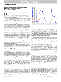
Parkinsonism and Related Disorders xxx (2015) 1e3
Contents lists available at ScienceDirect
Parkinsonism and Related Disorders
journal homepage: www.elsevier.com/locate/parkreldis
Letter to the Editor
Neuronavigation-guided transcranial magnetic stimulation of the dentate nucleus
improves cerebellar ataxia: A sham-controlled, double-blind n ¼ 1 study
Keywords:
Transcranial magnetic stimulation
Dentate nucleus
Ataxia
Lesions of the cerebellum can lead to hypotonia, ataxia, intention tremor, and dystonia [1], which can be disabling and resistant
to conventional treatments. The cerebrocerebellum receives input
exclusively from the cerebral cortex (via pontine nuclei) and projects to the motor, premotor, and prefrontal cortices and to the
ventrolateral nucleus of the thalamus via the dentate nucleus. It
is involved in motor planning, motor learning, and fine-tuning of
the motor activity, mainly by evaluating disparities between intention and action during the execution of movement. Cerebellar projections modulate activity in the contralateral primary motor cortex
(M1) through dentothalamocortical projections. Acute ischemic
damage to the deep cerebellar nuclei is known to result in ataxia
and loss of excitatory input to the contralateral M1 cortex [1]. However, chronic cerebellar ischemic lesions have recently been associated with reemerging increase in intracortical inhibition in the
contralesional M1, leading to marked inter-hemispheric asymmetry in cortical excitability, which could account for part of the functional impairment seen after stroke [2].
We report a case of post-surgical unilateral cerebellar infarction
leading to dystonia, intention tremor, and ataxia treated by inhibitory low-frequency neuronavigated repetitive transcranial magnetic stimulation (rTMS) to the healthy cerebellar hemisphere.
The aim was to decrease the functional cortical asymmetry related
to chronic cerebellar infarction [2]. We hypothesized that this treatment would balance the functional asymmetry seen between both
M1 after chronic cerebellar and attenuate clinical symptoms.
A 50-year-old female underwent a resection of a right acoustic
neuroma in 2006, which was complicated by ischemia of the right
cerebellar hemisphere (Fig. 1A). She developed severe axial and
appendicular ataxia, mostly on the right, with significant impairment in functioning. A few months later, she developed bilateral
involuntary hand and cervical dystonia (documented by electroneuromyographic study), which was refractory to medical treatment. Several treatments were attempted for the cerebellar
tremor, including: physical therapy, benzodiazepines, biperidene,
beta-adrenergic blocking agents, and primidone, without
satisfactory symptomatic improvement. The patient declined
deep brain stimulation for symptomatic tremor control. Because
of the refractory nature of the symptoms, the subject consented
to neuronavigated low-frequency TMS to the healthy dentate nucleus. A double-blind sham-controlled trial of 1 Hz neuronavigated
rTMS was performed on the left dentate nucleus with a refrigerated
double-cone rTMS coil (cooled DB-80, Magventure® TonikaElektronik, Denmark), which allows for the stimulation of deep
structures up to 4e5 cm from the coil center. The dentate nucleus
lay 50 mm deep from the skull surface, a distance similar to that
of the skull vertex to the leg area at M1 (48 mm) (Fig. 1B). We
detected the “hot spot” for M1 distal leg representation (peroneus
longus muscle), and then its rest motor threshold was measured.
The left dentate nucleus was then targeted by neuronavigation
and a 1500-pulse session of either active or sham stimulation was
performed at 90% of the rest motor threshold of the leg M1 area.
Two (active or sham) stimulation sessions were randomly performed four weeks apart. Clinical assessment and video recording
were performed using validated tools before stimulation and one
week after by movement disorder specialist blinded to the study.
After the active session, there was improvement in tremor (Fahn,
Tolosa, Marin Tremor Rating Scale before ¼ 38/144; after ¼ 24/
144 37% reduction) and in cerebellar ataxia (scale for the assessment and rating of ataxia [SARA] before ¼ 25.5/40, after ¼ 17/40)
(see video). The Patient Global Impression of Change (measured
on a seven-point Leikert scale ranging from 1 to 7) was 6 after
the active session (i.e. moderately improved) and 3 after the
sham procedure (i.e. no change). There was no change in her dystonia after active treatment (Unified Dystonia Rating Scale ¼ 9/112
before, and 9/112 after active stimulation). There was no clinical
improvement after sham stimulations using the same rating scales
Table 1.
Supplementary data related to this article can be found online at
http://dx.doi.org/10.1016/j.parkreldis.2015.05.010.
The dentate nucleus has a tonic facilitatory influence on the motor cortex and controls multi-joint movements [3]. In primates,
deep cerebellar nuclei have a primarily facilitatory effect on excitability in the contralateral primary motor cortex [1]. In rats,
hemi-cerebellectomy decreases intracortical facilitation and blocks
the facilitatory effects of somatosensory stimulation on M1 excitability [4]. In humans, TMS or electrical stimulation of the cerebellum given 5e7 ms before a TMS pulse is administered to the
contralateral M1 results in M1 inhibition, which is reflected in
decreased MEP amplitudes [3]. This is either due to preferential
excitation of Purkinje cells, which inhibit the dentate nucleus, or
to a direct disruptive effect of the TMS pulse upon output axons
http://dx.doi.org/10.1016/j.parkreldis.2015.05.010
1353-8020/© 2015 Elsevier Ltd. All rights reserved.
Please cite this article in press as: R.G. Cury, et al.Neuronavigation-guided transcranial magnetic stimulation of the dentate nucleus improves
cerebellar ataxia: A sham-controlled, double-blind n ¼ 1 study, Parkinsonism and Related Disorders (2015), http://dx.doi.org/10.1016/
j.parkreldis.2015.05.010
2
Letter to the Editor / Parkinsonism and Related Disorders xxx (2015) 1e3
Fig. 1. Brain MRI. (A) Axial T1-weighted image showing cerebellar infarction in the right hemisphere and a partially resected right acoustic neuroma (arrow). (B) Sagittal FLAIR
image reconstruction of the dentate nucleus and the leg area at M1, showing a similar distance of both areas from the skull surface.
Table 1
Evaluation of dystonia, tremor and ataxia after active and sham rTMS of the dentate
nucleus.
Active rTMS
UDRS
SARA
FTMTRS
%changea
Sham rTMS
Before
After
Before
After
09/112
25,5/40
38/144
09/112
17/40
24/144
09/112
25,5/40
38/144
09/112
25,5/40
38/144
none
33%
37%
UDRS: Unified Dystonia Rating Scale; SARA: Scale for the assessment and rating of
ataxia; FTMTRS: Fahn, Tolosa, Marin Tremor Rating Scale.
a
%change after Active rTMS.
rTMS study aiming at ataxia and tremor control. Despite limitations
of sham rTMS, especially in crossover designs, and the fact that offtarget cerebellar parenchyma was certainly stimulated due to
inherent properties of rTMS coils, we obtained positive results. In
our patient, the clinical improvement after active rTMS lasted for
about 10 days and peaked from the sixth to the seventh day before
gradually fading away. The reasons for these long-lasting effects
remain uncertain. The study protocol was safe and well tolerated,
and it provides sham-controlled evidence for larger studies using
repetitive sessions of deep rTMS in this patient population.
Conflict of interest
exiting the cerebellum via the dentate nucleus. In fact, cerebellar
infarctions result in acutely increased contralateral inhibition of
M1, which probably reflects the increase in M1 intracortical inhibition related to the loss of dentate-cortical facilitatory projections.
Consequently, there is a documented reduction in blood flow in
the healthy cerebral hemisphere in cases of unilateral cerebellar
infarction (crossed hemispheric diaschisis) [5]. However, chronic
infarctions have been associated with decreased intracortical inhibition in the contralesional M1 [2], leading to an abnormal asymmetry in cortical excitability between both cerebral hemispheres.
Based on these findings, low-frequency TMS has been employed
to decrease (or inhibit) the activity of neurons in the remaining
overactive cerebellar hemisphere, which would simulate the effects
of dentatotomy on spasticity and dystonia.
This study has some limitations. It is difficult to produce an
effective sham condition using TMS, especially using crossover designs. Although for the sham procedure the double-cone coil
created noise and bumps similar to an actual coil, there was a
lack of electric sensation in the scalp. Despite this drawback, this
same approach has been used in numerous studies in the rTMS
literature, providing results analogous to those using specific
sham coils [6,7]. Another point that has to be considered is that
while we ensured that the pulses reached the actual depth of the
dentate nucleus by obtaining MEP's of the distal leg area in M1
and showing the similar depth between both targets, other areas
of brain tissue were concomitantly stimulated with a less focused
and less intense induced electric current. The presence of these
“side-stimulations” is intrinsic to the rTMS method and its clinical
role can only be assessed in future specific studies.
This was the first controlled, neuronavigation-guided deep-
The authors report no conflicts of interest or financial
disclosures.
Funding sources for study
This project was not funded.
References
[1] R.N. Holdefer, L.E. Miller, L.L. Chen, J.C. Houk, Functional connectivity between
cerebellum and primary motor cortex in the awake monkey, J. Neurophysiol. 84
(2000) 585e590.
[2] S.N. Farias da Guarda, L.G. Cohen, M. da Cunha Pinho, F.I. Yamamoto,
P.E. Marchiori, M. Scaff, et al., Interhemispheric asymmetry of corticomotor
excitability after chronic cerebellar infarcts, Cerebellum Lond Engl. 9 (2010)
398e404.
[3] J. Liepert, T. Kucinski, O. Tüscher, F. Pawlas, T. B€
aumer, C. Weiller, Motor cortex
excitability after cerebellar infarction, Stroke J. Cereb. Circ. 35 (2004)
2484e2488.
[4] N.O. Ben Taib, O.B.T. Nordeyn, M. Manto, M. Mario, M. Pandolfo, P. Massimo, et
al., Hemicerebellectomy blocks the enhancement of cortical motor output associated with repetitive somatosensory stimulation in the rat, J. Physiol. 567
(2005) 293e300.
€nmezog
"lu, B. Sperling, T. Henriksen, P. Tfelt-Hansen, N.A. Lassen, Reduced
[5] K. So
contralateral hemispheric flow measured by SPECT in cerebellar lesions:
crossed cerebral diaschisis, Acta Neurol. Scand. 87 (1993) 275e280.
#-Obadia, R. Peyron, P. Mertens, F. Mauguie
$re, B. Laurent, L. Garcia-Lar[6] N. Andre
rea, Transcranial magnetic stimulation for pain control. Double-blind study of
different frequencies against placebo, and correlation with motor cortex stimulation efficacy, Clin. Neurophysiol. 117 (2006) 1536e1544.
[7] R. Galhardoni, G.S. Correia, H. Araujo, D.T. Fernandes, H.H. Kaziyama,
M.A. Marcolin, D. Bouhassira, M.J. Teixeira, D.C. de Andrade, Repetitive Transcranial Magnetic Stimulation (rTMS) in Chronic Pain: a review of the literature,
Arch. Phys. Med. Rehabil. 96 (2015) 156e172, http://dx.doi.org/10.1016/
j.apmr.2014.11.010.
Please cite this article in press as: R.G. Cury, et al.Neuronavigation-guided transcranial magnetic stimulation of the dentate nucleus improves
cerebellar ataxia: A sham-controlled, double-blind n ¼ 1 study, Parkinsonism and Related Disorders (2015), http://dx.doi.org/10.1016/
j.parkreldis.2015.05.010
3
Letter to the Editor / Parkinsonism and Related Disorders xxx (2015) 1e3
Rubens Gisbert Cury
Movement Disorders Center, Department of Neurology, School of
~o Paulo, Sa
~o Paulo, Brazil
Medicine, University of Sa
Guilherme Lepski
Functional Neurosurgery Division, Department of Neurology, School of
~o Paulo, Sa
~o Paulo, Brazil
Medicine, University of Sa
Transcranial Magnetic Stimulation Laboratories, Psychiatry Institute,
~o Paulo, Sa
~o Paulo, Brazil
University of Sa
Daniel Ciampi de Andrade*
Transcranial Magnetic Stimulation Laboratories, Psychiatry Institute,
~o Paulo, Sa
~o Paulo, Brazil
University of Sa
^ncer do Estado de Sa
~o Paulo Octavio Frias de Oliveira,
Instituto do Ca
~o Paulo, Brazil
Sa
Manoel J. Teixeira, Ricardo Galhardoni, Victor Rossetto Barboza,
Eduardo Alho
Transcranial Magnetic Stimulation Laboratories, Psychiatry Institute,
~o Paulo, Sa
~o Paulo, Brazil
University of Sa
Catharina Maria Seixas
~o Paulo, Brazil
School of Medicine, University of Campinas, Sa
^ncer do Estado de Sa
~o Paulo Octavio Frias de Oliveira,
Instituto do Ca
~o Paulo, Brazil
Sa
*
#as de Carvalho Aguiar, 255, 5"
Corresponding author. Av. Dr. Ene
#sar, 05403-900, S~
Andar, Sala 5084 e Cerqueira Ce
ao Paulo, SP,
Brazil.
E-mail address: [email protected] (D. Ciampi de Andrade).
3 December 2014
Please cite this article in press as: R.G. Cury, et al.Neuronavigation-guided transcranial magnetic stimulation of the dentate nucleus improves
cerebellar ataxia: A sham-controlled, double-blind n ¼ 1 study, Parkinsonism and Related Disorders (2015), http://dx.doi.org/10.1016/
j.parkreldis.2015.05.010
Baixar