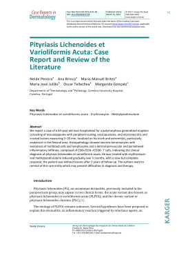
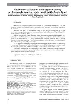
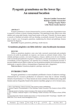
Case Report
Oral lichen planus
Oral lichen planus: two case reports in
male patients
Líquen plano oral: relato de dois casos em pacientes masculinos
Abstract
Purpose: Lichen planus is a chronic systemic disease that commonly involves the mucosa of
the oral cavity, most often in the absence of skin lesions. Although relatively frequent, oral
lichen planus is the target of much controversy, mainly in relation to its pathogenesis and
possible potential for malignancy. Thus, the objective of this paper is to report two cases of
lichen planus in male patients and to discuss the main aspects of this disease in relation to
etiopathogenesis and treatment.
Case description: Physical and histopathological examinations were carried out to confirm the
diagnosis of oral lichen planus. An elixir of dexamethasone was prescribed in the first case,
and in the second case, topical 0.05% clobetasol propionate was prescribed. After fifteen days,
both patients presented regression of the lesion.
Maiara de Moraes a
Felipe Rodrigues de Matos a
Joabe dos Santos Pereira a
Ana Myriam Costa de Medeiros a
Éricka Janine Dantas da Silveira a
Post-Graduate Program, Oral Pathology, Federal
University of Rio Grande do Norte, Natal, RN,
Brazil
a
Conclusion: Although the occurrence of oral lichen planus is common, an accurate diagnosis
is necessary so that the correct treatment can be established.
Key words: Lichen planus; oral mucosa; pathogenesis; diagnosis; treatment
Resumo
Objetivo: O líquen plano é uma doença crônica sistêmica comum que envolve a mucosa
oral, na maioria das vezes na ausência de lesões em pele. Embora relativamente comum, o
líquen plano oral é alvo de muita controvérsia, especialmente em relação à sua patogênese
e possibilidade de uma eventual transformação maligna. Assim, o objetivo deste trabalho é
relatar dois casos de líquen plano em pacientes do sexo masculino e discutir os principais
aspectos dessa lesão em relação a etiopatogênese e tratamento.
Descrição do caso: Exames físicos e histopatológicos foram realizados para confirmar o
diagnóstico de líquen plano oral. Foi prescrito elixir de dexametasona no primeiro caso e no
segundo caso, foi prescrito o uso tópico de propionato de clobetasol 0,05%. Após quinze
dias, os pacientes retornaram com regressão da lesão.
Conclusão: Embora a ocorrência de líquen plano oral é comum, o diagnóstico preciso é
necessário para que o correto tratamento possa ser realizado.
Palavras-chave: Líquen plano; mucosa oral; patogênese; diagnóstico; tratamento
Correspondence:
Éricka Janine Dantas da Silveira
Universidade Federal do Rio Grande do Norte –
Departamento de Odontologia
Av. Senador Salgado Filho, 1787 – Lagoa Nova
Natal, RN – Brasil
59056-000
E-mail: [email protected]
Received: August 11, 2009
Accepted: November 17, 2009
Conflict of Interest Statement: The authors state
that there are no financial and personal conflicts of
interest that could have inappropriately influenced
their work.
Copyright: © 2010 Moraes et al.; licensee
EDIPUCRS. This is an Open Access article distributed
under the terms of the Creative Commons AttributionNoncommercial-No Derivative Works 3.0 Unported
License.
208
Rev. odonto ciênc. 2010;25(2):208-212
Moraes et al.
Introduction
Lichen planus is a chronic inflammatory disease that affects
skin and mucosa. It is one of the most common dermatological
conditions involving the oral cavity. Its prevalence in the
general population is around 1% to 2%, and there are a large
number of cases in females (1-3).
The oral manifestations of lichen planus have been described
in the literature and can be classified into 3 types: reticular
lesions, including white lines, papules and plaques; atrophic
or erythematous lesions; and erosive lesions, including
ulcers and bullous lesions. Whereas reticular forms are
usually asymptomatic and sometimes discovered during a
routine oral clinical examination, the erythematous forms are
painful, causing discomfort to the patient (1,2). According
to Sousa et al. (3), clinically, oral lichen planus has specific
and clearly identifiable features, usually presenting in one
of two main forms – the reticular and the erosive forms,
although the other forms are not rare.
Since 1869, when the lesion was first described, the
etiopathogenesis of the lesion is still uncertain and is a
subject of many studies. Sugerman et al. (4) believe that
specific and non-specific mechanisms may be involved in
the etiopathogenesis. The specific mechanism involves the
presentation of antigen by keratinocytes and death caused by
cytotoxic T lymphocytes, and the non-specific mechanism
includes degranulation of mast cells and activation of matrix
metalloproteinases (1).
Lichen planus has potential for malignant transformation (2),
and the risk of malignant transformation varies between 0.4
and 5% in a period of observation from 0.5 to 20 years (5).
However, many controversies remain with regard to the risk
of malignant transformation, to the clinical form with the
greatest potential for malignancy, and about therapies used
for the treatment of OLP (5).
At microscopic examination, the lesion shows an intense
inflammatory infiltrate that is predominantly lymphocytic
immediately underlying the epithelium and organized into a
dense band-like pattern. The epithelium may be acanthotic,
and the cells of the basement layer may show hydropic
degeneration and loss of continuity (1).
As there is still no definitive treatment for many injuries,
drugs have been used to alleviate the painful effects and
the size of lesions and thus, to improve the oral health of
the patient. The drugs used have local or systemic actions,
and their major active components are corticosteroids such
as triamcinolone, fluocinolone acetonide and fluocinonida.
Elixirs of dexamethasone, clobetasol and triamcinolone are
used in patients with oral involvement (6). Aiming to better
understand the clinical behavior, the histopathology, and the
main treatments for patients with OLP, we propose to present
two case reports in male patients and to discuss issues in
their management.
Description of the cases
Case report 1
A 55-year-old, black, male patient was referred to the
Oral Diagnosis service complaining of rough tongue. The
patient reported that the change was noted one and a half
months earlier. He said that he had painful symptoms during
the ingestion of hot foods and that these symptoms had
been previously treated with different topical medicines,
including myconazol, nystatin solution and benzidamine
hydrochloride; however, the lesion did not regress. The
subject reported habits of smoking and drinking alcohol
beverages. The medical history did not have relevant
facts.
At clinical examination, the lesion was localized on the
dorsum of the tongue and was of the papular form (Fig. 1A),
and other lesions were identified in the left and right buccal
mucosa (Fig. 1B) that showed up as white striations; no
ulcer was present. The clinical diagnosis was oral lichen
planus. For confirmation of this diagnosis, an incisional
biopsy of this lesion was requested. An anti-HCV test was
also requested, and the result was negative.
Fig. 1. Lesion in the dorsum of the tongue with papular form (A) and in the right buccal mucosa with white
striations (B).
Rev. odonto ciênc. 2010;25(2):208-212
209
Oral lichen planus
The histopathological characteristics of the specimen
revealed a fragment of oral mucosa lined by stratified
squamous epithelium, with areas of ortho or parakeratosis,
acanthosis, spongiosis, focus of hydropic degeneration and
degeneration of the basement layer. The underlying lamina
propria was formed by fibrous connective tissue with variable
density and presented an intense inflammatory infiltrate,
mainly lymphocytic, situated in the subepithelial region and
arranged in a band-like pattern (Fig. 2). For treating the
painful symptoms, an elixir of dexamethasone, taken three
times a day for 15 days, was prescribed. After 15 days, a
decrease in the size of the two lesions was observed, with
complete remission of the symptoms observed previously.
The patient was asked to come again to the oral diagnosis
service in one year in order to follow his progress.
Fig. 2. Photomicrograph showing a lymphocytic infiltrate in
band-like pattern (H-E staining x 200).
Fig. 3. Lesions in lip (A), upper eyelids (B) and inferior member (C).
210
Rev. odonto ciênc. 2010;25(2):208-212
Case Report 2
A 42-year-oldmale patient was referred to the Oral Diagnosis
service complaining of lip lesions. The patient denied feeling
pain; however, he felt esthetically disturbed. He said that the
lesion had appeared about three months earlier and that he
had been treated with pomade and vitamin B. The previous
medical history revealed that the subject suffered from
arterial hypertension. At anamnesis, the patient reported
having suffered an anger episode and a bought of fidgeting,
and he thought that these events influenced the appearance
of the lesions. Considering his testimony, his emotional
stress was determined to be a potential causal factor to the
appearance of the lesions. We requested anti-HCV test, and
it was negative.
During the oral clinical exam, the presence of lesions on
the lower and upper lips was observed (Fig. 3A); additional
lesions were observed on the upper eyelids (Fig. 3B) and on
his body, mainly affecting the superior and inferior members
(Fig. 3C). These lesions were diverse in size and were white
plaque-like with normal consistence from the tissue. The
lesions did not show mobility or secondary signals that could
be added to the description; no ulcer was present. In view
of the facts, the clinical diagnosis was lichen planus, and,
in order to confirm the diagnosis by microscopical exam,
the patient was submitted to incisional biopsy of the lower
lip lesion.
The microscopic findings were the same as those for
the previous case, and in focal areas, the presence of
basophilic degeneration from collagenous fibers was
observed. The patient was treated topically with 0.05%
clobetasol propionate for fifteen days for regression of the
lesions. The evaluation of skin lesions was conducted by a
dermatologist.
Moraes et al.
Discussion
Lichen planus (LP) is a chronic autoimmune disease with
an unknown etiology that is marked by the invasion of
lymphocytic infiltrate within the epithelial tissue inducing
epithelial cell apoptosis and chronic inflammation (5). The
main areas involved are the skin and oral cavity, but it can
also occur in the vaginal mucous, scalp and nails (5-6).
The prevalence of oral lichen planus (OLP) in the general
population ranges between 0.5-2.6% (6).The median age
group affected ranges from the third to sixth life decades;
the occurrence in children is uncommon, and the disease
is more common in females (2). The patients in this case
study were males of 55 and 42 years, which fall into the age
range in which lichen planus is most commonly reported.
The precise etiology of this condition is unknown, but, in
their review, Ismail et al. (6) reported a list of causative
and exacerbating factors for OLP and oral lichen planus
reactions such as, drugs (anti-malarial, diuretics, gold salts,
antiretroviral), dental materials (dental amalgam, composite
and resin-based materials, metals), chronic liver disease and
hepatitis C virus, genetics and tobacco chewing. In our cases,
there were no situations like these that could have provoked
oral lichen planus reactions.
There is numerous evidence that cell-mediated immunity,
possibly initiated by endogenous or exogenous factors
in individuals genetically predisposed to the disease, is
crucial for the pathogenesis of the disease. Activated T
lymphocytes and increased production of cytokines result in
increased expression of the intercellular adhesion molecule
(ICAM-1) and the major histocompatibility complex type
II by keratinocytes, which leads to tissue destruction. This
process results in immune vacuolar degeneration, lysis of
cells in the basal layer, and, finally, dissolution of the cells
of the basal layer (7).
Stress was identified as one of the most frequent causes
of acute exacerbation of the disease (7,8). A recent study
suggests that patients with OLP exhibit higher levels of
anxiety and depression compared with control groups. In
addition to the discomfort that is caused by the lesion, many
patients are concerned about a possible malignancy and the
contagious nature of the lesion, which is favored by the
lack of educational materials available to individuals with
the disease. Therefore, the education of patients with OLP
can minimize their anxiety (7). In the case of Patient 2,
during the anamnesis he reported a change in his emotional
condition, and we noticed a certain anxiety in both patients
that may be related to the absence of knowledge about the
disease.
Six clinical forms of OLP have been described: reticular,
papular, plaque-like, erosive, atrophic and bullous (6,7). A
more simple clinical classification consists of three types
of lesions: reticulated lesions, including rows, plaques and
whitish papules; atrophic or erythematous lesions; and erosive
lesions, including ulcerations and bullous lesions. Whereas
the reticular lesions are asymptomatic, the erythematous and
erosive ones induce discomfort (7).
Clinically, the lesions in the oral cavity are usually multiple
and bilateral (5,6,8). OLP can involve any place in the oral
cavity; however, the main locals involved are the buccal
mucosa, gingival and tongue (6,8). The most common
clinical presentation is whitish striae in a reticulated pattern
(5,8). In the cases presented here, the anatomical areas of
the lesion were the back of the tongue, buccal mucosa and
lower lip, yonder cutaneous signs. The lesions in the buccal
mucosa had a striae shape and were reticulated, whitish, and
bilateral, and those from the tongue had a whitish papule
shape. The lip lesions were in whitish and purple plaques,
and there were whitish plaques on the inferior limbs.
The cases of LP that are restricted to oral mucosa, i.e., with
minimal involvement of the skin, occur in 15% of all cases.
Detailed reports of simultaneous occurrence of LP in the
oral cavity and skin are uncommon (5). In the patient in
Case 2, lesions were identified on the lips and on the skin
of the lower limbs. Cases with lichen planus isolated to a
single site are rare, (7) and the cases reported in this case
study corroborate the literature because the lesions involved
multiple sites.
Differential diagnoses include lichenoid eruptions associated
with medications, lichenoid lesions associated with contact
with restorative materials, leukoplakia, lupus erythematosus
and graft versus host disease (GVHD) (6).
The diagnosis of OLP is based on clinical and
histopathological findings. Classic histopathologic features
include the presence of a lymphocytic infiltrate in the
subepithelial region in band-like patterns, liquefactive
degeneration of the basal layer, Civatte´s bodies, which are
the presence of numerous eosinophilic colloid bodies along
with interface-epithelial tissue packs, variable degrees of
focal ortho or parakeratosis and irregular acanthosis (7,8).
The histopathological features were consistent with the
diagnosis of lichen planus, as the main findings were the
presence of band-like subepithelial lymphocytic infiltration
and degeneration of the basal layer.
The management of patients with OLP is very important.
The recognition of the form of OLP and its location and
questions about the clinical factors that may modify the
clinical appearance, such as Candida sp. infection and the
use of medications or irritation due to the use of prosthesis,
should be evaluated. A regular follow-up of the patient with
OLP should be done (8). The choice of treatment depends on
the severity and the discomfort. Unfortunately, there is no
treatment to permanently resolve the lesions. Drugs are used
to improve the condition of the patient. These medicaments
may be local or systemic. The active components are
corticosteroids such as triamcinolone, fluocinolone
acetonide and fluocinonida. An elixir of dexamethasone,
clobetasol and triamcinolone has been used in patients with
oral involvement (3). The propaedeutic used by our service
was the elixir of dexamethasone 0.1 mg/ml for intra-oral
lesions, and clobetasol propionate 0.5 g/g in the form of a
cream was prescribed for lesions in the lip. The patient with
skin manifestations was sent to a medical dermatologist for
evaluation and treatment of the skin lesions.
Rev. odonto ciênc. 2010;25(2):208-212
211
Oral lichen planus
According to Ismail et al. (6), maintenance of good oral
hygiene can enhance healing and lessen symptoms, and
exacerbating factors should be minimized or removed.
Surgical management, including cryosurgery and carbon
dioxide (CO2) laser, has been performed on OLP lesions,
but surgical excision is not recommended as the first-choice
treatment due to the inflammatory condition, which can
recur (6).
An undesirable complication of OLP is the development
of squamous cell carcinoma (SCC). Many studies have
been focused on this potential malignant transformation of
OLP (8), but the potential for malignancy of these
lesions is still controversial. The frequency of malignant
transformation ranges from 0.4 to 5%, with the highest rates
in the erythematous and erosive lesions (6,7).
Krutchkoff and Eisenberg (9) used the term lichenoid
dysplasia to describe lesions that resemble OLP but that
are dysplastic. Recent study suggests genetic changes such
as loss of heterozygosity (LOH) in dysplastic OLP that
are also seen in SCC. Zhang et al. (10) found a low loss
of heterozygosity (6%) in OLP without dysplasia when
compared with dysplastic OLP (40% for dysplasia average).
These findings support that epithelial dysplasia in OLP is an
indication to the risk of malignancy of the lesion (10).
Lichen planus is an autoimmune mucocutaneous disease that
does not have an effective treatment and that most frequently
causes significant discomfort and pain for the patient. A
suitable protocol for lichen planus includes the correct
identification of lesions by biopsy and histopathological
analysis and the use of anti-inflammatory drugs as a treatment.
When lichen planus occurs in the skin, patients should
always be referred to dermatologists; in other words, there
is a very important role of the multiprofessional actuation
to treat lichen planus, and regular clinical monitoring is
important because of the risk of malignant transformation
reported by some authors.
References
1. Bornstein MM, Hakimi B, Persson GR. Microbiological findings in
subjects with asymptomatic oral lichen planus: a cross-sectional
comparative study. J Periodontol 2008;79:2347-55.
2. McCartan BE, Healy CM. The reported prevalence of oral lichen
planus: a review and critique. J Oral Pathol Med 2008;37:447-53.
3. Sousa FA, Rosa LE. Oral lichen planus: clinical and histopathological
considerations. Braz J Otorhinolaryngol 2008;74:284-92.
4. Sugerman PB, Savage NW, Walsh LJ, Zhao ZZ, Zhou XJ, Khan A,
Seymour GJ, Bigby M. The patogenesis of oral lichen planus. Crit
Ver Oral Biol Med 2002;13:350-65.
5. Scully C, Carrozzo M. Oral mucosal disease: Lichen planus. Br J
Oral Maxillofac Surg 2008;46:15-21.
6. Ismail SB, Kumar SK, Zain RB. Oral lichen planus and lichenoid
reactions: etiopathogenesis, diagnosis, management and malignant
transformation. J Oral Sci 2007;49:89-106.
212
Rev. odonto ciênc. 2010;25(2):208-212
7. Einsen D. The clinical features, malignant potential, and systemic
associations of oral lichen planus: a study of 723 patients. J Am
Acad Dermatol 2002;46:207-14.
8. Epstein JB, Wan LS, Gorsky M, Zhang L. Oral lichen planus: progress
in understanding its malignant potential and the implications for
clinical management. Oral Surg Oral Med Oral Pathol Oral Radiol
Endod 2003;96:32-7.
9. Krutchkoff DJ, Eisenberg E. Lichenoid dysplasia: a distinct
histopathologic entity. Oral Surg Oral Med Oral Pathol 1985;60:
308-15.
10.Zhang L, Cheng X, Li Y, Poh C, Zeng T, Priddy R, Lovas J, Freedman
P, Daley T, Rosin MP. High frequency of allelic loss in dysplastic
lichenoid lesions. Lab Invest 2000;80:233-7.
Baixar