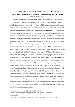
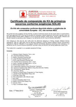
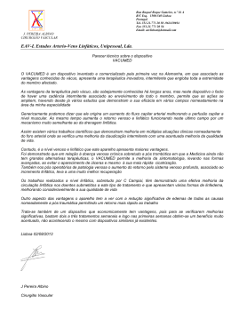
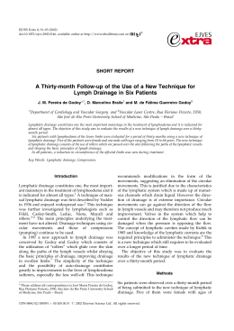
RSBO Revista Sul-Brasileira de Odontologia
ISSN: 1806-7727
[email protected]
Universidade da Região de Joinville
Brasil
Gadelha Vasconcelos, Marcelo; Câmara Santos, Bruna; Peixoto Lemos, Luciana Cristina; Fachetti
Ribeiro, Betania; Paraíso Iglesias, Déborah Pitta; Gadelha Vasconcelos, Rodrigo; Costa de Medeiros,
Ana Myriam
Oral lymphangioma: case report
RSBO Revista Sul-Brasileira de Odontologia, vol. 8, núm. 3, julio-septiembre, 2011, pp. 352-356
Universidade da Região de Joinville
Joinville, Brasil
Available in: http://www.redalyc.org/articulo.oa?id=153021326018
How to cite
Complete issue
More information about this article
Journal's homepage in redalyc.org
Scientific Information System
Network of Scientific Journals from Latin America, the Caribbean, Spain and Portugal
Non-profit academic project, developed under the open access initiative
ISSN:
Printed version: 1806-7727
Electronic version: 1984-5685
RSBO. 2011 Jul-Sep;8(3):352-6
Case Report Article
Oral lymphangioma: case report
Marcelo Gadelha Vasconcelos1
Bruna Câmara Santos2
Luciana Cristina Peixoto Lemos2
Betania Fachetti Ribeiro3
Déborah Pitta Paraíso Iglesias1
Rodrigo Gadelha Vasconcelos1
Ana Myriam Costa de Medeiros1
Corresponding author:
Marcelo Gadelha Vasconcelos
Avenida Praia dos Búzios, n. 9067 – Ponta Negra
CEP 59092-200 – Natal – RN – Brasil
E-mail: [email protected]
1
2
3
Program of Post-Graduation in Dentistry, Federal University of Rio Grande do Norte – Natal – RN – Brazil.
School of Dentistry, Federal University of Rio Grande do Norte – Natal – RN – Brazil.
Program of Post-Graduation in Dentistry, Federal University of Paraiba – Joao Pessoa – PB – Brazil.
Received for publication: September 28, 2010. Accepted for publication: December 6, 2010.
Keywords:
lymphangioma;
pathology; lymphatic
vessels.
Abstract
Introduction: Lymphangioma is a change of lymphatic vessels that
frequently affects the head and neck region. Its occurrence at oral
cavity is rare and it is most commonly identified at the anterior twothirds of the tongue. At this location, it is clinically characterized
as transparent and generally grouped vesicles, which can be red or
purple. The deep lesions appear as nodular masses of variable color
and superficial texture. It can be classified according to the size of
vessels into three types: capillary, cavernous, and cystic lymphangioma.
Several types of treatment have been suggested; and the most commonly
used treatments are: surgical excision, application of carbon dioxide
laser, cryotherapy using liquid nitrogen, and sclerosing agents.
Objective and case report: To describe a case of oral lymphangioma
diagnosed in a 17-year-old female patient. The lesion was presented
as multiple vesicles of soft consistency with thin epithelial lining and
color ranging from translucent to yellow-reddish, involving the soft
palate and the left retromolar region. Incisional biopsy confirmed the
hypothesis of cavernous lymphangioma. Patient was followed-up for
one year without signs of lesion relapse. Conclusion: Through this
clinical case report and literature review, this study emphasizes the
relevance of the clinical and histopathological features that should be
considered to confirm the clinical hypothesis and indicate the proper
therapeutic for oral lymphangiomas.
RSBO. 2011 Jul-Sep;8(3):352-6 – 353
Introduction
Lymphangioma is a benign hamartomatous
tumor of lymphatic channels, with a marked
pred i lect ion for t he head a nd neck reg ion,
at subma ndibula r a nd pa rot id a rea [2, 10].
Approximately half of the cases are present since
birth, and about 90% is developed before 2 years
old; it is rarely diagnosed in adults [11]. The lesion
is classified according to the vessels diameter into:
capillary or microcystic, cavernous or macrocystic,
and cystic or hygroma lymphangioma [7, 13].
At oral cavity, it presents a rare occurrence,
and it is more frequently located at the anterior
two-thirds of the tongue; although cases in palate,
gingiva, jugal mucosa, lips, and mandibular
alveolar ridge have already been described [2, 5,
12]. Clinically, its location is superficial, seen as
transparent and generally grouped vesicles, which
can be red or purple due to secondary hemorrhages.
The deep lesions appear clinically as nodular masses
of variable color and superficial texture [13].
There are several therapeutic modalities for the
treatment of oral lymphangioma. Surgical excision
is mostly indicated when vital structures are not
involved [7, 8]. Other treatments include the use
of sclerosing agents, application of carbon dioxide
laser and liquid nitrogen cryotherapy. However,
studies have evidenced that such methods are not
very efficient due to the possibility of lesion relapse
[3, 7, 8].
The aim of this study was to report a case of
lymphangioma in oral cavity treated by surgical
incision as well as to compa re t he clinica lpathological data found with the information
described in literature.
Case report
A 17-year-old female patient, brown skin, was
referred to the Clinic of Stomatology of the School
of Dentistry of the Federal University of Rio Grande
do Norte, complaining about blisters within oral
cavity for one year, which disrupted during feeding
and were related commonly to episodes of bleeding.
At clinical intraoral examination, we observed
exophytic vesicular lesions with thin epithelial lining
and color ranging from translucent to yellow-reddish,
of soft consistency and sessile insertion, giving to
the epithelial surface a granular aspect (figures 1
and 2). Clinically, such lesion was diagnosed as
lymphangioma and hemangyoma.
To confirm the diagnosis, an incisional biopsy
was executed, and the sample was referred to the
Service of Pathological Anatomy of the Discipline
of Oral Pathology of the Federal University of
Rio Grande do Norte for anatomopathological
examination. The histological cuts were examined
and stained in hematoxylin and eosin (HE), revealing
lymphatic vessels of great diameter distributed in
a conjunctive tissue poorly organized. Such vessels
were mainly located at subepithelial position,
occupying the lamina propria, and presented lumens
partially filled by proteic fluid content, erythrocytes,
and few lymphocytes (figures 3 and 4). Therefore, the
histopathological diagnosis confirmed the clinical
and surgical hypothesis of lymphangioma.
Patient was submitted to surgical treatment for
lesion enucleation. The procedure was facilitated
due to the superficial location of the lymphatic
vessels. After 1 year of following-up, we did not
observe the lesion relapse.
Figure 1 – Vesicular lesions of thin epithelial lining (some
disrupted) with granular ulcerated aspect on the soft
palate and retromolar region
Figure 2 – Detail of the vesicular lesions and ulcerated
areas in soft palate
Vasconcelos et al.
354 – Oral lymphangioma: case report
Figure 3 – Lymphatic vessels in subepithelial position
(HE 100X)
Figure 4 – Dilated lymphatic vessels partially filled
by proteic fluid content, erythrocytes and few
lymphocytes
Discussion
Lymphangiomas are vascular malformations. It
is hypothesized that they develop from congenital
obstruction or sequestration of the primitive
lymphatic cells that present proliferative capacity
and are commonly capable of accumulating fluids,
which give to them a cystic appearance. Because
these cells do not anastomose efficiently with
bigger lymphatic vessels, they then provoke areas
of lymphatic blockage [4, 10].
These lesions comprises about 30% of the lesions
of lymphatic vessel origin and present markedly
prediction by the neck and head area and common
at the cervical region, mainly at the posterior
triangle of the neck (upper middle third of clavicle,
anterior edge of trapezius muscle, posterior border
of sternocleidomastoid) [1]. The anterior triangle
may also be involved and the submandibular and
parotid regions are the more commonly associated
areas to lymphangioma development [2, 8]. The
lesions have been reported in several organs, and
only 1% occurs in the gastrointestinal tract [6].
Oral cavity rarely represents an occurrence site.
When oral cavity is affected, the most prevalent
site is the tongue, mostly restricted to its anterior
third; but the lesions can be extended and involve
great extensions of the tongue [1, 6]. Brennan et
al. [3] revised 49 cases of oral lymphangioma and
found that 17 were located in tongue, while only one
case was described at retromolar region and one
at soft palate. Therefore, the case described here
shows an uncommon site of oral lymphangioma
[2, 5], involving the retromolar region, extending
to the soft palate.
Lymphangioma is observed since birth or is
manifested before 2 years of age; consequently,
these lesions diagnosis is rarely performed in adult
patients [8]. In the case reported here, the lesion
developed in a patient older than the common age
group, who had 17 years-old at the moment of
the diagnosis and had reported that the problem
persisted by at least one year.
Clinically, oral lymphangioma is manifested
as clear vesicles giving the mucosa a granular
aspect. Such vesicles, which normally present a
translucent aspect due to the superficial location of
the lymphatic vessels, may occasionally assume a
red or blue aspect because of the rupture of blood
capillary into the lymphatic inner spaces [6, 10,
13]. In this case, the clinical lesions appearance is
in agreement with the form classically described
for oral lymphangiomas: grouped translucent
vesicles with some presenting reddish color. It
was observed that the vesicles characterizing
the granular aspect of the jugal mucosa surface
sometimes were disrupted, conferring the mixed
aspect (ulcerated and vesiculous) to the lesion. In
some cases the rupture of great part of the vesicles
can determine whether the lymphangioma would be
clinically manifested as a predominantly ulcerated
lesion [6].
The deep lesions generally are show n as
masses of diffuse growth, which depending on the
anatomical location may determine damages such as
tissue swelling, obstruction of upper airways, pain,
tongue’s extrusion, sialorrhea, deformity of jaws,
as well as difficulties in mastication, speech, and
during oral hygiene [6, 7]. Some reports described
RSBO. 2011 Jul-Sep;8(3):352-6 – 355
that lymphangiomas assume great proportions
and can measure 14 cm. Often, the fast growth
of the lesion occurs due to the association with a
secondary tumor or an infection in the respiratory
tract, which determine an increase of the lymph
production, blockage of the lymphatic drainage or
infection due to the tumor [10].
Contrastively, in this case, the lesions were
located more superficially and demonstrated
well-defined limits, which normally, according
to Leboulanger et al. [7], are not associated to
discomfort related to the lesion. Despite of the
asymptomatic feature described by the patient,
the lesion was associated with bleeding episodes
perceived during and/or after the meals. Therefore,
the presence of secondary hemorrhage inside some
lymphatic spaces accounted for the purple vesicles,
which can be misplaced by hemangioma [10].
Lymphangiomas are classified into three types:
simple (capillary or microcystic), composed of small
vessels as capillaries; cavernous (macrocystic),
comprising great and dilated lymphatic vessels
which often become macroscopically visible, and
cystic (hygroma cystic), in which the vessels occupy
great cystic spaces, commonly greater than 1 cm
[7, 13].
In oral cavity, the dense conjunctive tissue and
the presence of the skeletal muscles do not allowed
the vessels great expansion. Consequently, the most
common intraoral type is cavernous lymphangioma
[9]. Notwithstanding, because one lesion can display
the three aforementioned vessel diameters, this
classification is not routinely applied [10].
The microscopic examination of the histological
cuts stained in HE found the presence of vascular
spaces exhibiting a thin endothelial lining with
a single cell layer. This finding corroborated
the descriptions of Yaita et al. [14]. The authors
still added that these vessels did not present
an external layer of smooth muscle cells and
the immunohistochemical profile demonstrate a
weak expression for CD-1 and strong expression
for D2-40, a marker to be used in cases which
the differential diagnosis with blood vessels is
necessary. In this case, such lymphatic vessels
presented great lumens partially filled by proteic
fluid material, erythrocytes and few lymphocytes.
The preferential location of these vessels was
lamina propria, projecting on the oral mucosa
epithelium, compatible with either the translucent
or red vesicles, clinically identified [10, 14]. Areas
of epithelial discontinuity corroborate the clinical
findings of ulceration, previously described.
W h i le mu lt iple t he r a p e ut ic mo d a l it ie s
have been described for the treatment of deep
ly mpha n g ioma s, such a s su rg ica l excision,
cryotherapy, electrocauterization, eschlerotherapy,
steroids administration, embolization, and laser
therapy [14], surgical excision has been the best
alternative for lesions presenting localized growth
[2,6]. Because the lesions described in this case
report exhibited a superficial location, we opted for
their surgical removal. After one year of follow-up,
no sign of lesion relapse was identified.
Conclusion
Oral lymphangiomas are uncommon lesions
occurring at the dorsal region of the tongue, and
rarely at the retromolar and soft palate area.
Superficial and localized lesions can be treated
by conservative surgical excision with low relapse
rates. Therefore, this condition knowledge is of
fundamental importance for its correct diagnosis
and proper therapeutic indication.
References
1. Best SR, Coelho DH, Ahrens WA, Atez G,
Sasaki CT. Laser excision of multiple esophageal
lymphangiomas: a case report and review
of the literature. Auris Nasus Larynx. 2008
Jun;35(2):300-3.
2. Bozkaya S, Ugar D, Karaca I, Ceylan A, Uslu S,
Baris E et al. The treatment of lymphangioma in
the buccal mucosa by radiofrequency ablation: a
case report. Oral Surg Oral Med Oral Pathol Oral
Radiol Endod. 2006 Nov;102(5):e28-31.
3. Brennan TD, Miller AS, Chen
S. Lymphangiomas of the oral cavity: a
clinicopathologic, immunohistochemical, and
electron-microscopic study. J Oral Maxillofac
Surg. 1997 Sep;55(9):932-5.
4. Huang HY, Ho CC, Huang PH, Hsu SM.
Coexpression of VEGF-C and its receptors,
VEGFR-2 and VEGFR-3, in endothelial cells
of lymphangioma. Implication in autocrine or
paracrine regulation of lymphangioma. Lab Invest.
2001 Dec;81(12):1729-34.
5. Iamaroon A, Pongsiriwet S, Srisuwan S,
Krisanaprakornkit S. Lymphangioma of the tongue.
Int J Paediatr Dent. 2003 Jan;13(1):62-3.
6. Jian XC. Surgical management of lymphangiomatous
or lymphangiohemangiomatous macroglossia. J
Oral Maxillofac Surg. 2005 Jan;63(1):14-9.
Vasconcelos et al.
356 – Oral lymphangioma: case report
7. Leboulanger N, Roger G, Caze A, Enjolras O,
Denoyelle F, Garabedian E. Utility of radiofrequency
ablation for haemorrhagic lingual lymphangioma. Int
J Pediatr Otorhinolaryngol 2008 Jul;72(7):953-8.
8. Mandel L. Parotid area lymphangioma in an
adult: case report. J Oral Maxillofac Surg. 2004
Oct;62(10):1320-3.
9. Mosca RC, Pereira GA, Mantesso A. Cystic
hygroma: characterization by computerized
tomography. Oral Surg Oral Med Oral Pathol Oral
Radiol Endod. 2008 May;105(5):e65-9.
10. Neville BW, Damm DD, Allen CM, Bouquot JE.
Tumores dos tecidos moles. In: Patologia oral e
maxilofacial. 3.
���������������������������������������
ed. Rio de Janeiro: Elsevier; 2009.
p. 509-72.
11. Queiroz AM, Silva RA, Margato LC, NelsonFilho P. Dental care manegement of a young
patiet with extensive lymphangioma of the tongue:
a case report. Spec Care Dentist. 2006 JanFeb;26(1):20-4.
12. Stănescu L, Georgescu EF, Simionescu C,
Georgescu I. Lymphangioma of the oral cavity. Rom
J Morphol Embryology. 2006;47(4):373-7.
13. Weiss SW, Goldblum JR. Tumors of lymph
vessels. In: Soft tissue tumors. 4. ed. St. Luis:
Mosby; 2001. p 955-67.
14. Yaita T, Onodera K, Xu H, Ooya K.
Histomorphometrical study in cavernous
lymphangioma of the tongue. Oral Dis. 2007
Jan;13(1):99-104.
Baixar