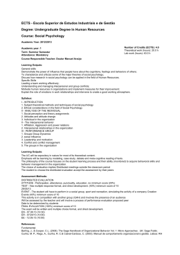
MONITORIZATION OF ALVEOLAR DEPOSITED SURFACE AREA OF
NANOPARTICLES AND ULTRAFINE PARTICLES IN DIFFERENT ENVIRONMENTS
Albuquerque, P.1; Gomes, J. 2,3; Bordado, J. 3, Reis, M.4
1ESTESL
2ISEL
– Escola Superior de Tecnologia de Saúde de Lisboa – Área Científica de Saúde Ambiental - Instituto Politécnico de Lisboa, Av. D. João II, Lote 4.69.01, 1990-096 Lisboa, Portugal
– Instituto Superior de Engenharia de Lisboa - Área Departamental de Engenharia Química – Instituto Politécnico de Lisboa, R. Conselheiro Emídio Navarro, 1959-007 Lisboa, Portugal
3IBB – Instituto de Biotecnologia e Bioengenharia / Instituto Superior Técnico – Universidade Técnica de Lisboa, Av. Rovisco Pais, 1049-001 Lisboa, Portugal
4Instituto de Medicina Preventiva, Faculdade de Medicina de Lisboa, Universidade de Lisboa, Av. Prof. Egas Moniz, 1649-028 Lisboa, Portugal
Email: [email protected]
INTRODUCTION
Current workplace exposure limits are based on particle mass, but this criteria could not be adequate in this case as nanoparticles are characterized by very large surface area,
which has been pointed out as the distinctive characteristic that could even turn out an inert substance into another substance exhibiting very different interactions with
biological fluids and cells. In fact, nanoparticles have far more surface area for the equivalent mass of larger particles, which increases the chance they may react with body
tissues. Thus, it has been claimed that surface area should be used for nanoparticle exposure and dosing. As a result, assessing exposure based on the measurement of
particle surface area is of increasing interest. It is well known that lung deposition is the most efficient way for airborne particles to enter the body and cause adverse health
effects.
If nanoparticles can deposit in the lung and remain there, have an active surface chemistry and interact with the body, then, there is potential for exposure.
It was showed that surface area plays an important role in the toxicity of nanoparticles and this is the metric that best correlates with particle-induced adverse health effects.
The potential for adverse health effects seems to be directly proportional to particle surface area. The objective of the study is to identify and validate methods and tools for
measuring nanoparticles during production, manipulation and use of nanomaterials.
MATERIALS AND METHODS
A Nanoparticle Surface Area Monitor (TSI 3550 – Fig. 1), was used for assessing exposure to
nano particles produced and manipulated in laboratory and industrial facilities. This
equipment indicates the human lung-deposited surface area of particles expressed as
square micrometers per cubic centimeter of air (μm2/cm3), corresponding to
tracheobronchial (TB) and alveolar (A) regions of the lung.
Fig. 1 - TSI Model 3550 - Nanoparticles Surface Area Monitor (NSAM) *,
measures surface deposited area of nanoparticles in alveolar and
tracheobronquial tracts of human lungs, based on diffusion charging.
Fig. 2 - TSI Model 3034 Scanning Mobility Particle
Sizer Spectrometer
(SMPS)**
Also, granulometry of particles was measured in the nano range using a Scanning
Mobility Particle Size Spectrometer, (TSI 3034 - Fig. 2). Particles were sampled using a
Nanometer Sampler Analyser, (TSI 3089 – Fig. 3) and observed further on using scanning
electronic microscopy.
The obtained results clearly demonstrated the existence of airborne nanoparticles, as
shown in figures 1 and 2, and also allowed the determination of lung-deposited surface
area of nanoparticles, as well as the dose per unit lung mass and unit lung area, in the
analyzed environments [1-4].
Fig. 3 - TSI Model 3089 Nanometer Aerosol
Sampler (NAS) *
RESULTS
Table 1. Measurement results over a typical week (May, 2011)
Sampling
conditions
Baseline
Monday
Tuesday
Wednesday
Thursday
Friday
Saturday
Sunday
Average deposited
area (µm2/cm3)
34.1 ± 5.0
57.6 ± 5.7
89.2 ± 8.0
87.1 ± 7.5
82.2 ± 7.2
75.0 ± 6.9
35.0 ± 4.5
34.9 ± 4.0
Range of values
(µm2/cm3)
25.5 – 50.3
14.7 – 343.3
27.4 – 510.5
31.1 – 421.1
23.2 – 245.7
17.8 – 511.1
6.91 – 365.5
4.94 – 252.2
TWA for 8h
(µm2/cm3)
1.07
172.8
267.6
261.4
165.2
143.6
107.4
104.7
Total deposited area
(µm2)
5.12 x 10⁵
8.29 x 10⁷
1.28 x 10⁸
1.25 x 10⁸
4.72 x 10⁷
4.70 x 10⁷
4.26 x 10⁷
4.10 x 10⁷
Dose per lung area
(µm2/m2)
6.40 x 10³
1.04 x 10⁶
1.61 x 10⁶
1.57 x 10⁶
5.90 x 10⁵
4.60 x 10⁵
3.40 x 10⁵
3.20 x 10⁵
Fig. 5 – Observations on
collected particles by
transmission electron
microscopy (TEM)
Fig. 4 – Size distribution of particles during a
typical week day (May 2011)
CONCLUSIONS
Previous studies (Fissan et al., 2007; Kuhlbisch et al., 2000; Ntziachristos et al., 2007) confirmed evidence that diffusion chargers are useful and reliable instruments for
measuring ambient aerosol concentrations in different environments and that their signal can be combined with the number concentration of particles to provide an estimate of
the mean diameter in real time.
The study clearly demonstrated the existence of ultrafine particles due to automobile traffic, which could be confirmed by the measurements of size distribution and
morphology of sampled particles. Also, this seems to be consistent with observations of ultrafine particles concentrations in other major towns. Mainly during week days,
observed concentrations can be as high as 2,6 times the measured baseline level.
It should be noted that, although measured parameters such as the deposited area and the dose per lung area, are elevated when compared with baseline values, mainly for
week days where automobile traffic is more intense, they cannot, at this stage, be ascertained as toxicity indicators. Nevertheless, they point out for important contamination of
potentially hazardous particles released from automobile traffic in urban environments. Data obtained in this study is a basic information allowing to understand the relationship
between exposure to ultrafine particles in urban atmospheres and health affections, which can be taken as the basis for epidemiologic studies.
As ultrafine particles can have a significant lifetime in urban air, possible effects on health cannot be neglected.
References:
[1] Albuquerque, P., Gomes, J., Bordado, J., Journal of Air & Waste Management Association, 62 (2012) 373-380.
[2] Gomes, J., Albuquerque, P., Miranda, R., Vieira, M., Journal of Toxicology and Environmental Health – A, 75 (2012) 747-755.
[3] Gomes, J., Albuquerque, P., Miranda, R., Santos, T., Vieira, M., Inhalation Toxicology, 24 (2012) 774-781
[4] Bordado, J., Gomes, J., Albuquerque, P., Journal of Air & Waste Management Association, 62 (2012) 1170-1180.
* Co-financed: Autoridade para as Condições de Trabalho (ACT) – Project 035 APJ/09 - “Nanopartículas em
Ambientes Interiores e Efeitos na Saúde Humana”
** Thanks to Departamento de Engenharia Mecânica, Faculdade de Ciências e Tecnologia da Universidade de Coimbra .
Baixar