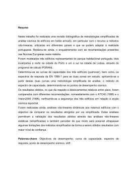
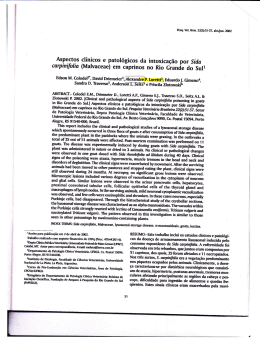
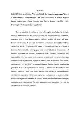
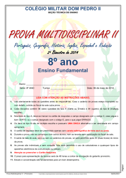
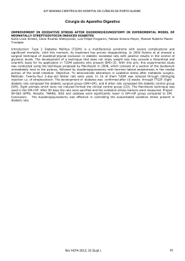
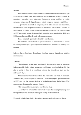
ANDREA MAYUMI KOROISHI ATIVIDADE ANTIFÚNGICA IN VITRO DOS EXTRATOS E NEOLIGNANAS DE Piper regnellii CONTRA DERMATÓFITOS Dissertação apresentada ao Programa de Pós-Graduação em Ciências Farmacêuticas (área de concentração – Produtos naturais e sintéticos biologicamente ativos), da Universidade Estadual de Maringá para a obtenção do grau de Mestre em Ciências Farmacêuticas. Orientador: Prof. Dr. Benedito Prado Dias Filho Co-orientador: Prof. Dr. Diógenes Aparício Garcia Cortez Maringá 2006 Este trabalho foi realizado no Departamento de Farmácia e Farmacologia da Universidade Estadual de Maringá, sob orientação do Prof. Dr. Benedito Prado Dias Filho, e contou com o apoio financeiro parcial do Conselho Nacional de Desenvolvimento Científico e Tecnológico (CAPES), Fundação Araucária e Programa de Pós-graduação em Ciências Farmacêuticas da Universidade Estadual de Maringá. Aos meus pais, Toshiharu Koroishi e Marly Mieco Ishizu Koroishi, ao meu irmão Edson Hideki Koroishi, ao meu namorado Daniel Rodrigues Silva, aos meus avôs Munenobu Ishizu, Siyone Koroishi e Tissato Koroishi pela paciência, incentivo, carinho e amor. AGRADECIMENTOS Ao professor Dr. Benedito Prado Dias Filho, meus sinceros agradecimentos, não apenas pela orientação firme e segura demonstrada na elaboração deste trabalho, mas também pelo incentivo, confiança e amizade nesses anos de convivência. Ao coordenador do Programa de Pós-graduação em Ciências Farmacêuticas da Universidade Estadual de Maringá, Prof. Dr. Celso Vataru Nakamura. Ao Prof. Dr. Diógenes Aparício Garcia Cortez, pela co-orientação e amizade. Aos professores Dr. Benício Alves de Abreu Filho e Dra. Tânia Ueda-Nakamura pela amizade, pelos conhecimentos transmitidos e pela confiança depositada. A aluna de iniciação científica e grande amiga Simone Rochtaschel Foss pelo apoio na elaboração de todo o trabalho. Aos meus amigos que apoiaram em todos os momentos Cecília Valente Truite, Elza Yamaguti, Jean Colacite, Nilza de Lucas Rodrigues Bittencourt, Raíssa Bocchi Pedorso. Aos meus colegas que me auxiliaram no Laboratório de Microbiologia Aplicada a Produtos Naturais e Sintéticos e pela grande amizade Amanda Bortoluci da Silva, Denise de Oliveira Scoaris, Eliana Harue Endo, Ivens Camargo Filho, Adriana Oliveira dos Santos, Érika Ravazzi Franco Ramos, Heloísa Bressan Gonçalvezs, Kelly Ishida, Rafael Eidi Yamamoto, Simone Evellyn Daniel Hernandes, Michele Cristina Vendrametto, Patrícia Mayumi Honda, Thelma Onozato. Aos técnicos do Laboratório de Microbiologia e Farmacognosia Adimir Arantes, Adriana R. Barra Vieira, Maria Manzoti, Marcio Guilhermetti, Marinete Martinez, Prisciliana Carvalho, Rosana Monteiro, Zelita Rodrigues pela amizade. Às minhas grandes amigas Ângela Delly Cembranel, Patrícia Nishimura, Solange Pinoti Primo pelo incentivo e companheirismo. Ao Programa de Pós-graduação em Ciências Farmacêuticas desta Universidade e ao corpo técnico/administrativo, pela oportunidade de realização deste trabalho, apoio e serviços prestados, em especial a Helena e a Sônia, secretárias do Departamento de Farmácia e Farmacologia. E a todos que contribuíram de alguma forma para a realização deste trabalho, muito obrigada. “A coisa mais bela que o homem pode experimentar é o mistério. É essa a emoção fundamental que está na raiz de toda a ciência e de toda a arte.” (Albert Einstein) RESUMO O presente estudo foi desenvolvido para avaliar a atividade antifúngica dos extratos hidroalcoólicos das folhas de Piper regnellii, comumente conhecida como pariparoba, amplamente distribuída em regiões tropicais e subtropicais. As micoses superficiais estão presentes em regiões cutâneas, pêlos, unhas, e os agentes patogênicos compreendem os fungos dermatófitos (Microsporum, Trichophyton e Epidermophyton), Piedraia hortai, Corynebacterium sp., Malassezia furfur (Pitiríase versicolor), Nocardia minutíssima (Eritrasma), Cladosporium vernecki (Tinea nigra), Aspergillus peniciloides (Tinea albigena), e além disso, as leveduras, Rhodotorula, Torulopsis, Cryptococcus, Candida, Trichosporon, Klöckera.. Foram obtidos extratos brutos hidroalcoólicos das folhas de Piper regnellii a 50, 70 e 90%, sendo o mais ativo o extrato bruto 90% frente aos fungos dermatófitos T. rubrum, T. mentagrophytes, M. canis e M. gypseum (CIM= 15,6, 15,6, 15,6 e 62,5 µg/mL, respectivamente) e inativo a fungos não dermatófitos Aspergillus niger (CIM>1000 µg/mL) e levedura, Candida albicans (CIM>1000µg/mL). O modelo biológico escolhido foi T. rubrum por ser mais sensível as drogas. Baseado nestes resultados, o extrato hidroalcoólico 90% foi fracionado por cromatografia em coluna com ensaios bio-dirigidos, sendo a fração clorofórmio mais ativo com CIM=6,2 µg/mL. A fração ativa foi então submetida à cromatografia em camada delgada preparativa, foram obtidos seis bandas, e a mais ativa F2C, apresentou CIM de 3,1 µg/mL, esta foi caracterizada em cromatografia líquida de alta eficiência e foram obtidas três substâncias, o conocarpano (1), eupomatenóide-5 (3) e eupomatenóide-6 (4), ambas com CIM igual a 100 µg/mL. Para o entendimento do mecanismo de ação foram feitos ensaios de inibição da germinação de esporos, inibição do crescimento de hifas, invasão em unhas e de citotoxicidade. O extrato bruto 90% inibiu a germinação de esporos e crescimentos de hifas a 7,8 µg/mL e no ensaio de citotoxicidade, cerca de 98% das células estavam viáveis a 50 µg/mL, indicando assim que o extrato bruto hidroalcoólico 90% é seletivo às células fúngicas. Além disso, o extrato ativo foi fungicida na concentração de 1,2 mg/mL no ensaio de invasão em unhas. Em conclusão, os dados in vitro podem ser úteis no uso de folhas de Piper regnellii para o tratamento de infecções por dermatófitos, e em termos de conservação, indicando que pode ser usado sem danos à planta. Palavras-chave: Piper regnellii, Piperaceae, eupomatenóide, antidermatófito, neolignanas LISTA DE ILUSTRAÇÕES Figure 1 Effect of different concentrations of the hydroalcoholic (90%) extract of P. regnelli leaves on growth of M. canis (A) and T. rubrum (B). The spore suspensions (5 l) were mixed with 20 l of various concentrations of crude extract (0.03, 0.07, 0.15, 0.32, 0.64, 1.28, 2.5 and 5.0g/ml) and added on petri dishes containing SDA, as described in Materials and methodos. The minimal concentration of crude extract that resulted in inhibition of both dermatophytes was 1.28 g. Data correspond to one representative experiment out of three………………………………………52 Figure 2 Spore germination inhibition of hydroalcoholic (90%) extract against T. rubrum: (A), Control; (B) and (C) treated with 3.9 and 7.8 g/ml of hydroalcoholic (90%) extract, respectively. Magnification x 20. Similar results were obtained in the different analyses…………………………………………………………………………..53 Figure 3 Growth of T. rubrum on cover slips after treatment with different concentrations of P. regnellii leaves extracts. (A) 7.8µg/mL 50% hydroalcoholic extract; (B) 15.62µg/mL 70% hydroalcoholic extract; (C) 7.8 g/ml 90% hydroalcoholic extract; (D) 0.3 g/ml Nystatin. Data correspond to one representative experiment out of three………………………………………………………………………54 Figure 4 Light microscopy (A-B) and scanning electron microscopy (C-D) of nail fragments treated (A and C) and untreated (B and D) with hydroalcoholic (90%) extract at a concentration of 1.2 g/ml. Similar results were obtained in the different analyses……………………………………………………………………………55 Figure 5 Cytotoxic assay. The hydroalcoholic (90%) extract was also evaluated for its potential toxic effects on Vero cell monolayer. After 48 h of incubation with 50 g/ml of crude extract (A) and 5 g/ml of Nystatin (B) 98 and 100 % of the cells were still viable, respectively. (C) Control. These results indicate that hydroalcoholic (90%) extract is selectively toxic to the fungal cells. Similar results were obtained in the different analyses…………………………………………………………………56 Figure 6 (A) Thin layer chromatography plates of hydroalcoholic extracts (50, 70 and 90%, v/v) and fractions (F1 to F9) obtained from active hydroalcoholic (90%) extract. (B). Preparative thin layer chromatography of the chloroform fraction being visualized, 6 bands were scraped from the other set, fractionated by thin-layer chromatography (F2A-F2F), assayed for antifungal activity and used for bioassayguided isolation. Similar results were obtained in the different analyses……………………………………………………………………………57 Figure 7 HPLC on reverse-phase resin Microsorb C-18. (A) The sub-fraction F2C with antifungal activity was applied in a reverse-phase column Microsorb- MV 100-5 C-18 (250 x 4.6) equilibrated and eluted with. Acetonitrile:water (60:40, v/v) containing 2% acetic acid: flow-rate of 1.0 ml/min; temperature: 30 °C; detection: 280 nm. (B) The standard mixture of neolignans: conocarpan (1), eupomatenoid-6 (2) and eupomatenoid-5 (3). Data correspond to one representative experiment out of three…………………………………………………………….…………...…58 Figure 8 Structures of the neolignans isolated from P. regnellii. (1) conocarpan, (2) eupomatenoid-3, (3) eupomatenoid-5, (4) eupomatenoid-6..…………………………….......59 LISTA DE TABELAS TABELA 1 Minimal inhibitory concentration (MIC) of hydroalcoholic crude extracts of leaves from P. regnellii………………………………………………….….60 TABELA 2 Antifungal activity of fraction obtained from hydroalcoholic extract (90%) of leaves from P. regnellii against dermatophyte T. rubrum…………………..61 TABELA 3 Minimal concentration of sub-fractions obtained from fraction F2 (chloroform) required for inhibition of spore germination of dermatophyte Trichophyton rubum………….……………………………………………………………62 SUMÁRIO 1 REVISÃO BIBLIOGRÁFICA............................................................................................14 1.1 PLANTAS MEDICINAIS..................................................................................................14 1.2 FAMÍLIA PIPERACEAE...................................................................................................15 1.3 FUNGOS DERMATÓFITOS.............................................................................................19 1.4 MICOSES SUPERFICIAIS................................................................................................19 1.5 AGENTES ANTIFÚNGICOS............................................................................................20 2 OBJETIVOS.........................................................................................................................25 REFERÊNCIAS...................... ……………………………………………………………26 3 ARTIGO: IN VITRO ANTIFUNGAL NEOLIGNANS FROM ACTIVITY OF EXTRACTS AND Piper regnellii AGAINST DERMATOPHYTES……………………….…………………………...33 4 ANEXO……………………………….………………………………………………….52 1- REVISÃO BIBLIOGRÁFICA 1.1 - PLANTAS MEDICINAIS No planeta existe uma grande variedade de plantas, cerca de 250 – 500 mil espécies, contudo a maioria ainda desconhecida cientificamente e pouco mais de 5% estudada fitoquimicamente, e uma porcentagem menor avaliada sob os aspectos biológicos (CECHINEL FILHO e YUNES, 1998). Até o início do século XIX, a atividade terapêutica das plantas medicinais utilizadas na época pouco se diferenciava dos medicamentos (SCHENKEL et al, 2004). As plantas utilizadas na medicina popular em todo o mundo, e em especial na América Latina, têm uma grande importância no atendimento primário à saúde. Entretanto, poucas plantas foram validadas do ponto de vista farmacológico, fitoquímico, biológico ou clínico (YUNES e CECHINEL FILHO, 2001). É importante ressaltar que plantas utilizadas na medicina popular com finalidades terapêuticas, bem como os estudos de plantas com fins de preservação ambiental, têm contribuído, e muito, para a descoberta de novos fármacos que recentemente são utilizados em tratamentos clínicos. Espécies nativas dos gêneros Baccharis sp. (carqueja), Bauhinia sp. (pata-de-vaca), Cecropia sp.(embaúba), Maytenus sp. (espinheira santa), Mikania sp. (guaco) e Passiflora sp. (maracujazeiro) são de uso popular, além disso, outras espécies nativas do Brasil foram inicialmente usadas com finalidade terapêutica por comunidades indígenas e caboclas (REIS et al, 2004). Nos séculos IX e X, descobriram compostos ativos de Valeriana officinalis com ação sedativa, entre os compostos ativos estão os ésteres do ácido isovalérico. Em 1763, Salix alba foi estudada devido ao uso das cascas para combater a febre e a dor, descobriu-se que tinha efeito analgésico. Em 1828, Buchner isolou a salicilina (glicosídeo do álcool salicílico). Em 1860, Kolbe e Lauteman sintetizaram o ácido salicílico e seu sal sódico a partir do fenol, e assim, ocorreu a primeira produção de salicilatos em 1874. Felix Hofman, em 1898, acetilou o grupo hidroxila em posição orto, e descobriu o ácido acetil salicílico, este foi o primeiro fármaco sintético a partir de uma molécula derivada de uma planta (YUNES e CECHINEL FILHO, 2001). Portanto se faz necessário à ampliação dos estudos com plantas que sejam tanto de uso popular ou não, pesquisando assim, novas moléculas com mais seletividade, baixa toxicidade e alto potencial farmacológico. 1.2 – FAMÍLIA PIPERACEAE A família Piperaceae pertence à classe Magnolipsida, subclasse Magnoliidae, subordem Nymphaeiflorae e ordem Piperales (MAcRAE; TOWERS, 1984; SANTOS et al., 2001 apud PESSINI, 2003). Está presente desde o México até o sudeste da Argentina (FIGUEIREDO, 2000). A família Piperaceae é constituída pelos gêneros Peperomia, Ottonia, Pothomorphe e Piper, e é um exemplo por possuir propriedades medicinais largamente empregadas pela população (COSTA, 1972). Arrigoni-Blank et al (2004) verificou que o extrato aquoso de partes aéreas de Peperomia pellucida inibiu o edema induzido por carragenina em animais, e que isso, poderia ser pela inibição de diferentes efeitos e mediadores químicos da inflamação. Verificaram também que esta espécie tem um efeito analgésico, provavelmente envolvido no mecanismo de síntese de prostaglandinas. Pothomorphe umbellatta tem sido usada como agente analgésico, diurético e antiespasmódico, antinflamatório, antimalárico, asma e distúrbios gastrointestinais. Estudos têm mostrado que uma concentração de 500mg/Kg de extrato hidroetanólico de P. umbellatta inibiu o edema de pata induzido por carragenina durante quatro horas, efeito similar ao da indometacina, comprovando assim, o uso popular no tratamento de distúrbios inflamatórios (PERAZZO, 2005). Estudos anteriores mostraram que a piperovatina, uma amida encontrada em Ottonia frutescens, apresentou atividade biológica de anestesia, além de outras características como um leve efeito de salivação e queimação na língua (MAKAPUGAY et al, 1983). A piperovatina também foi encontrada em Piper alatabaccum (FACUNDO, 2005) O gênero Piper tem cerca de 700 espécies amplamente distribuído em regiões tropicais e subtropicais, tanto no hemisfério norte quanto no sul, além disso, é usado popularmente como medicinal (PARMAR et al, 1997). P. sarmentosum, conhecida popularmente como “Cha-plu” na Indonésia, Malásia e Tailândia, possui amidas ativas contra a Mycobacterium tuberculosis e Plasmodium falciparum, a sarmentina e 1-piperetil pirrolidina (RUKACHAISIRIKUL, 2004). A presença de metil-éster ácido lancefólico e chalcona pinocembrina de P. lanceaefolium foram ativos contra Candida albicans, comprovando o uso popular na Colômbia (LÓPEZ et al, 2002). P. dilatatum, usado popularmente pelos índios Kuna no Panamá, apresentou compostos isolados a partir do extrato diclorometano fracionado em cromatografia em coluna de sílica gel e purificado em coluna de Sephadex gel LH 20. Os compostos obtidos foram ativos contra um fungo patogênico de plantas, o Cladosporium cucumerinum (TERREAUX et al, 1998). Foi relatado também a presença de diversas classes de substâncias em outras espécies de Piper como neolignanas e lignanas em P. clarkii (PRASAD et al, 1995), ciclohexanos oxigenados em P. cubeb (KOUL et al, 1996), fenilpropanóides e neolignanas em P. regnellii (BENEVIDES et al, 1999), neolignanas em P. aequale (MAXWELL et al, 1999). Holetz et al (2002) em um estudo de avaliação de plantas utilizadas popularmente para o tratamento de doenças infecciosas, verificaram que P. regnellii apresenta CIM de 7,8µg/mL para S. aureaus, 15,6µg/mL para B. subtilis, 250µg/mL para P. aeruginosa, 125µg/mL para Candida krusei. N O O N 4 MeO 1 2 O O N OCH3 O 3 H H 4 OBr C H3C H H OMe OAc OAc H AcO AcO H O O OMe O H OH 6 5 7 H3C H3C O OH H3CO O OH 8 9 Figura 1 (1) Piperovatina (Ottonia frutescens), (2) sarmentina e (3) 1-piperetil pirrolidina (P. sarmentosum), (4) 4’-metóxi flavona (P. clarkii), (5) piperenol C (P. cubeb), (6) dilapiol (P. regnellii), (7) conocarpano, (8) eupomatenóide-6, (9) eupomatenóide-5 (P. aequale). 1.3. FUNGOS DERMATÓFITOS Os fungos dermatófitos pertencem ao reino Fungi, divisão Deuteromicetes, classe Coelomicetes, ordem Moniliales, família Moniliaceae e gêneros Trichophyton, Microsporum e Epidermophyton. Os fungos dermatófitos se reproduzem assexuadamente pelo processo de gemulação, ou seja, as células se desprendem das hifas, sofrem diferenciação e disseminam pelo meio ambiente, essas células são chamadas de conídios que são geneticamente iguais às células parentais (TORTORA, 2000). Em locais com nutrientes apropriados, presença de água e oxigênio, os conídios se hidratam rapidamente e ocorrem alterações nas propriedades da superfície como o aumento na adesão com outros conídios e ao substrato (OSHEROV; MAY, 2001). Quando cultivados in vitro, as condições de crescimento são a temperatura de 28°C, umidade e meio de cultura apropriado. Os dermatófitos além de consumir carboidratos, proteínas, polipeptídeos, aminoácidos, uréia, são capazes também de hidrolisar a gelatina e a queratina. Outros nutrientes são essenciais como o fósforo, cloro, enxofre, sódio, potássio, cálcio e magnésio que estão presentes em sais como o fosfato monopotássico, fosfato dipotássico, cloreto de cálcio, sulfato de magnésio, cloreto de sódio e cloreto de potássio (ESTEVES, 1990). 1.4 - MICOSES SUPERFICIAIS As micoses superficiais se localizam em regiões cutâneas e seus anexos, como pêlos, unhas e cabelos. Entre os agentes que causam esta patologia estão os dermatófitos, além disso, Piedraia hortai, Corynebacterium sp., Malassezia furfur (Pitiríase versicolor), Nocardia minutíssima (Eritrasma), Cladosporium vernecki (Tinea nigra), Aspergillus peniciloides (Tinea albigena), e além disso, as leveduras, Rhodotorula, Torulopsis, Cryptococcus, Candida, Trichosporon, Klöckera. Os fungos dermatófitos parasitam a pele e seus anexos para consumir a queratina, incluem as espécies dos gêneros Microsporum, Trichophyton e Epidermophyton (LACAZ, 1984). De acordo com Costa et al (1999), um estudo feito com indivíduos da cidade de Goiânia, o pé foi o local mais acometido pela infecção (30%), seguida da região crural (17%), corpo (16%), couro cabeludo (13%), unha do pé (12%) e unha da mão (12%). Nesse mesmo estudo, os fungos mais freqüentemente isolados foram T. rubrum e T. mentagrophytes, sendo a espécie M. canis o maior agente etiológico das lesões de couro cabeludo. Segundo Esteves (1990), T. rubrum pode ser transmitido através de várias gerações e em vários membros da mesma família. Este microorganismo também pode estar presente em vários locais, como roupas, calçados, toalhas, escovas de cabelo, sabonete e piscinas. Entre as micoses superficiais está a onicomicose, e os fungos patogênicos mais comuns são o T. rubrum e T. mentagrophytes. Este tipo de micose, caracteriza-se como unhas friáveis, corroídas, secas, escamosas, com estrias longitudinais, podendo haver separação do limbo e do leito da unha. Outros fungos das espécies Microscporum, Epidermophyton, Candida albicans, Rhodotorula sp., Aspergillus sp., Penicillium sp., Fusarium oxysporum, entre outros, também podem causar esta infecção de unha (LACAZ, 1984). Além disso, a micose superficial pode ocorrer em várias partes do corpo, como região inguinal (tinea cruris), pescoço, tronco e abdômen (tinea versicolor), perigenital e axilar (eritrasma), no couro cabeludo (tinea capitis e tinea tonsurans), barba (tinea barbae), entre outros. 1.5 - AGENTES ANTIFÚNGICOS Os agentes antifúngicos atuais são classificados em fármacos sistêmicos (orais ou parenterais), fármacos orais para infecções mucocutâneas, e fármacos tópicos para infecções mucocutâneas (SHEPPARD et al, 2001). Para o tratamento de micoses superficiais utiliza-se a griseofulvina cujo mecanismo de ação é interferir nos microtúbulos fúngicos. No grupo dos azóis, o clotrimazol e miconazol de uso tópico são os mais comuns para tratar micoses superficiais. A terbinafina tem ação fungicida, porém, interfere na síntese de ergosterol agindo sobre a enzima esqualeno epoxidase, ocorre o acúmulo de esterol esqualeno que é tóxico ao microorganismo (SHEPPARD et al, 2001). Um outro azol, o 1-amino-6-metil-4-fenilpirazol-[3,4,-d]-1,2,3triazol promoveu alterações extra e intracelulares no fungo (MARES et al, 1999). De acordo com Rashid et al (1995), a infecção de unhas pelo T. rubrum foi baixo quando tratadas com terbinafina. Segundo Zaug (1995), verificou que o uso de esmalte contendo amorolfina em pacientes com onicomicose apresentou cura ou melhora clínica. Gupta (2000) verificou também o uso de esmalte para tratamento de onicomicose, porém, em sua formulação continha ciclopirox 8% confirmou a eficácia deste antifúngico tópico. Diterpenos, derivados do ácido p-cumárico e flavonas de Baccharis grisebachii (Asteraceae) apresentaram atividade antifúngica entre 100 e 125µg/mL contra T. rubrum, 50 250µg/mL contra T. mentagrophytes (FERESIN et al, 2003) Segundo Apisariyakul et al (1995), o óleo turmérico de Curcuma longa (Zingiberaceae) inibiu o crescimento de Microsporum gypseum com CIM de 459,6µg/mL, T. mentagrophytes e T. rubrum com CIM variando de 229,8 – 919,2µg/mL e Epidermophyton floccosum com CIM entre 114,9 – 229,8µg/mL. Aljabre et al (2005) através do método de difusão em ágar consideraram a porcentagem do crescimento fúngico como sendo controle 100%, e o CIM foi considerado quando a inibição estiver na faixa de 80 a 100%. A porcentagem de inibição para T. rubrum foi de 32,5 a 48 para o extrato de Nigella sativa (Ranunculaceae) (2,5mg/mL), 0 – 32,8 para timoquinona (0,062mg/mL) e 39,6 – 52,1 para griseofulvina (0,00095mg/mL). A partir de folhas de Hyptis ovalifolia (Lamiaceae) foram extraídos o óleo essecial, frações aquosas, metanólicas e hexânico. O extrato metanólico teve amplo espectro de atividade, o qual inibiu 90% dos dermatófitos isolados, a concentração que inibiu foi menor ou igual a 1000µg/mL (SOUZA et al, 2003). Ghahfarokhi et al (2004) utilizaram extrato aquoso de cebola (Allium cepa L.) para verificar a inibição do crescimento que foi de 78,12 e 53,19% na concentração de 3,12% de extrato, respectivamente para T. mentagrophytes e T. rubrum. A inibição foi determinada pela pesagem do peso micelial através dos filamentos retidos durante a filtragem. A seiva de Croton urucuran (Euphorbiaceae) foi testada através do ensaio de difusão em disco de papel e o método de diluição em caldo. No teste de difusão, produziu zonas de inibição na concentração de 3mg/mL e com diâmetro variando entre 21,8 – 26,9mm contra T. tonsurans, T. rubrum, M. canis e E. floccossum. Enquanto no método de diluição, a CIM foi de 2,5mg/mL, com exceção de T. tonsurans que foi de 1,25mg/mL (GURGEL, 2005). Além disso, a família Fabaceae também tem atividade antidermatofítica, como exemplo, a Psoralea corylifolia. Extratos obtidos a partir da utilização de solventes com polaridades diferentes, como o éter de petróleo, éter dietílico, benzeno, clorofórmio, acetona, metanol, etanol e água em soxlet a 50 – 55°C. O extrato metanólico teve atividade de 250µg como atividade máxima, apresentando um halo de inibição de 28mm de diâmetro. Posteriormente, este extrato, foi submetido à cromatagrafia em camada delgada, e apresentou seis diferentes bandas (PRASAD et al, 2004). A pesquisa de novos agentes antifúngicos a partir de plantas usadas popularmente com a finalidade de se tratar algum tipo de enfermidade, como cicatrização, doenças de pele, tumores, inflamação, entre outras, tem aumentado devido à toxicidade dos antifúngicos sintéticos. Schmourlo et al (2005) através de um screening de plantas pelo método da decocção ou caldo, verificaram que a Xanthosoma sagittifolitum L. Schott apresentou atividade inibitória de 100ng/mL contra T. rubrum. Cl OCH3 O CH3 C O N O H3CO Cl N 2 OCH3 1 N Cl N Cl CH2 O CH CH2 Cl 3 Cl CH3 CH3 CH3 N CH3 4 CH3 OH N O CH3 N C(CH3)3 6 5 Figura 2 Antifúngicos sintéticos. (1) Griseofulvina, (2) Clotrimazol, (3) Miconazol, (4) Amorolfina, (5) Ciclopirox, (6) Terbinafina COOH OCH3 O OH OH O H3CO O 1 O 2 Figura 3 Antifúngicos naturais (1) Ácido O-hexano-3-onil-éter trans-ferúlico (Baccharis grisebachii), (2) Metil-éster ácido lancefólico (Piper lanceaefolium) 2- OBJETIVOS Com base no uso de diversas plantas pela população com finalidade terapêutica, o presente trabalho teve como objetivo: - Pesquisar extratos brutos em diversas alcoolaturas de Piper regnellii, conhecida popularmente como pariparoba, para a atividade antifúngica contra fungos filamentosos e leveduras; - Purificação das frações ativas; - Isolamento e identificação das substâncias isoladas; - Determinar os mecanismos de ação e alterações morfológicas causadas pelas ações dos extratos, frações e substâncias isoladas, através de ensaios de inibição de germinação de esporos, crescimento de hifas, microscopia eletrônica de varredura e microscopia de fluorescência; - Verificar a invasão em unhas e tratamento, por microscopia óptica e microscopia eletrônica de varredura; 3- REFERÊNCIAS ALJABRE, S. H. M.; RANDHAWA, M. A.; AKHTAR, N.; ALAKLOBY, O. M.; ALQURASHI, A. M.; ALDOSSARY, A. Antidermatophyte activity of ether extrac of Nigella sativa and its active principle, thymoquimone. Journal of Ethnopharmacology, Limerick, v. 101, p. 116-119, 2005. APISARIYAKUL, A.; VANITTANAKOM, N.; BUDDHASUKH, D. Antifungical activity of turmeric oil extracted from Curcuma longa (Zingiberaceae). Journal of Ethnopharmacology, Limerick, v. 49, p. 163-169, 1995. ARRIGONI-BLANK, M. F.; DMITRIEVA, E. G.; FRANZOTTI, E. M.; ANTONIOLLI, A. R.; ANDRADE, M. R.; MARCHIORO, M. Anti-inflammatory and analgesic activity of Peperomia pellucida (L.) HBK (Piperaceae). Journal of Ethnopharmacology. São Cristóvão, 2004. BENEVIDES, P. J. C.; SARTORELLI, P.; KATO, M. Phenylpropanoids and neolignans from Piper regnellii. Phytochemistry, New York, v. 52, p. 339-343, 1999. CECHINEL FILHO, V.; YUNES, R. A. Estratégias para a obtenção de compostos farmacologicamente ativos a partir de plantas medicinais, conceitos sobre modificação estrutural para otimização da atividade. Química Nova, São Paulo, v. 21, n.1, p. 99-105, 1998. COSTA, A. F. Farmacognosia (Farmacognosia Experimental). Lisboa: Fundação Calouste Gulbenkian, 1972. v. III, 380 p. COSTA, T. R.; COSTA, M. R.; SILVA, M. V.; RODRIGUES, B. A.; FERNANDES, O. F. L.; SOARES, A. J.; SILVA, M. R. R. Etiologia e epidemiologia das dermatofitoses em Goiânia, GO, Brasil. Revista da Sociedade Brasileira de Medicina Tropical, Rio de Janeiro, v. 32, n. 4, p. 367-371, jul-ago, 1999. ESTEVES, J. A.; CABRITA, J. D.; NOBRE, G. N. Micologia Médica. 2. ed. Lisboa: Fundação Caloueste Gulbenkian, 1990. 1058 p. FACUNDO, V. A.; SILVEIRA, A. S. P.; MORAIS, S. M. Constituints of Piper alatabaccum Trel & Yuncker (Piperaceae). Biochemical systematics and ecology, Oxford, v.33, p. 753756, 2005. FERESIN, G. E.; TAPIA, A.; GIMENEZ, A.; RAVELO, A.G.; ZACCHINO, S.; SORTINO, M.; SCHMEDA-HIRSCHMANN, G. Constituents of the Argentinian medicinal plant Baccharis grisebachii and their antimicrobial activity. Journal of Ethnopharmacology, Limerick, v. 89, p. 73-80, 2003. FIGUEIREDO, R. A.; SAZIMA, M. Polination biology of Piperaceae species in Southeastern Brazil. Annals of Botany, Oxford, v.85, p. 455-460, 2000. GHAHFAROKHI, M. S.; GOODARZI, M.; ABYANEH, M. R.; AL-TIRAIHI, T.; SEYEDIPOUR, G. Morphological evidences for onion-induced growth inhibition of Trichophyton rubrum and Trichophyton mentagrophytes, Fitoterapia, Amsterdam, v. 75, p. 645-655, 2004. GUPTA, A. K.; FLECKMAN, P.; BARAN, R. Ciclopirox nail lacquer topical solution 8% in the treatment of toenail onychomycosis. Journal of the American Academy of Dermatology, St. Louis, v. 43, n. 4, p. S70-S80, october 2000. GURGEL, L. A.; SIDRIM, J. J. C.; MARTINS, D. T.; CECHINEL FILHO, V.; RAO, V. S. In vitro antifungal activity of dragon’s blood from against dermatophytes. Journal of Ethnopharmacology, Limerick, v. 97, p. 409-412, 2005. HOLETZ, F. B.; PESSINI, G. L.; SANCHES, N. R.; CORTEZ, D. A. G.; NAKAMURA, C. V.; DIAS FILHO, B. P. Screening of some plants used in brazilian folk medicine for the treatment of infectious diseases. Memórias do Instituto Oswaldo Cruz, Rio de Janeiro, v. 97, n. 7, p. 1027-1031, out., 2002. KOUL, J. L.; KOUL, S. K.; TANEJA, S. C.; DHAR, K. L. Oxygenated cyclohexanes from Piper cubeb. Phytochemistry, Limerick, v. 41, n. 4, p. 1097-1099, 1996. LACAZ, C. S.; PORTO, E.; MARTINS, J. E. C. Micologia Médica. 7. ed. São Paulo: Sarvier, 1984. p. 479. LÓPEZ, A.; MING, S. D.; TOWERS, G. H. N. Antifungal activity of benzoic acid derivatives from Piper lanceaefolium. Journal of Natural Products, Cincinnati, v. 65, n. 1, p. 62-64, 2002. MAcRAE, W. D.; TOWERS, G. H. N. Biological activities of lignans. Phytochemistry, New York, v. 23, n. 6, p. 1207-1220, 1984. MAKAPUGAY, H. C.; SOERJARTO, D. D.; KINGHORN, D.; BORDAS, E. Piperovatine, the tongue-numbin principle of Ottonia frutescens. Journal of Ethnopharmacology, Limerick, v.7, p. 235-238, 1983. MARES, D.; ROMAGNOLI, C.; SACCHETI, G.; VICENTINI, C. B.; BRUNI, A. Alterations in spore production in Trichophyton rubrum treated in vitro with 1-amino-6methyl-4-phenylpyrazolo[3,4-d]-1,2,3-triazole. Mycoses, Berlin, v.42, p. 549-554, 1999. MAWELL, A.; DABIDEEN, D.; REYNOLDS, W. F.; McLEAN, S. Neolignans from Piper aequale. Phytochemistry, Limerick, v. 50, p. 499-504, 1999. OSHEROV, N.; MAY, G. S. The molecular mechanisms of conidial germination. FEMS Microbiology Letters, Amsterdam, v. 199, p. 153-160, 2001. PARMAR, V. S.; JAIN, S. C.; BISHT, K. S.; JAIN, R.; TANEJA, P.; JHA, A.; TYAGI, O. D.; PRASAD, A. K.; WENGEL, J.; OLSEN, C. E.; BOLL, P. M. Phytochemistry of the genus Piper. Phytochemistry, New York, v. 46, n. 4, p. 597-673, 1997. PERAZZO, F. F.; SOUZA, G. H. B.; Lopes, W.; CARDOSO, L. G. V.; CARVALHO, J. C. T.; NANAYAKKARA, D.; BASTOS, J. K. Anti-inflammatory and analgesic properties of water-ethanolic extract from Photomorphe umbellata (Piperaceae) aerial parts. Journal of Ethnopharmacology, Limerick, v. 99, p. 215-220, 2005. PESSINI, G. L. Estudo fitoquímico, botânico e avaliação da atividade antimicrobiana da espécie vegetal Piper regnellii (Miq.) C. CD. var. pallescens (C. DC.) Yunck (Piperaceae). 2003. 136f. Dissertação (Mestrado em Ciências Farmacêuticas) – Departamento de Farmácia e Farmacologia da Universidade Estadual de Maringá, Maringá, 2003. PRASAD, A. K.; TYAGI, O. M.; WENGEL, J.; BOLL, P. M.; OLSEN, C. E.; BISHT, K. S.; SINGH, A.; SARANGI, A.; KUMAR, R.; JAIN, S. C.; PARMAR, V. S. Neolignans and a lignan from Piper clarkii. Phytochemistry, New York, v. 39, n. 3, p. 655-658, 1995. PRASAD, N. R.; ANANDI, C.; BALASUBRAMANIAN, S.; PUGALENDI, K. V.; Antidermatophytic activity of extracts from Psoralea corylifolia (Fabaceae) correlated with the presence of a flavonoid compound. Journal of Ethnopharmacology, Limerick, v. 91, p. 21-24, 2004. RASHID, A.; SCOTT, E. M.; RICHARDSON, M. D. Inhibitory effect of the ternafine on the invasion of nails by Trichophyton mentagrophytes. Journal of the American Academy of Dermatology, St. Louis, v. 33, n. 5, p. 718-723, november 1995. REIS, M. S.; MARIOT, A.; STEENBOCK, W. Diversidade e domesticação de plantas medicinais. In: SIMÕES, C. M. O.; SCHENKEL, E. P.; GOSMANN, G.; MELLO, J. C. P.; MENTZ, L. L.; PETROVICK, P. R. Farmacognosia da Planta ao Medicamento. 5. ed. Florianópolis: UFSC; Porto Alegre: UFRGS, 2004, p. 75-90. RUKACHAISIRIKUL, WONGVEIN, C.; T.; SIRIWATTANAKIT, RUTTANAWEANG, P.; P.; SUKCHAROENPHOL, WONGWATTANAVUCH, K.; P.; SUKSAMRARN, A. Chemical constituents and bioactivity of Piper sarmentosum. Journal of Ethnopharmacology, Limerick, v. 93, p. 173-176, 2004. SCHENKEL, EL. P.; GOSMANN, G.; PETROVICK, P. R. Produtos de Origem Vegetal e o Desenvolvimento de Medicamentos. In: In: SIMÕES, C. M. O.; SCHENKEL, E. P.; GOSMANN, G.; MELLO, J. C. P.; MENTZ, L. L.; PETROVICK, P. R. Farmacognosia da Planta ao Medicamento. 5. ed. Florianópolis: UFSC; Porto Alegre: UFRGS, 2004, p. 371-400. SHEPPARD, D.; LAMPIRIS, H. W. Agentes antifúngicos. In: KATZUNG, B. G. Farmacologia Básica & Clínica. 8. ed. Rio de Janeiro: Guanabara Koogan, 2001. SCHMOURLO, G.; MENDONÇA-FILHO, R. R.; ALVIANO, C. S.; COSTA, S. S. Screening of antifungal agents using ethanol precipitation and bioautography of medicinal and food plantas. Journal of Ethnopharmacology, Limerick, v. 96, p. 563-568, 2005. SOUZA, K. H. L.; OLIVEIRA, C. M. A.; FERRI, P. H.; OLIVEIRA JÚNIOR, J. G.; SOUZA JÚNIOR, A. H. Antimicrobial activity of Hyptis ovalifolia towards dermatophytes. Memórias do Instituto Oswaldo Cruz, Rio de Janeiro, v. 98, n. 7, p. 963-965, out, 2003. TERREAUX, C.; GUPTA, M. P.; HOSTETTMANN, K. Antifungal benzoic acid derivatives from Piper dilatatum. Phytochemistry, New York, v. 49, n. 2, p. 461-464, 1998. TORTORA, G. J.; FUNKE, B. R.; CASE, C. L. Microbiologia. 6. ed. Porto Alegre: Artmed Editora, 2000.827 p. ZAUG, M. Amorolfine nail lacquer: clinical experience in onychomycosis. Journal of the European Academy of Dermatology and Venereology, Amsterdam, v. 4, p. S23-S30, 1995. YUNES, R. A.; CECHINEL FILHO, V. Plantas Medicinais sob a Ótica da Química Medicinal Moderna. Chapecó: Argos, 2001, p. 523. In vitro antifungal activity of extracts and neolignans from Piper regnellii against dermatophytes Antidermatophyte activity of extracts and neolignans from Piper regnellii Andrea M. Koroishia, Simone R. Fossb, Diógenes A. G. Cortezc, Tânia Ueda Nakamurad, Celso Vataru Nakamurad, Benedito P. Dias Filhod* a Programa de Pós-graduação em Ciências Farmacêuticas. b c CNPq fellowship Departamento de Farmácia e Farmacologia. d Departamento de Análises Clínicas, Universidade Estadual de Maringá, Av. Colombo, 5790, 87020-900 Maringá, PR Corresponding author Phone: + 55 44 3261 4955. Fax: + 55-44-32614860, e-mail address: [email protected] ABSTRACT The present study was done to evaluate the antifungal activity of the hydro alcoholic leaves extracts of Piper regnellii, popularly known as pariparoba, which is widely distributed at tropical and subtropical regions. Superficial mycosis are present at cutaneous regions, nails and hair, and the pathogenic agents comprehend the dermatophytes fungi (Microsporum, Trichophyton and Epidermophyton), Piedraia hortai, Corynebacterium sp., Malassezia furfur (Pityriasis varicolored), Nocardia minutíssima (Erythrasma), Cladosporium vernecki (Tinea nigra), Aspergillus peniciloides (Tinea albigena), besides that, the yeasts, Rhodotorula, Torulopsis, Cryptococcus, Candida, Trichosporon, Klöckera. 50, 70 and 90% crude hydroalcoholic extracts of Piper regnellii leaves were obtained, being the last one the most active against the dermatophytes fungi T. rubrum, T. mentagrophytes, M. canis and M. gypseum (MIC= 15.6, 15.6, 15.6 and 62.5 µg/mL, respectively) and inactive against the non dermatophytes fungi Aspergillus niger (MIC>1000 µg/mL) and the yeast Candida albicans (MIC>1000µg/mL). The biological model chosen was T. rubrum because it is more sensible to drugs. Based on the results, the 90 % hydroalcoholic extract was fractionated by column chromatography with bioassays-guided, being the chloroform fraction the most active with a MIC=6.2 µg/mL. The active fraction was than submitted to preparative thin-layer chromatography and six bands were obtained, being F2C the most active with a MIC of 3.1 µg/mL, this one was characterized by high performance liquid chromatography and three compounds were obtained, conocarpan (1), eupomatenoid-5 (3) and eupomatenoid-6 (4), both with MIC equal to 100 µg/mL. For a better understanding of the mechanism of action, spore germination inhibition, hyphal growth inhibition, inhibitory effect on the invasion of nails and cytotoxicity assays were done. The 90 % crude extract inhibited the spore germination and hyphal growth at 7.8 µg/mL and at the cytotoxicity assay 98 % of the cells were viable at 50 µg/mL, indicating that 90 % hydroalcoholic crude extract is selective for fungal cells. Besides that, the active extract was fungicidal at 1.2 mg/mL at the nails invasion assay. In conclusion, the in vitro data can be helpful to the use of Piper regnellii leaves for the treatment of dermatophytes infections and in terms of conservation; it could be used without any detrimental effect for the plant. Key words: Piper regnellii, Piperaceae, eupomatenoid, conocarpan, antidermatophyte, neolignans. 1. Introduction The dermatophytes belonging to three genera, Trichophyton, Microsporum and Epidermophyton, have the ability to invade keratinized tissues, such as hair, skin or nails, of humans and other animals (Weitzman and Summerbell, 1995). As a result of the tissue invasion, these fungi cause dermatitis named dermatophytosis. Forms of the disease include tinea corporis, tinea pedis and onychomycosis. The advent of HIV infection and immunosuppression induced by organ transplants or cancer chemotherapy lead to increased predisposition to fungal infections (Walsh et al., 2004). Many antifungal drugs including imidazoles, butenafine and terbinafine, have been used clinically for the topical treatment of dermathophytosis (Watanabe, 1999). In addition, triazoles, griseofulvin and terbinafine are used as oral antifungal drugs for systemic therapy of severe dermatophytosis (Lesher, 1999). The prolonged duration of treatment, drug toxicity and interactions, fungal resistance and high costs are the major reasons for discontinue (de Pauw, 2000; Bennett et al., 2000). These factors render the development of new more efficient and safe antifungal drugs a requirement. The advent of synthetic antimicrobials, in the mid of the last century, lead to lack of interest in plants as a natural source for antimicrobial drugs (Cowan, 1999). In the recent years the situation has changed and the field of ethnobotanical research is raising (McCutcheon et al., 1992). Plants which have been used as medicines over hundreds of years, constitute an obvious choice for study. Piper regnellii of Piperaceae family is an herbaceous plant found in tropical and subtropical regions of the world (Cronquist, 1981). Leaf and root are used as crude extracts, infusions or plasters to treat wounds, reduction of swellings and skin irritations (Yuncker, 1972, 1973; Corrêa 1984). The search for active constituents from different Piper species has been intensified in recent years, especially due to the finding that several species have been shown to have a number of biological activities. Phytochemical study of P. regnellii roots has shown the accumulation of several phenylpropanoids and dihydrobenzofuran neolignans including (+)-conocarpan as major compound. This compound displays a variety of biological activities including anti-PAF (Pan et al., 1987), antifungical (Nair and Burke, 1990) and insecticidal activity (Boll et al., 1994; Chauret et al., 1996). In a screening of Brazilian medicinal plants, we reported the antimicrobial activity of the hydroethanolic extract obtained fromthe leaves of P. regnellii against the bacteria Staphylococcus aureus and Bacillus subtilis and against the yeasts Candida krusei and Candida tropicalis (Holetz et al., 2002). More recently, we reported the chemical composition and the antibacterial activity of ethanolic extracts fractions from leaves of P.regnellii as well as of the bioactivity-directed isolates identified as conocarpan (1), eupomatenoid-3 (2), eupomatenoid-5 (3) and eupomatenoid-3 (4) by spectroscopic analysis and by comparison with literature data (Pessini et al., 2003). In the present study the in vitro antidermatophyte activity of hydroalcoholic extracts and fractions of crude extract from the leaves of P regnellii and the bioassay-guided isolation of active compounds are described. 2. Material and methods 2.1. Plant material The leaves of Piper regnellii (Miq.) C. CD. var. pallescens (C. DC.) Yunck. were collected in August 2001 in Horto of Medicinal Plants “Profª. Irenice Silva" in the Campus of Universidade Estadual de Maringá. The plant material was identified by Marilia Borgo of the Botanical Department of Universidade Federal do Paraná, and a voucher specimen (no. HUM 8392) is deposited at the Herbarium of Universidade Estadual de Maringá, Paraná, Brazil. 2.2. Preparation of extracts The air-dried and powdered leaves of plant (210 g) were extracted with hydroalcoholic (50, 70 and 90%, v/v) by maceration method at room temperature for 5 days at dark room. The ethanol-water extracts were filtered, evaporated under vaccum at 40 ºC, lyophilized and kept in a freezer at about -10 °C. The extracts were assayed against yeast, dermatophyte and non-dermatophyte fungi as described below. 2.2.1. Isolation of the active compound The active hydroalcoholic extract (90%) from leaves of P. regnellii was submitted to vacuum chromatography over on silica gel eluted with hexane (F1) (1000 mL), chloroform (F2) (1400 mL), chloroform/ethyl acetate 19:1 v/v (F3)(1000 mL), chloroform/ethyl acetate 9:1 v/v (F4) (700 mL), chloroform/ethyl acetate 1:1 v/v (F5)(500 mL), ethyl acetate (F6) (500 mL), acetone (F7) (700 mL), methanol (F8) (1400 mL) and methanol/water 9:1 v/v (F9) (1800 mL). The resulting fractions were assayed for antifungal activity (Table 1). The active chloroform fraction was lyophilized and fractionated by preparative thin-layer chromatography (TLC) on 0.25 mm layers on silica gel G (Merck) using hexane-ethyl acetate (65:35, v/v) as solvent. Preparative TLC plates were run in duplicate and one set was used as the reference chromatogram. Spots and bands were visualized by UV irradiation (254 and 365 nm) and vanillin/sulphuric acid (2%) spray reagent. After being visualized, 6 bands were scraped from the preparative TLC plates and assayed for antifungal activity. The active band was dissolved in methanol and characterized by HPLC and spectroscopic methods (UV, NMR) and by comparison with literature data. The HPLC was carried out in using a Shimadzu LC-10 liquid chromatograph equipped with quaternary pump (LC-10 AD), manual injection valve (Caplucs) with loop of 20 µl, degasser (DGU – 14A), thermostatted column compartment (CTO-10ASvp) and a UV-Vis detector (SPD-10Avp), controlled by CLASS LC-10 Software. In the chromatographic analysis was used MetaSil ODS column, 5 µm, 250 x 4.6 mm, maintained at 30°C. The separation was carried out in isocratic system, using as mobile phase a mixture of acetonitrile-water (60:40, v/v) containing 2% acetic acid, with flow rate of 1.0 ml/min. The detection was carried out at 280 nm and the running time was 25 min. The sample injection volume was 20 µl. Three determinations were carried out for each sample. Conocarpan (1), eupomatenoid-5 (3) and eupomatenoid-6 (4) were used as reference to the corresponding peak in the sample extracts. For determination of the UV spectra the absorbance was recorded in the range of 200-300 mm in a Beckman DB-DC spectrophotometer. The NMR spectra were obtained in a Bruker ARX400 (9.4 T) and Varian Gemini 300 (7.05T), using deuterated solvent, TMS as internal standard and constant temperature of 298K. 2.3. Microorganisms used and growth conditions The test species used for this investigation were: Microsporum canis, Microsporum gypseum, Trichophyton mentagrophytes, Trichophyton rubrun, Aspergillus niger and Candida albicans. The fungi were maintained on Sabouraud dextrose agar (SDA) slants at 10 °C and subcultured monthly throughout this study. 2.4. Antifungal activity assay The antifungal activity of extracts, fractions and active compounds of P. regnellii was studied by microbroth dilution, spore germination inhibition, hyphal growth inhibition and inhibitory effect on the invasion of nails. 2.4.1. Microbroth dilution assay The antifungal assay was performed by microdilution techniques in sterile flat botton microplates (NCCSL 1999, 2000). Each well contained appropriate test samples, RPMI and approximately 104 spores or 105 yeasts in a total volume of 100 µl. The plates were incubated at 28ºC, for 24, 48 and 72 h, depending on the period of incubation time required for a visible growth; 24 h for Candida albicans, 48 h for Aspergillus niger and 72 h for the dermatophytes. Two susceptibility endpoints were recorded for each isolated. The MIC was defined as the lowest concentration of compounds at which the microorganism tested did not demonstrate visible growth. Minimum fungicidal concentration (MFC) was defined as the lowest concentration yielding negative subcultures or only one colony. 2.4.2. Conidium germination inhibition Fungi were grown on Sabouraud dextrose agar (SDA, Difco Laboratories, Detroit, MI) plates for 7-14 days, after which time spores were harvested from sporulating colonies and suspended in sterile ion solution. The concentrations of conidium in suspension were determined using a hemacytometer and adjusted to 1.0 x 10⁵ spores/ml. Various concentrations of the test samples in 90 l were prepared in 96-well flat bottom micro-culture plates by double dilution method. The wells were prepared in duplicates for each concentration. The wells were inoculated with 10 l of spore suspension containing 20003000 spores. The plates were incubated at 28 C for 20-30 h and then examined for spore germination under inverted microscope. For quantification, spores were considered germinated if they had a germ tube at least twice the length of the spore. For comparative purposes in some experiments, the spore suspension (5 l) was mixed with 20 l of various concentrations of extracts, fractions or active compounds and added on petri dishes containing SDA. After incubation at 28 C for 48h, the plates were photographed for the examination of inhibitory results 2.4.3. Hyphal growth inhibition Sub-inhibitory concentrations of crude extracts in 500 l were prepared in 24-well flat bottom micro-culture plates by double dilution method on which round cover slips were placed. The wells were prepared in duplicates for each concentration. The wells were inoculated with 100 l of spore suspension containing 2000-3000 spores. The plate were incubated at 28 C for 20-30 h. Cover slips were carefully removed and washed in PBS, pH 7.2, with light manual shaking. The cover slips with the adhered cells were fixed in absolute methanol and air-dried. The cells were stained with 5 mg of Calcofluor White M2R (Sigma, St. Louis, Mo.) in 50mL of H20 for 5 min, rinsed in H2O and mounted on a slide with synthetic resin (Araldite 502™). Slides were observed by using a Zeiss-fluorescent microscope. 2.4.4. Inhibitory effect on the invasion of nails Distal fragments of normal human fingernails were collected from a healthy volunteer who was not receiving antifungal therapy (Macura et al., 2003). The nails fragments were crumbled into pieces approximately 2 x 2 mm2 in diameter and autoclaved at 121 C for 15 min and place into sterile test tube. Then, nails fragments were saturated with various concentrations of the test samples for 1 hour, inoculated on the surface with 50 l of spore suspension, placed in a humidified atmosphere, and then incubated at 28 C for 7-14 days. Two methods were used to assess nail invasion. For light microscopy, nails fragments were examined by placing then on a glass microscope slide and clarified with DMSO prepared as follows: DMSO 40.0, KOH 20.0, H2O 60.0. The nail fragments used to prepare the microscope slide were washed thoroughly (twice in saline) to wash out the fungi growing on the surface, if present. Then, the preparations were inspected under a light microscope and searched for a presence or a lack of hyphae ingrown into the nails. For electron microscopy, nail fragments were inoculated; after growth had proceeded for the desired time, they were processed for scanning electron microscopy as indicated by Tanaka (1989), with the following modifications. After fixation, small drops of the sample were placed on a specimen support with poly-L-lysine. Postfixation was carried out with 1% osmium tetroxide in cacodylate buffer containing 0.8% potassium ferrocyanide and 5 mM CaCl2 for 30 min, with 1% tannic acid in cacodylate buffer for 30 min and with 1% osmium tetroxide for 30 min. Subsequently, the samples were dehydrated in graded ethanol, critical-point-dried in CO2, coated with chromium in a Penning sputter system in a high-vacuum chamber (Gatan-Model 681), and observed in a JEOL-JSM-6340F field-emission scanning electron microscope. Images were obtained using secondary electrons. 2.5 Cytotoxicity assay. The cytotoxicity assay was carried out, with some modifications, as previously described (Skehan et al., 1990; Skehan, 1995). Briefly, confluent Vero cell monolayers grown in 96-well cell culture plates were incubated with a ten-fold serial dilution of extracts, fractions and active compounds – starting with a concentration of 1000 g/ml – for 48 h at 37C and 5% CO2. At that time, cultures fixed with 10% trichloroacetic acid for 1 h at 4C were stained for 30 min with 0.4% sulforhodamine B (SRB) in 1% acetic acid, and subsequently washed 5 times with deionised water. Bound SRB was solubilised with a 200 l 10 mM unbuffered Tris-base solution. Absorbance was read in a 96-well plate reader. The dye was removed by four washes with 1% acetic acid. Protein-bound was extracted with 10 mM Tris. The cytotoxicity was expressed as a percentage of the optical density of the control. 3. Results and discussion The present study was designated to evaluate the antifungal activities of hydroalcoholic extracts (50, 70 and 90%) from P. regnellii leaves. The results of antifungal activities of the extracts by using both microdilution assays are summarised in Table 1. It was considered that if the extract displayed a MIC equal or less than 100 g/ml, the antifungal activity was strong; from 100 to 500 g/ml the antifungal activity was moderate; from 500 to 1000 g/ml the antifungal activity was weak; over 1000 g/ml they were considered inactive. Different results were obtained for the studied extracts against yeast, dermatophyte and non-dermatophyte fungi. The 90% hydroalcoholic extract of P. regnellii leaves presented a strong activity against the dermatophyte fungi T. mentagrophytes, T. rubrum, M. canis and M. gypseum with MICs of 15.6 g/ml, 15.6µg/mL, 15.6 g/ml and 62.2µg/mL, respectively. Both 50 and 70% hydroalcoholic extracts showed a moderated activity against the standard dermatophytes (MICs=125 and 250 g/ml). The minimal fungicidal concentrations were within one twofold dilution of the MICs for these organisms. Under the conditions employed here, all extracts were virtually inactive against the yeast C. albicans and non-dermatophyte fungus A.niger (MIC> 1000 g/ml). For comparative purposes, the spore suspension of T. rubrum and M. canis were mixed with various concentrations of 90% hydroalcoholic extract and added on petri dishes containing SDA. After incubation, the plates were examined and photographed for the examination of inhibitory results. A representative view of petri dishes in this assay is show in Fig. 1. The minimal concentration of hydroalcoholic extract that resulted in inhibition of both dermatophytes growth was 1.28 µg/mL. This assay was designed to achieve maximal sensitivity with mininal consumption of reagents and it could be used in a bioassay-guided fractionation. In order to determine potential action sites in fungal cells which might explain the inhibitory activity of the crude extract, we studied the effect of the hydroalcoholic extracts on the spore germination, hyphal growth and inhibitory effect on the invasion of nails. The cells were analyzed microscopically in order to determine the minimum concentration of the 90% hydroalcoholic extract which inhibits spore germination of T. rubrum (Fig. 2). The percent spore germination inhibition increased with the increase in concentration of the extract. The minimum concentration of the hydroalcoholic extract required to completely inhibit (100%) spore germination of T. rubrum after 12 h incubation was 7.8 g/ml. This is particularly noteworthy because the MIC of 90% hydroalcoholic extract was found to be 15.6 µg/mL. During the course of this work, T. rubrum growth inhibition was routinely checked. The growth forms were directly evaluated on the glass surface by fluorescence staining with the optical brightener Calcofluor. Difference in the intensity of colonization or adhesion of T. rubrum on the cover slips can be demonstrated by the hydroalcoholic group by Figs 3A-C and for the positive control by Fig 3D. Difference in the morphology of inhibited hyphae was apparent between fungi treated with hydroalcoholic extracts and those treated with Nystatin. This difference may be of great importance for understanding the mechanism by which hydroalcoholic crude extract and other antifungal plant extracts inhibit fungal growth. The penetration of antifungal drugs through the nail plate is a crucial requirement for successful topical treatment of dermathophytosis. The thickness of the nails and its relatively compact construction make it a formidable barrier to the entry of topically applied drugs. Futhermore, the nail plate barrier property appears to remains intact after long periods of aqueous immersion (Waters et al., 1981). For this purpose, nails fragments were saturated with hydroalcoholic extracts and then inoculated with spore suspension of T. rubrum. On light microscopy and scanning electron microscopy of nail fragments not exposed to hydroalcoholic extract of P. reginelli leaves, well-formed and extensive mycelial growth was seen (Fig 4 B and D). On nail fragments exposed to hydroalcoholic extract at concentrations more than 1.2 mg/ml and then inoculated with spore suspension, growth was not seen. Hydroalcoholic extract of P. regnellii leaves appeared to diffuse rapidly through the nail and remained active as seen by its fungicidal activity. Studies of alcohol permeability patterns in general reflect nail plate behavior and show how other organic substances of low molecular weight might penetrate the nail (Walters et al., 1983). To ensure successful topical therapy, an important characteristic of antifungal agent is its fungicidal potency even against non-growing cells. This property is very important because fungal nail infections occur under conditions that do not promote optimal growth for the pathogen. Under certain conditions, dermatophytosis can be complicated by secondary bacterial infections. Therefore, we investigated whether the hydroalcoholic extract exerts, in addition to its antifungal effects, a significant antibacterial activity against Gram-negative and Grampositive bacteria. The hydroalcoholic extract showed moderate activity on both Staphylococcus aureus and Bacillus subtilis with MIC of 180 g/ml (data not shown). In contrast to the relatively low MIC for gram-positive bacteria, gram-negative bacteria were not inhibited by hydroalcoholic extract at concentrations 1000 µg/ml. This was to be expected, because the outer membrane of gram-negative bacteria is known to present a barrier to the penetration of numerous antibiotic molecules, and the periplasmic space contains enzymes that are able of breaking down molecules introduced from outside. The hydroalcoholic extract was also evaluated for its potential toxic effects on human cells. After 48 h of incubation with 50 g/ml of crude extract 98 % of the cells were still viable, respectively. These results indicate that hydroalcoholic extract is selectively toxic to the fungal cells (Fig. 5). To obtain some information on the active components, the hydroalcoholic extract was fractionated on silica gel in to nine fractions. In this bioassay-guided fractionation, when the MIC was over 100 g/ml the extracts or isolated compound were considered inactive. The hexane (F1), chloroform (F2), chloroform:ethyl acetate (19:1) (F3) and chloroform:ethyl acetate (9:1) (F4) fractions were activity against T. rubrum with MIC ranged from 6.2 g/ml to 50 g/ml (Table 2). The minimal concentrations required for inhibition of spore germination ranged from 3.1 g/ml to 50 g/ml. The chloroform:ethyl acetate (4:1) (F5), chloroform:ethyl acetate (1:1) (F6), acetone (F7), methanol (F8) and methanol:water (1:1) (F9) showed no activity against the organisms tested (MIC >100 g/ml). The active chloroform fraction was lyophilized and fractionated by thin-layer chromatography. TLC plates were run in duplicate and one set was used as the reference chromatogram (Fig.6-A). After being visualized, 6 bands were scraped from the TLC plates (Fig. 6-B) and assayed for antifungal activity (Table 3). The active band was dissolved in methanol and characterized by HPLC (Fig 7) and spectroscopic methods. Structures were established by comparison with literature data. (Chauret et al., 1996; Achenbach et al., 1987; Snider et al., 1997) and identified as conocarpna (1), eupomatenoid-5 (3), and eupomatenóide-6 (4) (Figure 8), respectively. The pure compounds showed moderate activity on T. rubrum with MIC of 100 g/ml (data not shown) Benevides et al. (1999) related the first phytochemical investigation carried out on the specie P. regnellii on the chemistry of lignans/neolignans. Among the isolated compounds from the ethyl acetate extracts of the roots of P. regnellii, are the neolignans conocarpan (1), eupomatenoid-3 (2), eupomatenoid-5 (3) and eupomatenoid-6 (4). Benzofuran neolignans represent a sub-class with a variety of biological activities including anti-PAF, antifungal and insecticidal activity. Several compounds of this class have been isolated from Piperaceae species and in case of P. regnellii, phytochemical studies of its roots showed the accumulation of several phenylpropanoids and benzofuran neolignans including conocarpan as the major compound (Sartorelli et al., 2001) In recent years, a number of plants of Piper species have been found to possess antifungal activity (Candida albicans, Cryptococcus neoformans, Saccharomyce cerevisae, Caldosporium sphaerospermum, C. cladiosporiodes, Microsporum gypseum and Tricophyton mentagrophytes) which has allowed the isolation of the active principles followed by their characterization as benzofuran neolignans in leaves of P. fulvescens (Freixa et al., 2001), benzoic acid derivatives in leaves of P. dilatatum, and pyrrolidyne and piperidine amides in leaves and stems of P. hispidum (Alécio et al., 1998; Navickiene et al., 2000), seeds and leaves of P. tuberculatum (Navickiene et al., 2000; Silva et al., 2002) and of the leaves of P. arboretum (Silva et al., 2002). Pessini et al (2003) isolated and identified the neolignans conocarpan (1), eupomatenoid-3 (2), eupomatenoid-5 (3) and eupomatenoid-6 (4) from the hydroethanolic extract of the leaves of P. regnellii. Moreover, it was evaluated the antimicrobial activity of the compounds, that demonstrated potential activity, with exception of the compound eupomatenoid-3, that was inactive against bacteria and yeast. The compounds eupomatenoid5 (3) and eupomatenoid-6 (4) were active only against bacteria. In the present work, comparing the activity of the active chloroform fraction obtained from hydroalcoholic crude extract with that of isolated compounds, it is clear that the former had greater antifungal activity against T. rubrum. Synergy is a popular concept in the field of herbal medicine, suggesting that plant containing compounds potentiating each other’s action. Possible synergy would explain many failed attempts to isolate single, active compounds from medicinal plants. Solid, mechanistically supported evidence for this concept, however, has been lacking. It is hoped that this study will stimulated investigations at the molecular level of possible medicinal plant synergisms. Although the present study investigated the in vitro antidermatophyte activity, the results substatiate the ethnobotanical use of the hydroalcoholic crude extract for the treatment of various fungi-related disease. However, in vitro data may be helpful in determining the potential usefulness of the plants for treatment of dermatophyte infections. In terms of conservation, the results show that leaf material is useful for antifungal uses, and could be used without any detrimental effect on the plant. Acknowledgements This study was supported by Conselho Nacional de Desenvolvimento Científico e Tecnológico (CNPq), Capacitação e Aperfeiçoamento de Pessoal de Nível Superior, (Capes), Fundação Araucária, and Programa de Pós-graduação em Ciências Farmacêuticas da Universidade Estadual de Maringá. The authors would like to thank Marinete Martinez Vicentin for skillful technical assistance. Reference Achenbach, H., Grob, J., Dominguez, A. X., Cano, G., Star, J.V., Brussolo, L.D.C., Muñoz, G., Salgado, F. López, L.. 1987. Lignans and neolignans and norneolignans from Krameria cystisoides. Phytochemistry 26, 1159-1166. Alécio, A.C., Bolzani, V.S., Young, M.C.M., Kato, M.J., Furlan, M. 1998. Antifungal amides from leaves of Piper hispidum. J. Nat. Products 61, 637-639. Benevides, P.J.C., Sartorelli, P., Kato, M.J.1999. Phenylpropanoids and neolignans from P. regnellii. Phytochemistry 52, 339-343. Bennett, M.L., Fleischer Jr, A.B., Loveless, J.W., Fledman, S.R. 2000. Oral griseofulvin remains the treatment of choice for tinea capitis in children. Paediatric Dermatology 17, 304–309. Boll, P.M., Parmar, U.S., Tyagi, O.D., Prasad, A., Wengei, J. 1994. Pure Appl. Chem. 66, 2339. Chauret, D.C., Bernad, C.B.,Arnason, J.T., Durst, T. 1996. Inseticidal neolignans from Piper decurrens. J. Nat. Products, 59, 152-155. Corrêa, M.P. 1984. Dicionário das Plantas Úteis do Brasil e das Exóticas Cultivadas. Ministério da Agricultura, Instituto Brasileiro de Desenvolvimento Florestal, Brasília, 687 pp. Cowan, M.M. 1999. Plant products as antimicrobial agents. Clinical Microbiology Review 12 , 564–582. Cronquist, A. 1981. A integrated system of classification Columbia University, New York, 670 pp. de Pauw, B.E. 2000. Is there a need for new antifungal agents? Clinical Microbiology and Infection 6, 23–28. Freixa, B., Vila, R., Ferro, E.A., Adzet, T., Cañiguera, L.S. 2001. Antifungal principles from Piper fulvens. Planta Medica 67, 873-875. Holetz, F.B., Pessini, G.L., Sanches, N.R., Cortez, D.A.G., Nakamura, C.V., Dias Filho, B.P. 2002. Screening of some plants used in the Brazilian folk medicine for the treatment of infectious diseases. Memorias do Instituto Oswaldo Cruz 97, 1027-1031. Lesher, J.L. 1999. Oral therapy of common superficial fungal infections of the skin. Journal of the American Academy of Dermatology 40, S31-34. Macura, A.B., Macura-Biegun, A., Pawlik, B. 2003. Susceptibility of fungal infections of nails in patients with primary antibody deficiency. Comparative Immunology, Microbiology & Infectious Diseases 26, 223-232. McCutcheon, A.R., Ellis, S.M., Hancock, R.E.W., Tower, G.N.H. 1992. Antibiotic screening of medicinal plants of the Ethnopharmacology 37, 213–223. British Columbian native peoples. Journal of Nair, M.G., Burke, B.A. 1990. Antimicrobial Piper metabolite and related compounds. J. Agric. Food. Chem 38, 1093-1096. National Committee for Clinical Laboratory Standards (NCCLS). Performance standards for antimicrobial susceptibility testing: eight international supplement M100-S14, NCCLS, Wayne, PA, 2004. National Committee for Clinical Laboratory Standards (NCCLS). Reference Method for Broth Diluition Antifungical Susceptibility Testing of Yeasts. Approved Standard M27-A, NCCLS, Wayne, PA, 1997. Navickiene, H.M.D., Alécio, A.C., Kato, M.J., Bolzani, V.S., Young, M.C.M., Cavalheiro, A.J., Furlan, M. 2000. Antifungal amides from Piper hispidum and Piper tuberculatum. Phytochemistry 55, 621-626. Pan, J.X., Hensens, O.D., Zink, D.L., Chang, M.N., Hwang, S.B. 1987. Lignans with platelet activating factor antagonist activity from Magnolia biondii. Phytochemistry 26, 13771379. Pessini, G.L., Dias Filho, B.P., Nakamura, C.V., Cortez, D.A.G. 2003. Antibacterial activity of extracts and neolignans from Piper regnellii (Miq.) C. DC. var. pallescens. Memorias do Instituto Oswaldo Cruz. 98, 1115-1120. Sartorelli, P., Benevides, P.J.C, Ellensohn, R.M., Rocha, M.V.A.F., Moreno, P.R.H., Kato, M.J. 2001. Enantioselective conversion of p-hidroxypropenylbenzene to (+)-conocarpan in Piper regnellii. Plant Science 161,1083-1088. Silva, R.V., Navickiene, H.M.D., Kato, M.J., Bolzani, V.S., Méda, C.I., Young, M.C.M., Furlan M. 2002.Antifungal amides from Piper arboretum and Piper tuberculatum. Phytochemistry 59, 521-527. Skehan, P., 1995. Assays of cell growth and cytotoxicity. In: Cell Growth and Apoptosis. A Practical Approach (Studzinski, E. D.), p.169. Oxford University Press, New York. Skehan, P., Storeng, R., Scudiero, D., Monks, A., McMahon, J., Vistica, D., Warren, J.T., Bokesch, H., Kenney, S. and Boyd, M.R., 1990. New colorimetric cytotoxicity assay for anti-cancer-drug screening. Journal of the National Cancer Institute 82, 1107-1112. Snider, B.B., Han, L., Xie, C. 1997. Síntesis of 2,3-dihydrobenzofurans by Mn(OAc)3-based oxidative cycloaddition of 2-cyclohexenoes with alienes. Synthesis of (±)-conocarpan. J. Organic. Chem. 62, 6978-6984. Tanaka, K., 1989. High resolution scanning electron microcopy of the cell. Biology of the Cell 65, 89-98. Walsh, T.J., Groll, A., Hiemenz, J., Fleming, R., Roilides, E., Anaissie, E. 2004. Infections due to emerging and uncommon medically important fungal pathogens Clinical Microbiology and Infection 10, 48–66 Walters, K.A., Flynn, G.L., Marvel, J.A. 1981. Physicochemical characterization of the human nail. I. Pressure-scaled apparatus for measuring nail plate permeabilities. J. Ivest Dermatol, 76, 76-79. Walters, K.A., Flynn, G.L., Marvel, J.A. 1983. Physicochemical characterization of the human nail: permeation pattern for water and the homologous alcohols and differences with respects to the stratum corneum. J. Pharm Pharmacol 35, 28-33. Watanabe, S. 1999. Present state and future direction of topical antifungals. Japaneses Journal of Medical Mycology 40, 151-155. Weitzman, I., Summerbell, R.C. 1995. The dermatophytes. Clinical Microbiology Reviews 8, 240-259. Yuncker, T.G., 1972. The Piperaceae of Brazil I-Piper. Hoehnea 2, 19-366 Yuncker, T.G., 1973. The Piperaceae of Brazil II-Piper. Hoehnea 3, 29-284 LEGEND AND FIGURES 5 0.03 0.03 0.07 5 0.07 2.5 0.15 2.5 1.28 0.15 0.64 1.28 0.32 0.32 0.64 A B Figure 1. Effect of different concentrations of the hydroalcoholic (90%) extract of P. regnelli leaves on growth of M. canis (A) and T. rubrum (B). The spore suspensions (5 l) were mixed with 20 l of various concentrations of crude extract (0.03, 0.07, 0.15, 0.32, 0.64, 1.28, 2.5 and 5.0g/ml) and added on petri dishes containing SDA, as described in Materials and methodos. The minimal concentration of crude extract that resulted in inhibition of both dermatophytes was 1.28 g. Data correspond to one representative experiment out of three. A B C Figure 2. Spore germination inhibition of hydroalcoholic (90%) extract against T. rubrum: (A) Control; (B) and (C) treated with 3.9 and 7.8 g/ml of hydroalcoholic (90%) extract, respectively. Magnification x 20. Similar results were obtained in the different analyses A B C D Figure 3. Growth of T. rubrum on cover slips after treatment with different concentrations of P. regnellii leaves extracts. (A) 7.8µg/mL 50% hydroalcoholic extract; (B) 15.62µg/mL 70% hydroalcoholic extract; (C) 7.8 g/ml 90% hydroalcoholic extract; (D) 0.3 g/ml Nystatin. Data correspond to one representative experiment out of three. A C B D Figure 4. Light microscopy (A-B) and scanning electron microscopy (C-D) of nail fragments treated (A and C) and untreated (B and D) with hydroalcoholic (90%) extract at a concentration of 1.2 g/ml. Similar results were obtained in the different analyses A B C Figure 5. Cytotoxic assay. The hydroalcoholic (90%) extract was also evaluated for its potential toxic effects on Vero cell monolayer. After 48 h of incubation with 50 g/ml of crude extract (A) and 5 g/ml of Nystatin (B) 98 and 100 % of the cells were still viable, respectively. (C) Control. These results indicate that hydroalcoholic (90%) extract is selectively toxic to the fungal cells. Similar results were obtained in the different analyses. A 50 % B 70 % 90 % F1 F2 F3 F4 F5 F6 F7 F8 F9 F2A F2B F2C F2D F2E F2F Figure 6. (A) Thin layer chromatography plates of hydroalcoholic extracts (50, 70 and 90%, v/v) and fractions (F1 to F9) obtained from active hydroalcoholic (90%) extract. (B). Preparative thin layer chromatography of the chloroform fraction being visualized, 6 bands were scraped from the other set, fractionated by thin-layer chromatography (F2A-F2F), assayed for antifungal activity and used for bioassay-guided isolation. Similar results were obtained in the different analyses 2500 1 2 3 0 500 mAU 1000 1500 2000 A 2 3 4 5 6 Minutes 7 8 1 9 2 10 11 12 11 12 3 0 250 500 B 1 mAU 750 1000 1250 1500 0 0 1 2 3 4 5 6 Minutes 7 8 9 10 Figure 7. HPLC on reverse-phase resin Microsorb C-18. (A) The sub-fraction F2C with antifungal activity was applied in a reverse-phase column Microsorb- MV 100-5 C-18 (250 x 4.6) equilibrated and eluted with. acetonitrile/water (60:40, v/v) containing 2% acetic acid: flow-rate of 1.0 ml/min; temperature: 30 °C; detection: 280 nm. (B) The standard mixture of neolignans: conocarpan (1), eupomatenoid-6 (2) and eupomatenoid-5 (3). Data correspond to one representative experiment out of three. H3C H 3C O O O O OH 1 2 H 3C H 3C H3CO O OH O OH 3 4 Figure 8. Structures of the neolignans isolated from P. regnellii. (1) conocarpan, (2) eupomatenoid-3, (3) eupomatenoid-5, (4) eupomatenoid-6. Table 1. Minimal inhibitory concentration (MIC) of hydroalcoholic crude extracts of leaves from P. regnellii. MIC (g/ml) Fungi Yeast Candida albicans Hydroalcoholic extracts (%, v/v) Positive control 50 70 90 Nistatin >1000 >1000 >1000 5 >1000 >1000 >1000 2.5 250 250 15.62 0.30 15.62 250 62.5 62.5 125 250 15.62 15.62 62.5 0.30 0.30 0.30 Non-dermatophytre Aspegillus niger Dermatophyte Trichophyton mentagrophytes Trichophyton rubrum Microsporum canis Microscorum gypseum Table 2 Antifungal activity of fraction obtained from hydroalcoholic extract (90%) of leaves from P. regnellii against dermatophyte T. rubrum Antifungal activity (µg/ml) against T. rubrum Fractions (solvent) F1 Hexane F2 Chloroform F3 Chloroform:ethyl acetate (19:1) F4 Chloroform:ethyl acetate (9:1) F5 Chloroform:ethyl acetate (4:1) F6 Chloroform:ethyl acetate (1:1) F7 Acetone F8 Methanol F9 Methanol:water (1:1) Minimal inhibitory concentrationa 6.2 6.2 50 50 >100 >100 >100 >100 >100 Minimal concentration required for inhibition of spore germinationb 6.2 3.1 50 12.5 >100 100 >100 >100 >100 Table 3. Minimal concentration of sub-fractions obtained from fraction F2 (chloroform) required for inhibition of spore germination of dermatophyte Trichophyton rubum Sub-fractions F2A F2B F2C F2D F2E F2F Positive control (Nystatin) a Minimal concentration (µg/ml) required for inhibition of spore germinationa 12.5 50.0 3.1 12.5 25.0 50 0.2 For quantification, cells were considered germinated if they had a germ tube at least twice the length of the cell. H3C O OH 2 Espectro de RMN 1H do conocarpano (300MHz, CDCl3) H 3C O OH Espectro de RMN 1H do eupomatenóide-6 (300MHz, CDCl3) H3C H3CO O OH Espectro de RMN 1H do eupomatenóide-5 (300MHz,CDCl3) Livros Grátis ( http://www.livrosgratis.com.br ) Milhares de Livros para Download: Baixar livros de Administração Baixar livros de Agronomia Baixar livros de Arquitetura Baixar livros de Artes Baixar livros de Astronomia Baixar livros de Biologia Geral Baixar livros de Ciência da Computação Baixar livros de Ciência da Informação Baixar livros de Ciência Política Baixar livros de Ciências da Saúde Baixar livros de Comunicação Baixar livros do Conselho Nacional de Educação - CNE Baixar livros de Defesa civil Baixar livros de Direito Baixar livros de Direitos humanos Baixar livros de Economia Baixar livros de Economia Doméstica Baixar livros de Educação Baixar livros de Educação - Trânsito Baixar livros de Educação Física Baixar livros de Engenharia Aeroespacial Baixar livros de Farmácia Baixar livros de Filosofia Baixar livros de Física Baixar livros de Geociências Baixar livros de Geografia Baixar livros de História Baixar livros de Línguas Baixar livros de Literatura Baixar livros de Literatura de Cordel Baixar livros de Literatura Infantil Baixar livros de Matemática Baixar livros de Medicina Baixar livros de Medicina Veterinária Baixar livros de Meio Ambiente Baixar livros de Meteorologia Baixar Monografias e TCC Baixar livros Multidisciplinar Baixar livros de Música Baixar livros de Psicologia Baixar livros de Química Baixar livros de Saúde Coletiva Baixar livros de Serviço Social Baixar livros de Sociologia Baixar livros de Teologia Baixar livros de Trabalho Baixar livros de Turismo
Baixar