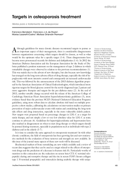
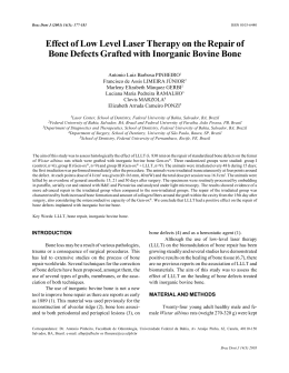
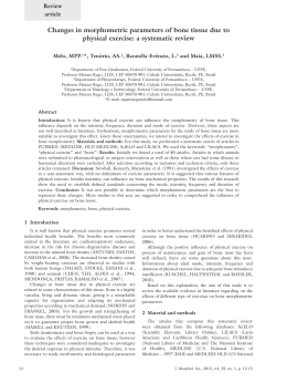
Transfusion and Apheresis Science 30 (2004) 139–144
intl.elsevierhealth.com/journals/tras
Use of autologous fibrin-platelet glue and bone fragments
in maxillofacial surgery
Gabriella Giannini
a,*
, Valbonesi Mauro a, Tel Agostino b, Bindi Gianfranco
b
a
b
Department of Immunohematology and Blood Transfusion, San Martino University Hospital,
Largo Rosanna Benzi 10, 16132 Genoa, Italy
Department of Maxillofacial Surgery, San Martino University Hospital, Largo Rosanna Benzi 10, 16132 Genoa, Italy
Accepted 30 November 2003
Abstract
The use of autologous cryo-platelet gel and bone fragments in maxillofacial surgery is described. Cryo-platelet gel
has been used successfully in 5 patients who underwent surgery for maxillary or mandibular problems. The level of
improvement was scored, arbitrarily, from 0 to 4. Very favourable results were seen in our 5 patients. The glue
preparation is very easy and inexpensive and the glue creates excellent and stable hemostasis. From a general point of
view, we add additional anecdotal evidence supporting the utility of fibrin-platelet glue in terms of reduced infections
and length of hospital stay, which suggest the need for well planned and controlled trials to show if there is benefit and
when and this treatment modality should be used in maxillofacial surgery.
2004 Elsevier Ltd. All rights reserved.
Keywords: Autologous fibrin-platelet glue; Growth factors; Wound healing
1. Introduction
Methods for preparation, in the blood bank, of
biological glues are many ranging from preparation of fibrin glue to the preparation of platelet
gels. Their use depends mostly on the expected
results. The main advantage of fibrin glue is its
tensile strength which helps in wound healing and
cessation of hemorrhage. Platelet derived glues are
*
Corresponding author. Tel.: +39-010352859/5553928; fax:
+39-0105556807.
E-mail address: [email protected]
(G. Giannini).
somehow complementary since they have a poor
tensile strength but promote tissue regeneration
due to their content of growth factors. The many
potential uses for fibrin glue in the field of oral and
maxillofacial surgery were first set forth in the
literature by Matras in 1982 [1]. She described fibrin sealant as a material possessing the qualities
of tissue sealing, hemostasis, and promotion of
wound healing. In this article and an update in
1985 [2], Matras presented case reports on the use
of fibrin glue in microneural and microvascular
anastomoses, as a hemostatic agent in soft tissue
deficits, as a replacement for suture material in the
split thickness skin graft procedure, in the reapproximation of comminuted bone fragments in
complicated fractures and in dural closure. The
1473-0502/$ - see front matter 2004 Elsevier Ltd. All rights reserved.
doi:10.1016/j.transci.2003.11.009
140
G. Giannini et al. / Transfusion and Apheresis Science 30 (2004) 139–144
fibrin sealant developed by Matras is currently
marketed in Europe under the trade name of Tisseel (Immuno, Vienna, Austria); it has recently
gained FDA approval in the United States but this
was delayed because of the earlier reported risk of
viral transmission associated with its use. The risk
of infectious disease transmission from commercially available fibrin glue systems has led to the
development of numerous autologous techniques
for preparing this useful material [3,4]. However,
these autologous techniques generally require patient interaction with the blood bank at least 3
days before the planned surgery and advanced coordination by the surgeon. Clerical error by the
blood bank still places the patients at risk for a
potential transfusion reaction or infectious disease
complication. Finally, the patient making the
donation must meet the blood bankÕs criteria for
weight, hemoglobin concentration, age and general health. The most recently published reports
in the oral and maxillofacial surgery literature
regarding the use of fibrin glue have been in the
area of mandibular reconstruction where Tayapongsak et al. [5,6] have reported success in using
the glue as an adhesive carrier for autogenous
particulate cancellous bone and marrow in conjunction with both allogeneic cribs and alloplastic
trays.
Our blood bank is involved in both the production of cryo-platelet gels and their application during surgery. Results were compared with
those of a historical group in terms of infection
rate, length of hospital stay (LHOS) and clinical
effect.
2. Materials and methods
Our blood bank is involved in a program for
collecting platelet and plasma immediately preoperatively for the purpose of producing cryoplatelet glue to be used intraoperatively. Briefly,
autologous platelets anticoagulated with ACD-A
were collected according to the dry-platelet collection technique employing an Excel apparatus
(Dideco Spa Mirandola, Italia), as previously described [7]. From 3.6 to 5 · 1011 platelets were
collected in 20–25 ml of plasma, along with 450–
500 ml of autologous plasma which was subsequently used to obtain 40–50 ml of cryoprecipitate.
After collection the plasma unit was immediately
frozen at )80 C in a mechanic refrigerator, and
the frozen plasma was then put at 4 C for 18–24 h
for spontaneous thawing. Cryodepleted plasma
was removed and the residual cryoprecipitate dissolved in 40–50 ml of plasma and frozen for subsequent use. The apheresis platelet concentrate
was frozen at )80 C, without any further
manipulation.
Immediately before the end of the surgery, 10
ml of platelet concentrate is mixed with 10 ml of
cryoprecipitate, 3 ml of Botropase (Ravizza Farmaceutici Spa Milano, Italia) and 3 ml of calcium
gluconate in a sterile Petri plastic dish. The Petri
dish content is evenly mixed to get a gel-like
material in 2–4 min.
The cryo-platelet gel was used in maxillofacial
surgery to cover osteotomy and interplantation of
bone blocks from the iliac crest or from the skull.
At each operation 5–6 fragments of cancellous
bone were obtained and used immediately. Particular attention was paid to the presence of red
bone marrow because of the theoretical presence
of mesenchemal progenitor cells.
Between December 2002 and September 2003, 5
patients, after an appropriate informed consent,
underwent surgery for maxillary or mandibular
problems.
Our case-list includes 3 male and 2 female patients with a mean age of 46.6 years (range 30–65
years). The diagnosis was severe maxillary bone
atrophy for 3 patients, maxillary skeletal deformity (progenia) for 1 patients and right hemimandibular pathological fracture for the last
patient (Table 1).
Table 1
Patient data
Patient
Sex/age
Diagnosis
1
2
3
F/65
M/30
M/42
4
5
M/45
F/51
Severe maxillary bone atrophy
Maxillary skeletal deformity (progenia)
Right hemimandibular pathological
fracture
Severe maxillary bone atrophy
Severe maxillary bone atrophy
G. Giannini et al. / Transfusion and Apheresis Science 30 (2004) 139–144
3. Case reports and results
The results obtained after surgery for three out
of our 5 patients are documented photographically
(Fig. 1). Patients 4 and 5 are not documented since
the results were the same as for case 1.
Case 1
Diagnosis: severe maxillary bone atrophy.
Intervention: maxillary bone autologous
reconstruction with iliac crest block insertion
after Le Fort osteotomy I and complete maxillary mobilization (Fig. 1A–D).
Case 2
Diagnosis: maxillary skeletal deformity (progenia).
Intervention: upper maxillary advancement
and mandibular setback (Fig. 2A–D).
Case 3
Diagnosis: right hemimandibular pathological fracture.
141
Intervention: horizontal maxillary branch
reconstruction with bone implants from skull
(calvaria) (Fig. 3A–E).
Comparing the results in these 5 patients with
the results observed in a group of 20 consecutive
comparable patients undergoing surgery for the
same indications a reduction in LHOS of 3 days
was seen along with the absence of infection
which, in contrast, were seen in patients in the
control group. The level of improvement was
scored, on an arbitrary bases, from 0 (no improvement) to 4 (complete healing) (Table 2).
4. Discussion
The process of wound healing is complex and
involves a series of interactions between platelets
and macrophages. In the first 2 days following
Fig. 1. (A) Le Fort upper maxillary osteotomy I with drawdown and interplantation of bone blocks from the iliac crest. (B) Wide cover
of bone reconstruction through fibrin-platelet glue, before suture. (C) Automated tomography with overview projection: note the
complete absence of the alveolar process of the maxilla. (D) Post-surgical automated tomography: whole maxillary volume bone
reconstruction.
142
G. Giannini et al. / Transfusion and Apheresis Science 30 (2004) 139–144
Fig. 2. (A) Upper maxillary osteotomy with putting forward. (B) Cover of osteotomy and method of fixation (plate in titanium) with a
large quantity of fibrin-platelet glue. (C) PatientÕs profile before intervention. (D) PatientÕs profile after intervention.
injury, growth factors are produced and released
by platelets. Thereafter, this function is taken over
by macrophages. Within the a-granules of the
human platelet are multiple growth factors that
are released when platelets are activated and degranulated. These include platelet derived growth
factor (PDGF), transforming growth factor (TGFb), fibroblast growth factor (FGF), epidermal
growth factor (EGF), platelet derived angiogenesis
factor and b-thromboglobulin. PDGF seems to be
the first growth factor present in the wound, and it
initiates connective tissue healing, including bone
regeneration and repair. The most important specific activities of PDGF include mitogenesis (an
increase in the cell populations of healing cells),
angiogenesis (endothelial mitoses into functioning
capillaries), and macrophage activation (debridement of the wound site and a second-phase source
of grown factors for continued repair and bone
regeneration). TGF, when released by platelet
degranulation or actively by macrophages, acts as
a paracrine growth factor (i.e., growth factors secreted by one cell exerting their effect on an adjacent second cell), affecting mainly fibroblasts,
marrow stem cells and the preosteoblasts. However, each of these target cells has the ability to
synthesize and secrete its own TGF proteins to act
on adjacent cells in a paracrine fashion or act on
itself as an autocrine growth factor. TGF therefore
represents a mechanism for sustaining a long-term
G. Giannini et al. / Transfusion and Apheresis Science 30 (2004) 139–144
143
Fig. 3. (A) Orthopantogram showing right hemimandibular pathological fracture. (B) Orthopantogram after bone implant reconstruction (calvaria). (C) Spongy-cortical spear from cranial theca (parietal bone). (D) Mandibular reconstruction with bone implants.
(E) Cover and fibrin-platelet glue insertion into bone gaps.
Table 2
Results obtained in the 5 patients (LHOS: length of hospital stay)
Pt
LHOS days
Infection Yes/No
Use of antibiotics Yes/No
Result at 30 days
1
2
3
4
5
3
3
3
3
3
No
No
No
No
No
Yes
Yes
Yes
Yes
Yes
Excellent
Excellent
Excellent
Excellent
Excellent
healing
healing
healing
healing
healing
Score 0/4
of
of
of
of
of
soft
soft
soft
soft
soft
tissues
tissues
tissues
tissues
tissues
4
4
4
4
4
144
G. Giannini et al. / Transfusion and Apheresis Science 30 (2004) 139–144
healing and bone regeneration module and may
even evolve into a bone-remodelling factor over
time. The most important function of TGF seems
to be the chemotaxis and mitogenesis of osteoblast
precursor; it also has the ability to stimulate
osteoblast deposition of collagen matrix of wound
healing and of bone. In our study, a purified
autologous platelet releasate has been prepared by
stimulating human platelets to release the contents
of their a-granules. Use of a platelet releasate in
wound healing has advantages. The growth factors
that are released are identical to and in the same
proportion as the growth factors normally brought
to the wound by the platelets. Preparation of a
platelet releasate is relatively easy and inexpensive
because the platelets can be harvested from peripheral blood. They readily release the contents of
their a-granules when stimulated with Ca gluconate and batroxobin. The use of autologous
material eliminates the risk of transmission of viral
disease; the formula for fibrin-platelet glue preparation is simple and standardized.
No study was carried out on platelet quality or
viability and the platelets were frozen without any
cryoprotectant since, in reality, their function after
thawing is the provision of phospholipid membranes along with the provision of preformed
growth factors for incorporation into the gel.
In the Department of Maxillofacial Surgery, we
have successfully used this technique in conjunction with ablative surgical procedures of the maxillofacial region, and for maxillary and mandibular
reconstruction. Also, we have adopted the practice
of using cryo-platelet glue to obtain hemostasis
after anterior iliac bone harvest. The use of particulate cancellous bone and marrow (PCBM)
grafts to reconstruct severe maxillary bone atrophy
is a well-established technique with proven success
[8]. The importance of adequately particulating,
condensing, and adapting the bone graft material
has been shown [9]. Even with complete and adequate dissection of the recipient bed, the PCBM
graft can be displaced through soft tissue manip-
ulation during closure. By applying autologous
platelet gel to the PCBM, adequate initial consolidation of the graft is obtained. This consolidation
allows for manipulation of the flap without displacement of the bone graft. Besides the obvious
mechanical benefits of using autologous cryoplatelet glue with PCBM grafts, physiologic benefits may also be assumed. The introduction of fibrin
and the release of the numerous growth factors
should enhance the healing potential of the autologous bone graft perhaps stimulating the stem cells
which are potentially present. Nonetheless, from a
clinical point of view, based on comparison with
the results obtained in these or comparable patients
seen at our facility who were treated conventionally, we are convinced of the absolute utility of
fibrin-platelet glue in terms of reduced infections
and length of hospital stay.
References
[1] Matras H. The use of fibrin glue in oral and maxillofacial
surgery. J Oral Maxillofac Surg 1982;40:617.
[2] Matras H. Fibrin seal: the state of the art. J Oral Maxillofac
Surg 1985;43:605.
[3] Saltz R, Sierra D, Feldman D, et al. Experimental and
clinical applications of fibrin glue. Plast Reconstr Surg 1991;
88:1016.
[4] Hartman AR, Galankis DK, Honig MP, et al. Autologous
whole plasma fibrin gel: intraoperative procurement. Arch
Surg 1992;127:357.
[5] Tayapongsak P, OÕBrien DA, Monteiro CB, et al. Autologous fibrin adhesive in mandibular reconstruction with
particulate cancellous bone and marrow. J Oral Maxillofac
Surg 1994;52:161.
[6] Tayapongsak P, Pijut LJ, Scheffer RB. Inferior border cribs
in mandibular reconstruction. Oral Surg Med Oral Pathol
Oral Radiol Endod 1995;79:10.
[7] Valbonesi M, Bruni R, et al. Dry platelets with the Dideco
Excel apparatus. Transfus Sci 1999;20:101–6.
[8] Boyne PJ. Autogenous cancellous bone and marrow transplants. Clin Orthop 1970;73:199.
[9] Marx RE. The science and art of reconstructing the jaws and
temporomandibular joints. In: Bell WH, editor. Modern
practice in orthognathic and reconstructive surgery. Philadelphia, PA: Saunders; 1992. p. 1448–531.
Download