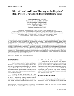
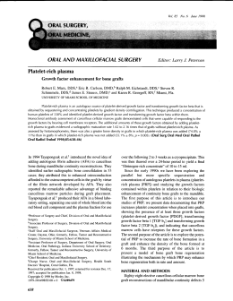
DEAR AUTHOR: This file contains the following: 1. 2. 3. 4. Author letter Reprint order form Page proof of your article and list of author queries Customer satisfaction survey After printing the PDF file, please read the page proofs carefully and e-mail a summary of the requested changes to me at [email protected] OR FAX any pages with corrections to me at 410-691-6235, attention: Dawn Shaw 1. Clearly indicate changes or corrections in pen in the margins of the page proofs. PLEASE NOTE: Only changes that are essential to the accuracy of the article will be allowed. Excessive or unreasonable changes are subject to approval by the Editorial Office and cannot be guaranteed. Fees resulting from extensive author changes will be the responsibility of the authors. 2. Answer all author queries (indicated as AQ:1, AQ:2, AQ:3, etc, in margins of the proofs and listed on the last page of the PDF proof). the 3. Check figures and tables carefully against legends and text citations, and check for size, numbering, and orientation of figures. If there is a correction to be made to a figure, please submit the corrected version electronically to avoid delay. If your figure(s) will be published in color, you will receive an Iris proof of your figure(s) by mail. Please carefully inspect the figure(s) and check that the result is acceptable and close to the original. Please notify Kara Barrick at 410-528-4329 or [email protected] as soon as possible if the reproduction is not acceptable. 4. Review all author/contributor names, titles, and affiliations. Proofread all numbers, equations, sums, and percentages carefully, and indicate whether sums are intentionally rounded off. PLEASE NOTE: No authors may be added or removed without an explanation to and approval from the Editorial Office. 5. PROOFS MUST BE RETURNED WITHIN 48 HOURS TO AVOID ANY DELAYS IN THE PUBLICATION OF YOUR ARTICLE. If you are not making any changes, please e-mail a message stating that there are no corrections or write “no changes” on the first page of your proof, sign and date it, and fax the page to me at the number give below. Failure to respond implies your approval to publish the proofs without additional changes. If you would like to order reprints of your article, simply complete the reprint order form. Please follow the instructions on the enclosed form; e-mail [email protected] or call 800-341-2258 with any questions. Thank you, Dawn Shaw Proof Manager Cadmus Professional Communications 940 Elkridge Landing Road Linthicum, MD 21090 phone: 410-691-6294 fax: 410-691-6235 Author(s) Name____________________________________________________________ 2004 Author Reprint Rates In addition to using this form to order reprints, Title of Article______________________________________________________________ ID200001 Article #__________________Publication Mo/Yr_________No. of pgs. in Article_________ it is to be used to calculate any additional Payment must be received before reprints can be shipped. Payments accepted in the form of a check or credit card; purchase orders are accepted for orders billed to a U.S. address. publication fees your article may incur. Publication fees include color separation charges MC and page charges. Prices are subject to change VISA Discover Am Express without notice. Quantities over 500 copies--- Account #_______________________________________________ Exp. Date__________ contact our Healthcare Dept. at 410-528-4426. Name_____________________________________________________________________ Outside the U.S. dial 4420-7981-0700. Street_____________________________________________________________________ City_____________________________________State_____________ Zip_____________ Fax or mail your order Lippincott Williams & Telephone_____________________________Signature____________________________ Wilkins, Author Reprints Dept, 351 W. Camden Street, Baltimore, MD 21201-2436. Reprint Cost Quantity of Reprints = ______________________ $__________ Covers (Optional) Plain: $21.00 per 100 $___________ Printed: $82.00 for the first 100 copies; $21.00 each add’l 100’s $___________ Fax: 410-528-4434. Rapid Ordering can be accessed at http://www.lww.com/periodicals/authorreprints. A confirmation of your order will be Color Fees (If your article contains color figures, use Rapid Ordering.) e-mailed to you. Separation Cost (You may have included color figures in your article. The separation costs to publish those figures will be included on the reprint invoice. $___________ For questions regarding reprints or publication fees please e-mail us at [email protected] or Reprint Color Cost ($70.00/100 reprints) $___________ contact us at 1-800-341-2258. Shipping Add $5.00 per 100 reprints for orders shipping within the U.S. and $20.00 per 100 reprints for orders shipping outside the U.S. $__________ Tax U.S. and Canadian residents add the appropriate tax, or submit a tax exempt form. $__________ Pgs/Qty 100 200 300 400 500 Up to 4 $208 $257 $303 $360 $405 5 to 8 $375 $441 $510 $585 $654 9 to 12 $537 $628 $720 $812 $906 13 to 16 $702 $818 $928 17 to 20 $859 $997 $1,041 $1,154 Shipping Information Ship:___________copies to: $1,130 $1,270 $1,413 Name_____________________________________________________________________ 21 to 24 $1,023 $1,186 $1,345 $1,491 $1,666 Address___________________________________________________________________ 25 to 28 $1,210 $1,390 $1,574 $1,786 $1,960 Dept/Rm___________________________________________________________________ 29 to 32 $1,367 $1,574 $1,785 $1,996 $2,211 __________________________________________________________________________ Phone #___________________________________________________________________ Lippincott Williams & Wilkins, Baltimore, MD balt5/ziy-id/ziy-id/ziy00305/ziy2754-05z martines Sⴝ3 8/5/05 9:03 4/Color Figure(s): F1,2 Art: ID200001 Input-ds Cocaine Associated With Onlay Bone Graft Failure: A Clinical and Histologic Report Jamil Awad Shibli, DDS, MS, PhD,* Elcio Marcantonio, DDS, MS, PhD,† Luis Carlos Spolidorio, DDS, MS, PhD,‡ and Elcio Marcantonio, Jr., DDS, MS, PhD§ ocaine is an alkaloid extracted from the Erythroxylon plant that grows in some countries of South America and Africa. Cocaine hydrochloride is made by dissolving cocaine alkaloid in hydrochloric acid to produce a water-soluble salt that can be dissolved in water and injected, inhaled, or snorted through the nasal mucosa. Snorting cocaine causes necrosis of the nasal cartilage and nasal septum, perforation of the septum, rhinitis, nose bleeding, an impaired sense of smell, and foul breath. Cocaine anesthetizes the mucosa of the mouth and throat. Thus, cocaine smokers are not aware that the hot vapors and other impurities that they inhale burn the tissues of the oropharynx and tracheobronchial tree.1 In addition, users of cocaine may rub the drug on the gingival tissue because of the similarity of the mucosal architecture, and abundant vascularity between nasal and oral mucosa.2 Kapila and Kashani3 reported a gingival recession and dental erosion associated with local application of cocaine; the intense vasoconstrictive effect of cocaine use has been deemed responsible for these effects.4,5 Thus, the aim of this case report is to describe an unusual onlay bone graft C *Assistant Professor, Department of Periodontology, Dental Research Division, Guarulhos University, Guarulhos, SP, Brazil. †Professor, Department of Oral and Maxillofacial Surgery, Dental School of Araraquara, State University of São Paulo (UNESP), Araraquara, SP, Brazil. ‡Associated Professor, Department of Pathology and Physiology, Dental School of Araraquara, State University of São Paulo (UNESP), Araraquara, SP, Brazil. §Professor, Department of Periodontology, Dental School of Araraquara, State University of São Paulo (UNESP), Araraquara, SP, Brazil. ISSN 1056-6163/05/01403-001 Implant Dentistry Volume 14 • Number 3 Copyright © 2005 by Lippincott Williams & Wilkins DOI: 10.1097/01.id.0000173329.81754.58 This patient report presents an unusual onlay bone graft failure following local cocaine application. Three months after the bone grafting procedure performed in the anterior maxilla for bone volume augmentation, the bone graft was totally exposed in the oral cavity as a result of the rubbing of cocaine on the gingival tissue that covered the bone graft. A histologic view of the removed bone fragment presented not only an area of necrosis but also ample spaces filled with necrosis material and resorption areas. Dental practitioners need to be aware of this phenomenon because such patients often do not report the use of drugs, particularly cocaine. (Implant Dent 2005;14:1–●●●) Key Words: autogenous bone graft, cocaine, dental implants, guided bone regeneration/failure necrosis associated with local application of cocaine. solution. The lingual cortex was preserved, and a collagen sponge, used as a hemostatic dressing, was placed in the donor area. The wound was closed with multilayered sutures following bone graft fixation. The corticocancellous bone block was stored in saline solution until fixing at the recipient site. The buccal cortex of the recipient site of the edentulous area was accurately débrided from any soft tissue left during flap elevation, and the cortex was perforated with a round bur to increase bleeding of the recipient bed for the graft. The bone block, which was also perforated, was then fixed in the area with a small-diameter titanium alloy screw. Small gaps between the bone graft and the alveolar crest were filled with cancellous bone from the bone trap. The periosteum at the base of the flap was carefully incised to allow stretching of the mucosa and tension-free adaptation of the wound margins. A removable, soft tissuesupported prosthesis was generously adjusted and relined with tissue conditioner. The patient was instructed to use his prosthesis for cosmetic purposes rather than for function. CASE REPORT Patient A male patient, aged 27 years, presented with a missing upper central incisor with deficient buccal/ palatal dimension and solicited dental implant treatment. The central incisor had been extracted 1 year before as a result of a fractured root (inadequate post design). The patient was healthy and without any significant medical history. Onlay Bone Graft The bone augmentation was performed as an outpatient procedure, with the patient under local anesthesia. The oral mucosa of the recipient implant site was incised palatally to the defect in the alveolar ridge. A fullthickness flap was elevated to expose the maxillary defect. The autogenous bone graft was obtained from the chin. According to the required quantity of bone, a corticocancellous block was outlined with a fissure bur assembled on a straight handpiece, under abundant irrigation with saline IMPLANT DENTISTRY / VOLUME 14, NUMBER 3 2005 <zdoi;10.1097/01.id.0000173329.81754.58> • <zjs;Clinical Science Clinical Science and Techniques> and Techniques> 1 • <zjss; balt5/ziy-id/ziy-id/ziy00305/ziy2754-05z martines Sⴝ3 8/5/05 9:03 4/Color Figure(s): F1,2 Art: ID200001 Input-ds No osteocytes were observed in lacunae (Fig. 2C). Some areas of the fragment presented external and internal bone resorption characterized by deep projections in various sizes and shapes. Some bone debris was seen around the bone fragment, characterized as necrotic bone tissue. DISCUSSION Fig. 1. A, Clinical view of the completely exposed onlay bone graft. B, Aspect of alveolar mucosa immediately after removal of bone graft. Maintenance Phase F1 During the first postoperative month, clinical complications were not observed, and the prosthesis was relined with tissue conditioner again. The patient returned during the third postoperative month, reporting exposure of the bone graft (Fig. 1A). During the clinical examination, it was observed that the bone graft was totally exposed in the oral cavity, although the patient did not have pain or any other complication. The bone block was partially rotated to the occlusal aspect and presented some mobility. With the patient under local anesthesia, the titanium alloy screw was removed, and the bone block was stored in 4% neutral formalin until processing. Following bone graft removal, the bone implant bed was cleaned with gauze soaked in 0.12% chlorhexidine (Fig. 1B). The patient was asked about the possible reasons for the cause of bone graft failure, such as late infection or compression from the temporary soft tissue-supported prosthesis. However, after a long conversation, the patient related the use of cocaine rubbing on the gingival tissue that covered the bone graft. The patient did not report how many times he had used the drug on the mucosa or why he chose this area. In addition, the information 2 Fig. 2. A, Histologic view showing necrotic material (*) in ample space remaining from titanium screw access showed in Fig. 1A (hematoxylin and eosin, original magnification ⫻100). B, Ample spaces (arrowheads) and parallel lamellae. External resorption with necrotic tissue (*) (hematoxylin and eosin, original magnification ⫻100). C, Empty lacunae (arrowheads), with ample spaces filled with necrotic tissue (*) (hematoxylin and eosin, original magnification ⫻100). about the drug use was omitted by the patient from his dental medical history. Histologic Processing and Evaluation The bone block was harvested, fixed in formalin, and decalcified in Morse solution. Following decalcification, routine histologic processing and paraffin embedding were performed, and 5-m thick tissue blocks were obtained with a longitudinal plane. The sections were stained with hematoxylin and eosin. The bone fragment was filled with a necrotic tissue (Fig. 2A). Bone tissue presented parallel lamellae and ample spaces, which were sometimes empty or filled with necrotic tissue (Fig. 2B). COCAINE ASSOCIATED WITH ONLAY BONE GRAFT FAILURE The procedure of guided bone regeneration (GBR) has been described as a predictable treatment for the regeneration of lost tissues. Nevertheless, some failures have been reported.6 Factors such as bone graft stability, size of the bone graft, peripheral sealing between bone graft and recipient bone, blood supply, and access to bone forming cells have been noted to be critical for a successful outcome.7 In addition, factors inherent to the patient, such as smoking and systemic health, as well as local conditions (e.g., provisional restoration, tension of the mucoperiosteal flap) are also crucial to predictability of the GBR. Fig. 1 shows the presence of sharp edges on the donor bone. However, we cannot confirm that these edges were left in the donor bone before that bone graft exposure. In addition, the bone graft had a slight movement in an occlusal direction, probably a result of mobility after bone graft exposure. It may be speculated that there were other mitigating factors, such as poor fit of the donor bone, failure to stabilize the donor bone, sharp edges left on the donor bone, and the lack of vascular channels that may jeopardize the GBR. However, we cannot confirm the exact cause of GBR failure. The cocaine probably potentiated the association of the failure of GBR. In addition, the patient himself admitted to using cocaine on the bone graft site. In this report, an unusual bone graft failure caused by the use of cocaine on the autogenous bone graft is presented. The alveolar mucosa under the exposed bone graft showed an aspect of clinical inflammation. Necrosis and ulceration of mucosal tissue interfaces of the nose from contact with snorted cocaine have also been noted. Some investigators8,9 have reported desquamations, ulcerations, F2 balt5/ziy-id/ziy-id/ziy00305/ziy2754-05z martines Sⴝ3 8/5/05 9:03 4/Color Figure(s): F1,2 Art: ID200001 Input-ds and necrosis of the areas where cocaine was rubbed. In this case, histologic examinations of the exposed bone graft revealed severe superficial necrosis consistent with ischemic necrosis, which was in agreement with a previous report. 10 Following bone block removal, the patient was referred to a local center that provides medical and psychologic treatment for drug users, before initiating new GBR for further dental implant placement. In addition, the use of gingival and alveolar mucosa as transport sites for cocaine administration appears to be fairly common.2 In conclusion, dental practitioners need to be aware of this phenomenon to recognize it because such patients often do not report the use of drugs, particularly cocaine. Disclosure The authors claim to have no financial interest in any company or any of the products mentioned in this article. REFERENCES 1. Underdahl JP, Chiou AG. Preseptal cellulitis and orbital wall destruction secondary to nasal cocaine abuse. Am J Ophthalmol. 1988;125:266-268. 2. Yukna RA. Cocaine periodontitis. Int J Periodontics Restorative Dent. 1991;11: 72-79. 3. Kapila YL, Kashani H. Cocaineassociated rapid gingival recession and dental erosion. A case report. J Periodontol. 1997;68:485-488. 4. Millard DR, Mejia FA. Reconstruction of the nose damage by cocaine. Plast Reconstr Surg. 2001;107:419-424. 5. Schweitzer VG. Osteolytic sinusitis and pneumomediastinum: Deceptive otolaryngologic complications of cocaine abuse. Laryngoscope. 1986;96:206-210. 6. Zitzmann NU, Schärer P, Marinello CP. Factors influencing the success of GBR. Smoking, timing of implant placement, implant location, bone quality and provisional restoration. J Clin Periodontol. 1999;26:673-682. 7. Lundgren AK, Sennerby L, Lundgren D. Guided jaw-bone regeneration using an experimental rabbit model. Int J Oral Maxillofac Surg. 1998;27:135-140. 8. Dello Russo NM, Temple HV. Cocaine effects on gingiva. J Am Dent Assoc. 1982;104:13. 9. Quart AM, Small CB, Klein RS. The cocaine connection. Users imperil their gingiva. J Am Dent Assoc. 1991;122: 85-87. 10. Gargiulo AV Jr, Toto PD, Gargiulo AW. Cocaine induced-gingival necrosis. Periodontal Case Rep. 1985;7:44-45. Reprint requests and correspondence to: Jamil Awad Shibli, DDS, MS, PhD Universidade Guarulhos Centro de Pós-Graduação Pesquisa e Extensão-CEPPE Praça Tereza Cristina 1-Centro 07023-070 Guarulhos, SP. Brazil FAX: 55-11-6464-1758 E-mail: [email protected] Abstract Translations [German, Spanish, Portugese, Japanese] AUTOR(EN): Jamil Awad Shibli, DDS, MS, PhD*, Elcio Marcantonio, DDS, MS, PhD**, Luis Carlos Spolidorio, DDS, MS, PhD***, Elcio Marcantonio, Jr., DDS, MS, PhD****. *Assistenzprofessor, Abteilung für Periodontologie, Fachbereich zahntechnische Forschung, Guarulhos Universität, Guarulhos, SP, Brasilien. **Professor, Abteilung für Gesichts- und Kieferchirurgie, zahnmedizinische Fakultät von Araraquara, staatliche Universität São Paulo (UNESP), Araraquara, SP, Brasilien. ***A.O. Professor, Abteilung für Pathologie und Physiologie, zahnmedizinische Fakultät von Araraquara, staatliche Universität São Paulo (UNESP), Araraquara, SP, Brasilien. ****Professor, Abteilung für Periodontologie, zahnmedizinische Fakultät von Araraquara, staatliche Universität São Paulo (UNESP), Araraquara, SP, Brasilien. Schriftverkehr: Jamil Awad Shibli, DDS, Universidade Guarulhos, Centro de PósGraduação, Pesquisa e Extensão-CEPPE, Praça Tereza Cristina, 1 - Centro. 07023-070 Guarulhos, SP, Brasilien. Fax: ⫹55 11 6464 – 1758. eMail: [email protected] Kokain-induziertes Versagen einer Spananlagerungsbehandlung: eine klinischer und histologischer Bericht ZUSAMMENFASSUNG: Der vorliegende Patientenbericht weist das ungewöhnliche Versagen einer Spananlagerung nach lokaler Kokainanwendung auf. Drei Monate nach der Knochengewebstransplantation zur Anreicherung des Knochenvolumens im vorderen Oberkiefer lag das Knochentransplantat in der Mundhöhle vollkommen offen, verursacht durch das Reiben des Kokains auf dem das Knochentransplantat bedeckenden Zahnfleischgewebe. Die histologische Untersuchung des Knochenfragments wies nicht nur einen teilweisen Gewebstod auf, sondern auch viele Räume mit nekrotischem Materialinhalt sowie Resorptionsbereiche auf. Zahnärzte müssen sich dieses Phänomens bewusst werden, insbesondere da viele Patienten den Gebrauch derartiger Drogen, besonders bei Kokain, nicht offen eingestehen. SCHLÜSSELWÖRTER: Autogenes Knochengewebstransplantat, Kokain, Zahnimplantate, geleitete Knochengewebsregeneration / Versagen der geleiteten Knochengewebsregenerationsbehandlung IMPLANT DENTISTRY / VOLUME 14, NUMBER 3 2005 3 balt5/ziy-id/ziy-id/ziy00305/ziy2754-05z martines Sⴝ3 8/5/05 9:03 4/Color Figure(s): F1,2 Art: ID200001 Input-ds AUTOR(ES): Jamil Awad Shibli, DDS, MS, PhD*, Elcio Marcantonio, DDS, MS, PhD**, Luis Carlos Spolidorio, DDS, MS, PhD***, Elcio Marcantonio, Jr., DDS, MS, PhD****. 4 *Profesor Asistente, Departamento de Periodontologı́a, División de Investigación Dental, Universidad Guarulhos, Guarulhos, SP, Brasil. **Profesor, Departamento de Cirugı́a Oral y Maxilofacial, Escuela Dental de Araraquara, Universidad Estatal de San Pablo (UNESP), Araraquara, SP, Brasil. ***Profesor Asociado, Departamento de Patologı́a y Fisiologı́a, Escuela Dental de Araraquara, Universidad Estatal de San Pablo (UNESP), Araraquara, SP, Brasil. ****Profesor, Departamento de Periodontologı́a, Escuela Dental De Araraquara, Universidad Estatal de San Pablo (UNESP), Araraquara, SP, Brasil. Correspondencia a: Jamil Awad Shibli, DDS, Universidade Guarulhos, Centro de Pos-Graduaçao, Pesquisa e Extensão-CEPPE, Praça Tereza Cristina, 1–Centro, 07023-4 070 Guarulhos, SP, Brazil. Fax: ⫹ 55 11 6464-1758, Correo electr4 ónico: [email protected] AUTOR(ES): Jamil Awad Shibli, CirurgiãoDentista, Mestre em Ciência, PhD*, Elcio Marcantonio, Cirurgião-Dentista, Mestre em Ciência, PhD**, Luis Carlos Spolidorio, Cirurgião-Dentista, Mestre em Ciência, PhD***, Elcio Maracantonio, Jr., CirurgiãoDentista, Mestre em Ciência, PhD****. *Professor Assistente, Depto. de Periodontologia, Divisão de Pesquisa Dentária, Universidade de Guarulhos, Guarulhos, SP, Brasil. **Professor, Depto. de Cirurgia Oral e Maxilofacial, Escola de Odontologia de Araraquara, Universidade Estadual Paulista (UNESP), Araraquara, SP, Brasil. ***Professor Associado, Depto. de Patologia e Fisiologia, Escola de Odontologia de Araraquara, Universidade Estadual Paulista (UNESP), Araraquara, SP, Brasil. ****Professor, Depto. de Periodontologia, Escola de Odontologia de Araraquara, Universidade Estadual Paulista (UNESP), Araraquara, SP, Brasil. Correspondência para: Jamil Awad Shibli, DDS, Universidade de Guarulhos, Centro de Pós-Graduação, Pesquisa e ExtensãoCEPPE, Praça Tereza Cristina, 1–Centro, 07023-070 Guarulhos, SP, Brasil. Fax: ⫹55 11 6464-1758, E-mail: [email protected] 4 La asociación de la cocaı́na con la falla de un injerto de hueso en la incrustación con recubrimiento: Informe clı́nico e histológico ABSTRACTO: Este informe del paciente presenta una falla inusual de un injerto de hueso en la incrustación con recubrimiento luego de la aplicación local de cocaı́na. Tres meses después del procedimiento de injerto de hueso, realizado en el maxilar anterior para aumentar el volumen del hueso, el injerto de hueso fue totalmente expuesto en la cavidad oral debido al contacto de la cocaı́na con el tejido gingival que cubrı́a al injerto de hueso. Una vista histológica del fragmento del hueso removido presentó no solamente un área de necrosis, sino también amplios espacios llenados con material de necrosis y áreas de reabsorción. Los profesionales dentales necesitan estar al tanto de este fenómeno, ya que dichos pacientes a menudo no informan el uso de drogas, la cocaı́na en particular. PALABRAS CLAVES: injerto autógeno del hueso, cocaı́na, implantes dentales, falla/ regeneración del hueso guiado Falha no Enxerto Ósseo Onlay Associado a Cocaı́na: Relato Clı́nico e Histológico RESUMO: O relato do paciente apresenta uma falha incomum no enxerto ósseo onlay após aplicação local de cocaı́na. Três meses após o procedimento de enxerto ósseo, realizado na maxila anterior para aumento do volume ósseo, o enxerto ósseo foi totalmente exposto na cavidade oral devido à esfregação de cocaı́na no tecido gengival que cobria o enxerto ósseo. Uma visão histológica do fragmento ósseo removido apresentou não só uma região de necrose, mas também amplos espaços cheios de material de necrose e regiões de reabsorção. Os dentistas em exercı́cio precisam ter consciência desse fenômeno, já que esses pacientes freqüentemente não relatam o uso de drogas, particularmente cocaı́na. PALAVRAS-CHAVE: enxerto ósseo autógeno, cocaı́na, implantes dentários, regeneração/falha guiada de osso COCAINE ASSOCIATED WITH ONLAY BONE GRAFT FAILURE JOBNAME: AUTHOR QUERIES PAGE: 1 SESS: 1 OUTPUT: Thu Aug 4 15:53:57 2005 /balt5/ziy⫺id/ziy⫺id/ziy00305/ziy2754⫺05z AUTHOR QUERIES AUTHOR PLEASE ANSWER ALL QUERIES 1
Download