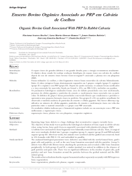
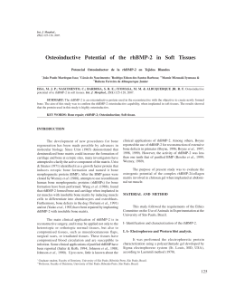
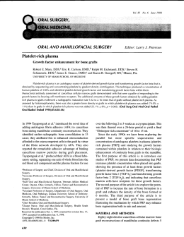
Braz Dent J (2003) 14(3): 177-181
LLLT and inorganic bovine bone graft
177
ISSN 0103-6440
Effect of Low Level Laser Therapy on the Repair of
Bone Defects Grafted with Inorganic Bovine Bone
Antonio Luiz Barbosa PINHEIRO1
Francisco de Assis LIMEIRA JÚNIOR2
Marleny Elizabeth Márquez GERBI2
Luciana Maria Pedreira RAMALHO3
Clovis MARZOLA4
Elizabeth Arruda Carneiro PONZI5
1Laser Center, School of Dentistry, Federal University of Bahia, Salvador, BA, Brazil
University of Bahia, Salvador, BA, Brazil and Federal University of Paraiba, João Pessoa, PB, Brazil
3Department of Diagnostics and Therapeutics, School of Dentistry, Federal University of Bahia, Salvador, BA, Brazil
4Department of Surgery, School of Dentistry, University of São Paulo, Bauru, SP, Brazil
5School of Dentistry, Federal University of Pernambuco, Recife, PE, Brazil
2Federal
The aim of this study was to assess histologically the effect of LLLT (λ 830 nm) on the repair of standardized bone defects on the femur
of Wistar albinus rats which were grafted with inorganic bovine bone Gen-ox®. Three randomized groups were studied: group I
(control, n=6); group II (Gen-ox®, n=9) and group III (Gen-ox® + LLLT, n=9). The animals were irradiated every 48 h during 15 days;
the first irradiation was performed immediately after the procedure. The animals were irradiated transcutaneuosly at four points around
the defect. At each point a dose of 4 J/cm2 was given (Ø~0.6 mm, 40 mW) and the total dose per session was 16 J/cm2. The animals were
killed by an overdose of general anesthetic 15, 21 and 30 days after surgery. The specimens were routinely processed by embedding
in paraffin, serially cut and stained with H&E and Picrosirius and analyzed under light microscopy. The results showed evidence of a
more advanced repair in the irradiated group when compared to the non-irradiated groups. The repair of the irradiated group was
characterized by both increased bone formation and amount of collagen fibers around the graft within the cavity from the 15th day after
surgery, also considering the osteoconductive capacity of the Gen-ox®. We conclude that LLLT had a positive effect on the repair of
bone defects implanted with inorganic bovine bone.
Key Words: LLLT, bone repair, inorganic bovine bone.
INTRODUCTION
Bone loss may be a result of various pathologies,
trauma or a consequence of surgical procedures. This
has led to extensive studies on the process of bone
repair worldwide. Several techniques for the correction
of bone defects have been proposed, amongst them, the
use of several types of grafts, membranes, or the association of both techniques.
The use of inorganic bovine bone is not a new
tool to improve bone repair as there are reports as early
as 1889 (1). This material was used previously for the
reconstruction of alveolar ridge (2), bone loss associated to both periodontal and periapical lesions (3), on
bone defects (4) and as a hemostatic agent (1).
Although the use of low-level laser therapy
(LLLT) on the biomodulation of bone repair has been
growing steadily and several studies have demonstrated
positive results on the healing of bone tissue (6,7), there
are no previous reports on the association of LLLT and
biomaterials. The aim of this study was to assess the
effect of LLLT on the healing of bone defects treated
with inorganic bovine bone.
MATERIAL AND METHODS
Twenty-four young adult healthy male and female Wistar albinus rats (weight 270-320 g) were kept
Correspondence: Dr. Antonio Pinheiro, Faculdade de Odontologia, Universidade Federal da Bahia, Av Araújo Pinho, 62, Canela, 40110-150
Salvador, BA, Brasil. e-mail: [email protected] or [email protected]
Braz Dent J 14(3) 2003
178
A.L.B. Pinheiro et al.
under natural conditions of light, humidity and temperature at the Animal Experimentation Laboratory of
the School of Dentistry of the Federal University of
Bahia, Brazil. The animals were fed with laboratory
diet and water ad libitum. The animals were divided
into three groups: group I: control (N=6); group II:
inorganic bovine bone (n=9); group III: inorganic bovine bone + LLLT (n=9). Each group was then divided
into three subgroups (15, 21 and 30 days).
Under intraperitoneal general anesthesia (10%
chloral hydrate, 0.4 ml/100 g), the right leg of the
animals was shaved and the femur exposed. Standardized 3 mm2 cavities were created on the superior third
of the lateral side of the bone. In group I, the periosteum
was repositioned and sutured with catgut and the skin
closed with nylon. In groups II and III, the cavities were
completely filled with inorganic bovine bone (Genox®; Baumer S/A, Mogi Mirim, SP, Brazil). All wounds
were routinely sutured as described previously. The
animals of group III were submitted to 7 sessions of
LLLT (λ 830 nm, 40 mW, Ø~0.60 mm, CW; Thera
Lase, DMC Equipamentos, São Carlos, SP, Brazil), at
48-h intervals. The irradiation was performed transcutaneously and the first session was performed immediately after surgery. A dose of 4 J/cm2 was applied to 4
points around the defect giving a total of 16 J/cm2 per
session and a total treatment dose of 112 J/cm2. This
was based on previous studies (8) recommending doses
ranging from 1.8 to 5.4 J/cm2. The animals were killed
15, 21 and 30 days after surgery by an overdose of
general anesthetic. Samples were taken and kept on 4%
buffered paraformaldeyde solution for 5 days. The
samples were decalcified with 10% nitric acid and
routinely stained with H&E and Picrosirius (9).
RESULTS
Light microscopy showed that at day 15 the
cavity of group I (control) was filled by bone marrow
and a few bone trabeculae. The cortical defect was
more pronounced 21 days after surgery and nearly
closed at day 30 (Figure 1).
In group II (Gen-ox®), the graft filled nearly the
entire cavity and was encircled by collagen fibers 15
days after surgery. Immature bone trabeculae were
observed at the graft surface but no newly formed bone
around the graft could be seen at this time. Up to day 30
after surgery, the graft particles were evenly distributed
in the cavity and were partially or totally encircled by
newly formed bone. Newly formed bone trabeculae
were seen initiating repair of the cortical region (Figure
2).
Fifteen days after surgery, specimens of group
III (Gen-ox® + LLLT) showed that the graft filled most
of the cavity and was also encircled either by collagen
fibers or mineralized newly formed bone. At the cortical region, some neoformed bone trabeculae around
some particles of the graft and a dense amount of
collagen fibers could be observed. Twenty-one days
after surgery, an increased amount of neoformed bone
could be seen and the graft was encircled by collagen
fibers. At the end of the experimental period, the cavity
was still filled by the graft and the area was densely
filled by collagen fibers (Figure 3).
DISCUSSION
It seems clear from the results that Gen-ox® has
osteoconductivity because all grafted specimens showed
Figure 1. Photomicrography of control group. Left: At day 15, there was discrete bone formation at the cortical defect (arrow). Middle:
At day 21, the deposition of new bone at the cortical region was more evident (arrow). Right: At day 30, there was a complete union
of the cortical region (arrow). H&E. Original magnification: approximately 40X.
Braz Dent J 14(3) 2003
LLLT and inorganic bovine bone graft
bone neoformation around the particles. It is interesting
to observe that most specimens showed the particles of
the graft enclircled by collagen fibers and filling the
center of the cavity at day 15. At day 21, these particles
were unevenly distributed and were encircled by a
variable amount of newly formed bone. The level of
bone neoformation did not change much up to day 30 in
most groups.
It is accepted that although this material has
osteoconductivity, the repair of the defect may be slow
because of the need for the graft to be reabsorbed,
slowing down the process. In this study, the presence of
macrophages and giant cells around the particles, as
observed previously (2,10), may explain the reduction
of the amount of particles throughout the experimental
time. On the other hand, a previous report suggested
that this type of material may also be incorporated into
the newly formed bone (4,5).
It is clear that the use of the graft prevents the
179
formation of fibrosis of the lesion and also protects the
cavity (4) and acts as a framework for the deposition of
neoformed bone (11).
Although the biomodulatory effect of LLLT on
bone regeneration was observed previously under different experimental conditions and the results observed
in these studies (6,7) are aligned with the ones of the
present investigation, others have found no effect
(12,13). This may be due to several aspects of the
design of the experiment.
It is important to also consider the systemic
effect of LLLT (14), which was not considered in some
previous reports in which no effects of LLLT were
found (16). Other reports used very low doses (12). The
use of inappropriate wavelengths may also result in
negative results (12). Higher wavelengths are more
resistant to dispersion than lower ones and deeply
penetrate the skin (15). It has been reported that 632.8
nm laser light penetrates 0.5-1 mm before losing 37%
Figure 2. Photomicrography of group II. Left: At day 15, cortical repair was observed and also the presence of delicate newly formed
bone within the cavity near the graft surface (arrowheads). At days 21 (middle) and 30 (right), newly formed bone could be seen around
the graft (arrowheads). Left panel: H&E; middle and right panels: Picrosirius. Original magnification: approximately 40X.
Figure 3. Photomicrography of group III. Left: At day 15, complete cortical repair was observed (arrowhead). At days 21 (middle) and
30 (right), newly formed bone could be seen (arrowheads) and also collagen fibers encircling the graft (arrows); Left panel: H&E;
middle and right panels: Picrosirius. Original magnification: approximately 40X.
Braz Dent J 14(3) 2003
180
A.L.B. Pinheiro et al.
of its intensity (16). On the other hand, infrared wavelengths penetrate 2 mm before losing the some percentile
of energy. This is a clear indication for the use of
infrared laser light on bone tissue. However, systemic
effects may not be disregarded when visible laser light
is used.
The presence of large amounts of collagen fibers
on irradiated specimens was the major finding in these
groups and this may represent an early effect of the
LLLT on repair as other studies demonstrated increased
collagen production following LLLT (17). As collagen
is an important component of the extracellular matrix of
bone and increased amounts, as seen in this study, may
indicate a positive effect of LLLT on bone healing
despite the fact that in some specimens there was no
difference in the amount of newly formed bone between the irradiated group and controls. It is necessary
to consider that a large amount of collagen fibers will
represent an increased bone neoformation after mineralization of the matrix. The mechanism by which LLLT
interferes in collagen synthesis is not fully understood;
however, it may be because of alterations in the genetic
regulation or in the modulation of enzymatic activity
involved in the metabolism of the collagen as suggested
previously (17).
It is uncertain whether biomodulation of bone
formation is an overall effect on mesenchymal cells or
a direct stimulation of osteoblasts. It is possible that the
observed results in irradiated specimens are due to an
increased release of growth factors, mainly fibroblast
growth factor, which is found in bone tissue and acts on
differentiated cells increasing both cell proliferation
and secretion of components of the matrix (18).
The doses used in this study are in agreement
with several previous reports that suggested that 1-5 J/cm2
induces positive effects on both bone and soft tissues
(6-8,17). It is important to note that four points of
irradiation were used to fractionate the total dose per
session. The points of irradiation around the defect
were chosen because the results of irradiation of the
graft would be uncertain. The presence of the particles
would make the diffusion of light into the tissues more
difficult.
A total dose per session of 16 J/cm2 is in accordance with the clinical parameters recommended by
Pinheiro et al. (8). The literature shows that biomodulatory effects are dose dependent (19). It is also recognized that other factors such as the phase of cell growth
Braz Dent J 14(3) 2003
(20) and the frequency and number of sessions (7) also
influence the final result of the use of LLLT.
When groups II and III were compared, it was
evident that bone neoformation was similar at day 15.
However, at days 21 and 30, bone formation was more
evident in both the core of the cavity and in the cortical
region. Although irradiated specimens showed a dense
deposition of collagen fibers around the particles in the
core of the defect and at the cortical region, the same
was not detected in control specimens, especially in the
core of the defect.
We conclude that LLLT had a positive biomodulatory effect on the repair of bone defects implanted
with inorganic bovine bone.
RESUMO
O objetivo deste trabalho foi avaliar histologicamente a influência
da radiação laser não-cirúrgica (λ 830 nm) sobre o reparo ósseo
de defeitos padronizados em fêmur de ratos Wistar albinus e
submetidos a implante de osso bovino inorgânico Gen-ox®.
Foram estabelecidos três grupos de animais: grupo I (controle,
n=06); grupo II (Gen-ox®, n=09) e grupo III (Gen-ox® + LLLT,
n=09). Os animais foram irradiados a cada 48 horas, sendo a
primeira irradiação realizada imediatamente após a cirurgia. Os
animais foram irradiados transcutaneamente em quatro pontos
em torno da ferida. Cada ponto recebeu uma dose de 4 J/cm2
(Ø~0,6 mm, 40 mW) e a dose total por sessão foi de 16 J/cm2. Os
sacrifícios foram realizados 15, 21 e 30 dias após a cirurgia. Os
espécimes foram removidos e fixados para procedimento laboratorial, sendo corados com H&E e Picrosirius e analisados em
microscopia de luz. Os resultados mostraram evidência de um
reparo ósseo mais avançado nos espécimes irradiados em relação
aos não irradiados. O reparo nos espécimes irradiados foi
caracterizado por uma maior neoformação óssea, bem como por
uma maior proliferação de fibras colágenas no interior do defeito
já a partir de 15 dias após a cirurgia, também considerando a
capacidade osteocondutiva do Gen-ox®. Conclui-se que a LLLT
resultou num efeito de biomodulação positiva sobre o reparo do
defeito ósseo submetido a implante de osso bovino inorgânico.
REFERENCES
1. Rios ALBB, Barbosa CEM, Abi Rached RSG, Gabrielli MFR,
Okamoto T. Comportamento biológico de implantes de osso
bovino anorgânico em arco zigomático de ratos. Estudo
histológico. Rev Odontol Univ Est São Paulo 1996;25:87-101.
2. Pinholt EM, Bang G, Haanaes HR. Alveolar ridge augmentation
in rats by Bio-Oss. Scand J Dent Res1991;99:154-161.
3. Simonpietri JJ, Novaes AB, Batista Júnior EL, Filho EJ. Guided
tissue regeneration associated with bovine-derived anorganic
bone in mandibular class II furcation defects. 6-month results at
re-entry. J Periodontol 2000;71:904-911.
4. Batista PS. Avaliação microscópica do processo de reparo em
cavidades ósseas submetidas a implante de osso liofilizado bovino
LLLT and inorganic bovine bone graft
5.
6.
7.
8.
9.
10.
11.
12.
(Bio-Oss®) em fêmur de ratas. [Master's thesis]. Porto Alegre:
Faculdade de Odontologia, Pontifícia Universidade Católica do
Rio Grande do Sul; 1999. 85p.
Batista Júnior EL, Novaes Júnior AB, Simonpietri JJ, Batista FC.
Use of bovine-derived anorganic bone associated with guided
tissue regeneration in intra-bony defects. Six-month evaluation at
re-entry. J Periodontol 1999;70:1000-1007.
Pinheiro ALB, Oliveira MAM, Martins PPM. Biomodulação da
cicatrização óssea pós-implantar com o uso da laserterapia nãocirúrgica: Estudo por microscopia eletrônica de varredura. Rev
FOUFBA 2001;22:12-19.
Silva Júnior AN, Pinheiro ALB, Oliveira MG, Weismann R,
Ramalho LM, Nicolau RA. Computerized morphometric assessment of the effect of low-level laser therapy on bone repair: an
experimental animal study, J Clin Laser Med Surg 2002;20:8387.
Pinheiro ALB. Low-level laser therapy in management of disorders of the maxillofacial region. J Clin Laser Med Surg
1997;15:181-183.
Junqueira LCU, Bignolas G, Brentani RR. Picrosirius staining
plus polarization microscopy, a specific method for collagen
detection in tissue sections. Histochem J 1979;11:447-455.
Sanches MG, Okamoto T, Carvalho ACP. Processo de reparo em
feridas de extração dental após implante de “osso anorgânico” Estudo histológico em ratos. Rev Fac Odont Araçatuba
1972;1:83-90.
Garbin CM, Garbin NM. Enxertos Ósseos e Regeneração Tecidual
Guiada: Noções Básicas. São Paulo: Pancast, 1994.
David R, Nissan M, Cohen I, Soudry M. Effect of low-power He-
13.
14.
15.
16.
17.
18.
19.
20.
181
Ne laser on fracture healing in rats. Lasers Surg Med
1996;19:458-464.
Lagan KM, Clements BA, McDonough S, Baxter GD. Low intensity laser therapy (830 nm) in the management of minor postsurgical wounds: a controlled clinical study. Lasers Surg Med
2001;28:27-32.
Rockhind S, Rousso M, Nissan M, Villarreal M, Barr-Nea L,
Rees DG. Systemic effects of low-power laser irradiation on the
peripheral and central nervous system, cutaneous wounds and
burns. Lasers Surg Med 1989;9:174-182.
Kolárová H, Ditrichová D, Wagner J. Penetration of the laser
light into the skin in vitro. Lasers Surg Med 1999;24:231-235.
Basford JR. Low-intensity laser therapy: still not an established
clinical tool. Lasers Surg Med 1995;16:331-342.
Reddy GK, Stehno-Bittel L, Enwemeka CS. Laser photostimulation of collagen production in healing rabbit achilles tendons.
Lasers Surg Med 1998;22:281-287.
Sigurdsson TJ, Lee MB, Kubota K, Turek TJ, Wozney JM,
Wikesjö UME. Periodontal repair in dogs: recombinant bone
morphogenetic protein-2 significantly enhances periodontal regeneration. J Periodontol 1995;66:131-138.
Kipshidze N, Nikolaychik V, Keelan MH, Shankar LR, Khanna
A, Komowsky R, Leon M, Moses J. Low-power helium:neon
laser irradiation enhances production of vascular endothelial
growth factor and promotes growth of endothelial cells in vitro.
Lasers Surg Med 2001;28:355-364.
Osawa Y, Shimizu N, Kariya G, Abiko Y. Low-power laser
irradiation stimulates bone nodule formation at early stages of
cell culture in rat calvarial cells. Bone 1998;22:347-354.
Accepted November 26, 2002
Braz Dent J 14(3) 2003
Baixar