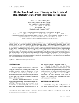
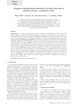
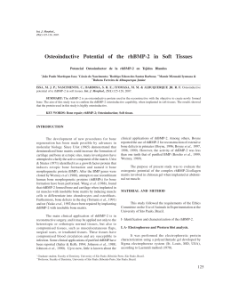
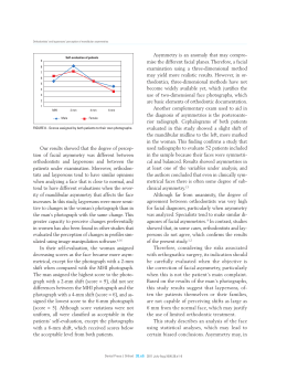
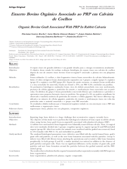
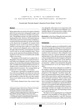
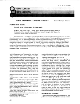
Original Article
Cone Beam Computed Tomography for Assessment of Root Length and
Marginal Bone Level during Orthodontic Treatment
Henrik Lunda; Kerstin Gröndahla,b; Hans-Göran Gröndahla,b
ABSTRACT
Objective: To evaluate the accuracy and precision of cone beam computed tomography (CBCT)
with regard to measurements of root length and marginal bone level in vitro and in vivo during the
course of orthodontic treatment.
Materials and Methods: Thirteen patients (aged 12–18 years) from an ongoing study and a dry
skull were examined with CBCT using multiplanar reformatting for measurements of root length
and marginal bone level. For in vivo evaluation of changes in root length, an index according to
Malmgren et al was used, along with a modification of this method.
Results: The in vitro mean difference between physical and radiographic measurements was
0.05 mm (SD 0.75) for root length and 20.04 mm (SD 0.54) for marginal bone level. In vivo the error
was ,0.35 mm for root length determinations and ,0.40 mm for marginal bone level assessments.
Conclusion: Despite changes in tooth positions, the CBCT technique yields a high level of reproducibility, enhancing its usefulness in orthodontic research. (Angle Orthod. 2010;80:466–473.)
KEY WORDS: CBCT; Root length; Marginal bone level; Orthodontic treatment
radiographs are fraught with uncertainties, particularly
in the anterior jaw regions. In individuals with Angle
Class II malocclusions, optimal visualization of both
jaws is difficult.11
Computed tomography (CT) has become the method
of choice for demanding imaging tasks. Higher costs
and higher radiation doses have prevented CT from
becoming a standard tool in dental imaging and, thus, in
studies of OIIRR, despite its inherent capabilities. In the
1990s CBCT12 was introduced in dentistry. Dosimetric
examinations have shown considerably lower doses
from CBCT than from CT,13 although they vary between
CBCT units and parameters used.14,15 Because of the
high spatial resolution, low radiation doses, and relative
affordability of this technique, it has gained widespread
acceptance in dentistry16 and in orthodontics.17 The
ability to provide distortion-free slice images of single
roots provides excellent possibilities to study root
resorption.
Our study aims to evaluate in vitro the accuracy and
precision of CBCT with regard to measurements of root
length and marginal bone level as well as the precision
with which such measurements can be made in vivo.
INTRODUCTION
Orthodontically induced inflammatory root resorption
(OIIRR) is an adverse effect of orthodontic treatment1
that might jeopardize an otherwise positive treatment
outcome. The incidence of OIIRR among individuals
having undergone orthodontic treatment differs between studies as a result of the techniques used to
quantify the degree of OIIRR.2
In most studies of OIIRR intraoral radiography has
been used.3 Even with efforts to obtain periodically
identical radiographs,4,5 intraoral radiography has
shortcomings.6–9 Follin and Lindvall10 found that in
order to be observed in intraoral radiographs, the
resorption has to cause a shortening of the root.
Panoramic radiography has also been used to assess
root shortening. This technique is sensitive to patient
positioning, and even under optimal conditions the
a
Department of Oral and Maxillofacial Radiology, Sahlgrenska Academy, University of Gothenburg, Sweden.
b
Department of Oral and Maxillofacial Radiology, Institute for
Postgraduate Dental Education, Jönköping, Sweden.
Corresponding author: Henrik Lund, DDS, Department of Oral
and Maxillofacial Radiology, Sahlgrenska Academy, University
of Gothenburg, PO Box 450, SE 405 30 Göteborg, Sweden
(e-mail: [email protected]).
MATERIALS AND METHODS
Subjects and Study Material
Accepted: September 2009. Submitted: July 2009.
G 2010 by The EH Angle Education and Research Foundation,
Inc.
Angle Orthodontist, Vol 80, No 3, 2010
To evaluate accuracy and precision of root length and
marginal bone level assessments in vitro we used a dry
466
DOI: 10.2319/072909-427.1
467
CBCT ASSESSMENT OF ROOT LENGTH AND BONE LEVEL
human skull. For the in vivo assessment of precision we
used radiographs from patients in an ongoing study. The
study comprised 13 patients, 7 boys and 6 girls (aged
12–18 years), participating in a study of root shortening
and marginal bone level alterations during orthodontic
treatment. All were diagnosed with a Class I malocclusion with crowding and were subjected to treatment
comprising extraction of one premolar in each jaw
quadrant and fixed appliances in both jaws. Informed
consent from the patients’ parents and ethical approval
from the Ethics Committee at the Sahlgrenska Academy, University of Gothenburg, were obtained. Subjects
were randomly chosen from patients having completed
three radiographic examinations (baseline, 6-month,
and end-point). Two teeth per subject and tooth group
(first molar, premolar, canine, and lateral and central
incisors) and one root from multi-rooted teeth were
randomly chosen, providing representative samples of
roots from all tooth groups.
For in vitro dry skull assessments five teeth were
chosen from the upper right and five from the lower left
quadrant as having no periodontal bone loss and
normal root lengths. Incisors, canines, second premolars, and first molars were chosen, corresponding to
the teeth examined in the patients.
Examination
A 60 3 60–mm volume was examined using 3D
Accuitomo FPD (J. Morita Mfg Corp, Kyoto, Japan), a
360u rotation, and exposure parameters of 17.5 seconds, 75 kV, and 4-5.5 mA, based on subject size.
Examinations were made so that the incisors to the first
molars became contained in one volume. To mimic soft
tissues the dry skull was immersed in water contained in
a Plexiglas bowl, placed on a stand so that it could be
positioned as would be a patient’s head.
Data Processing
Primary data reconstructions were made by acquisition software (i-Dixel-3DX, 3D, Version 1.691, J
Morita Mfg Corp) at the Accuitomo workstation
providing axial, frontal, and sagittal views. Secondary
reconstruction was then made using the i-Dixel
software to obtain a slice thickness and interval of
r
Figure 1. Measurement procedure for assessment of root length and
marginal bone level by means of reformatted images in axial,
sagittal, and coronal planes. (a) Root length—measurement from a
reference line between buccal and palatal cemento-enamel junction
(CEJ) to apex. The levels of CEJ and root apex were assessed using
a combination of axial, sagittal, and coronal images. (b) Buccal and
palatal/lingual bone levels—measurement in frontal and/or axial
views from a reference line at CEJ, according to Figure 1a, to the
buccal and palatal/lingual marginal bone crest, respectively. (c)
Mesial and distal bone levels—measurement from a reference line at
CEJ to the mesial and distal marginal bone crest, respectively. An
adjusted axial view was used to determine the level of the reference
line between the mesial and distal CEJ.
Angle Orthodontist, Vol 80, No 3, 2010
468
Figure 2. Index according to Malmgren et al.18 1 indicates irregular
root contour; 2, minor resorption; 3, severe resorption; and 4,
extreme resorption.
0.5 mm. Using DICOM export axial slices were sent to
PACS for later reformatting.
LUND, GRÖNDAHL, GRÖNDAHL
Figure 3. Modified index for describing slanted surface resorptions at
buccal, palatal/lingual, and mesial and distal surfaces at the
root apex.
Workstation/Equipment
Index
A workstation with Sectra IDS5 (Sectra Imtec AB,
Linköping, Sweden) PACS Multi Planar Reconstruction was used for reformatting and viewing of axial
slices. The workstation comprised a DELL computer
(Optiplex 755, DELL AB, Stockholm, Sweden) with a
graphic card (NVIDIA GeForce 6800 Series GPU 32bit, Matrox MED2mp-DVI) and 20-inch flat-panel
monitors. All measurements were performed on one
of two monochromatic screens (OLÒRIN Medic Line
ML 187D TFT-LCD, Olorin AB, Kungsbacka, Sweden)
with a resolution of 1280 3 1024 pixels.
For in vivo assessments of root length we also used
an index by Malmgren et al.18 The index (Figure 2)
comprises four scores, thus: 1 5 irregular root contour,
2 5 root resorption apically amounting to ,2 mm
(minor resorption), 3 5 root resorption from 2 mm to M
of the original root length (severe resorption), and 4 5
root resorption .M of the original root length (extreme
resorption). Score 1 was excluded because irregular
root contour was noted at teeth already at baseline (ie,
before the start of treatment). To describe a surface
resorption of the buccal, palatal/lingual, mesial, or
distal part of the apex that did not result in root
shortening a modification of the index was used
(Figure 3).
Reformatting and Measurements
Reformatting was completed so that the long axis of
the tooth/root became parallel to the axes of two
perpendicular, vertical image planes. This provided
optimal visualization of the tooth/root in axial, coronal,
and sagittal planes. Root length measurements were
made along the axis of the root. The marginal bone
level was assessed perpendicular to a line between
the buccal and lingual cemento-enamel junction (CEJ)
and the mesial and distal CEJ, respectively (Figure 1).
One observer made all measurements with intervals of
at least 2 days. The patient radiographs were
measured twice and those of the skull five times.
Direct measurements on the latter were performed
after the radiographs had been taken. The bone level
was marked with a thin pencil and each tooth was
gently removed. The bone level and root length were
measured five times using a digital caliper (Clas
Ohlson, Sweden AB, resolution 0.01 mm; accuracy,
60.03 mm for distances .100–200 mm). Measurements were taken from the CEJ to the most apical part
of the bone level at the buccal, palatal/lingual, mesial,
and distal aspects of the tooth. The root length was
measured along the axis of the root, perpendicular to
an imaginary line between the buccal and palatal/
lingual CEJ.
Angle Orthodontist, Vol 80, No 3, 2010
Statistics
For in vitro root length measurements we used
descriptive statistics presenting mean values and
standard deviations for the direct physical measurements vs those from radiographs. For comparisons
between direct and radiographic marginal bone level
measurements we studied their mean deviations from
zero and associated standard deviations. The Student’s paired t-test was used for significance testing,
with P , .05 as the cutoff value. The measurement
error of in vivo assessments of root length and bone
level from radiographs was calculated from duplicate
determinations using the formula s 5 !gd2/2n,
according to Dahlberg,19 where d 5 difference between duplicate determinations and n 5 number of
determinations. To evaluate differences between
reading 1 and 2 of baseline, 6-month, and end-point
examinations, the Student’s paired t-test was used,
and results were considered significant at P , .05.
Intraexaminer reliability using index scores was evaluated using Kappa statistics. All analyses were
performed using SPSS (version 15.0; SPSS Inc,
Chicago, Ill).
CBCT ASSESSMENT OF ROOT LENGTH AND BONE LEVEL
469
Figure 4. Mean values and standard deviations of in vitro root length measures obtained by direct physical and radiographic means. * indicates a
significant difference at P , .05.
Figure 5. Mean differences and standard deviations between physical and radiographic in vitro bone level measurements. * indicates a
significant difference at P , .05.
Angle Orthodontist, Vol 80, No 3, 2010
470
LUND, GRÖNDAHL, GRÖNDAHL
Figure 6. Precision of radiographic in vivo assessments of root length and bone level at baseline, 6-month, and end-point examinations.
RESULTS
In Vitro Assessments
Root length and marginal bone level measurements
are shown in Figures 4 and 5. The mean difference in
length determinations between physical and radiographic measurements was 0.05 mm (SD 0.75; range,
21.01 mm to 1.74 mm). For some roots a significant
difference at the P , .05 level was found (Figure 4).
The mean difference between physical and radiographic measurements of the marginal bone levels
was 20.04 mm (SD 0.54; range, 21.53 mm to
1.92 mm). A significant difference at the P , .05 level
was found for some of the teeth/surfaces investigated
(Figure 5).
In Vivo Assessments
Precision of root length and bone level assessments
for radiographic examinations at baseline, 6-month,
and end-point are shown in Figure 6. The error for root
length and bone level measurements was ,0.35 mm
and ,0.40 mm, respectively, and statistically insignificant (P . .05). Intraexaminer reliability for assessAngle Orthodontist, Vol 80, No 3, 2010
ment of root length/resorption and apical surface
resorption was done using the index by Malmgren et
al18 at 6-month and end-point examinations, as shown
in Figures 7 and 8. The Kappa-value was 1.0 for the 6month examination and 0.77 for the end-point examination. Using the modified index it was 0.75 for the 6month and 0.78 for the end-point examinations,
respectively.
DISCUSSION
New methods in management and care need to be
evaluated before being clinically applied. Fryback and
Thornbury20 described a hierarchy of levels at which a
method can be evaluated. At lower levels technical
efficacy, such as resolution and diagnostic accuracy,
can be evaluated. At the highest level the efficacy from
a societal point of view is evaluated. Our study
concerns lower levels of this hierarchy, but it must be
pointed out that a method with poor efficacy on a lower
level may have no value at higher ones. It is
noteworthy that what we describe is the assessment
of a method that may be valuable as an aid when
evaluating a clinical procedure.
CBCT ASSESSMENT OF ROOT LENGTH AND BONE LEVEL
Figure 7. Intraobserver agreement between readings 1 and 2 at 6month and end-point examinations using an index by Malmgren et al.18
That orthodontic treatment may induce root resorption is known, but as a result of the absence of good
methods to study its extent, in-depth scientific studies
are scarce. It is for this reason that an evaluation,
471
albeit on a low level, of a new method that can
visualize parts of teeth and roots needs to be done.
The in vitro comparison of root length between direct
and radiographic measurements showed statistically
significant mean differences for some roots. Although
the largest difference was 1.74 mm, 50% of them were
below 0.5 mm. Recent studies21–25 of the linear
accuracy of CBCT radiographs of dry skulls report
mean differences between physical and radiographic
measurements in the range of 0.07–0.26 mm. The
larger mean differences we found at specific roots
might be explained by difficulties in reproducing the
direct measurements, as a result of anatomical
conditions, rather than by inconsistencies in the
multiplanar reconstructions.
The marginal bone level assessments also showed
significant mean differences between direct physical
and radiographic measurements for some teeth and
surfaces. A major factor influencing the assessments,
especially the direct ones, might be related to
identification of the CEJ, which in dried teeth can be
less well demarcated. Also, use of a pencil to mark the
bony crest on the root surface may have induced
errors. Mol and Balsundaram26 report a mean difference between CBCT and physical measurements of
1.27 mm (SD 1.43) when quantifying periodontal bone
defects in vitro. Others27,28 have reported differences
ranging from 0.19 mm to 0.41 mm. In contrast, using a
measuring object with inserted metal balls to investigate accuracy and precision of tomograms, Lund et
al29 showed a mean difference between physical and
radiographic measurements that in some cases was
less than the voxel size (0.125 mm). Hence, anatomical structures are likely to be more difficult to
unequivocally define than are artificial ones.
In the clinical part of our study the largest measurement error (0.32 mm) for root length assessment was
found at the 6-month examination. This might be
explained by an initial remodeling of the root apex,
which would make it more diffuse and harder to
identify. The precision of bone level assessment at
buccal and palatal/lingual surfaces also showed the
largest measurement error at the 6-month examination. Artifacts due to metallic orthodontic brackets,
wires, and bands may have influenced the identification of the CEJ.30
The intraobserver agreement when using the index
of Malmgren et al18 was high for the examination at the
6-month mark, which can be explained by the large
number of unaffected roots. At the end-point examination the agreement was somewhat lower, probably
as a consequence of a higher frequency of apical
remodeling. In contrast, the intraobserver agreement
regarding surface resorption at the root apex was
lower at both the 6-month and end-point examinations.
Angle Orthodontist, Vol 80, No 3, 2010
472
LUND, GRÖNDAHL, GRÖNDAHL
These changes may occur as an early response to
tooth movements. Being subtle, they can be more
difficult to identify. However, the intraobserver agreement for the modified index was still high.
Intraoral radiography still is the number one choice
for root length assessments during orthodontic treatment.3 Despite efforts to obtain periodically identical
radiographs, the technique has fundamental shortcomings as a result of the irradiation geometry in
particular.8,9 The CBCT technique for assessment of
root length and bone level during orthodontic treatment
has many advantages over intraoral radiography. It
makes it possible to create scenes similar to previous
ones despite changes that may have occurred during
treatment. It also displays buccal and palatal/lingual
marginal bone not visible in intraoral radiographs.31
Today many CBCT machines are on the market, and
more are likely to come. It must be noted that they differ
with respect to, for example, geometric resolution,
which may influence both measurement accuracy and
precision. Also, when larger volumes are examined, as
may often be the case in orthodontics, geometric
resolution will decrease. Thus, the results we present
are valid for the machine we have used or others with
similar geometric resolution. The reason we have used
a CBCT unit yielding high geometric resolution is that it
will be used to study the effects of orthodontic treatment
with respect to root shortening and marginal bone level
changes. A disadvantage of the CBCT technique is that
the radiation dose is higher than that of intraoral
radiography.15 This leads to special considerations
regarding treatment follow-ups and controls, especially
in healthy young individuals, such as those undergoing
orthodontic treatment. Efforts must be made to follow
the ALARA (As Low As Reasonably Achievable)
principle.32 Despite that, the CBCT technique is a
valuable instrument in orthodontic research.
CONCLUSIONS
N Cone beam computed tomography performed with a
machine providing high-resolution images combined
with multiplanar reconstructions provides a method
for both accurate and precise assessment of root
shortening and bone level changes during orthodontic treatment.
N Despite changes in tooth positions, cone beam
computed tomography generally yields a high level
of reproducibility, thus enhancing its usefulness in
orthodontic research.
Figure 8. Intraobserver agreement between readings 1 and 2 at 6month and end-point examinations using a modified index for
description of slanted surface resorption.
Angle Orthodontist, Vol 80, No 3, 2010
REFERENCES
1. Brezniak N, Wasserstein A. Orthodontically induced inflammatory root resorption. Part I: The basic science aspects.
Angle Orthod. 2002;72:175–179.
CBCT ASSESSMENT OF ROOT LENGTH AND BONE LEVEL
2. Brezniak N, Wasserstein A. Root resorption after orthodontic treatment: Part 2. Literature review. Am J Orthod
Dentofacial Orthop. 1993;103:138–146.
3. Brezniak N, Wasserstein A. Orthodontically induced inflammatory root resorption. Part II: The clinical aspects. Angle
Orthod. 2002;72:180–184.
4. Brezniak N, Goren S, Zoizner R, Dinbar A, Arad A,
Wasserstein A, Heller M. The use of an individual jig in
measuring tooth length changes. Angle Orthod. 2004;74:
780–785.
5. Gegler A, Fontanella V. In vitro evaluation of a method for
obtaining periapical radiographs for diagnosis of external
apical root resorption. Eur J Orthod. 2008;30:315–319.
6. Brezniak N, Goren S, Zoizner R, Dinbar A, Arad A,
Wasserstein A, Heller M. A comparison of three methods
to accurately measure root length. Angle Orthod. 2004;74:
786–791.
7. Brezniak N, Goren S, Zoizner R, Shochat T, Dinbar A,
Wasserstein A, Heller M. The accuracy of the cementoenamel junction identification on periapical films. Angle
Orthod. 2004;74:496–500.
8. Katona TR. Flaws in root resorption assessment algorithms:
role of tooth shape. Am J Orthod Dentofacial Orthop. 2006;
130:698, e19–e27.
9. Katona TR. The flaws in tooth root resorption assessment
algorithms: the role of source position. Dentomaxillofac
Radiol. 2007;36:311–316.
10. Follin ME, Lindvall AM. Detection of lingual root resorptions
in the intraoral radiographs. An experimental study. Swed
Dent J. 2005;29:35–42.
11. Leach HA, Ireland AJ, Whaites EJ. Radiographic diagnosis
of root resorption in relation to orthodontics. Br Dent J. 2001;
190:16–22.
12. Arai Y, Tammisalo E, Iwai K, Hashimoto K, Shinoda K.
Development of a compact computed tomographic apparatus
for dental use. Dentomaxillofac Radiol. 1999;28:245–248.
13. Tsiklakis K, Donta C, Gavala S, Karayianni K, Kamenopoulou V, Hourdakis CJ. Dose reduction in maxillofacial imaging
using low dose cone beam CT. Eur J Radiol. 2005;56:
413–417.
14. Lofthag-Hansen S, Thilander-Klang A, Ekestubbe A,
Helmrot E, Gröndahl K. Calculating effective dose on a
cone beam computed tomography device: 3D Accuitomo
and 3D Accuitomo FPD. Dentomaxillofac Radiol. 2008;37:
72–79.
15. Ludlow JB, Davies-Ludlow LE, Brooks SL, Howerton WB.
Dosimetry of 3 CBCT devices for oral and maxillofacial
radiology: CB Mercuray, NewTom 3G and i-CAT. Dentomaxillofac Radiol. 2006;35:219–226.
16. Suomalainen A, Salo A, Robinson S, Peltola J. The 3DX
multi image micro-CT device in clinical dental practice.
Dentomaxillofac Radiol. 2007;36:80–85.
17. Kau CH, Richmond S, Palomo JM, Hans MG. Threedimensional cone beam computerized tomography in
orthodontics. J Orthod. 2005;32:282–293.
473
18. Malmgren O, Goldson L, Hill C, Orwin A, Petrini L, Lundberg
M. Root resorption after orthodontic treatment of traumatized teeth. Am J Orthod. 1982;82:487–491.
19. Dahlberg G. Statistical Methods for Medical and Biological
Students. New York, NY: Interscience Publications; 1940:
122–132.
20. Fryback DG, Thornbury JR. The efficacy of diagnostic
imaging. Med Decis Making. 1991;11:88–94.
21. Kobayashi K, Shimoda S, Nakagawa Y, Yamamoto A.
Accuracy in measurement of distance using limited conebeam computerized tomography. Int J Oral Maxillofac
Implants. 2004;19:228–231.
22. Loubele M, Maes F, Schutyser F, Marchal G, Jacobs R,
Suetens P. Assessment of bone segmentation quality of
cone-beam CT versus multislice spiral CT: a pilot study.
Oral Surg Oral Med Oral Pathol Oral Radiol Endod. 2006;
102:225–234.
23. Mischkowski RA, Pulsfort R, Ritter L, Neugebauer J,
Brochhagen HG, Keeve E, Zöller J. Geometric accuracy of
a newly developed cone-beam device for maxillofacial
imaging. Oral Surg Oral Med Oral Pathol Oral Radiol
Endod. 2007;104:551–559.
24. Pinsky HM, Dyda S, Pinsky RW, Misch KA, Sarment DP.
Accuracy of three-dimensional measurements using conebeam CT. Dentomaxillofac Radiol. 2006;35:410–416.
25. Stratemann SA, Huang JC, Maki K, Miller AJ, Hatcher DC.
Comparison of cone beam computed tomography imaging
with physical measures. Dentomaxillofac Radiol. 2008;37:
80–93.
26. Mol A, Balasundaram A. In vitro cone beam computed
tomography imaging of periodontal bone. Dentomaxillofac
Radiol. 2008;37:319–324.
27. Mengel R, Candir M, Shiratori K, Flores-de-Jacoby L. Digital
volume tomography in the diagnosis of periodontal defects:
an in vitro study on native pig and human mandibles.
J Periodontol. 2005;76:665–673.
28. Misch KA, Yi ES, Sarment DP. Accuracy of cone beam
computed tomography for periodontal defect measurements. J Periodontol. 2006;77:1261–1266.
29. Lund H, Gröndahl K, Gröndahl H-G. Accuracy and precision of linear measurements in cone beam computed
tomography Accuitomo tomograms obtained with different
reconstruction techniques. Dentomaxillofac Radiol. 2009;38:
379–386.
30. Sanders MA, Hoyjberg C, Chu CB, Leggitt VL, Kim JS.
Common orthodontic appliances cause artifacts that degrade the diagnostic quality of CBCT images. J Calif Dent
Assoc. 2007;35:850–857.
31. Vandenberghe B, Jacobs R, Yang J. Diagnostic validity (or
acuity) of 2D CCD versus 3D CBCT-images for assessing
periodontal breakdown. Oral Surg Oral Med Oral Pathol Oral
Radiol Endod. 2007;104:395–401.
32. Brooks SL. CBCT dosimetry: orthodontic considerations.
Semin Orthod. 2009;15:14–18.
Angle Orthodontist, Vol 80, No 3, 2010
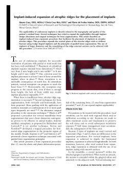
Baixar