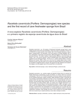
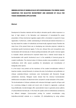
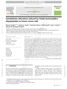
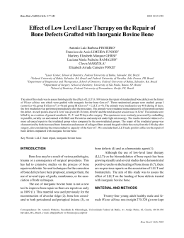
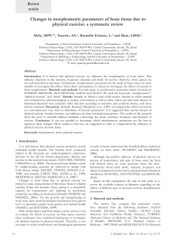
Biomaterials 21 (2000) 153}159
Platelet derived growth factor releasing chitosan sponge
for periodontal bone regeneration
Yoon Jeong Park!, Yong Moo Lee", Si Nae Park!, Seung Yoon Sheen",
Chong Pyoung Chung", Seung Jin Lee!,*
!Department of Industrial Pharmacy, College of Pharmacy, Ewha Womans University, 11-1, Daehyun-Dong, Seodaemun-Ku, Seoul 120-750, South Korea
"Department of Periodontology, College of Dentistry, Seoul National University, Seoul, South Korea
Received 11 February 1999; accepted 12 July 1999
Abstract
With an aim of improving bone regeneration, chitosan sponge containing platelet-derived growth factor-BB (PDGF-BB) were
developed. For fabrication of chitosan sponge, chitosan solution was freeze-dried, crosslinked and freeze-dried again. PDGF-BB was
incorporated into the chitosan sponge by soaking chitosan sponge into the PDGF-BB solution. Release kinetics of PDGF-BB, cell
attachment, proliferation capacity and bony regenerative potentials of PDGF-BB-loaded chitosan sponge were investigated.
Prepared chitosan sponge retained porous structure with 100 lm pore diameter that was suitable for cellular migration and growth.
Release rate of PDGF-BB could be controlled by varying initial loading content of PDGF-BB to obtain optimal therapeutic e$cacy.
PDGF-BB-loaded chitosan sponge induced signi"cantly high cell attachment and proliferation level, which indicated good cellular
adaptability. PDGF-BB-loaded chitosan sponge demonstrated marked increase in new bone formation and rapid calci"cation.
Degradation of the chitosan sponge was proceeded at defect site and subsequently replaced with new bone. Histomorphometric
analysis con"rmed that PDGF-BB-loaded chitosan sponge signi"cantly induced new bone formation. These results suggested that
chitosan sponge and PDGF-BB-loaded chitosan sponge may be bene"cial to enhance periodontal bone regeneration. ( 1999
Elsevier Science Ltd. All rights reserved.
Keywords: Chitosan sponge; Periodontal bone regeneration; Platelet-derived growth factor-BB; Controlled release
1. Introduction
Periodontal regenerative therapy with bone-substituting materials has gained favorable clinical e$cacy by
enhancing osseous regeneration in periodontal bony defects [1}9]. As bone-substituting materials, bone powder
[1,2], calcium phosphate ceramic [3,4], modi"ed forms
of hydroxyapatite (Osteogent) [5,6], and hard tissue
replacement polymer (HTR polymert) [7}9] have demonstrated their periodontal bony regenerative potency.
Bone-substituting materials should ful"ll several requirements such as biocompatibility, osteogenecity, malleability, biodegradability [1}3,7]. However, these materials
revealed some drawbacks including bone resorption, immune response, disease transmission, low biodegradability, poor adaptation. Several attempts have been made to
* Corresponding author. Tel.: 82-2-3277-3043; fax: 82-2-3277-2851.
E-mail address: [email protected] (S.J. Lee)
overcome these problems; use of biodegradable polymers
and combination of ceramics with bioactive polymers
such as collagen and polylactides [10}12]. These attempts have been aimed at developing new bone substitutes that resemble bone more closely than other
materials. However, these techniques have some limitations in inducing bone regeneration within whole therapeutic period. These materials act only as physical
sca!olds for bone tissue. Bone formation after grafting
these materials occurred over a period of several months
or years [1,4,6,7,10]. Additional function to these materials should be imparted to shorten bone forming period
and enhancing bone forming e$cacy. As an e!ective
approach, drug incorporation within these materials
might be suggested to obtain this purpose.
Early healing concept of bone formation by some
bioactive agents such as #urbiprofen and tetracycline has
been recently suggested in periodontal regeneration
[13,14]. Especially, growth factors including plateletderived growth factor-BB (PDGF-BB), insulin-like
0142-9612/00/$ - see front matter ( 1999 Elsevier Science Ltd. All rights reserved.
PII: S 0 1 4 2 - 9 6 1 2 ( 9 9 ) 0 0 1 4 3 - X
154
Y.J. Park et al. / Biomaterials 21 (2000) 153}159
growth factor (IGF), transforming growth factor-b
(TGF) function as modulators of chemotaxis, proliferation, and di!erentiation of pluripotent cells concerning
bone regeneration [15,16]. Overall healing periods might
be signi"cantly shortened by using these agents. It would
thus be more advantageous for the substituting material
to have release-controlling capacity of bioactive agents.
This combination of controlled drug delivery concept
with bone substitute technique can be highly bene"cial
for bone regeneration.
In this study, chitosan sponge were developed as an
osteoconductive material which induces or stimulates
bone formation. Chitosan is a biodegradable cationic
polysaccharide composed of N-acetylglucosamine residues which is known to accelerate wound healing and
bone formation [17]. Many previous reports corroborated enhanced wound healing and hemostatic e!ect of
chitosan [18}20]. In addition to these biomedical applicability, chitosan can regulate release of bioactive
agents [21}23]. Platelet-derived growth factor-BB
(PDGF-BB) was used in this study. PDGF-BB is reported as a potential mediator of bone regeneration [24].
It stimulates proliferation and di!erentiation of mesenchymally derived cells including "broblasts, smooth
muscle cells, ligament cells and osteoblasts [24}26]. Also,
it has revealed signi"cant neoosteogenic e!ect in beagle
dogs and monkeys [27}29]. However, as yet, extremely
high dose of PDGF-BB (above 10 lg/ml) has been applied in clinical trials owing to rapid clearance of
PDGF-BB in vivo and inability to maintain therapeutic
concentration during implantation period [25}28].
Therefore, controlled PDGF-BB delivery from the
sponge may be highly bene"cial for the treatment of
periodontal disease.
The objective of this study is to develop growth factor
releasing chitosan sponge matrices as a osteoconductive
materials for bone regeneration. This paper reports on
the fabrication of chitosan sponge, release kinetics of
growth factor, osteoblast attachment and proliferation
and bone regeneration capacity of growth-factor-loaded
chitosan sponge.
2. Materials and methods
2.1. Fabrication of chitosan sponge
Chitosan solution was freeze-dried, crosslinked with
tripolyphosphate and freeze-dried again to obtain
spongeous matrix. Constant amount of PDGF-BB (Genzyme Co., Cambridge, USA) solution was added to
chitosan sponge, kept overnight at 43C and freeze-dried.
PDGF-BB-loaded chitosan sponge was prepared to be of
plate form (1]1]0.1 cm in size, ca. 30 mg by weight)
containing 100, 200 and 400 ng of PDGF-BB per each
sponge, respectively. Surface and cross-section of the
sponge were examined by using a scanning electron
microscope (SEM) (Hitachi Model S-2460N, Hitachi
Ltd., Tokyo, Japan). The sponge was cut into small
fragments for calvarial defect "lling.
2.2. Release experiments
To determine the release kinetics of PDGF-BB from
the sponge, 125I-labeled PDGF-BB was utilized as
a tracer. Consistent ratio of 125I-labelled PDGF-BB
(5 lCi, Amersham Co., UK) was diluted with non-radioactive PDGF-BB to have "nal radioactivity of 1 lCi.
These radioactive mixtures were incorporated into the
chitosan sponge by the content of 200}400 ng per each
sponge. Each chitosan sponge was immersed in glass
vials containing pH 7.4 phosphate bu!er as releasing
medium (10 ml). The sealed vials were placed in a shaking
water bath at 373C and shaken at a frequency of 15 rpm.
At predetermined time interval over a period of 4 weeks,
samples were withdrawn from the vials and replenished
with fresh medium. The concentration of released
PDGF-BB in the samples was assayed by using a gamma
counter (Cobra II, Packard Instrument Company, CT,
USA).
2.3. Isolation of fetal rat calvarial osteoblastic cells
The cell isolation experiment was performed using the
method described by Bellows et al. [30]. The calvaria of
17}19 day-old fetal rats (Sprague-Dawley) were obtained
and all soft tissue was removed including the periosteum.
The frontal and parietal bones were then minced into
pieces approximately 2]2 mm using a sterile scalpel.
Bone cell isolation was performed with an enzymatic
digestion: Dulbecco's modi"ed Eagle's medium (DMEM;
Gibco, Grand Island, NY, USA) containing 3 mg/ml
dispase and 0.5 mg/ml collagenase (Sigma Chemical
Company, St. Louis, MO, USA). Following 20 min exposure, the media and released cells were removed and
discarded. The same enzymes were then used in a 3 h
digest. Contents of this digest were spun down, then the
pellet was resuspended and the resulting cells were incubated at 373C in an atmosphere containing 5% CO ,
2
95% air, with 99% relative humidity. Osteoblasts were
maintained in growth medium containing DMEM, 15%
heat-inactivated fetal bovine serum (FBS), 1% penicillin}streptomycin, 1% fungizone, HEPES bu!er (15 mM),
sodium pyruvate (1 mM) and ascorbic acid (5 lg/ml). Isolated cells were characterized by alkaline phosphatase
staining and Von Kossa staining for mineralization.
2.4. Osteoblastic cell attachment and proliferation
Osteoblastic cells (ca. 105 cells/cm2) were placed on
top of each chitosan sponge. Tissue culture plate (polystyrene) well was used as a control. The cultures were
Y.J. Park et al. / Biomaterials 21 (2000) 153}159
placed in the incubator for 1 day, and upon removal were
washed with phosphate bu!ered saline (PBS) and trypsinized. Aliquots of the resulting dissociated cell suspensions were counted on a Coulter counter multisizer
(Model 0646, Coulter Electronics, Hialeah, FL, USA).
Only counts between 8 and 32 lm in diameter were used.
Cell proliferation was also determined by cell counts as
described above after 1, 3, 7 and 10 days in culture. In this
experiment, six replicate samples were examined.
Attached and/or proliferated osteoblastic cells were
"xed with 2.5% glutaraldehyde (Sigma, MO, USA) in
0.1 M PBS (pH 7.4) for 30 min and then rinsed with 0.1 M
PBS. The cells were stained with 1 ml of cold 1% osmium
tetroxide (Polyscience, WI, USA), placed on ice for
30}40 min, rinsed with deionized water, and stored in
a deep freezer (!703C). The "xed and stained cell samples were freeze-dried and sputter-coated with gold and
were examined by using a SEM.
155
3. Results and discussion
3.1. Fabrication of chitosan sponge
Prepared chitosan sponge demonstrated porous structure with ca. 100 lm in pore diameter (Fig. 1). Since the
average size of bone cells is 10 lm [31], bone cells were
expected to easily migrate into the chitosan sponge and
to properly proliferate within the sponge. The sponge
showed brittle property at dried state, but provided good
malleability after being wetted with saline.
3.2. Release of PDGF-BB from chitosan sponge
Fig. 2 demonstrates the release of PDGF-BB from
chitosan sponge. Initial burst release was observed with
2.5. Bone regeneration capacity of PDGF-BB-loaded
chitosan sponge
In surgical procedure, 24 male Sprague-Dawley rats
(250 gm in average body weight) were divided into three
groups of eight each. The three groups included untreated sample group, chitosan-sponge-treated group,
and PDGF-BB-loaded chitosan-sponge-treated group.
Sprague-Dawley rats were anaesthetized by intraperitoneal injection of ketamine 30 mg/kg body weight).
A linear incision was formed after wiping the surgical site
with 0.5% chlorhexidine. A craniotomy defect (8 mm in
diameter) was prepared using a trephine needle in a dental handpieces supplemented with physiologic saline.
After dissecting the calvarial disc, chitosan sponge,
PDGF-BB-loaded chitosan sponge were placed into the
defect, and soft tissues and skins were then closed. Rats
were sacri"ced 4 weeks after implantation. Retrieved
specimens were "xed in formalin solution, decalci"ed in
5% nitric acid solution and embedded in para$n. Coronal sections (5 lm in thickness) were prepared and
stained with Goldner-Masson trichrome for photomicrography. Microscopical examination was undertaken
using Olympus BH-2 light microscope (Olympus Optical
Co., Osaka, Japan). Histomorphometric analysis for
these specimens was performed using a microscope
coupled with a high resolution video camera and a semiautomated image analysis system.
Fig. 1. Scanning electron microscopy of cross-section of a chitosan
sponge.
2.6. Statistical analysis
All measurements were collected in six replicate and
expressed as means$standard deviation. A two tailed
unpaired t-test was employed to assess the statistical
signi"cance of results for all measurements.
Fig. 2. Release pro"le of PDGF-BB from chitosan sponge. E!ects of
initial PDGF-BB loading on release rate: (j) 100 ng loaded, (v) 200 ng
loaded, (m) 400 ng loaded.
156
Y.J. Park et al. / Biomaterials 21 (2000) 153}159
rapid release during the "rst day, followed by sustained
release up to 6 days and a levelling o! of the release rate.
The burst e!ect indicates rapid water uptake of chitosan
sponge and dissolution of the exposed PDGF-BB particles at the surface of the chitosan sponge. As the
PDGF-BB-loading content increased, release rate increased correspondingly for the 6 days. Rapid release at
the initial step and maintenance of proper concentration
at the local site could be favored for growth factor delivery. Furthermore, considering the chemotactic e!ect of
PDGF-BB, the initial burst release of PDGF-BB is perhaps advantageous in bone cell migration, mitogenesis
and proliferation. Especially, consistent release of
PDGF-BB at the rate of 6 ng/day was observed from
200 ng PDGF-BB-loaded chitosan sponge up to 6 days
after initial burst of 100 ng. The release of PDGF-BB
after 6 days maintained therapeutic level (1}2 ng) for
3 weeks. PDGF-BB is reported to increase mitogenesis
and chemotaxis of bone cells proportionally to its concentration within 0.1}100 ng/ml range [32]. The range of
release rate of PDGF-BB is anticipated to stimulate
regeneration of bony defects during implantation periods. Also, increased release rate as the increase in loading
content revealed that the release rate of PDGF-BB can
be controlled for optimum therapeutic e$cacy in bone
regenerative therapy.
3.3. Osteoblastic cell attachment and proliferation
One of the important requirements for bone-substituting materials would be adaptation to a wide variety of
bone tissue defects. Bone-substituting materials appear
to have been suited in those case where wound "lling
could be obtained over the wound site. Cellular attachment and migration over the bone substituting material
surface are essential to obtain e!ective wound "lling and
bone tissue adaptation [33,34]. For this reason, the
authors intended to examine the cellular attachment and
proliferation onto chitosan sponge.
Fig. 3(a) shows the SEM of the initial attachment of
osteoblastic cells to chitosan sponge 1 day after incubation. Attached cells showed rounded and stellate morphology that is typically observed in initially attached
osteoblastic cells [35]. Higher density and amount of
cells were observed from PDGF-BB-loaded chitosan
sponge (Fig. 3(b)). Alkaline phosphatase activity con"rmed the viability of attached osteoblasts over the
sponge (data not shown). Fig. 4 demonstrates the proliferation level to chitosan sponge after 10 day incubation
period. Chitosan sponge demonstrated increase in bone
cell attachment and proliferation to compare with that of
control, polystyrene dishes. The surface of polystyrene
dish has been known to have good cellular attachment
and show rapid cellular con#uency in incubation
period [33]. The extent in cellular attachment and proliferation implies that chitosan sponge have good cellular
Fig. 3. Scanning electron microscopy of osteoblastic cells cultured on
chitosan sponge 1 day after incubation: (a) attached cells onto chitosan
sponge and (b) onto PDGF-BB-loaded chitosan sponge.
Fig. 4. Proliferation pro"le of osteoblastic cells cultured on chitosan
sponge for 10 day incubation period: (j) control polystyrene dish, (v)
chitosan sponge, (m) PDGF-BB-loaded chitosan sponge. Data are
presented as mean$SD, N"6; H: P(0.05, as compared with
chitosan sponge at same time point.
Y.J. Park et al. / Biomaterials 21 (2000) 153}159
157
adaptability. Moreover, PDGF-BB-loaded chitosan
sponge revealed signi"cantly increased cell attachment
and proliferation. High degrees of cell attachment and
proliferation were observed from PDGF-BB-loaded
chitosan sponge in contrast to that from a control or
unloaded chitosan sponge. Controlled release of PDGFBB from chitosan sponge e!ectively stimulated osteoblastic cells. PDGF-BB has been known to stimulate
proliferation, chemotaxis and collagenous protein synthesis of osteoblastic cells and ligament "broblasts [26].
Chitosan was reported to enhance the proliferation of
progenitor cells [36]. The combination of chitosan and
PDGF-BB may be highly bene"cial to increase cellular
proliferating response.
3.4. Bone regeneration by PDGF-BB-loaded
chitosan sponge
Figs. 5}8 show regenerative potential of chitosan
sponge and PDGF-BB-loaded chitosan sponge in rat
calvarial defect. In chitosan sponge untreated defect at
4 weeks (Fig. 5), connective tissue spanned the host bone
margin and defect was completely "lled with "brous
connective tissue (CT). Negligible amount of new bone
was observed. In contrast, osteogenesis was signi"cantly
increased in chitosan sponge treated defect at 4 weeks
(Fig. 6). Marked formation of inner and outer bone tables
was observed. At this period, degradation of chitosan
sponge was proceeded at defect site and degraded site
was "lled with "brous tissue and new bone. Bone growth
took place centripetally toward the sponge. Although
new bone was rapidly formed, complete reunion of new
bone was not occurred. Other studies suggested that
chitosan interfere with the function of inhibitory cells
Fig. 5. Histologic evaluation of rat calvarial defects with no treatment
of chitosan sponge after 4 weeks implantation. Edge of original host
bone margins was indicated by the arrow (B). Fibrous bridges was
formed between host bones and indicated by the star (w). The mark CT
depicts connective tissue (Goldner}Masson trichrom staining, original
magni"cation]40).
Fig. 6. Histologic evaluation of chitosan sponge in rat calvarial
defects after 4 weeks implantation (]40). Chitosan sponge was
degraded and indicated by the arrow (C). The marks OB and NB depict
old bone and new bone, respectively (Goldner}Masson trichrom
staining).
Fig. 7. Histologic evaluation of PDGF-BB-loaded chitosan sponge
in rat calvarial defects after 4 weeks implantation. (a) lower magni"cation (]40), (b) higher magni"cation (]200). Chitosan sponge
was degraded and indicated by the asterisk (*). The marks OB and NB
depict old bone and new bone, respectively (Goldner}Masson trichrom
staining).
158
Y.J. Park et al. / Biomaterials 21 (2000) 153}159
Fig. 8. Histomorphometric data (area of new bone in a defects) for
calvarial defects treated with PDGF-BB loaded or unloaded chitosan
sponge after 4 weeks implantation. 1: untreated defect, 2: chitosan
sponge treated defect, 3: PDGF-BB-loaded chitosan sponge treated
defect. Data are presented as mean$SD, N"6; H,HH: P(0.05, as
compared with control at same time point;##: P(0.05, as compared
with chitosan sponge at same time point.
including "broblasts and stimulate the function of
osteoprogenitor cells such as osteoblasts, which may
enhance osteogenesis [37,38]. Especially, Klokkevold
reported that chitosan potentiates the di!erentiation of
osteoprogenitor cells and can facilitate bone formation
[34]. In present study, degraded chitosan sponge were
embedded in newly formed osseous tissue without
"brous tissue invasion. This may be explained by inhibitory e!ect of chitosan against "broblasts while stimulating osteoblast activity.
Fig. 7 shows the result of bone regenerative capacity of
PDGF-BB-loaded chitosan sponge. Thick osteoid tissue
and new bone formation was noticeably increased
(Fig. 7(a)). Rapid calci"cation of new bone was observed
and new bone formation was still continued nearby the
chitosan sponge. Prominent bony bridges consolidating
calvarial defects were also observed. Bony contour was
completely restored with new bone and neoosteogenesis
was still proceeded. Newly formed bone tissue were
placed at the site that was previously occupied by the
sponge. Higher magni"cation (Fig. 7(b)) of this specimen
shows the di!used neoosteogenesis through the remnants
of chitosan sponge. No separation between new bone and
the sponge by "brous encapsulation could be observed.
Bone regenerative e!ects of PDGF-BB-loaded chitosan
sponge at 4 weeks are quantitatively shown by histomorphometric data (Fig. 8).
This study aimed to test chitosan sponge carrying
PDGF-BB in restoring osseous tissue to a critical sized
defect of 8 mm in diameter in the rat calvaria. Osseous
regeneration with normal contouring was evident histologically in chitosan sponge treated defect. Furthermore, PDGF-BB-carrying chitosan sponge showed
much improved bone regenerative capacity histologically
and histometrically. Histometric data proposed that
PDGF-BB-loaded chitosan sponge provide a better response than that of PDGF-BB-unloaded chitosan
sponge. The potency of PDGF-BB at wound healing and
tissue regeneration has been extensively investigated;
however, the obstacle to obtain optimal therapeutic e$cacy is its short biological half-life [25}28]. For this
reason, extremely high amount of growth factor has been
administered to compensate the loss of biologic activity.
Therefore, the development of delivery system of
PDGF-BB that release PDGF-BB in controlled manner
is essential for optimal clinical e$cacy. The experimental
results suggested that chitosan sponge properly function
as local PDGF-BB delivery devices and physical bone
substituting materials.
4. Conclusion
PDGF-BB-loaded porous chitosan sponge was e!ective for controlled PDGF-BB release to obtain bone
regenerative e!ects. Chitosan sponge, as an osteoconductive material which induces or stimulates bone formation, showed su$cient cellular adaptability and high
bone healing e$cacy. Especially, PDGF-BB in combination with chitosan sponge signi"cantly enhanced bone
healing and regeneration. PDGF-BB-loaded chitosan
sponge may be a valuable tool in periodontal bone regenerative therapy.
Acknowledgements
This work was supported by the grant from Korea
Ministry of Science and Technology (MOST) d98-N102-01-A-13, Korea.
References
[1] Peltzman B, Bowers GM, Reddi AH, Berquist JJ. Treatment of
furcation involvements with "bronectin and intraoral autogenous
bone grafts: preliminary observation. Int J Periodont Restorative
Dent 1988;8(5):51}63.
[2] Sepe WW, Bowers GM, Lawrence JJ. Clinical evaluation of
freeze-dried bone allografts in periodontal osseous defects, Part
II. J Periodontol 1978;49:9}14.
[3] Legeros RZ. Calcium phosphate materials in restorative dentistry: a review. Adv Dent Res 1988;2:164}80.
[4] Peplassi EM, Bissada NF, Greenwell H, Farah CF. Doxycyclinetricalcium phosphate composite graft facilitates osseous healing
in advanced periodontal furcation defects. J Periodontol 1991;
62:106}15.
[5] Lin FH, Lin CC, Liu HC, Huang YY, Wang CY, Lu CM. Sintered
porous DP-bioactive glass and hydroxyapatite as bone substitute.
Biomaterials 1994;15(13):1087}98.
[6] Kenney EB, Lekovic V, Elbaz JH, Kovavvic K, Caranze FA,
Takei Jr HH. The use of a porous hydroxyapatite implant in
periodontal defects. II. Treatment of class II furcation lesions in
lower molars. J Periodontol 1988;59:67}72.
Y.J. Park et al. / Biomaterials 21 (2000) 153}159
[7] Yukna RA. Clinical evaluation of HTR polymer bone replacement grafts in human mandibular class II molar furcations. J Periodontol 1994;65:342}9.
[8] Suzuki JB, Babcock-Goodmans, Phillips B. Comparison of clinical healing of human periodontal defects with HTR synthetic
grafts. J Dent Res 1989;68:409 (Abstr. 1822).
[9] Amler MH, LeGeros RZ. Hard tissue replacement (HTR) polymer as an implant material. J Biomed Mater Res 1990;24:1079}89.
[10] Horisaka Y, Okamoto Y, Matsumoto N, Yoshimura Y, Hirano A,
Nishida M, Kawada J, Yamashita K, Takagi T. Histological
changes of implanted collagen material during bone induction.
J Biomed Mater Res 1994;28:97}103.
[11] Robinson BP, Hollinger JO, Szachowicz EH, Brekke J. Calvarial
bone repair with porous D,L-polylactide. Otolaryngol Head Neck
Surg 1995;112:707}13.
[12] Rovira A, Amedee J, Bareille R, Rabaud M. Colonization of
a calcium phosphate/elastin-solubilized peptide-collagen composite material by human osteoblasts. Biomaterials 1996;
17:1335}540.
[13] Drury G. E!ect of topical tetracycline on bone regeneration
following freeze-dried bone allografts. J Dent Res 1980;59:364}70.
[14] Je!coat MK, Williams RC, Reddy MS, English R, Goldhaber P.
Flubiprofen treatment of human periodontitis. J Periodont Res
1988;23:381}5.
[15] Park YJ, Ku Y, Chung CP, Lee SJ. Controlled release of plateletderived growth factor from porous poly(L-lactide) membranes for
guided tissue regeneration. J Control Release 1998;51:201}11.
[16] Hollinger J. Factors for osseous repair and delivery: Part I.
J Cranofac Surg 1993;4(2):102}8.
[17] Muzzarelli RAA. Biochemical signi"cance of exogenous chitins in
animals and patients. Carbohydr Polym 1993;20:7}16.
[18] Klokkevold PR, Lew DS, Ellis DG, Bertoami CN. E!ect of
chitosan on lingual hemostasis in rabbits with platelet dysfunction induced by epoprostenol. J Oral Maxillofac Surg 1992;
50:41}5.
[19] Muzzarelli RAA, Zucchini C, Ilari P, Pugnaloni A, MattioliBelmonte M, Biagini G, Castaldini C. Osteoconductive properties
of methylpyrrolidinone chitosan in an animal model. Biomaterials 1993;14:925}9.
[20] Muzzarelli RAA, Mattioli-Belmonte M, Tietz C, Biagini R,
Ferioli G, Brunelli MA, Fini M, Giardino R, Ilari P, Biagini G.
Stimulatory e!ect on bone formation exerted by a modi"ed
chitosan. Biomaterials 1994;15(13):1075}81.
[21] Berscht PC, Nies B, Liebendorfer A, Kreuter J. Incorporation of
basic "broblast growth factor into methylpyrrolidone chitosan
#eeces and determination of the in vitro release characteristics.
Biomaterials 1994;15(8):593}600.
[22] Chandy T, Sharma CP. Chitosan matrix for oral sustained delivery of ampicilline. Biomaterials 1993;14:939}44.
[23] Shiraishi S, Imai T, Otagiri M. Controlled release of indomethacin by chitosan}polyelectrolyte complex: optimization
and in vivo/in vitro evaluation. J Control Release 1993;25:217}25.
159
[24] Cho MI, Lin WL, Genco RJ. Platelet-derived growth factor
modulated guided tissue regenerative therapy. J Periodontol
1995;66:522}30.
[25] Lynch SE, Castella GR, Williams RC, Kiritsy CP, Howell TH,
Reddy MS, Antoniades HN. The e!ects of short-term application
of a combination of platelet-derived and insulin-like growth
factors on periodontal wound healing. J Periodontol 1991;
62:458}67.
[26] Canalis E, McCarthy TL, Centrella M. E!ects of platelet-derived
growth factor on bone formation in vitro. J Cell Physiol
1989;140:530}7.
[27] Park JB, Matsuura M, Han KY, Norderyd O, Lin WL, Genco RJ,
Cho MI. Periodontal regeneration in class III furcation defects of
beagle dogs using guided tissue regenerative therapy with plateletderived growth factor. J Periodontol 1995;66:462}77.
[28] Rutherford RB, Niekrash CE, Kennedy JE, Charette MF. Platelet-derived and insulin-like growth factors stimulate regeneration
of periodontal attachment in monkeys. J Periodont Res 1992;
27:285}90.
[29] Lynch SE, Buser D, Hernandez RA, Webber HA, Stich H, Fax
CH, Williams RC. E!ects of the platelet-derived growth factor/insulin-like growth factor-I combination on bone regeneration around titanium dental implants. Results of a pilot study in
beagle dogs. J Periodontol 1991;62:710}6.
[30] Bellows CG, Aubin JE, Heershe JNM, Antoz ME. Mineralization
of bone nodules formed in vitro enzymatically released rat calvaria cell population. Calci"ed Tissue Int 1986;38:143}54.
[31] Ishaug SL, Yaszemski MJ, Bizios R, Mikos AG. Osteoblast function on synthetic biodegradable polymers. J Biomed Mat Res
1994;28:1445}53.
[32] Matsuda N, Lin WL, Kumar NM, Cho MI, Genco RJ. Mitogenic,
chemotactic and synthetic responses of rat periodontal ligament
"broblastic cells to polypeptide growth factors in vitro. J Periodontol 1992;63:515}25.
[33] Muzzarelli R, Baldassarre V, Conti F, Ferrera P, Biagini B.
Biological activity of chitosan: Ultrastructural study. Biomaterials 1988;9:247}52.
[34] Klokkevold PR, Vandemark L, Kenney EB, Bernard GW. Osteogenesis enchanced by chitosan (poly-N-acetyl glucosaminoglycan) in vitro. J Periodontol 1996;67:1170}5.
[35] Glass JR, Dickerson KT, Kim S, Polarek JW. Characterization of
a hyaluronic acid}Arg}Gly}Asp peptide cell attachment matrix.
Biomaterials 1996;17:1101}8.
[36] Van der Lei B, Wildevuur ChRH. Improved healing of microvascular PTFE prostheses by induction of a clot layer: an experimental study in rats. Plast Reconstr Surg 1989;84:960}8.
[37] Malette WG, Quigley HJ, Adickes ED. Chitin in nature and
technology. In: Muzzarelli R, Jeuniaux C, Gooday GW, editors.
Chitosan e!ect in nature and technology. New York: Plenum
press, 1986. p. 435}42.
[38] Muzzarelli R, Biagini G, Pugnaloni A. Reconstruction of periodontal tissue with chitosan. Biomaterials 1989;10:598}603.
Download