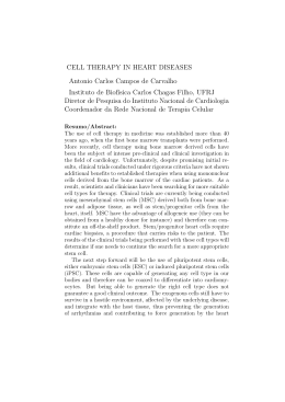
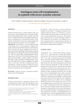
RELATO DE CASO
CASE REPORT
Hyperfibrotic myelodysplasia: case report with response to steroid
therapy
Recebido em 13/07/01
Aceito para publicação em 30/10/01
Mielodisplasia hiperfibrótica: relato de caso com resposta à terapia com corticosteróides
Maura Romeo1
Maria de Lourdes Lopes Ferrari Chauffaille2
Daniella Marcia Maranhão Bahia1
Maria Regina Regis Silva3
key
y words
Hyperfibrotic
Steroid therapy
resumo
Contexto: A fibrose de medula óssea é encontrada em algumas doenças hematológicas clonais,
incluindo síndromes mieloproliferativas, leucemias agudas e síndromes mielodisplásicas. Nas
síndromes mielodisplásicas, uma nova entidade clinicopatológica com acentuado aumento das
fibras de reticulina tem sido sugerida, e o termo “síndrome mielodisplásica hiperfibrótica” é usado
para sua definição. A biópsia de medula óssea mostra aumento das fibras de reticulina e hiperplasia
megacariocítica com displasia. O diagnóstico diferencial com mielofibrose primária é difícil, e casos
híbridos podem ocorrer. Pacientes com síndromes mielodisplásicas hiperfibróticas que respondem à
terapia com corticosteróide têm sido descritos. Na maioria dos casos a remissão é apenas
hematológica, mas resolução da fibrose de medula óssea já ocorreu em um paciente. Planejamento:
Relato de caso. Relato: Paciente do sexo masculino, 62 anos de idade, apresentou-se, em junho de
1995, com história de seis meses de letargia e dispnéia. Ao exame, mostrou-se pálido, sem
hepatoesplenomegalia. A concentração de hemoglobina foi de 3g/dl, com acentuada anisocitose,
mas sem “células em lágrimas”. Os aspirados de medula óssea mostraram-se “secos”, enquanto a
biópsia revelou hipercelularidade e fibrose grau IV, obliterando a arquitetura habitual. Os
megacariócitos estavam em número aumentado, com morfologia anômala, reagindo com
anticorpos antifator VIII e CD31. Com a introdução de prednisona 1mg/kg em junho de 1996, os
sintomas reduziram, a hemoglobina elevou-se e a fibrose cedeu para grau II, tornando-se independente de transfusões até janeiro de 1999. Então, a hemoglobina caiu para 6g/dl e, com a
introdução de prednisona, houve pronta elevação da hemoglobina.
unitermos
Síndromes mielodisplásicas
Hiperfibrótica
Corticoterapia
149
Jornal Brasileiro de Patologia e Medicina Laboratorial
Context: Bone marrow fibrosis is observed in different clonal hematological disorders including
myeloproliferative diseases, acute leukemias and myelodysplastic syndromes. In myelodysplastic
syndrome a new clinical-pathological entity with significant increase in reticulin fibers has been
suggested, and the term hyperfibrotic myelodysplasia was used to define it. Bone marrow
biopsy shows increased reticulin fibers, megakaryocytic hyperplasia and dysplasia. Differential
diagnosis with primary myelofibrosis may be difficult and hybrid cases may occur. Patients with
hyperfibrotic myelodysplastic syndrome responding to treatment with steroids have been
reported. In the majority of cases there was only hematological remission, although resolution
of fibrosis occurred in one patient. Design: Case report. Case report: A 62-year old male
presented in June 95 with a 6-month history of lethargy and dispnea. On examination he was
pale without hepato-splenomegaly. Hemoglobin concentration was 3g/dL with marked
anisocytosis without teardrop cells. Bone marrow aspirates resulted in dry tap. Bone marrow
biopsy showed hypercellularity with increased fibrosis (grade IV) obliterating the normal
marrow architecture. Megakaryocytes were increased in number, with abnormal morphology.
Monoclonal antibodies against factor VIII and CD31 revealed that both were expressed in
megakaryocytes. Prednisone (1mg/Kg) was introduced in June 1996, after what his symptoms
lessened and hemoglobin increased. Bone marrow fibrosis decreased (grade IV to grade II). He
has become transfusion independent till Jan/1999, when hemoglobin fell to 6g/dL and
prednisone was reintroduced with a prompt rise in hemoglobin concentration.
Rio de Janeiro, v. 38, n. 2, p. 149-151, 2002
Myelodysplastic syndromes
abstract
1. M.D. Disciplina de
Hematologia e Hemoterapia da
Universidade Federal de São
Paulo/Escola Paulista de
Medicina, São Paulo, Brasil.
2. M.D, Ph.D. Professora da
Disciplina de Hematologia e
Hemoterapia da Universidade
Federal de São Paulo/Escola
Paulista de Medicina, São Paulo,
Brasil.
3. M.D. Professora do
Departamento de Patologia
Aplicada da Universidade Federal
de São Paulo/Escola Paulista de
Medicina, São Paulo, Brasil.
Hyperfibrotic myelodysplasia
Introduction
Rio de Janeiro, v. 38, n. 2, 2002
Bone marrow fibrosis is observed in different clonal
hematological disorders including myeloproliferative
diseases, acute leukemias and myelodysplastic syndromes
(MDS) (2). In the last one, an increase in reticulin fibers of
varying degrees occurs in 50% of primary and in 80% of
cases secondary to treatment with alkylating agents and
radiotherapy. About 20% of MDS cases show significant
increase in reticulin fibers with or without collagen
formation (3).
Jornal Brasileiro de Patologia e Medicina Laboratorial
150
In 1988, Pagliuca et al. (4) reported 10 cases of primary
MDS with pronounced marrow fibrosis and used the term
“hyperfibrotic myelodysplasia” to define them. Bone
marrow biopsy showed increase in reticulin fibers,
megakaryocytic hyperplasia and dysplasia. Bone marrow
aspirates are generally hypocellular or dry and classification
according to FAB is not possible. When adequate bone
marrow aspirate is obtained it shows dysplasia of one, two
or all three cell lines with characteristic morphological
changes of MDS (2). On examination, hepatic or splenic
enlargement is absent or discrete (4).
Romeo et al.
fibers) obliterating the normal marrow architecture (grade IV fibrosis) (3) (Figure 1). One year after the diagnosis
he was transfusion dependent (04 packed red cells/month),
without response to further treatment (B6, folic acid, and
erythropoetin). In June 1996, prednisone (1mg/Kg) was
started and maintained for 4 weeks. His symptoms lessened
and hemoglobin concentration increased from 6g/dL to
13,2 g/dL. Bone marrow biopsy revealed reduction of
marrow fibrosis (from grade IV to II) (3) (Figure 2). He
became transfusion independent till Jan/1999 when
hemoglobin fell to 6g/dL and packed red cell transfusions
were necessary. At that moment there were no signal of
infection or other cause for the anemia. Prednisone was
restarted, maintained for 4 weeks and there was a prompt
rise in hemoglobin concentration.
Watts et al. (5), in 1991, reported cases of hyperfibrotic
MDS that responded to treatment with steroids. In the
majority of the cases there was just hematological
remission, but one patient has shown resolution of marrow
fibrosis following steroid therapy.
Case report
A 62-year old male presented in June 1995 with a sixmonth history of lethargy and dyspnea on exertion. On
clinical examination he was pale, without hepato or
splenomegaly. Hemoglobin concentration was 3g/dL,
white blood cell (WBC) count 4 x 10⁹/L and platelets
160 x 109/L. The differential WBC count was: neutrophils
65%, eosinophils 2%, basophils 0%, lymphocytes 28%
and monocytes 5%. Dysmorphic erythrocytic changes
included marked anisocytosis without teardrop cells. The
neutrophils were agranular with many pseudo Pelger-Hüet
forms. Bone marrow aspirates resulted in a dry tap. Bone
marrow biopsy showed a hypercellular marrow with
reduced erythroid elements. Megakaryocytes were
increased in number, having abnormal morphology.
Monoclonal antibodies against factor VIII and CD31
revealed expression of both in megakaryocytes. Micromegakaryocytes were present. There was a great increase
in reticulin fibers (Gomori’s silver impregnation for reticulin
Figure 1 – Bone marrow biopsy showing a great increase in reticulin fibers by
Gomori’s silver impregnation (grade IV fibrosis)
Figure 2 – Bone marrow biopsy showing reduction of marrow fibrosis
(from grade IV to II)
Romeo et al.
Discussion
Fibrosis of the marrow as a consequence of clonal
hematopoietic disorder with a proeminent megakaryocytic
component is possibly a result of stimulation by plateletderived polypeptide factors. These factors include plateletderived growth factor and transforming growth factor beta
in addition to platelet factor-IV, which inhibits collagen
breakdown. Maybe the action of steroids in this process
could justify the response of these patients to steroid
therapy (2, 5)
The patient presented in this paper is a typical case of
hyperfibrotic MDS. Dysplasia observed in erythroid and
granulocytes elements associated with absent hepatosplenomegaly support this diagnosis. Histopathology of
marrow did not permit in the present case differentiation
between hyperfibrotic-MDS and myelofibrosis, but
erythroid hypoplasia observed had already been reported
before in hyperfibrotic-MDS patients (4).
MDS treatment is divided into supportive care,
appropriate for older patients and those with indolent
disease; and aggressive chemotherapy and stem cell transplantation for selected patients. The International Prognostic Scoring System (1) can help identify patients who
are likely to have an indolent course and are best treated
with supportive measures.
Further studies correlating pathologic, cytogenetic and
molecular data with clinical outcome are necessary to
provide more information for proper classification and
treatment of this disease.
Acknowledgements
We thank Prof. Maria Claudia Zerbini, M.D, Ph.D., for
helpful comments and review.
4. Pagliuca, A. et al. Myelofibrosis in primary myelodysplastic
syndrome: a clinic-morphological study of 10 cases. Br. J.
Haematol., 71: 499-504, 1989.
5. Watts, E.J. et al. Hyperfibrotic myelodysplasia: a report of three
cases showing haematological remisson following
treatment with prednisolone. Br. J. Haematol., 78: 120-4,
1991.
Endereço para correspondência
Maria de Lourdes Chauffaille
Disciplina de Hematologia e Hemoterapia
da Universidade Federal de São Paulo
Escola Paulista de Medicina
Rua Botucatu 740 – Prédio dos
Ambulatórios – 3º andar
CEP 04023-900 – São Paulo-SP
Tel.: (55-11) 5576-4237
Fax: (55-11) 5571-8806
e-mail: [email protected]
151
Jornal Brasileiro de Patologia e Medicina Laboratorial
References
1. Greenberg, P. et al. International Scoring System for evaluating
prognosis in myelodysplastic syndrome. Blood, 89: 207988, 1997, erratum Blood, 91: 1100, 1998.
2. Lambertenghi-Deliliers, G. et al. Myelodysplastic syndrome with
increase marrow fibrosis: a distinct clinical-pathological
entity. Br. J. Haematol., 78:161-6, 1991.
3. Manoharan, A.; Horsley, R. & Pitney, W.R. The reticulin content
of bone marrow in acute leukemia in adults. Br. J. Haematol.,
43: 185-90, 1979.
Rio de Janeiro, v. 38, n. 2, 2002
Hyperfibrotic MDS is a clinic-pathological entity and
should be distinguished from the classical MDS subgroups
defined by FAB cooperative group in 1982. Differential
diagnosis between hyperfibrotic MDS and primary
myelofibrosis is difficult (2) and some authors suggest
that hybrid cases, called hyperfibrotic MDS-myelofibrosis,
may occur. Megakaryocytic hyperplasia with dysplasia
and increased marrow fibrosis are common to both.
Presence of dysplasia of all three lineages and modest or
absent hepatosplenomegaly, support the diagnosis of
hyperfibrotic MDS. In equivocal cases ferrokinetic studies
are recommended to exclude extramedullary haematopoesis regarded as sine qua non of idiopathic
myelofibrosis (2).
Hyperfibrotic myelodysplasia
Baixar