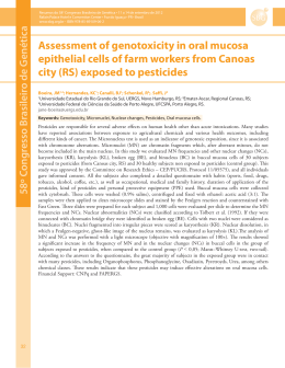
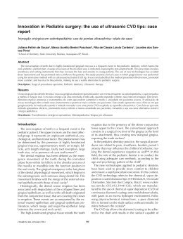
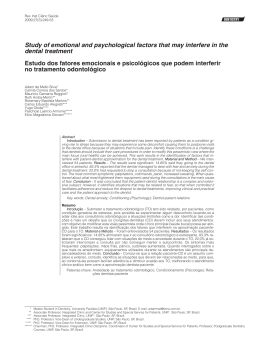
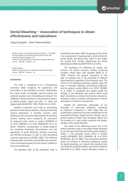
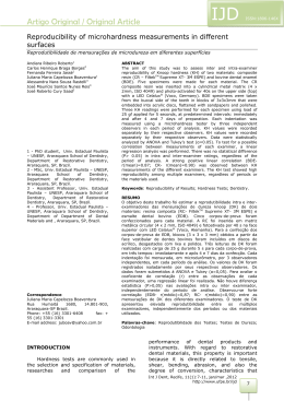
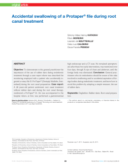
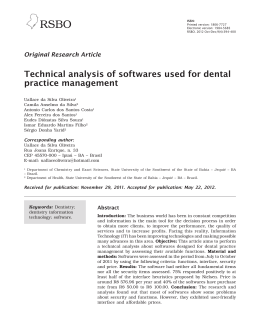
ISSN:
Versão impressa: 1806-7727
Versão eletrônica: 1984-5685
RSBO. 2011 Apr-Jun;8(2):182-8
Original Research Article
Nuclear anomalies in the buccal cells of children
under dental treatment
Elisângela de Fátima Rezende1
Maria Cristina Mendes-Costa1
Johnson Campideli Fonseca1
Alex de Oliveira Ribeiro1
Corresponding author:
Maria Cristina Mendes-Costa
University Center of Lavras
506, Padre José Poggel St. – Centenario
Zip code 37200-000 – Lavras – MG – Brazil
E-mail: [email protected]
1
Center of Researches in Health and Biological Science, University Center of Lavras – Lavras – MG – Brazil.
Received for publication: August 17, 2010. Accepted for publication: November 3, 2010.
Keywords:
genotoxicity;
micronucleus tests;
dental materials.
Abstract
Introduction: Since some dental materials may be aggressive
to a person’s body, studies involving such materials seem to
be necessary. Objective: This study was conducted to evaluate
the genotoxicity of dental materials through micronucleus (MN)
test. Material and methods: Exfoliated buccal cells of 4-to-12
year-old children, who were on some type of dental treatment,
were collected either before or after the treatment ending. Each
sample was composed of 1,000 cells per patient. Student’s t
test was used for comparison. Results: The dental materials
were divided into 3 groups, as follows: cement, monomers, and
their combination. Treatments using monomer + cement-based
materia ls were found to increase sig ni fica nt ly t he number
of binucleated (BN) cells, (p < 0.05) which indicate several
degenerative nuclear changes. Conclusion: The combination
of cement-based dental material with monomers increases the
cytotoxic action of dental materials.
RSBO. 2011 Apr-Jun;8(2):182-8
Introduction
Dental materials may produce ag gressive
effects caused by monomers release and/or other
organic and inorganic components [31]. As most
dental materials release small portions of several
substances on both the pulp and oral cavity, it
is essential to prove their biocompatibility and
toxicological profile. Appropriate regulation should
ensure that the genotoxicity be either abolished or
decreased [17].
The main dental materials used in pediatric
dental treatment are: glass ionomer cements (GIC),
zinc oxide - eugenol (ZOE), and resin composites
(RC). GIC maintains the surrounding environment
suitable for remineralization as well as interferes
with the bacteria metabolism by fluoride releasing
[2, 26]. They are employed as pulp capping, occlusal
sealant, and restorative material. Also, hydrophilic
monomers can be incorporated to GIC, so-called
resin-modified glass ionomers (RMGICs). The
polymerization reactions make monomers into
polymers. Consequently, the residual monomers
released may cause intense cytotoxic effects [9,
34]. ZOE is often used in low-cost, easy-dealing
temporary restoration [7]. Its effects can vary, as
follows: point mutation inducer [27]; cytotoxicity
[18, 27]; antimicrobial [6, 31]. RC is commonly
used in permanent restoration. It presents less
abrasion, offering easy handling. It also can be
found in different colors to match to the tooth to
be restored [3]. These materials may have some
genotoxic potential. An increasing number of
dental restorations have been performed over the
past decade with significantly increase in local
and systemic adverse effects such as cytogenetic
changes [29].
Chromosome damages are widely used as
biomarkers in monitoring human exposure to
carcinogenic agents [8, 20, 36]. Binucleated (BN) cells
may indicate several kinds of degenerative nuclear
changes [10]. BN are cells which have two similarsized nuclei (almost the same size). Such nuclei are
not overplaced, but may be side by side. They have
the same color, an intact nuclear membrane, and
are within the cytoplasm [14]. Micronuclei (MN) are
free round or egg-shaped corpuscles, about 1/3 to
1/16 of the nucleus size [4, 5, 25]. They are usually
found beside the main nucleus and are similar in
shape, color, and chromatinic body distribution
183
[14]. Such structures are a result of chromosome
fragments or entire acentric chromosomes which
are lost during a cell division. For this reason they
are not included in the daughter cells’ nuclei, thus
remaining in the cytoplasm of interphase cells [5,
10, 16, 35]. MN takes 7-16 or 30 days to be formed
[6, 28, 33, 37, 38]. Such period is related to the
amount of time that the basal cells take to reach
the surface and exfoliate.
It is extremely important to know how the
materials act in human tissues because they are
in close contact with oral mucosa [30]. This study
aims to evaluate the genotoxicity of dental materials
used in pediatric treatment through micronucleus
(MN) test of exfoliated buccal cells.
Material and methods
Study Design
T he st udy wa s approved by t he Et h ics
Committee of Unilavras (CA AE, 0036/06). The
sample comprised 72 children divided into control
group (n = 40) and test group (n = 32). Inclusion
criteria during this study were as follows: children
who had never undergone any dental treatment;
children aged 2-12 years (mean age of five years).
There were no children with neurological diseases
and related genetic alterations. Their participation
in the research was previously authorized by their
parents or children’s legal guardians.
Exclusion criteria comprised children with
damages in oral mucosa that preclude the collection
of cells and whose parents had not signed the
clarified consent form or did not want to participate
in the study.
Test g roup pat ients were div ided into 3
experimental subgroups according to the dental
material used, as follows: Group I - Cement-type
materials: glass ionomer cement and zinc oxide
cement + eugenol (n = 8); Group II - Monomerbased materials: composite resin, dental sealant, and
adhesives (n = 13); Group III: combination of both
(n = 11). The genetic material used was obtained
by collecting oral mucosa cells by scrubbing the
surface with a wood stick. The first collection was
done before the treatment in order to find out the
basal index of both micronucleus and binucleus
cells likely to be found in the mucosa.
184 –
Rezende et al.
Nuclear anomalies in the buccal cells of children under dental treatment
One month after the treatment, a new sample
collection was performed in the treated patients
when they returned for either a follow-up or new
treatment appointment This time period was
chosen because is related to the mitoses that the
altered cells would suffer, thus showing clearly
their micronucleus formation. The control group
consisted of patients’ oral mucosa cells before the
insertion of any type of dental material.
Genotoxicity evaluation (micronucleus test)
After the inner part of the cheek was scrubbed
by using a wood stick [10, 38], the epithelial
cells collected from buccal mucosa were smeared
onto clean microscope glass slides fixed in 70%
alcohol, air dried, and stained with 2% acetic
orcein [15], for 30’ [23]. A light microscope at
100 X magnification on coded slides was used
for MN analysis and then microphotographed
(Samsung SDC-312). Two thousand cells per
patient were analyzed, as follows: 1,000 before
and 1,000 after the treatment [19, 24]. After
bot h t he micronucleus a nd t he bi nucleated
cells were quantified, the frequency analysis of
both alterations was performed only in the non
fragmented, overplaced or overcrowded cells with
untouched nuclei, according to the acceptance
criteria, as described by Fenech et al. (2000).
Data statistic analysis was performed by Student’s
t test [19] for non-parametric data.
Figure 1 – Mean values of BN and MN average in control
and experimental groups
Normal, binucleated, and micronucleated
cells were found in a ll studied groups. The
binucleated cells (figure 2) presented two nuclei,
nearly the same size instead of overplaced. They
showed the same color, their nuclear membrane
was intact and within the same cytoplasm, as
described by Fenech et al. (2003). On the other
hand, the nucleus of the micronucleated cells
(figure 3) was shorter than the main nucleus
diameter, round or oval in shape, separated
from the main nucleus [14]. Diler and Ergene
[11] analyzed nuclear anomalies in buccal cells of
calcite factory workers and found micronucleus
a nd binucleated cells sug gest ing sig nifica nt
cytogenetic damage.
Results and discussion
The epithelial tissue of the oral cavity was
collected for the test, because it is in close contact
with dental materials and is constantly renewed
[1]. In addition to this, it can act as a tool for
biomonitoring human populations exposed to
genotoxic agents [12, 22].
The M N avera ge f requency i n a hea lt hy
population is about 1 to 3 out of 1,000 cells [12].
However, in our study, nearly 1% of the control
patients exhibited MN score of 7 to 8 before treatment
(figure 1). This suggests that those children had
probably been influenced by genotoxic agents [1,
22, 31].
Figure 2 – Binucleated cell RSBO. 2011 Apr-Jun;8(2):182-8
Figure 3 – Micronucleated cell
The cement materials did not caused decrease
and/or increase of BN or MN cells in Group I
patients (table I). The treatment with monomers
materials (Group II) did not significantly alter the
frequency of MN, because pre- and post-treatment
MN frequency score was similar (table I).
It is important to consider that both MNs and
BNs have a time-determined formation. The dental
185
material constantly releases small portions of its
own substances in the oral cavity [17], especially
during the first 30 days after its insertion [2].
Therefore, the time period of this study was enough
for all the possible variations appear.
The increase of the frequency of BN for Group
III was significantly influenced by the cement +
monomer combination (table I). Similarly results
were found in people working in oil and oil-products
stations [11] and in between smoking and exposure
to benzene workers [12].
Ma ny materia ls, pa rt icula rly root ca na l
sealers, remain in contact with vital tissues
for a long period, when cellular aggression by
chemical, physical and mechanical elements may
occur, being the respiratory system one of the
first cells to be affected. Some aggressor agents,
particularly chemical substances, block important
enzyme systems of the protein synthesis and/or the
generation of ATP; others lead to the generation
of harmful intracellular products or, still, act
directly destroying vital structural components of
the cell [30]. This may justify the appearance of
binucleated cells (Group III). Senne et al. (2009)
analyzed zinc oxide-eugenol cements and resin,
finding that all tested sealers were cytotoxic.
Reis et al. [24] studied the genotoxic effect of
ethanol on oral mucosa cells and observed that
the frequency means of micronucleated cells and
micronuclei were significantly higher in the group
of exposed individuals, when compared to the
control group.
Table I – Evaluation of BN and MN in Groups I, II and III
Group
I – Treatment
I – Control
II – Treatment
II – Control
III – Treatment
III – Control
BN
Mean ± SE
t Test 5%
2.180 ± 0.714 0.0922 (NS)
1.355 ± 0.760
2.019 ± 0.375 0.0531 (NS)
1.441 ± 0.541
2.413 ± 0.644 0.0069 (S)
1.588 ± 0.177
MN
Mean ± SE
1.330 ± 0.122
1.299 ± 0.356
1.696 ± 0.674
1.790 ± 0.486
1.821 ± 0.504
1.791 ± 0.722
t Test 5%
0.8928 (NS)
0.7913 (NS)
0.9371 (NS)
(NS) Not significant
(S) Significant. The treatment values were significantly different from the control at p < 0.05
(SE) Standard Error
Celik et al. (2003) and Fenech et al. (1999) state that binucleated cells indicate cytotoxicity. The
resin-modified glass ionomer cements (RMGIC) have hydrophilic monomers incorporated to the glass
ionomer cement. When RMGICs are subpolymerized, they convert monomers into polymers. In such case
186 –
Rezende et al.
Nuclear anomalies in the buccal cells of children under dental treatment
the released residual monomers may produce an
intense cytotoxic effect [34]. The RMGIC cytotoxic
effect was noticed in MDPC-23 odontoblastic
cells culture [9]. When combined, the chemical
composition of cements + monomers becomes
similar to that of RMGIC. For this reason, group
III materials were found to be related to cytotoxic
damages. Such results were reached by comparing
the number of BNs found either before or after
the exposure to dental material. Although BNs
are not directly involved with DNA, they interfere
with the late events occurring in cell division
[11, 39]. Because their consequences are still
unknown, further cytogenetic studies regarding to
the perpetuation of such cells in the oral mucosa
are necessary.
In order to analyze genotoxicity or cytotoxicity
in lymphocyte culture, the use of cytochalasin B is
needed. This substance paralyses the cytokinesis,
which promotes the nucleus division, resulting in
a two-nucleus cell [13, 41]. The cement + monomer
combination used in this study showed significant
result for BN, suggesting a direct inf luence on
cytokinesis similarly to cytochalasin B. Its action
can be explained by the inhibition of telophase,
consequently the cell reaches the epithelial surfaces
with two nuclei. Because the action of the evaluated
substances interferes directly into the cell instead
of the gene, a cytogenetic damage occurs [11].
Biological markers may express the dose
amount of exposure to carcinogens and their
interaction with macromolecules, as DNA [24].
Consequently, a greater emphasis should be given
to methods that detect the genotoxic human activity.
The biomarkers may be used for the prevention
of serious diseases, and detection of high-risk
patients.
References
Conclusion
8. Cerqueira EM, Gomes-Filho IS, Trindade S,
Lopes MA, Passos JS, Machado-Santelli GM.
Genetic damage in exfoliated cells from oral
mucosa of individuals exposed to X-rays during
panoramic dental radiographs. Mutat Res. 2004
Aug;562(1-2):111-7.
It can be concluded that a significant BN
increase was noticed when Group III materials
were used, suggesting that the combination of
a cement-based dental material with monomers
intensify the cytotoxic action. The combination of
MN analysis with other nuclear anomalies, such
as BN, was found to enhance the sensitiveness
as well as the evaluation of both cytotoxicity and
genotoxicity in scrubbed cells of the exfoliated
buccal mucosa.
1. Arlett CF, Ashby J, Fielder RJ, Scott D.
Micronuclei origins, applications and methodologies
– a workshop sponsored by the Heath and Sfety
Executive held in Manchester. Mutagenesi. 1989
May;4(6):482-5.
2. Bernardo PC, Rodrigues CR, Souza Paiva JA,
Singer TM, Sañudo A. Avaliação clínica de um
cimento de ionômero de vidro utilizado como
selante oclusal. Pes Odontol Bras. 2000 JanMar;14(1):53-7.
3. Bianchi EC, Aguiar PR, Poggi MR, Salgado MH,
Freitas CA, Bianchi ARR. Estudo do desgaste
abrasivo das resinas compostas disponíveis
no mercado brasileiro. Mat Res. 2003 AprJun;6(2):255-64.
4. Bindu L, Balarm P, Mathew A, Remani P,
Bhattathiri VN, Nair MK. Radiation-induced
changes in oral carcinoma cells – a multiparametric
evaluation. Cytopathol. 2003 Oct;14(5):287-93.
5. Blochingm M, Hofmann A, Lautenschläger CH,
Berghaus A, Grummt T. Exfoliative cytology of
normal buccal mucosa to predict the relative risk
of cancer in the upper aerodigestive tract using the
MN-assay. Oral Oncol. 2000 Nov;36(6):550-5.
6. Carrano AV, Natarajan AT. Considerations
for population monitoring using cytogenetics
techniques ICPEMC publication 14. Mutat Res.
1988;204:379-406.
7. Cassanho ACA, Fernandes AM, Oliveira LD,
Carvalho CAT, Jorge AOC, Koga-Ito CY. In vitro
activity of zinc oxide-eugenol and glass ionomer
cements on Candida albicans. Braz Oral Res. 2005
Apr-Jun;19(2):134-8.
9. Coimbra LR, Giro EMA, Aranha AMF,
Costa CAS. Citotoxicidade de cimentos de
ionômero de vidro restauradores sobre células
de linhagem odontoblástica. Rev Odont Ciên.
2006;21(54):338-45.
RSBO. 2011 Apr-Jun;8(2):182-8
10. Çelik A, Çavas T, Ergene-Gözükara S.
Cytogenetic biomonitoring in petrol station
attendants: micronucleus test in exfoliated buccal
cells. Mutagenesis. 2003 Sep;18(5):417-21.
11. Diler SB, Ergene S. Nuclear anomalies in the
buccal cells of calcites factory workers. Genet Mol
Biol. 2010 Jun;33(2):374-8.
12. Fenech M, Holland N, Chang WP, Zeiger E,
Bonassi S. The Human Micronucleus Project.
An international collaborative study on the
use of micronucleus technique for measuring
DNA damage in humans. Mutat Res. 1999
Jul;428(1-2):271-83.
13. Fenech M. The in vitro micronucleous
technique. Mutat Res. 2000 Nov;455(1-2);81-95.
14. Fenech M, Chang WP, Kirsch-Volders M,
Holland N, Bonassi S, Zeiger E. HUMN project:
detailed description of the scoring criteria for
the cytokinesis-block micronucleus assay using
isolated human lymphocyte cultures. Mutat Res.
2003 Jan;534(1-2):65-75.
15. Guerra M, Souza MJ. Como observar
cromossomos: um guia de técnicas em citogenética
vegetal, animal e humana. 1. ed. Ribeirão Preto:
Funpec; 2002. 131 p.
16. Heddle JA, Cimino MC, Hayashi M, Romagna F,
Shelby MD, Tucker JD et al. Micronuclei as an index
of cytogenetic damage: past, present, and future.
Environ Mol Mutagen. 1991 Aug;18(4):277-91.
17. Heil J, Reifferscheid G, Waldmann P, Leyhausen
G, Geurtsen W. Genotoxicity of dental materials.
Mutat Res. 1996 Jul;368(3-4):181-94.
18. Hume WR. Effect of eugenol on respiration and
division in human pulp, mouse fibroblasts and liver
cells in vitro. J Dent Res. 1984 Nov;63(11):1262-5.
19. Kehdy FSG, Cerqueira EMM, Bonjardim MB,
Camelo RM, Castro MCL. Study of the cytogenetic
effects of occupational exposure to pesticides on
sanitation workers in Belo Horizonte, Brazil. Genet
Mol Res. 2007 Sep;6(3):581-93.
20. Marcon F, Zijno A, Crebelli R, Carere A,
Veidebaum T, Peltonen K et al. Chromosome
damage and aneuploidy detected by interphase
multicolour FISH in benzene-exposed shale oil
workers. Mutat Res.1999 Sep;445(2):155-66.
187
21. Munerato MC, Sinigaglia M, Reguly ML,
Andrade HHR. Genotoxic effects of eugenol,
isoeugenol and safrole in the wing spot test
of Drosophila melanogaster. Mutat Res. 2005
Apr;582(1-2):87-94.
22. Pastor S, Gutiérrez S, Creus A, Xamena N,
Piperakis S, Marcos R. Cytogenetic analysis of
Greek farmers using the micronucleous assay
in peripheral lymphocytes and buccal cells.
Mutagenesis. 2001 Nov;16(6):539-45.
23. Pires NM, Souza IRP, Prates HT, Faria TCL,
Pereira Filho IA, Magalhães PC. Effect of leucaena
aqueous extract on the development, mitotic index
and peroxidase activity in maize seedlings. Rev
Bras Fisiol Veget. 2001 Apr;13(1):55-65.
24. Reis SRA, Sadigursky M, Andrade MGS, Soares
LP, Espírito Santo AR, Vilas Boas DS. Genotoxic
effect of ethanol on oral mucosa cells. Pesq Odontol
Bras. 2002 Jul-Sep;16(3):221-5.
25. Ribeiro LR, Salvatori MF, Marques EK. Mutagênese
ambiental. Canoas: Ulbra; 2003. 356 p.
26. Sacramento PA, Papa AMC, Carvalho FG,
Puppin-Rontani RM. Propriedades antibacterianas
de materiais forradores – revisão de literatura. Rev
Odontol Unesp. 2008;37(1):59-64.
27. Sarrami N, Pemberton MN, Thornhill MH,
Theaker ED. Adverse reactions associated with
the use of eugenol in dentistry. Braz Dent J. 2002
Sep;14(5):257-9.
28. Sarto F, Tomanin R, Giacomelli L, Canova
A, Raimondi F, Ghiotto C et al. Evaluation
of chromosomal aberrations in lymphocytes,
oral mucosa and hair root cells of patients
under antiblastic therapy. Mutat Res. 1990
Feb;228(2):157-69.
29. Schweikl H, Hiller KA, Bolaya C, Kreissl M,
Kreismanna W, Nusser A et al. Cytotoxic and
mutagenic effect of dental composite materials.
Biomat. 2005 May;26:1713-9.
30. Senne MI, Lemos N, Fidel SR, Fidel RAS.
Evaluation of the cytotoxicity of three root canal
sealers used in obturation of radicular canals
system. RSBO. 2009 Mar;6(1):71-6.
188 –
Rezende et al.
Nuclear anomalies in the buccal cells of children under dental treatment
31. Silva J, Erdtmann B, Henriques JAP. Genética
toxicológica. Porto Alegre: Alcance; 2003. 442 p.
32. Spahl W, Budzikiewicz H. Qualitative analysis
of dental resin composites by gas and liquid
chromatography/mass spectrometry. Fresenius J
Analy Chem. 1994 Nov-Dec;350(12):684-91.
33. Squier CA, Finkelstein MW. Mucosa bucal. In:
Ten Cate AR. Histologia bucal. Desenvolvimento,
estrutura e função. 5. ed. Rio de Janeiro:
Guanabara Koogan; 2001. p. 323-39.
34. Stanislawski L, Daniau X, Lauti A, Goldberg
M. Factors responsible for pulp cell cytotoxicity
induced by resin-modified glass ionomer cements.
J Biomed Mater Res. 1999 May;49(3):277-88.
35. Stich HF, Curtis JR, Parida BB. Application of
micronucleus test to exfoliated cells of high cancer
risk groups: tabacco chewers. Int J Cancer. 1982
Nov;30(5):553-9.
36. Stich HF, Rosin MP. Quantitating the synergistic
effect of smoking and alcohol consumption with the
micronucleus test on human buccal mucosa cells.
Int J Cancer. 1983 Mar; 31(3):305-8.
37. Stich HF, Rosin MP. Micronuclei in exfoliated
cells as a tool for studies in cancer risk
and cancer intervention. Cancer Lett. 1984
Apr;22(3):241-53.
38. Tolbert PE, Shy CM, Allen JW. Micronuclei and
other nuclear anomalies in buccal smears: a field
test in snuff users. American J Epidemiol. 1991
May;134(8):840-50.
39. Tolbert PE, Shy CM, Allen JW. Micronúcleo
e outras anomalias no espaço oral: métodos e
desenvolvimento. Mutat Res. 1992;271:69-77.
40. Vasconcelos KRF, Veiga Júnior VF, Rocha WC,
Bandeira MFCL. Avaliação in vitro da atividade
antibacteriana de um cimento odontológico à base
de óleo-resina de Copaifera multijuga Hayne. Braz
J Pharmacog. 2008 Dec;18(l):733-8.
41. Zalacain M, Sierrasesúnaga L, Datiño A.
El ensayo de micronúcleos como medida de
inestabilidad genética induzida por agentes
genotóxicos. Anales. 2005 [cited 2007 Sep 30];
28(2). Available from: URL:www.cfnavarra.es/
salud/anales/textos/vol28/n2/revis2a.html.
Baixar