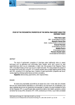
original article
Accidental swallowing of a Protaper® file during root
canal treatment
Simony Hidee Hamoy KATAOKA1
Flávio MOREIRA2
Lisandro de BOUTTEVILLE3
Celso Luiz CALDEIRA4
Oscar Faciola PESSOA5
ABSTRACT
high endoscopy and a CT scan. He remained asymptomatic after these two early interventions, was monitored over
four days, through X-rays of chest and abdomen, until the
foreign body was eliminated. Conclusion: General practitioners who do endodontics should be aware of the risks
involved in swallowing and/or accidental aspiration of foreign bodies during endodontic treatment, and know how to
avoid this problem by adopting a simple measure, the use
of rubber dam.
Objective: To demonstrate to the general practitioner the
importance of the use of rubber dam during endodontic
treatment through a case report where was described the
monitoring employed with a patient who accidentally ingested a rotary file S1 ProTaper® (Dentsply Maillefer, Switzerland) during the root canal preparation. Case report:
A 28 years-old patient underwent root canal treatment
without rubber dam and during the root canal therapy,
swallowed a ProTaper® S1. He was accompanied to the
hospital where, at first, was performed a gastrointestinal
Keywords: Deglutition. Rubber dams. Root canal preparation.
How to cite this article: Kataoka SHH, Moreira F, Boutteville L, Caldeira CL,
Pessoa OF. Accidental swallowing of a Protaper® ile during root canal treatment.
Dental Press Endod. 2011 July-Sept;1(2):89-93.
1
PhD Candidate, Discipline of Endodontics, Department of Esthetic Dentistry, School of
Dentistry, Universidade de São Paulo, São Paulo, SP, Brazil.
2
Discipline of Endodontics, School of Dentistry, Centro Universitário do Pará, Belém, PA, Brazil.
3
Discipline of Orthodontics, School of Dentistry, Centro Universitário do Pará, Belém, PA, Brazil.
4
Professor Doutor Discipline of Endodontics, Department of Esthetic Dentistry, School of
Dentistry, Universidade de São Paulo, São Paulo, SP, Brazil.
5
Professor Doutor Discipline of Endodontics, School of Dentistry, Centro Universitário do Pará,
Belém, PA, Brazil.
© 2011 Dental Press Endodontics
» The authors report no commercial, proprietary, or inancial interest in the
products or companies described in this article.
Received: July 7, 2011. Accepted: July 20, 2011.
Contact address: Oscar Faciola Pessoa
Travessa 9 de Janeiro, 927 – 66.065-510 – São Braz – Belém/PA - Brazil
E-mail: [email protected]
89
Dental Press Endod. 2011 July-Sept;1(2):89-93
[ original article ] Accidental swallowing of a Protaper® ile during root canal treatment
Introduction
The placement of the rubber dam is considered the
first step of security before endodontic procedures.1
During the management of patients at the time of clinical procedures, the use of rubber dam serves to prevent
ingestion or inhalation of the irrigation syringes, isolation clamps, drills and endodontic files.2
Currently rare, the ingestion of endodontic instruments during treatment can result in clinical complications and, therefore, even in legal proceedings.3
Grossman4 noted that 87% of foreign bodies passed
to the digestive tract. In contrast, 13% were aspirated
into the respiratory tract.
More serious complications caused by ingestion of
instruments include impaction, obstruction or perforation of the digestive or respiratory tract.5 However, 1%
or less requires surgical intervention.6
In this case reported, the patient accidentally swallowed an endodontic file during root canal treatment of
the lower left first molar without a rubber dam.
Case description
Male patient, 28 years-old, submitted to endodontic treatment of left mandibular first molar, without the
use of absolute isolation, swallowed a Protaper® S1 file,
25 mm in length. After referral to the Emergency Room,
chest X-rays were performed 24 hours after the accident
and medical recommendation was implemented. 12
hours after the radiographies, a computed tomography
(CT) was performed, suggesting that the instrument was
found in his stomach (Fig 1). By the gastroenterologist
advice, the patient underwent an upper gastrointestinal
videoendoscopy in order to remove the instrument by
laparoscopy, but it was unsuccessful. The patient was
then referred to the endoscopist, who warned about
the fact of a possible overlap of the bowel, creating a
false impression of a foreign body in the stomach. It
was then requested an abdominal radiography, which
was performed 2 hours after the laparoscopic surgery,
noting that the instrument was located close to the angle of the cervix or spleen (Fig 2). The medical advice
Figure 1. Stomach computer tomography.
Figure 2. Abdominal X-ray showing the S1 ile located close to the angle
of the spleen.
© 2011 Dental Press Endodontics
90
Dental Press Endod. 2011 July-Sept;1(2):89-93
Kataoka SHH, Moreira F, Boutteville L, Caldeira CL, Pessoa OF
was to perform radiographic monitoring every 12
hours, without food restrictions, discarding the need
for hospitalization. After 15 hours the instrument
was in the large intestine descending colon (Fig 3).
Radiographs of the 2 subsequent days showed that
the instrument was located in the pubis and rectum,
respectively (Figs 4 and 5). The foreign body was
expelled naturally by the patient 72 hours after the
last radiography, and he did not want to do other
radiography to confirm the file elimination.
Figure 3. Abdominal X-ray in the large intestine descending colon.
Figure 4. Abdominal X-ray, S1 ile in the pubis (lateral view).
© 2011 Dental Press Endodontics
Figure 5. Abdominal X-ray, S1 ile in the pubis.
91
Dental Press Endod. 2011 July-Sept;1(2):89-93
[ original article ] Accidental swallowing of a Protaper® ile during root canal treatment
instruments, increased visibility and also prevention of
the root canal system against contamination.7 When the
instrument is lost in the oropharynx, it is immediately
essential to determine its location and further penetration (into the respiratory or digestive tract), as when foreign bodies aspirated or ingested are not diagnosed or
treated properly, serious complications can occur.
The examination and radiographic monitoring is
mandatory for the differential diagnosis of the location,
nature and size of the foreign body. Radiolucent objects
often require endoscopy, computed tomography or simply physical monitoring.8 In this case, the radiopacity
of the instrument allowed to follow its path through the
abdominal X-ray without being necessary to submit the
patient to the supine position. Because of the shape and
sharp edge of the endodontic files, they present a high
risk of perforation.9 However, usually the files that penetrate into the gastrointestinal tract are asymptomatic
and atraumatic, and on average they are expelled within 4 days to 2 weeks.10 In this case report, the patient
was also asymptomatic and the file expulsion time was
short. The file position over the gastrointestinal tract,
from the stomach until the rectal evacuation, did not result in complications.
Figure 6. Abdominal X-ray, S1 ile in the rectum.
Conclusion
Dentists should be aware of the risks involving the
accidental swallowing and aspiration of foreign bodies of dental origin during endodontic treatment and
this type of accident can be avoided through the use
of rubber dam.
Discussion
Security during treatment is an important component, and the use of rubber dam is undoubtedly essential to avoid accidents. Furthermore, the isolation of
the tooth to be treated has many purposes, including
the patient protection from aspiration or swallowing of
© 2011 Dental Press Endodontics
92
Dental Press Endod. 2011 July-Sept;1(2):89-93
Kataoka SHH, Moreira F, Boutteville L, Caldeira CL, Pessoa OF
References
1. Fishelberg G. Hook D. Patient safety during endodontic therapy using
current technology: a case report. J Endod. 2003;29(10):683-4.
2. Lambrianidis T. Beltes P. Accidental swallowing of endodontic
instruments. Endod Dent Traumatol. 1996;12(6):301-4.
3. Kuo SC. Chen YL. Accidental swallowing of an endodontic ile. Int
Endod J. 2008;41(7)617-22.
4. Grossman LI. Prevention in endodontic practice. J Am Dent Assoc.
1971;82(2):395-6.
5. Reilly J, Thompson J, MacArthur C, Pransky S, Beste D, Smith M,
et al. Pediatric aerodigestive foreign body injuries are complications
related to timeliness of diagnosis. Laryngoscope 1997;107(1):7-20.
© 2011 Dental Press Endodontics
6. Webb WA. Management of foreign bodies of the upper
gastrointestinal tract. Gastroenterology. 1988 Jan;
94(1):204-16.
7. Cohen S. Burns R. Pathways of the pulp. 7th ed. St. Louis: CV
Mosby; 1998.
8. Samarasam I, Chandran S, Shukla V, Mathew G. A missing
denture’s misadventure! Dis Esophagus. 2006;19(1):53-5.
9. Rosenberg R. Hazards of endodontics without the rubber dam:
Reports of three cases. Ann Dent. 1965;24:29-32.
10. Lyons MF II, Tsuchida AM. Foreign bodies of the
gastrointestinal tract. Med Clin North Am. 1993;77:1101-14.
93
Dental Press Endod. 2011 July-Sept;1(2):89-93
Baixar