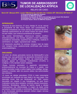
RELATO DE CASO
Choque Hemorrágico como
Manifestação Inicial de Tumor
Estromal Gastrointestinal
Vinícius Grando Gava*
Raul Pruinelli **
Mulher de 43 anos é admitida no serviço de emergência por
apresentar quadro de hemorragia digestiva (hematêmese e enterorragia). Negava episódios prévios, bem como utilização crônica de
medicamentos. Previamente hígida. Ao exame: pálida, sudorética
com freqüência cardíaca de 130 bpm, pressão arterial de 90/50
mmHg. Realizado ressuscitação volêmica vigorosa (cristalóide e
três unidades de concentrado de hemácias) e iniciado omeprazol.
Hemoglobina 4,7 g/dl. Endoscopia digestiva alta demonstrou sangue
vermelho vivo refluindo em grande quantidade, distalmente à
segunda porção duodenal, em segmento não passível de avaliação
endoscópica. Submetida a laparotomia por instabilidade hemodinâmica. Presença de tumoração exofítica ao nível do ângulo de
Treitz (foto). Realizado enterectomia com entero-entero anastomose
termino-terminal. Resolução do choque. Evolução pós-operatória
sem intercorrências. Anatomopatológico: nódulo transmural de
1,7X4,0 cm, com protrusão intra e extraluminal. Envolvimento
transmural por neoplasia fusocelular com focos de degeneração
mixóide e ulceração. Achados sugestivos de tumor estromal gastrointestinal (GIST). Imunohistoquímica C-kit positivo – perfil imunohistoquímico próprio de GIST. Seis meses após a cirurgia a
paciente encontra-se bem e sem sinais de recorrência.
Apesar de raro, o GIST é o tumor mesenquimal mais freqüente
do trato gastrointestinal. Os GISTs são provavelmente originários
das células intersticiais de Cajal, localizadas próximas ao plexo
mioentérico e presumivelmente o marcapasso da peristalse intestinal.
Anteriormente confundidos com leiomiomas e leiomiossarcomas,
suas características imunofenotípicas (a mais notável delas o
marcador immunohistológico KIT (CD 117) – presente em 95% dos
casos) hoje permitem sua correta distinção (1). Os GISTs apresentam
uma leve predominância no sexo masculino, com uma média de
idade de 60 anos. Podem ocorrer em qualquer local ao longo do
trato gastrointestinal, mas se encontram mais freqüentemente no
estômago (65%) ou intestino delgado (25%). Cólon e reto são a
localização de 5 a 10% e esôfago em cerca de 5%. Locais infreqüentes
incluem omento, mesentério e retroperitônio. Sua apresentação é
variável, cerca de 70% dos pacientes tem sintomas, 20% são assintomáticos e 10% são achados em autópsias. Tumores sintomáticos
tendem a ser maiores e seus sintomas usualmente são inespecíficos e relacionados ao seu efeito de massa ou sangramento.
Quando os GISTs erodem para o lúmen do trato gastrointestinal
podem induzem hemorragia significativa ou sangramento oculto
nas fezes. Sua ruptura para o peritônio pode causar hemorragia
com risco de morte. No presente caso aplicam-se as diretrizes
habituais de investigação e manejo da hemorragia digestiva, bem
como os princípios para adequada ressecção de um GIST. No
manejo cirúrgico, mesmo na urgência deve-se obedecer aos seguintes
princípios, conforme ditar a condição clínica do paciente (2):
a) Considerar que a cirurgia representa a única chance de cura.
b) A exploração abdominal deve ser completa, observar principalmente o peritônio e fígado (locais freqüentes de metástase). c) A
técnica de ressecção deve ser meticulosa, os GISTs são freqüentemente friáveis, sua cápsula rompe facilmente podendo ser causa de
* Residente de Oncologia Cirúrgica
** Chefe do PRM de Oncologia Cirúrgica do Hospital Nossa Senhora da
Conceição. Porto Alegre - RS, Brasil.
sangramento e aumentando a chance de recorrência. d) A simples
enucleação do tumor deve ser evitada. e) Ressecções segmentares
do estômago ou intestino devem ser realizadas a fim de se obterem
margens microscópicas negativas. f) Margem macroscópica de 1 a
2 cm é suficiente. g) Linfadenectomia raramente é indicada, pois o
comprometimento linfonodal é incomum. Em séries que incluem
cirurgias eletivas, a ressecção macroscópica completa é possível
em cerca de 85% dos pacientes com tumores localizados. Margens
microscópicas negativas são obtidas em 70 a 95% desses pacientes.
Entretanto, após a ressecção completa, pelo menos 50% dos pacientes
apresentam recorrência (1). O adequado seguimento pós-operatório ainda é desconhecido. A utilização da tomografia computadorizada de abdome e pelve tem sido recomendada a cada 3 a 6
meses inicialmente, conforme o potencial de malignidade do tumor,
e anualmente a seguir (1-3). O advento do imatinib (inibidor específico
da tirosina quinase) trouxe grande avanço no tratamento da doença
metastática , com taxa de resposta superior a 50% e estabilização
da doença em mais de 70% dos casos (4, 5). Ensaios clínicos estão em
andamento avaliando o uso do imatinib como terapia neoadjuvante
bem como a duração de sua manutenção.
Figura 1
REFERÊNCIAS
1. Gold JS, DeMatteo RP. Combined Surgical and Molecular
Therapy: The Gastrointestinal Stromal Tumor Model. Ann
Surg 2006;244:176-184.
2. DeMatteo RP, Brennan MF. Gastrointestinal Stromal Tumors.
In: Cameron JL. Current Surgical Therapy. 8 th ed. Philadelphia,
PA: Elsevier Mosby; 2004. p. 100-3.
3. Blay JY, Bonvalot S, Casali P, et al. Consensus meeting for
the management of gastrointestinal stromal tumors: Report of
the GIST Consensus Conference of 20-21 March 2004, under
the auspices of ESMO. Ann Oncol. 2005;16:566 -578.
4. Joensuu H, Roberts PJ, Sarlomo-Rikala M, et al. Effect of the
tyrosine kinase inhibitor STI571 in a patient with a metastatic
gastrointestinal stromal tumor. N Engl J Med. 2001;344:1052-1056.
5. Demetri GD, Von Mehren M, Blanke CD, et al.: Efficacy and
safety of imatinib mesylate in advanced gastrointestinal
stromal tumors. N Engl J Med. 2002;347:472-480.
Rev. Bras. Oncologia Clínica 2006 . Vol. 3 . N.º 7 (Jan/Abr) 37-38
| 37
Case Report
Hemorrhagic Shock as the
Initial Presentation Of
Gastrointestinal Stromal Tumor
Vinícius Grando Gava*
Raul Pruinelli **
A 43 year-old-woman present to the emergency department
for sustaining gastrointestinal hemorrhage (hematemesis and
enterorrhagia). She denies previous episodes, as well as chronic
medical usage. She was previously health. Physical examination
revealed: pallor, diaphoresis, a cardiac rate of 130 bpm, and a
blood pressure of 90/50 mmHg. Vigorous volemic resuscitation
was undertaken (crystalloid and three blood units) as well as
omeprazole. Hemoglobin: 4.7 g/dl. Upper gastrointestinal
endoscopy showed great amount of bright red blood, distal to the
second portion of the duodenum, not amenable to endoscopic
evaluation. Due to hemodynamic instability the patient underwent
laparotomy. An exophytic tumor at the Treitz level was found.
The patient was submitted to a small bowel resection with end-toend anastomosis.The shock was controlled and the post-operative
recovery was uneventful. Histopathologic study: 1.7X4.0 cm
transmural nodule, with intra and extraluminal protrusion,
transmural involvement by fusocelular neoplasia with myxoid
degeneration foci and ulceration.The findings are suggestive of
gastrointestinal stromal tumor (GIST).Immunohistochemical
panel: positive C-KIT. Six months after the surgical procedure the
patient is doing well with no signs of recurrence.
Despite its rarity, GIST is the most common mesenquimal
tumor of the gastrointestinal tract. GISTs are probably originated
from the interstitial cells of Cajal, located in and around the
myenteric plexus and thought to function as the intestinal
peristalsis pacemaker. Previously misclassified as leiomyomas or
leiomyosarcomas,
its
ultra
structural
features
and
immunophenotypical markers (the most notable is KIT (CD 117) –
with at least 95% staining positive) now distinct this entity (1).
GIST has a slight male predominance, with a median age of
approximately 60 years. GIST can occur anywhere along the
gastrointestinal tract but most commonly arises in the stomach
(65%) or small intestine (25%). Colon and rectum are the location of
5 to 10% and another 5% are found in the esophagus. Uncommonly,
GISTs can develop in the omentum, mesentery, or retroperitoneum.
The clinical presentation varies widely. Approximately 70% of
patients are symptomatic, 20% are asymptomatic, and 10% of the
cases are detected at autopsy. Symptomatic tumors tend to be larger
and the symptoms are most commonly related to mass effect or
bleeding. When GISTs erode into the lumen of the GI tract, they can
induce significant hemorrhage or occult blood loss. Its rupture into
the peritoneum can be life-threatening. In the present case,
guidelines for the assessment and management of gastrointestinal
bleeding apply, as well as the surgical resection principles.
Regarding the surgical management, even in the emergency setting,
one should obey the following principles, according to the clinical
condition of an individual patient (2):
a) Consider the surgery as the unique chance of cure; b) The
abdominal exploration must be complete with particular attention
to the peritoneum and the liver (frequent sites of metastasis); c)
The surgical resection technique must be meticulous because
GISTs are often friable, its capsule is easily torn with bleeding and
* Surgical Oncology Resident
** Chief of the Surgical Oncology - Hospital Nossa Senhora da Conceição,
Porto Alegre - RS, Brazil.
3 8 | Rev. Bras. Oncologia Clínica 2006 . Vol. 3 . N.º 7 (Jan/Abr) 37-38
tumor rupture ensuing, increasing the recurrence rate; d) Simple
enucleation of the tumor should be avoided; e) Segmental
resection of the stomach or intestine should be performed to
achieve negative microscopic margins; f) A 1 to 2 cm grossly
margin is enough; g) Lymphadenectomy is rarely indicated
because lymph node metastasis are rare.
Considering that, in series including elective surgical
procedures, complete gross resection is possible in approximately 85%
of patients with primary localized tumors. Negative microscopic
margins are achieved in 70 to 95% of these completely resected cases.
Although after a complete resection of a localized GIST, at least 50% of
patients develop recurrence (1). The adequate follow up strategy is
still unknown. Computed tomography scans of the abdomen and
pelvis every 3 to 6 months according to the malignant potential of the
tumor have been advocated. The interval can be extended after the
third year (1-3). The development of imatinib brought great advance
in the treatment of the metastatic disease, with a response rate of
more than 50% and stabilization of the disease in more than 70% (4,
5). Ongoing trials are evaluating the expanding use of imatinib as
neoadjuvant therapy as well as how long should it be used.
Figura 1
REFERENCES
1. Gold JS, DeMatteo RP. Combined Surgical and Molecular
Therapy: The Gastrointestinal Stromal Tumor Model. Ann
Surg 2006;244:176-184.
2. DeMatteo RP, Brennan MF. Gastrointestinal Stromal Tumors.
In: Cameron JL. Current Surgical Therapy. 8 th ed. Philadelphia,
PA: Elsevier Mosby; 2004. p. 100-3.
3. Blay JY, Bonvalot S, Casali P, et al. Consensus meeting for
the management of gastrointestinal stromal tumors: Report of
the GIST Consensus Conference of 20-21 March 2004, under
the auspices of ESMO. Ann Oncol. 2005;16:566 -578.
4. Joensuu H, Roberts PJ, Sarlomo-Rikala M, et al. Effect of the
tyrosine kinase inhibitor STI571 in a patient with a metastatic
gastrointestinal stromal tumor. N Engl J Med. 2001;344:1052-1056.
5. Demetri GD, Von Mehren M, Blanke CD, et al.: Efficacy and
safety of imatinib mesylate in advanced gastrointestinal
stromal tumors. N Engl J Med. 2002;347:472-480.
Baixar
pdf