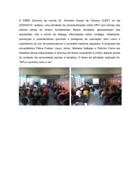
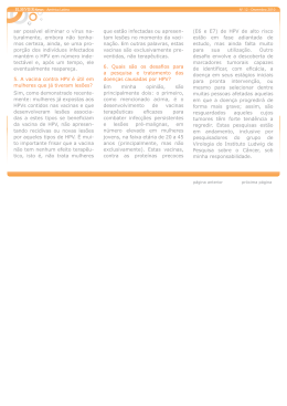
UNIVERSIDADE FEDERAL DE PERNAMBUCO
LABORATÓRIO DE IMUNOPATOLOGIA KEIZO ASAMI
PÓS-GRADUAÇÃO EM BIOLOGIA APLICADA À SAÚDE
CORRELAÇÃO DA INFECÇÃO POR PAPILLOMAVIRUS HUMANO (HPV)
COM POLIMORFISMOS DE DOIS GENES DE CITOCINAS: FATOR DE
NECROSE TUMORAL (TNF) ALFA E INTERLEUCINA (IL) 18 EM
PACIENTES COM E SEM LESÃO INTRAEPITELIAL CERVICAL.
MAYARA COSTA MANSUR FERNANDES
Recife
2012
MAYARA COSTA MANSUR FERNANDES
CORRELAÇÃO DA INFECÇÃO POR PAPILLOMAVIRUS HUMANO (HPV)
COM POLIMORFISMOS DE DOIS GENES DE CITOCINAS: FATOR DE
NECROSE TUMORAL (TNF) ALFA E INTERLEUCINA (IL) 18 EM
PACIENTES COM E SEM LESÃO INTRAEPITELIAL CERVICAL.
Dissertação apresentada ao Programa de
Pós-Graduação em Biologia Aplicada à
Saúde – Universidade Federal de
Pernambuco, como requisito final para a
obtenção do grau de Mestre em Biologia
Aplicada à Saúde.
ORIENTADOR: Prof. Dr. Sergio Crovella
CO-ORIENTADOR: Prof. Dr. Paulo Roberto Eleutério de Souza
Recife
2012
RESUMO
O Papillomavirus humano (HPV) é responsável por afetar anualmente 500 mil
mulheres com câncer cervical invasivo. Fatores de risco podem facilitar a
persistência do vírus da cérvice uterina. Polimorfismos genéticos em regiões
regulatórias e codificadoras de genes de citocinas estão associadas a
patogênese de um vasto número de doenças humanas. Este trabalho objetivou
determinar se existe relação entre os polimorfismos existentes na região G308A do gene TNFα e nas regiões -G137C e -C607A do gene IL18 na
susceptibilidade a infecção pelo HPV e na progressão das lesões intraepitelial
cervical. O estudo foi realizado com 122 mulheres HPV+ e 132 mulheres HPV(controle). Os polimorfismos dos genes TNFα e IL18 foram analisados pela
técnica Specific Sequence Polymosphism (PCR-SSP) e analisadas em gel de
agarose a 1,5%. As análises estatísticas para verificar a significância do estudo
dos genótipos foram realizadas utilizando o programa BioEstat 5.0. Os
resultados mostraram uma prevalência de 49,18% da infecção pelo HPV-16 e
70,49% delas apresentaram lesão cervical de alto grau. Em relação aos
polimorfismos houve associação do alelo mutante na região -308A do gene
TNFα e -607A do gene IL18 com a susceptibilidade a infecção pelo HPV
(p=0,0008; p<0,0001, respectivamente), mas não foi verificada relação destes
genes com a susceptibilidade ao desenvolvimento das lesões (p>0,05). Porém
não foi encontrada associação significativa em relação a região na região -137
do gene IL-18. Estes resultados sugerem dois possíveis marcadores genéticos
de susceptibilidade a infecção pelo HPV na população em estudo e que estes
não podem ser usados como marcadores de progressão da lesão cervical.
Palavras Chave: Papillomavirus Humano, Neoplasia Intraepitelial Cervical,
Polimorfismos de única base, Fator de Necrose tumoral alfa, Interleucina 18.
ABSTRACT
The Human Papillomavirus (HPV) is responsible for affecting anually 500
thousand women with invasive cervical cancer. Risk factors may facilitate the
persistence of the virus of the uterine cervix. Genetic polymorphisms in
regulatory and coding regions of cytokine genes are associated with the
pathogenesis of a large number of human diseases. This study aimed to
investigate whether there is a relationship between existing polymorphisms in
the region -G308A of the gene TNFα and in regions -G137C and C607A of the
IL18 gene in susceptibility to HPV infection and progression of cervical
intraepithelial lesions. The study was conducted with 122 women HPV+ and,
132 women HPV- (control). The polymorphisms of the TNFα and IL18 genes
were analyzed using the technic of the Specific Sequence Polymosphism (PCRSSP) and analyzed on agarose gel 1.5%. The Statistical analyzes to check the
significance of the study of the genotypes were performed using the BioEstat
5.0 program. The results showed a prevalence of 49.18% of HPV-16 infection
and 70.49% of that total had high-grade cervical lesions. Regarding the
polymorphisms there was an association of the mutant allele in the region 308A of the gene TNFα and -607A of the IL18 gene with susceptibility to HPV
infection (p = 0.0008, p <0.0001, respectively), but no relationship was found
these genes with susceptibility to the development of lesions (p> 0.05). But no
significant association was found with regard to the region -137 region of the
gene IL18. These results suggest two possible genetic markers of susceptibility
to HPV infection in the studied population and that these can not be used as
markers of progression of cervical injury.
Key-words: Human Papillomavirus, Cervical Intraepithelial Neoplasia, Single
Nucleotide Polymorphism, Tumour Factor Necrosis alpha, Interleukin 18
LISTA DE ABREVIATURAS E SIGLAS
CCI - Câncer Cervical Invasivo
DNA - Ácido desoxiribonucléico
E- Early (precoce)
HPV - Papillomavírus Humano
ICTV - The International Committee on the Taxonomy of Viruses (Comitê
Internacional de Taxonomia Viral)
IFN - Interferon
IL – Interleucina
L – Late (tardia)
MHC - Complexo de Histocompatibilidade Principal
NIC - Neoplasia Intraepitelial Cervical
NK - Natural killers (Matadoras Naturais)
SI - Sistema Imunológico
SIL – Lesão Intraepitelial Cervical
SNP - Single nucleotide polymorphism (Polimorfismo de única Base)
Th – Linfócito T helper (Linfócito T auxiliary)
TNF - Tumour Necrosis Factor (Fator de Necrose Tumoral)
LCR - Long Control Region (Região Longa de Controle)
UV – Ultravioleta
LISTA DE FIGURAS
Figura 1. - Representação esquemática do genoma do HPV contendo a
distribuição das regiões precoce, tardia e regulatória, conforme as unidades de
pares de bases cda molécula circular e DNA do vírus (Muñoz et al. 2006)
13
Figura 2. - Ciclo de infecção do HPV no epitélio estratificado (Adaptado de
Doorbar, 2005)
14
LISTA DE TABELAS
Artigo - Table 1 – Sequences of primers for identification and typing of HPV
used in the study
Artigo - Table 2 – Distribution genotypic and allelic frequencies of the
TNFG308A among health controls and patients
54
Artigo - Table 3 - Distribution genotypic and allelic frequencies of the IL18
among health controls and patients
56
55
SUMÁRIO
Revisão de Literatura
1. Câncer Cervical
10
2. Papillomavírus Humano
11
2.1 Classificação do HPV
11
2.2 Estrutura Viral
12
2.3 Lesões e cânceres causados pelo HPV
13
2.4 Fatores de risco associados com o HPV
15
3. Polimorfismos de Única Base
15
4. Mecanismos da Resposta Imune
16
4.1 Resposta Imune Inata
16
4.2 Resposta Imune Adaptativa
17
4.3 Interleucina 18 (IL-18) e o gene IL18
18
4.4 Fator de necrose Tumoral alfa (TNF-α) e o gene TNFα
19
Objetivos
22
Referências Bibliográficas
23
Artigo Científico
30
Abstract
32
Background
33
Methods
36
Results
42
Discussion
44
Conclusions
47
Contribution of Authors
47
Conflict of Interest
48
Abbreviations
49
Acknowledgments
50
References
50
Anexo I
54
Anexo II
57
Considerações Finais
58
REVISÃO DE LITERATURA
1. Câncer Cervical
O câncer cervical invasivo (CCI), a segunda neoplasia maligna mais
comum em todo o mundo, perdendo apenas pelo câncer de mama, é
responsável por cerca de 500 mil novos casos anuais, dos quais
aproximadamente metade resultam em mortes. A maioria dos casos (em torno
de 80%) ocorre em países em desenvolvimento, sendo nesses países o tipo
mais incidente nas mulheres. Infecções pelo Papillomavirus Humano (HPV)
representam cerca de 5% de todos os tipos de cânceres [1,2,3].
Nos Estados Unidos é considerada a infecção transmitida sexualmente
mais comum [4], com uma estimativa de 6.2 milhões de novos casos em
homens e mulheres, onde 74% dos novos casos ocorrem entre 15 e 24 anos. É
altamente prevalente em mulheres sexualmente ativas, sendo responsável por
60% a 80% das infecções em adolescentes neste país [4,5,6,7]. Na Europa,
surgem anualmente 33.000 novos casos de câncer, resultado em 15.000
mortes na faixa etária compreendida entre 15-44 anos [8]. No leste europeu a
incidência e mortalidade do câncer cervical é ainda maior, comparadas com a
Europa Ocidental [9,10].
No Brasil, a ocorrência deste câncer prevista para 2012 é de 17.540
novos casos segundo estimativa bienal do Instituto Nacional do Câncer, com
um risco aproximado de 17 casos a cada 100 mil mulheres. Sem considerar os
tumores de pele não melanoma, o câncer do colo do útero é o mais incidente
na Região Norte (24/100.000). Nas regiões Centro-Oeste (28/100.000) e
Nordeste (18/100.000) ocupam a segunda posição mais freqüente, na região
10
Sudeste (15/100.000) a terceira, e Sul (14/100.000), a quarta posição. Este
comportamento evidencia a urbanização e desenvolvimento do Sudeste
Brasileiro, que se reflete em maior informação da população com relação aos
métodos de prevenção e detecção precoce do CC. A dificuldade de acesso
explicaria a alta taxa de incidência de lesões em estádio avançado na região
Norte [11].
2. Papillomavírus Humano
2.1 Classificação do HPV
Anteriormente agrupados na família Papovaviridae e subfamília
Papillomavirus, devido à semelhança nas características morfológicas com o
gênero Polyomavirus, foram designados pelo Comitê Internacional de
Taxonomia de Virus (ICTV) como uma família própria, Papillomaviridae [12]. Os
papilomavírus foram identificados em uma grande variedade de espécies de
vertebrados, além de humanos, sendo específicos para seus respectivos
hospedeiros.
A classificação do HPV pode ser feita de diversas maneiras. Estes
vírus não são classificados como sorotipos, mas como genótipos com base na
seqüência DNA [13,14]. Quanto ao tropismo pela superfície epitelial que
tem predileção, são cutaneotrópicos ou mucosotrópicos, e podem induzir a
transformação neoplásica dessas células. Nestes, a infecção pelo HPV pode
causar verrugas no epitélio cutâneo, enquanto que na região anogenital esses
vírus podem causar verrugas genitais e os vários tipos de câncer em homens e
mulheres [15].
11
Podem se classificar também de acordo seu potencial para
oncogênese. Baseados em dados filogenéticos e em relação a sua presença
em doenças cervicais malignas ou benignas, podem ser classificados como de
baixo risco e alto risco. Os de baixo risco (HPV tipo 6, 11, 13, 40, 42-44, 54, 61,
62, 70, 72, 74, 81), incluindo os condilomas genitais, dos quais os tipos 6 e 11
são os mais prevalentes e os de alto risco oncogênico (16, 18, 31, 33, 35, 39,
45, 51, 52, 56, 58, 59, 68, 73, 82) [16,17].
2.2 Estrutura Viral
Devido à estreita relação com o câncer cervical, estudos são
direcionados na determinação das sequências de HPV. Até o momento, mais
de 200 tipos de HPV foram identificados baseados no DNA do vírus,
apresentando variedades entre os genomas [12].
A família Papillomaviridae insere um grupo de pequenos vírus não
envelopados, apresentando uma conformação icosaédrica, com uma molécula
de DNA circular com uma longa fita dupla [17,18]. O genoma possui menos de
8000 pares de bases, contendo três regiões: genes precoces (do inglês, early)
e tardios (do inglês, late), e entre estas duas regiões está a região reguladora
que não codifica genes (LCR de Long Control Region) (Figura 1).
12
Figura 1. Representação esquemática do genoma do HPV contendo a
distribuição das regiões precoce, tardia e regulatória, conforme as unidades de
pares de bases cda molécula circular e DNA do vírus (Muñoz et al. 2006)
A região early é o local onde estão presentes os genes responsáveis
pela produção de proteínas envolvidas no controle da replicação do DNA viral
(E1, E2, E4, E5, E6 e E7). A região late contém os genes L (L1 e L2),
responsável pela formação as proteínas do capsídeo protéico viral. Os
conjuntos de genes late e early são separados por uma região reguladora de
cerca de 1000pb que não codifica proteínas, mas contém promotores
necessários para a regulação da expressão gênica e origem de replicação do
genoma [18].
2.3 Lesões e cânceres causados pelo HPV
A persistência da infecção pelo HPV especialmente com um dos tipos
oncogênicos pode causar transformação no epitélio [19,20] (Figura 2). O
câncer cervical se desenvolve a partir lesões pré-malignas não-invasivas
denominada neoplasia intraepitelial cervical (NIC) ou lesão intraepitelial
escamosa (SIL).
13
Essas lesões pré-malignas são classificados histologicamente em base
de atipia progressiva de células epitelias: NIC I corresponde à displasia
leve,NIC II displasia moderada e NIC III para ambos displasia acentuada e
carcinoma in situ [21].
Figura 2. Ciclo de infecção do HPV no epitélio estratificado (Adaptado de
Doorbar, 2005)
O risco de progressão de lesões pré-malígnas para formas graves é
de 16% e 34% para NIC I e II respectivamente, e 22% para NIC III ou câncer
invasivo [22,23]. Na lesão de grau I (NIC I), 60%-75% dos casos não são
confirmados no segundo exame, regredindo espontaneamente [21].
O Papillomavirus humano é o agente causal principal para o
desenvolvimento de 99,7% dos casos de câncer cervical. Em outros tipos de
cânceres, estima-se que esse vírus é responsável por 90% dos casos de
câncer anal 40% dos cânceres dos órgãos genitais externos (vulva, vagina e
14
pênis), pelo menos 12% dos cânceres orofaríngeos e pelo menos 3% dos
cânceres bucais [12,24].
2.4 Fatores de risco associados com o HPV
A presença de um tipo oncogênico do HPV na região cervical é
considerada uma causa necessária para o desenvolvimento da NIC e do CC,
mas não exclusivamente. Caracterizada como uma doença multifatorial, o HPV
atua em conjunto a cofatores ambientais e fatores imunológicos e genéticos
próprios do hospedeiro para que ocorra a progressão para o câncer cervical
invasivo[25,26,27,28].
Fatores ambientais relacionados com CC incluem fatores hormonais
(uso de contraceptivos orais), multiparidade, coinfecção com outros vírus
(herpes simplex 2 - HSV-2) e bactérias ou com outras doenças sexualmente
transmissíveis, (Chlamydia trachomatis e HIV) e hábitos do hospedeiro como
fumo,
radiação
ultravioleta
(UV)
e
radiação
ionizante
e
os
fatores
dietéticos. Outros cofatores não-ambientais sendo também considerados
incluem os relacionados à resposta imune do hospedeiro e as relacionadas
com o próprio vírus, como o genótipo de HPV, coinfecção com outros tipos de
HPV, as variantes de HPV, carga viral e integração viral [29].
3. Polimorfismos de Única Base (SNP)
As variações que ocorrem no genoma são em sua maioria formas de
polimorfismos de nucleotídeo único (SNP), com uma frequência a cada 300 1000 pares de base em todo o genoma e surgem como resultado de defeitos
na replicação do DNA. Assim, os SNPs são mutações de ponto que originam
15
duas formas alélicas. Essas variações nos genes são muito úteis para pesquisa
de marcadores genéticos humanos e podem ser usadas para identificar genes
de susceptibilidade de doenças humanas complexas [30,31,32].
4. Mecanismos da Resposta Imune
O Sistema Imunológico (SI) é responsável pelo reconhecimento,
eliminação e remoção de substâncias estranhas, células mortas e/ou
danificadas, além da destruição de células mutantes e cancerosas. É bastante
especializado em gerar uma variedade enorme de células e moléculas capazes
de reconhecer e eliminar especificamente substâncias que não pertencem
naturalmente ao sistema de defesa [33,34].
Várias moléculas solúveis e circulantes ou também aquelas que
permanecem aderidas a membranas celulares agem conjuntamente em uma
rede dinâmica extraordinariamente adaptável, capaz de produzir uma resposta
imune efetora e eficaz contra invasores de estruturas complexas. De acordo
com a função exercida, a resposta imune pode ser caracterizada conforme a
atividade relacionada em reconhecimento e resposta.
Funcionalmente,
a
imunidade
possui
dois
caminhos
principais
envolvidos na eliminação de partículas estranhas: a imunidade inata e a
imunidade adquirida, que apresentam aspectos diferentes quanto ao sistema
de reconhecimento, células envolvidas e mecanismos de ação.
4.1 Resposta Imune Inata
A imunidade inata é a primeira linha de defesa e está presente na
maioria dos organismos. Os receptores das células do sistema imune inato
foram desenvolvidos durante o processo evolucionário [34,35].
16
Fazem parte dessa resposta barreiras físicas epitélios e mucosas e
moléculas
secretadas
em
fluidos
biológicos
como
citocinas.
Células
especializadas nesta resposta são as matadoras naturais (natural killers),
células dendríticas, macrófagos, linfócitos T citotóxicos e neutrófilos [35,36].
A imunidade inata é ativada por lesão celular ou óbito e se manifesta
como uma inflamação, a resposta vascular local da lesão. Durante a
inflamação, efetores solúvel e celular imunes inatos são recrutados. Células
parenquimatosas são recrutados para o local e fagócitos locais são, então,
ativado para secretarem citocinas inflamatórias e moléculas de defesa [14]. É
formado por um sistema de reconhecimento com menor especificidade contra
patógenos e esses componentes estão previamente formados antes da entrada
e contato com a substância estranha [33,34,37].
4.2 Resposta Imune Adaptativa
O
sistema
imune
adaptativo
é
especifico
contra
antígenos,
necessitando de um primeiro contato com este para ser construída. Agrupa os
linfócitos T, que são os grandes efetores dessa resposta e os fagócitos, com as
proteínas
relacionadas
a
essas
células
(complexo
principal
de
histocompatibilidade - MHC e os receptores de células T e B) [33,37].
Quando o hospedeiro tem contato com microorganismo patogênico
(vírus ou bactéria, por exemplo), células apresentadoras de antígenos
capturam os patógenos invasores e processam internamente seus antígenos.
As células T reconhecem antígenos que tenham sido transformadas em
peptídeos curtos, vinculado ao Complexo Principal de Histocompatibilidade
(MHC), e é apresentado ligado ao receptor de membrana celular [38].
17
Dois grandes subconjuntos de células T são identificados pelos
marcadores de superfície: CD4 ou CD8. Células T CD4 + reconhecem
antígenos apresentados por MHC de classe II; as células T CD8 + reconhecem
os antígenos apresentados pela classe I do MHC. Células T CD4 + são
resultado da ativação na secreção de uma variedade de pequenas proteínas,
ou citocinas, que ajudam a regular outras células [39,40].
As citocinas da resposta imune à infecção são geralmente classificadas
como imunoestimulante T helper 1 (Th1) ou citocinas do tipo T helper 2 (Th2).
Uma resposta Th1 favorecerá a produção de células T citotóxicas efetoras que
seria importante na remoção de todas as células infectadas pelo vírus
(citocinas tipo Th1- como interferon (IFN), o fator de necrose tumoral (TNF),
interleucina (IL) 2, e IL-12, enquanto uma resposta Th2 (IL-4, IL-5, IL-6, IL-8 e
IL-10) vai facilitar a estimulação das células B com a geração de anticorpo para
neutralização [41].
4.3. Interleucina 18 (IL-18) e o gene IL18
Interleucina (IL) -18 exerce atividade tanto na resposta inata como na
imunidade adaptativa [42,43,44,45]. Pertence à superfamília IL-1 decitocinas
próinflamatórias e o gene IL18 está situado na região 11q22.2-22.3
[44].Anteriormente denominada como fator de indução do interferon, devido a
identificação desta propriedade [46], sabe-se que tem ação pleiotropica
induzindo a expressão de quimiocinas e citocinas, além de moléculas de
adesão como a IL-8, TNF-α, VEGF e ICAM-1 [47,48,49,50].
Em cultura de adipócitos humanos pode-se verificar que a IL-18 é
secretada pelas células de gordura e a TNFa estimula a expressão de IL-18 e
18
seus receptores semelhantes aos seus efeitos sobre a inflamação em outros
genes relacionados [51].
A IL-18 desempenha um papel primordial na resposta imune, sendo
responsável pela diferenciação e ativação das células T helper (Th) em
subgrupos, de acordo com o perfil de citocinas [52]. Atua como um indutor para
a diferenciação e proliferação de células Th1 na presença de IL-12,
estimulando a produção de IFN e TNF nos linfócitos T e natural killer (NK)
[52,53,54]. A IL-18 e a IL-12 atuam em sinergismo na ativação de células NK,
células B e T [55,56]. Em contrapartida, a IL-18 pode ainda estimular a
resposta imune do tipo Th2, na ausência da IL-12 [54,57].
Vários polimorfismos no promotor do gene IL18 têm sido associados
com diferentes doenças inflamatórias e auto-imunes [58-63]. Cinco posições
diferentes de polimorfismos de único nucleotídeo na região promotora foram
identificados: -656 G / T, -607 C / A, -137 G / C, 113 G / T, e 127 C / T [64]. No
entanto, somente em SNPs nas posições -137 e -607 foram confirmados como
tendo um impacto sobre a atividade do gene IL18 em estudos anteriores [65].
4.4 Fator de Necrose Tumoral Alfa (TNF- α) e o gene TNFα
O Fator de Necrose Tumoral alfa (TNF-α) é uma potente citocina Th1
próinflamatória
liberado
principalmente
por
monócitos
e
macrófagos
estimulados. O gene se encontra dentro do complexo de histocompatibilidade
maior no cromossomo 6p21.3 [66,67],e é composto de quatro éxons dispostos
ao longo de cerca de 3 kb do DNA [68].
O TNF-α foi originalmente identificado e isolado por duas atividades
características: a capacidade de induzir necrose hemorrágica de certos
19
tumores e da capacidade de induzir caquexia durante os estados de infecção
crônica [69]. Em infecções severas, o TNF-α é produzido em larga escala e
causa anormalidades sistêmicas clinica e patológica [70].
A principal função fisiológica do TNF-α é estimular o recrutamento de
neutrófilos e monócitos para os locais da infecção e ativa essas células para
erradicar microrganismos [70]. Possui amplos efeitos biológicos, incluindo
proteção contra a infecção, vigilância contra tumores e estimula respostas
inflamatórias [68], além de desempenhar um papel essencial no sistema
imunológico [71]. O TNF-alfa estimula a síntese de outras citocinas
próinflamatórias e a adesão de moléculas. Além disso, tem mostrado mediar a
carcinogênese através da indução, proliferação, invasão e metástase de
células tumorais [72]. Em situações agudas, a produção local de TNF-α é
claramente benéfica. Ele aumenta a expressão de moléculas de adesão no
endotélio vascular a fim de permitir que células imunológicas, particularmente
os neutrófilos e macrófagos, se desloquem para os sítios onde ocorreram dano
tecidual e infecção [69].
Polimorfismos genéticos de citocinas, especialmente em regiões
reguladoras, podem estar relacionados com a quantidade de citocinas
produzida [73]. O nível de produção desta citocina varia com o genótipo do
indivíduo que secreta, podendo produzir níveis baixo, médio ou alto de TNF-α.
Existem vários polimorfismos localizados em diferentes sítios do
promotor do gene responsável pela síntese da TNF alfa. Foram identificados na
região -1031 (T→C), -863 (C→A), -857 (C→A), -851 (C→T), -419 (G→C), -376
(G→A), - 308 (G→A), -238 (G→A), -162 (G→A) e – 49 (G→A). Polimorfismos
20
de nucleotídeo único na região -308 e -238 do promotor do gene TNF têm sido
comumente estudados [69,74,75].
O polimorfismo no -308 supostamente afeta a expressão do gene e têm
sido implicados na regulação da transcrição do TNFα. Podem ser encontradas
numa determinada população duas formas alélicas, onde uma permanece com
o alelo G, comum, da guanina ou quando ocorre substituição desse alelo pelo
A (adenina) [76,77].
Segundo Cabrera et al. (1995) mostra que as células que contém o
alelo A aumentam a transcrição de seis a nove vezes mais que aquelas que
contém o alelo G [74,75,79]. Outros trabalhos relataram que in vitro, o alelo G
está relacionado com a reduzida produção de TNF-α, enquanto os indivíduos
com alelo A produzem mais TNF-a [74,80,81,82].
21
OBJETIVOS
Objetivo Geral
Verificar a relação entre os polimorfismos existentes na região (-308 G/A) do
gene TNFα e nas regiões (-137 G/C) e (-607 C/A) do gene IL18 com a infecção
pelo HPV e com a progressão da lesão intraepitelial cervical.
Objetivos Específicos
• Detectar e genotipar o HPV nas amostras analisadas.
• Determinar o polimorfismo dos genes TNFα e IL18 pela metodologia da
Specific Sequence Polymosphism (PCR-SSP).
• Associar a presença da infecção pelo HPV com o surgimento de lesões
intraepiteliais cervicais.
• Correlacionar os polimorfismos dos genes TNFα e IL18 com
susceptibilidade a infecção por HPV e o aparecimento de lesões
intraepiteliais.
22
REFERÊNCIAS BIBLIOGRÁFICAS
[1] Parkin DM, Bray F, Ferlay J, et al. Global cancer statistics, 2002. CA
Cancer J Clin 2005;55:74–108.
[2] Freeman H, Wingrove B. Excess Cervical Cancer Mortality: A Marker for
Low Access to Health Care in Poor Communities. NIH Pub. No. 05-5282.
Rockville, MD: National Cancer Institute, Center to Reduce Cancer Health
Disparities, 2005.
[3] Gargiulo F, De Francesco MA, Schreiber C, Ciravolo G, Salinaro F, B.
Valloncini B, Manca N. Prevalence and distribution of single and multiple
HPV infections cytologically abnormal cervical samples from Italian.
women Virus Research 125 (2007) 176–182.
[4] Weinstock H, Berman S, Cates JrW. Sexually transmitted diseases
among American youth: incidence and prevalence estimates, 2000.
Perspect Sex Reprod Health 2004;36(1):6–10.
[5] Dunne EF, Unger ER, Sternberg M, McQuillan G, Swan DC, Patel SS, et al.
Prevalence of HPV infection among females in the United States. JAMA
2007;297(8):813–9.
[6] Huang CM. Human papillomavirus and vaccination. Mayo Clin Proc
2008;83(6):701–6, quiz 706–707.
[7] Moscicki AB. Impact of HPVinfection in adolescent populations. J
Adolesc Health 2005;37:S3–9.
[8] ECDC (2008) Guidance for the introduction of HPV vaccines in EU
countries. From http://ecdc.europa.eu/pdf/HPVreport.pdf
[9] Arbyn M, Raifu AO, Autier P et al. Burden of cervical cancer in Europe:
estimates for 2004. Ann Oncol 2007; 18:1708–1715.
[10] Prymula R, Anca I, André F, Bakir M, Czajka H, Lutsar I, Mészner Z,
Salman N, Šimurka P, Usonis V. Central European Vaccination Advisory
Group (CEVAG) guidance statement on recommendations for the
introduction of HPV vaccines Eur J Pediatr (2009) 168:1031–1035
[11] MINISTÉRIO DA SAÚDE. INCA. Estimativa 2012: Incidência de Câncer no
Brasil. Rio de Janeiro, 2012. [Acesso em 12/2012]. Disponível em
[http://www.inca.gov.br/estimativa/2012/index.asp?ID=5]
[12]Burd EM. Human Papillomavirus and Cervical Cancer. Clin. Microbiol.
Rev. Jan. 2003, p. 1–17 Vol. 16, No. 1
[13] de Villiers EM, Fauquet C, Broker TR, Bernard HU, zur Hausen H.
Classification of papillomaviruses. Virology 2004;324:17–27.
23
[14] Stanley M. Immunobiology of HPV and HPV vaccines. Gynecologic
Oncology 109 (2008) S15–S21
[15] Bernard HU. The clinical importance of the nomenclature, evolution
and tazonaomy of human papillomaviruses. J Clin Virol, 32S (2005) S43–
S51
[16] Albring L, Brentano JE, Vargas, VRA. O câncer do colo do útero, o
Papilomavírus Humano (HPV) e seus fatores de risco e as mulheres
indígenas Guarani: estudo de revisão. RBAC,2006; 38(2): 87-90.
[17] Magi JC, Brito SEM, Grecco ETO et al. Prevalência de papilomavirus
humano (HPV) anal, genital e oral, em ambulatório geral de
coloproctologia. Rev bras. Coloproct., 2006:26(3): 233-238.
[18] Muñoz N, Castellsagu´e X, de Gonz´alez AB, Gissmann L. HPV in the
etiology of human cancer. Vaccine 24S3 (2006) S3/1–S3/10
[19] Cogliano V, Baan R, Straif K, Grosse Y, Secretan B, El Ghissassi F.
Carcinogenicity of human papillomaviruses. Lancet Oncol 2005;6(April
(4)):204.
[20] IARC. IARCmonographs onthe evaluationof
tohuman.Human Papillomaviruses 2005:90.
carcinogenic
risk
[21] Wright TC, Knrman RJ: A critical review of the morphologic
classification systems of preinvasive lesions of the cervix: The scientific
basis for shifting the paradigm. Papilloma Virus Report 1994:5:175-182.
[22] Piato S. Tratado de ginecologia. Porto Alegre: Artes Médicas; 1997.
[23] Brasil. Ministério da Saúde. Instituto Nacional de Câncer. Neoplasia intra–
epitelial cervical – NIC. Rev Bras Cancerol 2000;46(4):355-7.
[24] Antonsson A, Erfurt C, Hazard K, et al. Prevalence and type spectrum of
human papillomaviruses in healthy skin samples collected in three
continents. J Gen Virol 2003; 84:1881–6.
[25] Bosch FX, Lorincz A, Muñoz N, Meijer CJLM, Shah KV. The causal
relation between human papillomavirus and cervical cancer, J. Clin. Pathol.
55 (2002) 244–265.
[26] Ylitalo N, Josefsson A, Melbye M, Sorensen P, Frisch M, Andersen PK,
Spare ´n P, Gustafsson M, Magnusson P, Ponte ´n J, Gyllensten U, Adami HO.
A prospective study showing long-term infection with human papillomavirus 16
before the development of cervical carcinoma in situ, Cancer Res. 60 (2000)
6027–6032.
24
[27] Santos AM, Sousa H, Catarino R, Pinto D, Pereira D, Vasconcelos A,
Matos A, Lopes C, Medeiros R. TP53 codon 72 polymorphism and risk for
cervical cancer in Portugal, Cancer Genet. Cytogenet. 159 (2005) 143–147.
[28] Craveiro R, Costa S, Pinto D, Salgado L, Carvalho L, Castro C, Bravo I,
Lopes C, Silva I, Medeiros R. TP73 alterations in cervical carcinoma. Cancer
Genet. Cytogenet. 150 (2004) 116–– 121.
[29] Xavier Castellsague, F.Xavier Bosch, Nubia Muñoz. Environmental cofactors in HPV carcinogenesis. Virus Research 89 (2002) 191-199.
[30] Johnson GCL, Todd JA: Strategies in complex disease mapping. Curr
Opin Genet Dev 10:330, 2000.
[31] Sachidanandam R, Weissman D, Schmidt SC, Kakol JM, Stein LD, Marth
G, et al. A map of human genome sequence variation containing 1.42
million single nucleotide polymorphisms. Nature 2001;409:928e33.
[32] Hanchard NA. Genetic susceptibility and single-nucleotide
polymorphism. Seminars in Fetal & Neonatal Medicine (2005) 10, 283e289
[33] Goldsby, RA.; Kindt, TJ.; Osborne, BA. Kuby Imunologia, 4ªed. Ed.
Revinter, 2002: 480-495.
[34] Lehner, T. Innate and adaptive mucosal immunity in protection against
HIV infection. Vaccine, v.21 Suppl 2, Jun 1, p.S68-76. 2003.
[35] Sierra S, Kupfer B. et al. Basics of the virology of HIV-1 and its
replication. J Clin Virol 2005: 34 (4): 233-44.
[36] Magnadottir B. Innate Immunity of fish (overview). Fish Shellfish
Immunol, v.20, n.2. Feb, p. 137-151, 2006.
[37] Kindt TJ, Goldsby RA, Osborne BA, Kuby J. Immunology. New H.
Freeman 2007, XXII, 574, A – 531, G – 512, An – 527, I – 527 p.p.
[38] Inaba K., Metlay JP, Crowley MT, Steinman RM. Dendritic cells pulsed
with protein antigens in vitro can prime antigen-specific, MHC-restricted T
cells in situ. J. Exp. Med. 1990; 172, 631–640
[39] Spellberg B, Edwards Jr JE. Type 1/Type 2 immunity in infectious
diseases. Clin Infect Dis 2001;32:76–102.
[40] Zur Hausen H. Papillomaviruses and cancer: from basic studies to
clinical application. Nat Rev, Cancer 2002;2:342–50.
[41] Niedergang F, Didierlaurent A, Kraehenbuhl JP, Sirard JC. Dendritic cells:
the host Achille’s heel for mucosal pathogens? Trends Microbiol
2004;12:79–88.
25
[42] Okamura H, Tsutsui H, Komatsu M, Yutsudo A, Hakura T, Tanimoto K,
Torigoe T. Okura, Y. Nakada, K. Konishi, S. Fukuda, T. Kurimoto, Cloning of a
new cytokine that induces INF-c production by T cells, Nature 378 (1995)
88–91.
[43] Tsutsui H, Nakanishi K, Higashino K, Okamura H, Miyazawa Y, Kaneda K,
IFN-c-inducing factor upregulates Fas ligand-mediated cytotoxic activity
of murine natural killer cell clones, J. Immunol. 156 (1996) 3967–3973.
[44] Okamura, H, Tsutsui, H Kashiwamura, S, Yoshimoto, T, Nakanishi, K.
Interleukin-18: a novel cytokine that augments both innate and acquired
immunity. Adv. Immunol 1998; 70, 281–312.
[45] Dinarello CA. IL-18: a TH1-inducing, proinflammatory cytokine and new
member of the IL-1 family, J. Allergy Clin. Immunol. 103 (1999) 11–24.
[46] Parnet P, Garka KE, Bonnert TP, Dower SK, Sims JE. IL-1Rrp is a novel
receptor-like molecule similar to the type I interleukin-1 receptor and its
homologues T1/ST2 and IL-1R AcP. J Biol Chem 1996;271:3967-70.
[47] Kohka H, Yoshino T, Iwagaki H, Sakuma I, Tanimoto T, Matsuo Y,
Kurimoto M, Orita K, Akagi T, Tanaka N. Interleukin-18/interferon-gammainducing factor, a novel cytokine, up-regulates ICAM-1 (CD54) expression
in KG-1 cells, J. Leukoc. Biol. 64 (1998) 519–527.
[48] Park CC, Morel JC, Amin MA, Connors MA, Harlow LA, Koch AE.
Evidence of IL-18 as a novel angiogenic mediator, J.Immunol. 167 (2001)
1644–1653.
[49] Puren AJ, Fantuzzi G, Gu Y, Su MS, Dinarello CA. Interleukin-18
(IFNgamma-inducing factor) induces IL-8 and IL-1beta via TNFalpha
production from non-CD14+ human blood mononuclear cells, J. Clin.
Invest. 101 (1998) 711–721.
[50] Wood S, Wang B, Jenkins JR, Trayhurn P. The pro-inflammatory
cytokine IL-18 is expressed in human adipose tissue and strongly
upregulated by TNFa in human adipocytes. Biochemical and Biophysical
Research Communications 337 (2005) 422–429.
[51] Wang B, Jenkins JR, Trayhurn P. Expression and secretion of
inflammation-related adipokines by human adipocytes differentiated in
culture: integrated response to TNF-alpha, Am. J. Physiol. Endocrinol.
Metab. 288 (2005) E731–E740
[52] Gracie JA, Robertson SE,McInnes IB. Interleukin-18. J Leukoc Biol
2003;73:213–24.
[53] Esfandiari E, McInnes IB, Lindop G, Huang FP, Field M, Komai-Koma M,
Wei X, Liew FY. A proinflammatory role of IL-18 in the development of
spontaneous autoimmune disease. J Immunol 2001;167:5338–47.
26
[54] Nakanishi K, Yoshimoto T, Tsutsui H, Okamura H. Interleukin-18
regulates both Th1 and Th2 responses. Annu Rev Immunol 2001;19:423–74.
[55] Torre D, Speranza F, Giola M, Matteelli A, Tambini R, Biondi G. Role of
Th1 and Th2 cytokines in immune response to uncomplicated
Plasmodium falciparum Malaria. Am Society for Microbiol. 9: 348-351, 2002.
[56] Perkmann T, Winkler H, Graninger W, Kremsner PG, Winkler S.
Circulating levels of the interleukin (IL)-4 receptor and of IL-18 in patients
with Plasmodium falciparum malaria. Cytokine. 29: 153-158, 2005.
[57] Tominaga K, Yoshimoto T, Torigoe K, Kurimoto M, Matsui K, Hada T,
Okamura H, Nakanishi K: IL-12 synergizes with IL-18 or IL-1beta for
IFNgamma production from human T cells. Int Immunol 2000, 12:151-160.
[58] Imboden M, Nicod L, Nieters A, Glaus E, Matyas G, Bircher AJ,
Ackermann-Liebrich U, Berger W, Probst-Hensch NM, SAPALDIA Team: The
common Gallele of interleukin-18 single-nucleotide polymorphism is a
genetic risk factor for atopic asthma: The SAPALDIA Cohort Study. Clin Exp
Allergy 2006, 36:211-218.
[59] Tiret L, Godefroy T, Lubos E, Nicaud V, Tregouet DA, Barbaux S, Schnabel
R, Bickel C, Espinola-Klein C, Poirier O, Perret C, Münzel T, Rupprecht HJ,
Lackner K, Cambien F, Blankenberg S, Athero Gene Investigators: Genetic
analysis of the interleukin-18 system highlights the role of the interleukin18 gene in cardiovascular disease. Circulation 2005,112:643-650.
[60] Mojtahedi Z, Naeimi S, Farjadian S, Omrani GR, Ghaderi A: Association
of IL-18 promoter polymorphisms with predisposition to Type 1 diabetes.
Diabet Med 2006, 23:235-239.
[61] Sugiura T, Maeno N, Kawaguchi Y, Takei S, Imanaka H, Kawano Y,
Terajima-Ichida H, Hara M, Kamatani N: A promoter haplotype of the
interleukin-18 gene is associated with juvenile idiopathic arthritis in the
Japanese population. Arthritis Res Ther 2006, 8:R60.
[62] Tamura K, Fukuda Y, Sashio H, Takeda N, Bamba H, Kosaka T, Fukui S,
Sawada K, Tamura K, Satomi M, Yamada T, Yamamura T, Yamamoto Y,
Furuyama J, Okamura H, Shimoyama T. IL18 polymorphism is associated
with an increased risk of Crohn’s disease. J Gastroenterol 2002, 37:111116.
[63] Sivalingam SP, Yoon KH, Koh DR, Fong KY. Single-nucleotide
polymorphisms of the interleukin-18 gene promoter region in rheumatoid
arthritis patients: protective effect of AA genotype. Tissue Antigens 2003,
62:498-504.
27
[64] Giedraitis V, He B, Huang WX, Hillert J. Cloning and mutation analysis
of the human IL-18 promoter: a possible role of polymorphisms in
expression regulation. J. Neuroimmunol. 2001;112, 146–152.
[65] Kalina U, Ballas K., Koyama N, Kauschat D, Miething C, Arnemann J,
Martin H, Hoelzer D, Ottmann OG. Genomic organization and regulation of
the human interleukin-18 gene. Scand. J. Immunol 2000; 52, 525–530.
[66] Kirkpatrick A., Bidwell J., Van den Brule A.J.C., Meijer C.J.L.M., Pawade J.
and Glew S. TNFa polymorphism frequencies in HPV-associated cervical
dysplasia. Gynecologic Oncology 2004:92: 675–679.
[67] Duarte, I; Santos, A; Sousa, H; Catarino, R; Pinto, D; Matos, A; Pereira, D;
Moutinho, J; Canedo, P; Machado, CM; Medeiros, R. G-308A TNF-α
polymorphism is association with na increase risk of invasive cervial
câncer. Biochemical and Biophysical Research Communications 334 (2005)
588-592
[68] Laing KJ,Wang T, Zou J, et al. Cloning and expression analysis of
rainbow trout Oncorhynchus mykiss tumour necrosis factor-alpha. Eur J
Biochem 2001;268:1315–22.
[69] Maqsood, M.E. et al. Tumor necrosis factor alpha – 308 gene locus
promoter polymorphism: An analysis of association with health and
disease. Biochimica et Biophysica Acta 2009: 1792: 163 – 172
[70] Abbas A K, Lichtman A H. Cellular and Molecular Immunology. Fifth
edition. Saunders. 576pp. 2003
[71] Barth RJ Jr, Camp BJ, Martuscello TA, Dain BJ, Memoli VA. The cytokine
microenvironment of human colon carcinoma. Lymphocyte expression of
tumor necrosis factor- á and interleukin-4 predicts improved survival.
Cancer 1996; 78: 1168-1178.
[72] Castelletti, C.H.M. Possíveis regiões gênicas de interesse para o
desenvolvimento do papillomavírus humano. Tese de Mestrado,
Universidade Federal de Pernambuco (UFPE), Brasil, 2006, 99p.
[73] Nishimura M, Mizuta I, Mizuta E, Yamasaki S, Ohtac M, Kaji R, Kuno
S.Tumor necrosis factor gene polymorphisms in patients with sporadic
Parkinson's disease. Neuroscience Letters 2001: 311: 1-4.
[74] Kroeger KM, Carville KS, Abraham LJ. The 308 tumor necrosisfactoralpha promoter polymorphism affects transcription. Mol Immunol
1997;34(5):391– 9.
[75] Wilson AG, Symons JA, McDowell TL, McDevitt HO, Duff GO. Effects of a
polymorphism in the human tumor necrosis factor alpha promoter on
transcriptional activation. Proc Natl Acad Sci U S A 1997;94(7):3195–9.
28
[76] Bouma G, C’ rusius JBA, Oudkerk Pool M, Kolkman JJ, Von Blomberg
BM E, Kostense PJ, Giphart MJ, Schreuder GM, Meuwissen SGM and
Pena A S. Secretion of tumour necrosis factor alpha and lymphotoxin
alpha in relation to polymorphisms in the TNF genes and HLA-DR
alleles relevance for inflammatory bowel disease. Surtd.I. Imtttutt. 1996:
43, 45&463.
[77] Fernandes APM, Goncalves MAG, Simões RT, Mendes-Junior CT, Duarte
G, Donadi EA. A pilot case–control association study of cytokine
polymorphisms in Brazilian women presenting with HPV-related cervical
lesions. European Journal of Obstetrics & Gynecology and Reproductive
Biology 2008;140: 241–244
[78] Cabrera, M., M-A. Shaw, C. Sharples et al. Polymorphism in tumor
necrosis factor genes associated with mucocutaneous leishmaniasis. J.
Exp. Med. 1995; 182:1259–1264.
[79] Hildesheim A, Schiffman M, Scott DR et al. Human leukocyte antigen
class I/II alleles and development of human Papillomavirus-related
cervical neoplasia: results from a case-control study conducted in the
United States. Cancer Epidemiol Biomarkers Prev 1998;7:1035—41.
[80] Turner DM, Grant SC, Lamb WR, et al. A genetic marker of high TNF-a
production in heart transplant recipients. Transplantation 1995;60:1113—7.
[81] Braun J, Yin Z, Spiller S et al. Low secretion of tumour necrosis factor
alpha but no other Th1 or Th2 cytokines by peripheral blood mononuclear
cells correlates with chronicity in reactive arthritis. Arthritis Rheum 1999;
42:2039—44.
[82] Chen G, Wilson R, Wang SH, Zheng HZ, Walker JJ, McKillop JH. Tumour
necrosis factor-alpha (TNF-a) gene polymorphism and expression in preeclampsia. Clin Exp Immunol 1996;104:154—9.
29
Artigo Científico a ser submetido à revista Virology Journal, cujo fator de
impacto é 2,55
30
Correlation of the infection by HPV with polymorphisms of two genes of
cytokine: Tumor Necrosis Factor (TNF) alpha and Interleukin (IL) 18 in
patients with and without Cervical Intraepithelial Lesion.
Mayara C. Mansur Fernandes¹; Sérgio F. de Lima Júnior¹; Trícia Ruschelle N.
M. Marques²; Diêgo Henrique T. de Araújo²; Sandra de A. Heráclio3; Melânia M.
Ramos Amorim3, Paulo Roberto E. de Souza2, Sérgio Crovella1.
1
Laboratório de Imunopatologia Keizo Asami (LIKA), Universidade Federal de
Pernambuco (UFPE), Av. Professor Moraes Rego, Campus Universitário, s/n,
50670-901 Recife, PE, Brasil,
2
Departamento de Biologia, Universidade Federal Rural de Pernambuco
(UFRPE), Rua Dom Manoel de Medeiros, Dois Irmãos, s/n, 52171-900 Recife,
PE, Brasil
3
Instituto de Medicina Integral Professor Fernando Figueira (IMIP), Rua dos
Coelhos, nº 300, Boa Vista, 50070-550 Recife, PE, Brasil,
E-mail: Mayara C. Mansur Fernandes* - [email protected]; Sérgio F.
de Lima Júnior - [email protected]; Trícia Ruschelle N. M. Marques [email protected];
Diêgo
Henrique
[email protected];
Sandra
[email protected];
Melânia
[email protected]
-
Paulo
de
M.
Roberto
T.
de
A.
Heráclio
Ramos
E.
Araújo
de
-
Amorim
-
Souza
–
[email protected]; Sérgio Crovella - [email protected] .
* Corresponding Author
Rua Estevão de Sá, 291, apartamento 204, Várzea, Recife-PE Cep 50740-270.
31
ABSTRACT
Background: The Human Papillomavirus (HPV) is responsible for affecting
anually 500 thousand women with cervical cancer. Risk factors may facilitate
the persistence of the virus of the uterine cervix. Genetic polymorphisms in
regulatory and coding regions of cytokine genes are associated with the
pathogenesis of a large number of human diseases. This study aimed to
investigate whether there is a relationship between existing polymorphisms in
the region -G308A of the gene TNFα and in regions -G137C and C607A of the
IL18 gene in susceptibility to HPV infection and progression of cervical
intraepithelial lesions.
Methods: The study was conducted with 122 women HPV+ and, 132 women
HPV- (control). The polymorphisms of the TNFα and IL18 genes were analyzed
using the technic of the Specific Sequence Polymosphism (PCR-SSP) and
analyzed on agarose gel 1.5%. The Statistical analyzes to check the
significance of the study of the genotypes were performed using the BioEstat
5.0 program.
Results: The results showed a prevalence of 49.18% of HPV-16 infection and
70.49% of that total had high-grade cervical lesions. Regarding the
polymorphisms there was an association of the mutant allele in the region 308A of the gene TNFα and -607A of the IL18 gene with susceptibility to HPV
infection (p = 0.0008, p <0.0001, respectively), but no relationship was found
these genes with susceptibility to the development of lesions (p> 0.05). But no
significant association was found with regard to the region -137 region of the
gene IL18.
Conclusion: These results suggest two possible genetic markers of
susceptibility to HPV infection in the studied population and that these can not
be used as markers of progression of cervical injury.
Key-words: Human Papillomavirus, Cervical Intraepithelial Neoplasia, Single
Nucleotide Polymorphism, Tumour Factor Necrosis alpha, Interleukin 18
32
BACKGROUND
The cervical cancer is the second most common maligner neoplasm in
the world, losing to the mamma cancer only, it is responsible for about 500
thousand new cases per year, whose approximately half ends in death.
Developing countries retain 80% of the new cases, making this cancer the most
incident in women. HPV infections represent nearly 5% of all types of cancers
[1].
In Brazil, the National Institute of Cancer estimates that in 2012 the
occurrence will be 17.540 new cases, with an estimated risk of 17 cases per
100 thousand women [2].
The Human Papillomavirus (HPV), Papillomaviridae family, is the main
causal
etiological
agent
for
the
development
of
99.7%
of
cervical
carcinogenesis cases worldwide. The presence of one oncogenic type of HPV
in the cervical region is considered the central cause not isolated to the
development of Cervical Intraepithelial Neoplasm (CIN) and Invasive Cervical
Cancer (ICC) [3].
The persistence of the HPV infection especially with one of those
oncogenic types can cause epithelial changes [4,5]. The cervical cancer
develops from premalignant not-invasive lesions, called Cervical Intraepithelial
Neoplasm (CIN) or Squamous Intraepithelial Lesions (SIL).
The HPV acts together to environmental cofactors and immunological
and genetic factors the host own for the occurrence of progression into cervical
cancer [6,7,8].
33
The variations that occur in the genome are mostly caused by a singlenucleotide polymorphisms (SNPs) or point mutations that originate two allelic
forms. These genetic variations are considered as very helpful for the human
genetic markers research and it can be used to identify susceptibility genes of
complex human diseases [8,9].
Cytokine genetic polymorphisms, especially in regulatory regions, can be
related with the amount of cytokines produced [10].
The Tumor Necrosis factor-alpha (TNF-α) is a strong cytokine Th1, proinflammatory mainly released by stimulated monocytes and macrophages. The
gene is found within the major histocompatibility complex in the chromosome 6
(p21.3) [11,12], and consists of four exons disposed throughout the 3kb of the
DNA [13].
The principal physiological function of TNF-α is to stimulate the
recruitment of neutrophils and monocytes to the infection locations and activate
these cells to eradicate microorganism [14]. It has large biologic effects,
including protection against the infection, surveillance against tumors and
stimulates inflammatory responses [13], besides fulfill an essential role in
immune system [15]. The TNF-α stimulates the synthesis of others proinflammatory cytokines and the molecular adhesion. Moreover, it has showed to
mediate the carcinogenesis through induction, proliferation, invasion and tumor
cells metastasis [16]. In acute situations, the local production of TNF-α is clearly
beneficial. It increases the expression of adhesion molecules in the vascular
endothelium to allow that immunology cells, in particular monocytes and
neutrophils, move to the sites where occurred tissue damage and infection [17].
There are many polymorphisms localized in different promoter sites of the gene
34
responsible for the TNF-α synthesis. The Single nucleotide polymorphisms in
region -308 and -238 of the TNF-α gene promoter has been commonly studied
[17,18,19].
The polymorphism in -308 affects the gene expression and is involved in
the regulation of the TNF-α transcription. Two alleles forms can be found in a
particular population, where one remains with the G allele (guanine) or when
occurs a replacement of this one by the A allele (adenine) [20,21].
The IL-18 interleukin plays a primordial role in the immune response,
being responsible for the differentiation and T helper (Th) cell activation into
subgroups, according with the cytokines profile [24]. Acts like an inductor for the
differentiation and proliferation of Th1 cells in IL-12 presence, stimulating the
production of IFN and TNF in T lymphocytes and natural killer (NK) [22,23,24].
The IL-18 and IL-12 act in synergism on the activation of NK cells, B and T cells
[25,26]. On the other hand, the IL-18 can still stimulate the immune response of
Th2 type, in the absence of IL-12 [24,27]. It has pleiotropic action inducing the
expression of chemokines and cytokines, as well as adhesion molecules like IL18, TNF-α, VEGF and ICAM-1 [28].
Several polymorphisms in the promoter of IL18 gene have been
associated with different autoimmune and inflammatory diseases [29,30]. Five
different positions of single nucleotide polymorphisms in the promoter region
were identified: -656 G / T, -607 C / A, -137 G / C, 113 G / T, and 127 C / T [31].
However, only in SNPs at positions -137 and -607 were confirmed as having an
impact on the activity of IL18 gene in previous studies [34].
The role of genes polymorphism of TNFα and IL18 genes on the
susceptibility to many infectious diseases has been well documented, but the
35
functional importance about these polymorphisms is still uncertain. The
polymorphism study may help in understanding the progression of cervical
disease and HPV viral infection control, also allows a gene therapy according to
the genotype of the patient.
Therefore, this study aimed to determine the existing polymorphisms in
TNFα (-308 G/A), IL18 (-137 G/C) and (-607 C/A) gene regions in patients
infected by HPV and healthy, and correlating these polymorphisms with the
level of cervical intraepithelial lesion.
METHODS
Design and study site
In the present work, was carried out a population-based cross-sectional
study to compare detection rates of HPV infections and genotyping of existing
polymorphism in region -308 of the TNF-alpha gene and regions -137 and -607
of IL-18 gene in patients with or without precursor lesions of the cervix.
The patients were treated at the Women’s Laboratory, a member unit of
the Public Health Central Laboratory of Pernambuco State (LACEN –
Laboratório Central de Saúde Pública do Estado de Pernambuco), at the
Ambulatory of Lower Genital tract Pathology (PTGI – Ambulatório de patologia
do trato genital inferior) of the Woman Attention Center (Centro de Atenção a
mulher) and gynecological oncology infirmary at the Institute of Integral
Medicine Professor Fernando Figueira (IMIP – Instituto de Medicina Integral),
during the period from October 2008 to November 2009. The study was
previously approved by the local Ethics Committee in Research (nº355/08) and
all included patients agreed to participate, signing the Terms of free and
Informed Consent.
36
The study was conducted at the Laboratory of Genetics, Biochemistry
and DNA sequencing Professor Tania Falcão of the Universidade Federal Rural
de Pernambuco.
Study Population
The population studied was composed of women with cancer cytology
performed in accredited municipal networks and / or state, presenting with
lesions of low and high grade / ASCUS / AGUS uterine cervical cancer is valid
for one year and had not been submitted to treatment prior approval of the
cervix in the last six months. The control group consisted of healthy women with
no history of neoplastic disease.
Collecting the samples
The samples were collected from scraping the cervical region, with the
aid of brushes like the suitable cytobrush type. The brushes were immediately
placed in maintenance buffer containing 1.5 mL of TE buffer (10 mM Tris-HCl
and 1 mM EDTA pH 8.0) and later stored at -20 ° C. The samples were sent to
the Laboratory of Biochemical Genetics, and DNA sequencing Professor Tania
Falcão from Universidade Federal Rural de Pernambuco where they proceeded
the DNA extractions and that were analyzed later.
Genomic DNA extraction
The genomic DNA extraction was made from 500μL of cells scraped from
the cervical region using the Nucleospin Tissue ® kit (Macherey-Nagel)
following the manufacturer's instructions.
37
Sample quality
Were used initiators primers PC04 and GH20, with the annealing
temperature of 55 ° C, which flank a sequence around 268 bp of the β-globin
gene, producing a reaction product to verify the absence of inhibitors and the
integrity and quality of extracted DNA.
HPV detection by conventional PCR
For HPV detection was carried out a PCR reaction mixture containing
15μL: 1X enzyme buffer(Tris-HCl 10 mM, KCl 50 mM), 1,5mM MgCl2, 100μM
dNTP (dATP, dGTP, dCTP, dTTP), 1pmol/μl of each specific primer (MY09 and
MY11), 0.2 U of Taq DNA polymerase and 200 ng of DNA. For this reaction was
used the kit "GoTaq ® Hot Start Polymerase" (PROMEGA).
The conditions of cycling for PCR were: 94 ° C for 4 minutes, followed by
40 cycles at 94 ° C for 30 seconds, annealing at 55 ° C for 30 seconds and
extension at 72 ° C for 30 seconds, followed by an additional extension at 72° C
for 8 minutes. The expected fragment size for this reaction was 450pb.
The negative samples for the first set of primers were submitted to a new
amplification reaction with internal primers GP5 + and GP6 +. The reaction was
performed to a final volume of 15μl containing 1x PCR buffer, 200μM dNTP, 3.5
mM MgCl2, 1pmol/μl of each specific primer (GP5 + and GP6 +), 1U of Taq
DNA polymerase and 200 ng of DNA. For this reaction we used the kit "GoTaq
® Hot Start Polymerase" (PROMEGA).
The protocol of amplification used with the system GP5 + / GP6 + was
the following: 94°C for 4 minutes, followed by 35 cycles of denaturation at 94°C
for 1 min, annealing at 45°C for 2 min and extension at 72°C for 1.5 minutes,
38
followed by a additional extension at 72°C for 4 minutes in the presence of a
band of 150bp with primers GP5 + and GP6 +.
The primers information are presented in Table 1.
HPV typing
The reactions for HPV typing were performed using specific primers for
the identification of the most frequent high risk HPV in the world population (16
and 18).
The reaction mixture contained 1X Master Mix Colorless (PROMEGA)
and 1µM primer, ~ 200 ng DNA and nuclease-free water in qsp. The conditions
for cycling were: 94°C for 5 min, followed by 35 cycles of 95°C for 30 s, 57°C for
30 s and 72°C for 60s, and a final extension of 72°C for 10 min [35]. After
amplification was observed the fragment of 499bp to the HPV 16 and 172bp to
the HPV 18.
Samples sequencing
After confirmation of positive samples for HPV and typing with specific
primers for HPV 16 and HPV 18, sequencing reactions were performed with the
other samples not genotyped using the kit DyEnamic ET Dye Terminator Cycle
sequencing kit (GE Healthcare) for such 8μl Dye Terminator reagent, 0.5 pmol
of primer MY11 and 200ƞm of the amplicon to a final volume of 20μl.
The sequencing PCR followed the protocol of 30 cycles of amplification:
94 ° C for 20 seconds, 50 ° C for 15 seconds and 60 ° C for 60 seconds.
Immediately after the reaction was carried out the purification reaction of
samples according to the manufacturer's recommendations.
39
Then the samples were taken to the automated DNA sequencer
MegaBACE 1000 DNA Sequencer. At the end of the process the results were
compared with the various genotypes already found through the online platform
BLASTn
of
the
National
Center
for
Biotechnology
Information
(http://blast.ncbi.nlm.nih.gov/Blast.cgi) in order to determine the HPV genotype
found in the survey.
TNFα and IL18 genes polymorphisms analysis
The detection of genotypes of the polymorphisms in TNFα and IL18
genes were made using the technique of polymerase chain reaction (PCR),
method based on Specific Sequence Polymosphism (PCR-SSP) described by
Perrey and his collaborators in 1999 [38]. The PCR was performed using an
ordinary primer (generic) and two special primers of specific sequences for the
nitrogenous base present in allele, for each gene region.
The polymorphism identification was performed with two reaction
mixtures, differing from each other according to the primer used. Patients who
only amplified with the mixture containing the wild primer were considered
homozygous normal, whereas those samples that amplified only with the
mutant primer were genotyped homozygous polymorphic. When bands were
observed in both reaction mixtures, were called heterozygous.
The amplification reaction for analysis of TNFα gene polymorphism was
performed in a final volume of 15μl. The reaction mixture contained 200 ng of
DNA from clinical specimen (vaginal secretion), 1x enzyme tampon Taq
Platinum® (Invitrogen Life Technologies), 200 µM deoxynucleotide triphosphate
(dNTP), 2.5 mM MgCl2, 0.03 U DNA Taq Platinum DNA polymerase (Invitrogen,
40
Life Technologies) and 1 pmol / μl aliquot of each of the common and specific
primers (G or A).
The reaction conditions were: initial heating of 95°C for 4 min in the
denaturation step of the double-stranded DNA, followed by 40 cycles of
denaturation at 94°C for 30 seconds, annealing at 60.8°C for 30 seconds and
final extension at 72°C for 8 minutes. The reaction was kept at 4°C. The
reactions were run in automatic thermal cycler Mastercycler Gradient
(Eppendorf).
For detection of IL18, in both regions was carried out a reaction mixture
for PCR containing Master Mix Colorless PROMEGA 2X, 1µM of allele-specific
primers, 4.5 µL of nuclease-free water and the DNA template.
The cycling conditions for the PCR of the IL18- 137 G/C were: 94°C for 2
min, followed by 5 cycles at 94°C for 20 seconds and 68°C for 60 seconds, 25
cycles of 94°C for 20 seconds, 62°C for 20 seconds and 72°C for 40 seconds,
according to the protocol of J. Arimitsu and his collaborators in 2006 [37].
The cycling conditions for PCR of the IL18 - 607 C/A were: 94°C for 2
min, followed by 7 cycles at 94°C for 20 seconds, 64°C for 60 seconds and
72°C for 40 seconds, 25 cycles of 94°C for 20 seconds, 57°C for 20 seconds
and 72°C for 40 seconds.
The expected fragments were 261bp for the IL18- 137 G/C and 301bp for
the region of - 607 C/A.
Electrophoresis
41
The samples were applied in agarose gel 1.5% colored with Syber Green
and subjected to electrophoretic separation in 1X TBE running buffer with a
voltage of 70V and amperage of 100 mA, for approximately 40 min. The
separation of the amplification product was visualized in ultraviolet light. The
expected PCR fragments were compared with the molecular weight marker
100bp ladder (Promega).
Statistical Analysis
Data analysis was performed using the BioEstat 5.0 software. The study
was in the cross-sectional type with independents samples consisting of
nominal data (genotype). The influence of each polymorphism on the risk for
developing cervical disease was estimated by odds ratio (OR) using a
confidence interval of 95% for the parameters.
Allele frequencies were estimated by gene counting method. The HardyWeinberg test was used to verify if the studied groups were in balance. The
prevalence of different genotypes in patients and controls was analyzed by the
χ² test and by the Fisher exact test in contingency tables. P-value under or
equal to 0.05 were considered statistically significant.
RESULTS
Stratification of samples with lesion
The samples belonging to the group with lesion were stratified according
to the degree of cervical intraepithelial neoplasia: 20.49% (25/122) of patients
42
had low-grade lesions (CIN I), 70.49% (86/122) were with high-grade lesions
(CIN II / III) and 9.02% (11/122) had ICC.
HPV typing
The HPV genotyping identified 49.18% (60/122) of patients with HPV 16
infection, 15.57% (19/122) with HPV 18, 16.39% (20/122) were coinfected with
both and 18.85% (23/122) were positive for other HPV types and coinfections.
Analysis of the TNFα gene polymorphism
The polymorphisms in the promoter region of the TNFα gene were
calculated to check their relationship with the presence of infection and
susceptibility to development of cervical lesions (Table 2).
The low producer GG genotype showed to be very representative in
healthy controls (53.03%) when compared the frequency of this genotype in the
group of patients according to the degree of cervical lesion (32%, 27.91%,
45.45% ).
In Table 2, there is the distribution of genotypic and allelic frequencies to
the polymorphism in the -308 region of the TNFα gene. There was significant
difference regarding to the group with HPV infection compared with the control
group (p = 0.0008, p = 0.0011, respectively), but there was no correlation
between the polymorphism and susceptibility to development of cervical lesion
(p = 0 , 5654, p = 0.6646).
Analysis of the IL18 gene polymorphism
43
Regarding to the IL18 gene were analyzed two regions -137 and -607
(Table 3). In relation the region -137 wasn't observed any statistically significant
association for both the genotype frequencies and allele frequencies (p> 0.05).
For the region -607 was observed a statistically significant difference
regarding the genotype distribution between HPV + individuals and healthy
control subjects (p <0.0001). The same was not observed when related to the
development of cervical lesion (p> 0.05).
DISCUSSION
In the present study it was found high frequency of HPV types of high risk
16 and 18 in accordance with the Brazilian population where these two types
are the most prevalents [39]. Another relevant data is associated with
socioeconomic status of patients that reflects smaller information of the
population with respect to methods of prevention and early detection of ICC.
This would explain the high rate of high-grade lesions and cancer (79.51%).
In this article we found an association between the HPV infection and
polymorphism in the -308 region of the TNFα gene. However, we found no
literature on this type of association for polymorphism. But, only one study
showed a relationship between this polymorphism and the degree of cervical
lesion and other studies have linked this polymorphism with cervical cancer.
Kirkpatrick et al. [11] suggest that low secretor phenotype (GG) present
in 95% of patients with CIN 1 are giving protection to severe cervical disease
(CIN 2 and 3), but this result was not confirmed since only 32% of the patients
had GG genotype. Still in this context, Fernandes et al. [21] found no
44
relationship between this polymorphism and the presence of cervical lesions in
Brazilian women.
As the number of cervical cancer patients in this study was small, it was
not possible be performed statistical analysis in relation to this group separately.
However, Govan et al. [36] studied two ethnic groups in South Africa, one of
mixed and one of black people regarding the association with cervical cancer
and found no significant differences in the distribution of these polymorphisms.
Despite the works refer to different stages of cervical disease, as they also
found no association with cancer, we found no association in relation to lesion
progression.
Stanczuk et al. [40] in a study realized with the population of Zimbabwe
in cervical cancer patients was verified that low producer genotype (GG) was
present in 72% of patients infected by HPV. In this same population the A allele,
rarely appeared in 1% of patients and 2% of healthy women. However, no
significant differences in alleles distribution between patients with cervical
cancer and healthy women.
On the other hand, Duarte et al. [12] in a study made in Portugal in
cervical cancer patients, we observed that the AA genotype was more frequent
in patients than in controls (3.6% and 1.6% respectively), but these results were
not statistically significant , despite the frequency of this allele has slight
increase in group with the disease, probably due to reduced number of
genotype AA, because this genotype is considered rare. Our results showed, in
like manner, a higher percentage of AA genotype in the patients (9.02% 11/122) than control (3.79% - 5/132).
45
On our work, we haven't found any statistically significant association
when related the polymorphism of the region
-137 of the IL18 and the
progression for the lesions. The polymorphism on the -607 region was showed
associated with susceptibility to infection and persistence of HPV in the uterine
cervix. No studies were found relating the IL18 polymorphisms with
susceptibility to the lesions CC precursors caused by HPV.
Sáenz-López et al. [41] found no statistically significant association
between polymorphisms in regions -607 or -137 of the IL18 gene and the risk of
renal cell cancer. However, suggest that the low secretor genotypes of both
regions can contribute to the onset of disease and the aggressiveness of the
renal tumor installed.
Bushley et al. [42] verified that study conducted in Hawaiian women the
presence of at least one variant allele (GC / CC) IL18 in the region -137
demonstrated a decrease in risk of progression to last stages of ovarian cancer,
compared women with the GG genotype. In contrast, the high frequency of G
allele was associated with progression and metastasis of ovarian cancer.
Liu et al. [43] found significant differences in genotype distribution and
allele polymorphism -137 G / C IL18 gene between patients and controls. The
GC and CC genotypes (-137) were associated with an increased risk of prostate
cancer when compared with the GG genotype.
Nikiteas et al. [44] observed that the frequency of the mutant allele (CA /
AA) at position -607 of IL18 in patients with colorectal cancer was significantly
higher than in healthy controls, indicating a high risk for developing colorectal
cancer.
46
Migita et al. [45] observed that the presence of the C allele at position 607 (C / C + C / A) was associated with increased risk of hepatocellular
carcinoma in patients infected with HBV. On the other hand, the genotype AA of
region -607 was associated with risk reduction of HBV infection.
The ethnic diversity of the Brazilians, in Pernambuco state in particular
seems to interfere with the frequency of polymorphisms in the population, as
verified by Alves-Silva et al. [46]: a mixture of white (about 40%), africanAmericans (about 40%) and Amerindian (about 20%).
CONCLUSIONS
In the present study, we can conclude that TNFα and IL18 at position (607) genes polymorphism were associated with HPV infection, but there were
no statistically significant association with progression to cervical intraepithelial
neoplasia by this genes. No significant association was found with respect to 137 region of the IL18 gene.
These results suggest two possible genetic markers of susceptibility to
HPV infection in this population and that these can not be used as markers of
progression of cervical injury.
CONTRIBUTION OF AUTHORS
MCMF designed the study, coordinated and carried out the experiments
and drafted the manuscript; SFLJ participated in the experiments of DNA
extraction and sequencing of the samples and performed the data analysis;
TRNMM contributed to the clinical diagnosis and contributed in the preparation
of manuscript; DHTA contributed to the clinical diagnosis; MMRA SAH and
47
performed the sample collection; PRES conceived the study, performed the
data analysis and reviewed the manuscript; SC corrected the manuscript.
All authors have read and have approved the final manuscript.
CONFLICT OF INTEREST
No conflict of interest to declare.
48
ABBREVIATIONS
Atypical glandular cells of undetermined significance - AGUS
Atypical squamous cells of undetermined significance - ASCUS
Cervical Cancer CC
Cervical intraepithelial neoplasia - CIN
Deoxynucleotide Triphosphate (dNTP),
Human Papillomavirus -HPV
Interleukin 12 – IL-12
Interleukin 18 - IL-18
Instituto de Medicina Integral Professor Fernando Figueira - IMIP
Major histocompatibility complex – MHC
Natural Killer - NK
Odds Ratio (OR)
Patologia do Trato Genital Inferior - PTGI
Polymerase Chain Reaction - PCR
Specific Sequence Polymosphism – SSP
Single Nucleotide Polimorphism - SNPs
TE (Tris-HCl e EDTA)
Tumor factor Necrosis alpha - TNF-α
T helper cells - Th
49
ACKNOWLEDGMENTS
This research was supported by The National Council of Scientific and
Technological Development (CNPQ), Brazil.
REFERENCES
[1] Parkin DM, Bray F, Ferlay J, et al: Global cancer statistics, 2002. CA
Cancer J Clin 2005, 55:74–108.
[2] BRASIL. INCA - Instituto Nacional do Câncer. Ministério da Saúde.
Disponível
em:
<http://www.inca.gov.br/estimativa/2012/index.asp?ID=5>.
Acesso em: Dezembro de 2011.
[3] Wolschick NM, Consolaro MEL, Suzuki LE, Boer CG: Câncer do colo do
útero: tecnologias emergentes no diagnóstico, tratamento e prevenção da
doença. Rev. Bras. Anal. Clin 2007, 39 (Suppl 2):123-129.
[4] Lowy DR, Howley PM: Fields virology. In: Knipe DM, Howley PM, editors.
Papillomaviruses. Philadelphia, USA: Lippincott, Williams and Wilkins; 2001:
2231–64.
[5] Doorbar J: Molecular biology of papillomavirus infection in the cervical
cancer. Clin Sci Lon 2006, 110 (Suppl 5): 525-541.
[6] Ylitalo N, Josefsson A, Melbye M, Sorensen P, Frisch M, Andersen PK,
Spare´n P, Gustafsson M, Magnusson P, Ponte´n J, Gyllensten U, Adami H-O:
A prospective studyshowing long-term infection with human
papillomavirus 16 before the development of cervical carcinoma in situ.
Cancer Res 2000, 60: 6027–6032.
[7] Santos AM, Sousa H, Catarino R, Pinto D, Pereira D, Vasconcelos A, Matos
A, Lopes C, Medeiros R: TP53 codon 72 polymorphism and risk for cervical
cancer in Portugal. Cancer Genet Cytogenet 2005, 159: 143–147.
[8] Johnson GCL, Todd JA: Strategies in complex disease mapping. Curr
Opin Genet Dev 2000, 10:330.
[9] Sachidanandam R, Weissman D, Schmidt SC, Kakol JM, Stein LD, Marth G,
et al: A map of human genome sequence variation containing 1.42 million
single nucleotide polymorphisms. Nature 2001, 409:928-33.
[10] Nishimura M, Mizuta I, Mizuta E, Yamasaki S, Ohtac M, Kaji R, Kuno
S:Tumor necrosis factor gene polymorphisms in patients with sporadic
Parkinson's disease. Neuroscience Letters 2001, 311:1-4.
[11] Kirkpatrick A., Bidwell J., Van den Brule A.J.C., Meijer C.J.L.M., Pawade J.
and Glew S: TNFa polymorphism frequencies in HPV-associated cervical
dysplasia. Gynecologic Oncology 2004, 92: 675–679.
[12] Duarte, I; Santos, A; Sousa, H; Catarino, R; Pinto, D; Matos, A; Pereira, D;
Moutinho, J; Canedo, P; Machado, CM; Medeiros, R: G-308A TNF-α
polymorphism is association with na increase risk of invasive cervial
câncer. Biochemical and Biophysical Research Communications 2005, 334
588-592.
[13] Laing KJ, Wang T, Zou J, et al: Cloning and expression analysis of
rainbow trout Oncorhynchus mykiss tumour necrosis factor-alpha. Eur J
Biochem 2001, 268:1315–22.
50
[14] Abbas AK, Lichtman AH: Cellular and Molecular Immunology. Fifth
edition. Saunders. 576pp. 2003
[15] Barth RJ Jr, Camp BJ, Martuscello TA, Dain BJ, Memoli VA: The cytokine
microenvironment of human colon carcinoma. Lymphocyte expression of
tumor necrosis factor- á and interleukin-4 predicts improved survival.
Cancer 1996, 78: 1168-1178.
[16] Castelletti, CHM: Possíveis regiões gênicas de interesse para o
desenvolvimento do papillomavírus humano. Tese de Mestrado,
Universidade Federal de Pernambuco (UFPE), Brasil, 2006, 99p.
[17] Maqsood, ME et al. Tumor necrosis factor alpha – 308 gene locus
promoter polymorphism: An analysis of association with health and
disease. Biochimica et Biophysica Acta 2009, 1792:163–172.
[18] Kroeger KM, Carville KS, Abraham LJ: The 308 tumor necrosisfactoralpha promoter polymorphism affects transcription. Mol Immunol 1997,
34(Suppl 5):391– 9.
[19] Wilson AG, Symons JA, McDowell TL, McDevitt HO, Duff GO: Effects of a
polymorphism in the human tumor necrosis factor alpha promoter on
transcriptional activation. Proc Natl Acad Sci U S A 1997, 94(Suppl7):3195–
9.
[20] Bouma G, C’ rusius JBA, Oudkerk Pool M, Kolkman JJ, Von Blomberg
BM E, Kostense PJ, Giphart MJ, Schreuder GM, Meuwissen SGM and
Pena A S: Secretion of tumour necrosis factor alpha and lymphotoxin
alpha in relation to polymorphisms in the TNF genes and HLA-DR
alleles relevance for inflammatory bowel disease. Surtd.I. Imtttutt. 1996,
43:45-463.
[21] Fernandes APM, Goncalves MAG, Simões RT, Mendes-Junior CT, Duarte
G, Donadi EA: A pilot case–control association study of cytokine
polymorphisms in Brazilian women presenting with HPV-related cervical
lesions. European Journal of Obstetrics & Gynecology and Reproductive
Biology 2008, 140:241–244.
[22] Gracie JA, Robertson SE, McInnes IB: Interleukin-18. J Leukoc Biol 2003,
73:213–24.
[23] Esfandiari E, McInnes IB, Lindop G, Huang FP, Field M, Komai- Koma M,
Wei X, Liew FY: A proinflammatory role of IL-18 in the development of
spontaneous autoimmune disease. J Immunol 2001, 167:5338–47.
[24] Nakanishi K, Yoshimoto T, Tsutsui H, Okamura H: Interleukin-18
regulates both Th1 and Th2 responses. Annu Rev Immunol 2001, 19:423–
74.
[25] Torre D, Speranza F, Giola M, Matteelli A, Tambini R, Biondi G: Role of
Th1 and Th2 cytokines in immune response to uncomplicated
Plasmodium falciparum Malaria. Am Society for Microbiol 2002, 9: 348-351.
[26] Perkmann T, Winkler H, Graninger W, Kremsner PG, Winkler S:
Circulating levels of the interleukin (IL)-4 receptor and of IL-18 in patients
with Plasmodium falciparum malaria. Cytokine 2005, 29: 153-158.
[27] Tominaga K, Yoshimoto T, Torigoe K, Kurimoto M, Matsui K, Hada T,
Okamura H, Nakanishi K: IL-12 synergizes with IL-18 or IL-1beta for
IFNgamma production from human T cells. Int Immunol 2000, 12:151-160.
[28] Wood S., Wang B, Jenkins JR, Trayhurn P: The pro-inflammatory
cytokine IL-18 is expressed in human adipose tissue and strongly
51
upregulated by TNFa in human adipocytes. Biochemical and Biophysical
Research Communications 2005, 337:422–429.
[29] Tamura K, Fukuda Y, Sashio H, Takeda N, Bamba H, Kosaka T, Fukui S,
Sawada K, Tamura K, Satomi M, Yamada T, Yamamura T, Yamamoto Y,
Furuyama J, Okamura H, Shimoyama T: IL18 polymorphism is associated
with an increased risk of Crohn’s disease. J Gastroenterol 2002, 37:111116.
[30] Sivalingam SP, Yoon KH, Koh DR, Fong KY: Single-nucleotide
polymorphisms of the interleukin-18 gene promoter region in rheumatoid
arthritis patients: protective effect of AA genotype. Tissue Antigens 2003,
62:498-504.
[31] Giedraitis V, He B, Huang WX, Hillert J: Cloning and mutation analysis
of the human IL-18 promoter: a possible role of polymorphisms in
expression regulation. J Neuroimmunol 2001, 112:146–152.
[32] Kalina U, Ballas K., Koyama N, Kauschat D, Miething C, Arnemann J,
Martin H, Hoelzer D, Ottmann OG: Genomic organization and regulation of
the human interleukin-18 gene. Scand J Immunol 2000, 52:525–530.
[33] Manos MM, Ting Y, Wright DK, Lewis AJ, Broker TR, Wolinsky SM: Use of
polymerase chain reaction amplification for the detection of genital
human papillomaviruses. In: Furth M, Greaves M, editors. Molecular
diagnostics of human cancer. Cold Spring Harbor: Cold Spring Harbor
Laboratory; 1989. p. 209-14. (Cancer Cells Series, 7)
[34] de Roda Husman AM, Walboomers JM, Van Den Brule AJ, Meijer CJ,
Snijders PJ: The use of general primers GP5 and GP6 elongated at their 3'
ends with adjacent highly conserved sequences improves human
papillomavirus detection by PCR. J Gen Virol 1995, 76 (Suppl 4): 1057-62.
[35] Karlsen F, Kalantari M, Jenkins A, Pettersen E, Kristensen G, Holm R,
Johansson B, Hagmar B: Use of Multiple PCR Primer Sets for Optimal
Detection of Human Papillomavirus. Journal of Clinical Microbiology. Sept
1996, 2095–2100.
[36] Govan VA, Constant D, Hoffman M, Williamson A: The allelic distribution
of -308 Tumor Necrosis Factor-alpha gene polymorphism in South African
women with cervical cancer and control women. BMC Cancer 2006, 6:24.
[37] Arimitsu J. et al. IL-18 gene polymorphisms affect IL-18 production
capability by monocytes: Biochemical and Biophysical Research
Communications 342 (2006) 1413–1416
[38] Perrey C, Turner SJ, Pravica V, Howell WM, Hutchinson IV: ARMS-PCR
methodologies to determine IL-10, TNF-alpha, TNF-beta and TGF-beta 1
gene polymorphisms. Transpl Immunol. 1999, 7(Suppl2):127-8.
[39] Freitas TP, Carmo BB, Paula FDF, Rodrigues LF, Fernandes AP,
Fernandes PA: Molecular detection of HPV 16 and 18 in cervical samples of
patients from Belo Horizonte, Minas Gerais, Brazil. Rev Inst Med Trop S
Paulo 2007, 49(Suppl 5):297-301.
[40] Stanczuk GA, Sibanda EM, Tswana AS, Bergstrom S: Polymorphism at
the −308-promoter position of the tumor necrosis factor-alpha (TNF-α)
gene and cervical cancer. Int J Gynecol Cancer 2003, 13:148-153.
[41] Saenz-López P, Carretero R, Vazquez F, Martin J, SÂnchez E, Tallada M,
Garrido F, CÔzar JM, Francisco Ruiz-Cabello F: Impact of interleukin-18
polymorphisms-607 and -137 on clinical characteristics of renal cell
carcinoma patients. Human Immunology 2010, 71:309–313
52
[42] Bushley AW, Ferrell R, McDuffie K, Terada KY, Carney ME, Thompson PJ,
Wilkens LR, Tung K, Ness RB, Goodman, MT. Polymorphisms of interleukin
(IL)-1a, IL-1h, IL-6, IL-10, and IL-18 and the risk of ovarian cancer.
Gynecologic Oncology 2004, 95:672–679.
[43] Liu, Yunguang, Lin, Na, Huang, Li , Xu, Qunqing, Pang, Guangfu, Genetic
polymorphisms of the interleukin-18 gene and risk of prostate cancer,
DNA and Cell Biology 2007, 26(Suppl 8):613-18.
[44] Nikiteas N, Yannopoulos A, Chatzitheofylaktou A And Tsigris C:
Heterozygosity for Interleukin-18 –607 A/C Polymorphism is Associated
with Risk for Colorectal Cancer. Anticancer Research 2007, 27:3849-3854
[45] Migita K, Sawakami-Kobayashi K, Maeda Y, Nakao K, Kondoh S, Sugiura
M, Kawasumi R, Segawa O, Tajima H, Machida M, Nakamura M, Yano K, Abiru
S, Kawasaki E, Yatsuhashi H, Eguchi K, And Ishibashi H: Interleukin-18
promoter polymorphisms and the disease progression of Hepatitis B
virus-related liver disease. Translational research the journal of laboratory
and clinical medicine 2009, 153: 2, 91-96.
[46] Alves-Silva J, da Silva Santos M, Guimarães PE, et al: The ancestry of
Brazilian mtDNA lineages. Am J Hum Genet 2000, 67:444–461.
53
ANEXO I
Tabelas do Artigo
54
55
56
ANEXO II
Parecer do Comitê de ética em Pesquisa envolvendo seres humanos do Centro
de
Ciências
da
Saúde
da
Universidade
Federal
de
Pernambuco
CEP/CCS/UFPE.
57
CONSIDERAÇÕES FINAIS
No
presente
trabalho
foi
possível
verificar
a
associação
dos
polimorfismos dos genes TNFα e IL18 (-607) com a infecção pelo HPV na
população pernambucana.
A importância deste estudo deve-se ao fato de ter sido o primeiro a
analisar a relação do gene IL18 (-607) com as lesões cervicais e o câncer
cervical invasivo e ampliar o conhecimento da TNFα e sua relação com as
neoplasias intraepiteliais cervicais, ainda escasso na literatura.
Sugerimos para os próximos trabalhos fazer uma associação a outros
cofatores que possam estar envolvidos na infecção viral bem como expandir o
perfil das citocinas estudadas de cada paciente e assim, trazer novas
informações para a população de Pernambuco – Brasil.
58
Baixar