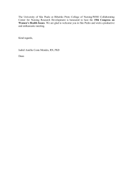
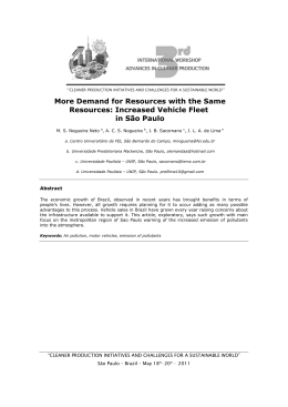
Published once every two months
J Bras Pneumol. v.41, number 1, p. 1-104 January/February 2015
Editor-in-Chief
Rogerio Souza - University of São Paulo, São Paulo, Brazil
Executive Editors
Associação Brasileira
de Editores Científicos
Bruno Guedes Baldi - University of São Paulo, São Paulo, Brazil
Caio Júlio Cesar dos Santos Fernandes - University of São Paulo, São Paulo, Brazil
Carlos Roberto Ribeiro de Carvalho - University of São Paulo, São Paulo, Brazil
Carlos Viana Poyares Jardim - University of São Paulo, São Paulo, Brazil
Publication Indexed in:
Latindex, LILACS, Scielo
Brazil, Scopus, Index
Copernicus, ISI Web of
Knowledge, MEDLINE and
PubMed Central (PMC)
Associate Editors
Available in Portuguese and
English from:
www.jornaldepneumologia.com.br
or www.scielo.br/jbpneu.
Afrânio Lineu Kritski - Federal University of Rio de Janeiro, Brazil
Álvaro A. Cruz - Federal University of Bahia, Salvador, Brazil
Celso Ricardo Fernandes de Carvalho - University of São Paulo, São Paulo, Brazil
Fábio Biscegli Jatene - University of São Paulo, São Paulo, Brazil
Geraldo Lorenzi-Filho - University of São Paulo, São Paulo, Brazil
Ilma Aparecida Paschoal - State University at Campinas, Campinas, Brazil
José Alberto Neder- Federal University of São Paulo, São Paulo, Brazil
José Antônio Baddini Martinez - University of São Paulo, Ribeirão Preto, Brazil.
Renato Tetelbom Stein - Pontifical Catholic University of Rio Grande do Sul, Porto Alegre, Brazil
Sérgio Saldanha Menna Barreto - Federal University of Rio Grande do Sul, Porto Alegre, Brazil
Editorial Council
I
N
T
E
R
N
A
T
I
O
N
A
L
Alberto Cukier - University of São Paulo, São Paulo, Brazil
Ana C. Krieger - New York University School of Medicine, New York, NY, USA
Ana Luiza de Godoy Fernandes - Federal University of São Paulo, São Paulo, Brazil
Antonio Segorbe Luis - University of Coimbra, Coimbra, Portugal
Brent Winston - Department of Critical Care Medicine, University of Calgary, Calgary, Canada
Carlos Alberto de Assis Viegas - University of Brasília, Brasília, Brazil
Carlos M. Luna - Hospital de Clinicas, University of Buenos Aires, Buenos Aires, Argentina
Carmem Silvia Valente Barbas - University of São Paulo, São Paulo, Brazil
Chris T. Bolliger - University of Stellenbosch, Tygerberg, South Africa
Dany Jasinowodolinski - Federal University of São Paulo, São Paulo, Brazil
Douglas Bradley - University of Toronto, Toronto, ON, Canada
Denis Martinez - Federal University of Rio Grande do Sul, Porto Alegre, Brazil
Emílio Pizzichini - Universidade Federal de Santa Catarina, Florianópolis, SC
Frank McCormack - University of Cincinnati School of Medicine, Cincinnati, OH, USA
Geraldo Lorenzi-Filho - University of São Paulo, São Paulo, Brazil
Gustavo Rodrigo - Departamento de Emergencia, Hospital Central de las Fuerzas Armadas, Montevidéu, Uruguay
Irma de Godoy - São Paulo State University, Botucatu, Brazil
Isabela C. Silva - Vancouver General Hospital, Vancouver, BC, Canadá
J. Randall Curtis - University of Washington, Seattle, Wa, USA
John J. Godleski - Harvard Medical School, Boston, MA, USA
José Dirceu Ribeiro - State University at Campinas, Campinas, Brazil
José Miguel Chatkin - Pontifical Catholic University of Rio Grande do Sul, Porto Alegre, Brazil
José Roberto de Brito Jardim - Federal University of São Paulo, São Paulo, Brazil
José Roberto Lapa e Silva - Federal University of Rio de Janeiro, Rio de Janeiro, Brazil
Kevin Leslie - Mayo Clinic College of Medicine, Rochester, MN, USA
Luiz Eduardo Nery - Federal University of São Paulo, São Paulo, Brazil
Marc Miravitlles - Hospital Clinic, Barcelona, España
Marcelo Alcântara Holanda - Federal University of Ceará, Fortaleza, Brazil
Marli Maria Knorst - Federal University of Rio Grande do Sul, Porto Alegre, Brazil
Marisa Dolhnikoff - University of São Paulo, São Paulo, Brazil
Mauro Musa Zamboni - Brazilian National Cancer Institute, Rio de Janeiro, Brazil.
Nestor Muller - Vancouver General Hospital, Vancouver, BC, Canadá
Noé Zamel - University of Toronto, Toronto, ON, Canadá
Paul Noble - Duke University, Durham, NC, USA
Paulo Francisco Guerreiro Cardoso - Pavilhão Pereira Filho, Porto Alegre, RS
Paulo Pego Fernandes - University of São Paulo, São Paulo, Brazil
Peter J. Barnes - National Heart and Lung Institute, Imperial College, London, UK
Renato Sotto-Mayor - Hospital Santa Maria, Lisbon, Portugal
Richard W. Light - Vanderbili University, Nashville, TN, USA
Rik Gosselink - University Hospitals Leuven, Bélgica
Robert Skomro - University of Saskatoon, Saskatoon, Canadá
Rubin Tuder - University of Colorado, Denver, CO, USA
Sonia Buist - Oregon Health & Science University, Portland, OR, USA
Talmadge King Jr. - University of California, San Francisco, CA, USA
Thais Helena Abrahão Thomaz Queluz - São Paulo State University, Botucatu, Brazil
Vera Luiza Capelozzi - University of São Paulo, São Paulo, Brazill
BRAZILIAN THORACIC SOCIETY
Office: SCS Quadra 01, Bloco K, Asa Sul, salas 203/204. Edifício Denasa, CEP 70398-900, Brasília, DF,
Brazil. Tel. +55 61 3245-1030/+55 0800 616218. Website: www.sbpt.org.br. E-mail: [email protected]
The Brazilian Journal of Pulmonology (ISSN 1806-3713) is published once every two
months by the Brazilian Thoracic Society (BTS). The statements and opinions contained in
the editorials and articles in this Journal are solely those of the authors thereof and not
of the Journal’s Editor-in-Chief, peer reviewers, the BTS, its officers, regents, members, or
employees. Permission is granted to reproduce any figure, table, or other material published
in the Journal provided that the source for any of these is credited.
BTS Board of Directors (2013-2014 biennium):
President: Dr. Renato Maciel - MG
Secretary-General: Dr. Paulo Henrique Ramos Feitosa - DF
Director, Professional Advocacy: Dr. Jose Eduardo Delfini Cançado - SP
CFO: Dr. Saulo Maia Davila Melo - SE
Scientific Director: Dr. Miguel Abidon Aide - RJ
Director, Education and Professional Practice: Dr. Clystenes Odyr Soares Silva - SP
Director, Communications: Dra. Simone Chaves Fagondes - RS
President, BTS Congress 2016: Marcus Barreto Conde - RJ
President Elect (2017/2018 biennium): Fernando Luiz Cavalcanti Lundgren - PE
Chairman of the Board: Jairo Sponholz Araújo (PR)
AUDIT COMMITTEE:
Active Members: Clóvis Botelho (MT), Benedito Francisco Cabral Júnior (DF), Rafael de Castro Martins (ES)
Alternates: Maurício Meireles Góes (MG), Alina Faria França de Oliveira (PE), Paulo Cesar de Oliveira (MG)
COORDINATORS, BTS DEPARTMENTS:
Programmatic Initiatives – Alcindo Cerci Neto (PR)
Thoracic Surgery – Darcy Ribeiro Pinto Filho (RS)
Sleep–disordered Breathing – Marcelo Fouad Rabahi (GO)
Respiratory Endoscopy – Mauro Musa Zamboni (RJ)
Pulmonary Function – John Mark Salge (SP)
Imaging – Bruno Hochhegger (RS)
Lung Diseases – Ester Nei Aparecida Martins Coletta (SP)
Clinical Research – Oliver Augusto Nascimento (SP)
Pediatric Pulmonology – Paulo Cesar Kussek (PR)
Residency – Alberto Cukier (SP)
COORDINATORS, BTS SCIENTIFIC COMMITTEES:
Asthma – Emilio Pizzichini (SC)
Lung Cancer – Teresa Yae Takagaki (SP)
Pulmonary Circulation – Carlos Viana Poyares Jardim (SP)
Advanced Lung Disease – Dagoberto Vanoni de Godoy (RS)
Interstitial Diseases – José Antônio Baddini Martinez (SP)
Environmental and Occupational Respiratory Diseases – Ana Paula Scalia Carneiro (MG)
COPD – Roberto Stirbulov (SP)
Epidemiology – Frederico Leon Arrabal Fernandes (SP)
Cystic Fibrosis – Marcelo Bicalho of Fuccio (MG)
Respiratory Infections and Mycoses – Mauro Gomes (SP)
Pleura – Roberta Karla Barbosa de Sales (SP)
International Relations – José Roberto de Brito Jardim (SP)
Smoking – Luiz Carlos Corrêa da Silva (RS)
Intensive Care – Marco Antônio Soares Reis (MG)
Tuberculosis – Fernanda Carvalho de Queiroz Mello (RJ)
ADMINISTRATIVE SECRETARIAT OF THE BRAZILIAN JOURNAL OF PULMONOLOGY
Address: SCS Quadra 01, Bloco K, Asa Sul, salas 203/204. Edifício Denasa, CEP 70398-900, Brasília, DF,
Brazil. Tel. +55 61 3245-1030/+55 0800 616218.
Assistant Managing Editor: Luana Maria Bernardes Campos. E-mail: [email protected]
Circulation: 1,100 copies
Distribution: Free to members of the BTS and libraries
Printed on acid-free paper
SUPPORT:
Published once every two months
J Bras Pneumol. v.41, number 1, p. 1-104 January/February 2015
EDITORIAL
1 - 2015—another step along the road in a 40-year journey...
2015 — mais um passo em um caminho de 40 anos...
Rogério Souza
ARTIGO ESPECIAL / SPECIAL ARTICLE
3 - A workshop on asthma management programs and centers in Brazil: reviewing and
explaining concepts
Programas e centros de atenção a asmáticos no Brasil; uma oficina de trabalho: revisitando e
explicitando conceitos
Rafael Stelmach, Alcindo Cerci-Neto, Eduardo Vieira Ponte, Gerardo Alves,
Ildely Niedia Araujo-Costa, Laura Maria de Lima Belizário Facury Lasmar,
Luci Keiko Kuromoto de Castro, Maria Lucia Medeiros Lenz, Paulo Silva, Alberto Cukier,
Alexssandra Maia Alves, Aline Silva Lima-Matos, Amanda da Rocha Oliveira Cardoso,
Ana Luisa Godoy Fernandes, Bruno Piassi de São-José, Carlos Antônio Riedi, Deborah Schor,
Décio Medeiros Peixoto, Diego Djones Brandenburg, Elineide Gomes dos Santos Camillo,
Faradiba Sarquis Serpa, Heli Vieira Brandão, João Antonio Bonfadini Lima, Jorge Eduardo Pio,
Jussara Fiterman, Maria de Fátima Anderson, Maria do Socorro de Lucena Cardoso,
Marcelo Tadday Rodrigues, Marilyn Nilda Esther Urrutia Pereira, Marti Antila, Solange Valle,
Sonia Maria Martins, Vanessa Gonzaga Tavares Guimarães, Yara Arruda Marques Mello,
Wenderson Clay Correia de Andrade, William Salibe-Filho, Zelina Maria da Rocha Caldeira,
Zuleid Dantas Linhares Mattar, Álvaro Augusto Souza da Cruz-Filho, Paulo Camargos
ARTIGOS ORIGINAIS / ORIGINAL ARTICLES
16 - Negative impact of asthma on patients in different age groups
Impacto negativo da asma em diferentes faixas etárias
Marcela Batan Alith, Mariana Rodrigues Gazzotti, Federico Montealegre, James Fish,
Oliver Augusto Nascimento, José Roberto Jardim
23 - Endobronchial ultrasound-guided transbronchial needle aspiration for lung cancer
staging: early experience in Brazil
Punção aspirativa por agulha guiada por ultrassom endobrônquico no estadiamento do câncer de
pulmão: experiência inicial no Brasil
Viviane Rossi Figueiredo, Paulo Francisco Guerreiro Cardoso, Márcia Jacomelli,
Sérgio Eduardo Demarzo, Addy Lidvina Mejia Palomino, Ascédio José Rodrigues,
Ricardo Mingarini Terra, Paulo Manoel Pego-Fernandes, Carlos Roberto Ribeiro Carvalho
31 - Preoperative predictive factors for intensive care unit admission after pulmonary resection
Fatores preditivos pré-operatórios de internação em unidade de terapia intensiva após ressecção
pulmonar
Liana Pinheiro, Ilka Lopes Santoro, João Aléssio Juliano Perfeito, Meyer Izbicki,
Roberta Pulcheri Ramos, Sonia Maria Faresin
39 - Chronic intermittent hypoxia increases encoding pigment epithelium-derived factor
gene expression, although not that of the protein itself, in the temporal cortex of rats
Hipóxia intermitente crônica aumenta a expressão gênica, mas não proteica, de pigment
epithelium-derived factor, no córtex temporal de ratos
Guilherme Silva Julian, Renato Watanabe de Oliveira, Vanessa Manchim Favaro,
Maria Gabriela Menezes de Oliveira, Juliana Cini Perry, Sergio Tufik, Jair Ribeiro Chagas
48 - Community-acquired pneumonia: economics of inpatient medical care vis-à-vis clinical
severity
Pneumonia adquirida na comunidade: economia de cuidados médicos em regime de
internamento, em relação à gravidade clínica
Vojislav Cupurdija, Zorica Lazic, Marina Petrovic, Slavica Mojsilovic, Ivan Cekerevac,
Nemanja Rancic, Mihajlo Jakovljevic
Published once every two months
J Bras Pneumol. v.41, number 1, p. 1-104 January/February 2015
58 - Diagnostic accuracy of the Bedside Lung Ultrasound in Emergency protocol for the
diagnosis of acute respiratory failure in spontaneously breathing patients
Acurácia diagnóstica do protocolo de ultrassom pulmonar à beira do leito em situações de
emergência para diagnóstico de insuficiência respiratória aguda em pacientes com ventilação
espontânea
Felippe Leopoldo Dexheimer Neto, Juliana Mara Stormovski de Andrade,
Ana Carolina Tabajara Raupp, Raquel da Silva Townsend, Fabiana Gabe Beltrami, Hélène Brisson,
Qin Lu, Paulo de Tarso Roth Dalcin
ARTIGO DE REVISÃO / REVIEW ARTICLE
65 - Chronic rhinosinusitis and nasal polyposis in cystic fibrosis: update on diagnosis and
treatment
Rinossinusite crônica e polipose nasossinusal na fibrose cística: atualização sobre diagnóstico e
tratamento
Suzie Hyeona Kang, Paulo de Tarso Roth Dalcin, Otavio Bejzman Piltcher,
Raphaella de Oliveira Migliavacca
77 - Risk factors associated with adverse reactions to antituberculosis drugs
Fatores de risco associados às reações adversas a medicamentos antituberculose
Laíse Soares Oliveira Resende, Edson Theodoro dos Santos-Neto
RELATO DE CASO / CASE REPORT
90 - Video-assisted thoracoscopic implantation of a diaphragmatic pacemaker in a child with
tetraplegia: indications, technique, and results
Implante de marca-passo diafragmático por videotoracoscopia em criança com tetraplegia:
indicações, técnica e resultados
Darcy Ribeiro Pinto Filho, Miguel Lia Tedde, Alexandre José Gonçalves Avino,
Suzan Lúcia Brancher Brandão, Iuri Zanatta, Rafael Hahn
CARTAS AO EDITOR / LETTER TO THE EDITOR
95 - Nonadherence to treatment in lung transplant recipients: a matter of life and death
Falta de adesão ao tratamento em pacientes submetidos a transplante pulmonar: uma questão de
vida ou morte
André Nathan Costa, Elaine Marques Hojaij, Liliane Saraiva de Mello, Felipe Xavier de Melo,
Priscila Cilene Leon Bueno de Camargo, Silvia Vidal Campos, Jose Eduardo Afonso Junior,
Rafael Medeiros Carraro, Ricardo Henrique de Oliveira Braga Teixeira
98 - Anxiety, depression, and motivation for smoking cessation in hospitalized patients with
and without cancer
Motivação para cessação do tabagismo, ansiedade e depressão em pacientes internados com e sem
neoplasia
Igor Bastos Polonio, Meiryelle Landim Franco, Marina Angélica Mendes Tegon,
Célia Beatriz Gianotti Antoneli
101 - Incidence of spontaneous subdural hematoma in incident cases of pulmonary arterial
hypertension: a registry of cases occurring over a five-year period
Incidência de hematomas subdurais espontâneos em casos de pacientes com hipertensão arterial
pulmonar: análise de um registro de cinco anos
Luis Felipe Lopes Prada, Francisca Gavilanes, Rogério Souza
103 - Hibernoma: an uncommon cause of a pleural mass
Hibernoma: uma causa incomum de massa pleural
Edson Marchiori, Gláucia Zanetti, Bruno Hochhegger
Editorial
2015—another step along the road in a 40-year journey...
2015 — mais um passo em um caminho de 40 anos...
Rogério Souza
This is a special year for the Brazilian Journal
of Pulmonology (BJP); in October, the BJP will
turn 40.(1) Dates such as this are particularly
significant, given that they lead us to reflect on
many of the aspects related to the existence of
the BJP: its history; its current relevance; and
its prospects.
The BJP was founded in 1975, and the fact that
it continues its journey today is undoubtedly due
to the continuous efforts of numerous colleagues
who, directly or indirectly, have sought to make the
Journal function as a great common denominator
linking all members of the respiratory medicine
community. This process has been spearheaded
by a group of brave editors whose efforts have
led the BJP to great achievements: Manoel Lopes
dos Santos (1975-6); Bruno Carlos Palombini
(1976-8); Carlos Frazzatto Junior (1978-82);
José Roberto de Brito Jardim (1982-6); Miguel
Bogossian (1986-90); Nelson Morrone (1990-4);
Carlos Alberto de Castro Pereira (1994-8); Thais
Helena Abrahão Tomaz Queluz (1998-2002);
Geraldo Lorenzi Filho (2002-4); José Antônio
Baddini Martinez (2004-10); and, most recently,
Carlos Roberto Ribeiro Carvalho (2010-4), all
of whom have contributed to making the BJP
the most important scientific journal in the
respiratory field in Latin America and one of the
most important scientific journals in Brazil. Such
a feat gives us a measure of the responsibility
we all have to our Journal henceforward.
In recent decades, the BJP was indexed for
SciELO (2002) and, subsequently, for PubMed/
MEDLINE (2006), which has greatly increased
the visibility of the Journal and therefore the
number of articles submitted, particularly after
it was indexed for the Thomson Reuters Institute
for Scientific Information Web of Science and its
first impact factor appeared in Journal Citation
Reports.(2) We have recently been penalized by
having our impact factor temporarily suspended,
a fact that will affect us in the next evaluation.(3)
However, we have to bear in mind that this metric
associated with scientific publication should be
understood within its surrounding context rather
than as a single number. In particular, the BJP has
http://dx.doi.org/10.1590/S1806-37132015000100001
two very clear functions: to be a publication of
quality, providing a vehicle for scientific production
in the field of respiratory medicine, especially
in Brazil but also worldwide, and to be the
major instrument for the continuing education
of pulmonologists in Brazil. For the latter mission,
the commitment is to reach into the daily lives
of its readers through review articles that reflect
the necessities of daily clinical practice in the
respiratory field. It is for the dissemination of
scientific production that we will use common
editorial metrics, such as the impact factor.
Such metrics, despite their limitations, allow
editorial policy planning, with course changes
when necessary, in order to increase the national
and international visibility of the Journal, thereby
attracting the very best science in the field. To
that end, several steps have been taken with a
view toward increasing the international exposure
of the BJP, among which is the fact that, since
2014, the Journal has been available on PubMed
Central, an open archive of the U.S. National
Institutes of Health National Library of Medicine,
making it possible to access the full-text articles
published by the BJP (in English) directly via the
PubMed search function.(4)
We will now make all articles accepted by the
BJP available online ahead of print, in order to
maximize the exposure time of our publications.
In parallel, all BJP content will be screened for
plagiarism with the same tools used by the
major scientific journals in the medical field.
With this screening, along with the exposure,
the trustworthiness of the content of the BJP
will also increase significantly.
Although the responsibility of managing the
BJP is huge, standing on the shoulders of those
who have made the Journal so solid makes the
path much smoother. The aforementioned changes
are aimed at preparing us for further growth. In
addition, another very important change deserves
special mention. The board of associate editors
of the BJP has grown significantly. In advance, I
thank all the editors who have embarked on the
project for the next four-year term of editorship
of the BJP, which begins now. The participation
J Bras Pneumol. 2015;41(1):1-2
2
Souza R
of such a significant body of researchers in the
editorship of the Journal undoubtedly allows
a closer relationship between the BJP and
major research groups, in Brazil and around
the world. It also allows the BJP to play its
aforementioned role in the continuing education
of respiratory medicine professionals working in
Brazil, particularly those who are not affiliated
with educational institutions. The selection of
the topics of greatest interest to all kinds of
readers will come to be shared by a group that
is representative of the various fields of study
in respiratory medicine, potentially enhancing
the scope of the Journal.
The ultimate goal for the next four years is
to increase the representativeness of the BJP,
not only via its role of disseminating scientific
knowledge, which is to be measured by various
existing metrics, but also via the role it plays in
the education of health professionals in Brazil,
which is to be measured by the interest in the
different areas of the Journal, on the basis of
the number of logins/downloads and requested
J Bras Pneumol. 2015;41(1):1-2
reprints. Let us hope that the celebrations of
and reflections on the last 40 years of BJP
history will lead us steadily toward a 2015 full
of achievements.
Rogério Souza
Editor-in-Chief of the Brazilian Journal
of Pulmonology
References
1. Santos ML. Brazilian Journal of Pulmonology: thirty
years of history. J Bras Pneumol. 2005;31(5):i. http://
dx.doi.org/10.1590/S1806-37132005000500001
2. Carvalho CR. Publications in the Brazilian Journal of
Pulmonology. J Bras Pneumol. 2013;39(1):1-4. http://
dx.doi.org/10.1590/S1806-37132013000100001
3. Carvalho CR. The Brazilian Journal of Pulmonology and
international databases. J Bras Pneumol. 2013;39(5):52931. http://dx.doi.org/10.1590/S1806-37132013000500001
4. Carvalho CR, Baldi BG, Jardim CV, Caruso P, Souza R.
New steps for the international consolidation of the
Brazilian Journal of Pulmonology. J Bras Pneumol.
2014;40(4):325-6.
http://dx.doi.org/10.1590/
S1806-37132014000400001
http://dx.doi.org/10.1590/S1806-37132015000100001
Special Article
A workshop on asthma management programs and
centers in Brazil: reviewing and explaining concepts*
Programas e centros de atenção a asmáticos no Brasil; uma
oficina de trabalho: revisitando e explicitando conceitos
Rafael Stelmach, Alcindo Cerci Neto, Ana Cristina de Carvalho Fernandez Fonseca,
Eduardo Vieira Ponte, Gerardo Alves, Ildely Niedia Araujo-Costa, et al.
Abstract
Objective: To report the results of a workshop regarding asthma management programs and centers (AMPCs) in
Brazil, so that they can be used as a tool for the improvement and advancement of current and future AMPCs.
Methods: The workshop consisted of five presentations and the corresponding group discussions. The working
groups discussed the following themes: implementation of asthma management strategies; human resources
needed for AMPCs; financial resources needed for AMPCs; and operational maintenance of AMPCs. Results: The
workshop involved 39 participants, from all regions of the country, representing associations of asthma patients
(n = 3), universities (n = 7), and AMPCs (n = 29). We found a direct relationship between a lack of planning
and the failure of AMPCs. Based on the experiences reported during the workshop, the common assumptions
about AMPCs in Brazil were the importance of raising awareness of managers; greater community participation;
interdependence between primary care and specialized care; awareness of regionalization; and use of medications
available in the public health system. Conclusions: Brazil already has a core of experience in the area of asthma
management programs. The implementation of strategies for the management of chronic respiratory disease
and their incorporation into health care system protocols would seem to be a natural progression. However,
there is minimal experience in this area. Joint efforts by individuals with expertise in AMPCs could promote the
implementation of asthma management strategies, thus speeding the creation of treatment networks, which
might have a multiplier effect, precluding the need for isolated centers to start from zero.
Keywords: Asthma; Academic medical centers; Area health education centers; Health planning organizations;
Regional medical programs; Managed care programs.
Resumo
Objetivo: Relatar os resultados de uma oficina de trabalho sobre programas e centros de atenção a asmáticos
(PCAAs) no Brasil para que possam servir como instrumento para melhoria e avanço dos PCAAs existentes
e criação de novos. Métodos: A oficina de trabalho constituiu-se de cinco apresentações e discussões em
grupos. Os grupos de trabalho discutiram os seguintes temas: implementação de uma linha de cuidado em
asma; recursos humanos necessários para os PCAA; recursos necessários para financiar os PCAA; e manutenção
do funcionamento dos PCAAs. Resultados: A oficina envolveu 39 participantes de todas as regiões do país,
representando associações de asmáticos (n = 3), centros universitários (n = 7) e PCAAs (n = 29). Evidenciou-se
uma relação direta entre a ausência de planejamento e o insucesso dos PCAAs. Com base nas experiências
brasileiras elencadas durante a oficina, as premissas comuns foram a importância da sensibilização do gestor,
maior participação da comunidade, interdependência entre a atenção primária e a especializada, observação da
regionalização e utilização dos medicamentos disponíveis no sistema público de saúde. Conclusões: O Brasil
já tem um núcleo de experiências na área programática da asma. A implementação de uma linha de cuidado
em doenças respiratórias crônicas e sua inclusão nas redes de saúde parecem ser o caminho natural. Porém, a
experiência nessa área ainda é pequena. Agregar pessoas com experiência nos PCAAs na elaboração da linha de
cuidado em asma encurtaria tempo na criação de redes de atenção com possível efeito multiplicador, evitando
que se partisse do zero em cada local isolado.
Descritores: Asma; Centros médicos acadêmicos; Centros educacionais de áreas de saúde; Organizações de
planejamento em saúde; Programas médicos regionais; Programas de assistência gerenciada.
*Study carried out in the Department of Pulmonology, Heart Institute, Hospital das Clínicas, Faculdade de Medicina da Universidade
de São Paulo – HC-FMUSP, University of São Paulo School of Medicine Hospital das Clínicas – São Paulo, Brazil.
Correspondence to: Rafael Stelmach. Avenida Enéas de Carvalho Aguiar, 44, 5º andar, Divisão de Pneumologia, CEP 05403-000,
São Paulo, SP, Brasil.
Tel. 55 11 3285-3407. E-mail: [email protected]
Financial support: This study was funded with unrestricted educational grant support from the Programa para Controle da Asma
na Bahia/Global Initiative for Asthma (ProAR, Bahia State Asthma Control Program/GINA) and by the pharmaceutical company
Chiesi Farmacêutica.
Submitted: 21 August 2014. Accepted, after review: 17 November 2014.
http://dx.doi.org/10.1590/S1806-37132015000100002
J Bras Pneumol. 2015;41(1):3-15
4
Stelmach R, Cerci Neto A, Fonseca ACCF, Ponte EV, Alves G, Araujo-Costa IN, et al.
Introduction
Some asthma patient management programs
and centers that are currently in operation in
Brazil are coming of age. In an editorial, Holanda(1)
reported results of questionnaires on Asthma
Management Programs and Centers (AMPCs) in
Brazil, completed by 16 members of the Brazilian
Thoracic Association (BTA) or regional affiliates.
At the time, 14 AMPCs (87.5%) confirmed that
they were in regular operation, 10 of which had
been established in the 1990s. Looking back, the
responses regarding asthma management seem
alarming: inhaled medications were unavailable
(there were only oral medications); treatment
demand was higher than treatment availability;
and there were no outpatient clinics specializing
in asthma.
Many AMPCs were created as a result of
the dissemination of the first national and
international guidelines for the management of
asthma, published in the same decade. Therefore,
those guidelines prompted the holding of the First
and Second Brazilian Conferences on Asthma in
1997 and 1999, respectively. The unavailability of
inhaled corticosteroids (ICs) in public institutions
contradicted the cornerstone of the treatment of
persistent asthma, derived from the guidelines.
Pioneers of that time include the AMPCs in the
cities of Belo Horizonte, Fortaleza, and São Paulo,
which already showed that educating patients
decreased the number of hospitalizations and
improved patient quality of life.(1,2)
One group of authors(2) prepared a timeline
of the evolution of public policies and AMPCs
in Brazil since 1996, showing that, in 1998, the
National Drug Policy was created, which led to the
dispensation of medications for asthma control.
This provision stimulated and gave support to
the creation of new programs and required the
implementation of referral centers.(2) It is not
by chance that the Carta de Salvador (Salvador
Charter),(3) urging the implementation of the
National Asthma Control Program, was formulated
in 2001. The cities of Porto Alegre, Goiânia,
Londrina, Niterói, Salvador, Feira de Santana, Rio
de Janeiro, and Vitória established their AMPCs at
the same time as decrees regulating the allocation
of federal resources to health care services were
being issued.(2) At that time, in addition to ensuring
care for asthma patients, some AMPCs stood out
for their scientific production, others stood out for
providing training to professionals from different
J Bras Pneumol. 2015;41(1):3-15
areas, and others stood out for expanding to
small municipalities, decentralizing activities.
In 2007, an editorial(4) presented an account
of the decade and highlighted the need for
professional training and funding to advance
the quality of care by improving AMPCs. Examples
of asthma programs in Brazil were obtained in
2008, by analyzing the responses to the forms sent
to BTA members and members of the Brazilian
Association of Allergists and Immunologists.(5) Of
55 services that reported having a systematized
program, 11 (20%) did not respond to the structural
questions and 27 (49%) were excluded from the
analysis (17 treated severe asthma and 10 had
had the program for less than two years). All 17
programs analyzed received public funding for
their maintenance: 4 (23%) received state funding
exclusively, whereas 13 (77%) received state and
municipal funding. There were no programs in
northern Brazil. All 17 programs had referral
centers with specialists, and 47% developed
educational activities (lectures or individual
visits) within the community. In addition, 47%
provided home visits by nurses, and 41% adopted
public health strategies, such as family health
care, outreach, humanizing practices, and visits
by community health care agents. That study(5)
showed that, from 2003 onward, the number
of programs increased significantly, as a result
of the availability of full public funding for the
purchase of asthma medications.
Some successful programs no longer exist
because of political and administrative changes.
However, it can be stated that there has been no
dissemination of programs across the country.
There are still fewer than five dozen programs,
as mentioned above. Most are supported by the
dedication of some individuals—experts in their
field—and often with resources from funding
agencies, partnerships with the private sector,
or both. In the majority of localities, they have
not become programs or management strategies
of the municipal or state departments of health,
especially since, as yet, the Brazilian National
Ministry of Health itself has not prioritized
management strategies for chronic respiratory
diseases.
Between 1991 and 2010, the epidemiologic
picture changed, as a result of the growth of
the Brazilian population at a rate of 20 million
per decade. The population jumped from 146.8
million in 1991 to 190.7 million in 2010.(6) This
http://dx.doi.org/10.1590/S1806-37132015000100002
A workshop on asthma management programs and
centers in Brazil: reviewing and explaining concepts
means that there was an increase in the number
of asthma patients. Conversely, the number of
hospitalizations for asthma via the Sistema Único
de Saúde (SUS, Brazilian Unified Health Care
System) decreased from 400,000 per year to fewer
than 200,000 per year between 2000 and 2012,(7)
with a disproportionate decrease of 30% in the
expenditures for such hospitalizations (110 million
Brazilian reals vs. 80 million Brazilian reals). It
is correct to state that the asthma centers and
programs helped this reduction in the number of
hospitalizations. In addition, they encouraged the
changes made by the laws and national regulations
that initially led to the decentralization of payment
of costs of asthma and rhinitis medications, as
well as to the publication of the Primary Care
Guidebook – rhinitis , asthma, and COPD(8) and,
more recently, of the revised Clinical Protocol and
Therapeutic Guidelines – asthma.(9) Because of the
high prevalence of COPD, which is a mandatory
differential diagnosis for asthma, COPD care was
combined with asthma care in adults in some
centers, in addition to being standardized at
the federal level.(10) Given the regulation of the
basic (i.e. municipal) and specialized (i.e. state)
components of pharmaceutical care, as well as
the free provision of basic asthma medications
at enrolled pharmacies since 2012, it can be
stated that there already is adequate public
funding.(2) However, the population’s demands
for more resources/materials for the treatment
of chronic respiratory diseases have become a
reality for public and private health, including
via litigation.(11) Another change in public health
has been the increased value placed on the
family health program strategy. According to
the Brazilian National Ministry of Health,(12) half
of the Brazilian population receive some care
through that strategy, and one of the current
developments is the controversial program of
importation of physicians. Conversely, several
national and international studies have shown
that asthma is not controlled in more than 50% of
the patients evaluated,(13) which can be confirmed
by the still low use of ICs.(14)
What to do to provide care for those who
remain unassisted and to continue to decrease
asthma-related morbidity and mortality in Brazil
is the question. The Global Initiative for Asthma
(GINA)(15) recommends that, by 2015, there should
be fewer than 100,000 hospitalizations per year
for asthma in Brazil. It must be borne is mind
http://dx.doi.org/10.1590/S1806-37132015000100002
5
that, despite the decrease in the number of
hospitalizations, official statistics show that the
number of deaths from asthma (3,000 deaths per
year) has remained unchanged.(7) How to continue
the history of AMPCs, making them a reference
in education and care, is the challenge ahead.
How to multiply them (physically or conceptually)
is a challenge and a requirement. In an attempt
to answer these questions, it was proposed that
a workshop on AMPCs in Brazil be held. The
objective of the present study was to report the
results of this workshop so that they can be used
as a tool for the improvement and advancement
of current centers and programs, as well as for
the establishment of new ones.
Methods
Using a list of AMPCs that were identified
in surveys conducted in 2000, 2008, and 2013
(the last survey has not been published), four
coordinators of AMPCs that have been in operation
since the 1990s selected health professionals
with related activities throughout Brazil. The
geographic distribution, lifetime (continuity),
and infrastructure of AMPCs were taken into
consideration, as were their scientific production,
training of staff specializing in chronic respiratory
diseases, and successful or innovative experiences
in the area. Some professionals involved in tertiary
care (severe asthma), as well as individuals who
were in charge of associations of asthma patients
and had health training, were also selected. In
addition, physicians, nurses, and pharmacists
who were directly involved in the processes in
AMPCs and who preferably did not perform
program management were invited. Two workshop
coordinators made a list of 48 guests after analysis
of AMPCs, as well as of associations of patients
in the states of Rio de Janeiro and São Paulo.
The objectives proposed for the workshop
were as follows:
• Compile successful experiences in
AMPCs in Brazil and the difficulties
in implementing asthma management
strategies in the SUS.
• Outline the current state of initiatives in
asthma in Brazil in their various phases
(planning and regional integration;
professional training and standardization;
funding and management; expansion and
consolidation; and national guidelines
for asthma programs).
J Bras Pneumol. 2015;41(1):3-15
6
Stelmach R, Cerci Neto A, Fonseca ACCF, Ponte EV, Alves G, Araujo-Costa IN, et al.
• Find and propose solutions to problems
associated with the development of the
AMPCs that are already in operation in
the country.
• Develop a practical manual for the
implementation of program activities and
centers of excellence for the treatment
of asthma.
The workshop agenda was designed to favor
group work. The coordinators defined the proposed
themes for discussion. Presentations on specific
issues served as a basis for the group discussions.
Each group had two coordinators, who were in
charge of recording the discussions and reporting
them at plenary sessions. Chart 1 presents a
summary of the workshop program.
The group discussions were recorded in
real time by a company specializing in editing
for events. The results were systematized by
combining the group reports, the notes made
by the (workshop and group) coordinators, and
the text prepared by the editing company. The
results are presented by group discussion topic.
Results
Of 48 guests, 39 attended the event, which was
held in the city of São Paulo and lasted eight hours.
All regions of the country were represented (the
Northern, Central West, Northeastern, Southern,
and Southeastern regions had 1, 2, 8, 11, and
17 representatives, respectively). The group of
professionals consisted of 13 pulmonologists,
8 pediatric pulmonologists, 6 allergists/
immunologists, 6 pediatricians (2 of whom were
allergologists), 2 family/community physicians,
3 pharmacists, and 1 nurse. The associations of
asthma patients had 3 representatives present.
Seven professionals represented university
(secondary/tertiary) referral centers, and 29
represented AMPCs. Although three of those
Chart 1 - Outline of the workshop on asthma management programs and centers.
Outline content
Opening remarks
Situation at the time of the event
Asthma and Public Health – a current overview
Asthma management program activities – lines of conduct
Primary care in respiratory programs: matrix-like planning in asthma/COPD
Difficulties of/solutions for highly demanded asthma programs
Formation of working groups based on guiding principles
Difficulties in implementing asthma management strategies
Lack of planning
Unawareness of the regional context
Inadequate regional integration models
Lack of partnerships
Securing human resources for asthma programs
Importance of training
Who should be trained?
Is it important to create regional protocols?
How can the community be involved in the process?
The role of referral centers in the development of human resources
Seeking financial resources for asthma programs through public/private/academic sector cooperation
What are the current funding sources?
How can access to those resources be gained?
How and who should manage the resources?
Where and how should the resources be spent? (education, training, medications?)
Operational maintenance of asthma programs
The asthma program is operational, what now?
How can the experience be expanded and replicated?
Is it worthwhile to include other diseases?
Presentation of the conclusions of the working groups
Debates
J Bras Pneumol. 2015;41(1):3-15
http://dx.doi.org/10.1590/S1806-37132015000100002
A workshop on asthma management programs and
centers in Brazil: reviewing and explaining concepts
centers/AMPCs had private management, all
were directed to SUS.
The results of the study groups were
systematized and are described as follows:
Difficulties in implementing asthma
management strategies
Lack of planning
• Strategic planning is critical in
implementing asthma management
strategies, in order to minimize potential
implementation difficulties and maximize
community and public manager awareness.
• Through planning, public managers are
committed to political and financial
support, even when there are local
political changes.
• In order to maintain the support from
public managers, asthma management
strategies should have an appropriate
cost-benefit ratio and prioritize the
free provision of outpatient asthma
medications, because this reduces
hospitalization expenditures and increases
the productivity of patients and health
professionals.
• It is important to estimate the number
of patients who will be reached by the
intervention. Management strategies
that prioritize primary care help a large
volume of patients who, individually, do
not use the health care system very often.
When management strategies prioritize
secondary care, they will help a smaller
volume of patients who individually use
many health resources. In the long run,
no level of care should be excluded, and
there should be mechanisms to allow
patients to seek treatment networks or
webs according to the behavior of their
disease.
• Planning focused on the development of
continuous data collection tools provides
indicators that can be used to measure
the benefits and impact of those activities
in public health.
• Civil society and medical societies must
work together to avoid setbacks in the
treatment of asthma patients, and,
http://dx.doi.org/10.1590/S1806-37132015000100002
7
therefore, public awareness should be
raised.
• Medical specialty societies and patient
associations have a very positive record in
fostering the implementation of asthma
management strategies.
• Currently, asthma programs are organized
around individuals, and there is a high
risk of loss of continuity.
Regional context unawareness /
inadequate regional integration models
• Asthma management strategies should
respect the heterogeneity of the country.
There is already a minimal health care
system that reaches almost the entire
country—primary care clinics (PCCs) and
family health program teams/strategies—
which should be prioritized.
• Asthma management strategies should take
advantage of the structure that is available.
Specialized centers are indispensable
to training primary care teams without
experience in asthma and to supporting
patients who are refractory to treatment,
while also making that structure available
to smaller municipalities (in the region).
Lack of partnerships
• In order to implement and maintain
asthma management strategies, it is
necessary to have support from the
public/state sector, the private sector
(i.e., schools, pharmaceutical industries,
the media, and health insurance plans),
and the third sector (i.e., universities,
religious institutions, nongovernmental
organizations, foundations, associations,
etc.).
• The physical structure, the purchase of
medications, and the health teams are the
responsibility of municipalities and states.
The private sector and the third sector can
contribute by disseminating information,
providing technological knowledge, and
facilitating public/state sector activities.
These contributions can mainly take the
form of scientific meetings for health
teams, donation of spirometers, support for
forming associations of asthma patients,
media dissemination of information about
J Bras Pneumol. 2015;41(1):3-15
8
Stelmach R, Cerci Neto A, Fonseca ACCF, Ponte EV, Alves G, Araujo-Costa IN, et al.
asthma to the lay public, and commitment
for selling medications to the public sector
at the lowest price possible.
• The use of volunteers should be encouraged.
Presentations in schools and communities
given by volunteers have significant impact,
according to experiences in the city of
São Paulo. It is of note that volunteer
activities are not regular and should be
planned as short-term.
Securing human resources for AMPCs
Importance of training
• Continuing training of all categories
of health professionals at all levels of
care is necessary so that professionals
working in asthma care know how to
identify, classify, and manage patients
appropriately, thereby reducing asthmarelated morbidity and mortality, as well as
improving the quality of life of patients
and their family members.
• In addition to health professionals, it is of
paramount importance to train patients
and their family members in recognizing
the disease, the periods of exacerbation,
and the forms of treatment, therefore
preventing complications.
• Currently, the focus of public health is
the Family Health Program Strategy,
which is responsible for the holistic care
of individuals. The objective is to train
primary care professionals so that they
can identify people with asthma, classify
them in accordance with the clinical
protocols, and manage them, in order
to reduce the number of unnecessary
referrals to secondary care and not to
place an additional burden on the health
system.
• In addition to training, it is important
that the network be under continuing
supervision for the maintenance
of appropriate care, with qualified
referrals, well-defined network flows,
and monitoring of local indicators.
Who should be trained?
• Training should be attended by the
multidisciplinary team together as a
J Bras Pneumol. 2015;41(1):3-15
group, and a piece of information that
is shared by all categories should be
used in demonstrating the role of each
individual in patient care. Techniques
such as matrix-like planning can make
the function of each professional in daily
practice clearer. Any trained professional
can be a local tutor.
• Professionals at referral centers should
also be trained to receive, accommodate,
and treat patients in accordance with
specialized care protocols.
• The creation of regional centers could
enable training and consulting anywhere
in the country through partnerships,
preventing extensive traveling within
the country. Professionals from societies,
institutions, and universities who are
associated with the asthma problem in
our communities can be part of those
centers.
Is it important to create regional
protocols?
• With regard to the creation of regional
protocols, it is of note that the current
national and international patient care
guidelines are applicable to all centers.
Indeed, It is necessary to create flows and
organize health care services according
to local contexts.
How can the community be involved in
the process?
• A community that is aware of the risks and
costs of illness, and that knows that there
are resources to reduce them, mobilizes
with public authorities to try to improve
care for users.
• Community involvement should be broad,
considering individuals with or without
asthma, but with an emphasis on the
asthma community, with includes patients
and their family members.
• Knowledge about asthma and asthma
management strategies should be
disseminated widely, and this can be
made possible through local health
councils and through local/regional
media (television, radios, churches, and
schools). The dissemination of information
http://dx.doi.org/10.1590/S1806-37132015000100002
A workshop on asthma management programs and
centers in Brazil: reviewing and explaining concepts
through widely accessed electronic media
such as social networks reaches large
populations.
• Emphasize to patients that they are
responsible for demanding complete,
quality care, which includes professionals
who provide appropriate care, provision
of necessary materials, and continuing
education. This responsibility cannot be
assumed only by health professionals.
• Children should start receiving asthma
education early, which, in addition to
strengthening family ties in terms of
education, encourages adherence to
treatment.
The role of referral centers in the
development of human resources
• During training, referral centers are
responsible for passing on information
regarding patient referral flow and
asthma management strategy steps to the
entire system. This knowledge should be
shared by all professionals working in the
system, from primary care professionals to
professionals working in referral centers,
whether they are regional/secondary or
tertiary centers.
• Referral centers are also responsible for
providing information to primary care
professionals and for, together with those
professionals, defining criteria for patient
referral to/from referral centers and criteria
for patient follow-up. Standardization of
patient follow-up and management, as
well as of referral and counter-referral
models, facilitates dialogue.
Seeking financial resources for asthma
programs through public/private/
academic sector cooperation
What are the current funding sources?
• From a broad perspective, funding is
partially taken care of, especially when
it comes to medications. The lack of
funding implies discontinuity of activities
and loss of motivation.
• The federal government makes asthma
medications available through the
special drugs program and the “Popular
http://dx.doi.org/10.1590/S1806-37132015000100002
9
Pharmacy” program. Some municipalities
contribute by making ICs and short-acting
bronchodilators available at PCCs.
• There is a need for an alignment between
what is proposed in the guidelines/
strategies and what in fact there is to
treat patients and control asthma.
How can access to those resources be
gained?
• The group discussions evidenced the need
for the current forms of funding and for
alternatives to be further explained in
a document prepared by public health
specialists with a deep understanding
of the subject. This document would
be produced by specialist societies or
organizations/associations related to
asthma.
• There are forms and sources of funding
that are unknown to the group.
• The provision of special medications,
with the addition of long-acting β2
agonists, was an advance; however, in
some programs, this provision is not far
reaching, because it is focused on people
(it is not standardized).
How and who should manage the
resources?
• Social control is very important because
it fosters the continuity of the program.
• The raising and allocation of resources
should be based on technical rather than
on political criteria.
• A joint management strategy with
the family health program should be
pursued. Since primary care is a priority
and resources are directed to it, it is
not feasible to have resources available
for the asthma program unless there
is integrated patient care. There is no
money specifically for asthma, but there
is money for primary care.
• In addition, there are funding sources
other than the federal government, such as
state and municipal foundations, through
which it is possible to obtain additional
resources.
J Bras Pneumol. 2015;41(1):3-15
10
Stelmach R, Cerci Neto A, Fonseca ACCF, Ponte EV, Alves G, Araujo-Costa IN, et al.
Where and how should the resources be
spent?
• It was concluded that there is no
conflict between the recommendations
in national and international guidelines
and the medications provided to patients.
Medications should be addressed in all
stages of planning guidelines in the
program.
• Funding for asthma education is as
necessary as is funding for other activities.
The resource exists, but it is imperative
to understand and propose ways to apply
for it.
Operational maintenance of asthma
programs
The present work group chose to merge the
three questions that should be included in the
program—”The asthma program is operational,
what now?”; “How can the experience be expanded
and replicated?”; and “Is it worthwhile to include
other diseases?—into a single theme, which is
described below.
Strategies for maintaining, expanding,
and replicating asthma programs
• AMPCs are characterized as a set of
pre-defined activities, objectives, and goals
that will meet the needs of a population.
• The maintenance of AMPCs requires
the following: knowledge of the local
context; multidisciplinary coordination
and institutional programs; greater
awareness and continuing updating of
professionals; dissemination of activities;
and participation of the population.
• It was identified that comprehensiveness
of care is important in individual care;
however, when it comes to programs,
maintaining the focus on care for people
with asthma is critical. There should be
integration with programs targeted at
other conditions, such as smoking and
COPD. For instance, smoking family
members of children with asthma should
be advised and referred to smoking
cessation groups. The management
of allergic rhinitis, however, should be
addressed in asthma clinical protocols,
J Bras Pneumol. 2015;41(1):3-15
•
•
•
•
•
•
given the prevalent association between
rhinitis and asthma.
Knowledge of the local context, together
with definition of the area of operation
and delimitation of the target population,
favors the maintenance of the process.
Cultural issues should be part of that
knowledge and should guide activities
that respect diversity.
Program coordination should be
multidisciplinary (pharmacists, family
physicians, specialists, physical therapists,
etc.), which provides different perspectives
on the program and facilitates informed
planning decisions based on the local
context.
Personal
ownership—”So-and-so’s
program”—should be avoided. The
program, whenever possible, must have
a name of its own and institutional
guidelines.
Epidemiological data, such as prevalence
and impact of asthma, should be used
to raise awareness of and update
professionals, being part of the educational
process.
Protocols or guidelines should be
adapted for local use on the basis of
current strategies, such as those of
GINA and of the BTA. These protocols
should include the different resources,
whether structural or human, of primary
care (PCCs), secondary care (specialty
outpatient clinics), or tertiary care
(emergency rooms and hospitals), with
well-defined referral criteria and with an
emphasis on treatment networks.
The activities of the program and
the assessment indicators should be
disseminated to the population, managers,
and directly involved professionals, as
well as to the academic community
(through conferences, symposia and
publications). The information should
be clear and objective. It is considered
important to include communication
techniques in the training of professionals.
These individuals and groups can pass on
technical information to the population,
both individually and collectively, in health
care clinics, local health councils, schools,
associations of asthma patients, etc. The
http://dx.doi.org/10.1590/S1806-37132015000100002
A workshop on asthma management programs and
centers in Brazil: reviewing and explaining concepts
dissemination can be achieved through
electronic means, newsletters, and more
objective reports.
• It is important to provide managers with
updated local epidemiological data and
results for cost reduction and improvement
in the quality of life of patients and their
family members, as well as data regarding
the involvement of the population.
Discussion
The results of the present study confirm
the importance of planning. We found a direct
relationship between a lack of planning and the
failure of asthma programs in their various phases
(design, implementation, and maintenance). In
the experiences reported during the workshop,
there were shared assumptions in the planning
phase of AMPCs in Brazil: greater awareness
of managers; greater community participation;
interdependence between primary care and
specialized care; awareness of the regional context;
and use of medications available in the public
health system for the treatment of asthma. This
is consistent with the medical literature,(16,17) but
there are some differences in the hierarchy of
these assumptions.
The literature shows that one of the essential
conditions for the implementation and maintenance
of asthma programs is previous planning. In
2012, one group of authors(16) highlighted the
importance of and the need for the creation of a
planning group that, from the beginning, involves
all segments that will play a role in the asthma
programs, including managers. This critical step
of the process should be guided by knowledge
of possible difficulties of the health care system,
which are accessed through established indices.(16,18)
The GINA guidelines for improving care in
asthma(16) and the recommendations published
by the BTA(17) are currently two of the major
sources of technical information and methods for
implementation of asthma programs. However,
hearing the players themselves in their work
processes, which was made possible by the present
study, revealed a scenario is that differs from
those guidelines/recommendations in some
aspects and complies with them in others. The
workshop format in the present study allowed a
very productive exchange of information among
the various initiatives in Brazil and contributes to
making up for the lack of scientific publications.
http://dx.doi.org/10.1590/S1806-37132015000100002
11
The results of the present study made it
clear that the population is the major player in
the process, because, in addition to being the
object of the intervention, it is the major element
that should initiate and monitor the program
implementation process. Various studies have
shown that interventions in the community,
in order to provide greater educational and
scientific support, lead to improved results.
(19)
Social participation does not consist only
of a monitoring role, but also of joint and
multidisciplinary initiatives, such as dissemination
of social knowledge.
It is also clear from the results that awareness
of and decision making by public managers are
determining factors for the success or failure
of program activities. This is a characteristic
of Brazilian society and is not related directly
to health, but rather to politics,(20) especially
since the asthma interventions in the community
that are mentioned in the present study(5) were
designed in state or municipal departments of
health or in public universities. It is essential to
understand that the role of the programs is both
technical and political, which requires knowledge
of legislation, system organization, public health
strategies, etc.(17) The issue of participation of
managers permeates continuity solutions that
can directly affect program maintenance. The
involvement of collegiate bodies or the conversion
of programs into state/municipal laws or decrees,
such as the newly launched Programa Respira
Minas (Breathe, Minas Program),(21) reduces the
possibility of discontinuity.
The understanding that the various levels of
care should work jointly and in a coordinated
manner was reinforced. The notion of
interdisciplinary and multidisciplinary teams
assuming a role in management strategies is
internationally well known, and the singularity
of professional categories is recognized, using
these unique characteristics in order to improve
team efficacy.(18,22,23)
Referral centers are support centers for patients
who are more severely ill or for those who require
technological or therapeutic resources. In addition,
they can be responsible for training activities
and for continuing and permanent education.
In the models in Brazil, referral centers are also
centers of program planning and management.
Currently, managers invest most health resources
in primary care, which can cause imbalance in
J Bras Pneumol. 2015;41(1):3-15
12
Stelmach R, Cerci Neto A, Fonseca ACCF, Ponte EV, Alves G, Araujo-Costa IN, et al.
management strategies. The balance between the
different levels of care is an essential condition
for any management strategy.(17) According to the
results of the present study, in centers where there
are no specialists, it is necessary that specialists
be available regionally.
Another important component in the
implementation of AMPCs is the process
of education through training of all health
professionals(24) and of patients themselves. One
group of authors showed that the implementation
of education programs leads to a reduction
in asthma attacks, decreasing the number of
hospitalizations and emergency room visits, as
well as bringing about improvement in the quality
of life of asthma patients.(25)
Continuing multidisciplinary training of primary
care professionals has been shown to have a
direct relationship with fewer asthma-related
hospitalizations and better-informed referrals to
specialties(19-22) The vast majority of primary care
professionals in Canada preferred training based
on a combination of didactic lecture and clinical
case discussion.(26) More rational prescription of
medications, well-structured action plans, and
the proper use of spirometry led to improved care
for asthma patients. In addition, according to the
participants in the present study, professional
training should address management strategies,
especially referral and counter-referral criteria
and flows.
The Clinical Protocol and Therapeutic
Guidelines(9) available in the SUS includes almost
all therapeutic classes for asthma treatment, but
their indications are for different situations than
those recognized by national and international
guidelines.(15,27) Nevertheless, the workshop
reiterated that it is not necessary to create new
clinical protocols (also known as guidelines for
asthma), but only to adapt them to local contexts
and medical contexts.
There are resources for the purchase of
medications (municipal resources) and for medical
specialties (mostly state resources).(28,29) The
lack of resources—or the unawareness of their
availability—is often cited as a limiting factor for
the development of program implementation, but
there are various activities that can be developed
without financial resources.(16)
The involvement of other professionals and
the transformation of programs into institutional
policies rather than people-centered policies
J Bras Pneumol. 2015;41(1):3-15
are key factors for success. Success has already
been achieved in asthma programs in the cities
of Belo Horizonte and Salvador,(30-33) where there
are several publications reporting national and
international indicators of quality.
It was concluded that, even without appropriate
dissemination, Brazil already has a core of
experience in the area of asthma management
programs, through local and regional activities, as
well as activities in universities. Despite the fact
that the movement for the creation of asthma
programs has contributed to the current design
of funding for treatment of the disease and
has certainly influenced the epidemiological
change regarding the disease, there has been no
proliferation of AMPCs. Since 2003, the number
of AMPCs in operation has remained virtually
unchanged. Although this is not a phenomenon
occurring just in Brazil,(16) the national experience
of AMPCs is sufficiently mature and has a critical
mass of experienced professionals to come up
with proposals(5) for change.
The implementation of national strategies for
the management of respiratory diseases and their
incorporation into health care system protocols
would seem to be a natural progression. However,
there is minimal experience in management
strategies in this area. Joint efforts by individuals,
such as the present workshop participants, with
expertise in AMPCs and availability to go to
interested centers, who could act as facilitators
to developing standards and methods and who
could raise awareness of managers, would speed
the creation of treatment networks and have a
multiplier effect, thus precluding the need for
isolated centers to start from zero.
References
1. Holanda MA. Asmáticos brasileiros: o tratamento desejado.
J Pneumol. 2000;26(3):vii-ix. http://dx.doi.org/10.1590/
S0102-35862000000300001
2. Amaral LM, Palma PV, Leite IC. Evolution of public
policies and programs for asthma control in Brazil
from the perspective of consensus guidelines. J Bras
Pneumol. 2012;38(4):518-25. http://dx.doi.org/10.1590/
S1806-37132012000400015
3. Silva LC, Freire LM, Mendes NF, Lopes AC, Cruz A. Carta
de Salvador. J Pneumol. 2002;28 Suppl 1:S2. http://
dx.doi.org/10.1590/S0102-35862002000700002
4. Cerci Neto A, Zamboni MM, Holanda MA. Open letter in
favor of the creation of asthma programs in Brazil. J Bras
Pneumol. 2007;33(2):ix-x. http://dx.doi.org/10.1590/
S1806-37132007000200001
5. Cerci Neto A, Ferreira Filho OF, Bueno T. Brazilian
examples of programs for the control of asthma. J Bras
http://dx.doi.org/10.1590/S1806-37132015000100002
A workshop on asthma management programs and
centers in Brazil: reviewing and explaining concepts
Pneumol. 2008;34(2):103-6. http://dx.doi.org/10.1590/
S1806-37132008000200007
6. Instituto Brasileiro de Geografia e Estatística [homepage
on the Internet]. Brasília: IBGE [cited 2014 Jun 1].
Estatística população 2012. Available from: http://www.
ibge.gov.br/home
7. DATASUS [homepage on the Internet]. Brasília: Ministério
da Saúde [cited 2014 Jun 1]. Informações epidemiológicas
de morbidade (TABNET) e mortalidade. Available from:
http://www2.datasus.gov.br/DATASUS
8. Brasil. Ministério da Saúde. Secretaria de Políticas de
Saúde. Departamento de Atenção Básica. Doenças
Respiratórias Crônicas: cadernos de atenção básica, 25.
Série A. Normas e Manuais Técnicos. Brasília: Ministério
da Saúde; 2010.
9. Brasil. Ministério da Saúde. Secretaria de Atenção à
Saúde. Portaria no. 709, de 17 de dezembro de 2010.
Aprova o Protocolo Clínico e Diretrizes Terapêuticas Asma. Brasília: Diário Oficial da União, 22 dez 2010;
n. 244, Sec 1:99.
10. Brasil. Ministério da Saúde. Secretaria de Atenção à
Saúde. Portaria no. 609, de 6 de junho de 2013. Aprova
o Protocolo Clínico e Diretrizes Terapêuticas - Doença
Pulmonar Obstrutiva Crônica. Brasília: Diário Oficial da
União, 6 jun 2013; n. 108, Sec 1:36.
11. Pepe VL, Figueiredo TA, Simas L, Osorio-de-Castro
CG, Ventura M. A judicialização da saúde e os novos
desafios da gestão da assistência farmacêutica. Ciên
Saude Coletiva. 2010;15(5):2405-14. http://dx.doi.
org/10.1590/S1413-81232010000500015
12. Brasil. Ministério da Saúde. Departamento de Atenção
Básica. [homepage on the Internet]. Brasília: DAB [cited
2014 Jun 1]. Atenção Básica e a Saúde da Família.
Available from: http://dab.saude.gov.br/atencaobasica.php
13. Gold LS, Montealegre F, Allen-Ramey FC, Jardim J,
Smith N, Sansores R, et al. Level of asthma control
and healthcare utilization in Latin America. Allergy.
2013;68(11):1463-6. http://dx.doi.org/10.1111/all.12237
14. Stelmach R. Estimativa do percentual de pacientes
tratados com corticosteroide inalado (Beclometasona
HFA) pelo Sistema Único de Saúde (SUS) e no sistema
Farmácia Popular em Minas Gerais, São Paulo e Rio
Grande do Sul. Comunicação pessoal. São Paulo: Chiesi
Farmacêutica; 2014.
15. Global Initiative for Asthma – GINA [homepage on the
Internet]. Bethesda: Global Initiative for Asthma. [cited
2014 Jun 1]. Available from: www.ginasthma.org
16. Boulet LP, FitzGerald JM, Levy ML, Cruz AA, Pedersen
S, Haahtela T, Bateman ED. A guide to the translation
of the Global Initiative for Asthma (GINA) strategy into
improved care. Eur Respir J. 2012;39(5):1220-9. http://
dx.doi.org/10.1183/09031936.00184511
17. Sociedade Brasileira de Pneumologia e Tisiologia. Pratica
Pneumológica: Ações Programáticas em Pneumologia.
Brasília: SBPT/GEN; 2012.
18. Haahtela T, Tuomisto LE, Pietinalho A, Klaukka T, Erhola
M, Kaila M, et al. A 10 year asthma programme in Finland:
major change for the better. Thorax, 2006;61(8):663-70.
http://dx.doi.org/10.1136/thx.2005.055699
19. Cerci Neto A, organizador. Asma em saúde pública. São
Paulo: Manole; 2007.
20. Brasil. Poder Legislativo. Lei Orgânica da Saúde no 8080,
de 19 de setembro de 1990. Brasília: Diário Oficial da
União, 20 set 1990; Sec 1:18055.
http://dx.doi.org/10.1590/S1806-37132015000100002
13
21. Governo do Estado de Minas Gerais. Secretaria do
Estado da Saúde. Aprova a instituição do Programa
Respira Minas, no âmbito do Estado de Minas Gerais.
Deliberação CIB-SUS/MG no 1.861, 01 de julho de 2014.
Belo Horizonte: Diário Oficial do Estado de Minas Gerais,
3 jul 2014; Sec 1:17.
22. Cerci Neto A. Mudança no perfil do manejo da asma em
uma cidade brasileira de médio porte após programa
estruturado: dados após quatro anos de implantação
[thesis]. Londrina: Universidade Estadual de Londrina;
2009.
23. Lozano P, Finkelstein JA, Carey VJ, Wagner EH, Inui
TS, Fuhlbrigge AL, et al. A multisite randomized trial of
the effects of physician education and organizational
change in chronic-asthma care health: outcomes of the
Pediatric Asthma Care Patient Outcomes Research Team
II Study. Arch Pediatr Adolesc Med. 2004;158(9):875-83.
http://dx.doi.org/10.1001/archpedi.158.9.875
24. Oliveira MA, Muniz MT, Santos LA, Faresin SM, Fernandes
AL. Custo-efetividade de programa de educação para
adultos asmáticos atendidos em hospital-escola de
instituição pública. J Bras Pneumol, 2002;28(2):71-7.
25. Mendez NH, Moralez DT, Campos JJ, Ortega JM, Vazquez
JU, Delgado V, et al. Results of an education program
for adult asthmatics [article in Spanish]. Rev Alerg Mex.
2001;48(2):42-4.
26. Lougheed MD, Moosa D, Finlayson S, Hopman WM,
Quinn M, Szpiro K, et al. Impact of a provincial asthma
guidelines continuing medical education project: The
Ontario Asthma Plan of Action’s Provider Education I
Asthma Care Project. Can Respir J. 2007;14(2):111-7.
27. Sociedade Brasileira de Pneumologia e Tisiologia.
Diretrizes da Sociedade Brasileira de Pneumologia e
Tisiologia para o Manejo da Asma 2012. J Bras Pneumol.
2012;38(Suppl 1) S1-S46.
28. Brasil. Ministério da Saúde. Portaria no 2.982, de 26 de
Novembro de 2009. Aprova as normas de execução e de
financiamento da Assistência Farmacêutica na Atenção
Básica. Brasília: Diário Oficial da União, 1 dez 2009;
no 229, Sec 1:120.
29. Brasil. Ministério da Saúde. Portaria no 2.981, de 26 de
Novembro de 2009. Aprova o Componente Especializado
da Assistência Farmacêutica. Diário Oficial da União, 1
dez 2009; no 229, Sec 1:71.
30. Ponte EV, Franco RA, Souza Machado A, Souza Machado
C, Cruz AA. Impacto de um programa para o controle
da asma grave na utilização de recursos do Sistema
Único de Saúde. J Bras Pneumol. 2007;33(1):15-9.
http://dx.doi.org/10.1590/S1806-37132007000100006
31. Lasmar L, Fontes MJ, Mohallen MT, Fonseca AC, Camargos
P. Wheezy child program: the experience of the belo
horizonte pediatric asthma management program. World
Allergy Organ J. 2009;2(12):289-95. http://dx.doi.
org/10.1097/WOX.0b013e3181c6c8cb
32. Cruz AA, Souza-Machado A, Franco RA, Souza-Machado C,
Ponte EV, Santos PM, et al. The impact of a program for
control of asthma in a low-income setting. World Allergy
Organ J. 2010;3(4):167-74. http://dx.doi.org/10.1097/
WOX.0b013e3181dc3383
33. Andrade WC, Camargos P, Lasmar L, Bousquet J. A
pediatric asthma management program in a low-income
setting resulting in reduced use of health service for
acute asthma. Allergy. 2010;65(11):1472-7. http://dx.doi.
org/10.1111/j.1398-9995.2010.02405.x
J Bras Pneumol. 2015;41(1):3-15
14
Stelmach R, Cerci Neto A, Fonseca ACCF, Ponte EV, Alves G, Araujo-Costa IN, et al.
About the authors
Rafael Stelmach
Tenured Professor. Department of Pulmonology, Heart Institute, Hospital das Clínicas, Faculdade de Medicina da Universidade de
São Paulo – HC-FMUSP, University of São Paulo School of Medicine Hospital das Clínicas – São Paulo, Brazil.
Alcindo Cerci-Neto
Professor. (Paraná) State University at Londrina; and Coordinator. Programa Respira Londrina (Breathe, Londrina Program),
Londrina, Brazil.
Ana Cristina de Carvalho Fernandez Fonseca
Pediatric Pulmonologist. Programa Criança que Chia (Wheezing Child Program), Belo Horizonte City Hall, Belo Horizonte, Brazil.
Eduardo Vieira Ponte
Adjunct Professor. Jundiaí School of Medicine, Jundiaí, Brazil.
Gerardo Alves
Coordinating Pharmacist. Programa de Atenção Integral à Criança e Adulto com Asma de Fortaleza – PROAICA, Integrated
Asthma Management Program for Chidren and Adults in Fortaleza – Fortaleza Municipal Department of Health, Fortaleza, Brazil.
Ildely Niedia Araujo-Costa
Coordinator. Asthma Patient Care Program, Federal University of Maranhão University Hospital, São Luís, Brazil.
Laura Maria de Lima Belizário Facury Lasmar
Adjunct Professor. Federal University of Minas Gerais; and Pediatric Pulmonologist. Centro Multidisciplinar para Asma de Difícil
Controle – CEMAD, Multidisciplinary Center for the Treatment of Difficult-to-Control Asthma – and Programa Criança que Chia
(Wheezing Child Program), Belo Horizonte City Hall, Belo Horizonte, Brazil.
Luci Keiko Kuromoto de Castro
Pediatrician. Programa Respira Londrina (Breathe, Londrina Program), Londrina, Brazil.
Maria Lucia Medeiros Lenz
Coordinating Physician. Asthma Program, Conceição Hospital Group, Porto Alegre, Brazil.
Paulo Silva
Coordinating Pulmonologist. Asthma Patient Management Program – RESPIRAÇÃO – Montenegro, Brazil.
Alberto Cukier
Tenured Professor. Department of Pulmonology. Heart Institute, University of São Paulo School of Medicine Hospital das Clínicas,
São Paulo, Brazil
Alexssandra Maia Alves
Children’s Pulmonologist. Fortaleza Municipal Department of Health, Fortaleza, Brazil.
Aline Silva Lima-Matos
Physician. Programa para Controle da Asma na Bahia – ProAR, Bahia State Asthma Control Program – Salvador, Brazil.
Amanda da Rocha Oliveira Cardoso
Physician. Programa Catavento (Pinwheel Program), Goiânia Municipal Department of Health, Goiânia, Brazil.
Ana Luisa Godoy Fernandes
Tenured Associate Professor. Department of Pulmonology, Federal University of São Paulo Paulista School of Medicine, São
Paulo, Brazil.
Bruno Piassi de São-José
Physician. Pulmonology Outpatient Clinic, Federal University of Minas Gerais Hospital das Clínicas, Belo Horizonte, Brazil.
Carlos Antônio Riedi
Adjunct Professor. Federal University of Paraná, Curitiba, Brazil.
Deborah Schor
Physician. Allergy Outpatient Clinic, Recife Allergology Center; and Volunteer Preceptor. Asthma Outpatient Clinic, Federal
University of Pernambuco Hospital das Clínicas, Recife, Brazil.
Décio Medeiros Peixoto
Adjunct Professor. Mother and Child Department, Federal University of Pernambuco, Recife, Brazil.
Diego Djones Brandenburg
Pediatric Pulmonologist. Porto Alegre Hospital de Clínicas, Porto Alegre, Brazil and Asthma Patient Management Program –
RESPIRAÇÃO – Montenegro, Brazil.
Elineide Gomes dos Santos Camillo
Pharmacist. Conceição Hospital Group, Porto Alegre, Brazil.
J Bras Pneumol. 2015;41(1):3-15
http://dx.doi.org/10.1590/S1806-37132015000100002
A workshop on asthma management programs and
centers in Brazil: reviewing and explaining concepts
15
Faradiba Sarquis Serpa
Physician. Asthma Program, Santa Casa de Misericórdia de Vitória School of Medical Sciences, Vitória, Brazil.
Heli Vieira Brandão
Assistant Professor. Department of Pediatrics, Bahia State University at Feira de Santana; and Coordinator. Feira de Santana
Asthma and Allergic Rhinitis Control Program, Feira de Santana, Brazil.
João Antonio Bonfadini Lima
Physician. Asthma Program, Porto Alegre Municipal Department of Health, Porto Alegre, Brazil.
Jorge Eduardo Pio
Medical Director in Respiratory Health Care. Rio de Janeiro Municipal Department of Health, Rio de Janeiro, Brazil.
Jussara Fiterman
Professor. Pontifícia Universidade Católica do Rio Grande do Sul – PUCRS, Pontifical Catholic University of Rio Grande do Sul –
School of Medicine, Porto Alegre, Brazil.
Maria de Fátima Anderson
Allergologist. Associação Brasileira de Asmáticos – ABRA, Brazilian Association of Asthma Patients – Rio de Janeiro, Brazil.
Maria do Socorro de Lucena Cardoso
Pulmonologist. Programa de Assistência e Controle da Asma – PACA, Asthma Care and Control Program – and Associate Professor.
Universidade Federal do Amazonas – UFAM, Federal University of Amazonas – Manaus, Brazil.
Marcelo Tadday Rodrigues
Adjunct Professor of Pulmonology. Universidade de Santa Cruz do Sul – UNISC, University of Santa Cruz do Sul – Santa Cruz do
Sul, Brazil; and Pulmonologist. Pereira Filho Ward, Irmandade da Santa Casa de Misericórdia de Porto Alegre – ISCMPA, Santa
Casa Sisters of Mercy Hospital of Porto Alegre – Porto Alegre, Brazil.
Marilyn Nilda Esther Urrutia Pereira
Medical Coordinator. Programa Infantil de Prevenção de Asma – PIPA, Children’s Asthma Prevention Program – Uruguaiana
Municipal Department of Health, Uruguaiana, Brazil.
Marti Antila
Allergist. Sorocaba Municipal Asthma Program, Sorocaba, Brazil.
Physician. Programa Respira Rio (Breathe, Rio Program), Rio de Janeiro, Brazil.
Sonia Maria Martins
Medical Coordinator. Grupo de Trabalho de Problemas Respiratórios – GRESP, Working Group on Respiratory Problems – Sociedade
Brasileira de Medicina de Família e Comunidade – SBMFC, Brazilian Society of Family and Community Medicine – Rio de Janeiro,
Brazil.
Vanessa Gonzaga Tavares Guimarães
Coordinator. Programa de Atendimento ao Paciente Asmático do Distrito Federal – PAPA-DF, Asthma Patient Management
Program in the Federal District of Brasília – and Supervisor. Residency Program in Pediatric Allergy and Immunology, Brasília
Mother and Child Hospital, Brasília, Brazil.
Yara Arruda Marques Mello
Director. Department of Allergy and Immunology, Edmundo Vasconcelos Hospital Complex; and Director. Associação Brasileira
de Asmáticos-São Paulo – ABRA, Brazilian Association of Asthma Patients-SP – São Paulo, Brazil.
Wenderson Clay Correia de Andrade
Pediatrician. Projeto Respirai (Breathe Project), Itabira Municipal Department of Health, Itabira, Brazil.
William Salibe-Filho
Attending Physician. Department of Pulmonology, ABC School of Medicine, Santo André, Brazil; and Professor. São Camilo
University Center School of Medicine, São Paulo, Brazil.
Zelina Maria da Rocha Caldeira
Pulmonologist. Niterói Municipal Health Foundation, Niterói, Brazil.
Pediatrician and Director of Government Policy and International Relations. Associação Brasileira de Asmáticos-São Paulo – ABRA,
Brazilian Association of Asthma Patients-SP – São Paulo, Brazil.
Álvaro Augusto Souza da Cruz-Filho
Associate Professor. Federal University of Bahia School of Medicine; and Coordinator. Center of Excellence in Asthma, Salvador, Brazil.
Paulo Camargos
Full Professor. Department of Pediatrics, Federal University of Minas Gerais, Belo Horizonte, Brazil.
http://dx.doi.org/10.1590/S1806-37132015000100002
J Bras Pneumol. 2015;41(1):3-15
Original Article
Negative impact of asthma on
patients in different age groups*
Impacto negativo da asma em diferentes faixas etárias
Marcela Batan Alith, Mariana Rodrigues Gazzotti, Federico Montealegre,
James Fish, Oliver Augusto Nascimento, José Roberto Jardim
Abstract
Objective: To evaluate the impact of asthma on patients in Brazil, by age group (12-17 years, 18-40 years, and
≥ 41 years). Methods: From a survey conducted in Latin America in 2011, we obtained data on 400 patients
diagnosed with asthma and residing in one of four Brazilian state capitals (São Paulo, Rio de Janeiro, Curitiba,
and Salvador). The data had been collected using a standardized questionnaire in face-to-face interviews. For
the patients who were minors, the parents/guardians had completed the questionnaire. The questions addressed
asthma control, number of hospitalizations, number of emergency room visits, and school/work absenteeism,
as well as the impact of asthma on the quality of life, sleep, and leisure. We stratified the data by the selected
age groups. Results: The proportions of patients who responded in the affirmative to the following questions
were significantly higher in the 12- to 17-year age group than in the other two groups: “Have you had at least
one episode of severe asthma that prevented you from playing/exercising in the last 12 months?” (p = 0.012);
“Have you been absent from school/work in the last 12 months?” (p < 0.001); “Have you discontinued your
asthma relief or control medication in the last 12 months?” (p = 0.008). In addition, 30.2% of the patients in
the 12- to 17-year age group reported that normal physical exertion was very limiting (p = 0.010 vs. the other
groups), whereas 14% of the patients in the ≥ 41-year age group described social activities as very limiting (p =
0.011 vs. the other groups). Conclusions: In this sample, asthma had a greater impact on the patients between
12 and 17 years of age, which might be attributable to poor treatment compliance.
Keywords: Asthma; Age groups; Quality of life.
Resumo
Objetivo: Avaliar o impacto da asma em pacientes segundo as faixas etárias de 12-17 anos, 18-40 anos e ≥
41 anos no Brasil. Métodos: Os dados de 400 pacientes com asma diagnosticada por um médico e residentes
de quatro capitais estaduais brasileiras (São Paulo, Rio de Janeiro, Curitiba e Salvador) foram obtidos em um
inquérito realizado em países da América Latina em 2011. Os dados foram coletados por meio de um questionário
padronizado em entrevista presencial com os pacientes ou com os pais/responsáveis daqueles < 18 anos. As
questões abordavam controle da asma, número de hospitalizações, número de consultas de urgência, absenteísmo
na escola/trabalho e impactos da asma na qualidade de vida, sono e lazer. Os dados foram estratificados pelas
faixas etárias selecionadas. Resultados: Em comparação com os grupos de pacientes adultos, houve uma
proporção significativamente maior no grupo 12-17 anos em relação a ter ao menos um episódio de asma grave
que impediu o paciente a continuar a jogar ou a se exercitar nos últimos 12 meses (p = 0,012), absenteísmo
escolar/trabalho nos últimos 12 meses (p < 0,001), e interrupção de medicação para controle ou prevenção da
asma nos últimos 12 meses (p = 0,008). Além disso, 30,2% dos pacientes na faixa etária 12-17 anos relataram
que esforços físicos normais eram atividades muito limitantes (p = 0,010 vs. outros grupos), enquanto 14% dos
pacientes do grupo ≥ 41 anos descreveram as atividades sociais como muito limitantes (p = 0,011 vs. outros
grupos). Conclusões: Nessa amostra, o impacto da asma foi maior nos pacientes com idade entre 12 e 17 anos
do que nos adultos, e isso pode ser atribuído à baixa aderência ao tratamento.
Descritores: Asma; Grupos etários; Qualidade de vida.
*Study carried out at the Federal University of São Paulo Paulista School of Medicine, São Paulo, Brazil.
Correspondence to: José Roberto Jardim. Rua Botucatu, 740, 3º andar, Pneumologia UNIFESP/EPM, CEP 04021-032, São Paulo,
SP, Brasil.
Tel. 55 11 5572-4301. E-mail: [email protected]
Financial support: None.
Submitted: 29 May 2014. Accepted, after review: 4 September 2014.
J Bras Pneumol. 2014;41(1):16-22
http://dx.doi.org/10.1590/S1806-37132015000100003
Negative impact of asthma on patients in different age groups
Introduction
According to the World Health Organization,
an estimated 235 million people worldwide have
asthma.(1) In Brazil, the prevalence of clinically
diagnosed asthma is approximately 20%, and
the frequency of active disease is 10%, rates
that are not very different from those found
in developed countries.(2) Asthma has a major
impact on public and private health care systems
in Brazil.
Asthma is one of the leading chronic diseases
of childhood and is considered the most prevalent
chronic respiratory disease in children and
adolescents. It is a potentially serious condition,
the prevalence of which has increased worldwide.(3)
It affects not only children but also adults, being a
global health problem. Fortunately, hospitalization
rates for asthma in individuals over 20 years of
age decreased by 49% between 2000 and 2010.
In 2011, the Brazilian Unified Health Care System
Department of Information Technology recorded
160,000 hospitalizations among patients of all
ages, asthma being therefore the fourth leading
cause of hospitalizations.(4) Factors associated
with an increased risk of symptom persistence
into adulthood include disease severity, atopy,
smoking, and being female.(5)
Because asthma is a chronic disease, patients
must adhere to medication and guidelines for
the self-management of asthma, which include
general information on asthma, written action
plans for asthma, and asthma symptom diaries.
All of the above are very effective in reducing
morbidity and mortality in asthma patients.(6)
The Global Initiative for Asthma classifies
asthma patients as having controlled, partly
controlled, or uncontrolled asthma on the basis of
symptoms, limitations in activities of daily living,
nocturnal awakenings, rescue medication use, and
pulmonary function data; in addition, it states
the importance of achieving and maintaining
clinical control of asthma as a treatment goal.(1,7,8)
To that end, it is necessary to know the extent
to which each goal is met; to identify barriers
to asthma control; and to determine whether
subgroups of patients stratified by age are at an
increased risk because of poor disease control.(1)
This knowledge would allow us to determine
the impact of asthma on the quality of life of
patients in different age groups and provide
appropriate guidance to each group. Our hypothesis
was that the level of asthma control was higher
http://dx.doi.org/10.1590/S1806-37132015000100003
17
in patients between 12 and 17 years of age
because they are cared for and supervised by
their parents/caregivers. The objective of the
present study was to evaluate the impact of
asthma on the quality of life of 400 asthma
patients residing in one of four Brazilian state
capitals, interviewed in person, and stratified
by age group (12-17 years, 18-40 years, and
≥ 41 years).
Methods
The Latin America Asthma Insight and
Management (LA AIM) survey was conducted
in 2011 in Argentina, Brazil, Mexico, Venezuela,
and Puerto Rico in order to explore and document
patient perception of asthma, as well as patient
knowledge of the disease and its forms of
treatment.(9) The present study is a cross-sectional
study using a subsample of the multicenter LA
AIM survey and focusing exclusively on the
patients residing in Brazil. The present study
was approved by the Research Ethics Committee
of the Federal University of São Paulo Hospital
São Paulo (Ruling no. 250.155).
Initially, 4,545 households in four Brazilian
cities (São Paulo, Rio de Janeiro, Curitiba, and
Salvador) were selected from a national probability
sample. If there were two or more individuals
with asthma in the household, one of them was
randomly selected. All of the individuals who
reported having physician-diagnosed asthma were
included. A home visit was scheduled over the
telephone, and individuals were interviewed by
a professional interviewer trained to administer
the questionnaire. A total of 400 individuals were
interviewed in person; those who were ≥ 18 years
of age were interviewed directly, as were the
parents/guardians of those who were between
12 and 17 years of age. The interviews lasted
approximately 35 minutes. The questionnaire
consisted of 53 questions addressing five major
asthma domains: symptoms; impact of asthma on
life; perception of asthma control; exacerbations;
and treatment/medication.(10)
In order to evaluate the impact of asthma
on the daily life of respondents, the questions
addressed the frequency of school or work absences
due to asthma, activity limitations because of
the disease, productivity levels on days when
experiencing an asthma attack, and the influence
of asthma on the quality of life. Respondents
were also asked whether they or their children
J Bras Pneumol. 2014;41(1):16-22
18
Alith MB, Gazzotti MR, Montealegre F, Nascimento OA, Fish J, Jardim JR
had been hospitalized or had been admitted
to the ICU in the last 12 months and, if so,
how many times. In addition, respondents were
asked whether they had sought a physician for
exacerbations, symptoms of worsening disease,
and severe asthma attacks in the previous year.
Respondents also answered questions regarding
their (or their children’s) treatment.
In the statistical analysis, categorical variables
are presented as absolute numbers and percentages,
and continuous variables are presented as mean
and standard deviation. The chi-square test was
used in order to compare categorical variables
among the age groups studied (12-17 years,
18-40 years, and ≥ 41 years), and the level of
significance was set at p < 0.05. Data analysis
was performed with the Statistical Package for
the Social Sciences, version 18.0 (SPSS Inc.,
Chicago, IL, USA).
Results
We evaluated 400 asthma patients residing in
one of four cities in Brazil: São Paulo (47.8%);
Rio de Janeiro (36.0%); Curitiba (7.0%); and
Salvador (9.2%). Of the 400 patients, 128 (32%)
were male and 272 (68%) were female. Most of
the patients ≥ 41 years of age were female (p =
0.011). Of the sample as a whole, approximately
half had pets in the household (p = 0.037).
There were no significant differences among
the three age groups regarding the presence of
smokers in the household or a history of rhinitis
or allergy (Table 1).
Regarding activities of daily living, the
proportion of patients who had had at least one
episode of severe asthma that prevented them
from playing/exercising in the last 12 months
was significantly higher in the 12- to 17-year
age group than in the other two groups. Of the
asthma patients in the 12- to 17-year age group,
67.4% had been absent from school or work in
the last 12 months (p < 0.001). There were no
significant differences among the groups regarding
the other activities of daily living (Table 2).
Regarding activities limited by asthma (Table
3) in the 12- to 17-year age group (n = 43), 13
(30.2%) reported that normal physical exertion
was limiting (p = 0.010), whereas 24 (14.0%)
of the patients in the ≥ 41-year age group (n
= 172) described social and daily activities as
limiting (p = 0.011 and p = 0.005, respectively).
J Bras Pneumol. 2014;41(1):16-22
There were no differences among the
groups regarding episodes of asthma requiring
hospitalization or emergency room visit (Table 4).
Of the patients in the 12- to 17-year age
group, 48.8% had discontinued their asthma
relief or control medication in the last 12 months
(p = 0.008; Table 5).
Discussion
The present study evaluated the impact of
asthma on patients in three different age groups
and showed that the disease had a greater impact
on the patients between 12 and 17 years of age
than on the adult patients. This finding was
inconsistent with our hypothesis that the level
of asthma control was higher in patients in the
12- to 17-year age group because they were cared
for and supervised by their parents/caregivers.
In our study, we found no significant
differences among the age groups regarding
the proportions of patients reporting the need
for hospitalization, emergency room visits, or
urgent physician visits for asthma; however, the
proportions were high in the three age groups,
ranging from 46.5% to 51.2% (Table 4). A
multicenter prospective observational study
involving a large cohort of asthma patients was
conducted in the USA with the objective of gaining
a better understanding of the natural history of
asthma in patients with severe or difficult-totreat asthma.(11) The study found at least one
hospitalization or emergency room visit in 5-15%
of the adults, in 10-17% of the adolescents,
and in 9-22% of the children,(11) a finding that
is inconsistent with those of the present study.
This shows that the level of asthma control is
low among asthma patients in Brazil.(10)
Of the asthma patients in the 12- to 17-year
age group in the present study, 67.4% had been
absent from school or work in the last 12 months
(p < 0.001). Another survey of asthma patients in
Latin America(12) sought to assess the quality of
asthma treatment and control in Latin America;
to determine how closely asthma management
guidelines were being followed; and to assess
patient perception of, knowledge of, and attitudes
toward asthma. In that survey, 79% of the adults
and 68% of the children reported that asthma
symptoms limited their activities of daily living;
in addition, 58% of the children and 31% of
the adults reported being absent from school
or work because of asthma.(12)
http://dx.doi.org/10.1590/S1806-37132015000100003
Negative impact of asthma on patients in different age groups
19
Table 1 - Demographic and clinical characteristics of 400 asthma patients interviewed in one of four Brazilian
state capitals and stratified by age group.a
Variable
Age group, years
p*
12-17
18-40
≥ 41
(n = 43)
(n = 185)
(n = 172)
Gender
Female
20 (46.5)
126 (68.1)
121 (70.3)
0.011
Male
23 (53.5)
59 (31.9)
51 (29.7)
Pets in the household
24 (55.8)
86 (46.5)
89 (52.0)
0.037
Smokers in the household
24 (55.8)
80 (43.2)
68 (39.5)
0.155
History of rhinitis or allergy
33 (76.7)
143 (77.3)
127 (73.8)
0.738
Level of asthma controlb
Controlled
3 (7.0)
24 (13.0)
10 (5.8)
0.197
Partly controlled
26 (60.5)
98 (53.0)
102 (59.3)
Uncontrolled
14 (32.6)
63 (34.1)
60 (34.9)
a
Values expressed as n (%). bIn accordance with the Global Initiative for Asthma criteria.(7) *Chi-square test.
Table 2 - Activities of daily living affected by asthma in 400 asthma patients interviewed in one of four
Brazilian state capitals and stratified by age group.a
Variable
Age group, years
p*
12-17
18-40
≥ 41
(n = 43)
(n = 185)
(n = 172)
Episode of severe asthma that prevented
23 (53.5)
76 (41.1)
52 (30.2)
0.012
the patient from playing or exercising
Episode of severe asthma that forced the
24 (55.8)
93 (50.3)
63 (36.6)
0.104
patient to leave school or work
School or work absenteeism
29 (67.4)
75 (40.5)
48 (27.9)
< 0.001
a
Values expressed as n (%). *Chi-square test.
Table 3 - Activities of daily living limited by asthma in 400 asthma patients interviewed in one of four
Brazilian state capitals and stratified by age group.a
Variable
Age group, years
p*
12-17
18-40
≥ 41
(n = 43)
(n = 185)
(n = 172)
Sports and recreation
12 (28.0)
24 (13.0)
34 (19.8)
0.197
Normal physical exertion
13 (30.2)
23 (12.5)
42 (24.5)
0.010
Social activities
5 (11.6)
10 (5.5)
24 (14.0)
0.011
Sleep
14 (32.6)
46 (24.9)
49 (28.5)
0.220
Daily activities
7 (16.3)
21 (11.4)
35 (20.3)
0.005
a
Values expressed as n (%). *Chi-square test.
A study conducted in California, USA, showed
that school-age children (4-17 years of age) with
daily or weekly asthma symptoms had a higher
rate of missing at least one week of school in the
last 12 months because of asthma (28%) than
did children who experienced asthma symptoms
less than once a month (15%). In adults with
asthma, the rate of missing at least one week
of work in the previous year because of asthma
was more than twice as high among those with
daily or weekly symptoms (12%) than among
http://dx.doi.org/10.1590/S1806-37132015000100003
those who experienced symptoms less than once
a month (5%).(13)
Work absenteeism reduces productivity and,
consequently, increases the indirect costs of
asthma. A study conducted in France and Spain
with the objective of describing costs and quality
of life in adult patients with asthma according to
the level of asthma control found that patients
with uncontrolled asthma increase asthma-related
costs in the two countries.(8)
J Bras Pneumol. 2014;41(1):16-22
20
Alith MB, Gazzotti MR, Montealegre F, Nascimento OA, Fish J, Jardim JR
Table 4 - Episodes of asthma requiring hospital admission, ICU admission, unscheduled urgent visit, or
emergency room visit in 400 asthma patients interviewed in one of four Brazilian state capitals and stratified
by age group.a
Variable
Age group, years
p*
12-17
18-40
≥ 41
(n = 43)
(n = 185) (n = 172)
Episode of severe asthma requiring hospital admission
28 (65.1) 115 (62.2) 85 (49.4)
0.071
Episode of severe asthma requiring ICU admission
2 (4.7)
11 (5.9)
13 (7.6)
0.914
Emergency visit to a physician’s office, a hospital, or a clinic 22 (51.2)
86 (46.5)
79 (45.9)
0.824
Emergency room visits or hospitalizations
13 (30.2)
56 (30.3)
40 (23.3)
0.297
Most commonly used health service
Private
6 (14.0)
15 (8.1)
20 (11.6)
0.197
Health insurance
15 (34.8)
56 (30.3)
36 (20.9)
PCC
11 (25.6)
77 (41.6)
83 (48.3)
Other
11 (25.6)
37 (20.0)
33 (19.2)
PCC: primary care clinic. aValues expressed as n (%). *Chi-square test.
Table 5 - Questions regarding treatment in 400 asthma patients interviewed in one of four Brazilian state
capitals and stratified by age group.a
Variable
Age group, years
p*
12-17
18-40
≥ 41
(n = 43)
(n = 185) (n = 172)
Had heard of peak flow meters
22 (51.2)
64 (34.6)
66 (38.4)
0.334
Owned a peak flow meter
4 (9.3)
4 (2.1)
8 (4.6)
0.248
Had a written action plan for asthma management
19 (44.2)
75 (40.5)
74 (43.0)
0.210
Had used asthma relief or control medication in the last 4
12 (27.9)
42 (22.7)
50 (29.1)
0.496
weeks
Had discontinued asthma relief or control medication in the 21 (48.8)
70 (37.8)
55 (31.9)
0.008
last 12 months
a
Values expressed as n (%). *Chi-square test.
Given that asthma is a chronic disease,
long-term treatment adherence is expected.
Drug therapy is more effective when patients
use corticosteroids regularly. However, many
patients refuse to undergo long-term treatment.
In addition, it is important to educate patients
regarding the use of action plans and PEF diaries.
This helps to reduce morbidity and mortality.(14-16)
In our study, there were no differences among
the groups regarding the use of asthma relief or
control medication (p = 0.496). However, 48.8%
of the participants between 12 and 17 years
of age had discontinued their asthma relief or
control medication in the last 12 months (p =
0.008). There were no differences among the three
groups of patients regarding their knowledge of
the peak flow meter: approximately half of the
patients knew what a peak flow meter was, but
only less than 5% had one. In addition, 40% of
the patients had received a written treatment
J Bras Pneumol. 2014;41(1):16-22
plan, although, ideally, all should have. In a
study conducted in India with the objective
of investigating self-management in asthma
patients, it was found that not all patients had
metered dose inhalers at home, and only 2%
had a peak flow meter and kept a PEF diary. In
addition, none of the patients had received a
written treatment plan from their physicians.(6)
In a study conducted in Michigan, USA,
with the objective of evaluating adherence to
treatment with inhaled corticosteroids in adult
asthma patients (in the 18-50 year age bracket),
adherence to treatment was found to be poor,
being associated with a worse prognosis.(17)
It is known that adherence to inhaled
corticosteroids is inadequate in Brazil and other
countries.(18-20) In a study conducted in the city
of Belo Horizonte, Brazil, with the objective of
evaluating the association between adherence to
beclomethasone and the level of asthma control in
http://dx.doi.org/10.1590/S1806-37132015000100003
Negative impact of asthma on patients in different age groups
children between 3 and 12 years of age, adherence
to treatment was found to be very low, being a
cause for concern.(21) This finding is inconsistent
with our hypothesis that the level of asthma
control was higher in patients between 12 and 17
years of age because they are presumably cared
for and supervised by their parents/caregivers.
Our study has some limitations. Although we
included patients residing in one four major cities
in Brazil, it is possible that the study population
does not represent the population with asthma
in Brazil. However, it is unlikely that studies with
this type of design can cover the entire population
of a country. In addition, the data used in the
present study were obtained from self-reports
rather than medical records. Another limitation
is the fact that parents/guardians completed the
questionnaires for the participants between 12
and 17 years of age, which could have introduced
an information bias.
In the present study, we found that asthma
had a greater impact on the patients between 12
and 17 years of age than on the adult patients.
Therefore, we believe that young patients require
appropriate counseling for a better understanding
of their disease and the importance of adherence
to treatment for asthma control. We also believe
that programs for parents/caregivers should be
developed so that these individuals can counsel
their children on the importance of medication
use. The opportunity to psychotherapy should also
be provided to young patients in order to improve
their treatment adherence and quality of life.
In conclusion, asthma has a greater impact on
young patients (between 12 and 17 years of age)
than on adults, which might be attributable to
poor treatment adherence. This shows that a more
specific approach is required in order to improve
treatment adherence among young patients.
The negative impact of asthma on such patients
includes lower participation in sports, physical
activity, social activities, and daily activities, as
well as school/work absenteeism. It is of note
that, in the present study, the youngest group
had the most contact with domestic animals,
which can be a cause of poorer asthma control.
References
1. Chipps BE, Zeiger RS, Dorenbaum A, Borish L, Wenzel
SE, Miller DP, et al. Assessment of asthma control
and asthma exacerbations in the epidemiology and
natural history of asthma: outcomes and treatment
regimens (TENOR) observational cohort. Curr Respir
http://dx.doi.org/10.1590/S1806-37132015000100003
21
Care Rep. 2012;1(4):259-69. http://dx.doi.org/10.1007/
s13665-012-0025-x
2. Stein RT. Pediatric asthma--the impact of hospital
admissions. J Bras Pneumol. 2006;32(5):xxv-xxvi. http://
dx.doi.org/10.1590/S1806-37132006000500002
3. Maia JG, Marcopito LF, Amaral AN, Tavares Bde F, Santos
FA. Prevalence of asthma and asthma symptoms among
13 and 14-year-old schoolchildren, Brazil. Rev Saude
Publica. 2004;38(2):292-9. http://dx.doi.org/10.1590/
S0034-89102004000200020
4. Ministério da Saúde do Brasil. Departamento de Informática
do SUS [homepage on the Internet]. Brasília: DATASUS
[cited 2013 May 25]. Morbidade hospitalar do SUS –
por local de internação – Brasil. Available from: http://
tabnet.datasus.gov.br/cgi/tabcgi.exe?sih/cnv/miuf.def
5. Sociedade Brasileira de Pneumologia e Tisiologia.
Diretrizes da Sociedade Brasileira de Pneumologia e
Tisiologia para o manejo da asma 2012. J Bras Pneumol.
2012;38(Suppl 1) S1-S46.
6. Kotwani A, Shendge S. Asthma self-management: a
study in an emergency room of a chest hospital in Delhi,
India. South Med Rev. 2012;5(2):20-5. PMid:23532570
7. Global Initiative for Asthma [homepage on the Internet].
Bethesda: NHLBI/WHO [update Dec 2011; cited 2013
May 18].; 2011. Available from: www.ginasthma.com
8. Doz M, Chouaid C, Com-Ruelle L, Calvo E, Brosa M,
Robert J, et al. The association between asthma control,
health care costs, and quality of life in France and
Spain. BMC Pulm Med. 2013;13:15. http://dx.doi.
org/10.1186/1471-2466-13-15
9. Maspero JF, Jardim JR, Aranda A, Tassinari C P, GonzalezDiaz SN, Sansores RH, et al. Insights, attitudes, and
perceptions about asthma and its treatment: findings from
a multinational survey of patients from Latin America.
World Allergy Organ J. 2013;6(1):19.
10. Gazzotti MR, Nascimento OA, Montealegre F, Fish J,
Jardim JR. Level of asthma control and its impact on
activities of daily living in asthma patients in Brazil.J Bras
Pneumol. 2013;39(5):532-8. http://dx.doi.org/10.1590/
S1806-37132013000500002
11. Dolan CM, Fraher KE, Bleecker ER, Borish L, Chipps B,
Hayden ML, et al. Design and baseline characteristics
of The epidemiology and natural history of asthma:
Outcomes and Treatment Regimens (TENOR) study: a
large cohort of patients with severe or difficult-to-treat
asthma. Ann Allergy Asthma Immunol. 2004;92(1):32-9.
http://dx.doi.org/10.1016/S1081-1206(10)61707-3
12. Neffen H, Fritscher C, Schacht FC, Levy G, Chiarella P,
Soriano JB, et al. Asthma control in Latin America: the
Asthma Insights and Reality in Latin America (AIRLA)
survey. Rev Panam Salud Publica. 2005;17(3):191-7.
http://dx.doi.org/10.1590/S1020-49892005000300007
13. Meng YY, Babey SH, Hastert TA, Lombardi C, Brown ER.
Uncontrolled asthma means missed work and school,
emergency department visits for many Californians. Policy
Brief UCLA Cent Health Policy Res. 2008;(PB2008-2):1-8.
14. Suissa S, Ernst P, Benayoun S, Baltazan M, Cai B. Low-dose
inhaled corticosteroids and the prevention of death from
asthma. N Engl J Med. 2000;343(5):332-6. http://dx.doi.
org/10.1056/NEJM200008033430504
15. Horne R. Compliance, adherence and concordance:
implication for asthma treatment. Chest. 2006;130(1
Suppl):65S-72S.
http://dx.doi.org/10.1378/
chest.130.1_suppl.65S
16. Gibson PG, Powell H, Coughlan J, Wilson A, Abramson
MJ, Haywood P, et al. Self-management education and
J Bras Pneumol. 2014;41(1):16-22
22
Alith MB, Gazzotti MR, Montealegre F, Nascimento OA, Fish J, Jardim JR
regular practitioner review for adults with asthma. Cochrane
Database Syst Rev. 2003;(1):CD001117. PMid:12535399
17. Williams LK, Pladevall M, Xi H, Peterson EL, Joseph C,
Lafata JE et al. Relationship between adherence to inhaled
corticosteroids and poor outcomes among adults with
asthma. J Allergy Clin Immunol. 2004;114(6):1288-93.
http://dx.doi.org/10.1016/j.jaci.2004.09.028
18. Bender B, Milgrom H, Rand C. Nonadherence in asthmatic
patients: is there a solution to the problem? Ann Allergy
Asthma Immunol. 1997;79(3): 177-85; quiz 185-6.
http://dx.doi.org/10.1016/S1081-1206(10)63001-3
19. Sabate E. Adherence to long-term therapies: evidence
for action. Geneva: World Health Organization; 2003.
20. Milgrom H, Bender B, Ackerson L, Bowry P, Smith B, Rand
C. Noncompliance and treatment failure in children with
asthma. J Allergy Clin Immunol. 1996;98(6 Pt 1):1051-7.
http://dx.doi.org/10.1016/S0091-6749(96)80190-4
21. Lasmar L, Camargos P, Champs NS, Fonseca MT,
Fontes MJ, Ibiapina C, et al. Adherence rate to
inhaled corticosteroids and their impact on asthma
control. Allergy. 2009;64(5):784-9. http://dx.doi.
org/10.1111/j.1398-9995.2008.01877.x
About the authors
Marcela Batan Alith
Researcher. Pulmonary Rehabilitation Center, Federal University of São Paulo Paulista School of Medicine; and Physical Therapist.
University of São Paulo University Hospital, São Paulo, Brazil.
Mariana Rodrigues Gazzotti
Researcher. Pulmonary Rehabilitation Center, Federal University of São Paulo Paulista School of Medicine; and Professor of
Physical Therapy. São Camilo University Center, São Paulo, Brazil.
Federico Montealegre
Former Medical Director. Merck, Sharp & Dohme Corp., Carolina, PR, USA; and Professor. University of Puerto Rico School of
Public Health, Reio Piedras, PR, USA.
James Fish
Global Scientific Affairs. Merck, Sharp & Dohme Corp., Whitehouse Station (NJ) USA.
Oliver Augusto Nascimento
Attending Pulmonologist. Federal University of São Paulo Paulista School of Medicine, São Paulo, Brazil.
José Roberto Jardim
Tenured Professor of Pulmonology, Federal University of São Paulo Paulista School of Medicine, São Paulo, Brazil.
J Bras Pneumol. 2014;41(1):16-22
http://dx.doi.org/10.1590/S1806-37132015000100003
Original Article
Endobronchial ultrasound-guided transbronchial needle
aspiration for lung cancer staging:
early experience in Brazil*,**
Punção aspirativa por agulha guiada por ultrassom endobrônquico no
estadiamento do câncer de pulmão: experiência inicial no Brasil
Viviane Rossi Figueiredo, Paulo Francisco Guerreiro Cardoso, Márcia Jacomelli,
Sérgio Eduardo Demarzo, Addy Lidvina Mejia Palomino, Ascédio José Rodrigues,
Ricardo Mingarini Terra, Paulo Manoel Pego-Fernandes,
Carlos Roberto Ribeiro Carvalho
Abstract
Objective: Endobronchial ultrasound-guided transbronchial needle aspiration (EBUS-TBNA) is a minimally
invasive, safe and accurate method for collecting samples from mediastinal and hilar lymph nodes. This study
focused on the initial results obtained with EBUS-TBNA for lung cancer and lymph node staging at three
teaching hospitals in Brazil. Methods: This was a retrospective analysis of patients diagnosed with lung cancer
and submitted to EBUS-TBNA for mediastinal lymph node staging. The EBUS-TBNA procedures, which involved
the use of an EBUS scope, an ultrasound processor, and a compatible, disposable 22 G needle, were performed
while the patients were under general anesthesia. Results: Between January of 2011 and January of 2014, 149
patients underwent EBUS-TBNA for lymph node staging. The mean age was 66 ± 12 years, and 58% were male.
A total of 407 lymph nodes were sampled by EBUS-TBNA. The most common types of lung neoplasm were
adenocarcinoma (in 67%) and squamous cell carcinoma (in 24%). For lung cancer staging, EBUS-TBNA was
found to have a sensitivity of 96%, a specificity of 100%, and a negative predictive value of 85%. Conclusions:
We found EBUS-TBNA to be a safe and accurate method for lymph node staging in lung cancer patients.
Keywords: Lung neoplasms; Neoplasm staging; Bronchoscopy; Endoscopic ultrasound-guided fine needle
aspiration; Lymph nodes.
Resumo
Objetivo: A endobronchial ultrasound-guided transbronchial needle aspiration (EBUS-TBNA, punção aspirativa por
agulha guiada por ultrassom endobrônquico) é um método seguro e preciso para a coleta de amostras de linfonodos
mediastinais e hilares. O presente estudo teve por objetivo avaliar os resultados iniciais com EBUS-TBNA para o
estadiamento linfonodal de câncer de pulmão em 3 hospitais acadêmicos no Brasil. Métodos: Análise retrospectiva
de pacientes com neoplasia de pulmão diagnosticada e submetidos a EBUS-TBNA para estadiamento linfonodal
mediastinal. Todos os procedimentos foram realizados sob anestesia geral. Utilizou-se um ecobroncoscópio, uma
processadora de ultrassom e agulhas 22 G descartáveis e compatíveis com o ecobroncoscópio. Resultados: Entre
janeiro de 2011 e janeiro de 2014, 149 pacientes foram submetidos ao estadiamento linfonodal com EBUSTBNA. A média de idade foi 66 ± 12 anos, sendo 58% do sexo masculino. Um total de 407 linfonodos foram
puncionados via EBUS-TBNA. Os tipos mais comuns de neoplasia brônquica foram adenocarcinoma (em 67%)
e carcinoma escamoso (em 24%). Para o estadiamento da neoplasia, o EBUS-TBNA apresentou sensibilidade de
96%, especificidade de 100% e valor preditivo negativo de 85%. Conclusões: A EBUS-TBNA mostrou-se um
método seguro e acurado no estadiamento linfonodal em pacientes com câncer de pulmão.
Descritores: Neoplasias Pulmonares; Estadiamento de neoplasias; Broncoscopia; Aspiração por agulha fina
guiada por ultrassom endoscópico; Linfonodos.
*Study carried out at the Heart Institute of the University of São Paulo School of Medicine Hospital das Clínicas, the São Paulo
State Cancer Institute, and Hospital Sírio-Libanês, São Paulo, Brazil.
Correspondence to: Paulo Francisco Guerreiro Cardoso. Disciplina de Cirurgia Torácica, Instituto do Coração, Hospital das Clínicas,
FMUSP, Rua Dr. Enéas de Carvalho Aguiar, 44, Bloco II, Andar Térreo (CAPI) Cerqueira César, CEP 05403-000, São Paulo, SP, Brasil.
Tel. 55 11 3221-2232. E-mail: [email protected]
Financial support: None.
Submitted: 7 November 2014. Accepted, after review: 17 December 2014.
**A versão completa, em português, deste artigo está disponível em: www.jornaldepneumologia.com.br
http://dx.doi.org/10.1590/S1806-37132015000100004
J Bras Pneumol. 2015;41(1):23-30
24
Figueiredo VR, Cardoso PFG, Jacomelli M, Demarzo SE, Palomino ALM, Rodrigues AJ, et al.
Introduction
Lung cancer is the leading cause of cancer
death, having accounted for an estimated 160,000
deaths in the United States in 2012.(1) In Brazil,
there are approximately 27,000 new cases of lung
cancer every year.(2) The prognosis depends on early
diagnosis, histology, and staging. The acquisition
of CT scans and positron emission tomography/CT
(PET/CT) scans represent important steps in the
lung cancer staging process. Mediastinal lymph
node sampling for cytology and histopathology is
essential for accurate staging, because it provides
the guidelines for treatment and can avoid
unnecessary surgery. Samples for lymph node
cytology and histopathology can be obtained
endoscopically (by conventional or ultrasoundguided bronchoscopic needle aspiration biopsy) or
surgically (by mediastinoscopy, mediastinotomy,
mediastinal sampling, or mediastinal lymph node
dissection).
Endobronchial ultrasound-guided transbronchial
needle aspiration (EBUS-TBNA), a minimally invasive
method, has been shown to be safe and accurate
for collecting samples from mediastinal and hilar
lymph nodes. In mediastinal lymph node staging
for lung cancer, EBUS-TBNA has been shown to
have an accuracy of 98%(3) and has proven to be
superior to CT and PET/CT, even in the absence
of mediastinal adenopathy (defined as a lymph
node diameter ≥ 10 mm) on the CT scan. Because
EBUS-TBNA is a nonsurgical procedure, it causes
less discomfort to the patient and can be performed
on an outpatient basis, resulting in treatment
costs that are lower than those associated with
mediastinoscopy.(4)
Prospective studies comparing EBUS-TBNA
and mediastinoscopy have shown that, in the
lymph node staging of non-small cell lung cancer,
there is a high level of concordance between the
two methods.(4,5) In addition, EBUS-TBNA has
the ability to identify contralateral hilar lymph
node metastases. This examination begins with
the N3 lymph nodes, progressing towards the N2
and N1 lymph node stations. Sampling includes
any lymph node station with nodes greater than
5 mm in diameter on their short axis.
Implementing EBUS-TBNA in Brazil has
required planning; training of medical and
nursing staff; changes to the physical structure
of facilities, a new operational protocol; and a
billing strategy suited to use of the procedure
in Brazil.(6) This study focused on the results
J Bras Pneumol. 2015;41(1):23-30
obtained with EBUS-TBNA for lung cancer staging
at three teaching hospitals in Brazil.
Methods
This was a retrospective cross-sectional study
that included patients ≥ 18 years of age diagnosed
with lung cancer who underwent EBUS-TBNA for
lymph node staging between January of 2011
and January of 2014. The study was approved by
the Research Ethics Committee of the University
of São Paulo School of Medicine Hospital das
Clínicas, in the city of São Paulo, Brazil (Protocol
no. 435.645).
Every EBUS-TBNA procedure was performed
by one of three experienced bronchoscopists,
all of whom had been trained in standard
and interventional bronchoscopy, with similar
levels of training in EBUS-TBNA, each having
performed the procedure in more than 50 cases.
In all instances, endobronchial ultrasound
scope was used (BF-UC180F; Olympus Medical
Systems, Tokyo, Japan). Ultrasound imaging was
generated by one of two different ultrasound
processors—EU-ME1 (Olympus Medical Systems);
or Prosound α5 (Aloka, Tokyo, Japan)—and we
used a disposable 22 G needle compatible with
the ultrasound scope—NA-201SX-4022 (Olympus
Medical Systems); ECHO-HD-22-EBUS-O (Cook
Medical, Winston-Salem, NC, USA); or GUS-4518-022 (Medi-Globe, Achenmühle, Germany). The
procedure was performed either in the operating
room or in the endoscopy suite. In most of the
procedures, a trained cytopathologist was present
to analyze the lymph node cytological aspirates
and determine whether the material collected was
satisfactory for diagnosis. The lymph node map
reported by Yasufuku et al.(7) was used in order to
locate the lymph node stations in all procedures.
As a means of checking the airways for
endoluminal lesions, the EBUS-TBNA was
preceded by conventional bronchoscopy under
local anesthesia (1% xylocaine instilled into
the airway). If any such lesions were identified,
biopsies were taken only at completion of the
EBUS-TBNA staging. The EBUS-TBNA followed
standardized steps, from the assessment of CT
and PET/CT images to the procedure itself.
Planning for the procedure included determining
which lymph node stations would be sampled
in a sequence of aspiration biopsies, performed
initially at the lymph node station farthest from
the tumor (N3 station), then moving on to the
http://dx.doi.org/10.1590/S1806-37132015000100004
Endobronchial ultrasound-guided transbronchial needle aspiration
for lung cancer staging: early experience in Brazil
lymph node stations closer to the tumor (N2 and
N1).(8) The procedure was performed with the
patient under general anesthesia, the airway being
maintained with a laryngeal mask or endotracheal
intubation. Prior to sampling, the mediastinal
lymph node stations were mapped, measured,
and photographed. The lymph node contents
were aspirated in order to collect cell aspirates
for cytology, and tissue samples for histology
were also collected. The collected material was
pushed out from the needle tract by the guidewire
onto glass slides or into a container. A single
drop of the material was placed on the glass
slide and a uniform smear was then produced.
The remaining material within the needle tract
was flushed out into 10% formaldehyde solution
for cell-block preparation. Solid samples were
placed into a separate container with 10%
formaldehyde solution for histopathology. In the
pathology department, fragments and sediment
were embedded in paraffin for histology and
immunohistochemistry.
The data collected are presented as absolute
numbers and percentages. Sensitivity, specificity
and negative predictive value for the detection
of lymph node metastasis were calculated as
follows:
sensitivity = tp / (tp+fn)
25
specificity = tn / (tn+fp)
positive predictive value = tp / (tp+fp)
negative predictive value = tn / (tn+fn)
where tp is true positive; tn is true negative; fp
is false positive; and fn is false negative. The
size difference between malignant and benign
lymph nodes was calculated using a z test. The
level of significance was set at p < 0.05.
Results
One hundred and forty-nine patients diagnosed
with lung cancer underwent EBUS-TBNA for
lymph node staging. The mean age was 66 ± 12
years, and 87 (58%) of the patients were male.
Figure 1 summarizes patient enrollment and the
results of the EBUS-TBNA. On the basis of the
EBUS-TBNA findings, the cancer was staged as
N0/N1 in 69 patients (46%) and as N2/N3 in
80 (54%). The histopathology and mediastinal
lymph node staging by EBUS-TBNA are described
in Table 1. The tumor histology showed that, in
our sample, the most common type of neoplasia
was adenocarcinoma, which was identified in
100 cases (67%), followed by squamous cell
carcinoma, in 36 (24%), small cell carcinoma,
in 7 (5%), carcinoid tumor, in 3 (2%), sarcoma,
in 2 (1%) and mucoepidermoid carcinoma, in 1
(0.5%). The EBUS-TBNA identified lung cancer
149 EBUS-TENA performed
407 lymph modes
53 lymph modes
unsatisfactory sample
69 N0/N1
204 lymph modes
42 N0/N1
not surgically
confirmed
224 lymph modes
4 false N0/N1
4 false benign
lymph modes
80 N2/N3
150 lymph modes
23 N0/N1
76 true benign
lymph modes
29 lymph modes
(diameter 5-10 mm)
20 lymph modes
(diameter 5-10 mm)
130 lymph modes
(diameter > 10 mm)
47 lymph modes
(diameter > 10 mm)
Figure 1 - Flow diagram of patient enrollment, lymph nodes sampled, and endobronchial ultrasound-guided
transbronchial needle aspiration (EBUS-TBNA) results.
http://dx.doi.org/10.1590/S1806-37132015000100004
J Bras Pneumol. 2015;41(1):23-30
26
Figueiredo VR, Cardoso PFG, Jacomelli M, Demarzo SE, Palomino ALM, Rodrigues AJ, et al.
Table 1 - Histopathology and mediastinal lymph
node staging by endobronchial ultrasound-guided
transbronchial needle aspiration.
Histopathology
Nº of patients (%)
Total
Adenocarcinoma 100 (67)
Squamous cell
36 (24)
carcinoma
Small cell
7 (5)
carcinoma
Carcinoid tumor
3 (2)
Sarcoma
2 (1)
Mucoepidermoid 1 (0,7)
carcinoma
Total
149 (100)
N0/N1
N2
N3
46 (46) 34 (34) 20 (20)
17 (47) 14 (39) 5 (14)
1 (14)
5 (72)
1 (14)
2 (67)
2 (100)
1 (100)
0
0
0
1 (33)
0
0
69 (46) 53 (36) 27 (18)
and N0/N1 lymph nodes in 69 patients, of
whom 27 (39%) underwent surgical lymph node
staging. There were 23 true-positive results and
4 false-negative results (3 adenocarcinomas and
1 carcinoid tumor). Of the remaining 42 patients
(those in whom the staging was not confirmed by
surgery), 32% were considered unfit for surgery,
17% had distant metastases, 4% had another
primary tumor outside the lung, and 13% were
lost to follow up. There was one procedurerelated complication—endobronchial bleeding
at the puncture site in a patient with small cell
carcinoma—which was controlled endoscopically.
There was no procedure-related mortality among
the patients in our sample.
A total of 407 lymph nodes with a diameter
≥ 5 mm were assessed by EBUS-TBNA, with an
average of 3.15 lymph nodes per patient. Fiftythree (13%) of the punctures were considered
unsatisfactory for analysis by the pathologist.
The lymph node stations sampled were 2R
(0.8%); 2L (0.4%); 4R (23.5%); 4L (14.0%); 7
(30.8%); 10R (6.0%); 10L (4.6%); 11R (8.8%); 11L
(10.4%); 12R (0.4%); and 12L (0.2%). Cytology
and histology samples are depicted in Figures 2
and 3, respectively.
The final cytologic diagnosis and diameter of
lymph nodes are described in Table 2. As far as
lymph node size is concerned, the comparison
between benign and malignant lymph nodes
showed that those > 10 mm in diameter were
more often malignant. Of the 407 lymph nodes
biopsied, 76 (19%) were classified as benign
on the basis of the surgical findings. Of those
76 lymph nodes, 47 (62%) had a diameter >
10 mm and 29 (38%) had a diameter of 5-10
J Bras Pneumol. 2015;41(1):23-30
Figure 2 - Fine needle aspiration cytology of a lymph
node with squamous carcinoma (thionin staining;
magnification, ×400). Courtesy of the Pathology
Laboratory, Heart Institute, University of São Paulo
School of Medicine Hospital das Clínicas, São Paulo,
Brazil.
Figure 3 - Cell block histopathology of a lymph
node with squamous carcinoma (Hematoxylin-eosin
staining; magnification, ×400). Courtesy of the
Pathology Laboratory, Heart Institute, University of
São Paulo School of Medicine Hospital das Clínicas,
São Paulo, Brazil.
mm, reactive lymphadenitis being identified in
75 and one testing positive for tuberculosis.
The finding of malignancy in lymph nodes that
had been classified as unsuspicious by other
imaging methods (PET/CT and CT scan) resulted
in a change in the management strategy in 5
patients (corresponding to 33% of the patients
in whom PET/CT and CT scans had raised no
suspicion about the mediastinal lymph nodes
in question). Lymph node diameters and final
cytologic diagnoses are shown in Table 2. For
the detection of lymph node metastasis in our
sample, EBUS-TBNA had a sensitivity of 96% (103
http://dx.doi.org/10.1590/S1806-37132015000100004
Endobronchial ultrasound-guided transbronchial needle aspiration
for lung cancer staging: early experience in Brazil
Table 2 - Final cytologic diagnosis in 407 lymph nodes.
Final cytologic
Nº of lymph nodes (%)
diagnosis
n (%)
Diameter
5-10
> 10
mm
mm
Positive for malignant
150 (37) 20 (13) 130 (87)*
cells
76 (19) 29 (38) 47 (62)*
Benigna
Negative for malignant 128 (31) 38 (30) 90 (70)
cellsb
False benign
4 (1)
1 (25)
3 (75)
Unsatisfactory sample
53 (13) 7 (13) 46 (87)
Total
407
91 (22) 316 (78)
*significant difference (p < 0.001; 95% CI: 0.137-0.363).
a
Confirmed by surgery. bNot confirmed by surgery.
of 117), a specificity of 100%, and a negative
predictive value of 85% (23 of 27).
Discussion
Preliminary results from the first three centers
in Brazil at which it was implemented have
demonstrated that the EBUS-TBNA procedure
is a safe and accurate method for staging lung
cancer. The preference for general anesthesia
over conscious sedation is based on the comfort
the former provides the patient as well as the
EBUS-TBNA team. Performing the cytopathology
in the room often adds time to the procedure
in exchange for more accurate results. However,
the routine use of general anesthesia increases
costs and procedural time (in the operating
room or endoscopy suite). As far as accuracy
is concerned, one study comparing deep and
conscious sedation for EBUS-TBNA cancer staging
showed that more lymph nodes were sampled
under deep sedation than under conscious
sedation, the diagnostic yields being 80% and
66%, respectively.(9) Because the EBUS-TBNA
procedure is not only technically demanding
but also labor-intensive, we believe that either
deep sedation or general anesthesia are necessary
in order to perform the procedure easily and
safely. In addition, instruction in EBUS-TBNA at
teaching hospitals is facilitated if the procedure
is performed under general anesthesia. At some
facilities, EBUS-TBNA is performed with a rigid
bronchoscope under general anesthesia.(10) At
others, it is performed through an endotracheal
tube under deep sedation or through a laryngeal
mask under general anesthesia.(11,12) At our facility,
http://dx.doi.org/10.1590/S1806-37132015000100004
27
the preference is for performing the procedure
through a laryngeal mask under general anesthesia.
Various strategies for approaching mediastinal
and hilar lymph nodes have been proposed. (8)
Lymph node puncture can be performed
concurrently with the lymph node mapping,
the lymph nodes with malignant characteristics,
such as diameter > 10 mm, spherical shape, welldefined margins, necrosis, heterogeneity, and
absence of hilum, being identified selectively.(13)
In our study, we elected to do the ultrasound
mapping of all accessible lymph nodes prior to
the puncture of the lymph node stations. As
recommended by other authors,(11,13) we sampled
lymph nodes ≥ 5 mm in diameter, which would
presumably have a greater impact on staging
and management.
We found that, among the lung cancer patients
evaluated in the present study, lymph nodes > 10
mm in diameter were more often malignant, as
reported in the literature. However, Herth et al.(14)
reported that, for the detection of metastatic
lymph nodes of 5-10 mm in diameter, EBUS-TBNA
has a sensitivity of 89% and a negative predictive
value of 98.9%. In our study, 13% of the lymph
nodes positive for malignancy were ≤ 10 mm
in diameter. That finding resulted in a change
in the management of the cancer in 5 patients
(33% of the patients in whom mediastinal lymph
nodes had been classified as unsuspicious on PET/
CT and CT scans). In this context, EBUS-TBNA
might play an important role in the approach
to mediastinal and hilar lymph nodes initially
considered metastatic or nonmetastatic solely
on the basis of their diameter, as determined
by other imaging methods. Our data show that
62% of the truly benign mediastinal lymph nodes
were > 10 mm in diameter, whereas 13% of the
true malignant mediastinal lymph nodes were
smaller than 10 mm in diameter. Another study
comparing CT, PET/CT, and EBUS-TBNA for the
detection of lymph node metastasis in a sample
of 102 patients with lung cancer showed that
the accuracy of CT, PET/CT, and EBUS-TBNA
was 60.8%, 72.5%, and 98%, respectively.(3)
In EBUS-TBNA samples, the lymph node
morphology can also suggest malignancy. Certain
characteristics, such as being round, having
heterogeneous density, showing necrosis, and
having well-defined margins can be suggestive of
malignancy. Conversely, having a diameter < 10
mm, having an oval shape, having homogeneous
J Bras Pneumol. 2015;41(1):23-30
28
Figueiredo VR, Cardoso PFG, Jacomelli M, Demarzo SE, Palomino ALM, Rodrigues AJ, et al.
density, showing no necrosis, having ill-defined
margins and the presence of a central hilar
structure are suggestive of a benign lymph node.(13)
The present study has certain limitation.
Because of the retrospective design and the
small number of patients, we cannot draw any
correlations between lymph node ultrasound
patterns and the presence of metastasis.
Nevertheless, given the high prevalence of
infectious granulomatous diseases in our patient
population, the recognition and differentiation
of lymph node ultrasound patterns might become
important for discerning between benign and
malignant disease.
We consider the presence of a cytopathologist
during the procedure essential to determining
whether the material collected is satisfactory (in
terms of volume and character) for diagnosis. A
cytopathologist can also contribute to the screening
and processing of samples for other procedures,
such as histochemistry, immunohistochemistry,
genetic mutation testing, and culture.(15)
It has been reported that the learning curve
for EBUS-TBNA is “steep”, the performance of
at least 50 procedures, under the supervision of
an experienced bronchoscopist, being required
in order to train a specialist.(7) In fact, in a
preliminary study involving only 50 patients,
we found that only 74% of the samples were
considered satisfactory for cytology.(16) Since
then, that proportion has increased considerably
as we have gained expertise and started using
on-site cytopathology during the EBUS-TBNA
procedures. Other authors have questioned whether
it is necessary to have a cytopathologist in the
room in order to improve the diagnostic accuracy
of EBUS-TBNA.(17) After our initial experience with
EBUS-TBNA was reported, improvements were
made in the technique, as well as in the collection,
preparation, and processing of samples, and team
experience was enhanced. Most importantly, the
presence of a cytopathologist in the room has
reduced the proportion of samples considered
unsatisfactory for diagnosis from 26% in the first
year of our experience to 13% at this writing.
There is considerable evidence that EBUSTBNA is a safe procedure, the reported rate of
complications—including minor complications such
as bronchospasm and endobronchial bleeding,
as well as more severe complications, such as
pneumomediastinum and mediastinitis—ranging
from 0.5% to 1.2%.(18) In the present study, we
J Bras Pneumol. 2015;41(1):23-30
observed only one procedure-related complication
among the 149 patients evaluated, and there
were no procedure-related deaths.
New methods aimed at improving EBUSguided sample collection have been reported.(19)
Such methods include the use of mini-forceps for
the collection of lymph node fragments through
small perforations on the bronchial wall.(19) In
addition, analysis of the genetic profile of the
tumors in the samples collected has recently been
included in EBUS-TBNA protocols.(20,21) Within
this context, changes in the procedure, as well
as in the collection and analysis of the samples,
are indicators of the ongoing technological
development of EBUS-TBNA.
In conclusion, the early results obtained
with EBUS-TBNA in Brazil indicate that it is
a safe and accurate procedure for lung cancer
staging. It is a minimally invasive procedure
whose results can have a significant impact
on the therapeutic strategy, the chance of a
patient undergoing unnecessary surgery being
higher when treatment decisions are based
on radiological findings alone. The success of
EBUS-TBNA depends on collaboration among the
members of a multidisciplinary team composed
of medical (bronchoscopist, cytopathologist and
anesthesiologist) and paramedical staff.
References
1. American Lung Association [homepage on the Internet].
Washington DC: The Association [cited 2014 Aug 30].
Lung Cancer Fact Sheet. Available from: http://www.lung.
org/lung-disease/lung-cancer/resources/facts-figures/
lung-cancer-fact-sheet.html
2. Instituto Nacional de Câncer. [homepage on the Internet].
Rio de Janeiro: Ministério da Saúde do Brasil [cited 2014
Aug 30]. Incidência de Câncer no Brasil: estimativa 2014.
Available from: http://www.inca.gov.br/estimativa/2014
3. Yasufuku K, Nakajima T, Motoori K, Sekine Y, Shibuya K,
Hiroshima K, et al. Comparison of endobronchial ultrasound,
positron emission tomography, and CT for lymph node
staging of lung cancer. Chest. 2006;130(3):710-8. http://
dx.doi.org/10.1378/chest.130.3.710
4. Yasufuku K, Pierre A, Darling G, de Perrot M, Waddell
T, Johnston M, et al. A prospective controlled trial
of endobronchial ultrasound-guided transbronchial
needle aspiration compared with mediastinoscopy for
mediastinal lymph node staging of lung cancer. J Thorac
Cardiovasc Surg. 2011;142(6):1393-400.e1. http://dx.doi.
org/10.1016/j.jtcvs.2011.08.037
5. Ernst A, Anantham D, Eberhardt R, Krasnik M, Herth
FJ. Diagnosis of mediastinal adenopathy-real-time
endobronchial ultrasound guided needle aspiration
versus mediastinoscopy. J Thorac Oncol. 2008;3(6):57782. http://dx.doi.org/10.1097/JTO.0b013e3181753b5e
http://dx.doi.org/10.1590/S1806-37132015000100004
Endobronchial ultrasound-guided transbronchial needle aspiration
for lung cancer staging: early experience in Brazil
6. Figueiredo VR, Jacomelli M, Rodrigues AJ, Canzian
M, Cardoso PF, Jatene FB. Current status and clinical
applicability of endobronchial ultrasound-guided
transbronchial needle aspiration. J Bras Pneumol.
2013;39(2):226-37.
http://dx.doi.org/10.1590/
S1806-37132013000200015
7. Endobronchial ultrasound: an atlas and practical guide.
Ernst A, Herth FJ, editors. Dordrecht; New York: Springer;
2009. 169 p.
8. Rusch VW, Asamura H, Watanabe H, Giroux DJ, Rami-Porta
R, Goldstraw P. The IASLC lung cancer staging project: a
proposal for a new international lymph node map in the
forthcoming seventh edition of the TNM classification for
lung cancer. J Thorac Oncol. 2009;4(5):568-77. http://
dx.doi.org/10.1097/JTO.0b013e3181a0d82e
9. Yarmus LB, Akulian JA, Gilbert C, Mathai SC,
Sathiyamoorthy S, Sahetya S, et al. Comparison of
moderate versus deep sedation for endobronchial
ultrasound transbronchial needle aspiration. Ann Am
Thorac Soc. 2013;10(2):121-6. http://dx.doi.org/10.1513/
AnnalsATS.201209-074OC
10. Nakajima T, Yasufuku K. The techniques of endobronchial
ultrasound-guided transbronchial needle aspiration.
Innovations (Phila). 2011;6(1):57-64. http://dx.doi.
org/10.1097/IMI.0b013e31820c91a7
11. Endobronchial ultrasonography. Kurimoto N, Fielding D,
Musani A, editors. Oxford: Wiley-Blackwell; 2011. 176
p. http://dx.doi.org/10.1002/9781444314366
12. Fujiwara T, Yasufuku K, Nakajima T, Chiyo M, Yoshida
S, Suzuki M, et al. The utility of sonographic features
during endobronchial ultrasound-guided transbronchial
needle aspiration for lymph node staging in patients
with lung cancer: a standard endobronchial ultrasound
image classification system. Chest. 2010;138(3):641-7.
http://dx.doi.org/10.1378/chest.09-2006
13. Herth FJ, Eberhardt R, Krasnik M, Ernst A. Endobronchial
ultrasound-guided transbronchial needle aspiration
of lymph nodes in the radiologically and positron
emission tomography-normal mediastinum in patients
29
with lung cancer. Chest. 2008;133(4):887-91. http://
dx.doi.org/10.1378/chest.07-2535
14. Monaco SE, Pantanowitz L, Khalbuss WE. Comparing
endobronchial ultrasound-guided fine needle
aspiration specimens with and without rapid on-site
evaluation. Cytojournal. 2012;9:2. http://dx.doi.
org/10.4103/1742-6413.92414
15. Tedde ML, Figueiredo VR, Terra RM, Minamoto H, Jatene
FB. Endobronchial ultrasound-guided transbronchial needle
aspiration in the diagnosis and staging of mediastinal
lymphadenopathy: initial experience in Brazil. J Bras
Pneumol. 2012;38(1):33-40. http://dx.doi.org/10.1590/
S1806-37132012000100006
16. Griffin AC, Schwartz LE, Baloch ZW. Utility of on-site
evaluation of endobronchial ultrasound-guided
transbronchial needle aspiration specimens. Cytojournal.
2011;8:20. http://dx.doi.org/10.4103/1742-6413.90081
17. Asano F, Aoe M, Ohsaki Y, Okada Y, Sasada S, Sato
S, et al. Complications associated with endobronchial
ultrasound-guided transbronchial needle aspiration: a
nationwide survey by the Japan Society for Respiratory
Endoscopy. Respiratory Res. 2013;14:50. http://dx.doi.
org/10.1186/1465-9921-14-50
18. Franke KJ, Bruckner C, Szyrach M, Ruhle KH, Nilius
G, Theegarten D. The contribution of endobronchial
ultrasound-guided forceps biopsy in the diagnostic workup
of unexplained mediastinal and hilar lymphadenopathy.
Lung. 2012;190(2):227-32. http://dx.doi.org/10.1007/
s00408-011-9341-0
19. Bulman W, Saqi A, Powell CA. Acquisition and processing
of endobronchial ultrasound-guided transbronchial needle
aspiration specimens in the era of targeted lung cancer
chemotherapy. Am J Respir Crit Care Med. 2012;185(6):60611. http://dx.doi.org/10.1164/rccm.201107-1199CI
20. Santis G, Angell R, Nickless G, Quinn A, Herbert A, Cane
P, et al. Screening for EGFR and KRAS mutations in
endobronchial ultrasound derived transbronchial needle
aspirates in non-small cell lung cancer using COLD-PCR.
PLoS One. 2011;6(9):e25191. http://dx.doi.org/10.1371/
journal.pone.0025191
About the authors
Viviane Rossi Figueiredo
Medical Director of the Bronchoscopy Department. Heart Institute of the University of São Paulo School of Medicine Hospital
das Clínicas, São Paulo, Brazil.
Paulo Francisco Guerreiro Cardoso
Attending Thoracic Surgeon. University of São Paulo School of Medicine Hospital das Clínicas, São Paulo, Brazil.
Márcia Jacomelli
Medical Supervisor of the Bronchoscopy Department. Heart Institute of the University of São Paulo School of Medicine Hospital
das Clínicas, São Paulo, Brazil.
Sérgio Eduardo Demarzo
Attending Physician. Bronchoscopy Department. Heart Institute of the University of São Paulo School of Medicine Hospital das
Clínicas, São Paulo, Brazil.
Addy Lidvina Mejia Palomino
Attending Physician. Bronchoscopy Department. Heart Institute of the University of São Paulo School of Medicine Hospital das
Clínicas, São Paulo, Brazil.
Ascédio José Rodrigues
Attending Physician. Bronchoscopy Department. Heart Institute of the University of São Paulo School of Medicine Hospital das
Clínicas, São Paulo, Brazil.
http://dx.doi.org/10.1590/S1806-37132015000100004
J Bras Pneumol. 2015;41(1):23-30
30
Figueiredo VR, Cardoso PFG, Jacomelli M, Demarzo SE, Palomino ALM, Rodrigues AJ, et al.
Ricardo Mingarini Terra
Professor. Department of Cardiorespiratory Diseases, University of São Paulo School of Medicine, São Paulo, Brazil.
Paulo Manoel Pego-Fernandes
Full Professor. Thoracic Surgery Section, Department of Cardiorespiratory Diseases, University of São Paulo School of Medicine,
São Paulo, Brazil.
Carlos Roberto Ribeiro Carvalho
Full Professor. Pulmonology Section, Department of Cardiorespiratory Diseases, University of São Paulo School of Medicine, São Paulo, Brazil.
J Bras Pneumol. 2015;41(1):23-30
http://dx.doi.org/10.1590/S1806-37132015000100004
Original Article
Preoperative predictive factors for intensive care
unit admission after pulmonary resection*
Fatores preditivos pré-operatórios de internação em unidade
de terapia intensiva após ressecção pulmonar
Liana Pinheiro, Ilka Lopes Santoro, João Aléssio Juliano Perfeito,
Meyer Izbicki, Roberta Pulcheri Ramos, Sonia Maria Faresin
Abstract
Objective: To determine whether the use of a set of preoperative variables can predict the need for postoperative
ICU admission. Methods: This was a prospective observational cohort study of 120 patients undergoing elective
pulmonary resection between July of 2009 and April of 2012. Prediction of ICU admission was based on the
presence of one or more of the following preoperative characteristics: predicted pneumonectomy; severe/very severe
COPD; severe restrictive lung disease; FEV1 or DLCO predicted to be < 40% postoperatively; SpO2 on room air at
rest < 90%; need for cardiac monitoring as a precautionary measure; or American Society of Anesthesiologists
physical status ≥ 3. The gold standard for mandatory admission to the ICU was based on the presence of one
or more of the following postoperative characteristics: maintenance of mechanical ventilation or reintubation;
acute respiratory failure or need for noninvasive ventilation; hemodynamic instability or shock; intraoperative
or immediate postoperative complications (clinical or surgical); or a recommendation by the anesthesiologist
or surgeon to continue treatment in the ICU. Results: Among the 120 patients evaluated, 24 (20.0%) were
predicted to require ICU admission, and ICU admission was considered mandatory in 16 (66.6%) of those 24. In
contrast, among the 96 patients for whom ICU admission was not predicted, it was required in 14 (14.5%). The
use of the criteria for predicting ICU admission showed good accuracy (81.6%), sensitivity of 53.3%, specificity
of 91%, positive predictive value of 66.6%, and negative predictive value of 85.4%. Conclusions: The use of
preoperative criteria for predicting the need for ICU admission after elective pulmonary resection is feasible and
can reduce the number of patients staying in the ICU only for monitoring.
Keywords: Thoracic surgery; Risk factors; Intensive care units.
Resumo
Objetivo: Avaliar se a utilização de um conjunto de variáveis pré-operatórias é capaz de antever a necessidade
de internação em UTI no pós-operatório. Métodos: Estudo de coorte observacional prospectivo, com 120
pacientes submetidos à ressecção pulmonar eletiva entre julho de 2009 e abril de 2012. A previsão de indicação
de internação em UTI indicação foi baseada na presença de uma ou mais das seguintes condições pré-operatórias:
previsão de pneumonectomia; DPOC grave/muito grave; doença restritiva grave; VEF1 ou DLCO previstos para
o pós-operatório < 40% do previsto; SpO2 em repouso e ar ambiente < 90%; necessidade de monitorização
cardíaca profilática; classificação da American Society of Anesthesiologists ≥ 3. O padrão ouro para internação
justificada em UTI foi baseado na presença de uma ou mais das seguintes condições pós-operatórias: manutenção
de ventilação mecânica ou reintubação; insuficiência respiratória aguda ou necessidade de ventilação não
invasiva; instabilidade hemodinâmica ou choque; intercorrências intraoperatórias ou no pós-operatório imediato
(cirúrgicas ou clínicas); indicação do anestesiologista ou cirurgião para a manutenção de tratamento na UTI.
Resultados: Dos 120 pacientes avaliados, houve previsão de necessidade de internação em UTI em 24 (20,0%),
sendo essa considerada justificada em 16 deles (66,6%) desses 24, ao passo que dos 96 pacientes sem previsão
de necessidade de internação em UTI, essa foi necessária em 14 (14,5%). A utilização dos critérios preditivos
para a internação em UTI mostrou boa acurácia (81,6%), sensibilidade de 53,3%, especificidade de 91%,
valor preditivo positivo de 66,6% e valor preditivo negativo de 85,4%. Conclusões: A utilização de critérios
pré-operatórios para a indicação de internação em UTI após ressecção pulmonar eletiva é factível e é capaz de
reduzir o número de pacientes que aí permanecem apenas para vigilância.
Descritores: Cirurgia torácica; Fatores de risco; Unidades de terapia intensiva.
*Study carried out in the Pulmonology Section, Department of Medicine, Universidade Federal de São Paulo/Escola Paulista de
Medicina – UNIFESP-EPM, Federal University of São Paulo Paulista School of Medicine – São Paulo, Brazil.
Correspondence to: Ilka Lopes Santoro. Rua Calixto da Mota, 106, apto. 33, Vila Mariana, CEP 04117-100, São Paulo, SP, Brasil.
Tel. 55 11 5549-1830. E-mail: [email protected]
Financial support: None.
Submitted: 27 August 2014. Accepted, after review: 22 November 2014.
http://dx.doi.org/10.1590/S1806-37132015000100005
J Bras Pneumol. 2015;41(1):31-38
32
Pinheiro L, Santoro IL, Perfeito JAJ, Izbicki M, Ramos RP, Faresin SM
Introduction
According to the 1997 American Thoracic
Society(1) statement regarding ICU allocation
decisions, the primary goal of the ICU is “to
preserve meaningful human life by protecting
and sustaining patients in a caring manner when
they are threatened by an acute critical illness
or injury or as a consequence of medical or
surgical therapy”. The 1999 update(2) adds that
the ICU serves to monitor and care for patients
with potentially severe physiological instability
requiring technical and/or artificial life support.
The European Respiratory Society and the
European Society of Thoracic Surgeons(3) do
not recommend systematic ICU admission after
thoracotomy. Patients who are estimated to be at
low risk for complications should be referred to
a dedicated thoracic surgery unit. Patients with
reduced cardiopulmonary reserve undergoing
complex resection and patients who are estimated
to be at moderate to high risk for complications
should be referred to a high-dependency unit,
if available, whereas patients requiring support
for organ failure should be admitted to the ICU.
It is known that among patients admitted to the
ICU after surgery, only a minority develops acute
complications requiring immediate intervention; the
majority is referred to the ICU for “surveillance” of
possible deterioration of their clinical condition or
simply for monitoring.(4-6) It should be noted that
patients referred to the ICU only for monitoring
may experience unfavorable outcomes, such as
increased stress due to the environment as well
as to sleep and family deprivation. In addition,
there is a substantial increase in hospital costs.(7)
Thoracic surgery always results in pulmonary
dysfunction, and depending on the degree of
impairment, there can be difficulty in extubating
the patient at the end of the surgical procedure
and need for prolonged mechanical ventilation. In
addition, many surgical candidates present with
comorbidities and/or compromised cardiopulmonary
reserve, which makes them more susceptible
to developing perioperative complications.(2,3)
However, the question regarding the proportion
of these patients who would benefit from the ICU
setting in the immediate postoperative period
also remains unanswered.
Few studies have bothered to determine
prognostic factors associated with the need for
ICU admission,(8-11) and in the literature there
is no consensus regarding indications for ICU
J Bras Pneumol. 2015;41(1):31-38
admission after pulmonary resection. Therefore,
we decided to design a study to investigate
whether the use of a set of preoperative variables
can predict the need for immediate postoperative
ICU admission.
Methods
A prospective observational cohort study of
patients referred to the outpatient preoperative
evaluation clinic of the Department of Pulmonology
of the Universidade Federal de São Paulo (UNIFESP,
Federal University of São Paulo) was carried out
between July of 2009 and April of 2012. This
study was approved by the UNIFESP Research
Ethics Committee (Ruling no. 410/09).
We included patients over 18 years of age
undergoing elective pulmonary resection, with
diagnosed or suspected benign or malignant
disease. The exclusion criteria were as follows:
incomplete preoperative evaluation; one
concomitant surgical procedure in addition
to pulmonary resection; parenchymal-sparing
procedures; and preoperative or intraoperative
death.
After giving written informed consent,
participants underwent a preoperative evaluation
consisting of clinical assessment and physical
examination, with the use of a structured
form. Pulmonary function was assessed by
using the modified algorithm presented in the
American College of Chest Physicians (ACCP)
guidelines,(12) with the following tests: pre- and
post-bronchodilator spirometry; pulse oximetry;
DLCO measurement; pH measurement and arterial
blood gas analysis; cardiopulmonary exercise
testing; and pulmonary perfusion mapping.
On completing the evaluation, patients were
categorized as being at high or acceptable risk.
All patients were operated on by the same
team of thoracic surgeons and were referred
to the ICU of the Pulmonology Section in the
immediate postoperative period, being subsequently
transferred to the thoracic surgery ward. They
were prescribed epidural or intravenous analgesia
by the pain team and received physiotherapy
until they were discharged.
Preoperative variables collected included age,
gender, surgical disease diagnosis, proposed surgery,
smoking, respiratory symptoms, comorbidities,
Charlson comorbidity index,(13) and American
Society of Anesthesiologists (ASA) physical status,(14)
as well as baseline and predicted postoperative
http://dx.doi.org/10.1590/S1806-37132015000100005
Preoperative predictive factors for intensive care unit admission after pulmonary resection
(ppo) values for FVC, FEV1, FEV1/FVC, DLCO,
and maximal oxygen uptake.
Intraoperative variables analyzed included
anesthesia time, procedure performed, number
of segments resected, and surgical and clinical
complications.
Postoperative variables analyzed included
pulmonary complications (prolonged mechanical
ventilation, acute respiratory failure [ARF],
pulmonary infection, atelectasis, bronchospasm,
and oxygen therapy at discharge); hemodynamic
complications (shock, hypotension, cardiac
arrhythmia, heart failure, and hypertensive crisis);
and surgical complications (bronchial fistula, air
leak for more than seven days, bleeding requiring
transfusion or reoperation, pneumothorax, and
empyema; Chart 1).(15-18)
Prediction of the need for ICU admission was
based on the presence of one or more of the
following preoperative characteristics: planned
pneumonectomy; severe/very severe COPD (FEV1/
FVC < 0.7 and FEV1 < 50% predicted); severe
restrictive lung disease (FVC < 50% predicted);
ppo-FEV1 or ppo-DLCO < 40%; SpO2 on room
air at rest < 90%; need for cardiac monitoring
as a precautionary measure; or ASA physical
status ≥ 3.
The gold standard for mandatory admission
to the ICU was based on the presence of one or
more of the following postoperative characteristics:
need for maintenance of invasive mechanical
33
ventilation after surgery or for reintubation;
ARF or need for noninvasive positive pressure
ventilation; hypotension with hemodynamic
instability or associated with signs of shock,
requiring blood transfusion as well as fluid
resuscitation; unresolved intraoperative or
immediate postoperative complications (clinical
or surgical); or a recommendation by the
anesthesiologist or surgeon to continue treatment
in the ICU.
Statistical analysis
Categorical variables were summarized as
absolute and relative frequencies (percentages).
Numerical variable data were expressed as mean,
standard deviation, median, and interquartile range.
Diagnostic performance of the ICU admission
prediction model was tested by calculating
sensitivity, specificity, positive predictive value,
negative predictive value, and accuracy, and was
measured against the gold standard for mandatory
ICU admission. All data were tabulated using
Microsoft® Excel 2007. For statistical calculations,
we used the Statistical Package for the Social
Sciences for Windows, version 19.0 (IBM SPSS
Statistics, IBM Corp., Armonk, NY, USA).
Results
Figure 1 depicts the flowchart of patient
selection. The clinical and functional characteristics
Chart 1 - Criteria for the diagnosis of complications observed after pulmonary resection.(15-18)
Complication
Criteria
PMV
Need for mechanical ventilation for more than 48 hours after surgery for treatment
of ARF.(15)
Pulmonary infection
Antimicrobial treatment for suspected pulmonary infection, associated with at least
one of the following criteria: onset of purulent secretions or change in character of
secretions; presence of a new opacity on chest X-ray or progression of a pre-existing
one; fever; or leukocytosis > 12,000 cells/mm3.(15,16)
Atelectasis
Chest X-ray findings consistent with onset or worsening of respiratory symptoms.
Bronchospasm
Wheezing on lung auscultation associated with shortness of breath and requiring
medications, as long as LHF and PTE are ruled out.
ARF
Clinical condition resulting from acutely impaired gas exchange and requiring invasive
ventilatory support.
Shock
Absolute hypotension (systolic blood pressure less than 90 mmHg) or relative hypotension
(a greater than 40 mmHg decrease in systolic blood pressure) associated with signs of
hypoperfusion, oliguria, pallor, cold skin, altered mental status, and metabolic acidosis,
with hemodynamic instability or requiring vasoactive medication.(17)
Hypotension
Hypotension requiring fluid resuscitation and blood transfusion.
Oxygen therapy
Need for home oxygen therapy after hospital discharge.(18)
PMV: prolonged mechanical ventilation; ARF: acute respiratory failure; LHF: left heart failure, and PTE: pulmonary
thromboembolism.
http://dx.doi.org/10.1590/S1806-37132015000100005
J Bras Pneumol. 2015;41(1):31-38
34
Pinheiro L, Santoro IL, Perfeito JAJ, Izbicki M, Ramos RP, Faresin SM
of the 120 patients included in the study are
shown in Table 1.
DLCO was measured in 31 patients (25.8%),
and its mean ± SD was 64.4 ± 19% predicted.
Maximal oxygen consumption was determined
in 4 patients who underwent cardiopulmonary
exercise testing, and its mean was 21 mL • kg‑1
• min‑1 or 74.5% predicted.
The mean anesthesia time was 6.1 ± 1.8
hours. The surgical procedures performed
Preoperative evaluation
n = 164
Excluded
n = 44
Loss to follow-up, n = 13
No spirometry, n = 1
Change in treatment, n = 11
Preoperative death, n = 3
Change in operative plan, n = 16
Included
n = 120
Figure 1- Flowchart of patient selection.
Table 1 - Clinical, functional, and histopathological
characteristics of the 120 patients undergoing pulmonary
resection.a
Characteristic
Result
Male gender
65 (54.2)
Age (years)
56.2 ± 12.3
2 (0-3)
Number of respiratory symptomsb
2 (1-3)
Number of comorbiditiesb
3 (2-4)
Charlson indexb
Smoker
75 (62.5)
ASA physical status
1
4 (3.3)
2
111 (92.5)
3
5 (4.2)
Functional parameter
FVC
88.4 ± 17.6
82.3 ± 19.2
FEV1
0.75 ± 0.1
FEV1/FVC
72.4 ± 19.3
ppo-FEV1
DLCO
64.4 ± 19.0
ppo-DLCO
53.4 ± 15.2
Histopathological diagnosis
Benign disease
41 (34.2)
Malignant disease
79 (65.8)
Primary lung cancer
52 (43.3)
Metastatic cancer
27 (22.5)
ASA: American Society of Anesthesiologists; and ppo:
predicted postoperative. aValues expressed as n (%) or
as mean ± SD, except where otherwise indicated. bValues
expressed as median (interquartile range).
J Bras Pneumol. 2015;41(1):31-38
were pneumonectomy (in 16 patients; 13.4%);
lobectomy (in 58; 48.3%); bilobectomy (in
1; 0.8%); trisegmentectomy (in 2; 1.6%);
segmentectomy (in 16; 13.4%); and other minor
operations (in 27; 22.5%).
Pathological examination of the surgical
specimens revealed benign disease in 41 patients
(34.1%) and malignant disease in 79 (65.9 %;
Table 1).
Thirty-nine patients (32.5%) had a total
of 89 complications. Of those, 64 (72%) were
clinical complications and 25 (28%) were surgical
complications. Hypotension requiring fluid
resuscitation and blood transfusion or shock
of various etiologies was the most common
clinical complication (21; 23.7%), followed by
pulmonary infection (15; 16.9%), ARF and/or
need for mechanical ventilation after surgery (10;
11.2%), arrhythmia (7; 7.9%), and bronchospasm
(3; 3.4%). There were two episodes (2.2%) of each
of the following complications: atelectasis; acute
pulmonary edema; need for oxygen therapy at
hospital discharge; and hypertensive crisis. Bleeding
was the most common surgical complication
(11; 12.3%), followed by bronchial fistula (5;
5.6%), empyema (4; 4.5%), prolonged air leak
(3; 3.4%), and pneumothorax (2; 2.2%).
The 30-day mortality rate was 2.5% (3/120),
and three deaths were due to septic shock from
pulmonary infection, which, in 2 cases, was
preceded by a bronchial fistula. The 3 patients
in question had a diagnosis of bronchiectasis
with aspergilloma. Another 4 deaths occurred
after 30 days, but during the same hospital stay:
2 occurred during an operation for bronchiectasis
and 2 occurred during an operation for cancer.
Among the 120 patients evaluated, 24 (20.0%)
were predicted to require ICU admission, and
the clinical criteria for this prediction are shown
in Table 2.
Among those 24 cases, there were 16 true
positives and 8 false positives. Of those 8 false
positives, 1 underwent surgical treatment of
bleeding bronchiectasis, had restrictive lung
disease, and had a ppo-DLCO of 33%, and 1
had severe COPD, with a preoperative FEV1 of
45% predicted and an SpO2 of 88%. In the
remaining 6, the proposed surgical procedure
was pneumonectomy. All false positives were
discharged from the ICU in the morning after
the procedure (Table 3).
http://dx.doi.org/10.1590/S1806-37132015000100005
Preoperative predictive factors for intensive care unit admission after pulmonary resection
Among the 96 patients for whom ICU admission
was not predicted, there were 96 true negatives
and 14 false negatives. Those 14 false negatives
were ASA physical status 2, had a mean age of
60 years, and had a mean ppo-FEV1 of 70%; in
addition, the proposed surgical procedure was
not pneumonectomy (Table 3).
The accuracy of the ICU admission prediction
model was 81.6% (98/120), with sensitivity of
53.3%, specificity of 91%, positive predictive
value of 66.6%, and negative predictive value
of 85.4%.
Analysis of the false negative subgroup revealed
that, in 2 cases, the operation was converted from
lobectomy to pneumonectomy intraoperatively;
2 patients developed arrhythmia, which was
promptly corrected, and 12 patients had shock
or hypotension requiring transfusion, the shock
being caused by intraoperative or immediate
postoperative bleeding in 10 (Table 4).
35
Table 2 - Factors for predicting the need for ICU
admission in 24 patients undergoing pulmonary
resection.
Reason
n (%)
Pneumonectomy
13 (10.8)
ASA physical status 3
5 (4.2)
Severe COPD
3 (2.5)
3 (2.5)
ppo-FEV1 or ppo-DLCO < 40%
Cardiac condition
2 (1.7)
2 (1.7)
SpO2 < 90%
Severe RLD
1 (0.8)
ASA: American Society of Anesthesiologists; ppo: predicted
postoperative; and RLD: restrictive lung disease.
Discussion
Table 3 - Distribution of the 120 patients undergoing
pulmonary resection by ICU admission status.
Predicted ICU Mandatory ICU admission, Total
admission, n
n (%)
(%)
Yes
No
Yes
16 (67)
8 (33)
24
No
14 (15)
82 (85)
96
Total
30
90
120
Studies on criteria for predicting ICU admission
after pulmonary resection are few in number
and have included mainly patients with lung
cancer. (7,10,11) When planning this study, we
decided to include patients with benign diseases
as well, because, in developing countries, surgical
procedures for the treatment of bronchiectasis
and other post-infectious pulmonary sequelae are
still very common. Benign diseases are known to
be more prevalent in patients younger than those
presenting with cancer, but this does not make
the rates of postoperative morbidity (18 to 46%)
and mortality (zero to 26.3%) any lower.(19-21)
In 2008, Brunelli et al.(10) developed and
validated the first risk scale for predicting the
need for ICU admission after pulmonary resection,
principally for patients with lung cancer. Of
the 1,297 participants, 82 (6.3%) required ICU
admission, and, using the logistic regression
model, those authors found that the independent
predictors of need for ICU admission were age
over 65 years, ppo-FEV1 < 65%, ppo-DLCO <
50%, cardiac comorbidities, and pneumonectomy.
Okiror et al.(11) performed external validation
of the scale developed in aforementioned study
and concluded that the scale of Brunelli et al. had
a moderate discriminatory power for predicting
the need for ICU admission. However, the criteria
used in the validation study were not the same
as those of the original study (emergency ICU
admission vs. elective ICU admission), which
affected the results of the validation study.
Therefore, when analyzing studies of prognostic
risk models, it is necessary to consider the
population they apply to, and when it comes
to validation studies, it is necessary to determine
whether the circumstances under which they
were carried out can be superimposed to those
of the original study. However, it is important
to bear in mind that most health care facilities
do not have the technological means to run
sophisticated models, nor do they follow strict
inclusion protocols.
What could then be done in this context?
Pieretti et al.(9) used a set of pre-established
criteria for predicting the need for ICU admission
and obtained satisfactory results. Therefore,
at our facility—i.e., a university hospital—we
decided to determine whether the use of a set
of preoperative clinical variables for predicting
the need for ICU admission would be able to
accomplish this goal.
The choice of the clinical criteria used for
predicting the need for ICU admission was based
on data in the literature and on our daily clinical
practice. Pneumonectomy accounts for the highest
morbidity and mortality rates among the various
possible resections.(20-23) ASA physical status 3
means that the patient has severe systemic
http://dx.doi.org/10.1590/S1806-37132015000100005
J Bras Pneumol. 2015;41(1):31-38
36
Pinheiro L, Santoro IL, Perfeito JAJ, Izbicki M, Ramos RP, Faresin SM
Table 4 - Characteristics of the patients for whom ICU admission was not predicted but who required it
(mandatory ICU admission).
Patient Age ASA physical
Disease
Resection
Reason for ICU admission
status
1
66
2
SCC; COPD; arrhythmia; SAH
Left lung
Arrhythmia
2
63
2
SCC
Left lung
Bleeding; HVC; APE; MV
3
58
2
DM; SAH; cystadenoma
LUL
Bleeding; HVC; MV; bronchial
fistula
4
70
2
Adenoca; RA; vesical tumor
RUL/seg. VI
Bleeding; hypotension
5
71
2
SCC; neolarynx
RUL
ARF; MV; shock
6
72
2
SCC
LLL/lingula
Bleeding; HVC; MV
7
77
2
Nodule in the RLL
Nodule resection Hypertensive crisis; arrhythmia
8
54
2
Nodule in the LSD
RUL
Bleeding; hypotension
9
63
2
Metastasis
Metastasectomy
Bleeding; HVC; APE
10
28
2
BE
RUL
Bleeding; hypotension
11
34
2
BE
RUL
Bleeding; HVC; MV
12
62
2
BE
ML/seg. VI
Shock
13
56
2
BE/aspergilloma
RUL
Bronchial fistula; MV;
hemothorax; HVC
14
63
2
BE/aspergilloma
LUL
Bleeding; HVC
ASA: American Society of Anesthesiologists; SCC: squamous cell carcinoma; SAH: systemic arterial hypertension; HVC:
hypovolemic shock; APE: acute pulmonary edema; MV: mechanical ventilation; DM: diabetes mellitus; LUL: left upper
lobe; RA: rheumatoid arthritis; RUL: right upper lobe; seg.: segment; ARF: acute respiratory failure; LLL: left lower
lobe; RLL: right lower lobe; BE: bronchiectasis; and ML: middle lobe.
disease resulting in functional limitation in
activities of daily living.(14,24-26) Values of < 50%
predicted for FEV1 and FVC, as determined by
spirometry, are associated with severe or very
severe disease. In a scenario of a patient with
reduced functional reserve, who will undergo
removal of nonfunctioning parenchyma, the use
of ppo-FEV1 or ppo-DLCO for predicting the need
for postoperative ICU admission corrects this
distortion, to some extent, and a threshold of 40%
has been established by the ACCP guidelines. (12)
An SpO2 on room air at rest < 90% indicates
reduced functional reserve and worsening of gas
exchange during the removal of still functioning
parenchyma. (28,29) Cardiac monitoring as a
precautionary measure, the need for which is
defined by the cardiologist, is recommended in
patients with reduced cardiovascular reserve.(12)
Patients were predicted to require ICU
admission because there was a major surgical
risk factor or because their clinical status was
compromised either by reduced cardiopulmonary
reserve or by comorbidities. However, the prediction
was incorrect in 8 patients (false positives), and
the predictive factors were ppo-DLCO < 40%,
severe COPD with an SpO2 on room air of 88%,
and planned pneumonectomy. This finding could
signal that, even in large operations, if there are
J Bras Pneumol. 2015;41(1):31-38
no other risk factors, the patient could be sent
to an intermediate care unit and not necessarily
to the ICU.
Among the 96 patients for whom the
need for ICU admission was not predicted,
the non-prediction was incorrect in 14 (false
negatives), a situation that is a greater cause
for concern than is predicting the need for ICU
admission when that is not the case, if the error
endangers the postoperative course of such
patients. All the complications that justified the
patient staying in the ICU occurred during or
soon after the surgical procedure, namely: shock
and/or hypotension requiring blood transfusion
as well as fluid resuscitation; and intraoperative
conversion from lobectomy to pneumonectomy.
And this scenario could not have been predicted
preoperatively.
Although these complications ended up
reducing the sensitivity of the criteria used,
which was 53.3%, the specificity and negative
predictive value of the criteria were encouraging,
91% and 85.4%, respectively, and their accuracy
was 81.7%.
These findings make us think that prediction
of the need for ICU admission after pulmonary
resection should consider the possibility of
major bleeding and not only the size of the
http://dx.doi.org/10.1590/S1806-37132015000100005
Preoperative predictive factors for intensive care unit admission after pulmonary resection
operation or the clinical status of the patient.
Most studies addressing prediction of the need for
ICU admission in pulmonary resection candidates
have focused mainly on patients with malignant
lung diseases,(7-11) and our study showed that,
among patients with bronchiectasis, the rate of
intraoperative bleeding was more than twice as
high as that among patients with lung cancer.
The major limitation of our study is its sample
size, which did not allow the internal validation
of the study. The inclusion of patients from a
single facility can limit the size of the population
sample and preclude the generalization of results.
However, it has several advantages: it is convenient
for the population that seeks our services; all
surgical procedures are performed by the same
surgical team; preoperative clinical assessment
is also performed by the same team and always
in the same way, regardless of whether that is a
clinical study or that is simply health care; and
the care provided in the ICU and the follow-up
care by physiotherapists and by the pain team of
the hospital are consistent with our peculiarities
and difficulties. Another minimal limitation is
the 53.3% sensitivity of the model, a value that
reduces its reliability in predicting the need for
ICU admission after pulmonary resection. However,
this model ensures high specificity and high
negative predictive value in predicting which
patients will not require ICU admission.
In conclusion, the use of composite measures
for predicting the need for ICU admission after
pulmonary resection is feasible and accurate, and
since this model uses clinical variables that do not
require high technology (planned pneumonectomy;
severe COPD; severe restrictive lung disease;
ppo-FEV1 or ppo-DLCO < 40%; SpO2 on room
air at rest < 90%; need for cardiac monitoring
as a precautionary measure; ASA physical status
≥ 3), it may have wide applicability in daily
clinical practice.
References
1. American Thoracic Society. Fair allocation of intensive care
unit resources. Am J Respir Crit Care Med. 1997;156(4
Pt 1):1282-301. Erratum in: Am J Respir Crit Care Med
1998;157(2):671.
2. Guidelines for intensive care unit admission, discharge,
and triage. Task Force of the American College of
Critical Care Medicine, Society of Critical Care Medicine.
Crit Care Med. 1999; 27(3):633-8. http://dx.doi.
org/10.1097/00003246-199903000-00048
3. Brunelli A, Charloux A, Bolliger CT, Rocco G, Sculier JP,
Varela G, et al. ERS/ESTS clinical guidelines on fitness
http://dx.doi.org/10.1590/S1806-37132015000100005
37
for radical therapy in lung cancer patients (surgery and
chemo-radiotherapy). Eur Respir J. 2009;34(1):17-41.
Erratum in: Eur Respir J. 2009;34(3):782. http://dx.doi.
org/10.1183/09031936.00184308
4. Thibault GE, Mulley AG, Barnett GO, Goldstein RL,
Reder VA, Sherman EL, et al. Medical intensive care:
indications, interventions, and outcomes. N Engl J
Med. 1980;302(17):938-42. http://dx.doi.org/10.1056/
NEJM198004243021703
5. Rosenthal GE, Sirio CA, Shepardson LB, Harper DL,
Rotondi AJ, Cooper GS. Use of intensive care units
for patients with low severity of illness. Arch Intern
Med. 1998;158(10):1144-51. http://dx.doi.org/10.1001/
archinte.158.10.1144
6. Ron A, Aronne LJ, Kalb PE, Santini D, Charlson ME. The
therapeutic efficacy of critical care units. Identifying
subgroups of patients who benefit. Arch Intern Med.
1989;149(2):338-41.
http://dx.doi.org/10.1001/
archinte.1989.00390020064013
7. Melley DD, Thomson EM, Page SP, Ladas G, Cordingley
J, Evans TW. Incidence, duration and causes of intensive
care unit admission following pulmonary resection for
malignancy. Intensive Care Med. 2006;32(9):1419-22.
http://dx.doi.org/10.1007/s00134-006-0269-4
8. Jordan S, Evans TW. Predicting the need for intensive care
following lung resection. Thorac Surg Clin. 2008;18(1):61-9.
http://dx.doi.org/10.1016/j.thorsurg.2007.11.003
9. Pieretti P, Alifano M, Roche N, Vincenzi M, Forti Parri SN,
Zackova M, et al. Predictors of an appropriate admission
to an ICU after a major pulmonary resection. Respiration.
2006;73(2):157-65.
10. Brunelli A, Ferguson MK, Rocco G, Pieretti P, Vigneswaran
WT, Morgan-Hughes NJ, et al. A scoring system predicting
the risk for intensive care unit admission for complications
after major lung resection: a multicenter analysis. Ann
Thorac Surg. 2008;86(1):213-8. http://dx.doi.org/10.1016/j.
athoracsur.2008.03.063
11. Okiror L, Patel N, Kho P, Ladas G, Dusmet M, Jordan S,
et al. Predicting risk of intensive care unit admission after
resection for non-small cell lung cancer: a validation
study. Interact Cardiovasc Thorac Surg. 2012;14(1):31-3.
http://dx.doi.org/10.1093/icvts/ivr060
12. Colice GL, Shafazand S, Griffin JP, Keenan R, Bolliger
CT; American College of Chest Physicians. Physiologic
evaluation of the patient with lung cancer being considered
for resectional surgery: ACCP evidenced-based clinical
practice guidelines (2nd edition). Chest. 2007;132(3
Suppl):161S-77S.
13. Charlson ME, Pompei P, Ales KL, MacKenzie CR. A
new method of classifying prognostic comorbidity
in longitudinal studies: development and validation.
J Chronic Dis. 1987;40(5):373-83. http://dx.doi.
org/10.1016/0021-9681(87)90171-8
14. Vacanti CJ, VanHouten RJ, Hill RC. A statistical analysis
of the relationship of physical status to postoperative
mortality in 68,388 cases. Anesth Analg. 1970;49(4):564-6.
http://dx.doi.org/10.1213/00000539-197007000-00010
15. Pereira ED, Fernandes AL, da Silva Anção M, de Araúja
Pereres C, Atallah AN, Faresin SM. Prospective assessment
of the risk of postoperative pulmonary complications in
patients submitted to upper abdominal surgery. Sao Paulo
Med J. 1999;117(4):151-60. http://dx.doi.org/10.1590/
S1516-31801999000400003
16. Canet J, Gallart L, Gomar C, Paluzie G, Vallès J, Castillo J,
et al. Prediction of postoperative pulmonary complications
J Bras Pneumol. 2015;41(1):31-38
38
Pinheiro L, Santoro IL, Perfeito JAJ, Izbicki M, Ramos RP, Faresin SM
in a population-based surgical cohort. Anesthesiology.
2010;113(6):1338-50. http://dx.doi.org/10.1097/
ALN.0b013e3181fc6e0a
17. Gaieski D, Parsons PE, Finlay G. Shock in adults: types,
presentation, and diagnostic approach. UptoDate [serial
on the Internet] 2014 Aug [cited 2014 Aug 27]. Available
from: http://www.uptodate.com/contents/shock-inadults-types-presentation-and-diagnostic-approach
18. Sociedade Brasileira de Pneumologia e Tisiologia.
Oxigenoterapia domiciliar prolongada (ODP). J Pneumol.
2000; 26(6):341-50. http://dx.doi.org/10.1590/
S0102-35862000000600011
19. Hiramatsu M, Shiraishi Y, Nakajima Y, Miyaoka E, Katsuragi
N, Kita H, et al. Risk factors that affect the surgical
outcome in the management of focal bronchiectasis in
a developed country. Ann Thorac Surg. 2012;93(1):24550. http://dx.doi.org/10.1016/j.athoracsur.2011.08.077
20. Miller DL, Deschamps C, Jenkins GD, Bernard A, Allen MS,
Pairolero PC. Completion pneumonectomy: factors affecting
operative mortality and cardiopulmonary morbidity. Ann
Thorac Surg. 2002;74(3):876-83; discussion 883-4.
http://dx.doi.org/10.1016/S0003-4975(02)03855-9
21. Sherwood JT, Mitchell JD, Pomerantz M. Completion
pneumonectomy for chronic mycobacterial disease. J
Thorac Cardiovasc Surg. 2005;129(6):1258-65. http://
dx.doi.org/10.1016/j.jtcvs.2004.12.053
22. Bernard A, Deschamps C, Allen MS, Miller DL, Trastek
VF, Jenkins GD, et al. Pneumonectomy for malignant
disease: factors affecting early morbidity and mortality.
J Thorac Cardiovasc Surg. 2001;121(6):1076-82. http://
dx.doi.org/10.1067/mtc.2001.114350
23. Algar FJ, Alvarez A, Salvatierra A, Baamonde C, Aranda
JL, López-Pujol FJ. Predicting pulmonary complications
after pneumonectomy for lung cancer. Eur J Cardiothorac
Surg. 2003;23(2):201-8. http://dx.doi.org/10.1016/
S1010-7940(02)00719-4
24. Saklad M. Grading of patients for surgical procedures.
Anesthesiology.
1941;2:281-4.
http://dx.doi.
org/10.1097/00000542-194105000-00004
25. Owens WD, Felts JA, Spitznagel EL Jr. ASA physical
status classifications: a study of consistency of ratings.
Anesthesiology. 1978;49(4):239-43. http://dx.doi.
org/10.1097/00000542-197810000-00003
26. Aronson WL, McAuliffe MS, Miller K. Variability in the
American Society of Anesthesiologists Physical Status
Classification Scale. AANA J. 2003;71(4):265-74.
27. Brunelli A, Refai M, Xiumé F, Salati M, Marasco R, Sciarra
V, et al. Oxygen desaturation during maximal stair-climbing
test and postoperative complications after major lung
resections. Eur J Cardiothorac Surg. 2008;33(1):77-82.
http://dx.doi.org/10.1016/j.ejcts.2007.09.028
28. Markos J, Mullan BP, Hillman DR, Musk AW, Antico
VF, Lovegrove FT, et al. Preoperative assessment as a
predictor of mortality and morbidity after lung resection.
Am Rev Respir Dis. 1989;139(4):902-10. http://dx.doi.
org/10.1164/ajrccm/139.4.902
29. Brunelli A, Kim AW, Berger KI, Addrizzo-Harris DJ.
Physiologic evaluation of the patient with lung cancer
being considered for resectional surgery: Diagnosis and
management of lung cancer, 3rd ed: American College
of Chest Physicians evidence-based clinical practice
guidelines. Chest. 2013;143(5 Suppl):e166S-90S. Erratum
in: Chest. 2014;145(2):437.
About the authors
Liana Pinheiro
Graduate Student. Department of Pulmonology, Universidade Federal de São Paulo/Escola Paulista de Medicina – UNIFESP-EPM,
Federal University of São Paulo Paulista School of Medicine – São Paulo, Brazil.
Ilka Lopes Santoro
Affiliate Professor. Department of Pulmonology, Universidade Federal de São Paulo/Escola Paulista de Medicina – UNIFESP-EPM,
Federal University of São Paulo Paulista School of Medicine – São Paulo, Brazil.
João Aléssio Juliano Perfeito
Deputy Dean for Undergraduate Programs. Universidade Federal de São Paulo – UNIFESP, Federal University of São Paulo – São
Paulo, Brazil.
Meyer Izbicki
Coordinator of the Pulmonary Function Laboratory. Department of Pulmonology, Universidade Federal de São Paulo/Escola Paulista
de Medicina – UNIFESP-EPM, Federal University of São Paulo Paulista School of Medicine – São Paulo, Brazil.
Roberta Pulcheri Ramos
Adjunct Professor. Department of Pulmonology, Universidade Federal de São Paulo/Escola Paulista de Medicina – UNIFESP-EPM,
Federal University of São Paulo Paulista School of Medicine – São Paulo, Brazil.
Sonia Maria Faresin
Affiliate Professor. Department of Pulmonology, Universidade Federal de São Paulo/Escola Paulista de Medicina – UNIFESP-EPM,
Federal University of São Paulo Paulista School of Medicine – São Paulo, Brazil.
J Bras Pneumol. 2015;41(1):31-38
http://dx.doi.org/10.1590/S1806-37132015000100005
Original Article
Chronic intermittent hypoxia increases encoding pigment
epithelium-derived factor gene expression, although not that
of the protein itself, in the temporal cortex of rats*,**
Hipóxia intermitente crônica aumenta a expressão gênica, mas não
proteica, de pigment epithelium-derived factor, no córtex temporal de ratos
Guilherme Silva Julian, Renato Watanabe de Oliveira, Vanessa Manchim Favaro,
Maria Gabriela Menezes de Oliveira, Juliana Cini Perry, Sergio Tufik, Jair Ribeiro Chagas
Abstract
Objective: Obstructive sleep apnea syndrome is mainly characterized by intermittent hypoxia (IH) during sleep, being
associated with several complications. Exposure to IH is the most widely used animal model of sleep apnea, short-term
IH exposure resulting in cognitive and neuronal impairment. Pigment epithelium-derived factor (PEDF) is a hypoxiasensitive factor acting as a neurotrophic, neuroprotective, and antiangiogenic agent. Our study analyzed performance on
learning and cognitive tasks, as well as PEDF gene expression and PEDF protein expression in specific brain structures,
in rats exposed to long-term IH. Methods: Male Wistar rats were exposed to IH (oxygen concentrations of 21-5%) for
6 weeks—the chronic IH (CIH) group—or normoxia for 6 weeks—the control group. After CIH exposure, a group of rats
were allowed to recover under normoxic conditions for 2 weeks (the CIH+N group). All rats underwent the Morris water
maze test for learning and memory, PEDF gene expression and PEDF protein expression in the hippocampus, frontal
cortex, and temporal cortex being subsequently assessed. Results: The CIH and CIH+N groups showed increased PEDF
gene expression in the temporal cortex, PEDF protein expression remaining unaltered. PEDF gene expression and PEDF
protein expression remained unaltered in the frontal cortex and hippocampus. Long-term exposure to IH did not affect
cognitive function. Conclusions: Long-term exposure to IH selectively increases PEDF gene expression at the transcriptional
level, although only in the temporal cortex. This increase is probably a protective mechanism against IH-induced injury.
Keywords: Sleep apnea, central; Disease models, animal; Cognition; Sleep; Sleep apnea, obstructive.
Resumo
Objetivo: A síndrome da apneia obstrutiva do sono caracteriza-se principalmente por episódios de hipóxia intermitente
(HI) durante o sono e associa-se a diversas complicações. A exposição à HI é o mais usado modelo animal de apneia
do sono, e protocolos de curta duração causam diversos prejuízos cognitivos e neuronais. Pigment epithelium-derived
factor (PEDF, fator derivado do epitélio pigmentado) é um fator neurotrófico, neuroprotetor e antiangiogênico sensível
à hipóxia celular. Nosso estudo analisou o desempenho em tarefas cognitivas e de aprendizagem, bem como a expressão
do gene PEDF e da proteína PEDF em estruturas cerebrais específicas em ratos expostos a HI de longa duração. Métodos:
Ratos Wistar foram expostos a HI (21-5% de oxigênio) durante 6 semanas — o grupo HI crônica (HIC) — ou a normóxia
durante 6 semanas — o grupo controle. Após a exposição à HIC, um grupo de ratos foi exposto a normóxia durante
2 semanas (o grupo HIC+N). Todos os animais foram submetidos ao labirinto aquático de Morris para avaliação de
memória e aprendizado; avaliou-se também a expressão do gene PEDF e da proteína PEDF no hipocampo e nos córtices
frontal e temporal. Resultados: Os grupos HIC e HIC+N apresentaram um aumento de expressão do gene PEDF no
córtex temporal, porém sem aumento dos níveis proteicos. A expressão do gene PEDF e da proteína PEDF manteve-se
inalterada nas demais estruturas. A exposição de longa duração à HI não afetou a função cognitiva. Conclusões: A
exposição de longa duração à HI aumenta seletivamente a expressão do gene PEDF ao nível transcricional, embora
apenas no córtex temporal. Esse aumento é provavelmente um mecanismo de proteção contra a HI.
Descritores: Apneia do sono tipo central; Modelos animais de doenças; Sono; Cognição; Apneia do sono tipo
obstrutiva.
*Study carried out in the Department of Psychobiology, Federal University of São Paulo Paulista School of Medicine, São Paulo,
and in the Department of Biosciences, Federal University of São Paulo, Baixada Santista Campus, Santos, Brazil.
Correspondence to: Jair Ribeiro Chagas. Rua Napoleão de Barros, 925, 3º andar, CEP 04024-002, São Paulo, SP, Brasil.
Tel/Fax: 55 11 2149-0144. E-mail: [email protected]
Financial support: This study received financial support from the Associação Fundo de Incentivo à Pesquisa (AFIP, Association for
the Incentive Funding of Research), the Conselho Nacional de Desenvolvimento Científico e Tecnológico (CNPq, National Council
for Scientific and Technological Development; Fellowship Grant nos. 558924/2008-5 to Juliana Cini Perry, 308652/2011-8 to
Maria Gabriela Menezes de Oliveira, 301974/2011-0 to Sergio Tufik, and 310040/2011-6 to Jair Ribeiro Chagas), the Coordenação
de Aperfeiçoamento de Pessoal de Nível Superior (CAPES, Office for the Advancement of Higher Education), and the Fundação
de Amparo à Pesquisa do Estado de São Paulo (FAPESP, São Paulo Research Foundation; Grant nos. 2011/15060-4 to Guilherme
Silva Julian, 2011/16011-6 to Renato Watanabe de Oliveira, and 2011/03791-3 to Vanessa Manchim Favaro).
Submitted: 8 August 2014. Accepted, after review: 11 November 2014.
**A versão completa, em português, deste artigo está disponível em: www.jornaldepneumologia.com.br
http://dx.doi.org/10.1590/S1806-37132015000100006
J Bras Pneumol. 2015;41(1):39-47
40
Julian GS, de Oliveira RW, Favaro VM, de Oliveira MGM, Perry JC, Tufik S, Chagas JR
Introduction
Methods
Obstructive sleep apnea (OSA) is the most
common sleep-related breathing disorder and
is a public health issue because of its high
prevalence. (1,2) It is characterized by recurrent
episodes of partial or complete upper airway
obstruction, leading to sleep fragmentation,
hypercapnia, and nocturnal intermittent hypoxia
(IH). A number of animal models of OSA have been
developed over the years,(3) most of which have
focused on IH. The IH model produces several
effects that are similar to those of OSA, including
cognitive impairment, changes in sleep architecture,
insulin resistance, and hypertension. (4-10) This
suggests that IH plays an important role in OSA,(11)
affecting even cognition.
Learning and cognition require a process
known as synaptic plasticity, which is the
ability of synapses to strengthen or weaken
their connections.(12) Several factors control
synaptic plasticity, including neurotransmitters
and neurotrophic factors that play an essential
role in the growth and survival of developing
neurons. One such factor is pigment epitheliumderived factor (PEDF), which has antiangiogenic,
neuroprotective, and neurotrophic activity.
As a neuroprotective agent, PEDF reduces
glutamate-mediated excitotoxicity(13-15) and
attenuates ischemic brain damage.(16) As
a neurotrophic agent, PEDF induces the
expression of other factors, such as brainderived neurotrophic factor, glial cell line-derived
neurotrophic factor, and nerve growth factor,(17)
and increases the formation of dendritic spines. (18)
The sensitivity of PEDF to hypoxic exposure has
been shown to vary,(19,20) with divergent results
regarding the relationship between PEDF and
oxygen levels.
Although the effects of short-term IH on
learning, cognition, memory, and neurotrophic
factors are known,(21) the effects of longterm IH remain unclear. Therefore, in order
to determine the relationships among PEDF,
chronic intermittent hypoxia (CIH), and memory,
as well as to improve the understanding of the
role of PEDF in CIH, the present study examined
spatial memory, PEDF gene expression, and
PEDF protein expression in a rat model of CIH.
Indeed, the effects of in vivo hypoxia models
on PEDF messenger RNA (mRNA) and protein
levels remain unknown.
In the present study, we used 45 adult male
Wistar Hannover rats provided by the Federal
University of São Paulo Centro de Desenvolvimento
J Bras Pneumol. 2015;41(1):39-47
de Modelos Experimentais para Medicina e Biologia
(CEDEME, Center for the Development of Biological
and Biomedical Models), located in the city of
São Paulo, Brazil. The study was approved by
the Animal Research Ethics Committee of the
Federal University of São Paulo, located in the
city of São Paulo, Brazil (Protocol nº 2025/11).
All animals were housed at 22°C on a 12/12-h
light/dark cycle (lights on at 7:00 a.m. and off at
7:00 p.m.) and were given ad libitum access to
food and water. The rats were randomly assigned
to the control group (n = 15); the CIH group
(n = 15), which comprised animals exposed to
IH for 6 weeks(22); or the CIH+N group, which
comprised animals exposed to 6 weeks of IH
followed by 2 weeks of recovery in normoxia.(22)
The Morris water maze (MWM) test was
performed in a separate room in a black circular
pool (of 200 cm in diameter by 40 cm in height)
filled with water at approximately 23°C to a depth
of 25 cm. For animal orientation, distinct visual
cues were placed on each wall of the room. A
black platform of 10 cm in diameter was placed
2 cm below the surface and fixed in the center
of the target quadrant.
Two separate tests were performed. The
first involved 8 control rats and the 15 rats
in the CIH group, and the second involved 7
control rats and the 15 rats in the CIH+N group.
Spatial learning sessions were conducted on five
consecutive days, in the last week of exposure
to CIH or CIH+N. During the test week, the rats
in the CIH group were exposed to IH for 8 h/
day. The spatial learning sessions consisted of
four 1-min trials for each animal, with a 1-min
interval between trials. Rats began the MWM
from different quadrants in the pool at the start
of each trial.
After the last training day, all rats underwent a
1-min trial of free swimming in the MWM without
the platform. The ratio between the time spent in
the target quadrant and the time spent in other
quadrants was used in order to determine spatial
memory. The tests were performed one day after
the last CIH exposure for the CIH group and after
the 2 weeks of normoxia for the CIH+N group.
Each group had its own control group. All trials
were analyzed by identifying contrast between
http://dx.doi.org/10.1590/S1806-37132015000100006
Chronic intermittent hypoxia increases encoding pigment epithelium-derived factor gene
expression, although not that of the protein itself, in the temporal cortex of rats
the (white) animal and the (black) tank, with the
Noldus EthoVision XT video tracking software,
version 7.0 (Noldus Information Technology
Inc., Leesburg, VA, USA).
Immediately after the MWM test, all rats were
euthanized by rapid decapitation. The brains
were rapidly removed and dissected to remove
the hippocampus, frontal cortex, and temporal
cortex. All tissues were rapidly dissected on dry
ice and stored at −80°C until RNA extraction. It
has been reported that CIH affects spatial memory
and learning,(6,9,23) which is why we studied PEDF
expression in the hippocampus, frontal cortex,
and temporal cortex (areas related to spatial
and visual learning).
For all structures, total RNA extraction was
performed with TRIzol® (Thermo Fisher Scientific
Inc., Waltham, MA, USA), in accordance with
the manufacturer instructions. After extraction,
RNA was treated with DNAse I (Thermo Fisher
Scientific Inc.), its quality and integrity being
evaluated by visualization of rRNA after agarose
gel electrophoresis. Quantitation was performed
by means of spectrophotometry at 260 nm
(NanoDrop, Wilmington, DE, USA), and purity
was estimated by a 260/280-nm ratio > 1.8. One
µg of RNA from each dissected structure was
reverse transcribed with the High Capacity cDNA
Reverse Transcription Kit (Applied Biosystems,
Foster City, CA, USA), in accordance with the
manufacturer instructions.
Each cDNA was used as a template for realtime PCR amplification with fluorescent-labeled
probes (TaqMan®; Applied Biosystems) and the
7500 Real-Time PCR System (Applied Biosystems)
for detection. The level of expression of the PEDF
gene (Rn00709999_m1) was determined by using
beta-actin (Rn00667869_m1) and glyceraldehyde3-phosphate dehydrogenase (Rn01775763_g1)
as housekeeping genes.(22) Each reaction was
performed in a final volume of 20 µL, i.e., 1 µL
of cDNA diluted in water and 19 µL of master
mix (1 µL of TaqMan® assay probe, 10 µL of
TaqMan® Universal PCR Master Mix, and 8 µL of
water), threshold cycle values being maintained
between 15.0 and 33.0. All samples were run in
triplicate, and average values were calculated.
For Western blotting analyses, all tissues were
homogenized in lysis buffer (50 mM Tris-HCl,
pH 7.4; 100 mM NaCl; 0.1% Triton X-100 [The
Dow Chemical Company, Midland, MI, USA];
1 mM EDTA; and a protease inhibitor cocktail
http://dx.doi.org/10.1590/S1806-37132015000100006
41
[Sigma-Aldrich, St. Louis, MO, USA]), 10 µL of
lysis buffer being used for each 1 mg of tissue.
After homogenization, the lysate was cleared by
centrifugation at 13,000 rpm for 10 min at 4°C,
the supernatant was collected, and supernatant
proteins were quantified by the Lowry method
(Bio-Rad Laboratories, Inc., Hercules, CA, USA).
One hundred micrograms of protein extract
from all brain structures were incubated at 95°C
for 10 min with sample buffer, subjected to
SDS-PAGE (10%), and transferred to a 0.2-µm
nitrocellulose membrane (Hybond ECL; GE
Healthcare, Chalfont St Giles, UK). After protein
transfer, the membrane was blocked in a solution
of 5% skim milk in TBS with Tween 20 for 2 h at
room temperature and incubated overnight at 4°C
with PEDF primary antibody (BioProducts MD,
LLC, Middletown, MD, USA) and glyceraldehyde3-phosphate dehydrogenase primary antibody
(Sigma-Aldrich) at 1:500 and 1:1,000,000 dilutions,
respectively. The membrane was developed with
goat anti-rabbit secondary antibody, labeled with
Alexa Fluor® 680 fluorescent dye (Thermo Fisher
Scientific Inc.), incubated for 1 h in blocking
buffer at 1:10,000 dilution, washed with TBS
with Tween 20, and scanned on an Odyssey
Infrared Imaging System (LI-COR Biosciences,
Lincoln, NE, USA). The images were analyzed
with the Odyssey Application Software, version
1.2 (LI-COR Biosciences).
All data were initially analyzed for normality
of distribution and homogeneity of variance
with the Kolmogorov-Smirnov test and Levene’s
test, respectively. When data were not normally
distributed or heterogeneity of variance was
identified, a Z score correction was performed,
and the groups were compared by one-way
ANOVA followed by Dunnett’s post hoc test,
when necessary. With regard to the Western
blotting results, the groups were compared by the
Kruskal-Wallis test because of the small number
of animals per group. With regard to the MWM
test results, the groups were compared by repeated
measures ANOVA, followed by Tukey’s post hoc
test. All data were expressed as mean ± standard
error of the mean. The level of significance was
set at p ≤ 0.05.
Results
All rats learned the MWM, the difference
between their performance during the training
phase and their performance during the acquisition
J Bras Pneumol. 2015;41(1):39-47
42
Julian GS, de Oliveira RW, Favaro VM, de Oliveira MGM, Perry JC, Tufik S, Chagas JR
phase being significantly different. A continuous
decrease in latency time shows the learning process
(F1,17 = 32.561, p < 0.001 for the control group
vs. the CIH group during the training phase; F1,23
= 38.916, p < 0.001 for the control group vs.
the CIH+N group during the training phase). As
can be seen in Figures 1 and 2, neither CIH nor
CIH+N had any effect on the learning process
(F1,17 = 1.393, p = 0.246 for the CIH group during
the acquisition phase vs. the CIH group during
the training phase; F1,23 = 1.837, p = 0.128 for
the CIH+N group during the acquisition phase
vs. the CIH+N group during the training phase),
with no evidence of learning impairment in either
group when compared with the control group.
Memory retention can be evaluated by removing
the platform (probe trials). A probe trial shows
whether animals learned the task and whether
50
Morris water maze Probe trial I
(Control and CIH groups)
A
Latency (s)
40
30
20
10
0
1
2
3
4
Training day
CIH
5
Time spent in each quadrant (s)
Morris water maze
(Control and CIH groups)
30
B
20
10
0
Target
Opposite
Right
Left
Quadrant
Control
Control
CIH
Figure 1 - Effects of chronic intermittent hypoxia (CIH) on spatial learning and memory. In A, comparison
between Wistar rats exposed to CIH (the CIH group) and control rats (the control group) in terms of their
performance during Morris water maze (MWM) testing. For 5 consecutive days, rats underwent four 1-min
trials, with a 1-min interval between trials. No statistically significant differences were observed. All data
are presented as mean ± SD. Two-way repeated measures ANOVA followed by Tukey’s post hoc test. In
B, comparison between the control and CIH groups in terms of the time spent in each MWM quadrant, in
order to evaluate spatial memory on day 6. No statistically significant differences were observed. All data
are expressed as mean ± standard error of the mean.
50
Morris water maze Probe trial II
(Control and CIH+N groups)
A
Latency (s)
40
30
20
10
0
1
2
3
4
Training day
Control
CIH + N
5
Time spent in each quadrant (s)
Morris water maze
(Control and CIH+N groups)
30
B
20
10
0
Target
Opposite
Right
Left
Quadrant
Control
CIH + N
Figure 2 - Effects of exposure to chronic intermittent hypoxia (CIH) followed by 2 weeks of normoxia
on spatial learning and memory. In A,, comparison between Wistar rats exposed to CIH plus two weeks
of normoxia (the CIH+N group) and control rats (the control group) in terms of their performance during
Morris water maze (MWM) testing. For 5 consecutive days, rats underwent four 1-min trials, with a 1-min
interval between trials. No statistically significant differences were observed. All data are presented as mean
± SD. Two-way repeated measures ANOVA followed by Tukey’s post hoc test. In B, comparison between the
control and CIH+N groups in terms of the time spent in each MWM quadrant, in order to evaluate spatial
memory on day 6. No statistically significant differences were observed. All data are expressed as mean ±
standard error of the mean.
J Bras Pneumol. 2015;41(1):39-47
http://dx.doi.org/10.1590/S1806-37132015000100006
Chronic intermittent hypoxia increases encoding pigment epithelium-derived factor gene
expression, although not that of the protein itself, in the temporal cortex of rats
43
Table 1 - Comparison of different chronic intermittent hypoxia protocols in terms of cognitive effects,
evaluation method, minimum oxygen concentration, cycle duration, and exposure duration.
Author
Year of
Cognitive effects
Evaluation
Minimum oxygen Cycle
Exposure
publication
method
concentration duration
duration
Gozal
2001
Impaired learning and
MWM task
10%
1.5 min
14 days
et al.(9)
spatial memory
Row
et al.(25)
2002
Impaired learning and
spatial memory
Goldbart
et al.(6)
2003
Row
et al.(23)
MWM task
10%
1.5 min
14 days
Impaired learning and
MWM task
spatial memory; impaired and CREB
CREB phosphorylation
phosphorylation
10%
1.5 min
1-30 days
2007
Impaired working
memory
Modified MWM
task
10%
1.5 min
1-14 days
Perry
et al.(28)
2008
Unaltered amygdaladependent memory
Inhibitory
avoidance task
10%
2 min
3-21 days
Wall
et al.(26)
2013
Reduced LTP
In vitro
measurement
of LTP
5%
1.5 min
8 days
Shiota
et al.(27)
2013
Unaltered learning and
spatial-memory
MWM task
5%
10 min
8 weeks
Julian
et al.(22)
-
Unaltered learning and
spatial memory
MWM task
5%
3 min
6 weeks and
6 weeks + 2
weeks recovery
MWM: Morris water maze; LTP: long-term potentiation; and CREB: cAMP response element binding protein.
they were able to retain learned information. On
probe trials I and II, no significant differences were
observed between the CIH and control groups
or between the CIH+N and control groups in
terms of the time spent in the target quadrant
(F1,20 = 0.122; p = 0.731 and F1,23 = 0.278; p =
0.603, respectively; Figures 1 and 2), showing
that neither CIH nor CIH+N affected learning
and memory retention processes.
Exposure to CIH did not affect PEDF gene
expression in the hippocampus and frontal cortex
(F2,21 = 1.408; p = 0.267 and F2,21 = 2.689; p =
0.091, respectively), mRNA levels having remained
unaltered after 6 weeks of IH. In addition, PEDF
gene expression remained unaltered after 6
weeks of IH followed by 2 weeks of recovery in
normoxia, showing that although hypoxia induces
angiogenesis, CIH and CIH+N did not affect
the expression of PEDF, which is an important
neurotrophic and antiangiogenic factor.
The relative mRNA expression of PEDF in
the temporal cortex increased 1.5-fold after
6 weeks of exposure to IH (F2,20 = 6.583; p =
0.006; Dunnett’s post hoc test: p = 0,004). After
2 weeks of recovery in normoxia, PEDF mRNA
did not return to normal, being 1.3-fold higher
in the CIH+N group than in the control group
(Dunnet’s post hoc test; p = 0.029; Figure 3).
This suggests that it takes a long time for the
http://dx.doi.org/10.1590/S1806-37132015000100006
effects of CIH to subside, or even that they are
irreversible.
There were no significant changes in PEDF
protein levels in the hippocampus (H(2) = 1.192; p
= 0.551), frontal cortex (H(2) = 0.38; p = 0.981),
or temporal cortex (H(2) = 2.577; p = 0.276)
of animals exposed to CIH or CIH+N. Although
PEDF gene expression in the temporal cortex
increased 1.5-fold, the protein levels remained
unaltered (Figure 4). This might be due to the
fact that Western blotting is less sensitive than
real-time PCR, which is a much more accurate
method.
Discussion
Short-term IH models have several neurological
implications: increased hippocampal and cortical
apoptosis(9); inhibition of cAMP response element
binding protein phosphorylation; and learning
and memory impairment.(6) Our results show
that exposure to CIH and CIH+N did not affect
learning and retention in a spatial memory task,
being inconsistent with those of studies involving
short-term exposure to IH.(6,9,23-26) This might
be due to differences in the hypoxia protocol
(including differences in cycle and exposure
duration) and in oxygen concentration. Studies
involving short-term (≤ 30-day) exposure to IH
J Bras Pneumol. 2015;41(1):39-47
44
Julian GS, de Oliveira RW, Favaro VM, de Oliveira MGM, Perry JC, Tufik S, Chagas JR
2.0
*
PEDF
fold change
1.5
*
1.0
0.5
0.0
HC
FC
Control
TC
CIH
CIH+N
250
200
(PEDF/GAPDH)
PEDF relative expression (%)
PEDF
Figure 3 - Relative pigment epithelium-derived factor
(PEDF) gene expression in the central nervous system,
glyceraldehyde-3-phosphate dehydrogenase and betaactin being used as housekeeping genes. PEDF gene
expression was found to be increased, although only
in the temporal cortex, in the chronic intermittent
hypoxia (CIH) and CIH plus 2 weeks of normoxia
(CIH+N) groups when compared with the control
group. *p < 0.05 in comparison with the control
group; one-way ANOVA followed by Dunnett’s post
hoc test. All data are expressed as mean ± standard
error of the mean. HC: hippocampus; FC: frontal
cortex; and TC: temporal cortex.
150
100
50
0
HC
FC
Control
TC
CIH
CIH+N
Figure 4 - Relative pigment epithelium-derived factor
(PEDF) protein expression in the central nervous
system, glyceraldehyde-3-phosphate dehydrogenase
(GAPDH) being used as loading control. There were
no significant differences in PEDF levels among the
brain structures studied (p < 0.05). There were no
significant differences in PEDF levels among the
control, chronic intermittent hypoxia (CIH), and CIH
plus 2 weeks of normoxia (CIH+N) groups (p < 0.05).
One-way ANOVA. All data are expressed as mean ±
standard error of the mean. HC: hippocampus; FC:
frontal cortex; and TC: temporal cortex.
and oxygen concentrations ranging from 21% to
10% have shown evidence of memory impairment
(Table 1). A comparison of the findings of the
J Bras Pneumol. 2015;41(1):39-47
studies described in Table 1 shows that cognitive
response varies according to the IH protocol.
Studies have shown that memory retention
processes are not affected by CIH. Shiota et al.(27)
demonstrated that exposure to IH for 8 weeks with
varying oxygen concentrations (ranging from 21%
to 5%) did not affect learning and retention in
the MWM task. Golbart et al.(6) evaluated spatial
reference memory in rats and demonstrated that
short-term exposure to IH selectively affects
memory and cAMP response element binding
protein phosphorylation, whereas long-term IH
exposure does not. Perry et al.(28) reported that
rats exposed to IH for 3 weeks did not show
impaired acquisition/retention in an inhibitory
avoidance task, an amygdala-dependent memory
task, or an activity chamber.
The fact that long-term exposure to IH does
not result in memory impairment might be due
to neural adaptation after short-term exposure to
IH; although short-term exposure to IH affects
learning and memory, long-term exposure does
not. Therefore, IH models might not be the best
animal models to study the cognitive effects of
OSA, because of a possible adaptation response
in animals.(29)
Our study showed that neither CIH nor CIH+N
affected PEDF gene expression or PEDF protein
expression in the hippocampus. These results
corroborate our behavioral results, which suggest
that the hippocampus was able to exert its spatial
learning function normally. Because PEDF has
important neurotrophic and neuroprotective
functions, PEDF levels are expected to remain
unaltered in cases of unaltered learning and
memory.
Our study showed that PEDF gene expression
in the temporal cortex increased 1.5-fold in the
CIH group and 1.3-fold in the CIH+N group. This
increase in PEDF gene expression is similar to
that observed in exposure to severe sustained
hypoxia (an oxygen concentration of 0.2%) in
vitro; increased PEDF gene expression can be
a cellular defense mechanism to ensure cell
survival under severe hypoxic conditions.(30) In
addition, a period of 2 weeks of recovery in
normoxia after exposure to CIH is not enough
to normalize PEDF gene expression, showing
that CIH-induced changes can be long-lasting.
(31)
Although PEDF gene expression increased in
the CIH and CIH+N groups, PEDF protein levels
did not change. Divergent gene and protein
http://dx.doi.org/10.1590/S1806-37132015000100006
Chronic intermittent hypoxia increases encoding pigment epithelium-derived factor gene
expression, although not that of the protein itself, in the temporal cortex of rats
expression profiles following severe sustained
hypoxia have been reported, protein levels having
remained unaltered and genetic expression having
increased 2.0-fold.(30)
The effects of CIH on PEDF gene expression
were not reversed after 2 weeks of recovery in
normoxia in the CIH+N group. This underscores the
fact that CIH has persistent effects on biochemical
and oxidative parameters in the brainstem and
forebrain, which are related to hypersomnolence.(31)
The unaltered protein levels in the present study
might also be due to increased PEDF catabolism,
given that matrix metalloproteinases 2 and 9 show
increased activity in hypoxic conditions and are
involved in PEDF degradation. Therefore, increased
PEDF levels followed by increased activity of
matrix metalloproteinases 2 and 9 might result
in unaltered PEDF protein levels.(32) Conversely,
VEGF, which is a potent angiogenic factor, is
expressed differently among brain regions, VEGF
levels remaining unaltered in the temporal cortex
and being increased in the frontal cortex after
exposure to IH. In addition, VEGF regulates PEDF
expression, supporting the idea of a negative
feedback loop in the protein.(33)
The present study has some limitations.
One is that CIH models simulate only one of
the four major characteristics of OSA. Another
limitation is that our CIH protocol included a
low number of IH events per hour (simulating
mild OSA). In summary, long-term exposure to
IH selectively increased PEDF gene expression
at the transcriptional level, although only in the
temporal cortex. In the hippocampus and frontal
cortex, PEDF gene expression remained unaltered.
Protein expression remained unaltered in all
structures. Exposure to CIH did not affect learning
and memory on the MWM task. This selective
increase in gene expression in the temporal cortex
might be a protective mechanism against the
neuronal injury caused by CIH. The results of
the present study suggest that the effects that
long-term exposure to IH has on memory are
reversible.
References
1. Tufik S, Santos-Silva R, Taddei JA, Bittencourt LR.
Obstructive sleep apnea syndrome in the Sao Paulo
Epidemiologic Sleep Study. Sleep Med. 2010;11(5):441-6.
http://dx.doi.org/10.1016/j.sleep.2009.10.005
2. Peppard PE, Young T, Barnet JH, Palta M, Hagen EW, Hla
KM. Increased prevalence of sleep-disordered breathing
http://dx.doi.org/10.1590/S1806-37132015000100006
45
in adults. Am J Epidemiol. 2013;177(9):1006-14. http://
dx.doi.org/10.1093/aje/kws342
3. Dematteis M, Godin-Ribuot D, Arnaud C, Ribuot C, StankeLabesque F, Pépin JL, et al. Cardiovascular consequences
of sleep-disordered breathing: contribution of animal
models to understanding the human disease. ILAR J.
2009;50(3):262-81. http://dx.doi.org/10.1093/ilar.50.3.262
4. Dumitrascu R, Heitmann J, Seeger W, Weissmann N,
Schulz R. Obstructive sleep apnea, oxidative stress and
cardiovascular disease: lessons from animal studies. Oxid
Med Cell Longev. 2013;2013:234631. http://dx.doi.
org/10.1155/2013/234631
5. Iyer SR. Sleep and type 2 diabetes mellitus- clinical
implications. J Assoc Physicians India. 2012;60:42-7.
6. Goldbart A, Row B., Kheirandish L, Schurr A, Gozal E, Guo
S., et al. Intermittent hypoxic exposure during light phase
induces changes in cAMP response element binding protein
activity in the rat CA1 hippocampal region: water maze
performance correlates. Neuroscience. 2003;122(3):58590. http://dx.doi.org/10.1016/j.neuroscience.2003.08.054
7. Carreras A, Kayali F, Zhang J, Hirotsu C, Wang Y, Gozal
D. Metabolic effects of intermittent hypoxia in mice:
steady versus high-frequency applied hypoxia daily during
the rest period. Am J Physiol Regul Integr Comp Physiol.
20121;303(7):R700-9.
8. Perry JC, Bergamaschi CT, Campos RR, Andersen ML,
Casarini DE, Tufik S. Differential sympathetic activation
induced by intermittent hypoxia and sleep loss in rats:
Action of angiotensin (1-7). Auton Neurosci. 2011;160(12):32-6 http://dx.doi.org/10.1016/j.autneu.2010.11.006
9. Gozal D, Daniel JM, Dohanich GP. Behavioral and
anatomical correlates of chronic episodic hypoxia during
sleep in the rat. J Neurosci. 2001;21(7):2442-50.
10. Polotsky VY, Rubin AE, Balbir A, Dean T, Smith PL,
Schwartz AR, et al. Intermittent hypoxia causes REM
sleep deficits and decreases EEG delta power in NREM
sleep in the C57BL/6J mouse. Sleep Med. 2006;7(1):7-16.
http://dx.doi.org/10.1016/j.sleep.2005.06.006
11. Sunderram J, Androulakis IP. Molecular mechanisms
of chronic intermittent hypoxia and hypertension. Crit
Rev Biomed Eng. 2012;40(4):265-78. http://dx.doi.
org/10.1615/CritRevBiomedEng.v40.i4.30
12. Poon VY, Choi S, Park M. Growth factors in synaptic
function. Front Synaptic Neurosci. 2013;5:6. http://
dx.doi.org/10.3389/fnsyn.2013.00006
13. Sanagi T, Yabe T, Yamada H. Adenoviral gene delivery
of pigment epithelium-derived factor protects striatal
neurons from quinolinic acid-induced excitotoxicity. J
Neuropathol Exp Neurol. 2010;69(3):224-33. http://
dx.doi.org/10.1097/NEN.0b013e3181cfc46f
14. Taniwaki T, Hirashima N, Becerra SP, Chader GJ,
Etcheberrigaray R, Schwartz JP. Pigment epitheliumderived factor protects cultured cerebellar granule
cells against glutamate-induced neurotoxicity.
J Neurochem. 1997;68(1):26-32. http://dx.doi.
org/10.1046/j.1471-4159.1997.68010026.x
15. Yabe T, Sanagi T, Yamada H. The neuroprotective role
of PEDF: implication for the therapy of neurological
disorders. Curr Mol Med. 2010;10(3):259-66. http://
dx.doi.org/10.2174/156652410791065354
16. Sanagi T, Yabe T, Yamada H. Gene transfer of PEDF
attenuates ischemic brain damage in the rat middle cerebral
J Bras Pneumol. 2015;41(1):39-47
46
Julian GS, de Oliveira RW, Favaro VM, de Oliveira MGM, Perry JC, Tufik S, Chagas JR
artery occlusion model. J Neurochem. 2008;106(4):184154. http://dx.doi.org/10.1111/j.1471-4159.2008.05529.x
17. Yabe T, Wilson D, Schwartz JP. NFkappaB activation
is required for the neuroprotective effects of pigment
epithelium-derived factor (PEDF) on cerebellar granule
neurons. J Biol Chem. 2001;276(46):43313-9. http://
dx.doi.org/10.1074/jbc.M107831200
18. Smith ND, Schulze-Hoepfner FT, Veliceasa D, Filleur S,
Shareef S, Huang L, et al. Pigment epithelium-derived
factor and interleukin-6 control prostate neuroendocrine
differentiation via feed-forward mechanism. J Urol.
2008;179(6):2427-34. http://dx.doi.org/10.1016/j.
juro.2008.01.081
19. Yang XM, Yafai Y, Wiedemann P, Kuhrt H, Wang Y-S,
Reichenbach A, et al. Hypoxia-induced upregulation
of pigment epithelium-derived factor by retinal glial
(Müller) cells. J Neurosci Res. 2012;90(1):257-66. http://
dx.doi.org/10.1002/jnr.22732
20. Fernández-Barral A, Orgaz JL, Gomez V, del Peso L,
Calzada MJ, Jiménez B. Hypoxia negatively regulates
antimetastatic PEDF in melanoma cells by a hypoxia
inducible factor-independent, autophagy dependent
mechanism. PLoS One. 2012;7(3):e32989. http://dx.doi.
org/10.1371/journal.pone.0032989
21. Xie H, Yung W. Chronic intermittent hypoxia-induced
deficits in synaptic plasticity and neurocognitive
functions: a role for brain-derived neurotrophic factor.
Acta Pharmacol Sin. 2012;33(1):5-10. http://dx.doi.
org/10.1038/aps.2011.184
22. Julian GS, de Oliveira RW, Perry JC, Tufik S, Chagas
JR. Validation of housekeeping genes in the brains of
rats submitted to chronic intermittent hypoxia, a sleep
apnea model. PLoS One. 2014;9(10):e109902. http://
dx.doi.org/10.1371/journal.pone.0109902
23. Row BW, Kheirandish L, Cheng Y, Rowell PP, Gozal D.
Impaired spatial working memory and altered choline
acetyltransferase (CHAT) immunoreactivity and nicotinic
receptor binding in rats exposed to intermittent hypoxia
during sleep. Behav Brain Res. 2007;177(2):308-14.
http://dx.doi.org/10.1016/j.bbr.2006.11.028
24. Shan X, Chi L, Ke Y, Luo C, Qian S, Gozal D, et al.
Manganese superoxide dismutase protects mouse cortical
neurons from chronic intermittent hypoxia-mediated
oxidative damage. Neurobiol Dis. 2007;28(2):206-15
http://dx.doi.org/10.1016/j.nbd.2007.07.013
25. Row BW, Kheirandish L, Neville JJ, Gozal D. Impaired spatial
learning and hyperactivity in developing rats exposed to
intermittent hypoxia. Pediatr Res. 2002;52(3):449-53.
http://dx.doi.org/10.1203/00006450-200209000-00024
26. Wall AM, Corcoran AE, O’Halloran KD, O’Connor JJ. Effects
of prolyl-hydroxylase inhibition and chronic intermittent
hypoxia on synaptic transmission and plasticity in the
rat CA1 and dentate gyrus. Neurobiol Dis. 2014;62:8-17.
http://dx.doi.org/10.1016/j.nbd.2013.08.016
27. Shiota S, Takekawa H, Matsumoto SE, Takeda K,
Nurwidya F, Yoshioka Y, et al. Chronic intermittent
hypoxia/reoxygenation facilitate amyloid-β generation
in mice. J Alzheimers Dis. 2013;37(2):325-33.
28. Perry JC, D’Almeida V, Lima MM, Godoi FR, Vital MA,
Oliveira MG, et al. Intermittent hypoxia and sleep
restriction: motor, cognitive and neurochemical alterations
in rats. Behav Brain Res. 2008;189(2):373-80. http://
dx.doi.org/10.1016/j.bbr.2008.01.014
29. Lavie L, Lavie P. Ischemic preconditioning as a possible
explanation for the age decline relative mortality in sleep
apnea. Med Hypotheses. 2006;66(6):1069-73. http://
dx.doi.org/10.1016/j.mehy.2005.10.033
30. Lange J, Yafai Y, Reichenbach A, Wiedemann P, Eichler
W. Regulation of pigment epithelium-derived factor
production and release by retinal glial (Müller) cells under
hypoxia. Invest Ophthalmol Vis Sci. 2008;49(11):5161-7.
http://dx.doi.org/10.1167/iovs.08-2201
31. Veasey SC, Davis CW, Fenik P, Zhan G, Hsu YJ, Pratico D,
et al. Long-term intermittent hypoxia in mice: protracted
hypersomnolence with oxidative injury to sleep-wake
brain regions. Sleep. 2004;27(2):194-201.
32. Notari L, Miller A, Martínez A, Amaral J, Ju M, Robinson G,
et al. Pigment epithelium-derived factor is a substrate for
matrix metalloproteinase type 2 and type 9: implications
for downregulation in hypoxia. Invest Ophthalmol Vis
Sci. 2005;46(8):2736-47. http://dx.doi.org/10.1167/
iovs.04-1489
33. Kalaria RN, Spoors L, Laude EA, Emery CJ, Thwaites-Bee D,
Fairlie J, et al. Hypoxia of sleep apnoea : cardiopulmonary
and cerebral changes after intermittent hypoxia in rats.
Respir Physiol Neurobiol. 2004;140(1):53-62. http://
dx.doi.org/10.1016/j.resp.2004.01.003
About the authors
Guilherme Silva Julian
Master’s Student. Department of Psychobiology, Federal University of São Paulo Paulista School of Medicine, São Paulo, Brazil.
Renato Watanabe de Oliveira
Master’s Student. Department of Psychobiology, Federal University of São Paulo Paulista School of Medicine, São Paulo, Brazil.
Vanessa Manchim Favaro
Doctoral Student. Department of Psychobiology, Federal University of São Paulo Paulista School of Medicine, São Paulo, Brazil.
Maria Gabriela Menezes de Oliveira
Associate Professor. Department of Psychobiology, Federal University of São Paulo Paulista School of Medicine, São Paulo, Brazil.
Juliana Cini Perry
Postdoctoral Researcher. Department of Psychobiology, Federal University of São Paulo Paulista School of Medicine, São Paulo, Brazil.
J Bras Pneumol. 2015;41(1):39-47
http://dx.doi.org/10.1590/S1806-37132015000100006
Chronic intermittent hypoxia increases encoding pigment epithelium-derived factor gene
expression, although not that of the protein itself, in the temporal cortex of rats
47
Sergio Tufik
Full Professor. Department of Psychobiology, Federal University of São Paulo Paulista School of Medicine, São Paulo, Brazil.
Jair Ribeiro Chagas
Associate Professor. Department of Psychobiology, Federal University of São Paulo Paulista School of Medicine, São Paulo; and
Department of Biosciences, Federal University of São Paulo, Baixada Santista Campus, Santos, Brazil.
http://dx.doi.org/10.1590/S1806-37132015000100006
J Bras Pneumol. 2015;41(1):39-47
Original Article
Community-acquired pneumonia: economics of
inpatient medical care vis-à-vis clinical severity*,**
Pneumonia adquirida na comunidade: economia de cuidados médicos em
regime de internamento, em relação à gravidade clínica
Vojislav Cupurdija, Zorica Lazic, Marina Petrovic, Slavica Mojsilovic,
Ivan Cekerevac, Nemanja Rancic, Mihajlo Jakovljevic
Abstract
Objective: To assess the direct and indirect costs of diagnosing and treating community-acquired pneumonia
(CAP), correlating those costs with CAP severity at diagnosis and identifying the major cost drivers. Methods: This
was a prospective cost analysis study using bottom-up costing. Clinical severity and mortality risk were assessed
with the pneumonia severity index (PSI) and the mental Confusion-Urea-Respiratory rate-Blood pressure-age
≥ 65 years (CURB-65) scale, respectively. The sample comprised 95 inpatients hospitalized for newly diagnosed
CAP. The analysis was run from a societal perspective with a time horizon of one year. Results: Expressed as
mean ± standard deviation, in Euros, the direct and indirect medical costs per CAP patient were 696 ± 531 and
410 ± 283, respectively, the total per-patient cost therefore being 1,106 ± 657. The combined budget impact
of our patient cohort, in Euros, was 105,087 (66,109 and 38,979 in direct and indirect costs, respectively). The
major cost drivers, in descending order, were the opportunity cost (lost productivity); diagnosis and treatment of
comorbidities; and administration of medications, oxygen, and blood derivatives. The CURB-65 and PSI scores
both correlated with the indirect costs of CAP treatment. The PSI score correlated positively with the overall
frequency of use of health care services. Neither score showed any clear relationship with the direct costs of CAP
treatment. Conclusions: Clinical severity at admission appears to be unrelated to the costs of CAP treatment.
This is mostly attributable to unwarranted hospital admission (or unnecessarily long hospital stays) in cases of
mild pneumonia, as well as to over-prescription of antibiotics. Authorities should strive to improve adherence
to guidelines and promote cost-effective prescribing practices among physicians in southeastern Europe.
Keywords: Pneumonia; Cost of illness; Costs and cost analysis; Health care costs; Hospitalization; Severity of
illness index.
Resumo
Objetivo: Avaliar os custos médicos diretos e indiretos de diagnóstico e tratamento para pacientes com pneumonia
adquirida na comunidade (PAC), correlacionando-os com a gravidade da PAC ao diagnóstico e identificando os
principais fatores de custo. Métodos: Análise de custos prospectiva utilizando custo bottom-up. A gravidade
clínica e o risco de mortalidade foram determinados através de pneumonia severity index (PSI) e a escala mental
Confusion-Urea-Respiratory rate-Blood pressure-age ≥ 65 years (CURB-65), respectivamente. A amostra foi composta
por 95 pacientes hospitalizados devido a PAC recém-diagnosticada. A análise foi realizada em uma perspectiva
social com um horizonte de tempo de um ano. Resultados: Expressos em média ± desvio-padrão em euros, os
custos médicos diretos e indiretos por paciente com PAC foram de 696 ± 531 e 410 ± 283, respectivamente, sendo,
portanto, o custo total por paciente de 1.106 ± 657. O impacto orçamentário combinado deste grupo de pacientes
em euros foi de 105.087 (66.109 e 38.979 nos custos diretos e indiretos, respectivamente). Os principais fatores de
custo, em ordem descendente, foram custo de oportunidade (perda de produtividade); diagnóstico e tratamento
de comorbidades; e administração de medicamentos, oxigênio e derivados do sangue. Os escores CURB-65 e PSI
correlacionaram-se com os custos indiretos do tratamento da PAC. O escore PSI correlacionou-se positivamente
com a frequência global no uso de serviços médicos. Nenhum dos escores mostrou uma relação clara com os
custos diretos do tratamento da PAC. Conclusões: A gravidade clínica na admissão parece não se correlacionar
com os custos do tratamento da PAC. Esses custos são principalmente causados por internações hospitalares
desnecessárias (ou por internação desnecessariamente prolongada) em casos de pneumonia leve, assim como
pela prescrição exagerada de antibióticos. As autoridades devem se esforçar para melhorar a adesão às diretrizes
e promover práticas de prescrição custo-efetivas entre os médicos do sudeste da Europa.
Descritores: Pneumonia; Efeitos psicossociais da doença; Custos e análise de custo; Custos de cuidados de
saúde; Hospitalização; Índice de gravidade de doença.
*Study carried out at the Faculty of Medical Sciences, University of Kragujevac, Kragujevac, Serbia.
Correspondence to: Mihajlo Jakovljevic. Faculty of Medical Sciences, University of Kragujevac, Svetozara Markovica 69, 34000 Kragujevac, Serbia.
Tel. 381 34 306-800. Fax: 381 34 306-800. E-mail: [email protected]; [email protected]
Financial support: This study received financial support from the Ministry of Education, Science, and Technological Development
of the Republic of Serbia (Grant no. OI 175 014). Publication of the results was not contingent upon the approval of or subject
to censorship by the Ministry.
Submitted: 18 August 2014. Accepted, after review: 5 December 2014.
**A versão completa, em português, deste artigo está disponível em: www.jornaldepneumologia.com.br
J Bras Pneumol. 2015;41(1):48-57
http://dx.doi.org/10.1590/S1806-37132015000100007
Community-acquired pneumonia: economics of inpatient medical care vis-à-vis clinical severity
Introduction
Community-acquired pneumonia (CAP) remains
an ongoing challenge for health care facilities
specializing in pulmonary medicine and critical care,
across the globe.(1) Its treatment is still demanding,
and outcomes remain unpredictable, despite the
unprecedented innovation in the development of
new antibiotics.(2) One major cause of the difficulties
experienced in and the frequently unsatisfactory
clinical outcomes of the treatment of CAP is
poor adherence to clinical practice guidelines,
as demonstrated in a recent study conducted in
Brazil.(3) In the context of increased expectations
that health care systems will deliver cost-effective
care, the financial burden of CAP has attracted
the attention of experts.(4) Various measures have
been proposed and implemented to contain the
costs related to the care of patients with CAP
while preserving satisfactory clinical outcomes.(5)
Among the most successful of such measures is
prioritizing outpatient diagnosis and treatment
over hospital admission. Commonly reported
results of this strategy are lower frequency of
hospital infections with multidrug-resistant bacteria
and fewer resources spent on the diagnosis and
treatment of associated comorbidities.(6)
In comparison with other countries in the
Western Balkans, one peculiarity of Serbia, which
is the largest health care market in the region,
is that its health care system is rather typical of
the broader Eastern European context.(7) Health
expenditures continue to grow faster than the
gross domestic product available among the
high- and upper middle-income economies of the
region.(8) The population continues to age while
incidence and prevalence rates of the leading
diseases common to the upper socioeconomic
classes are still rising.(9) Local data regarding the
financial burden of CAP remain scarce across
the region.(10) Many policy makers have begun
to realize that a more robust evidence base is
needed in order to make informed decisions on
resource allocation. In light of current weaknesses
of regional health financing, funding the quest
for knowledge of the local cost drivers of key
clinical conditions represents a valuable investment
in the future of emerging markets.(11)
Methods
This was a prospective non-interventional
clinical study with retrospective insight into
http://dx.doi.org/10.1590/S1806-37132015000100007
49
pneumonia-related resource use and the direct
costs of medical care, as well as indirect costs
associated with absenteeism and the related
productivity losses. Our aim was to assess the
direct and indirect costs of diagnosis and medical
treatment of patients with CAP, as well as to
determine whether those costs correlate with CAP
severity (stage) and clinical status at diagnosis.
The analysis was run from a societal perspective
with a time horizon of one year, and we used
bottom-up costing.
Patients were recruited from among inpatients
recently diagnosed with CAP and admitted to
the University of Kragujevac Center for Clinical
Medicine, in Kragujevac, Serbia. The Center, which
is one of several large tertiary care facilities in
the country, with 1,300 beds, provides specialty
care for central and western Serbia. Common
clinical practice at the facility is mostly in line
with current guidelines for the diagnosis and
treatment of respiratory infections.(12)
We included patients ≥ 18 years of age who
had received a diagnosis of CAP, confirmed by
laboratory tests and imaging. A diagnosis of
CAP was defined by signs of new infiltration on
chest X-rays, together with at least one of the
following symptoms: newly occurring cough;
abnormal body temperature (< 35.6°C or > 37.5°C);
and an abnormal blood cell count, defined as
leukocytosis with a “left shift” or neutropenia.
Patients who had a recent prior hospital admission
(within the last 15 days) for any reason were
excluded, as were those presenting with clinical
signs indicative of pulmonary tuberculosis and
those with severe pneumonia, requiring urgent
admission to the intensive care unit and assisted
ventilation.
Patients were enrolled consecutively over
a period of 16 months. They were assessed by
the attending physicians (internists who were
subspecialists in pulmonology). The majority of the
eligible patients (those meeting the study criteria)
were recruited. Annually, there are approximately
200 hospital admissions for CAP, as well as up
to 500 cases of CAP treated on an outpatient
basis, at the target facility. Approximately 30%
of the inpatients initially included were lost to
follow up, due to loss of contact; a change
of permanent residence and contact data; or
voluntary withdrawal from the study.
At admission, the clinical evolution and severity
of the infection were assessed with the pneumonia
J Bras Pneumol. 2015;41(1):48-57
50
Cupurdija V, Lazic Z, Petrovic M, Mojsilovic S, Cekerevac I, Rancic N, Jakovljevic M
severity index (PSI),(13) as well as the score on the
scale known as CURB-65, an acronym based on
the key terms of each risk factor evaluated (i.e.,
mental Confusion, Urea, Respiratory rate, Blood
pressure, and age ≥ 65 years), which is used in
order to quantify mortality risk.(14) Both of these
scales are physician-oriented and have been widely
adopted as reliable and valid clinical instruments.
They are used worldwide to inform decisions
regarding treatment, as well as to evaluate the
likely prognosis.
Follow-up and observation took place from
September of 2012 through December of 2013.
The patient sample was representative of the
population of central Serbia. Clinical background
data were obtained by the attending physicians
during prospective clinical follow-up. The results
of a variety of laboratory tests and other diagnostic
measures, as well as physical examination
findings, are provided for the first and last day
of hospitalization. We also determined 30-day
mortality after discharge.
Patterns of resource use—frequency of physical
examinations, bronchoscopies, sputum cultures,
blood cultures, chest X-rays, and blood tests,
as well as drug doses, etc.—were prospectively
reported by the attending physicians and nurses.
Direct medical costs were calculated on the basis
of data available in the University of Kragujevac
Center for Clinical Medicine electronic database
of discharge invoices. The database contained
exact prices of goods and services consumed in
relation to the diagnosis and treatment of CAP.
These prices were registered, at the time that the
services were provided, by the primary payer, the
National Health Insurance Fund of the Republic
of Serbia.(15) We calculated indirect costs using
Grossman’s human capital approach.(16) Pneumonia
caused absenteeism and resulted in opportunity
costs (lost productivity), as calculated on the
basis of the length of hospital stay; employment
status of the patient and caregiver; and history
of home care. We monetized lost work time on
the basis of monthly salaries by occupation,
according to the current official average values
published by the Republic of Serbia.(17)
All statistical analyses were performed with
the SPSS Statistics software package, version
19.0 (IBM Corporation, Armonk, NY, USA).
Continuous variables are presented as mean ±
standard deviation, with ranges or with 95%
confidence intervals. Categorical variables are
J Bras Pneumol. 2015;41(1):48-57
presented as proportions of the sample as a
whole. To test the significance of differences
between pre- and post-treatment values for
continuous variables, we used Student’s t-tests
for dependent samples or the Wilcoxon signedrank test, depending on the normality of the data
distribution (Kolmogorov-Smirnov test results).
The significance of differences between two
independent samples for continuous variables
was measured with Student’s t-tests or the MannWhitney U test, also depending on the normality
of the data distribution. For comparisons of
continuous variables among three or more groups,
we used one-way ANOVA or the Kruskal-Wallis
test, again depending on the normality of the
data distribution. Linear correlations between
key variables were tested by Spearman’s and
Pearson’s correlation coefficients. The strength
of each correlation was assessed according to
Cohen’s kappa (≥ 0.5 = extremely strong; 0.3-0.49
= moderate; and ≤ 0.29 = weak). Predictors of
fatal outcome were subjected to multiple logistic
regression, and the results are expressed as odds
ratios. The level of statistical significance was
set at p < 0.05.
Patient data were handled in accordance with
the Declaration of Helsinki and with Serbian
national legislation on biomedical research in
human subjects. The study was approved by the
Research Ethics Committee of the University of
Kragujevac Center for Clinical Medicine (Protocol
no. 01-2349).
Results
The study sample was well-balanced in terms
of the gender distribution, males and females
respectively accounting for 46 (48.5%) and 49
(51.5%) of the 95 patients evaluated. The mean
age was 63.46 ± 14.83 years (range, 23-92 years),
retired individuals comprising 52.6% of the sample.
The most significant risk factors were smoking and
low rates of vaccination (against influenza and
Streptococcus pneumoniae). In the vast majority
of the cases, the onset of the clinical symptoms
of CAP occurred at least 7 days before hospital
admission (Table 1). At admission, 11 (11.6%)
of the 95 patients exhibited a high degree of
clinical severity (advanced stage), with impaired
consciousness. The majority of patients had at
least one significant comorbidity disorder, the
most common comorbid condition being essential
hypertension. According to the PSI scores at
http://dx.doi.org/10.1590/S1806-37132015000100007
Community-acquired pneumonia: economics of inpatient medical care vis-à-vis clinical severity
admission, there was a clear predominance of
class III (score range, 71-90) and class IV (score
range, 91-130). On the basis of the CURB-65
scores at admission, 58 (61%) of the patients
were in the low-risk group and therefore might
have been candidates for outpatient treatment.
Nevertheless, all of the patients in our sample were
admitted to the hospital. The mean hospital stay
was two weeks. Thereafter, 52 (54.7%) of the 95
patients received home care, and the mean length
51
of follow-up treatment was approximately ten
days. In most cases, home care was provided by
a member of the immediate or extended family.
Although most of the patients recovered fully
after hospital discharge, five died during the
follow-up period. On the basis of the multiple
logistic regression analysis we concluded that
a single determinant, the CURB-65 score, is a
significant predictor of mortality (OR =12,60). All
of the patients who died had been classified as
Table 1 - Baseline demographic characteristics, lifestyle risk factors, clinical severity, and mortality risk in
a sample of patients hospitalized with community-acquired pneumonia in Serbia.
Variable
(N = 95)a
Age
63.46 ± 14.83 (23-92)
24.58 ± 5.36 (13.84-41.40)
Body mass index (kg/m2)
Occupation
Unknown, n (%)
19 (20)
Unemployed, n (%)
7 (7.4)
Homemaker, n (%)
3 (3.2)
Retired, n (%)
50 (52.6)
Unskilled laborer, n (%)
11 (11.6)
Health care worker, n (%)
1 (1.1)
Farmer, n (%)
1 (1.1)
Self-employed, n (%)
3 (3.2)
Nursing home occupant, n (%)
2 (2.1)
Current smoker, n (%)
44 (46.3)
Smoking history (pack-years)
All patients
18.32 ± 22.85 (0-100)
Smokers only
37.05 ± 19.49 (0.4-100)
Previous vaccination against influenza
1 (1.1)
1 (1.1)
Previous vaccination against Streptococcus pneumoniae
Previous hospitalization for pneumonia, n (%)
14 (14.7)
History of alcohol consumption, n (%)
10 (10.5)
Pneumonia severity index
Total score
85.08 ± 33.95 (13-182)
Class I (≤ 50), n (%)
15 (15.8)
Class II (51-70), n (%)
18 (18.9)
Class III (71-90), n (%)
22 (23.2)
Class IV (91-130), n (%)
32 (33.7)
Class V (> 131-395), n (%)
8 (8.4)
CURB-65 mortality risk (score)
Low (0-1), n (%)
58 (61.1)
Moderate (2), n (%)
29 (30.5)
Severe (3-5), n (%)
8 (8.4)
Length of hospital stay (days)
14.62 ± 7.081 (3-48)
Pre-admission duration of clinical signs and symptoms of CAP (days)
6.44 ± 5.60 (1-30)
Medical care provided by a family member, n (%)
52 (54.7)
Survival at 30 days after discharge
Yes, n (%)
90 (94.7)
No, n (%)
5 (5.3)
CURB-65: (scale based on) mental Confusion-Urea-Respiratory rate-Blood pressure-age ≥ 65 years; and CAP: communityacquired pneumonia. aResults presented as mean ± SD (range) except where otherwise indicated.
http://dx.doi.org/10.1590/S1806-37132015000100007
J Bras Pneumol. 2015;41(1):48-57
52
Cupurdija V, Lazic Z, Petrovic M, Mojsilovic S, Cekerevac I, Rancic N, Jakovljevic M
being in the CURB-65 moderate-risk or severerisk group.
Most of initial laboratory and blood test
results were far more concerning than were
those obtained at discharge, indicating successful
recovery. Clinical complications of pulmonary
infection were quite common. The most common
such complications were (in descending order)
respiratory insufficiency, PaO2 < 60 mmHg, anemia,
pleural effusion, and diarrhea (Table 2).
With regards to the economics of inpatient
treatment for CAP, the diagnostic tests most
frequently ordered by the attending physicians
at the facility under study were chest X-ray,
electrocardiography, laboratory analyses (hematology
and biochemistry), arterial blood gas analysis,
and spirometry (see Table 3). The medications
most often prescribed for the treatment of
CAP, according to the mean number of defined
daily doses (DDDs) per patient, were as follows:
levofloxacin (8.57 DDDs); methylprednisolone (5.04
DDDs); aminophylline (4.73 DDDs); ceftriaxone
(3.54 DDDs); fluticasone+salmeterol (0.73+2.20
DDDs); ceftazidime (2.47 DDDs); amikacin (2.03
DDDs); fenoterol+ipratropium bromide (0.76+1.60
DDDs); ertapenem (1.83 DDDs); and acetylcysteine
(1.81 DDDs). Antibiotics were the strongest single
contributor to the acquisition costs of medications.
Expressed as mean ± standard deviation (95%
CI), the direct and indirect costs per CAP patient,
in Euros, were 696 ± 531 (30 to 589) and 410 ±
283 (353 to 467), respectively, the total per to
patient cost therefore being 1,106 ± 657 (974
to 1,238). The combined budget impact of our
patient cohort was 105,087 (66,109 and 38,979
in direct and indirect costs, respectively). As can
be seen in Table 4, the major cost drivers were
as follows (values in Euros): general medical
care (32 ± 32 [26 to 39]); administration of
medications, oxygen, and blood derivatives (178
± 211 [135 to 220]); laboratory tests and imaging
(52 ± 53 [42 to 63]); consultations and surgical
interventions (30 ± 38 [22 to 38]); administrative
and supporting services (2 ± 20 [−2 to 6]); and
the diagnosis and treatment of comorbidities
(401 ± 304 [340 to 462]).
After dividing the patients into subgroups
according to their CURB-65 score (mortality risk)
at admission, we identified no significant cost
differentials among the low-, moderate-, and severerisk groups. Minor exceptions were oxygen and
hormonal preparations, the administration of which
J Bras Pneumol. 2015;41(1):48-57
was significantly more frequent among patients
in the CURB-65 moderate- and severe-risk groups
(p = 0.046 vs. the low-risk group). Paradoxically,
the cost of CAP treatment was highest for the
patients in the CURB-65 low-risk group. That
finding can be explained by the fact that most
of those cases were heavily dependent on home
nursing care after early discharge, increasing the
opportunity costs related to lost productivity (p =
0.002 vs. the moderate- and severe-risk groups).
There was a strong positive correlation between
the PSI and CURB-65 scores (r = 0.663). We also
found that the scores on the PSI and CURB-65
both correlated with the indirect costs of CAP
treatment (r = −0.339 and r = −0.360, respectively).
Greater CAP severity, as indicated by a higher
PSI score, correlated with the use of imaging
and laboratory tests (r = 0.177), as well as with
the administration of medications, oxygen and
blood derivatives (r = 0.257). The PSI score also
correlated positively with the overall per-patient
frequency of use of health care services (r = 0.354).
The overall costs of care were substantially
lower for CAP non-survivors than for CAP survivors.
That is primarily attributable to the shorter hospital
stays among the former (p = 0.049), although
the costs related to imaging, laboratory tests,
physician consultations, and surgical costs were
also significantly higher among the survivors
(p = 0.004). Length of hospital stay exhibited
strong positive correlations with direct, indirect,
and total costs (r = 0.493, r = 0.307, and r =
0.531, respectively).
Discussion
The results presented here are the fruit of
an attempt to analyze resource use, costs and
clinical practice patterns on CAP in southeastern
Europe,(10) which, to our knowledge, constitutes
the first such attempt. Similar data are readily
available for a number of high income economies.
In Switzerland, for example, the overall cost for
a single episode of CAP in a child or adolescent
is calculated to be 11,258 Swiss francs, or
approximately 11,000 Euros.(18) These costs vary
widely among economies. In Poland, the mean
cost for outpatient treatment of CAP is only 186
zlotys, or approximately 43 Euros.(19) To date, there
have been only a few cost-of-illness studies on
respiratory disorders in Eastern Europe, and most
of those have focused on COPD, confirming its
huge economic burden. Such studies have shown
http://dx.doi.org/10.1590/S1806-37132015000100007
Community-acquired pneumonia: economics of inpatient medical care vis-à-vis clinical severity
53
Table 2 - Clinical parameters, symptoms, comorbidities, and clinical complications in a sample of patients
hospitalized with community-acquired pneumonia in Serbia.
Category
Variable
(N = 95)
Body temperature (°C), mean ± SD (range)
38.19 ± 0.94 (36-40)
Respiratory rate (breaths/min), mean ± SD (range)
19.61 ± 4.73 (12-36)
Heart rate (bpm), mean ± SD (range)
94.45 ± 18.58 (55-150)
Systolic blood pressure (mmHg), mean ± SD (range)
124.37 ± 19.16 (75-170)
Diastolic blood pressure (mmHg), mean ± SD (range)
75.05 ± 10.30 (55-110)
Cough, n (%)
80 (84.2)
Clinical presentation
Productive cough, n (%)
49 (51.6)
Dyspnea, n (%)
55 (57.9)
Chest pain, n (%)
44 (46.3)
Impaired consciousness, n (%)
11 (11.6)
ICU admission, n (%)
9 (9.5)
Artificial ventilation (assisted breathing), n (%)
0 (0)
Coronary heart disease, n (%)
17 (17.9)
Heart failure, n (%)
21 (22.1)
Heart valves damage, n (%)
4 (4.2)
Essential hypertension, n (%)
58(61.1)
Asthma, n (%)
10 (10.5)
Comorbid disorders
COPD, n (%)
21 (22.1)
Kidney failure, n (%)
11 (11.6)
Liver failure, n (%)
2 (2.1)
Encephalopathy, n (%)
4 (4.2)
Diabetes mellitus, n (%)
29 (30.5)
Cancer (any malignancy), n (%)
6 (6.3)
Respiratory failure, n (%)
39 (41.1)
32 (33.7)
PaO2 < 60 mmHg, n (%)
Pleural effusion, n (%)
23 (24.2)
Pulmonary embolism, n (%)
1 (1.1)
Pneumothorax, n (%)
0 (0)
Heart failure, n (%)
17 (17.9)
Gastrointestinal bleeding, n (%)
1 (1.1)
21 (22.1)
Clinical complications Diarrhea, n (%)
Hemoptysis, n (%)
9 (9.5)
Empyema, n (%)
1 (1.1)
Leukopenia, n (%)
2 (2.1)
Anemia, n (%)
28 (29.5)
Platelet deficiency, n (%)
2 (2.1)
Stroke, n (%)
2 (2.1)
Lung abscess, n (%)
3 (3.2)
that the costs of treatment increase in parallel
with the degree of COPD severity, according to
the Global Initiative for Chronic Obstructive Lung
Disease clinical classification.(20) Intercountry
comparisons of the costs of COPD treatment in
Europe remain scarce, which hinders analysis
of the key cost drivers and unique national
health care settings.(21) Estimates of the annual
economic burden of CAP have exceeded common
expectations in some markets, ranging from 63
http://dx.doi.org/10.1590/S1806-37132015000100007
million New Zealand dollars (approximately 42
million Euros) in New Zealand to 440.7 million
pounds (approximately 574 million Euros) in
the United Kingdom.(21)
Our finding that the length of hospital stay
had a significant impact on the overall costs of
CAP treatment is supported by well-documented
evidence from other national settings.(22) The
strongest single cost driver in our sample was the
opportunity cost related to work absenteeism.
J Bras Pneumol. 2015;41(1):48-57
54
Cupurdija V, Lazic Z, Petrovic M, Mojsilovic S, Cekerevac I, Rancic N, Jakovljevic M
Table 3 - Patterns of resource use and frequency of pneumonia-related clinical interventions in a sample
of patients hospitalized with community-acquired pneumonia in Serbia.
Intervention
Per-patient frequency of use
Total number of
Patients undergoing the
examinations
examinations
Mean ± SD
(range)
n (%)
Electrocardiography
2.92 ± 2.56
0-14
277
94 (98.9)
Bronchoscopy
0.18 ± 0.39
0-1
17
17 (17.9)
Thoracocentesis
0.09 ± 0.39
0-2
9
6 (6.4)
Thoracic drainage
0.01 ± 0.10
0-1
1
1 (1.1)
Spirometry
1.12 ± 1.47
0-7
106
52 (54.6)
Chest CT
0.27 ± 0.45
0-1
26
26 (27.4)
Chest X-ray
4.08 ± 2.42
0-20
388
95 (100)
Blood culture
0.26 ± 0.64
0-2
25
15 (15.8)
Sputum culture
0.94 ± 1.45
0-6
89
40 (42.1)
Blood workup
2.35 ± 1.16
0-8
223
93 (97.9)
Serum biochemistry
2.34 ± 1.23
0-8
222
92 (96.8)
Routine coagulation tests
0.46 ± 1.13
0-9
44
26 (27.4)
Viral immunoassays
0.07 ± 0.26
0-1
7
7 (7.4)
Blood-gas analysis
2.20 ± 3.64
0-19
209
54 (56.8)
In developed countries, such a cost matrix is
common for the majority of noncommunicable
diseases, although it is less common for shortterm, communicable diseases. Our finding that the
diagnosis and treatment of major comorbidities
constituted the major cost driver could be explained
by the rising incidence and financial burden of
diseases common to the upper socioeconomic
classes in Eastern Europe. Local evidence strongly
supporting our findings can be found in other
studies, also conducted in Serbia, evaluating the
burdens of diabetes mellitus, COPD, addiction,
fertility disorders, hepatitis, and cancer.(23-27)
Our data, which were acquired in a prospective
manner within a methodologically appropriate
framework, show that only the PSI score was
predictive of the volume of health care services
consumed. The CURB-65 and PSI scores both
showed a satisfactory positive predictive value for
the opportunity costs related to lost productivity.
Nevertheless, we failed to identify any significant
correlations between either of those clinical
assessments of severity and the direct costs of
CAP treatment. The lack of any such correlation
is likely the result of poor physician adherence
to guidelines.(28) It seems that high prescription
rates and the overuse of diagnostics measures
(imaging and laboratory testing) are common in
cases of CAP that are treated in the early, mild
stages. Resource use and costs in more severe,
advanced cases of CAP, in which the outcome is
highly unpredictable, too frequently approach those
seen in mild cases. It is likely that this is primarily
J Bras Pneumol. 2015;41(1):48-57
attributable to long hospital stays, the routine
administration of multiple antibiotics, and the use
of the expensive parenteral medications preferred
by physicians in the region. An excellent recent
example of how adherence to clinical guidelines
can downsize treatment expenditures and generate
savings was provided in a controlled study of
alcohol addiction conducted by our group.(29)
Our findings in the present study underscore the
need for health care policies establishing stricter
supervision of standard clinical practice. Analysis
of the current state of the art of CAP treatment
in Serbia, the largest health care market in the
Western Balkans, indicates that the allocation of
resources is inefficient. Providing local physicians
with better evidence-based guidelines on costeffective medical interventions for pneumonia
would likely improve clinical outcomes and
generate savings.
The present study has some minor limitations.
Conducting a prospective cost-of-illness study in
parallel with a non-interventional clinical study in
one large university hospital precludes a substantial
increase in the sample size. A multicenter study
conducted across several countries in the region
might allow the knowledge of CAP treatment
costs to be expanded beyond its current limits.
Unfortunately, such an additional effort was not
within the scope of the present study. Nevertheless,
the results of our study, which we believe to be
the first of its kind conducted in southeastern
Europe, could lay the groundwork for improved
http://dx.doi.org/10.1590/S1806-37132015000100007
Community-acquired pneumonia: economics of inpatient medical care vis-à-vis clinical severity
55
Table 4 - Cost matrix of medical care for community-acquired pneumonia, including cost per patient and
total expenditures, in a sample of patients hospitalized with community-acquired pneumonia in Serbia,
2012-2013.
Variable
Per patient cost, in Eurosa
Total cost,
in Euros
Mean ± SD (range)
General medical care
32 ± 32 (26 to 39)
3,086
Nursing care
12.1 ± 12.2 (9.6 to 14.5)
1,147
Consumables
20.4 ± 21.0 (16.2 to 24.6)
1,939
Medications, oxygen, blood, and blood derivatives
178 ± 211 (135 to 220)
16,894
Anti-infective medicines for systemic use
165.4 ± 199.1 (125.3 to 205.4)
15,710
Blood and blood derivatives
1.1 ± 10.5 (−1.0 to 3.2)
103
Antiseptics and disinfectants
0.3 ± 0.4 (0.3 to 0.4)
31
Cholesterol-lowering drugs (statins, fibrates, etc.) and
0.2 ± 1.0 (0.0 to 0.4)
21
dietary supplements (vitamins and minerals)
Oxygen and systemic hormonal preparations (insulin,
10.8 ± 22.7 (6.3 to 15.4)
1,029
bisphosphonates, steroids, etc.)
Laboratory analysis and imaging diagnostics
52 ± 53 (42 to 63)
4,989
Laboratory analysis
29.8 ± 29.8 (23.9 to 35.8)
2,836
Simple X-rays
3.4 ± 2.7 (2.9 to 4.4)
327
Nuclear medicine diagnostics
0.3 ± 1.9 (−0.1 to 0.6)
26
Tools and consumables
0.1 ± 0.5 (0.0 to 0.2)
7
Cardiovascular interventional radiology
12.3 ± 35.6 (5.2 to 19.5)
1,173
Tools and consumables
0.4 ± 4.1 (−0.4 to 1.2)
40
Contrast agents, film, etc., for radiological services
6.1 ± 8.8 (4.3 to 7.9)
581
Consultations and surgical interventions
30 ± 38 (22 to 38)
2,858
Consultations
28.7 ± 37.3 (21.3 to 36.2)
2,731
Surgical interventions
0.9 ± 3.9 (0.2 to 1.7)
90
Dialysis and psychiatric treatment
0.4 ± 1.4 (0.1 to 0.7)
37
Administrative and supporting services
2 ± 20 (−2 to 6)
192
Diagnosis and treatment of comorbidities
401 ± 304 (340 to 462)
38,092
Direct costs
696 ± 531 (30 to 589)
66,109
Indirect costs
410 ± 283 (353 to 467)
38,979
Total costs
1,106 ± 657 (974 to 1,238)
105,087
cost-efficiency in the treatment of pneumonia
in the region.
As previously mentioned, the dominant cost
drivers were the treatment of comorbid disorders,
clinical complications, and the opportunity cost
related to lost productivity. In view of the high
incidence of respiratory infections in European
communities, health care authorities should
strive to improve adherence to guidelines and
promote cost-effective prescribing practices among
physicians in the region. Adopting long-term
strategies aimed at reshaping the mindset of
regional hospital staff would help contain costs
and improve clinical outcomes.
References
1. Polverino E, Torres MA. Community-acquired pneumonia.
Minerva Anestesiol. 2011;77(2):196-211.
http://dx.doi.org/10.1590/S1806-37132015000100007
2. Garre M, Potard M, Hiar I, Tonnelier JM, Gentric A.
Antimicrobial therapy of community-acquired pneumonia.
New antibiotics, abbreviated course treatment, costbenefit analysis. Med Mal Infect. 2001;31(4):174-80.
3. Silveira CD, Ferreira CS, Corrêa Rde A. Adherence to
guidelines and its impact on outcomes in patients
hospitalized with community-acquired pneumonia at
a university hospital. J Bras Pneumol. 2012;38(2):148-57.
4. Zhou QT, He B, Zhu H. Potential for cost-savings in the care
of hospitalized low-risk community-acquired pneumonia
patients in China. Value Health. 2009;12(1):40-6. http://
dx.doi.org/10.1111/j.1524-4733.2008.00410.x
5. Hoe LK, Keang LT. Hospitalized low-risk communityacquired pneumonia: outcome and potential for
cost-savings. Respirology. 1999;4(3):307-9. http://
dx.doi.org/10.1046/j.1440-1843.1999.00197.x
6. Mandell LA, Wunderink RG, Anzueto A, Bartlett JG,
Campbell GD, Dean NC, et al. Infectious Diseases Society
of America/American Thoracic Society consensus guidelines
on the management of community-acquired pneumonia
in adults. Clin Infect Dis. 2007;44, Suppl 2:S27-72
http://dx.doi.org/10.1086/511159
7. Jakovljevic MB. Resource allocation strategies in
Southeastern European health policy. Eur J Health
J Bras Pneumol. 2015;41(1):48-57
56
Cupurdija V, Lazic Z, Petrovic M, Mojsilovic S, Cekerevac I, Rancic N, Jakovljevic M
Econ. 2013;14(2):153-9. http://dx.doi.org/10.1007/
s10198-012-0439-y
8. Jakovljevic MB. Health Expenditure Dynamics in Serbia
1995-2012. Hospital Pharmacology. 2014;1(3):180-3.
9. Jakovljevic M, Jovanovic M, Lazic Z, Jakovljevic V, Djukic
A, Velickovic R, et al. Current efforts and proposals to
reduce healthcare costs in Serbia. Ser J Exp Clin Res.
2011;12 (4):161-3. http://dx.doi.org/10.5937/sjecr1104161J
10. Jakovljevic M, Cupurdija V, Lazic Z. Cost of illness
of community-acquired pneumonia. Review of the
literature and possible strategies in the Serbian health
care setting. Farmeconomia. Health economics and
therapeutic pathways. 2013;14(4):133-9.
11. Jakovljevic MB. The key role of leading emerging BRIC
markets for the future of global health care. Ser J Exp
Clin Res. 2014;15(3):139-43. http://dx.doi.org/10.5937/
sjecr1403139J
12. Woodhead M, Blasi F, Ewig S, Garau J, Huchon G, Ieven
M, et al. Guidelines for the management of adult lower
respiratory tract infections--full version. Eur Respir J.
2011;17 Suppl 6:E1-59.
13. Valencia M, Badia JR, Cavalcanti M, Ferrer M, Agustí
C, Angrill J, et al. Pneumonia severity index class
V patients with community-acquired pneumonia:
characteristics, outcomes, and value of severity scores.
Chest. 2007;132(2):515-22. http://dx.doi.org/10.1378/
chest.07-0306
14. Richards G, Levy H, Laterre PF, Feldman C, Woodward
B, Bates BM, et al. CURB-65, PSI, and APACHE II to
assess mortality risk in patients with severe sepsis
and community acquired pneumonia in PROWESS. J
Intensive Care Med. 2011;26(1):34-40. http://dx.doi.
org/10.1177/0885066610383949
15. National Health Insurance Fund [homepage on the
Internet]. Belgrade: Republic of Serbia [cited 2014 Aug
1]. Available from: http://www.eng.rfzo.rs/index.php/
about-us/about-us
16. Zweifel P. The Grossman model after 40 years. Eur J Health
Econ. 2012;13(6):677-82. http://dx.doi.org/10.1007/
s10198-012-0420-9
17. Trading Economics [homepage on the Internet]. New
York/Lisbon: Trading Economics [cited 2014 Aug 1].
Serbia Wages 2000-2015. Available from: http://www.
tradingeconomics.com/serbia/wages.
18. Keitel K, Alcoba G, Lacroix L, Manzano S, Galetto-Lacour
A, Gervaix A. Observed costs and health care use of
children in a prospective cohort study on communityacquired pneumonia in Geneva, Switzerland. Swiss Med
Wkly. 2014;144:w13925.
19. Jahnz-Rózyk K. Health economic impact of viral
respiratory infections and pneumonia diseases on the
elderly population in Poland. Pol Merkur Lekarski.
2010;29(169):37-40.
20. Lazic Z, Gajovıc O, Tanaskovic I, Milovanovic D, Atanasijevic
D, Jakovljevic M. GOLD Stage Impact on COPD Direct
Medical Costs in Elderly. J Health Behav & Pub Health.
2012;2(3):1-7.
21. Jakovljevic M, Lazic Z, Verhaeghe N, Jankovic S, Gajovic
O, Annemans L. Direct medical costs of COPD diagnosis
and treatment, Eastern vs. Western European country-examples of Serbia and Belgium. Farmeconomia. Health
economics and therapeutic pathways 2013;14(4):161-8.
22. Fine MJ, Pratt HM, Obrosky DS, Lave JR, McIntosh LJ,
Singer DE, et al. Relation between length of hospital
stay and costs of care for patients with communityacquired pneumonia. Am J Med. 2000;109(5):378-85.
http://dx.doi.org/10.1016/S0002-9343(00)00500-3
23. Biorac N, Jakovljevic MB, Stefanovic D, Perovic S, Jankovic
S. Assessment of diabetes mellitus type 2 treatment costs
in the Republic of Serbia. [Article in Serbian]. Vojnosanit
Pregl. 2009;66(4):271-6. http://dx.doi.org/10.2298/
VSP0904271B
24. Jovanovic M, Jakovljevic M. Inpatient detoxification
procedure and facilities: financing considerations from
an Eastern European perspective. Alcohol Alcohol.
2011;46(3):364-5. http://dx.doi.org/10.1093/alcalc/agr010
25. Jakovljevic M, Varjacic M, Jankovic SM. Cost-effectiveness
of ritodrine and fenoterol for treatment of preterm
labor in a low-middle-income country: a case study.
Value Health. 2008;11(2):149-53. http://dx.doi.
org/10.1111/j.1524-4733.2007.00222.x
26. Jakovljevic M, Mijailovic Z, Jovicic BP, Canovic P, Gajovic
O, Jovanovic M, et al. Assessment of viral genotype
impact to the cost-effectiveness and overall costs of
care for PEG-interferon-2α + ribavirine treated chronic
hepatitis C patients. Hepat Mon. 2013;13(6):e6750.
27. Jakovljevic MB. Oncology monoclonal antibodies
expenditure trends and reimbursement projections in
the emerging Balkan market. Farmeconomia. Health
economics and therapeutic pathways. 2014;15(1):27-32.
28. Conterno LO, Moraes FY, Silva Filho CR. Implementation
of community-acquired pneumonia guidelines at a public
hospital in Brazil. J Bras Pneumol. 2011;37(2):152-9.
http://dx.doi.org/10.1590/S1806-37132011000200004
29. Jakovljevic M, Jovanovic M, Rancic N, Vyssoki B, Djordjevic
N. LAT Software Induced Savings on Medical Costs of
Alcohol Addicts’ Care--Results from a Matched-Pairs
Case-Control Study. PLoS One. 2014;9(11):e111931.
http://dx.doi.org/10.1371/journal.pone.0111931
About the authors
Vojislav Cupurdija
Internist. Pulmonology Department, University of Kragujevac Center for Clinical Medicine, Kragujevac, Serbia.
Zorica Lazic
Full Professor. Faculty of Medical Sciences, University of Kragujevac, Kragujevac, Serbia; and Head. Pulmonology Department,
University of Kragujevac Center for Clinical Medicine, Kragujevac, Serbia.
Marina Petrovic
Vice-Dean. Faculty of Medical Sciences, University of Kragujevac, Kragujevac, Serbia; and Internist. Pulmonology Department,
University of Kragujevac Center for Clinical Medicine, Kragujevac, Serbia.
Slavica Mojsilovic
Internist. Pulmonology Department, University of Kragujevac Center for Clinical Medicine, Kragujevac, Serbia.
J Bras Pneumol. 2015;41(1):48-57
http://dx.doi.org/10.1590/S1806-37132015000100007
Community-acquired pneumonia: economics of inpatient medical care vis-à-vis clinical severity
57
Ivan Cekerevac
Head. Intensive Care Unit, University of Kragujevac Center for Clinical Medicine, Kragujevac, Serbia.
Nemanja Rancic
Assistant Professor. Centre for Clinical Pharmacology, Medical Faculty, Military Medical Academy, University of Defence, Belgrade,
Serbia.
Mihajlo Jakovljevic
Head. Graduate Program in Health Economics and Pharmacoeconomics, Faculty of Medical Sciences, University of Kragujevac,
Kragujevac, Serbia.
http://dx.doi.org/10.1590/S1806-37132015000100007
J Bras Pneumol. 2015;41(1):48-57
Original Article
Diagnostic accuracy of the Bedside Lung Ultrasound in
Emergency protocol for the diagnosis of acute respiratory
failure in spontaneously breathing patients*,**
Acurácia diagnóstica do protocolo de ultrassom pulmonar à beira do leito em situações de emergência
para diagnóstico de insuficiência respiratória aguda em pacientes com ventilação espontânea
Felippe Leopoldo Dexheimer Neto, Juliana Mara Stormovski de Andrade,
Ana Carolina Tabajara Raupp, Raquel da Silva Townsend, Fabiana Gabe Beltrami,
Hélène Brisson, Qin Lu, Paulo de Tarso Roth Dalcin
Abstract
Objective: Bedside lung ultrasound (LUS) is a noninvasive, readily available imaging modality that can complement
clinical evaluation. The Bedside Lung Ultrasound in Emergency (BLUE) protocol has demonstrated a high diagnostic
accuracy in patients with acute respiratory failure (ARF). Recently, bedside LUS has been added to the medical
training program of our ICU. The aim of this study was to investigate the accuracy of LUS based on the BLUE
protocol, when performed by physicians who are not ultrasound experts, to guide the diagnosis of ARF. Methods:
Over a one-year period, all spontaneously breathing adult patients consecutively admitted to the ICU for ARF
were prospectively included. After training, 4 non-ultrasound experts performed LUS within 20 minutes of patient
admission. They were blinded to patient medical history. LUS diagnosis was compared with the final clinical
diagnosis made by the ICU team before patients were discharged from the ICU (gold standard). Results: Thirty-seven
patients were included in the analysis (mean age, 73.2 ± 14.7 years; APACHE II, 19.2 ± 7.3). LUS diagnosis had a
good agreement with the final diagnosis in 84% of patients (overall kappa, 0.81). The most common etiologies
for ARF were pneumonia (n = 17) and hemodynamic lung edema (n = 15). The sensitivity and specificity of LUS
as measured against the final diagnosis were, respectively, 88% and 90% for pneumonia and 86% and 87% for
hemodynamic lung edema. Conclusions: LUS based on the BLUE protocol was reproducible by physicians who are
not ultrasound experts and accurate for the diagnosis of pneumonia and hemodynamic lung edema.
Keywords: Ultrasonography, interventional; Respiratory insufficiency; Intensive care units.
Resumo
Objetivo: O ultrassom pulmonar (USP) à beira do leito é uma técnica de imagem não invasiva e prontamente
disponível que pode complementar a avaliação clínica. O protocolo Bedside Lung Ultrasound in Emergency (BLUE,
ultrassom pulmonar à beira do leito em situações de emergência) demonstrou elevado rendimento diagnóstico
em pacientes com insuficiência respiratória aguda (IRpA). Recentemente, um programa de treinamento em USP
à beira do leito foi implementado na nossa UTI. O objetivo deste estudo foi avaliar a acurácia do USP baseado
no protocolo BLUE, quando realizado por médicos com habilidades básicas em ultrassonografia, para orientar
o diagnóstico de IRpA. Métodos: Ao longo de um ano, todos os pacientes adultos consecutivos respirando
espontaneamente admitidos na UTI por IRpA foram prospectivamente inclusos. Após treinamento, 4 operadores
com habilidades básicas em ultrassonografia realizaram o USP em até 20 minutos após a admissão na UTI,
cegados para a história do paciente. Os diagnósticos do USP foram comparados aos diagnósticos da equipe
assistente ao final da internação na UTI (padrão-ouro). Resultados: Foram inclusos na análise 37 pacientes
(média etária: 73,2 ± 14,7 anos; APACHE II: 19,2 ± 7,3). O diagnóstico do USP demonstrou concordância com
o diagnóstico final em 84% dos casos (kappa total: 0,81). As causas mais comuns de IRpA foram pneumonia
(n = 17) e edema pulmonar cardiogênico (n = 15). A sensibilidade e a especificidade do USP comparado ao
diagnóstico final foram de 88% e 90% para pneumonia e de 86% e 87% para edema pulmonar cardiogênico,
respectivamente. Conclusões: O USP baseado no protocolo BLUE foi reproduzível por médicos com habilidades
básicas em ultrassonografia e acurado para o diagnóstico de pneumonia e de edema pulmonar cardiogênico.
Descritores: Ultrassonografia de intervenção; Insuficiência respiratória; Unidades de terapia intensiva.
*Study carried out under the auspices of the Graduate Program in Respiratory Sciences, Federal University of Rio Grande do Sul, Porto Alegre,
Brazil; in the Intensive Care Unit, Ernesto Dornelles Hospital, Porto Alegre, Brazil; and in the Multidisciplinary Intensive Care Unit (Prof.
J.J. Rouby), Department of Anesthesiology and Critical Care, Pitié-Salpêtrière Hospital, Assistance Publique-Hôpitaux de Paris – AP-HP,
Public Assistance-Paris Hospitals – Université Pierre et Marie Curie – UPMC, Pierre and Marie Curie University – Paris 6, Paris, France.
Correspondence to: Felippe Leopoldo Dexheimer Neto. Avenida Ipiranga, 1801, Azenha, CEP 90880-481, Porto Alegre, RS, Brasil.
Tel. 55 51 9119-5508. E-mail: [email protected]
Financial support: Felippe Leopoldo Dexheimer Neto was supported by grants from the doctoral fellowship program of the
Coordenação de Aperfeiçoamento de Pessoal de Nível Superior (CAPES, Office for the Advancement of Higher Education)/Brazilian
Ministry of Education - BEX 6869/13-1.
Submitted: 25 July 2014. Accepted, after review: 13 November 2014.
**A versão completa, em português, deste artigo está disponível em: www.jornaldepneumologia.com.br
J Bras Pneumol. 2015;41(1):58-64
http://dx.doi.org/10.1590/S1806-37132015000100008
Diagnostic accuracy of the Bedside Lung Ultrasound in Emergency protocol for
the diagnosis of acute respiratory failure in spontaneously breathing patients
Introduction
Acute respiratory failure (ARF) is a critical
condition requiring dynamic evaluation and
interventions. Bedside lung ultrasound (LUS) is a
noninvasive, readily available imaging modality that
can complement physical examination and clinical
evaluation.(1,2) The main advantage of bedside
LUS is its immediate application to the diagnosis
of thoracic disorders. Other advantages include
delaying or even avoiding the need for patient
transportation to the radiology suite or for radiation
exposure and guiding life-saving therapies in extreme
emergency.(1,3-5) The use of LUS by emergency
physicians, intensivists, and pulmonologists has
been reported in many studies. (1,4-10)
The appeal for using LUS in ARF patients
is evident since LUS can detect lung aeration
changes in many life-threatening conditions, such
as acute lung edema, acute respiratory distress
syndrome, pneumonia, and pneumothorax. (4-6,10-14)
Recently, Lichtenstein and colleagues proposed a
diagnostic algorithm—the Bedside Lung Ultrasound
in Emergency (BLUE) protocol—to guide the
diagnosis of severe dyspnea.(15) The authors showed
that the diagnostic accuracy of LUS, as measured
against the final diagnosis made by the Intensive
Care Unit (ICU) team, was 90.5%. Similarly, Silva
et al. demonstrated that the diagnostic accuracy
of the LUS approach in ARF patients was higher
than was that of an initial routine evaluation
based on clinical, radiological, and biological
data (83% vs. 63%, p < 0.02).(16)
Since ultrasound is an operator-dependent
imaging modality and bedside LUS is a recently
developed tool, the reproducibility of findings
obtained by physicians who are not ultrasound
experts needs further validation. Indeed, the
original BLUE protocol was performed by highly
qualified ultrasound experts.(15) Recently, ultrasound
training has been added to the medical training
program in our ICU. As we were concerned
about the accuracy of bedside LUS performed
by physicians who are not ultrasound experts, we
therefore decided to investigate the diagnostic
accuracy of the BLUE protocol for ARF.
Methods
Patients
We conducted a prospective study of
all spontaneously breathing adult patients
http://dx.doi.org/10.1590/S1806-37132015000100008
59
consecutively admitted to our 23-bed clinicalsurgical ICU for ARF. This research was approved
by the institutional ethics committee (Protocol
no. 112/2011), which waived the requirement
for informed consent.
The inclusion criteria were age ≥ 18 years and
admission to the ICU for ARF, defined by one of
the following: a respiratory rate ≥ 30 breaths/
min; a PaO2 ≤ 60 mmHg; an oxygen saturation on
room air ≤ 90%, as measured by pulse oximetry;
or a carbon dioxide tension (PCO2) ≥ 45 mmHg
with an arterial pH ≤ 7.35. The exclusion criteria
were having required intubation before admission
and/or having a multiple diagnosis or a rare
(i.e., frequency < 2%) diagnosis, according to
the original protocol.(15)
Study design and LUS assessment
After attending 5 hours of theoretical training
and performing 10 supervised LUS examinations,
4 non-ultrasound experts participated in the
study. They were blinded to patient medical
history and were not involved in diagnostic or
therapeutic decisions. All patients were placed
in a semirecumbent position and were evaluated
with the same curvilinear probe with a range
of 3-5 MHz (Toshiba Tosbee®; Toshiba, Tokyo,
Japan). As a rule, LUS was performed within 20
minutes of admission, by one non-ultrasound
expert. Each patient underwent a bedside chest
X-ray at admission, which was interpreted by
a radiologist unblinded to medical history. The
initial clinical evaluation and diagnosis were
performed by the physicians responsible for patient
care. They were blinded to the LUS results but
were aware of the chest X-ray results. The final
diagnosis of the episode of ARF made by the
ICU team before patients were discharged from
the ICU was considered the gold standard. The
main diagnoses, including pneumonia, acute
hemodynamic lung edema, obstructive lung
disease (i.e., decompensated COPD or asthma),
and pneumothorax, were evaluated. Patients
with a multiple diagnosis or rare diseases were
excluded from the analysis, as in the original
BLUE protocol study.(15)
LUS images were recorded for each of six
quadrants in each hemithorax (upper and lower
parts of the anterior, lateral, and posterior chest
wall, delimited by anterior and posterior axillary
lines).(3,15) Each quadrant was classified on the
basis of worst findings into categories according
J Bras Pneumol. 2015;41(1):58-64
60
Dexheimer-Neto Fl, Andrade Jms, Raupp Act, Townsend Rs, Beltrami Fg, Brisson H, et al.
to the predominant profile (A, B, or C) in each
hemithorax, as previously described.(5,15)
The A, B, and C profiles were defined as
follows (Figure 1):
• A profile (A-lines): white (hyperechoic)
horizontal lines that are static and appear
at regular intervals.
• B profile (B-lines): hyperechoic vertical
artifacts that move in synchrony with
the respiratory cycle.
• C profile: consolidation image appearing
as a tissue structure containing white
points consisting of lung parenchyma.
To identify normal lung aeration, lung sliding
is a key ultrasound finding. It corresponds to the
regular movement of the pleural line (described as
a shimmering or bright white line) in regular cycles
in synchrony with each respiratory movement.
In accordance with the BLUE protocol
(Table 1),(15) a normal profile (bilateral lung
sliding with A-lines) should be combined with
screening for leg vein thrombosis. Deep venous
1
C
P
C
C
thrombosis was sought using the same probe. A
positive finding was the visualization of anatomic
echoic intraluminal thrombosis or the absence of
compression of femoral or popliteal veins. If there
were signs of leg vein thrombosis, pulmonary
embolism was the diagnosis; otherwise, the
normal pattern was suggestive of respiratory
dysfunction due to obstructive lung disease (i.e.,
decompensated COPD or asthma).(3,5,15) The absence
of lung sliding combined with the presence
of A-lines was suggestive of pneumothorax.
However, for this diagnosis, it was necessary
to identify the lung point (the point where it
is possible to identify both normal lung sliding
and its absence). (3,5,15) Also, the identification
of a normal anterior pattern associated with
the presence of pleural effusion and posterior
consolidation (A profile with posterior alveolar
syndrome, pleural syndrome, or both) or an
anterior or lateral consolidation (C profile) was
suggestive of pneumonia.(3,15,16)
P
C
A
A
2
B
B
3
C
C
Adapted from Dexheimer Neto et al.(9)
Figure 1 - Lung ultrasound findings (left), their schematic representation (center), and illustrative examples
(right). P: pleural line; c: ribs; A: A-lines; B: B-lines; and C: pulmonary consolidation.
• Panel 1: Normal lung aeration – A profile (A-lines): white (hyperechoic) horizontal lines that
are static and appear at regular intervals.
• Panel 2: Partial loss of lung aeration – B Profile (B-lines): hyperechoic vertical artifacts that
move in synchrony with the respiratory cycle.
• Panel 3: Total loss of lung aeration – C profile (consolidation): consolidation image appearing
as a tissue structure containing white points consisting of lung parenchyma.
J Bras Pneumol. 2015;41(1):58-64
http://dx.doi.org/10.1590/S1806-37132015000100008
Diagnostic accuracy of the Bedside Lung Ultrasound in Emergency protocol for
the diagnosis of acute respiratory failure in spontaneously breathing patients
61
Table 1 - Lung ultrasound profiles in acute respiratory failure.
Condition
Lung ultrasound finding
Pneumonia
AB profile, or consolidation, or A profile with posterior
alveolar syndrome, pleural syndrome, or both
Acute hemodynamic lung edema
B profile
Obstructive lung disease (i.e., decompensated COPD or
asthma)
A profile without DVT
Pneumothorax
A profile with a lung point and no lung sliding
Pulmonary embolism
A profile with DVT
DVT: deep vein thrombosis.
A B profile characterized by symmetric bilateral
B-lines suggested hemodynamic lung edema. (3,9,15)
However, B-line predominance without lung
sliding could also be suggestive of pneumonia.
The AB profile was characterized by asymmetric
findings between the hemithoraces, suggestive
of pulmonary infection as the etiology of ARF.
Statistical analysis
Categorical variables are expressed as numbers
and percentages, and continuous data are expressed
as mean ± standard deviation. The diagnostic
performance of LUS as measured against each
final diagnosis was assessed by calculation of
sensitivity, specificity, and predictive values by
using a standard formula. The completeness
and accuracy of reporting was assessed with the
Standards of Reporting of Diagnostic Accuracy
checklist.(17) In addition, the level of agreement
among observers for the ultrasound findings and
the final clinical diagnosis was evaluated with the
kappa reliability test: kappa values < 0 indicated
less than chance agreement; kappa values of 0.010.20 indicated slight agreement; kappa values of
0.21-0.40 indicated fair agreement; kappa values
of 0.41-0.60 indicated moderate agreement; kappa
values of 0.61-0.80 indicated substantial agreement;
and kappa values of 0.81-0.99 indicated almost
perfect agreement.(18) McNemar’s test was used
to compare within-subject diagnostic accuracy
between LUS and chest X-ray and between LUS
and the initial clinical evaluation. Statistical analysis
was performed with IBM-SPSS software, version
16 (IBM Inc., Armonk, NY, USA). All tests were
two-tailed, and a p value < 0.05 was considered
statistically significant.
Results
Forty-two consecutive patients admitted to
the ICU for ARF between October of 2011 and
http://dx.doi.org/10.1590/S1806-37132015000100008
November of 2012 were enrolled in the study.
As previously described, 5 patients with rare
diagnoses were excluded from the final analysis (2
patients with pulmonary fibrosis, 1 patient with
hypersensitivity pneumonitis, 1 with leptospirosis,
and 1 with abdominal compartment syndrome).
The baseline characteristics of the patients are
shown in Table 2.
Of the 37 medical patients, 70% were
transferred from the medical ward. The mean
hospital length of stay before ICU admission was
7.9 ± 7.7 days. Noninvasive or invasive positivepressure ventilation was required in 92% of the
patients (Table 2). The overall observed mortality
was 42%.
According to the final diagnosis, the most
common etiology of ARF was pneumonia (n = 17).
Fifteen patients were admitted for hemodynamic
lung edema, and 4 were admitted for obstructive
lung disease. There was only one patient with
pulmonary embolism (in this patient, LUS was
normal as expected, but it was not possible
to identify deep vein thrombosis) and none
with pneumothorax. The sensibility, specificity,
and predictive values are shown in Table 3.
Pulmonary embolism and pneumothorax were
not included because the number of patients
with these conditions was insufficient to perform
diagnostic performance analysis.
The BLUE protocol diagnosis made at admission
by physicians who are not ultrasound experts had a
perfect agreement with the final diagnosis in 84%
of the patients (overall kappa, 0.81). Agreements
between the 2 methods were 0.78 and 0.74 for
pneumonia and lung edema, respectively.
The diagnostic accuracy of LUS alone was
significantly higher than was that of chest X-ray
alone (84% vs. 43%; p = 0.01). No significant
difference was found between LUS and the
standard initial clinical evaluation (84% vs.
65%; p = 0.12).
J Bras Pneumol. 2015;41(1):58-64
62
Dexheimer-Neto Fl, Andrade Jms, Raupp Act, Townsend Rs, Beltrami Fg, Brisson H, et al.
Discussion
The main result of the present study is that
bedside LUS performed by physicians who are not
ultrasound experts allows the correct diagnosis of
the most common causes of ARF (pneumonia and
hemodynamic lung edema) with good sensitivity
and specificity, as measured against the final
diagnosis. The high overall diagnostic accuracy
of LUS (84%) and the good agreement (kappa
coefficient, 0.81) between LUS and the final
diagnosis confirmed the high diagnostic yield
of LUS. Indeed, the diagnostic accuracy of LUS
was higher than was that of chest X-ray.
The primary concern that led us to perform the
present study was operator bias, since different
operators could interpret ultrasonographic patterns
of lung differently. Gaining competence in a
skill over time is a well-recognized process,
Table 2 - Characteristics of the patients admitted to
the ICU for acute respiratory failure (n = 37).a
Characteristic
Result
Age, years
73.2 ± 14.7
Male gender
16 (43)
BMI
25.7 ± 4.7
APACHE II score
19.2 ± 7.3
Glasgow Coma score
12.7 ± 3.1
pH
7.32 ± 0.13
173.15 ± 108.2
PaO2/FiO2
50.9 ± 48
PCO2
Previous diseases:
Cancer
11 (30)
Heart disease
22 (59)
Heart Failure
7 (19)
Obstructive lung disease
8 (22)
Neurological disease
7 (19)
Chronic renal failure
4 (11)
Immediate trial of NIV
15 (41)
Success of NIV
6 (16)
Orotracheal intubation at admission
19 (51)
(without a previous NIV trial)
Spontaneous breathing
3 (8)
BMI: body mass index; APACHE: Acute Physiological and
Chronic Health Evaluation; PCO2: carbon dioxide tension;
and NIV: noninvasive ventilation. aData expressed as mean
± SD or n (%).
which has also been demonstrated for LUS.(19)
In most previous studies, a limited number of
investigators who were experts in LUS performed
the ultrasound examinations.(10,15,16) Lichtenstein
et al. reported a sensitivity and a specificity of
97% and 95%, respectively, for hemodynamic
lung edema and of 94% and 89%, respectively, for
pneumonia.(15) In our study, the values obtained
by physicians who are not ultrasound experts,
although slightly lower (86% and 87%, respectively,
for lung edema, and 88% and 90%, respectively,
for pneumonia), are close to those reported by
expert physicians. (10,15) This result indicates that
the BLUE protocol is feasible and reproducible.
Recently, Silva et al. compared the accuracy
of cardiothoracic ultrasound with that of an
initial clinical evaluation, as measured against
the final diagnosis made by an expert panel,
in 78 ARF patients.(16) The authors found that
the ultrasound approach was significantly more
accurate than was the initial clinical approach
(83% vs. 63%, respectively; p < 0.02).This finding
indicates that the use of LUS data could have
significantly improved the initial diagnosis.(16)
Similarly, it has been shown that therapeutic
management can be changed directly as a result
of information provided by LUS in up to 47% of
mechanically ventilated patients.(20) Interestingly,
our results show similar accuracy rates, with
accuracy being higher for LUS than for the initial
clinical evaluation (84% vs. 65%).(16) However,
our study was underpowered to find a significant
difference.
In this study, LUS accuracy was significantly
higher than was that of chest X-ray (84% vs. 43%;
p = 0.009). Indeed, bedside LUS has been shown
to have superior accuracy when evaluating patients
with atelectasis, pneumothorax, pneumonia, or
acute respiratory distress syndrome, compared
with chest X-ray.(2,4,9,14,21)
In an attempt to increase concordance, all
patients were evaluated in the same position and
with the same probe. There is no recommendation
for the duration of LUS training.(22-24) In the
present study, in order to homogenize the
interpretation of LUS findings, we arbitrarily
Table 3 - Diagnostic performance of bedside lung ultrasound for each diagnosis.
Diagnosis
Sensitivity Specificity Positive predictive value Negative predictive value
Pneumonia (n = 17)
88%
90%
88%
90%
Hemodynamic lung edema (n = 15)
85%
87%
80%
91%
Obstructive lung disease (n = 4)
67%
100%
100%
94%
J Bras Pneumol. 2015;41(1):58-64
http://dx.doi.org/10.1590/S1806-37132015000100008
Diagnostic accuracy of the Bedside Lung Ultrasound in Emergency protocol for
the diagnosis of acute respiratory failure in spontaneously breathing patients
chose a total of 5 hours of theoretical training
and 10 supervised ultrasound examinations. With
this training method, our operators were able
to individually achieve substantial diagnostic
agreement (kappa coefficient, 0.81).
Bedside LUS is rapidly becoming integral to
the evaluation of critically ill patients. However,
it is still not widely used in Brazil. Costs are
often regarded as major barriers.(22) In a study
conducted in Italy, the use of bedside LUS was
associated with a 26% reduction in the total
number of chest X-rays and a 47% reduction
in the total number of CT scans.(25)
The main limitations of this study are its small
sample size and the fact that it was conducted
in a single center. Because our results are based
mainly on the diagnoses of pneumonia and
hemodynamic lung edema, further studies are
needed to validate the BLUE protocol in the
diagnosis of other causes of ARF. In addition,
intra- and inter-operator variabilities were not
assessed. Furthermore, as we followed the original
BLUE protocol, our study did not incorporate
the diagnosis of pleural effusion as an etiology
of ARF, although LUS has great potential in the
diagnosis of this pattern.(5)
In conclusion, this study, conducted in an
ICU in Brazil, has demonstrated that the BLUE
protocol is feasible and can easily be implemented
in the ICU. After a brief training period, physicians
are able to diagnose the main causes of ARF
with accuracy.
Acknowledgments
We would like to thank Dr. Charlotte Arbelot
for having kindly authorized the reproduction
of images from her personnel database.
References
1. Koenig SJ, Narasimhan M, Mayo PH. Thoracic
ultrasonography for the pulmonary specialist. Chest.
2011;140(5):1332-41. http://dx.doi.org/10.1378/
chest.11-0348
2. Lichtenstein D, Goldstein I, Mourgeon E, Cluzel
P, Grenier P, Rouby JJ. Comparative diagnostic
performances of auscultation, chest radiography, and
lung ultrasonography in acute respiratory distress
syndrome. Anesthesiology. 2004;100(1):9-15. http://
dx.doi.org/10.1097/00000542-200401000-00006
3. Bouhemad B, Zhang M, Lu Q, Rouby JJ. Clinical review:
Bedside lung ultrasound in critical care practice. Crit
Care. 2007;11(1):205. http://dx.doi.org/10.1186/cc5668
4. Manno E, Navarra M, Faccio L, Motevallian M, Bertolaccini
L, Mfochivè A, et al. Deep impact of ultrasound in
the intensive care unit: the “ICU-sound” protocol.
http://dx.doi.org/10.1590/S1806-37132015000100008
63
Anesthesiology. 2012;117(4):801-9. http://dx.doi.
org/10.1097/ALN.0b013e318264c621
5. Volpicelli G, Elbarbary M, Blaivas M, Lichtenstein DA,
Mathis G, Kirkpatrick AW, et al. International evidencebased recommendations for point-of-care lung ultrasound.
Intensive Care Med. 2012;38(4):577-91. http://dx.doi.
org/10.1007/s00134-012-2513-4
6. Bouhemad B, Liu ZH, Arbelot C, Zhang M, Ferarri F,
Le-Guen M, et al. Ultrasound assessment of antibioticinduced pulmonary reaeration in ventilator-associated
pneumonia. Crit Care Med. 2010;38(1):84-92. http://
dx.doi.org/10.1097/CCM.0b013e3181b08cdb
7. Remérand F, Dellamonica J, Mao Z, Ferrari F, Bouhemad
B, Jianxin Y, et al. Multiplane ultrasound approach to
quantify pleural effusion at the bedside. Intensive Care
Med. 2010;36(4):656-64. http://dx.doi.org/10.1007/
s00134-010-1769-9
8. Soummer A, Perbet S, Brisson H, Arbelot C, Constantin JM,
Lu Q, et al. Ultrasound assessment of lung aeration loss
during a successful weaning trial predicts postextubation
distress*. Crit Care Med. 2012;40(7):2064-72. http://
dx.doi.org/10.1097/CCM.0b013e31824e68ae
9. Dexheimer Neto FL, Dalcin Pde T, Teixeira C, Beltrami
FG. Lung ultrasound in critically ill patients: a new
diagnostic tool. J Bras Pneumol. 2012;38(2):246-56.
http://dx.doi.org/10.1590/S1806-37132012000200015
10. Chavez MA, Shams N, Ellington LE, Naithani N, Gilman
RH, Steinhoff MC, et al. Lung ultrasound for the
diagnosis of pneumonia in adults: a systematic review
and meta-analysis. Respir Res. 2014;15:50. http://dx.doi.
org/10.1186/1465-9921-15-50
11. Bouhemad B, Brisson H, Le-Guen M, Arbelot C, Lu Q,
Rouby JJ. Bedside ultrasound assessment of positive
end-expiratory pressure-induced lung recruitment. Am J
Respir Crit Care Med. 2011;183(3):341-7. http://dx.doi.
org/10.1164/rccm.201003-0369OC
12. Caltabeloti FP, Monsel A, Arbelot C, Brisson H, Lu Q,
Gu WJ, et al. Early fluid loading in acute respiratory
distress syndrome with septic shock deteriorates lung
aeration without impairing arterial oxygenation: a lung
ultrasound observational study. Crit Care. 2014;18(3):R91.
http://dx.doi.org/10.1186/cc13859
13. Baldi G, Gargani L, Abramo A, D’Errico L, Caramella
D, Picano E, et al. Lung water assessment by lung
ultrasonography in intensive care: a pilot study. Intensive
Care Med. 2013;39(1):74-84. http://dx.doi.org/10.1007/
s00134-012-2694-x
14. Ding W, Shen Y, Yang J, He X, Zhang M. Diagnosis of
pneumothorax by radiography and ultrasonography:
a meta-analysis. Chest. 2011;140(4):859-66. http://
dx.doi.org/10.1378/chest.10-2946
15. Lichtenstein DA, Mezière GA. Relevance of lung ultrasound
in the diagnosis of acute respiratory failure: the BLUE
protocol. Chest. 2008;134(1):117-25. http://dx.doi.
org/10.1378/chest.07-2800
16. Silva S, Biendel C, Ruiz J, Olivier M, Bataille B, Geeraerts
T, et al. Usefulness of cardiothoracic chest ultrasound in
the management of acute respiratory failure in critical
care practice. Chest. 2013;144(3):859-65. http://dx.doi.
org/10.1378/chest.13-0167
17. Bossuyt PM, Reitsma JB, Bruns DE, Gatsonis CA, Glasziou
PP, Irwig LM, et al. Towards complete and accurate
reporting of studies of diagnostic accuracy: the STARD
initiative. BMJ. 2003;326(7379):41-4. http://dx.doi.
org/10.1136/bmj.326.7379.41
J Bras Pneumol. 2015;41(1):58-64
64
Dexheimer-Neto Fl, Andrade Jms, Raupp Act, Townsend Rs, Beltrami Fg, Brisson H, et al.
18. Viera AJ, Garrett JM. Understanding interobserver
agreement: the kappa statistic. Fam Med. 2005;37(5):360-3.
19. Tutino L, Cianchi G, Barbani F, Batacchi S, Cammelli R,
Peris A. Time needed to achieve completeness and accuracy
in bedside lung ultrasound reporting in intensive care
unit. Scand J Trauma Resusc Emerg Med. 2010;18:44.
http://dx.doi.org/10.1186/1757-7241-18-44
20. Xirouchaki N, Georgopoulos D. Impact of lung ultrasound
on clinical decision making in critically ill patients: response
to O’Connor et al. Intensive Care Med. 2014;40(7):1063.
http://dx.doi.org/10.1007/s00134-014-3316-6
21. Cortellaro F, Colombo S, Coen D, Duca PG. Lung
ultrasound is an accurate diagnostic tool for the diagnosis
of pneumonia in the emergency department. Emerg
Med J. 2012;29(1):19-23. http://dx.doi.org/10.1136/
emj.2010.101584
22. Bahner DP, Adkins EJ, Hughes D, Barrie M, Boulger
CT, Royall NA. Integrated medical school ultrasound:
development of an ultrasound vertical curriculum.
Crit Ultrasound J. 2013;5(1):6. http://dx.doi.
org/10.1186/2036-7902-5-6
23. Mosier JM, Malo J, Stolz LA, Bloom JW, Reyes NA, Snyder
LS, et al. Critical care ultrasound training: a survey of
US fellowship directors. J Crit Care. 2014;29(4):645-9.
http://dx.doi.org/10.1016/j.jcrc.2014.03.006
24. Noble VE, Lamhaut L, Capp R, Bosson N, Liteplo A,
Marx JS, et al. Evaluation of a thoracic ultrasound
training module for the detection of pneumothorax
and pulmonary edema by prehospital physician care
providers. BMC Med Educ. 2009;9:3. http://dx.doi.
org/10.1186/1472-6920-9-3
25. Peris A, Tutino L, Zagli G, Batacchi S, Cianchi G, Spina R,
et al. The use of point-of-care bedside lung ultrasound
significantly reduces the number of radiographs and
computed tomography scans in critically ill patients. Anesth
Analg. 2010;111(3):687-92. http://dx.doi.org/10.1213/
ANE.0b013e3181e7cc42
About the authors
Felippe Leopoldo Dexheimer Neto
Doctoral Fellow. Graduate Program in Respiratory Sciences, Federal University of Rio Grande do Sul, Porto Alegre, Brazil.
Juliana Mara Stormovski de Andrade
Intensivist. Intensive Care Unit, Ernesto Dornelles Hospital, Porto Alegre, Brazil.
Ana Carolina Tabajara Raupp
Intensivist. Intensive Care Unit, Ernesto Dornelles Hospital, Porto Alegre, Brazil.
Raquel da Silva Townsend
Intensivist. Intensive Care Unit, Ernesto Dornelles Hospital, Porto Alegre, Brazil.
Fabiana Gabe Beltrami
Medical Student. Federal University of Health Sciences of Porto Alegre, Porto Alegre, Brazil.
Hélène Brisson
Hospital Practitioner. Multidisciplinary Intensive Care Unit (Prof. J.J. Rouby), Department of Anesthesiology and Critical Care,
Pitié-Salpêtrière Hospital, Assistance Publique-Hôpitaux de Paris – AP-HP, Public Assistance-Paris Hospitals – Université Pierre
et Marie Curie – UPMC, Pierre and Marie Curie University – Paris 6, Paris, France.
Qin Lu
Hospital Practitioner. Multidisciplinary Intensive Care Unit (Prof. J.J. Rouby), Department of Anesthesiology and Critical Care,
Pitié-Salpêtrière Hospital, Assistance Publique-Hôpitaux de Paris – AP-HP, Public Assistance-Paris Hospitals – Université Pierre
et Marie Curie – UPMC, Pierre and Marie Curie University – Paris 6, Paris, France.
Paulo de Tarso Roth Dalcin
Associate Professor. Department of Internal Medicine, Federal University of Rio Grande do Sul, Porto Alegre, Brazil.
J Bras Pneumol. 2015;41(1):58-64
http://dx.doi.org/10.1590/S1806-37132015000100008
Review Article
Chronic rhinosinusitis and nasal polyposis in cystic fibrosis:
update on diagnosis and treatment*
Rinossinusite crônica e polipose nasossinusal na fibrose cística:
atualização sobre diagnóstico e tratamento
Suzie Hyeona Kang, Paulo de Tarso Roth Dalcin, Otavio Bejzman Piltcher,
Raphaella de Oliveira Migliavacca
Abstract
Although cystic fibrosis (CF) is an irreversible genetic disease, advances in treatment have increased the life
expectancy of CF patients. Upper airway involvement, which is mainly due to pathological changes in the
paranasal sinuses, is prevalent in CF patients, although many are only mildly symptomatic (with few symptoms).
The objective of this literature review was to discuss the pathophysiology and current therapeutic management
of chronic rhinosinusitis (CRS) in CF patients. The review was based on current evidence, which was classified
in accordance with the Oxford Centre for Evidence-Based Medicine criteria. When symptomatic, CRS with nasal
polyps can affect quality of life and can lead to pulmonary exacerbations, given that the paranasal sinuses can
be colonized with pathogenic bacteria, especially Pseudomonas aeruginosa. Infection with P. aeruginosa plays
a crucial role in morbidity and mortality after lung transplantation in CF patients. Although clinical treatment
of the upper airways is recommended as initial management, this recommendation is often extrapolated from
studies of CRS in the general population. When sinonasal disease is refractory to noninvasive therapy, surgery is
indicated. Further studies are needed in order to gain a better understanding of upper airway involvement and
improve the management of CRS in CF patients, with the objective of preserving lung function and avoiding
unnecessary invasive procedures.
Keywords: Nose diseases; Cystic fibrosis; Nasal polyps; Paranasal sinuses; Sinusitis.
Resumo
A fibrose cística (FC) é uma doença genética irreversível, mas os avanços no tratamento têm aumentado a
expectativa de vida dos pacientes. O acometimento das vias aéreas superiores, principalmente por alterações
patológicas dos seios paranasais, é prevalente nesses pacientes, embora muitos apresentem poucos sintomas.
O objetivo desta revisão é discutir a fisiopatologia e o manejo terapêutico atual da rinossinusite crônica (RSC)
na FC. A revisão fundamentou-se nas evidências mais recentes, classificadas em conformidade com os critérios
do Oxford Centre for Evidence-Based Medicine. Quando sintomática, a RSC com pólipos nasais pode afetar a
qualidade de vida e as exacerbações pulmonares, já que os seios paranasais podem ser colonizados por bactérias
patogênicas, principalmente a Pseudomonas aeruginosa. Essa bactéria tem papel crucial na morbidade e
mortalidade após o transplante pulmonar em pacientes com FC. Embora o tratamento clínico das vias aéreas
superiores seja indicado no manejo inicial, a indicação é muitas vezes extrapolada de estudos sobre RSC na
população geral. A cirurgia é a alternativa quando o quadro nasossinusal é refratário à terapia não invasiva.
Mais estudos são necessários para compreender melhor o acometimento das vias aéreas superiores e melhorar
o manejo da RSC na FC, a fim de preservar a função pulmonar e evitar procedimentos invasivos desnecessários.
Descritores: Doenças nasais; Fibrose cística; Pólipos nasais; Seios paranasais; Sinusite.
Introduction
Cystic fibrosis (CF) is an autosomal recessive
disease that is irreversible. It has been mapped
on the long arm of chromosome 7 (7q31), which
encodes the cystic fibrosis transmembrane
conductance regulator (CFTR) protein.
Approximately 1,000 CF-causing mutations
*Study carried out at the Porto Alegre Hospital de Clínicas, Federal University of Rio Grande do Sul School of Medicine, Porto
Alegre, Brazil.
Correspondence to: Suzie Hyeona Kang. Avenida Soledade, 569, sala 805-806, Torre Beta, Petrópolis, CEP 90470-340, Porto
Alegre, RS, Brasil.
Tel. 55 51 3378-9998. E-mail: [email protected]
Financial support: None.
Submitted: 23 June 2014. Accepted, after review: 6 November 2014.
http://dx.doi.org/10.1590/S1806-37132015000100009
J Bras Pneumol. 2015;41(1):65-76
66
Kang SH, Dalcin PTR, Piltcher OB, Migliavacca RO
have been identified, the most common being
ΔF508.(1) In general, CF presents as multisystem
impairment, characterized by progressive lung
disease, exocrine pancreatic insufficiency, liver
disease, intestinal motility disorder, male infertility,
and high concentrations of sweat electrolytes
as a result of mucus hyperviscosity.(2)
It is well established that CF patients have
upper airway involvement, and many develop
chronic rhinosinusitis (CRS), which has a negative
effect on their quality of life. Advances in treatment
have increased the life expectancy of CF patients
in recent years.(3) This increase in life expectancy
has increased the focus on the management of
comorbidities, including sinonasal disease. The
objective of the present review was to discuss
the pathophysiology, symptoms, diagnosis, and
therapeutic management of sinonasal disease in
CF patients, as well as the influence of CRS on
lung disease in such patients.
Methods
We searched the MEDLINE (PubMed), SciELO,
and Cochrane Library databases using the search
terms and Boolean operators (cystic fibrosis [Title])
AND (sinusites [Title/Abstract] OR paranasal sinuses
[Title/Abstract] OR upper airways [Title/Abstract])
in order to identify titles and abstracts of original
and review articles published between 1960 and
2013. The search was limited to articles in English,
Spanish, or Portuguese. We selected the most
recent and relevant articles in order to provide
an update on the treatment of sinonasal disease
in CF patients.
Criteria for the diagnosis of CRS and
nasal polyposis
According to the European Position Paper
on Rhinosinusitis and Nasal Polyps 2012,(4)
rhinosinusitis in adults is defined as inflammation
of the nose and paranasal sinuses with two or
more of the following symptoms:
• nasal congestion
• anterior or posterior nasal drip
• facial pain or pressure
• reduction or loss of smell
These symptoms can be accompanied by
endoscopic signs (of nasal polyps, mucopurulent
discharge primarily from the middle meatus,
edema/mucosal obstruction primarily in the
middle meatus, or any combination of the three),
J Bras Pneumol. 2015;41(1):65-76
radiographic changes in the paranasal sinuses,
or both. On the basis of its duration, the disease
can be classified as acute/intermittent (< 12
weeks with complete resolution of symptoms) or
chronic/persistent (≥ 12 weeks without complete
resolution of symptoms).
Nasal polyposis is considered to be a subgroup
of CRS.(4) In patients with CF, the finding of
extensive radiographic changes in the absence
of symptoms or endoscopic signs is common, its
true clinical significance being unclear.(5)
Epidemiology
Although CF has a varied phenotypic
presentation, pulmonary and sinonasal involvement
occurs in 90-100% of CF patients.(6) Approximately
80% of patients with CF have nasal obstruction,
25% have anosmia, and more than 50% complain
of rhinorrhea and headache.(7) The prevalence of
nasal polyposis in patients with CF appears to
depend on age, increasing during adolescence
and ranging from 6% to 48%.(8)
Pathogenesis of sinonasal disease in
CF patients
Although ciliary structure and beat frequency
are normal in CF patients, many factors appear
to contribute to impaired mucociliary clearance
in such patients. First, changes in the viscoelastic
properties of mucus, which are secondary to
abnormal chloride conductance, appear to have a
crucial role in the development of sinonasal disease.
Colonization with Pseudomonas aeruginosa,
which has a high affinity for the respiratory
mucosa, also appears to contribute to impaired
clearance. The presence of bacteria releases many
substances, including homolysine and phenazine
derivatives. They reduce ciliary beat frequency,
and chronic inflammation causes goblet cell
hyperplasia, squamous metaplasia, and hair cell
loss. Macroscopically, these factors lead to sinus
ostial obstruction, resulting in infected mucus
stasis, local inflammation, and impaired gas
exchange. Increased PaCO2 causes mucosal edema,
decreased ciliary function, and, consequently,
bacterial colonization.(1)
It has been suggested that there is a relationship
between the genotype-phenotype correlation and
CRS. A high risk of nasal polyposis has been
found in patients homozygous for ΔF508 or
http://dx.doi.org/10.1590/S1806-37132015000100009
Chronic rhinosinusitis and nasal polyposis in cystic fibrosis:
update on diagnosis and treatment
67
other severe mutations (Figure 1), although with
no clear correlation with the severity of CF.(9)
This hypothesis was not confirmed, and some
studies even showed improved lung function
and nutritional status in CF patients with nasal
polyposis.(10,11) Genetic studies have suggested
that the CFTR mutation responsible for CF is in
itself a predisposing factor for sinonasal disease,
showing an increased prevalence of CFTR mutations
in the general population with CRS.(12)
Sinonasal anatomic changes are common in
patients with CF. Several factors contribute to sinus
hypoplasia, including growth disorders secondary
to severe chronic infections and early inflammation
or changes in growth and embryogenesis caused
by a genetic mutation. Erosion of the lateral
nasal wall is assumed to be due to osteitis or
the pressure exerted by polyps or thick mucus
on the medial wall of the sinus, leading to the
formation of “pseudomucocele” (Figure 2).(13) In
pediatric patients, the aforementioned changes
should raise the suspicion of CF.(14) One study
showed that patients homozygous for ΔF508
were more likely to have frontal, maxillary, and
sphenoid sinus hypoplasia than were those with
other mutations in the CFTR protein.(14)
The reason why the prevalence of nasal
polyposis is high remains unclear, as does
the pathophysiology of the disease. Possible
explanations include atopy and nasal obstruction
causing impaired blood circulation.(15) Studies
conducted in the 1990s showed that the prevalence
of atopy was no higher in CF patients than
in the general population.(16) In addition, the
histopathology of CF differs from that of atopyrelated polyps by the absence of eosinophilic
infiltrate. It is of note that a unique etiology for
nasal polyposis does not explain the pathogenesis
of the disease, chronic inflammation probably
being the most relevant factor.(5)
Figure 1 - Nasal polyposis in a ΔF508 homozygous
adolescent with cystic fibrosis.
Figure 2 - Ethmoid sinus pseudomucocele in a
6-year-old child with cystic fibrosis.
http://dx.doi.org/10.1590/S1806-37132015000100009
Role of the upper airways in lung
disease
In patients with CF, sinonasal involvement
can exacerbate lung disease, the upper airways
serving as a bacterial reservoir. Postnasal drip
has been considered a major cause of lower
respiratory tract infections, possibly because
the bacterial florae in the paranasal sinuses and
lower airways are identical.(17)
Sinus obstruction secondary to thick,
impacted mucus contributes to the presence of
microorganisms throughout the respiratory tree.
Failure of the upper airways to filter, humidify,
and warm inhaled air can be an aggravating
factor in the deterioration of lung function as a
result of repeated infections. This allows chronic
colonization of the airways with pathogens such
as Pseudomonas spp., which compromise airway
immunity.(18)
J Bras Pneumol. 2015;41(1):65-76
68
Kang SH, Dalcin PTR, Piltcher OB, Migliavacca RO
Sinonasal disease and lung
transplantation
In lung transplant recipients, the major cause
of morbidity and mortality is P. aeruginosa
pneumonia, which is probably a consequence
of sinonasal colonization. One study showed
a significant correlation between bacterial
colonization of the paranasal sinuses and lung
graft infection (primarily with P. aeruginosa)
after functional endoscopic sinus surgery (FESS).
Patients who had undergone FESS before lung
transplantation were found to have lower paranasal
bacterial counts, which were correlated with
reduced bacterial cultures in bronchoalveolar
lavage fluid.(19) An early study showed that daily
catheter instillation of tobramycin via maxillary
sinus antrostomy resulted in negative cultures
for P. aeruginosa.(20) Another comparative study,
similar to the aforementioned study, showed
reduced recurrence of CRS after FESS and nasal
lavage with tobramycin, although no significant
differences were found regarding colonization
with P. aeruginosa.(17)
Diagnosis
History taking and physical examination
adolescent patients with CF have significant
complaints of sinonasal symptoms, despite
changes in imaging and endoscopic findings.(7)
This adaptation to sinonasal symptoms is due to
the absence of a healthy baseline for comparison.
When symptoms occur, nasal polyposis (Figure
3) with consequent nasal obstruction is the main
complaint. In addition, as can be seen in Figure
4, bulging of the lateral nasal wall can exacerbate
the obstruction.(8)
In a retrospective study of pediatric patients,
initial symptoms included nasal obstruction (in
62%), rhinorrhea (in 64%), and mouth breathing
(in 38%). Chronic complaints included cough
(in 60%), sleep disturbances (in 37%), headache
(in 32%), and anosmia (in 12%).(7) Headache is
more prevalent in adolescents and adults, often
becoming a chronic symptom.(3)
Physical examination can provide evidence
of sinonasal disease, including facial deformity,
broadening of the nasal root, hypertelorism, and
proptosis. Anterior rhinoscopy and endoscopic
examination can reveal congestion and hyperemia
of the nasal mucosa, abundant secretion, polyps,
and medial bulging of the lateral nasal wall.(7)
Imaging
The symptoms of rhinosinusitis are
underreported; only 10% of pediatric and
Imaging of the paranasal sinuses of CF
patients reveals specific features, such as
frontal and sphenoid sinus hypoplasia (Figure
Figure 3 - Widespread nasal polyposis and frontal
sinus hypoplasia in an adult cystic fibrosis patient who
had undergone nasal surgery and had no sinonasal
symptoms.
Figure 4 - Medial bulging of the lateral nasal wall
with obstructive septal deviation in an adult patient
with cystic fibrosis, causing symptoms of bilateral
nasal obstruction.
J Bras Pneumol. 2015;41(1):65-76
http://dx.doi.org/10.1590/S1806-37132015000100009
Chronic rhinosinusitis and nasal polyposis in cystic fibrosis:
update on diagnosis and treatment
5), demineralization of the uncinate process, and
medial bulging of the lateral nasal wall.(21) In
most CF patients, CT imaging shows opacified
paranasal sinuses after the age of 8 months.
There are fewer sinus pneumatization variants,
such as Haller cells and agger nasi cells. The
maxillary sinuses are usually reduced in size, and
the posterior ethmoid sinus usually grows faster
than does the anterior ethmoid sinus, causing
an inversion of their relationship throughout
the ethmoid labyrinth.(7) The presence of frontal
sinus agenesis (Figure 6) and maxilloethmoid
sinus opacification greater than 75% have been
proposed as pathognomonic criteria for CF.(22)
CT is the gold standard imaging modality,
especially for surgical planning. However, CT
findings are not useful as an outcome measure
for clinical or surgical interventions for CRS.(23)
Although nuclear magnetic resonance imaging
allows better differentiation among mucosa,
polyps, and retained secretions than does CT, it
does not clearly show bony structures.(24)
69
aeruginosa, the latter being the most responsible
Sputum examination and middle meatus
aspiration culture are used in order to guide
antibiotic therapy. The most prevalent pathogens
in CF patients are Staphylococcus aureus and P.
for the destruction of lung parenchyma. Pulmonary
colonization with P. aeruginosa has been
significantly correlated with the presence of
nasal polyposis, the prevalence of which has
been reported to increase with the duration of
colonization with the pathogen.(25) The phenotypic
change by P. aeruginosa (to mucoid growth in
macrocolonies, which inhibit phagocytosis) is
the main factor for persistent airway infection,
P. aeruginosa forming a biofilm and increasing
its resistance, despite an intense inflammatory
response.(26) Early identification of Pseudomonas
spp. infection is essential for the initiation of
eradication therapy, the objective of which is
to prevent or delay chronic infection with the
bacteria at a phase in which strains are more
susceptible to antibiotics.(27)
Other common bacteria in CF patients
include Haemophilus influenzae, Burkholderia
cepacia, Achromobacter xylosoxidans, and
Stenotrophomonas maltophilia.(16) Despite the
impaired mucociliary clearance inherent to CF,
patients with the disease are no more susceptible
to viral upper respiratory tract infections than
are those without it.(28)
Nonbacterial pathogens such as Aspergillus spp.
are also found in sinus aspirates from more than
40% of adults with CF, a finding that signifies
colonization more than it does invasive disease.(29)
Figure 5 - Axial CT scan of the sinuses showing
sphenoid sinus hypoplasia in a 25-year-old patient
with cystic fibrosis.
Figure 6 - Sagittal CT scan of the sinuses showing
frontal sinus aplasia in a 40-year-old patient with
cystic fibrosis.
Bacteriology of the paranasal
sinuses
http://dx.doi.org/10.1590/S1806-37132015000100009
J Bras Pneumol. 2015;41(1):65-76
70
Kang SH, Dalcin PTR, Piltcher OB, Migliavacca RO
In a study specifically aimed at detecting fungal
sinonasal disease in CF patients, 33.3% of the
cultures were positive for fungi, Candida albicans
being the most frequently isolated species. Other
isolated fungi included A. fumigatus, Bipolaris
spp., Exserohilum spp., and Penicillium spp.(30)
Clinical treatment
Conservative management is considered the
first step in the treatment of CRS in CF patients.
Various treatments with nasal corticosteroid sprays,
decongestants, antihistamines, and saline irrigation
are routinely used without a specific assessment
of their efficacy. Table 1 summarizes the therapies
used in CRS patients (with and without CF), the
levels of evidence and grades of recommendation
being based on the Oxford Centre for EvidenceBased Medicine 2011 Levels of Evidence.(31)
Nasal lavage
Normal saline (0.9% saline solution) or
hypertonic saline is used in order to wash
secretions, debris, and nasal crusts. Hypertonic
saline has the advantage of having an osmotic
decongestant effect on the nasal mucosa; however,
it causes mild, reversible ciliostasis.(32) A Cochrane
meta-analysis concluded that the quality of life
of patients with CRS was better with nasal lavage
than without it.(33) There are no studies of CF
patients with CRS, and these recommendations are
extrapolated from studies of patients without CF.
Although it is advocated that 7% saline solution
is more appropriate for CF patients because of its
mucolytic effect, resulting in improved quality
of life and reduced pulmonary exacerbations,(34)
the commercially available 3% saline solutions
are the most widely used (level of evidence: IV;
grade D recommendation).
Nasal decongestants
Nasal decongestants (oxymetazoline,
phenylephrine, and xylometazoline) reduce inferior
turbinate congestion but do not directly affect
the maxillary and ethmoid sinuses. Rebound
congestion can occur when nasal decongestants
are used for more than 1 week, causing physical
dependence and drug-induced rhinitis,(35) their
routine use therefore being contraindicated (level
of evidence: IV; grade D recommendation).
Nasal corticosteroids
We found only one randomized clinical trial
(RCT) of a topical nasal corticosteroid in CF
patients, who received 100 μg of betamethasone
nasal drops twice a day for 6 weeks. The
study showed a significant reduction in polyp
size. (36) Although CF polyps have a histological
predominance of neutrophils, which theoretically
do not respond to steroids, studies have shown
positive effects of steroid use, which are
Table 1 - Levels of evidence and grades of recommendation of studies on the treatment of chronic
rhinosinusitis in patients with and without cystic fibrosis.a
Treatment
Patients without CF
Patients with CF
CRSsNP
CRSwNP
CRS
0.9% saline nasal irrigation
Ia (A)
Ib (D)
IV (D)
3% saline nasal irrigation
Ia (A)
Ib (D)
IV (D)
7% hypertonic saline nebulization
N/A
N/A
N/A
Oral antibiotics < 4 weeks
II (B)
Ib/Ib(−)* (C)
ND
Oral antibiotics > 12 weeks
Ib (C)
III (C)
III (C)
Macrolides
Ib (C)
III (C)
III (C)
SDD
IIb (B)
Topical nasal antibiotics
Ib(−)b(A−)c
Systemic corticosteroids
IV (C)
Ia (A)
IV (D)
Nasal corticosteroids
Ia (A)
Ia (A)
Ib (A)
Recombinant human DNase
ND
ND
IIa (B)
Nasal decongestants
ND
ND
IV (D)
Leukotriene receptor antagonists
ND
Ib(−)*
ND
Ibuprofen
ND
N/A
IV (D)
FESS alone
III
III
III (B/C)
CF: cystic fibrosis; CRSsNP: chronic rhinosinusitis without nasal polyposis; CRSwNP: chronic rhinosinusitis with nasal
polyposis; ND: no data; and FESS: functional endoscopic sinus surgery. aIn accordance with the Oxford Centre for
Evidence-Based Medicine 2011 Levels of Evidence.(31) bIb(−): category Ib evidence from a study with a negative outcome.
c
(A−): grade A recommendation against use.
J Bras Pneumol. 2015;41(1):65-76
http://dx.doi.org/10.1590/S1806-37132015000100009
Chronic rhinosinusitis and nasal polyposis in cystic fibrosis:
update on diagnosis and treatment
probably due to the anti-inflammatory effect
of steroids (37) (level of evidence: Ib; grade A
recommendation).
Oral corticosteroids
Short-term use of oral corticosteroids in
the beginning of rhinosinusitis treatment with
antibiotics can improve the therapeutic effects;
however, this issue remains controversial. CRS
nearly always coexists with lung disease, and
patients receive multiple courses of antibiotics
for the treatment of pulmonary exacerbations.
The frequent use of antibiotics can explain the
reduced incidence of rhinosinusitis complications.(38)
A Cochrane review of RCTs of oral corticosteroid
use in CF patients showed slower progression of
lung disease, fewer hospitalizations for respiratory
exacerbations, and better quality of life but no
effects on sinonasal symptoms.(39) Although
oral corticosteroids are widely recommended
for patients with CRS, there is surprisingly little
evidence for oral corticosteroid use, especially
in CRS patients without nasal polyposis (level of
evidence: IV; grade D recommendation).
Recombinant human DNase
Recombinant human DNase reduces the viscosity
of secretions in the airways of CF patients by
cleavage of extracellular DNA. One study investigated
nasal inhalation of recombinant human DNase
in patients undergoing FESS, showing that there
was a reduction in mucosal edema more than
3 years after surgery, lower recurrence of nasal
polyps, and less need for sinonasal procedures
in the treatment group.(40) One RCT showed that
treatment with recombinant human DNase for 8
weeks improved nasal symptoms, as well as the
CT and endoscopic appearance of the paranasal
sinuses. However, the efficacy of recombinant
human DNase appears to depend on surgical
enlargement of the paranasal sinus ostia to allow
the drug to be delivered to the sinus mucosa(35)
(level of evidence: IIa; grade B recommendation).
Oral antibiotics
Antibiotics constitute an integral component of
the pharmacological management of rhinosinusitis
in CF patients and are generally used for a period
of 3-6 weeks.(16) Although the ideal duration of
treatment has yet to be defined, long-course
http://dx.doi.org/10.1590/S1806-37132015000100009
71
antibiotic therapy is recommended for CF patients
because of mucociliary function changes caused
by abnormal ion transport and the presence of
bacterial agents (P. aeruginosa and Streptococcus
pneumoniae) inducing a greater reduction in
mucociliary clearance.(41)
Although the choice of antibiotic therapy is
empirical, the selected treatment should provide
coverage for P. aeruginosa, which is one of the
most common pathogens found in the lower
and upper airways of CF patients. Drugs such
as ciprofloxacin and azithromycin are the most
widely used for prophylaxis and exacerbation
control.(6)
Long-term use of azithromycin reduces airway
inflammation and lung parenchymal destruction
in patients colonized with P. aeruginosa, a finding
that has been confirmed in RCTs.(42,43) Possible
mechanisms of action include direct effects
on the pathogen and the host. A decrease in
bacterial virulence, especially in P. aeruginosa
virulence, and a late bactericidal effect, as well
as a decrease in airway adherence of, motility
of, and biofilm production by Pseudomonas
spp., are the potential effects of the agent.(44)
Low doses of macrolides for prolonged periods
have been used in CRS patients because macrolides
play a role in modulating chronic inflammation.
Macrolides are promising in the treatment of CRS
because of their additional effect of reducing
IL-8 production and, consequently, the size
of nasal polyps (level of evidence: III; grade C
recommendation).(45)
Topical antibiotics
Tobramycin belongs to the aminoglycoside
class of antibiotics, and the use of inhaled
tobramycin in the treatment of chronic lower
airway infections with P. aeruginosa is well
established.(41) One systematic review showed
that there was insufficient evidence for the
widespread use of tobramycin in patients with
CRS, although tobramycin was reported to be
significantly beneficial in patients with CF and
CRS, especially in the postoperative management
of patients undergoing FESS.(46) Although the
use of inhaled colistin and aztreonam in the
treatment of the lower airways is based on strong
evidence, there are no studies on the use of
inhaled colistin and aztreonam in the treatment
of CRS in CF patients (level of evidence: IIb;
grade B recommendation).(5)
J Bras Pneumol. 2015;41(1):65-76
72
Kang SH, Dalcin PTR, Piltcher OB, Migliavacca RO
Leukotriene receptor antagonists
Leukotrienes are inflammatory mediators found
in various diseases of the respiratory tract. Patients
with rhinitis and severe corticosteroid-dependent
asthma with salicylate intolerance can be safely
treated with leukotriene receptor antagonists
(montelukast and zafirlukast). Because of their
anti-inflammatory activity, leukotriene receptor
antagonists are recommended for patients with
CRS with nasal polyposis as an alternative to oral
corticosteroids, being used in combination with
topical corticosteroids. A recent meta-analysis
showed a small improvement in sinonasal
symptoms in patients with CRS and nasal polyposis
without CF; however, the results obtained with
the use of leukotriene receptor antagonists in
combination with nasal corticosteroids were of
no clinical relevance. (47) One RCT showed that
montelukast reduces eosinophilic inflammation
in CF patients and has positive effects on lung
function, suggesting a beneficial role in preventing
remodeling and bronchiolar disease.(48) There are
currently no data regarding leukotriene receptor
antagonists and CRS in CF patients (no evidence
available).
Surgical treatment
Many CF patients do not respond satisfactorily
to the clinical management of CRS, 10-20%
undergoing paranasal sinus surgery. Many
eventually require revision surgery because of
chronic sinus disease.(49) The indication of routine
FEES for the treatment of sinonasal disease in CF
patients is controversial, given that the severity of
its clinical presentation is subject to multifactorial
influences. Patients who benefit most from surgery
are those who develop recurrent disease as a
result of an anatomic abnormality that obstructs
sinus drainage, particularly in the presence of
nasal polyposis.(42) Several patients present with
complete opacification of the maxillary sinus
and normal aeration of the ethmoid sinuses. In
such patients, advanced disease limited to the
maxillary sinus (similar to mucoceles) might cause
few symptoms, requiring no surgical treatment.(5)
Because of the chronic nature of the disease
and because the primary objective of surgical
treatment is symptom improvement, less invasive
procedures, such as polypectomy, have been
proposed. However, studies have shown that
early recurrence of nasal polyps is more likely in
J Bras Pneumol. 2015;41(1):65-76
patients undergoing polypectomy alone than in
those undergoing polypectomy and procedures
that are more extensive, such as intranasal
ethmoidectomy and antrostomy.(50,51) It has been
reported that FESS should be performed in CF
patients with persistent nasal obstruction after
clinical treatment; in those with endoscopic or
CT findings of anatomic obstruction; in those in
whom there is a correlation between sinonasal
symptoms and pulmonary exacerbations, especially
before lung transplantation; and in those in
whom symptoms such as facial pain and headache
affect quality of life.(22)
One study showed a reduction in
hospitalizations in CF patients in the first 6
months after FESS.(52) A recent systematic review
showed that FESS is safe in patients with CF, and
that there is improvement in subjective symptoms,
such as nasal obstruction, rhinorrhea, headache,
facial pain, and anosmia. However, the authors
found no improvement in pulmonary function
test results after surgery.(53) After that systematic
review, a prospective cohort study examined the
effects of FESS with adjuvant antibiotic therapy
on bacterial colonization of the lower airways 1
year after surgery. The results of the study showed
significantly decreased growth of pathogenic
bacteria in sputum cultures after FESS, particularly
in CF patients in whom sinonasal and sputum
cultures were positive for the same pathogen (level
of evidence: III; grade B/C recommendation).(54)
Future research
Intranasal gentamicin
Topical application of gentamicin appears to
reduce nasal potential difference in CF patients
as a result of the mechanism of correction of
CFTR allele expression. Nasal aminoglycosides
delivered via nebulization have been studied in
CRS patients without CF and have been found
to decrease nasal bacterial colonization and the
inflammatory response.(55)
Ibuprofen
Recent studies have described the therapeutic
effects of high doses of ibuprofen in the treatment
of progressive lung disease in children with CF.
Although ibuprofen was found to be beneficial
in a small case series of patients with CF and
http://dx.doi.org/10.1590/S1806-37132015000100009
Chronic rhinosinusitis and nasal polyposis in cystic fibrosis:
update on diagnosis and treatment
nasal polyposis, larger studies are needed in
order to evaluate its efficacy.(56)
CFTR modulators
New therapeutic strategies aimed at rescuing
CFTR activity have been approved for selected
groups of CF patients. Drugs that have undergone
clinical testing include ivacaftor (VX-770),
lumacaftor (VX-809), and ataluren (PTC124).
Ivacaftor resulted in significant improvement
in lung function in CF patients with the G551D
mutation and was recently approved by the U.S.
Food and Drug Administration for use in individuals
who are over 6 years of age and have specific
mutations.(57) To date, there have been no studies
examining the effects of CFTR modulators on
the sinonasal mucosa. However, given that these
new molecules attempt to “potentiate” defective
chloride channels, they are assumed to have a
beneficial effect on sinonasal disease in certain
CF patients.
Gene therapy
Although gene therapy is considered the
ultimate solution for CF, it is still under study.
There have been gene transfer studies targeting
the nasal cavities.(58) One RCT studied the CFTR
gene, which was transferred to the nasal mucosa
via an adeno-associated virus. The method rectified
abnormal nasal potential difference measurements
and reduced the recurrence of rhinosinusitis
during the first month.(59)
Balloon catheter sinuplasty
Balloon catheter sinuplasty is a new therapeutic
alternative for patients with CRS. It was introduced
in 2006 and has been shown to be as effective
as FESS. It has recently been evaluated for the
treatment of CRS in pediatric patients. Balloon
catheter sinuplasty has proven safe and effective
and has the advantage of not involving tissue
removal, sparing the mucosa.(60) Although there
have been no studies of balloon catheter sinuplasty
in patients with CF, the procedure is a less invasive
alternative for the treatment of CRS patients,
particularly pediatric patients.
Final considerations
Sinonasal disease is common in children and
adults with CF. In patients with CRS, the findings of
http://dx.doi.org/10.1590/S1806-37132015000100009
73
noneosinophilic nasal polyps and unusual bacteria
or specific radiographic findings are suggestive
of CF, even in the absence of gastrointestinal or
pulmonary symptoms. Children with CRS should
be considered CF patients until proven otherwise
and should always be screened for the disease.
When symptomatic, CRS with nasal polyposis
impairs the filtering function of the upper airways,
contributing to the colonization of the nasal
sinuses with pathogens such as P. aeruginosa.
The pathophysiology of CF predisposes the
sinonasal mucosa to chronic inflammation and
recurrent infections caused by mucus stasis and
anatomic changes that decrease sinus aeration. The
severity of sinonasal disease can affect pulmonary
status and contribute to pulmonary exacerbations.
When conservative treatment does not resolve the
symptoms and when sinonasal disease is related
to deterioration of lung disease, FESS plays an
important role. However, the diseased mucosa
remains after surgery, leading to high rates of
recurrence of CRS. Further studies on perioperative
management are therefore required, focusing
on the use of preoperative and postoperative
antibiotics and anti-inflammatory agents. Such
studies can help to improve the management of
CRS, thus preventing recurrence and avoiding
revision procedures.
References
1. Tandon R, Derkay C. Contemporary management of
rhinosinusitis and cystic fibrosis. Curr Opin Otolaryngol
Head Neck Surg. 2003;11(1):41-4. http://dx.doi.
org/10.1097/00020840-200302000-00009
2. Dalcin Pde T, Abreu E Silva FA. Cystic fibrosis in adults:
diagnostic and therapeutic aspects. J Bras Pneumol.
2008;34(2):107-17.
3. Mak GK, Henig NR. Sinus disease in cystic fibrosis. Clin
Rev Allergy Immunol. 2001;21(1):51-63. http://dx.doi.
org/10.1385/CRIAI:21:1:51
4. Fokkens WJ, Lund VJ, Mullol J, Bachert C, Alobid I,
Baroody F, et al. European Position Paper on Rhinosinusitis
and Nasal Polyps 2012. Rhinol Suppl. 2012;(23):3 p
preceding table of contents, 1-298.
5. Mainz JG, Koitschev A. Pathogenesis and management
of nasal polyposis in cystic fibrosis. Curr Allergy Asthma
Rep. 2012;12(2):163-74. http://dx.doi.org/10.1007/
s11882-012-0250-y
6. Oomen KP, April MM. Sinonasal manifestations in cystic
fibrosis. Int J Otolaryngol. 2012;2012:789572.
7. Brihaye P, Jorissen M, Clement PA. Chronic rhinosinusitis
in cystic fibrosis (mucoviscidosis). Acta Otorhinolaryngol
Belg. 1997;51(4):323-37.
8. Steinke JW, Payne SC, Chen PG, Negri J, Stelow EB, Borish
L. Etiology of nasal polyps in cystic fibrosis: not a unimodal
J Bras Pneumol. 2015;41(1):65-76
74
Kang SH, Dalcin PTR, Piltcher OB, Migliavacca RO
disease. Ann Otol Rhinol Laryngol. 2012;121(9):57986. http://dx.doi.org/10.1177/000348941212100904
9. Feuillet-Fieux MN, Lenoir G, Sermet I, Elie C, Djadi-Prat
J, Ferrec M, et al. Nasal polyposis and cystic fibrosis(CF):
review of the literature. Rhinology. 2011;49(3):347-55.
10. Marshak T, Rivlin Y, Bentur L, Ronen O, Uri N. Prevalence
of rhinosinusitis among atypical cystic fibrosis patients.
Eur Arch Otorhinolaryngol. 2011;268(4):519-24. http://
dx.doi.org/10.1007/s00405-010-1382-0
11. Cimmino M, Cavaliere M, Nardone M, Plantulli A, Orefice
A, Esposito V, et al. Clinical characteristics and genotype
analysis of patients with cystic fibrosis and nasal polyposis.
Clin Otolaryngol Allied Sci. 2003;28(2):125-32. http://
dx.doi.org/10.1046/j.1365-2273.2003.00677.x
12. Wang X, Moylan B, Leopold DA, Kim J, Rubenstein RC,
Togias A et al. Mutation in the gene responsible for cystic
fibrosis and predisposition to chronic rhinosinusitis in
the general population. JAMA. 2000;284(14):1814-9.
http://dx.doi.org/10.1001/jama.284.14.1814
13. Woodworth BA, Ahn C, Flume PA, Schlosser RJ. The
delta F508 mutation in cystic fibrosis and impact on
sinus development. Am J Rhinol. 2007;21(1):122-7.
http://dx.doi.org/10.2500/ajr.2007.21.2905
14. April MM, Tunkel DE, DeCelie-Germana J, Zeitlin
PL, Zinreich SJ. Computed Tomography (CT) Scan
Findings of the Paranasal Sinuses in Cystic Fibrosis.
Am J Rhinol. 1995;9(5):277-80. http://dx.doi.
org/10.2500/105065895781808892
15. Tos M, Mogensen C, Thomsen J. Nasal polyps in cystic
fibrosis. J Laryngol Otol. 1977;91(10):827-35. http://
dx.doi.org/10.1017/S0022215100084449
16. Ramsey B, Richardson MA. Impact of sinusitis in cystic
fibrosis. J Allergy Clin Immunol. 1992;90(3 Pt 2):54752. http://dx.doi.org/10.1016/0091-6749(92)90183-3
17. Moss RB, King VV. Management of sinusitis in cystic
fibrosis by endoscopic surgery and serial antimicrobial
lavage. Reduction in recurrence requiring surgery. Arch
Otolaryngol Head Neck Surg. 1995;121(5):566-72. http://
dx.doi.org/10.1001/archotol.1995.01890050058011
18. Davidson TM, Murphy C, Mitchell M, Smith C, Light
M. Management of chronic sinusitis in cystic fibrosis.
Laryngoscope. 1995;105(4 Pt 1):354-8. http://dx.doi.
org/10.1288/00005537-199504000-00002
19. Nunley DR, Grgurich W, Iacono AT, Yousem S, Ohori NP,
Keenan RJ, et al. Allograft colonization and infections
with pseudomonas in cystic fibrosis lung transplant
recipients. Chest. 1998;113(5):1235-43. http://dx.doi.
org/10.1378/chest.113.5.1235
20. Lewiston N, King V, Umetsu D, Starnes V, Marshall
S, Kramer M, et al. Cystic fibrosis patients who have
undergone heart-lung transplantation benefit from
maxillary sinus antrostomy and repeated sinus lavage.
Transplant Proc. 1991;23(1 Pt 2):1207-8.
21. Eggesbø HB, Søvik S, Dølvik S, Kolmannskog F. CT
characterization of inflammatory paranasal sinus disease
in cystic fibrosis. Acta Radiol. 2002;43(1):21-8. http://
dx.doi.org/10.1080/028418502127347592
22. Nishioka GJ, Cook PR, McKinsey JP, Rodriguez FJ.
Paranasal sinus computed tomography scan findings
in patients with cystic fibrosis. Otolaryngol Head Neck
J Bras Pneumol. 2015;41(1):65-76
Surg. 1996;114(3):394-9. http://dx.doi.org/10.1016/
S0194-5998(96)70208-5
23. McMurphy AB, Morriss C, Roberts DB, Friedman EM.
The usefulness of computed tomography scans in
cystic fibrosis patients with chronic sinusitis. Am J
Rhinol. 2007;21(6):706-10. http://dx.doi.org/10.2500/
ajr.2007.21.3104
24. Graham SM, Launspach JL, Welsh MJ, Zabner J. Sequential
magnetic resonance imaging analysis of the maxillary
sinuses: implications for a model of gene therapy in
cystic fibrosis. J Laryngol Otol. 1999;113(4):329-35.
25. Henriksson G, Westrin KM, Karpati F, Wikström AC,
Stierna P, Hjelte L. Nasal polyps in cystic fibrosis: clinical
endoscopic study with nasal lavage fluid analysis. Chest.
2002;121(1):40-7. http://dx.doi.org/10.1378/chest.121.1.40
26. Johansen HK, Aanaes K, Pressler T, Nielsen KG, Fisker J,
Skov M, et al. Colonisation and infection of the paranasal
sinuses in cystic fibrosis patients is accompanied by a
reduced PMN response. J Cyst Fibros. 2012;11(6):525-31.
http://dx.doi.org/10.1016/j.jcf.2012.04.011
27. Silva Filho LV, Ferreira Fde A, Reis FJ, Britto MC, Levy
CE, Clark O, et al. Pseudomonas aeruginosa infection
in patients with cystic fibrosis: scientific evidence
regarding clinical impact, diagnosis, and treatment.
J Bras Pneumol. 2013;39(4):495-512. http://dx.doi.
org/10.1590/S1806-37132013000400015
28. Ramsey BW, Gore EJ, Smith AL, Cooney MK, Redding GJ,
Foy H. The effect of respiratory viral infections on patients
with cystic fibrosis. Am J Dis Child. 1989;143(6):662-8.
29. Bargon J, Dauletbaev N, Köhler B, Wolf M, Posselt
HG, Wagner TO. Prophylactic antibiotic therapy is
associated with an increased prevalence of Aspergillus
colonization in adult cystic fibrosis patients. Respir
Med. 1999;93(11):835-8. http://dx.doi.org/10.1016/
S0954-6111(99)90270-6
30. Muhlebach MS, Miller MB, Moore C, Wedd JP, Drake AF,
Leigh MW. Are lower airway or throat cultures predictive
of sinus bacteriology in cystic fibrosis? Pediatr Pulmonol.
2006;41(5):445-51. http://dx.doi.org/10.1002/ppul.20396
31. Centre For Evidence-Based Medicine (CEBM) [homepage
on the Internet]. Oxford: CEBM [cited 2014 Jun 23].
The Oxford 2011 Levels of Evidence. Available from:
http://www.cebm.net/index.aspx?o=5653
32. Boek WM, Keles N, Graamans K, Huizing EH. Physiologic
and hypertonic saline solutions impair ciliary activity in
vitro. Laryngoscope. 1999;109(3):396-9. http://dx.doi.
org/10.1097/00005537-199903000-00010
33. Harvey R, Hannan SA, Badia L, Scadding G. Nasal saline
irrigations for the symptoms of chronic rhinosinusitis.
Cochrane Database Syst Rev. 2007;(3):CD006394.
34. Elkins MR, Bye PT. Inhaled hypertonic saline as a therapy
for cystic fibrosis. Curr Opin Pulm Med. 2006;12(6):445-52.
http://dx.doi.org/10.1097/01.mcp.0000245714.89632.b2
35. Mainz JG, Koitschev A. Management of chronic
rhinosinusitis in CF. J Cyst Fibros. 2009;8 Suppl 1:S10-4.
http://dx.doi.org/10.1016/S1569-1993(09)60005-9
36. Hadfield PJ, Rowe-Jones JM, Mackay IS. A prospective
treatment trial of nasal polyps in adults with cystic
fibrosis. Rhinology. 2000;38(2):63-5.
http://dx.doi.org/10.1590/S1806-37132015000100009
Chronic rhinosinusitis and nasal polyposis in cystic fibrosis:
update on diagnosis and treatment
37. Costantini D, Di Cicco M, Giunta A, Amabile G. Nasal
polyposis in cystic fibrosis treated by beclomethasone
dipropionate. Acta Univ Carol Med (Praha).
1990;36(1-4):220-1.
38. Jaffe BF, Strome M, Khaw KT, Shwachman H. Nasal
polypectomy and sinus surgery for cystic fibrosis--a 10
year review. Otolaryngol Clin North Am. 1977;10(1):81-90.
39. Cheng K, Ashby D, Smyth RL. Oral steroids for longterm use in cystic fibrosis. Cochrane Database Syst Rev.
2013;6:CD000407.
40. Raynor EM, Butler A, Guill M, Bent JP 3rd. Nasally inhaled
dornase alfa in the postoperative management of chronic
sinusitis due to cystic fibrosis. Arch Otolaryngol Head
Neck Surg. 2000;126(5):581-3. http://dx.doi.org/10.1001/
archotol.126.5.581
41. Gysin C, Alothman GA, Papsin BC. Sinonasal disease in
cystic fibrosis: clinical characteristics, diagnosis, and
management. Pediatr Pulmonol. 2000;30(6):481-9. http://
dx.doi.org/10.1002/1099-0496(200012)30:6<481::AIDPPUL8>3.0.CO;2-N
42. Jaffé A, Francis J, Rosenthal M, Bush A. Long-term
azithromycin may improve lung function in children
with cystic fibrosis. Lancet. 1998;351(9100):420. http://
dx.doi.org/10.1016/S0140-6736(05)78360-4
43. Saiman L, Marshall BC, Mayer-Hamblett N, Burns
JL, Quittner AL, Cibene DA, et al. Azithromycin in
patients with cystic fibrosis chronically infected with
Pseudomonas aeruginosa: a randomized controlled trial.
JAMA. 2003;290(13):1749-56. http://dx.doi.org/10.1001/
jama.290.13.1749
44. Luisi F, Gandolfi TD, Daudt AD, Sanvitto JP, Pitrez PM,
Pinto LA. Anti-inflammatory effects of macrolides in
childhood lung diseases. J Bras Pneumol. 2012;38(6):78696. http://dx.doi.org/10.1590/S1806-37132012000600016
45. Yamada T, Fujieda S, Mori S, Yamamoto H, Saito H.
Macrolide treatment decreased the size of nasal polyps and
IL-8 levels in nasal lavage. Am J Rhinol. 2000;14(3):143-8.
http://dx.doi.org/10.2500/105065800782102717
46. Lim M, Citardi MJ, Leong JL. Topical antimicrobials in
the management of chronic rhinosinusitis: a systematic
review. Am J Rhinol. 2008;22(4):381-9. http://dx.doi.
org/10.2500/ajr.2008.22.3189
47. Wentzel JL, Soler ZM, DeYoung K, Nguyen SA, Lohia S,
Schlosser RJ. Leukotriene antagonists in nasal polyposis:
a meta-analysis and systematic review. Am J Rhinol
Allergy. 2013;27(6):482-9. http://dx.doi.org/10.2500/
ajra.2013.27.3976
48. Schmitt-Grohé S, Eickmeier O, Schubert R, Bez C, Zielen
S. Anti-inflammatory effects of montelukast in mild cystic
75
fibrosis. Ann Allergy Asthma Immunol. 2002;89(6):599605. http://dx.doi.org/10.1016/S1081-1206(10)62108-4
49. Robertson JM, Friedman EM, Rubin BK. Nasal
and sinus disease in cystic fibrosis. Paediatr Respir
Rev. 2008;9(3):213-9. http://dx.doi.org/10.1016/j.
prrv.2008.04.003
50. Crockett DM, McGill TJ, Healy GB, Friedman EM, Salkeld
LJ. Nasal and paranasal sinus surgery in children with
cystic fibrosis. Ann Otol Rhinol Laryngol. 1987;96(4):36772. http://dx.doi.org/10.1177/000348948709600403
51. Cepero R, Smith RJ, Catlin FI, Bressler KL, Furuta GT,
Shandera KC. Cystic fibrosis--an otolaryngologic perspective.
Otolaryngol Head Neck Surg. 1987;97(4):356-60.
52. Rosbe KW, Jones DT, Rahbar R, Lahiri T, Auerbach AD.
Endoscopic sinus surgery in cystic fibrosis: do patients
benefit from surgery? Int J Pediatr Otorhinolaryngol.
2001;61(2):113-9.
http://dx.doi.org/10.1016/
S0165-5876(01)00556-0
53. Macdonald KI, Gipsman A, Magit A, Fandino M, Massoud
E, Witterick IJ, et al. Endoscopic sinus surgery in patients
with cystic fibrosis: a systematic review and meta-analysis
of pulmonary function. Rhinology. 2012;50(4):360-9.
54. Aanaes K, Johansen HK, Skov M, Buchvald FF, Hjuler
T, Pressler T, et al. Clinical effects of sinus surgery
and adjuvant therapy in cystic fibrosis patients - can
chronic lung infections be postponed? Rhinology.
2013;51(3):222-30.
55. Kobayashi T, Baba S. Topical use of antibiotics for
paranasal sinusitis. Rhinol Suppl. 1992;14:77-81.
56. Lindstrom DR, Conley SF, Splaingard ML, Gershan WM.
Ibuprofen therapy and nasal polyposis in cystic fibrosis
patients. J Otolaryngol. 2007;36(5):309-14. http://
dx.doi.org/10.2310/7070.2007.0049
57. Chaaban MR, Kejner A, Rowe SM, Woodworth BA. Cystic
fibrosis chronic rhinosinusitis: a comprehensive review.
Am J Rhinol Allerg. 2013;27(5):387-95. http://dx.doi.
org/10.2500/ajra.2013.27.3919
58. Graham SM, Launspach JL. Utility of the nasal model
in gene transfer studies in cystic fibrosis. Rhinology.
1997;35(4):149-53.
59. Wagner JA, Nepomuceno IB, Shah N, Messner AH,
Moran ML, Norbash AM, et al. Maxillary sinusitis
as a surrogate model for CF gene therapy clinical
trials in patients with antrostomies. J Gene Med.
1999;1(1):13-21.
http://dx.doi.org/10.1002/
(SICI)1521-2254(199901/02)1:1<13::AID-JGM6>3.3.CO;2-7
60. Sedaghat AR, Cunningham MJ. Does balloon
catheter sinuplasty have a role in the surgical
management of pediatric sinus disease? Laryngoscope.
2011;121(10):2053-4. http://dx.doi.org/10.1002/lary.21929
About the authors
Suzie Hyeona Kang
Doctoral Student. Graduate Program in Pulmonology, Federal University of Rio Grande do Sul School of Medicine, Porto Alegre, Brazil.
Paulo de Tarso Roth Dalcin
Associate Professor. Graduate Program in Pulmonology, Federal University of Rio Grande do Sul School of Medicine, Porto
Alegre, Brazil.
http://dx.doi.org/10.1590/S1806-37132015000100009
J Bras Pneumol. 2015;41(1):65-76
76
Kang SH, Dalcin PTR, Piltcher OB, Migliavacca RO
Otavio Bejzman Piltcher
Adjunct Professor. Department of Ophthalmology and Otolaryngology, Federal University of Rio Grande do Sul School of Medicine,
Porto Alegre, Brazil.
Raphaella de Oliveira Migliavacca
Otolaryngologist. Department of Otolaryngology and Head & Neck Surgery, Federal University of Rio Grande do Sul School of
Medicine Hospital de Clínicas, Porto Alegre, Brazil.
J Bras Pneumol. 2015;41(1):65-76
http://dx.doi.org/10.1590/S1806-37132015000100009
Review Article
Risk factors associated with adverse
reactions to antituberculosis drugs*
Fatores de risco associados às reações
adversas a medicamentos antituberculose
Laíse Soares Oliveira Resende, Edson Theodoro dos Santos-Neto
Abstract
This review sought to identify the available scientific evidence on risk factors associated with adverse reactions
to antituberculosis drugs. We performed a systematic review of studies published in the 1965-2012 period and
indexed in the MEDLINE and LILACS databases. A total of 1,389 articles were initially selected. After reading
their abstracts, we selected 85 studies. Of those 85 studies, 16 were included in the review. Risk factors for
adverse reactions to antituberculosis drugs included age > 60 years, treatment regimens, alcoholism, anemia,
and HIV co-infection, as well as sodium, iron, and albumin deficiency. Protective factors against hepatic adverse
effects of antituberculosis drugs included being male (combined OR = 0.38; 95% CI: 0.20-0.72) and showing
a rapid/intermediate N-acetyltransferase 2 acetylator phenotype (combined OR = 0.41; 95% CI: 0.18-0.90).
There is evidence to support the need for management of adverse reactions to antituberculosis drugs at public
health care facilities.
Keywords: Tuberculosis; Drug-related side effects and adverse reactions; Antitubercular agents; Review.
Resumo
Esta revisão buscou identificar a evidência científica disponível sobre os fatores de risco associados às reações
adversas a medicamentos (RAM) antituberculose. Foi realizada uma revisão sistemática de estudos publicados
entre 1965 a 2012 e indexados nas bases de dados MEDLINE e LILACS. Foram inicialmente selecionados 1.389
artigos. Após a leitura dos resumos, foram selecionados 85 estudos. Dos 85 estudos, 16 foram incluídos na
revisão. Os fatores de risco de RAM foram idade > 60 anos, esquemas de tratamento, alcoolismo, anemia,
coinfecção pelo HIV e deficiência de sódio, ferro e albumina. Os fatores de proteção contra RAM hepáticas
foram o sexo masculino (OR combinada = 0,38; IC95%: 0,20-0,72) e o fenótipo acetilador rápido/intermediário
da N-acetiltransferase 2 (OR combinada = 0,41; IC95%: 0,18-0,90). Há, portanto, evidências para subsidiar o
manejo de RAM antituberculose nos serviços de saúde pública.
Descritores: Tuberculose; Efeitos colaterais e reações adversas relacionados a medicamentos; Antituberculosos;
Revisão.
Introduction
There have been few technological advances
in the treatment of tuberculosis (TB). First-line
antituberculosis drugs (rifampin, isoniazid,
pyrazinamide, and ethambutol) constitute the
main therapeutic strategy to control the disease,
their efficacy being greater than 95% in susceptible
patients.(1)
The treatment regimen currently used in
developing countries is a fixed-dose, single-tablet
combination of four drugs (rifampin, isoniazid,
pyrazinamide, and ethambutol) in the intensive
phase of treatment in order to reduce primary
resistance to the isoniazid-rifampin combination
and improve patient adherence to treatment.(1,2)
However, treatment discontinuation and dropout
persist and result in increased morbidity and
mortality from TB.(3,4)
Negative outcomes of TB treatment pose
risks to individual and public health, prolonging
the infection and thus increasing the possibility
of transmission of multidrug-resistant bacilli.
Therefore, factors associated with treatment
failure have been studied in order to improve
treatment and prognosis.(5)
Adverse drug reactions (ADRs) are defined by
the World Health Organization.(6) The Brazilian
*Study carried out at the Federal University of Espírito Santo, Vitória, Brazil.
Correspondence to: Edson Theodoro dos Santos-Neto. Programa de Pós-Graduação em Saúde Coletiva, Centro de Ciências da
Saúde, Universidade Federal do Espírito Santo. Av. Marechal Campos, 1468, Maruípe, CEP 29043-260, Vitória, ES, Brasil.
Tel. 55 27 3335-7225. E-mail: [email protected]
Financial support: None.
Submitted: 23 May 2014. Accepted, after review: 5 January 2015.
http://dx.doi.org/10.1590/S1806-37132015000100010
J Bras Pneumol. 2015;41(1):77-89
78
Resende LSO, Santos-Neto, ET
National Ministry of Health divides adverse
reactions to antituberculosis drugs into two
large groups on the basis of their severity. Minor
adverse effects occur in 5-20% of cases and are
thus classified because they require no immediate
modification of the standard regimen and, in
most cases, call for measures that can be taken
at primary care clinics. Major adverse effects
are less common; that is, they occur in 3-8%
of cases and call for treatment discontinuation
or modification, as well as specialized care.(1,7)
According to the Brazilian guidelines on TB,
adverse reactions to antituberculosis drugs are
multifactorial. However, the major determinants
of adverse reactions to antituberculosis drugs are
the doses, the time of day at which the drugs
are administered, age (from the fourth decade
of life onward), nutritional status (body weight
loss > 15%), alcohol consumption (daily alcohol
intake > 101 mL), liver function, kidney function,
and co-infection with HIV.(1,8,9)
In recent decades, there has been an increasing
concern over patient adherence to antituberculosis
treatment.(10) Therefore, studies on this topic are
warranted, given that adverse reactions during
TB treatment are a major factor for treatment
nonadherence. The objective of the present
study was to identify scientific evidence on
risk factors associated with adverse reactions
to antituberculosis drugs.
Methods
This was a systematic review of studies retrieved
from electronic databases and examining risk
factors associated with adverse reactions to
antituberculosis drugs. The LILACS database
was searched for articles published between
January of 1982 and April of 2013, and the
MEDLINE database was searched for articles
published between January of 1965 and April
of 2013. The MEDLINE and LILACS databases
were searched with the use of MeSH and DeCS
terms, respectively, in combination with the
appropriate Boolean operators (OR, AND, and
NOT), as shown in Chart 1.
Because our database searches covered a long
period, there was no need for cross-referencing.
The LILACS database was concomitantly searched
for articles, theses, and dissertations, articles being
chosen whenever there was a duplicate entry.
The retrieved articles were initially screened
by the reading of their titles and abstracts. The
inclusion criteria were as follows: having an
available abstract; having been published in
English, Spanish, or Portuguese; being a study
of humans only; being an original, quantitative
study (i.e., systematic reviews, meta-analyses, and
case reports being excluded); having included
individuals over 10 years of age, given that the
treatment regimen recommended for children
differs from that recommended for adults;
having included individuals with non-multidrugresistant pulmonary TB caused by Mycobacterium
tuberculosis, including those with latent TB;
having involved at least one of the drugs that
constitute the treatment regimen recommended by
the World Health Organization and the Brazilian
National Ministry of Health (i.e., rifampin,
isoniazid, pyrazinamide, and ethambutol); and
Chart 1 - Search strategies used in order to retrieve articles from the databases.
Database
Search strategy
((“tuberculosis, pulmonary”[MeSH Terms]) AND ((“rifampin”[MeSH Terms] OR “rifampin”[All
Fields]) OR (“isoniazid”[MeSH Terms] OR “isoniazid”[All Fields]) OR (“pyrazinamide”[MeSH Terms]
OR “pyrazinamide”[All Fields]) OR (“ethambutol”[MeSH Terms] OR “ethambutol”[All Fields]) OR
(“antitubercular agents”[MeSH Terms] OR ((“antitubercular”[All Fields]) AND drug[All Fields])) OR
(RHZ[All Fields]) OR (RHZE[All Fields])) AND ((“toxicity”[Subheading] OR “toxicity”[All Fields])
MEDLINE
OR (“chemically induced”[Subheading] OR “chemically induced”[All Fields]) OR (“contraindic
ations”[Subheading] OR “contraindications”[All Fields]) OR (“complications”[Subheading] OR
“complications”[All Fields]) OR (“adverse effects”[Subheading] OR “adverse effects”[All Fields])
OR (“poisoning”[Subheading] OR “poisoning”[All Fields] OR “poisoning”[MeSH Terms])) AND
(abstract[text] AND (“1982/01/01”[PDAT] : “2013/04/30”[PDAT]) AND “humans”[MeSH Terms]
AND (English[lang] OR Portuguese[lang] OR Spanish[lang]) AND “adult”[MeSH Terms])
LILACS
“tuberculose pulmonar” [Descritor de assunto] and rifampicina or “isoniazida” or “pirazinamida” or
“etambutol” or “drogas antituberculose” or “RHZ” or “RHZE” [Descritor de assunto] and “reacao
adversa a medicamento” or “intoxicacao” or “toxicidade” or “/efeitos adversos” or efeito colateral
[Palavras]
J Bras Pneumol. 2015;41(1):77-89
http://dx.doi.org/10.1590/S1806-37132015000100010
Risk factors associated with adverse reactions to antituberculosis drugs
having described or referred to estimators of
the association between ADRs and risk factors.
For each criterion, there were two response
options: yes and no. In order to be included
in the review, an article had to meet all of the
inclusion criteria; that is, the response option
for each of the aforementioned criteria had
to be “yes”. When there was uncertainty as to
whether a given article met all of the inclusion
criteria, the article in question was read in its
entirety. A total of 85 articles (2 articles from
the LILACS database and 83 articles from the
MEDLINE database) were selected. Only 1 article
was unavailable in full text.
In most of the studies, statistical associations
between ADRs and their risk factors had not
been calculated. Therefore, the data were
processed with Epi Info, version 3.5.3, ORs being
calculated as measures of association between
risk factors and ADRs, with 95% CIs. In addition,
Pearson’s chi-square test (with Yates’ correction)
or two-tailed Fisher’s exact test was used (n <
5). Meta-analyses were performed by means of
the Mantel-Haenszel test and by calculating the
combined OR (cOR). For all analyses, the level
of significance was set at 5%.
Results
A total of 1,389 articles were retrieved. Of
those, 20 were retrieved from the LILACS database
and 1,369 were retrieved from the MEDLINE
database. There were 2 duplicate studies. The
duplicates were excluded from the analysis, and
1,387 studies were evaluated.
Eighty-four articles were selected to be read
in their entirety by two independent raters, who
took into account four criteria: 1) the study
did include individuals over 10 years of age,
given that most of the abstracts provided no
such information; 2) the study examined adverse
reactions to antituberculosis drugs using the
terms ADR(s), side effect(s), toxicity, or adverse
effect(s); 3) the study had a loss of less than 20%
of the sample for the analyses of interest; and
4) the study included measures of association
between ADRs and risk factors or allowed the
calculation of such measures. In order to be
included in the review, an article had to meet
all four criteria.
A total of 68 articles were excluded. Of those,
21 were excluded for not meeting the inclusion
criteria: much of the information that was unclear
http://dx.doi.org/10.1590/S1806-37132015000100010
79
in the abstracts was clarified only after the articles
were read in their entirety; 14 were excluded
because they included individuals under 10 years
of age; 6 were excluded for not having described
any adverse reaction to antituberculosis drugs;
21 were excluded because more than 20% of
the sample was lost; and 6 were excluded for
not including measures of association between
ADRs and risk factors or for not including data
that allowed the calculation of such measures.
This resulted in the inclusion of 16 articles in
the present review. The articles were selected
with the aid of Microsoft Office Excel 2010, and
the process is illustrated in Figure 1.
Most (63%) of the studies were conducted in
Asia, 2 were conducted in Latin America, 2 were
conducted in North America, 1 was conducted in
Europe, and 1 was conducted in Africa. Table 1
presents the general characteristics of the studies
included in the review.
Of the 16 studies included in the present
review, 56% had been conducted in the past 10
years, and 4 had been conducted in the 1980s
or 1990s. All of the studies were available in
English. In addition, 63% had a longitudinal
design, and only 2 were clinical trials.
The sample sizes were measured for the
proposed designs. The smallest sample consisted
of 100 individuals,(11,12) and the largest consisted
of 908 individuals.(13)
Two studies involved individuals with latent
TB receiving chemoprophylaxis. The treatment
given to individuals with active TB varied across
studies in terms of drugs, doses, frequency, and
duration.
The age of the participants varied widely
across studies, being unspecified in most.
(14-20)
Approximately 70% of the studies
involved inpatients,(11,14-18,20-25) others involved
outpatients,(21,26) and 1 involved an unspecified
population.(19)
Statistical analysis revealed that the risk
factors associated with adverse reactions to
antituberculosis drugs were as follows: gender;
race/ethnicity; nationality; age; weight; marital
status; treatment regimen; genetic factors; anemia;
co-infection with HIV, HBV, or HCV; diabetes;
liver disease; hypoalbuminemia; hyponatremia;
lymphopenia; and alcohol, tobacco, or illicit
drug use.
Table 2 presents the risk factors that were
significantly associated with ADRs. The ADRs were
J Bras Pneumol. 2015;41(1):77-89
Resende LSO, Santos-Neto, ET
1,389
retrieved articles
SELECTION
IDENTIFICATION
80
MEDLINE:
1,369 articles
LILACS:
20 articles
2 duplicate studies,
duplicates being excluded
MEDLINE:
1,286 excluded articles
Reading of the titles and
abstracts of 1,387 articles
LILACS: 18 excluded articles
ELIGIBILITY
88 excluded articles
1 article was not found
85
articles read in
their entirety
21 did not meet the selection criteria
14 included individuals < 10 years of age
6 described no ADRs
21 had a loss of more than 20% of the sample
6 included no measures of association
16 articles included
in the review
Figure 1 - Flowchart of article selection. ADRs: adverse drug reactions.
divided into gastrointestinal ADRs, neurological
ADRs, immune-mediated ADRs, hepatic ADRs,
and other ADRs.
Gastrointestinal ADRs included nausea,
vomiting, indigestion, diarrhea, and other,
unspecified, reactions. Co-infection with HIV
was the only risk factor for gastrointestinal
ADRs, with a significant association with the
development of diarrhea.(20)
Neurological ADRs included vertigo and other,
unspecified, reactions. However, no risk factors
were significantly associated with neurological
ADRs.
Immune-mediated ADRs included fever, herpes
zoster, Kaposi’s sarcoma, oral candidiasis, immune
reconstitution inflammatory syndrome, and other,
unspecified, reactions. Co-infection with HIV was
found to be a risk factor for Kaposi’s sarcoma
and oral candidiasis.(20) Fever was a common
ADR, being significantly associated with the
following risk factors: anemia; hypoalbuminemia;
hyponatremia; alcoholism; and co-infection with
HIV.(16)
Hepatic ADRs were the most investigated
ADRs, including jaundice, hepatitis, hepatotoxicity,
and drug-induced hepatotoxicity. The rifampinstreptomycin-isoniazid-pyrazinamide combination
taken daily for 3 months; the rifampinJ Bras Pneumol. 2015;41(1):77-89
streptomycin-isoniazid-pyrazinamide taken daily
for 3 months, followed by the streptomycinisoniazid-pyrazinamide combination taken twice
a week for 2 months; and the streptomycinisoniazid-pyrazinamide combination taken daily
for 3 months, followed by the streptomycinisoniazid-pyrazinamide combination taken twice
a week for 2 months were compared and were
found to be statistically significant protective
factors for jaundice.(13) In addition, primary
chemoprophylaxis was found to be a statistically
significant protective factor for hepatotoxicity.(21)
The association between genetic factors and
hepatic ADRs was evaluated in 5 studies. A slow
N-acetyltransferase 2 (NAT2) acetylator phenotype
was found to be a significant risk factor for
hepatitis.(25) In contrast, a rapid/intermediate
NAT2 acetylator phenotype was found to be
a protective factor for hepatotoxicity.(11,18) In
addition, a C/C genotype at rs2070401 in BACH1
and a G/A or A/A genotype at rs4720833 in MAFK
were found to be risk factors for drug-induced
hepatotoxicity.(12)
Only 3 studies showed a statistically significant
association between sociodemographic factors
and hepatic ADRs. One study showed that being
under 65 years of age is a protective factor
for hepatitis,(15) whereas another showed that
http://dx.doi.org/10.1590/S1806-37132015000100010
Risk factors associated with adverse reactions to antituberculosis drugs
81
Table 1 - Studies included in the systematic review, conducted in the 1965-2012 period.
Author Study design
Age
Sample
Study
Study period Study Treatment
group
(n)
population
setting
regimen
(years)
Ai
et al.(14)
Baghaei
et al.(15)
Case-control ≤ 60 and
study
> 60
Population< 65 and
based cohort
≥ 65
study
Barnes
et al.(16)
Cohort study
Cantalice
Filho
et al.(17)
Case-control
15-49
study
and ≥ 60
Cho
et al.(18)
HKCS/
BMRC(19)
Kelly
et al.(20)
Khalili
et al.(11)
Lee
et al.(21)
Cohort study
Clinical trial
≥ 15
51.2 ±
17.5
46.7 ±
18.4
≥ 15
639
June of 2006
Outpatients to March of
2007
China
761
Inpatients
January of
2006 to
January of
2008
Iran
RHZE for
6 months
161
Inpatients
June of 1984
to March of
1985
USA
No data
Χ2 and
Fisher’s
exact test
Inpatients
January of
1980 to
December of
1996
Brazil
RHZ for 2
months +
RH for 4
months
Χ2 and
Fisher’s
exact test
Inpatients
June of 2004
to December
of 2005
South
Korea
MannRHZE for
Whitney
2 months
test, Χ2,
+ HRE for
and Fisher’s
4 months
exact test
China
Relative
frequency
(%) and
SHRZ (3×/ absolute
week for 6 frequency
(n),
months)
unspecified
univariate
analysis
KaplanMeier
method,
SHRZ for
2 months/ unspecified
univariate
TH for 6
and
months
multivariate
analyses
581
132
620
Unspecified
October of
1984 to
October of
1986
187
Inpatients
November of
1991 to May
of 1993
Africa
Case-control
study
100
Inpatients
September
of 2006 to
March of
2009
Iran
Retrospective
cohort study
18-84
Unspecified
RHZE for
univariate
2 months
and
+ RH for
multivariate
4 months
analyses
Χ2, Fisher’s
exact test,
MannWhitney
test, and
logistic
regression
Mean,
Cohort study 34.9 and
41.7
18-86
Statistical
analysis
148
April of 1999
Outpatients/
(Latent
to March of
inpatients
TB)
2001
http://dx.doi.org/10.1590/S1806-37132015000100010
USA
RHZE for
2 months
+ RH for
4 months
Χ2
RZ for 2
months
Relative
frequency
(%) and
absolute
frequency
(n), relative
risk, and
multivariate
analysis
J Bras Pneumol. 2015;41(1):77-89
82
Resende LSO, Santos-Neto, ET
Table 1 - Continued...
Author Study design
Age
group
(years)
Sample
Study
Study period
(n)
population
Study
setting
Treatment
regimen
Statistical
analysis
Randomized
Nanashima
crosset al.(12)
sectional
study
22-94
100
Inpatients
May of 2005
to September Japan
of 2006
H (400
mg/day)
+ R (450
mg/day)
MannWhitney
test, Χ2,
Fisher’s
exact test,
and logistic
regression
Martínez
Sanchís
et al.(22)
≥ 10 to
≥ 64
198
(Latent Inpatients
TB)
December
of 1996 to
Spain
December of
2002
H
300 mg/
day (2 or
6 months)
Χ2, Fisher’s
exact test,
and logistic
regression
RHZE
Χ2 and
logistic
regression
Unspecified
univariate
analysis
and logistic
regression
Sharma
et al.(23)
Sirinak
et al.(24)
Teixeira
et al.(25)
Teleman
et al.(26)
Cohort study
Cohort study
Cohort study
Case-control
study
16-80
≥ 18
> 18
Retrospective
16-82
cohort study
No authors
listed(13)
Clinical trial
≥ 12
346
769
167
783
908
Inpatients
1996-2000
Inpatients
May of 2005
to September Thailand
of 2006
RHZE
Inpatients
1998-2008
MannWhitney
test, Χ2,
H (400
mg/day) + Fisher’s
exact test,
others
Student’s
t-test, etc.
Outpatients
MannWhitney
January of
RHZ for 9 test, Χ2,
1998 to
Singapore months + Fisher’s
December of
exact test,
E or S
1998
and logistic
regression
Outpatients
Relative
frequency
(%) and
absolute
frequency
(n),
unspecified
univariate
analysis
Unspecified
India
Brazil
India
R3/R5/Z5
TB: tuberculosis; HKCS: Hong Kong Chest Service; BMRC: British Medical Research Council; R: rifampin; H: isoniazid;
Z: pyrazinamide; E: ethambutol; S: streptomycin; T: thiocetazone; R3: rifampin-streptomycin-isoniazid-pyrazinamide
daily for 3 months; R5: rifampin-streptomycin-isoniazid-pyrazinamide daily for 3 months, followed by streptomycinisoniazid-pyrazinamide twice a week for 2 months; Z5: streptomycin-isoniazid-pyrazinamide daily for 3 months, followed
by streptomycin-isoniazid-pyrazinamide twice a week for 2 months; and Χ2: chi-square test.
being ≤ 60 years of age is a protective factor
for drug-induced hepatotoxicity.(26) Being male
was found to be a significant protective factor
for hepatotoxicity.(21)
Other ADRs included arthralgia, exanthema,
and unspecified reactions (kidney disorders,
jaundice, hearing loss, liver problems, and skin
rash). One study showed that age (< 60 years)
J Bras Pneumol. 2015;41(1):77-89
was a significant protective factor for unspecified
reactions.(14) The rifampin-streptomycin-isoniazidpyrazinamide combination taken daily for 3
months and the rifampin-streptomycin-isoniazidpyrazinamide taken daily for 3 months, followed
by the streptomycin-isoniazid-pyrazinamide
combination taken twice a week for 2 months
were protective factors for arthralgia; in contrast,
http://dx.doi.org/10.1590/S1806-37132015000100010
Risk factors associated with adverse reactions to antituberculosis drugs
83
Table 2 - Statistically significant associations between risk factors and adverse effects of antituberculosis
drugs in the reviewed studies, conducted in the 1965-2013 period.
Author
Risk factor
ADR
OR
95% CI
Χ2 (Yates’
Value
correction)
of p
or two-tailed
Fisher’s exact
test
HIV
Diarrhea
+
Diarrhea
−
Kelly
42
83
Present
2.63 1.17-6.37 5.461
0.019
et al.(20)
10
52
Absent
Hyponatremia
Fever +
Fever −
Two-tailed
Barnes
9
22
Present
16.6 3.95-146.80 Fisher’s exact 0.000
(16)
et al.
5
51
Absent
test
Hypoalbuminemia
Fever
+
Fever
−
Barnes
88
24
Present
6.11 2.74-13.68 23.545
0.000
et al.(16)
18
30
Absent
Alcoholism
Fever < 7 days
Fever > 7 days
Two-tailed
Barnes
10
23
Present
5.22 1.30-24.76 Fisher’s exact 0.014
(16)
et al.
4
48
Absent
test
Anemia
Fever < 7 days
Fever > 7 days
Two-tailed
Barnes
14
54
Present
∞
3.3804 to ∞ Fisher’s exact 0.000
et al.(16)
0
59
Absent
test
Hyponatremia
Fever
<
7
days
Fever
>
7
days
Barnes
9
22
Present
4.17 1.09-17.46 4.577
0.032
et al.(16)
5
51
Absent
Hypoalbuminemia Fever < 7 days
Fever > 7 days
Two-tailed
Barnes
14
54
Present
∞
1.03 to ∞ Fisher’s exact 0.034
(16)
et al.
0
19
Absent
test
HIV
Fever
+
Fever
−
Kelly
69
56
Present
2.59 1.31-5.19 7.850
0.005
et al.(20)
20
42
Absent
HIV
Oral candidiasis + Oral candidiasis −
Two-tailed
Kelly
19
106
Present
10.93 1.64-461.62 Fisher’s exact 0.004
(20)
et al.
1
61
Absent
test
HIV
Kaposi’s sarcoma +Kaposi’s sarcoma −
Two-tailed
Kelly
10
115
Present
∞
1.15
to
∞
Fisher’s exact 0.032
et al.(20)
0
62
Absent
test
Age
ADR +
ADR −
209
288
0.61
0.41-0.91 6.136
0.013
Ai et al.(14) < 60 years
77
65
> 60 years
Recent infection Hepatotoxicity + Hepatotoxicity −
Two-tailed
Lee
13
66
Present
13.39
1.89-577.38
Fisher’s exact 0.002
et al.(21)
1
68
Absent
test
Jaundice −
No authors Therapeutic regimen Jaundice +
18
279
R3
0.06 0.03-0.09 190.15
0.000
listed(13)
328
283
R5 and Z5
No authors Therapeutic
regimen
listed(13)
R5
R3 and Z5
Therapeutic
No authors
regimen
listed(13)
Z5
R3 and R5
NAT2
Rapid or
Cho
intermediate
et al.(18)
acetylator
Slow acetylator
Jaundice +
26
320
Jaundice −
281
281
0.08
Jaundice +
Jaundice −
0.01
2
302
344
260
Hepatotoxicity + Hepatotoxicity −
11
102
7
12
http://dx.doi.org/10.1590/S1806-37132015000100010
0.18
0.05-0.13
170.83
0.000
0.00-0.02
Two-tailed
Fisher’s exact
test
0.000
0.05-0.68
7.977
0.005
J Bras Pneumol. 2015;41(1):77-89
84
Resende LSO, Santos-Neto, ET
Table 2 - Continued...
Author
Risk factor
NAT2
Rapid or
intermediate
acetylator
Slow acetylator
C/C genotype
at rs2070401 in
BACH1
G/A or A/A
Nanashima
genotype at
(12)
et al.
rs4720833 in
MAFK
Present
Absent
NAT2
genotype
Teixeira
Slow acetylator
(25)
et al.
Others
Age
Baghaei
< 65 years
et al.(15)
≥ 65 years
Khalili
et al.(11)
Teleman
et al.(26)
Age
≤ 60 years
> 60 years
Therapeutic
No authors regimen
R3
listed(13)
R5 and Z5
Therapeutic
No authors regimen
R5
listed(13)
R3 and Z5
Therapeutic
No authors regimen
Z5
listed(13)
R3 and R5
ADR
Χ2 (Yates’
Value
correction)
of p
or two-tailed
Fisher’s exact
test
OR
95% CI
0.09
0.02-0.46
9.73
Two-tailed
2.04-90.86 Fisher’s exact
test
0.001
2.71
1.03-7.65
4.084
0.043
0.6
0.39-0.94
5.013
0.025
0.04
0.00-0.17
37.264
0.000
Hepatotoxicity + Hepatotoxicity −
5
31
9
5
Drug-induced
Drug-induced
hepatotoxicity + hepatotoxicity −
16
37
2
45
Hepatitis +
Hepatitis −
18
64
8
77
Hepatitis +
Hepatitis −
50
416
49
246
Drug-induced
Drug-induced
hepatotoxicity + hepatotoxicity −
26
613
29
368
10.322
0.001
Arthralgia +
137
345
Arthralgia −
160
266
0.66
0.49-0.88
8.164
0.004
Arthralgia +
133
349
Arthralgia −
174
252
0.55
0.41-0.74
17.158
0.000
Arthralgia +
212
270
Arthralgia −
92
334
2.85
2.11-3.87
49.89
0.000
ADR: adverse drug reaction; Χ2: chi-square test; R3: rifampin-streptomycin-isoniazid-pyrazinamide daily for 3 months;
R5: rifampin-streptomycin-isoniazid-pyrazinamide daily for 3 months, followed by streptomycin-isoniazid-pyrazinamide
twice a week for 2 months; Z5: streptomycin-isoniazid-pyrazinamide daily for 3 months, followed by streptomycinisoniazid-pyrazinamide twice a week for 2 months; and NAT2: N-acetyltransferase 2.
the
streptomycin-isoniazid-pyrazinamide
combination taken daily for 3 months, followed
by the streptomycin-isoniazid-pyrazinamide
combination taken twice a week for 2 months
was a risk factor for arthralgia.(13)
The results of our meta-analyses (Figure 2)
show that protective factors for hepatic ADRs
include showing a rapid/intermediate NAT2
acetylator phenotype (cOR = 0.41; 95% CI:
0.18-0.90), being 35 years of age or older (cOR
= 0.38; 95% CI: 0.20-0.72), and being male
(cOR = 0.38; 95% CI: 0.20-0.72).
J Bras Pneumol. 2015;41(1):77-89
Discussion
In the present review, one study(13) demonstrated
that a therapeutic regimen without rifampin was
a risk factor for arthralgia. This finding suggests
that rifampin can indirectly offer protection
against arthralgia.
Joint pain is considered to be a minor side
effect, and, when unrelated to hyperuricemia, it
is frequently associated with pyrazinamide and,
more rarely, isoniazid.(8) This is probably due to
pyrazinoic acid, which is the major metabolite
http://dx.doi.org/10.1590/S1806-37132015000100010
Male gender vs.
Hepatotoxicity
Male gender vs.
Hepatitis
NAT2 rapid or
intermediate
Age > 35 years vs.
acetylator
Hepatotoxicity vs. Hepatotoxicity
Risk factors associated with adverse reactions to antituberculosis drugs
-5
-4
-3
85
Khalili et al.(11)
Cho et al.(18)
Combined odds ratio
Martínez Sanchis et al.(22)
Lee et al.(21)
Combined odds ratio
Teixeira et al.(25)
Baghaei et al.(15)
Combined odds ratio
Martínez Sanchis et al.(22)
Lee et al.(21)
Combined odds ratio
-2
-1
0
1
2
3
4
5
Figure 2 - Meta-analysis of the factors associated with hepatic adverse reactions to antituberculosis drugs.
of pyrazinamide; pyrazinoic acid inhibits renal
tubular secretion of uric acid, thus increasing its
serum concentration and causing joint pain.(7) In a
multicenter study,(27) arthralgia was reported in 6
of 617 patients receiving rifampin, isoniazid, and
pyrazinamide but in none of the 445 patients who
received rifampin and isoniazid. However, most
(87.5%) of the studies included in the present
review involved different treatment regimens.
Therefore, the results obtained by combining
these studies can be misleading, and this is a
limitation of our review.
The present review included 2 studies
addressing latent TB rather than active TB.
One of the studies(22) showed that primary
chemoprophylaxis (the treatment of predisposed
individuals in order to prevent TB infection, i.e.,
before they present with positive tuberculin skin
test results) with 300 mg of isoniazid and 50
mg of pyridoxine for 2 months was a significant
protective factor for hepatotoxicity when compared
with secondary chemoprophylaxis (the treatment of
latent TB infection, i.e., the treatment of patients
with positive tuberculin skin test results without
disease), which lasted longer (i.e., 6 months).
The duration of drug exposure can be a
determinant of hepatic ADRs, given that longer
http://dx.doi.org/10.1590/S1806-37132015000100010
exposure to toxic metabolites translates to a
greater chance of severe injury. A study conducted
in 2000 in Barcelona, Spain, showed that the
duration of chemoprophylaxis was associated
with toxic effects; however, it was impossible
to establish a relationship with the type of drug
used.(28)
There is divergence across studies regarding
the association between HIV co-infection and
ADRs during TB treatment.(10) In patients with
TB/HIV co-infection, ADRs are generally related
to the immune system itself(29) and are due
to immunosuppression and drug metabolism
pathways, which often generate toxic compounds.
Therefore, the findings of one study,(20) in which
HIV co-infection was considered a risk factor
for diarrhea, are justifiable.
The interaction among antituberculosis
drugs can potentiate their toxic effects on the
gastrointestinal and hepatic systems.(22) Breen
et al.(30) found no difference between groups
of patients with and without HIV co-infection
in terms of the incidence of hepatotoxicity. In
another study,(29) co-infection with HIV was found
to be a risk factor for grade I hepatotoxicity,
defined as a three-fold increase in the lower
limit of normal for alanine aminotransferase.
J Bras Pneumol. 2015;41(1):77-89
86
Resende LSO, Santos-Neto, ET
With regard to the use of alcohol, much of
the alcohol ingested by humans is metabolized in
the liver by the enzyme alcohol dehydrogenase.
This enzyme converts alcohol to acetaldehyde,
which has toxic effects even at reduced
concentrations. (31,32) There is also evidence that
the induction of cytochrome P450 2E1 by ethanol
is related to the pathogenesis of alcoholic liver
disease. (33) Coca(29) reported that alcoholism is
a risk factor for hepatotoxicity. However, none
of the studies examining alcohol use as a risk
factor for hepatotoxicity showed a significant
association between the two.
Being male was found to be significantly
associated with hepatotoxicity, being a protective
factor rather than a risk factor.(21) This is possibly
due to the fact that androgen activity increases
(induces) hepatic microsomal enzyme activity,
which allows males to metabolize drugs more
effectively.(31,34) However, further studies on
gender-dependent variations in drug metabolism
are needed in order to draw more definitive
conclusions.
With regard to genetic factors, there was a
significant association between a slow NAT2
acetylator phenotype—NAT2 being the principal
enzyme responsible for metabolizing isoniazid—
and hepatotoxicity,(11,18) given that this phenotype
can generate more hepatotoxic metabolites.(31,35)
There is a possibility of information bias as a
result of combining results of studies evaluating
hepatotoxicity during antituberculosis treatment,
given that several studies employ no criteria
for determining the severity of hepatotoxicity,
which can be classified as grade I, II, III, or
IV hepatotoxicity on the basis of transaminase
levels.(29) One limitation of the present study is
that most of the reviewed studies presented no
information regarding the diagnostic criteria
for hepatotoxicity.
Gastrointestinal ADRs are the most common
ADRs during TB treatment.(8) They might be due
to the chemical effects of the antibacterial agents,
which have an effect not only on bacterial cells
but also on human cells.(31,36) They can therefore
cause tissue damage in the central nervous system,
peripheral nervous system, liver, and hematopoietic
system.(37) However, in the present review, most
of the risk factors for gastrointestinal ADRs were
not significantly associated with such reactions.
Many (37.5%) of the reviewed studies evaluated
the age of the participants; however, there
J Bras Pneumol. 2015;41(1):77-89
were differences across studies in terms of age
characterization, children and elderly individuals
being evaluated. According to two studies,(14,15)
elderly patients (over 60 or 65 years of age) are
more likely to have ADRs. This is due to the fact
that elderly individuals have a slower metabolism,
which is due to reduced enzymatic activity, reduced
hepatic clearance, and reduced availability of
essential endogenous cofactors. (31,34) However, in
one of the aforementioned studies,(14) the fact
that the ADRs observed in the participants were
not separately categorized limits this analysis.
Another limitation is related to the results of two
studies.(21,22) Although the combination of results
was numerically significant in the meta-analysis,
showing that being 35 years of age or older is
a protective factor for hepatic ADRs in patients
receiving TB treatment, it should be evaluated with
caution, given that the authors’ decision not to
divide the participants into different population
groups (e.g., elderly and non-elderly patients)
is a confounding factor in the analysis of the
results. The authors’ decision was based on the
small sample size, adults over 50 years of age
constituting less than 15% of the sample.
The studies included in the present review were
conducted in the 1986-2012 period, which is an
extended period of time. The major drawback of
early studies (i.e., those conducted in the 1980s
or in the 1990s) was their lack of methodological
rigor, with little or no information regarding
sample size calculation, losses to follow-up,
independent variables, statistical methods, ethical
issues, and study population characteristics.
Another problem is that, for none of the reviewed
studies, the year of publication corresponded to
the year in which the study had been conducted.
Among researchers who study publication bias,
some consider that the time elapsed between
the completion of a study and its publication
is an important factor and is related to results
without statistical significance.(10)
Most of the studies included in the present
review were observational studies. Only 2 clinical
trials were included.(13,19) Observational studies are
important because of their exploratory nature,
which allows inferences to be made. However, a
randomized clinical trial is the most appropriate
study design to evaluate the safety profile of a
given drug.(36,38)
Given that the incidence of TB is higher among
poor individuals, we expected to find, among
http://dx.doi.org/10.1590/S1806-37132015000100010
Risk factors associated with adverse reactions to antituberculosis drugs
the studies included in the present review, at
least one in which socioeconomic factors, such
as income and occupation, had been evaluated.
However, none of the reviewed studies examined
socioeconomic variables.
The highest TB incidence rates are concentrated
in African countries. However, India, China, and
Indonesia together accounted for more than 40%
of all TB cases in 2006.(39) This explains why many
(44%) of the studies included in the present
review were conducted in the aforementioned
countries.
The relationship between TB and socioeconomic
indicators appears to be associated with the
level of spatial aggregation and the particular
characteristics of geographic areas.(40) In a study
conducted in London, UK, it was found that for
each 1% increase in the proportion of households
with more than one person per room, the average
TB notification rate increased by 12%.(41) However,
the association between adverse reactions to
antituberculosis drugs and socioeconomic factors
has yet to be confirmed.
There is, however, an association between a
low income and malnutrition, the latter being
related to physiological changes. A low protein
diet (poor nutrition) is related to changes in
T-cell-mediated immune function, increasing
susceptibility to M. tuberculosis infection and
diseases.(40) This explains the results of one
study,(16) in which albumin, iron, and sodium
deficiency was a risk factor for fever, as was
the use of alcohol.
ADRs are more closely monitored in a hospital
setting, where complaints and symptoms are
continuously monitored. In addition, the hospital
setting allows fewer losses to follow-up and more
detailed data collection in longitudinal studies. (13)
In a study conducted in the outpatient clinic
of a teaching hospital in the city of São Paulo,
Brazil, the frequency of minor adverse reactions
was 41.1% and that of major adverse reactions
was 12.8%.(7) According to the authors of the
study, the difference between their findings and
those of other studies (in which the incidence
was lower) might be due to the fact that patients
treated at the outpatient clinic of the medical
teaching hospital are routinely screened for all
possible side effects. In the present review, most
of the study populations consisted of inpatients.
However, in several countries, the primary treatment
regimens are delivered on an outpatient basis.(8)
http://dx.doi.org/10.1590/S1806-37132015000100010
87
It can be assumed that ADRs are underreported
because of the difficulty in identifying such
reactions and the difficulty in monitoring
patients undergoing treatment. This limits the
generalization of the results of the present study.
Although patients are instructed to seek medical
attention at a primary care clinic should any
symptom arise, they rarely do in cases of mild
ADRs. In addition, there is a long interval between
medical visits, and ADRs are not usually reported
or treated properly.
Another limitation of the present review is
related to our methodological rigor; articles
that did not meet all of the inclusion criteria
were excluded from the review. In other reviews,
rating scales are used in order to assist in the
evaluation of the studies; therefore, rather than
being excluded for not meeting methodological
and statistical criteria, studies simply receive low
ratings on these items.(42)
Although some of the risk factors analyzed in
the present review were found to be significantly
associated with ADRs, most of the results did
not allow us to establish correlations, given that
outcomes and exposure were defined differently
across studies and therefore constituted an obstacle
to our meta-analysis.
Final considerations
In the present review, ADRs were significantly
associated with age, gender, treatment regimen,
alcoholism, HIV co-infection, genetic factors, and
nutritional deficiencies. Individual factors such
as showing a rapid/intermediate NAT2 acetylator
phenotype, being 35 years of age or older, and
being male are protective factors for hepatic ADRs
in patients receiving antituberculosis treatment.
The remaining results should be interpreted
cautiously, given that most of the data collected
precluded a meta-analysis and, consequently, an
evaluation of heterogeneity and external validity.
The present systematic review can guide future
studies aimed at achieving TB control for the
benefit of public health.
References
1. Brasil. Ministério da Saúde. Secretaria de Vigilância em
Saúde. Departamento de Vigilância Epidemiológica.
Manual de recomendações para o controle da tuberculose
no Brasil. Brasília: Ministério da Saúde; 2011.
2. Maciel EL, Guidoni LM, Favero JL, Hadad DJ, Molino LP,
Jonhson JL, et al. Adverse effects of the new tuberculosis
J Bras Pneumol. 2015;41(1):77-89
88
Resende LSO, Santos-Neto, ET
treatment regimen recommended by the Brazilian Ministry
of Health. J Bras Pneumol. 2010;36(2):232-8.
3. Awofeso N. Anti-tuberculosis medication side-effects
constitute major factor for poor adherence to tuberculosis
treatment. Bull World Health Organ. 2008;86(3):B-D.
4. Hepler CD, Strand LM. Opportunities and responsibilities in
pharmaceutical care. Am J Hosp Pharm. 1990;47(3):533-43.
5. Villa TC, Brunello ME, Arcêncio RA, Sassaki CM, Assis
EG, Gonzalez RI. Factors predicting unfavorable results
in tuberculosis treatment: an integrative literature review
(2001-2005) [Article in Portuguese]. Online Braz J Nurs
[serial on the Internet]. 2007 Oct [cited 2014 May
23];7(0):[about 13 p.]. Available from: http://www.
objnursing.uff.br/index.php/nursing/article/view/1098
6. World Health Organization. The importance of
pharmacovigilance: safety monitoring of medicinal
products. Geneva: World Health Organization; 2002.
7. Vieira DE, Gomes M. Adverse effects of tuberculosis
treatment: experience at an outpatient clinic of a
teaching hospital in the city of São Paulo, Brazil. J
Bras Pneumol. 2008;34(12):1049-55. http://dx.doi.
org/10.1590/S1806-37132008001200010
8. Brasil. Ministério da Saúde. Secretaria de Políticas de
Saúde. Departamento de Atenção Básica. Manual técnico
para o controle da tuberculose: cadernos de atenção
básica n°6. Brasília: Ministério da Saúde; 2002.
9. Conde MB, Melo FA, Marques AM, Cardoso NC,
Pinheiro VG, Dalcin Pde T, et al. III Brazilian Thoracic
Association Guidelines on tuberculosis. J Bras Pneumol.
2009;35(10):1018-48.
10. Brasil PE, Braga JU. Meta-analysis of factors related
to health services that predict treatment default by
tuberculosis patients. Cad Saude Publica. 2008;24
Suppl
4:s485-502.
http://dx.doi.org/10.1590/
S0102-311X2008001600003
11. Khalili H, Fouladdel S, Sistanizad M, Hajiabdolbaghi M,
Azizi E. Association of N-acetyltransferase-2 genotypes and
anti-tuberculosis induced liver injury; first case-controlled
study from Iran. Curr Drug Saf. 2011;6(1):17-22. http://
dx.doi.org/10.2174/157488611794479946
12. Nanashima K, Mawatari T, Tahara N, Higuchi N, Nakaura
A, Inamine T, et al. Genetic variants in antioxidant
pathway: risk factors for hepatotoxicity in tuberculosis
patients. Tuberculosis (Edinb). 2012;92(3):253-9. http://
dx.doi.org/10.1016/j.tube.2011.12.004
13. A controlled clinical trial of 3- and 5-month regimens in
the treatment of sputum-positive pulmonary tuberculosis
in South India. Tuberculosis Research Centre, Madras,
and National Tuberculosis Institute, Bangalore. Am Rev
Respir Dis. 1986;134(1):27-33.
14. Ai X, Men K, Guo L, Zhang T, Zhao Y, Sun X, et al.
Factors associated with low cure rate of tuberculosis in
remote poor areas of Shaanxi Province, China: a case
control study. BMC Public Health. 2010;10:112. http://
dx.doi.org/10.1186/1471-2458-10-112
15. Baghaei P, Tabarsi P, Chitsaz E, Saleh M, Marjani M,
Shemirani S, el al. Incidence, clinical and epidemiological
risk factors, and outcome of drug-induced hepatitis due
to antituberculous agents in new tuberculosis cases. Am
J Ther. 2010;17(1):17-22. http://dx.doi.org/10.1097/
MJT.0b013e31818f9eae
16. Barnes PF, Chan LS, Wong SF. The course of fever
during treatment of pulmonary tuberculosis.
Tubercle.
1987;68(4):255-60.
http://dx.doi.
org/10.1016/0041-3879(87)90065-1
J Bras Pneumol. 2015;41(1):77-89
17. Cantalice Filho JP, Bóia MN, Sant Anna CC. Analysis of the
treatment of pulmonary tuberculosis in elderly patients
at a university hospital in Rio de Janeiro, Brazil. J Bras
Pneumol. 2007;33(6):691-8. http://dx.doi.org/10.1590/
S1806-37132007000600013
18. Cho HJ, Koh WJ, Ryu YJ, Ki CS, Nam MH, Kim JW, et al.
Genetic polymorphisms of NAT2 and CYP2E1 associated
with antituberculosis drug-induced hepatotoxicity in
Korean patients with pulmonary tuberculosis. Tuberculosis
(Edinb). 2007;87(6):551-6. http://dx.doi.org/10.1016/j.
tube.2007.05.012
19. Hong Kong Chest Service/British Medical Research Council.
Acceptability, compliance, and adverse reactions when
isoniazid, rifampin, and pyrazinamide are given as a
combined formulation or separately during three-timesweekly antituberculosis chemotherapy. Am Rev Respir
Dis. 1989;140(6):1618-22. http://dx.doi.org/10.1164/
ajrccm/140.6.1618
20. Kelly PM, Cumming RG, Kaldor JM. HIV and tuberculosis
in rural sub-Saharan Africa: a cohort study with two year
follow-up. Trans R Soc Trop Med Hyg. 1999;93(3):28793. http://dx.doi.org/10.1016/S0035-9203(99)90025-1
21. Lee AM, Mennone JZ, Jones RC, Paul WS. Risk factors for
hepatotoxicity associated with rifampin and pyrazinamide
for the treatment of latent tuberculosis infection:
experience from three public health tuberculosis clinics.
Int J Tuberc Lung Dis. 2002;6(11):995-1000.
22. Martínez Sanchís A, Calpe Calpe JL, Llavador Ros G,
Ena Mu-oz J, Calpe Armero A. Primary prevention and
treatment of latent tuberculosis infection with isoniazid:
efficacy of a control program, 1997-2002 [Article in
Spanish]. Arch Bronconeumol. 2005;41(1):27-33. http://
dx.doi.org/10.1157/13070281
23. Sharma SK, Balamurugan A, Saha PK, Pandey RM,
Mehra NK. Evaluation of clinical and immunogenetic
risk factors for the development of hepatotoxicity
during antituberculosis treatment. Am J Respir Crit
Care Med. 2002;166(7):916-9. http://dx.doi.org/10.1164/
rccm.2108091
24. Sirinak C, Kittikraisak W, Pinjeesekikul D, Charusuntonsri
P, Luanloed P, Srisuwanvilai LO, et al. Viral hepatitis
and HIV-associated tuberculosis: risk factors and TB
treatment outcomes in Thailand. BMC Public Health.
2008;8:245. http://dx.doi.org/10.1186/1471-2458-8-245
25. Teixeira RL, Morato RG, Cabello PH, Muniz LM, Moreira
Ada S, Kritski AL, et al. Genetic polymorphisms of
NAT2, CYP2E1 and GST enzymes and the occurrence
of antituberculosis drug-induced hepatitis in Brazilian TB
patients. Mem Inst Oswaldo Cruz. 2011;106(6):716-24.
http://dx.doi.org/10.1590/S0074-02762011000600011
26. Teleman MD, Chee CB, Earnest A, Wang YT. Hepatotoxicity
of tuberculosis chemotherapy under general programme
conditions in Singapore. Int J Tuberc Lung Dis.
2002;6(8):699-705.
27. Klasco RK (Ed): DRUGDEX® System. [database on the
Internet]. Greenwood Village (CO): Thomson Micromedex.
c1974-2012 - [cited 2014 May 23]. Available from:
http://www.thomsonhc.com/hcs/librarian/ND_T/HCS/
ND_PR/Main/CS/BAB6CF/DUPLICATIONSHIELDSYNC/
B80A93/ND_PG/PRIH/ND_B/HCS/SBK/3/ND_P/Main/
PFActionId/hcs.common.RetrieveDocumentCommon/
DocId/0028/ContentSetId/31/SearchTerm/isoniazid%20/
SearchOption/BeginWith
28. Martínez Alfaro EM, Cuadra F, Solera J, Maciá MA, Geijo
P, Sánchez Martínez PA, et al. Evaluation of 2 tuberculosis
chemoprophylaxis regimens in patients infected with
human immunodeficiency virus. The GECMEI Group
http://dx.doi.org/10.1590/S1806-37132015000100010
Risk factors associated with adverse reactions to antituberculosis drugs
[Article in Spanish]. Med Clin (Barc). 2000;115(5):161-5.
http://dx.doi.org/10.1016/S0025-7753(00)71496-5
29. Coca MN. Hepatotoxicidade ao esquema rifampicina,
isoniazida e pirazinamida no tratamento da tuberculose
em pacientes com e sem a síndrome da imunodeficiência
humana adquirida [dissertation]. Dissertação (mestrado).
Belo Horizonte: Faculdade de Medicina da Universidade
Federal de Minas Gerais; 2009.
30. Breen RA, Miller RF, Gorsuch T, Smith CJ, Schwenk
A, Holmes W, et al. Adverse events and treatment
interruption in tuberculosis patients with and without
HIV co-infection. Thorax. 2006;61(9):791-4. http://
dx.doi.org/10.1136/thx.2006.058867
31. Katzung BG. Farmacologia básica e clínica. 10th ed.
São Paulo: McGraw Hill; 2010.
32. Mincis M, Mincis R. Doença hepática alcoólica. RBM
[serial on the Internet]. 2010 Jun [cited 2014 May
23];67(1):21-31. Available from: http://www.moreirajr.
com.br/revistas.asp?fase=r003&id_materia=4336
33. Guengerich FP. Role of cytochrome P450 enzymes in
drug-drug interactions. Adv Pharmacol. 1997;43:7-35.
http://dx.doi.org/10.1016/S1054-3589(08)60200-8
34. Matos LC, Martins B. Hepatites tóxicas: revisão da
literatura. Med Intern. 2005;4(12):239-58.
35. Possuelo LG. Estudo de polimorfismo presente no gene
que codifica N- acetiltransferase 2 e associação com
hepatotoxicidade em paciente com tuberculose tratados
com RHZ [thesis]. Porto Alegre: Universidade Federal
do Rio Grande do Sul; 2008.
89
36. Fuchs FD, Wannmacher L, Ferreira MB. Farmacologia
Clínica: Fundamentos da terapêutica racional. 3rd ed.
Rio de Janeiro: Guanabara Koogan; 2004.
37. Bisaglia JB, Santussi WM, Guedes AG, Gomes AP, Oliveira
PC, Siqueira-Batista R. Atualização terapêutica em
tuberculose: principais efeitos adversos dos fármacos. Bol.
Pneumol Sanit. [serial on the Internet]. 2003 Dec [cited
2014 May 23];11(2):53-9. Available from: http://scielo.
iec.pa.gov.br/scielo.php?script=sci_arttext&pid=S0103460X2003000200008&lng=pt
38. Medronho RA. Epidemiologia. 2nd ed. São Paulo:
Atheneu; 2009.
39. World Health Organization. A practical handbook on the
pharmacovigilance of medicines used in the treatment
of tuberculosis: enhancing the safety of the TB patient.
Geneva: World Health Organization; 2012.
40. San Pedro A, Oliveira RM. Tuberculosis and socioeconomic
indicators: systematic review of the literature [Article in
Portuguese]. Rev Panam Salud Publica. 2013;33(4):294301. http://dx.doi.org/10.1590/S1020-49892013000400009
41. Mangtani P, Jolley DJ, Watson JM, Rodrigues LC.
Socioeconomic deprivation and notification rates
for tuberculosis in London during 1982-91. BMJ.
1995;310(6985):963-6. http://dx.doi.org/10.1136/
bmj.310.6985.963
42. Sampaio RF, Mancini MC. Estudos de revisão sistemática:
um guia para síntese criteriosa da evidência científica.
Rev Bras Fisioter. [serial on the Internet]. 2007 Feb
[cited 2014 May 23];1(11):83-9. Available from:
http://www.scielo.br/scielo.php?script=sci_arttext&p
id=S1413-35552007000100013
About the authors
Laíse Soares Oliveira Resende,
Technical Assistant. Espírito Santo State Court of Justice, Vitória, Brazil.
Edson Theodoro dos Santos-Neto
Professor. Federal University of Espírito Santo, Vitória, Brazil.
http://dx.doi.org/10.1590/S1806-37132015000100010
J Bras Pneumol. 2015;41(1):77-89
Case Report
Video-assisted thoracoscopic implantation of a diaphragmatic
pacemaker in a child with tetraplegia:
indications, technique, and results*
Implante de marca-passo diafragmático por videotoracoscopia
em criança com tetraplegia: indicações, técnica e resultados
Darcy Ribeiro Pinto Filho, Miguel Lia Tedde, Alexandre José Gonçalves Avino,
Suzan Lúcia Brancher Brandão, Iuri Zanatta, Rafael Hahn
Abstract
We report the case of a child with tetraplegia after cervical trauma, who subsequently underwent diaphragmatic
pacemaker implantation. We reviewed the major indications for diaphragmatic pacing and the types of devices
employed. We highlight the unequivocal benefit of diaphragmatic pacing in the social and educational reintegration
of individuals with tetraplegia.
Keywords: Spinal cord injuries; Respiration, artificial; Pacemaker, artificial; Quadriplegia.
Resumo
Relatamos o caso de uma criança tetraplégica após trauma cervical que foi posteriormente submetida a implante
de marca-passo diafragmático. Revisamos as principais indicações da estimulação diafragmática e os tipos de
dispositivos empregados, assim como apontamos o inequívoco benefício da reinserção socioeducacional desses
indivíduos na sociedade.
Descritores: Traumatismos da medula espinal; Respiração artificial; Marca-passo artificial; Quadriplegia.
Introduction
The generic term “diaphragmatic pacemaker”
(DP) refers to a device that generates electrical
impulses delivered to the phrenic nerve in order to
produce diaphragm contractions that are aimed
at replacing mechanical ventilation in patients
with respiratory failure. The key requirement for
the use of this treatment approach is that the
phrenic nerve is preserved.
Electrical stimulation of the phrenic nerve was
described 200 years ago, and, since then, this
treatment approach has been investigated under
various conditions, such as asphyxia, cholera, polio,
and apnea.(1) However, the clinical use of DPs has
occurred only in recent decades, after the study
by Glenn & Phelps, who implanted a DP into
patients with spinal trauma and congenital central
hypoventilation syndrome. (2,3) Since then, there
have been many advances in diaphragm pacing,
also referred to as electrical(4) or electrophrenic(5)
ventilation.
There are two types of devices for stimulation
of the phrenic nerve, depending on the site
of implantation: directly on the phrenic nerve
or directly on the diaphragm. Phrenic nerve
pacemakers can be implanted on the nerve via
the cervical or thoracic route. The phrenic nerve
arises from the C3 and C4 nerve roots, and, more
distally, from the C5 nerve root. When implantation
is performed via the cervical route, there is the
risk of the electrode being positioned on the
nerve below its junction to the C5 nerve root.
The presence of a tracheostomy can increase the
risk of infection.(6) Therefore, the recommended
site for implantation is the intrathoracic segment
of the phrenic nerve, preferably by means of
video-assisted thoracoscopic surgery.
*Study carried out at the Caxias do Sul General Hospital, Caxias do Sul University Foundation, Caxias do Sul, Brazil.
Correspondence to: Darcy Ribeiro Pinto Filho. Hospital Geral de Caxias do Sul, Avenida Professor Antônio Vignolli, 255, Petrópolis,
CEP 95070-561, Caxias do Sul, RS, Brasil.
Tel/fax: 55 54 3218-7200, extension 240. E-mail: [email protected]
Financial support: None.
Submitted: 26 May 2014. Accepted, after review: 4 September 2014.
J Bras Pneumol. 2014;41(1):90-94
http://dx.doi.org/10.1590/S1806-37132015000100011
Video-assisted thoracoscopic implantation of a diaphragmatic pacemaker in a child
with tetraplegia: indications, technique, and results
In addition to implanting electrodes around
the phrenic nerve, it is necessary to create a
subcutaneous pocket to place a metallic extension
of the electrode.
On the international market, there are two
models of phrenic pacemakers: Mark IV® (Avery
Biomedical Devices, Commack, NY, USA), which is
monopolar (Figure 1),(7) and Atrostim® (Atrotech,
Tampere, Finland), which is quadripolar.(8,9)
Another device that has been developed
more recently is a true DP: NeuRx® (Synapse
Biomedical, Oberlin, OH, USA).(10,11) This device
is laparoscopically implanted directly on the
motor point of the diaphragm. Although it has
the theoretical advantage of being implanted via
a single route, this device has one disadvantage:
its electrodes are exteriorized through the skin
of the patient.
Such types of pacemakers have been used in
three groups of patients. The classic indication
is patients with respiratory failure after spinal
trauma or with respiratory failure due to central
lesions caused by tumors or strokes. Another
indication is patients with congenital central
hypoventilation syndrome (Ondine syndrome),(12)
particularly full-time ventilator-dependent patients,
so that they can gain mobility during daytime.
Finally, an indication that has yet to be proven
is patients with amyotrophic lateral sclerosis,
in the hope that DPs can delay the onset of
respiratory failure.(13)
Considering that the use of electrical
ventilation is still limited in Brazil, the objective
Figure 1 - Diaphragmatic pacemaker with the receiver
connected to the electrode.
http://dx.doi.org/10.1590/S1806-37132015000100011
91
of the present study was to report a case of
DP implantation in a child with spinal trauma,
not only to demonstrate the benefits obtained
from this treatment option (essentially, the social
reintegration of the patient and the technical
ease of video-assisted thoracoscopic implantation)
but also to demonstrate that DPs can be handled
without difficulty even outside the hospital setting.
Case report
A five-year-old male patient from the city of
Caxias do Sul, Brazil, presented with a history of
pedestrian-motor vehicle accident (in January
of 2010) and an upper cervical spine fracture,
at C3-C4. The spinal cord injury was confirmed
by axial CT and 3D reconstruction.
The patient underwent alignment and surgical
fixation of the corresponding vertebrae. He
developed tetraplegia and respiratory failure
requiring full-time mechanical ventilation,
and underwent tracheostomy, gastrostomy,
and cystostomy. In addition to the clinical
circumstances, social isolation was crucial to the
family’s decision to seek help. In social networks,
his suffering was shared with other patients
experiencing the same condition.
In April of 2013, implantation of a phrenic
pacemaker was recommended. The preoperative
evaluation included routine hematological tests,
arterial blood gas analysis, and chest X-ray. A
nerve conduction study of the phrenic nerve was
requested in order to assess its functional viability,
which could be impaired because of traumatic
ischemia, as well as to evaluate phrenic nerve/
diaphragm integrity, which is a key requirement for
implantation. The nerve conduction study revealed
proper nerve conduction, with a good response
of the diaphragm to cervical transcutaneous
electrical stimulation.
The surgical procedure for implantation of
the device was conducted by the team of the
Department of Thoracic Surgery of the General
Hospital of the University of Caxias do Sul
Foundation, in the city of Caxias do Sul, Brazil.
Because of the patient’s specific clinical status,
the anesthetic and surgical procedure demanded
precautions that are unusual in young patients.(14)
The patient was given general anesthesia with a
tracheal tube inserted through the tracheostomy
stoma, with the aid of bronchoscopy to achieve
unilateral ventilation during the procedure. With
the patient placed in the lateral decubitus position,
J Bras Pneumol. 2014;41(1):90-94
92
Pinto-Filho DR, Tedde ML, Avino AJG, Brandão SLB, Zanata I, Hahn R.
a 10-mm trocar was inserted in the midaxillary
line, sixth intercostal space, for introduction of a
fiber optic probe (30°). A 3-cm minithoracotomy
was performed in the anterior axillary line, fourth
intercostal space, for insertion of the components
of the Mark IV® system and for phrenic nerve
dissection.
Over the pericardium, the nerve was carefully
dissected to prevent rupture or ischemia. The
extent of dissection was kept as minimal as
possible so that the electrode could “embrace”
the nerve without causing excessive traction or
compression (Figure 2). At this point, the system
was connected, and DP stimulation was tested in
vivo to confirm diaphragm contraction (Figure 3).
Through the minithoracotomy, over the
pectoralis major muscle, a tunnel incision
was made to create a subcutaneous pocket in
which to place the device receiver, which was
connected to the electrode implanted on the
phrenic nerve. The system was tested again,
under direct thoracoscopic visualization, using
different voltages, to define the voltage that would
produce an as-isometric-as-possible contraction
bilaterally. Residual pneumothorax was evacuated
by performing Valsalva maneuvers, without the
need for pleural cavity drainage (Figure 3).
The possibility of performing the procedure
by means of video-assisted thoracoscopic surgery
reduced morbidity and accelerated postoperative
recovery. The procedure was uneventful, and the
patient was discharged on postoperative day 3.
Given that the use of mechanical ventilation
leads to diaphragm atrophy with conversion
of slow type I fibers to fast type II fibers,(15)
after three weeks, which is the time required
for surgical wound healing and for decrease
in the edema at the nerve-electrode interface,
the period of DP stimulation to condition the
diaphragm was started.
Figure 2 - Photograph showing the time of implantation
of the diaphragmatic pacemaker intraoperatively.
Figure 3 - Photograph showing the positioning of
the receivers and antenna intraoperatively.
J Bras Pneumol. 2014;41(1):90-94
Discussion
Although mechanical ventilation is the lifesustaining factor in patients with upper spinal
trauma, it also has negative characteristics, such
as diaphragm atrophy, barotrauma, tracheostomy
and tube wounds, speech difficulty, loss of smell,
etc. In addition, poorly ventilated posterior lung
segments, impaired mucociliary clearance, and
accumulation of excess secretion result in a high
frequency of respiratory infections, which are
the leading cause of death in such patients.(16)
Transition from the ventilator to the DP requires
a systematic progression, both for the child, so
that he/she can adapt as physiologically and
comfortably as possible, and the caregiver, who
should be able to identify signs of ventilatory
effort and fatigue. The child’s family was trained
to recognize such signs and quickly proved to
be able to handle the device.
Closure of the tracheostomy tube and
perception of the diaphragm muscle contraction
caused discomfort and anxiety, but such symptoms
were addressed and were improved at each session
of use of the equipment by the family itself.
It is known that, in diaphragm pacing with a
DP, there is a relationship between stimulation with
high frequencies and neural fatigue/degeneration.
Initially, stimulation occurred at low frequencies
(< 10 Hz) and at an RR of 12 to 15 breaths/
min. Accordingly, time off the ventilator was
gradually increased day by day, starting with a
5-minute session every hour.
http://dx.doi.org/10.1590/S1806-37132015000100011
Video-assisted thoracoscopic implantation of a diaphragmatic pacemaker in a child
with tetraplegia: indications, technique, and results
Tracheostomized mechanically ventilated
patients experience loss of smell, which prevents
them from differentiating foods and recognizing
their taste because of a shift in airflow caused by
positive pressure. After initiation of ventilation
with the DP, the patient regained normal olfactory
function, which also represents a gain in quality
of life.(17)
Dependence on mechanical ventilation and
the consequent reduction in mobility, which leads
to social isolation, are factors to be considered,
especially because, in general, patients are young,
previously healthy individuals who have been
abruptly deprived of autonomy. Psychological
trauma and hopelessness settle in and are issues
that need to be addressed by a specialized team.
In this scenario, the resumption of social and
educational interaction is the most celebrated
result of the procedure.
At this writing, the discharged patient is living
in his home, where he receives supportive care
from a multidisciplinary team, and has returned
to regular classes and started going to the movies
and on outings with friends and family.
More than one year after the procedure, the
child already remains off mechanical ventilation
for more than 10 uninterrupted hours, maintaining
a satisfactory respiratory pattern (tidal volume =
277 mL; RR = 18 breath/min; and SpO2 = 97%).
Full-time pacing can be achieved.(18)
Although DPs are a technology that is still
expensive in Brazil, they are available in the
country, and medical industry and medical research
point to an increase in the supply and quality
of device options.
In conclusion, the possibility of dispensing
with mechanical ventilation, even intermittently,
allowing the reintegration of such individuals into
society, as well as their inclusion in rehabilitation
programs that are more effective and their return
to school or work life, is the essence of this
therapeutic indication.
References
1. Glenn WW, Phelps ML. Diaphragm pacing by
electrical stimulation of the phrenic nerve.
Neurosurgery. 1985;17(6):974-84. http://dx.doi.
org/10.1227/00006123-198512000-00021
2. Glenn WW, Holcomb WG, Gee JB, Rath R. Central
hypoventilation; long-term ventilatory assistance
by radiofrequency electrophrenic respiration.
Ann Surg. 1970;172(4):755-73. http://dx.doi.
org/10.1097/00000658-197010000-00020
http://dx.doi.org/10.1590/S1806-37132015000100011
93
3. Glenn WW, Holcomb WG, McLaughlin AJ, O’Hare JM,
Hogan JF, Yasuda R. Total ventilatory support in a
quadriplegic patient with radiofrequency electrophrenic
respiration. N Engl J Med. 1972;286(10):513-6. http://
dx.doi.org/10.1056/NEJM197203092861004
4. Tedde ML, Onders RP, Teixeira MJ, Lage SG, Ballester
G, Brotto MW, et al. Electric ventilation: indications for
and technical aspects of diaphragm pacing stimulation
surgical implantation. J Bras Pneumol. 2012;38(5):566-72.
http://dx.doi.org/10.1590/S1806-37132012000500005
5. Stonnington HH. Electrophrenic respiration. Mayo Clin
Proc. 1979;54(10):690.
6. Sardenberg RA, Secaf LB, Pinotti AC, Taricco MA, Brock RS,
Younes RN. Diaphragmatic Pacing: unusual indication with
successful application. J Bras Pneumol. 2011;37(5):697-9.
http://dx.doi.org/10.1590/S1806-37132011000500020
7. Khong P, Lazzaro A, Mobbs R. Phrenic nerve stimulation:
the Australian experience. J Clin Neurosci. 2010;17(2)205-8.
http://dx.doi.org/10.1016/j.jocn.2009.06.012
8. Le Pimpec-Barthes F, Gonzalez-Bermejo J, Hubsch JP,
Duguet A, Morélot-Panzini C, Riquet M, et al. Intrathoracic
phrenic pacing: a 10-year experience in France. J Thorac
Cardiovasc Surg. 2011;142(2):378-83. http://dx.doi.
org/10.1016/j.jtcvs.2011.04.033
9. Hirschfeld S, Vieweg H, Schulz AP, Thietje R, Baer GA.
Threshold currents of platinum electrodes used for
functional electrical stimulation of the phrenic nerves
for treatment of central apnea. Pacing Clin Electrophysiol.
2013;36(6):714-8. http://dx.doi.org/10.1111/pace.12073
10. Onders RP, Dimarco AF, Ignagni AR, Aiyar H, Mortimer
JT. Mapping the phrenic nerve motor point: the key to
a successful laparoscopic diaphragm pacing system in
the first human series. Surgery. 2004;136(4):819-26.
http://dx.doi.org/10.1016/j.surg.2004.06.030
11. Onders RP. Phrenic nerve and diaphragm motor point
pacing. In: Patterson GA, Pearson FG, Cooper JD,
Deslauriers J, Rice TW, Luketich JD, et al. editors. Pearson’s
Thoracic and Esophageal Surgery. 3rd ed. Philadelphia,
PA: Churchill Livingstone Elsevier; 2008. p. 1445-57.
http://dx.doi.org/10.1016/B978-0-443-06861-4.50123-4
12. Weese-Mayer DE, Berry-Kravis EM, Ceccherini I, Keens TG,
Loghmanee DA, Trang H; et al. An official ATS clinical
policy statement: Congenital central hypoventilation
syndrome: genetic basis, diagnosis, and management.
Am J Respir Crit Care Med. 2010;181(6):626-44. http://
dx.doi.org/10.1164/rccm.200807-1069ST
13. Onders RP, Elmo M, Kaplan C, Katirji B, Schilz R.
Final analysis of the pilot trial of diaphragm pacing in
amyotrophic lateral sclerosis with long-term follow-up:
diaphragm pacing positively affects diaphragm respiration.
Am J Surg. 2014;207(3):393-7. discussion 397.
14. Tedde ML, Vasconcelos Filho P, Hajjar LA, de Almeida
JP, Flora GF, Okumura EM, et al. Diaphragmatic
pacing stimulation in spinal cord injury: anesthetic
and perioperative management. Clinics (Sao Paulo).
2012;67(11):1265-9
http://dx.doi.org/10.6061/
clinics/2012(11)07
15. Ayas NT, McCool FD, Gore R, Lieberman SL, Brown R.
Prevention of human diaphragm atrophy with short
periods of electrical stimulation. Am J Respir Crit Care
Med. 1999;159(6):2018-20. http://dx.doi.org/10.1164/
ajrccm.159.6.9806147
16. University of Alabama at Birmighan. National Spinal
Cord Injury Statistical Center [homepage on the Internet]
Birminghan: NSCISC. [cited 2012 Nov 30]. 2012 Annual
Report Complete Public Version. [Adobe Acrobat document,
J Bras Pneumol. 2014;41(1):90-94
94
Pinto-Filho DR, Tedde ML, Avino AJG, Brandão SLB, Zanata I, Hahn R.
104p. https://www.nscisc.uab.edu/PublicDocuments/
reports/pdf/2012%20NSCISC%20Annual%20Statistical%20
Report%20Complete%20Public%20Version.pdf
17. Adler D, J. Gonzalez-Bermejo J, Duguet A,
Demoule A, Le Pimpec-Barthes F, Hurbault A, et al.
Diaphragm pacing restores olfaction in tetraplegia.
Eur Respir J. 2009;34(2):365-70. http://dx.doi.
org/10.1183/09031936.00177708
18. Glenn WW, Holcomb WG, Shaw RK, Hogan JF, Holschuh
KR. Long-term ventilatory support by diaphragm pacing
in quadriplegia. Ann Surg. 1976;183(5):566-77. http://
dx.doi.org/10.1097/00000658-197605000-00014
About the authors
Darcy Ribeiro Pinto Filho
Head. Department of Thoracic Surgery, Caxias do Sul General Hospital, Caxias do Sul University Foundation, Caxias do Sul, Brazil.
Miguel Lia Tedde
Thoracic Surgeon. Department of Thoracic Surgery, Heart Institute, University of São Paulo School of Medicine Hospital das
Clínicas, São Paulo, Brazil.
Alexandre José Gonçalves Avino
Associate Thoracic Surgeon. Department of Thoracic Surgery, Caxias do Sul General Hospital, Caxias do Sul University Foundation,
Caxias do Sul, Brazil.
Suzan Lúcia Brancher Brandão
Associate Thoracic Surgeon. Department of Thoracic Surgery, Caxias do Sul General Hospital, Caxias do Sul University Foundation,
Caxias do Sul, Brazil.
Iuri Zanatta
General Surgeon. Caxias do Sul General Hospital, Caxias do Sul University Foundation, Caxias do Sul, Brazil.
Rafael Hahn
General Surgeon. Caxias do Sul General Hospital, Caxias do Sul University Foundation, Caxias do Sul, Brazil.
J Bras Pneumol. 2014;41(1):90-94
http://dx.doi.org/10.1590/S1806-37132015000100011
Letter to the Editor
Nonadherence to treatment in lung transplant
recipients: a matter of life and death
Falta de adesão ao tratamento em pacientes submetidos a
transplante pulmonar: uma questão de vida ou morte
André Nathan Costa, Elaine Marques Hojaij, Liliane Saraiva de Mello,
Felipe Xavier de Melo, Priscila Cilene Leon Bueno de Camargo,
Silvia Vidal Campos, Jose Eduardo Afonso Junior, Rafael Medeiros Carraro,
Ricardo Henrique de Oliveira Braga Teixeira
To the Editor:
Lung transplantation is a complex intervention,
requiring strict adherence to a very specific
medical regimen, which involves not only drug
taking but also a fairly restrictive daily routine.
The extent to which patients adhere to the
prescribed regimen plays a key role in achieving
optimal transplantation outcomes.(1) Therefore,
adherence to treatment is of great importance
in the care of lung transplant recipients. The
concept of adherence implies active participation
by patients, who must understand their disease
and the proposed treatment and strictly follow
the recommendations of the health care team.(1)
The World Health Organization proposes a close
partnership among physicians, multidisciplinary
staff, and patients, in order to improve treatment
adherence.(2) The recent death of an adolescent
female who underwent lung transplantation in
the Lung Transplantation Department of the
University of São Paulo School of Medicine Hospital
das Clínicas Instituto do Coração (InCor, Heart
Institute), located in the city of São Paulo, Brazil,
and who died because of treatment nonadherence
raised great concern about this issue, leading
us to revise our multidisciplinary approach to
patients and review the current knowledge of
treatment adherence.
An 18-year-old female patient underwent
double lung transplantation for end-stage cystic
fibrosis. Initial immunosuppressive therapy included
basiliximab and methylprednisolone, being followed
by maintenance treatment with cyclosporine
(adjusted to blood levels), mycophenolate, and
prednisone. The patient remained stable for a
period of one year and five months, after which
she presented with acute progressive shortness
of breath, hypoxemia, loss of lung function,
and diffuse ground-glass opacities on HRCT
http://dx.doi.org/10.1590/S1806-37132015000100012
scans. Although her outpatient prescription drugs
included cyclosporine, mycophenolate, prednisone,
itraconazole, and trimethoprim-sulfamethoxazole,
her cyclosporine blood levels were far below the
minimum target. When queried, her caregiver
admitted that, despite his efforts, she had not
been taking her medication as prescribed in the
past month and had been smoking narghile in
her spare time. An open lung biopsy revealed
grade A3 acute rejection, chronic airway rejection
or bronchiolitis obliterans (C1), chronic vascular
rejection (D), and organizing pneumonia. She was
treated with rabbit antithymocyte globulin and
corticosteroids but died as a result of alveolar
hemorrhage and multiple infectious complications.
Recent studies have shown that as many
as 25-50% of chronic disease patients can be
considered nonadherers, nonadherence being
temporary in some and permanent in others.(1,2)
Among transplant recipients, nonadherence rates
can be as high as 80%, especially in adolescent
patients.(3) Although different methods and
definitions of nonadherence (e.g., missed
medication doses, delayed medication use, and dose
modification) can influence the aforementioned
rates,(1,4) nonadherence is undoubtedly an issue
of great importance.
For lung transplant recipients, relief from
the incapacitating symptoms of chronic lung
disease comes at a price: long-term treatment; a
complex therapeutic regimen; drug side effects;
a restrictive diet; limited alcohol use; smoking
cessation; and constraints on peer socialization.
After the initial relief of the chronic symptoms,
some patients lose the motivation to follow
the strict rules required in order to maintain
the graft. Therefore, the immediate relief (or
alleviation of anxious feelings) can reduce or
J Bras Pneumol. 2015;41(1):95-97
96
Costa AN, Hojaij EM, Mello LS, Melo FX, Camargo PCLB, Campos SV, et al.
negate the positive effects of long-term graft
duration, in that it can lead to abandonment
of the use of the prescribed medication. Special
attention should be given to certain populations
of lung transplant recipients. Being an adolescent
is a predisposing factor for poor adherence to
treatment because of the characteristic feeling
of invulnerability in such individuals.(1) Most
of the patients at our facility are cystic fibrosis
patients, who are exactly in this age group and
whose psychological profile increases the risk of
nonadherence. However, noncompliance with
physician recommendations cannot be understood
simply as disobedience. Patients should not be
coerced, made to feel guilty, or punished, the
staff remaining in the comfortable position that
it is up to patients to do exactly as they are told.
Blaming patients for losing their graft because
of nonadherence is not the correct way to deal
with the situation. Health professionals should be
trained in adherence assessment and management,
because communication skills training with a focus
on adherence management results in significantly
higher adherence rates.(3,5)
Environmental factors that affect the extent to
which patients will adhere to the recommended
treatment should be identified.(5,6) Notable among
such factors are patient beliefs regarding their
disease and its treatment; transient emotional
and cognitive problems; the quality of social
support received; and an established relationship
with the health care team.(5) Strategies to improve
patient adherence to the therapeutic regimen
should be implemented on the basis of individual
characteristics and needs.(5) Finally, the health
care team and patients should form a cohesive
group.
Given that treatment nonadherence is a
complex and multidimensional problem, none
of the aforementioned efforts will ever produce
a one-size-fits-all solution to nonadherence.
Nevertheless, transplant teams should routinely
revise and improve their multidisciplinary approach
to patients. Further research and education are
needed in order to gain a better understanding
of how patient characteristics affect adherence
to treatment.
André Nathan Costa
Pulmonologist, Department of
Pulmonology, Instituto do Coração –
InCor, Heart Institute – University of
J Bras Pneumol. 2015;41(1):95-97
São Paulo School of Medicine Hospital
das Clínicas, São Paulo, Brazil
Elaine Marques Hojaij
Psychologist, Departments of
Psychology and Lung Transplantation,
Instituto do Coração – InCor, Heart
Institute – University of São Paulo
School of Medicine Hospital das
Clínicas, São Paulo, Brazil
Liliane Saraiva de Mello
Nurse, Department of Lung
Transplantation, Instituto do Coração
– InCor, Heart Institute – University of
São Paulo School of Medicine Hospital
das Clínicas, São Paulo, Brazil
Felipe Xavier de Melo
Pulmonologist, Department of
Pulmonology, Instituto do Coração –
InCor, Heart Institute – University of
São Paulo School of Medicine Hospital
das Clínicas, São Paulo, Brazil
Priscila Cilene Leon Bueno de Camargo
Pulmonologist, Department of
Pulmonology, Instituto do Coração –
InCor, Heart Institute – University of
São Paulo School of Medicine Hospital
das Clínicas, São Paulo, Brazil
Silvia Vidal Campos
Infectious Disease Specialist, Instituto
do Coração – InCor, Heart Institute
– University of São Paulo School of
Medicine Hospital das Clínicas, São
Paulo, Brazil
Jose Eduardo Afonso Junior
Pulmonologist, Department of
Pulmonology, Instituto do Coração –
InCor, Heart Institute – University of
São Paulo School of Medicine Hospital
das Clínicas, São Paulo, Brazil
Rafael Medeiros Carraro
Pulmonologist, Department of
Pulmonology, Instituto do Coração –
http://dx.doi.org/10.1590/S1806-37132015000100012
Nonadherence to treatment in lung transplant
recipients: a matter of life and death
InCor, Heart Institute – University of
São Paulo School of Medicine Hospital
das Clínicas, São Paulo, Brazil
Ricardo Henrique de Oliveira Braga Teixeira
Pulmonologist, Department of
Pulmonology; and Clinical Coordinator,
Lung Transplant Group, Instituto
do Coração – InCor, Heart Institute
– University of São Paulo School of
Medicine Hospital das Clínicas, São
Paulo, Brazil
References
1. Korb-Savoldelli V, Sabatier B, Gillaizeau F, Guillemain
R, Prognon P, Bégué D, et al. Non-adherence with drug
97
treatment after heart or lung transplantation in adults: a
systematic review. Patient Educ Couns. 2010;81(2):14854. http://dx.doi.org/10.1016/j.pec.2010.04.013
2. World Health Organization. Adherence to long-term
therapies: evidence for action. Geneva: WHO; 2003.
3. Dobbels F, Hames A, Aujoulat I, Heaton N, Samyn M.
Should we retransplant a patient who is non-adherent?
A literature review and critical reflection. Pediatr
Transplant.
2012;16(1):4-11.
http://dx.doi.
org/10.1111/j.1399-3046.2011.01633.x
4. Germani G, Lazzaro S, Gnoato F, Senzolo M, Borella V,
Rupolo G, et al. Nonadherent behaviors after solid organ
transplantation. Transplant Proc. 2011;43(1):318-23.
http://dx.doi.org/10.1016/j.transproceed.2010.09.103
5. Moraes AB, Rolim GS, Costa Jr AL. O processo de adesão
numa perspectiva analítico comportamental. Rev Bras
Ter Comp Cogn. 2009;11(2):329-45.
6. Morrissey PE, Flynn ML, Lin S. Medication
noncompliance and its implications in transplant
recipients. Drugs. 2007;67(10):1463-81. http://dx.doi.
org/10.2165/00003495-200767100-00007
Submitted: 01 June 2014. Accepted, after review: 13 June 2014.
http://dx.doi.org/10.1590/S1806-37132015000100012
J Bras Pneumol. 2015;41(1):95-97
Letter to the Editor
Anxiety, depression, and motivation for smoking cessation in
hospitalized patients with and without cancer
Motivação para cessação do tabagismo, ansiedade e depressão em
pacientes internados com e sem neoplasia
Igor Bastos Polônio, Meiryelle Landim Franco,
Marina Angélica Mendes Tegon, Célia Beatriz Gianotti Antoneli
To the Editor,
We read with interest the article by Almeida
et al.(1) published in the Brazilian Journal of
Pulmonology. The article addressed smoking habits
and nicotine dependence in patients with head
and neck cancer. Most of the patients showed high
or very high nicotine dependence, and patients
with advanced cancer smoked more cigarettes
per day than did those with initial cancer, a
finding that is paradoxical. However, this reveals
the behavior of that specific population, despite
their severe disease.
The importance of that finding is that smoking
is a chronic disease characterized by nicotine
dependence and, therefore, it is included in
the International Classification of Diseases.(2)
Smoking is one of the major risk factors for
the development of various types of cancer.
It is estimated that approximately 30% of all
malignant tumors are associated with tobacco
consumption. In patients with a diagnosis of
cancer, smoking accounts for poor response
to treatment, decreased survival and quality
of life, toxicity to treatment, increased cancer
recurrence, and the appearance of metastases.
Smoking cessation treatment in this population
is extremely difficult, because this population
requires specific counseling, psychotherapy, and
behavioral intervention, given that recurrence
of smoking is very high and pharmacological
treatment is not sufficient to maintain patient
abstinence for long periods.(3) Consequently,
knowing the profile of smokers with cancer is
essential to the development of specific strategies
for smoking cessation in this population. For this
reason, we conducted a prospective observational
study involving 50 individuals (smokers, former
smokers, and nonsmokers) hospitalized in the
clinical medicine ward of a tertiary hospital in
the city of São Paulo, Brazil, between February
and May of 2014. To that end, the following
J Bras Pneumol. 2015;41(1):98-100
instruments were administered: the Fagerström
Test for Nicotine Dependence(4); the Prochaska
& DiClemente Stages of Change Model(5); and
the Hospital Anxiety and Depression Scale.(6)
The study was approved by the Human Research
Ethics Committee of the Anhembi Morumbi
University, located in the city of São Paulo. Of
the 50 respondents, 18 (36%) reported being
nonsmokers, 15 (30%) reported being former
smokers, and 17 (34%) reported being smokers.
Therefore, most of the individuals analyzed have
or have had tobacco exposure. Regarding the
sociodemographic profile of the participants,
we found that most were male and married
and had a low level of education. As for age at
smoking initiation, the present study corroborates
the findings of previous studies,(7) showing that
smoking initiation occurs during adolescence,
with 58.3% of smokers having started smoking
before the age of 18 years, which indicates the
need for campaigns, specifically targeted at this
age group, to raise awareness of the harmful
effects of smoking among adolescents and young
adults. One ethnographic study(8) reported that
health concerns are the major motivating factor
for smoking cessation. Most of the former smokers
(72%) in the present study reported having quit
smoking of their own volition, regardless of
health problems, which suggests that further
research on the subject is needed.
According to Table 1, the sample was
homogeneous with regard to gender and age,
without statistically significant differences. For
statistical analysis, we chose to group smokers
and former smokers together, given that former
smokers remain at an increased risk for cancer
for several years after smoking cessation. In the
groups with and without cancer, there were 7 and
8 smokers, respectively. Most of the individuals
showed moderate dependence according to
http://dx.doi.org/10.1590/S1806-37132015000100013
Anxiety, depression, and motivation for smoking cessation in
hospitalized patients with and without cancer
99
Table 1 - Demographic data as well as data for the Hospital Anxiety and Depression Scale and the Fagerström
Test for Nicotine Dependence.
Variable
Smokers and former smokers (n = 31)
p*
With cancer (n = 19)
Without cancer (n = 12)
Mean (SD)
Median (range)
Mean (SD)
Median (range)
Age
59.8 (9.8)
59 (34-80)
59.8 (17.8)
61.5 (25-81)
0.623
12/7
(63.2/36.8)
11/1
(91.7/8.3)
0.086†
M/Fa
Anxiety
7.2 (4.0)
7.0 (2-15)
4.9 (2.4)
4.0 (2-10)
0.111
Depression
3.9 (3.5)
2.0 (0-13)
2.9 (2.0)
3.0 (0-6)
0.609
Fagerström
4.8 (2.0)
6.0 (1-7)
5.2 (1.5)
5.0 (3-7)
0.804
M/F: male/female. aValues expressed as n/n (%/%). *Mann-Whitney test. †Fisher’s exact test.
the Fagerström Test for Nicotine Dependence(4)
(70.6% of the smokers), and none showed high
dependence. There were no significant differences
in terms of anxiety or depression between the
two groups analyzed. We expected that, in the
patients with cancer, the levels of anxiety and
depression would be higher(9); however, many were
in the end stage of the disease and were probably
resigned. All of the patients with cancer knew
their diagnosis. Regarding anxiety, as determined
by the Hospital Anxiety and Depression Scale(6),
83% of the nonsmokers had scores indicating
possible anxiety, whereas among smokers and
former smokers, scores indicating unlikely
anxiety were obtained in 65.6% of the cases. The
statistical association was significant, suggesting
that nonsmokers were possibly more anxious.
Anxiety and depression are known to be greater
in smokers.(2,10) Our results could be explained by
the possible use of antidepressant and anxiolytic
medications by the population of smokers and
former smokers. The scale also addresses issues
related to depression. In our sample, most of
the nonsmokers (72%) and smokers (94%) had
scores indicating unlikely depression.
According to Table 2, 86.6% of the smokers
were in the precontemplation stage of change,
whereas 13.4% were in the contemplation stage
of change. Among those with cancer, 6 and 1
were in the precontemplation and contemplation
stages of change, respectively. Among those
without cancer, all were in the precontemplation
stage of change. This finding is very interesting,
given that these patients, many of whom had
severe disease, were not thinking about quitting
smoking (precontemplation stage of change).
This shows the importance of the approach of
physicians during hospitalization, providing clear
information about the risks of smoking and offering
specific treatment. It also indicates the need for
http://dx.doi.org/10.1590/S1806-37132015000100013
Table 2 - Stages of change in 15 smokers as per the
Prochaska & DiClemente Stages of Change Model.
Stage of change
Smokers
n
%
Precontemplation
13
86.6%
Contemplation
2
13.4%
public health interventions specifically targeted
at this population.
Igor Bastos Polonio
Professor of Pulmonology, Santa Casa
de São Paulo School of Medical Sciences
and Professor of Medical Practice,
Anhembi Morumbi University; Head,
Department of Pulmonology, Irmandade
da Santa Casa de Misericórdia de São
Paulo, São Paulo, Brazil
Meiryelle Landim Franco
Medical Student, Anhembi Morumbi
University School of Medicine, São
Paulo, Brazil
Marina Angélica Mendes Tegon
Medical Student, Anhembi Morumbi
University School of Medicine, São
Paulo, Brazil
Célia Beatriz Gianotti Antoneli
Full Professor of Oncology, Anhembi
Morumbi University School of Medicine
and University of Santo Amaro School
of Medicine, São Paulo, Brazil
References
1. Almeida AA, Bandeira CM, Gonçalves AJ, Araújo AJ.
Nicotine dependence and smoking habits in patients with
J Bras Pneumol. 2015;41(1):98-100
100
Polonio IB, Franco ML, Tegon MA, Antoneli CB
head and neck cancer. J Bras Pneumol. 2014;40(3):286-93.
http://dx.doi.org/10.1590/S1806-37132014000300012
2. Fiore MC, Bailey WC, Cohen SJ, Dorfman SF, Goldstein
MG, Gritz ER, et al. Treating tobacco use and dependence.
Clinical practice guideline. Rockville, MD: U.S. Department
of Health and Human Services, Public Health Service; 2000.
3. Toll BA, Brandon TH, Gritz ER, Warren GW, Herbst RS;
AACR Subcommittee on Tobacco and Cancer. Assessing
tobacco use by cancer patients and facilitating cessation:
an American Association for Cancer Research policy
statement. Clin Cancer Res. 2013;19(8):1941-8. http://
dx.doi.org/10.1158/1078-0432.CCR-13-0666
4. Fagerström KO, Schneider NG. Measuring nicotine
dependence: a review of the Fagerström Tolerance
Questionnaire. J Behav Med. 1989;12(2):159-82. http://
dx.doi.org/10.1007/BF00846549
5. DiClemente CC, Prochaska JO. Self-change and
therapy change of smoking behavior: a comparison
of processes of change in cessation and maintenance.
Addict Behav.1982;7(2):133-42. http://dx.doi.
org/10.1016/0306-4603(82)90038-7
6. Castro MM, Quarantini L, Batista-Neves S, Kraychete
D, Daltro C, Miranda-Scippa A. Validity of the hospital
anxiety and depression scale in patients with chronic
pain [article in Portuguese]. Rev Bras Anestesiol.
2006;56(5):470-7.
7. Corrêa PC. Tabagismo, hipertensão e diabetes - reflexões.
Rev Bras Clin Terap. 2003;29(1):19-24.
8. Falcão TJ, Costa Ido C. Smoking in a small city: an
ethnographic study to serve as a base for the creation of a
public health program. J Bras Pneumol. 2008;34(2):91-7.
9. Jadoon NA, Munir W, Shahzad MA, Choudhry ZS.
Assessment of depression and anxiety in adult cancer
outpatients: a cross-sectional study. BMC Cancer.
2010;10:594. http://dx.doi.org/10.1186/1471-2407-10-594
10. Goodwin RD, Wall MM, Choo T, Galea S, Horowitz
J, Nomura Y, et al. Changes in the prevalence of
mood and anxiety disorders among male and female
current smokers in the United States: 1990-2001. Ann
Epidemiol. 2014;24(7):493-7. http://dx.doi.org/10.1016/j.
annepidem.2014.01.014
Submitted: 20 June 2014. Accepted, after review: 30 June 2014.
J Bras Pneumol. 2015;41(1):98-100
http://dx.doi.org/10.1590/S1806-37132015000100013
Letter to the Editor
Incidence of spontaneous subdural hematoma in incident
cases of pulmonary arterial hypertension: a registry of cases
occurring over a five-year period
Incidência de hematomas subdurais espontâneos em casos de pacientes
com hipertensão arterial pulmonar: análise de um registro de cinco anos
Luis Felipe Lopes Prada, Francisca Gavilanes, Rogério Souza
To the Editor:
Imatinib, a tyrosine-kinase inhibitor, has
recently been tested to determine its safety
and efficacy for the treatment of pulmonary
arterial hypertension (PAH), specifically in a
study entitled Imatinib in Pulmonary Arterial
Hypertension, a Randomized, Efficacy Study
(IMPRES).(1) Experimental data suggest that
imatinib plays a role in controlling pulmonary
vascular remodeling, and this hypothesis had
been previously tested in isolated case reports. (2)
Nevertheless, the results of the IMPRES, a
randomized, double-blind, placebo-controlled
trial of imatinib mesylate as add-on therapy
for pulmonary arterial hypertension, clearly
demonstrated an increase in the occurrence of
one severe side effect—spontaneous subdural
hematoma.(1) The authors reported eight separate
cases in which patients developed spontaneous
subdural hematoma: two during the core study (in
which 103 patients were enrolled in the treatment
group) and six during the open-label, long-term
extension study (in which 144 patients opted
to be treated with imatinib). All of the patients
were using oral anticoagulants at target levels.
In patients with chronic myeloid leukemia, the
first study to investigate the efficacy of imatinib
showed no spontaneous subdural hematoma
but did identify thrombocytopenia in 4-24% of
the patients, depending on the dosage.(3) After
the use of imatinib became widespread, there
were some reports of spontaneous bleeding and
(more rarely) spontaneous subdural hematoma.(4)
A recent review of two randomized controlled
trials of targeted therapies in PAH, collectively
involving 564 patients, reported the occurrence of two
events of spontaneous subdural hematoma among
those patients, which translates to an incidence
of 0.3% (95% CI: 0.1-1.3).(5) In both of those
cases, the patients were using oral anticoagulants.
The risk of bleeding in PAH patients was further
http://dx.doi.org/10.1590/S1806-37132015000100014
evaluated in a study involving 218 patients with
chronic thromboembolic pulmonary hypertension,
connective tissue disease-associated PAH, and
idiopathic PAH.(6) All of the patients evaluated in
that study were receiving vitamin K antagonists.
The authors found that the incidence of bleeding
was highest in the patients with connective tissue
disease-associated PAH, although central nervous
system bleeding occurred in only one case (0.4%).
We have recently created a registry of incident
cases of PAH treated at a large referral center
in Brazil over a five-year period (2008-2013).(7,8)
During that period, 178 newly diagnosed cases
were included in the registry. During follow-up,
two patients presented with spontaneous subdural
hematoma, corresponding to an incidence of
1.1% (95% CI: 0.3-4.0): one was a female patient
with idiopathic PAH (baseline mean pulmonary
artery pressure of 50 mmHg; cardiac output of
4.3 L/min) who was using bosentan, and one was
a male patient with schistosomiasis-associated
PAH (baseline mean pulmonary artery pressure
of 55 mmHg; cardiac output of 2.71 L/min) who
was using sildenafil. Neither of those patients
were using an oral anticoagulant.
Our data provide the first prospectively collected
data on the incidence of spontaneous subdural
hematoma in patients with PAH managed at a
tertiary referral center. Our results underscore
the assertion that the events reported in the
IMPRES are not trivial and truly represent a
major cause for concern regarding the safety
of imatinib for use in PAH.
Luis Felipe Lopes Prada
Pulmonologist, Heart Institute,
University of São Paulo School of
Medicine Hospital das Clínicas, São
Paulo, Brazil
J Bras Pneumol. 2015;41(1):101-102
102
Prada LFL, Gavilanes F, Souza R
Francisca Gavilanes
Pulmonologist, Heart Institute,
University of São Paulo School of
Medicine Hospital das Clínicas, São
Paulo, Brazil
Rogério Souza
Associate Professor of Pulmonology,
University of São Paulo School of
Medicine, São Paulo, Brazil
References
1. Hoeper MM, Barst RJ, Bourge RC, Feldman J, Frost AE,
Galié N, et al. Imatinib Mesylate as add-on therapy for
pulmonary arterial hypertension: results of the randomized
IMPRES study. Circulation. 2013;127(10):1128-38. http://
dx.doi.org/10.1161/CIRCULATIONAHA.112.000765
2. Souza R, Sitbon O, Parent F, Simonneau G, Humbert
M. Long term imatinib treatment in pulmonary arterial
hypertension. Thorax. 2006;61(8):736. http://dx.doi.
org/10.1136/thx.2006.064097
3. Druker BJ, Talpaz M, Resta DJ, Peng B, Buchdunger
E, Ford JM, et al. Efficacy and safety of a specific
inhibitor of the BCR-ABL tyrosine kinase in chronic
myeloid leukemia. N Engl J Med. 2001;344(14);1031-7.
http://dx.doi.org/10.1056/NEJM200104053441401
4. Song KW, Rifkind J, Al-Beirouti B, Yee K, McCrae J,
Messner HA, et al. Subdural hematomas during CML
therapy with imatinib mesylate. Leuk Lymphoma.
2004;45(8):1633-6. http://dx.doi.org/10.1080/10428
190310001615666
5. Simonneau G, Hwang LJ, Teal S, Galie N. Incidence of
subdural hematoma in patients with pulmonary arterial
hypertension (PAH) in two randomized controlled clinical
trials. Eur Respir J. 2012;40(suppl 56):941.
6. Henkens IR, Hazenoot T, Boonstra A, Huisman MV,
Vonk-Noordegraaf A. Major bleeding with vitamin K
antagonist anticoagulants in pulmonary hypertension.
Eur Respir J. 2013;41(4):872-8. http://dx.doi.
org/10.1183/09031936.00039212
7. Alves JL Jr, Gavilanes F, Jardim C, Fernandes CJ, Morinaga
LT, Dias B, et al. Pulmonary arterial hypertension in the
southern hemisphere: results from a registry of incident
Brazilian cases. Chest. 2014 Oct 9. [Epub ahead of print]
http://dx.doi.org/10.1378/chest.14-1036
8. Gavilanes F, Alves JL Jr, Fernandes C, Prada LF, Jardim
CV, Morinaga LT, et al. Left ventricular dysfunction in
patients with suspected pulmonary arterial hypertension.
J Bras Pneumol. 2014;40(6):609-16. http://dx.doi.
org/10.1590/S1806-37132014000600004
Submitted: 23 June 2014. Accepted, after review: 30 June 2014.
J Bras Pneumol. 2015;41(1):101-102
http://dx.doi.org/10.1590/S1806-37132015000100014
Letter to the Editor
Hibernoma: an uncommon cause of a pleural mass
Hibernoma: uma causa incomum de massa pleural
Edson Marchiori, Gláucia Zanetti, Bruno Hochhegger
To the Editor:
Here, we report the case of a 37-year-old
asymptomatic male patient who was referred
because of abnormalities seen on a routine chest
X-ray. The physical examination findings and
laboratory test results were normal. A new chest
X-ray revealed a large opacity in the left lower
hemithorax. Chest CT revealed a heterogeneous
pleural mass in the left lower hemithorax (Fig.
1). The mass appeared to be an extrapulmonary
lesion arising from the chest wall. There were
no calcifications. Complete surgical excision
was performed. The gross specimen showed a
well-circumscribed, encapsulated, soft, brownto-yellow mass, measuring 10 × 9 × 5 cm (Fig.
2A). The microscopic findings were diagnostic
of a hibernoma (Fig. 2B). At this writing, the
patient remains asymptomatic and subsequent
follow-up evaluations have been unremarkable.
Hibernomas are rare benign tumors that take
their name from their histological similarity to the
brown fat of hibernating animals, but also seen in
the human fetus and to a diminishing degree with
age in adults.(1-3) The distribution of this tumor
follows the sites of persistence of brown fat. The
A
B
A
B
C
Figure 1 - Chest CT reconstructions in the axial,
coronal, and sagittal planes (A, B, and C, respectively),
showing a heterogeneous pleural mass with areas
of low-attenuation (fatty tissue, arrows) in the left
lower hemithorax.
http://dx.doi.org/10.1590/S1806-37132015000100015
Figure 2 - Photograph of the gross specimen (A),
demonstrating a well-circumscribed, encapsulated, soft,
brown-to-yellow mass measuring 10 × 9 × 5 cm. Below,
the photomicrograph (B) shows that, histologically,
the tumor consisted of two tumor cell types: cells with
granular intense eosinophilic cytoplasm; and clear
multivacuolated cells filled with lipid droplets, with
no evidence of cellular atypia or mitosis (hematoxylin
and eosin staining; magnification, ×200).
J Bras Pneumol. 2015;41(1):103-104
104
Marchiori E, Zanetti G, Hochhegger B
most common sites are the thigh, shoulder, back,
neck, thorax, upper extremity, abdomen, and
retroperitoneum. Intrathoracic locations include
the mediastinum and the pericardium. Tumors
involving the pleura are extremely uncommon. In
most cases, a hibernoma manifests as a painless
mass and is an incidental finding on physical
examination or imaging. Although these tumors
are always benign, they tend to grow to large sizes
and symptoms can arise from the compression of
adjacent structures. In individuals with a hibernoma,
significant weight loss has been described and is
attributed to excessive thermogenesis of the tumor
tissue responsible for the catabolism of circulating
lipids and carbohydrates into thermal energy.(4)
Complete surgical excision is the treatment of choice,
and the postoperative prognosis is excellent. There
have been no reports of recurrence or metastatic
disease in hibernoma patients.(1,2) The gross specimen
typically shows a well-encapsulated, firm, tan or
brown tumor. Microscopy reveals univacuolated
or multivacuolated fat cells with small, central
nucleoli.(1)
On CT scans, a hibernoma usually presents as a
heterogenous low-attenuation mass (with regions
of fat and soft tissue attenuation); on T1- and
T2-weighted magnetic resonance imaging, it is
seen as a hyperintense, heterogeneous mass. In
imaging studies, the main differential diagnoses
are lipoma and liposarcoma. Because of the
similarity of their fat content, hibernomas and
lipomas have comparable signal characteristics
on magnetic resonance imaging and CT scans.
Although hibernomas are more heterogeneous due
to their different composition, in terms of their
fibrous and vascular elements, histopathological
analysis is always necessary in order to make an
accurate diagnosis.(1,2)
Edson Marchiori
Full Professor Emeritus, Fluminense
Federal University, Niterói, Brazil;
and Associate Professor of Radiology,
Federal University of Rio de Janeiro,
Rio de Janeiro, Brazil
Gláucia Zanetti
Professor, Graduate Program in
Radiology, Federal University of Rio
de Janeiro, Rio de Janeiro, Brazil;
and Professor of Clinical Medicine,
Petrópolis School of Medicine,
Petrópolis, Brazil
Bruno Hochhegger
Chest Radiologist, Santa Casa Hospital
Complex in Porto Alegre; and Professor
of Radiology, Federal University of
Health Sciences of Porto Alegre, Porto
Alegre, Brazil
References
1. Little BP, Fintelmann FJ, Mino-Kenudson M, Lanuti M,
Shepard JA, Digumarthy SR. Intrathoracic hibernoma: a
case with multimodality imaging correlation. J Thorac
Imaging. 2011;26(2):W20-2. http://dx.doi.org/10.1097/
RTI.0b013e3181e35acd
2. Ugalde PA, Guilbault F, Vaillancourt R, Couture C.
Subpleural hibernoma. Ann Thorac Surg 2007;84(4):1376-8.
http://dx.doi.org/10.1016/j.athoracsur.2007.05.044
3. Kumazoe H, Nagamatsu Y, Nishi T, Kimura YN, Nakazono T,
Kudo S. Dumbbell-shaped thoracic hibernoma: computed
tomography and magnetic resonance imaging findings.
Jpn J Radiol. 2009;27(1):37-40. http://dx.doi.org/10.1007/
s11604-008-0289-9
4. Hertoghs M, Van Schil P, Rutsaert R, Van Marck E, Vallaeys
J. Intrathoracic hibernoma: report of two cases. Lung
Cancer. 2009;64(3):367-70. http://dx.doi.org/10.1016/j.
lungcan.2008.11.003
Submitted: 22 July 2014. Accepted, after review: 29 July 2014.
J Bras Pneumol. 2015;41(1):103-104
http://dx.doi.org/10.1590/S1806-37132015000100015
Instructions for Authors
The Jornal Brasileiro de Pneumologia (J Bras
Pneumol, Brazilian Journal of Pulmonology) ISSN1806-3713, published once every two months, is the
official organ of the Sociedade Brasileira de Pneumologia e
Tisiologia (Brazilian Thoracic Society) for the publication of
scientific papers regarding Pulmonology and related areas. After being approved by the Editorial Board, all articles
will be evaluated by qualified reviewers, and anonymity will
be preserved throughout the review process.
Articles that fail to present merit, have significant
errors in methodology or are not in accordance with the
editorial policy of the journal will be directly rejected by the
Editorial Board, with no recourse. Articles may be written
in Portuguese, Spanish or English. In the online version
of the Journal (www.jornaldepneumologia.com.br, ISSN1806-3756), all articles will be made available in Spanish
or Portuguese, as well as in English. Authors may submit
color figures. However, the cost of printing figures in color,
as well as any related costs, will be borne by the authors.
For further clarification, please contact the Journal
Secretary by e-mail or by telephone.
The Jornal Brasileiro de Pneumologia upholds the
World Health Organization (WHO) and International
Committee of Medical Journal Editors (ICMJE) policies
regarding the registration of clinical trials, recognizing
the importance of these initiatives for the registration and
international, open-access dissemination of information
on clinical trials. Therefore, as of 2007, the Journal only
accepts clinical trials that have been given an identification number by one of the clinical trials registries meeting
the criteria established by the WHO and the ICMJE. This
identification number must be included at the end of the
abstract. Within this context, the Jornal Brasileiro de
Pneumologia adheres to the definition of a clinical trial
as described by the WHO, which can be summarized as
“any study that prospectively assigns human beings to be
submitted to one or more interventions with the objective of evaluation the effects that those interventions have
on health-related outcomes. Such interventions include
the administration of drugs, cells and other biological
products, as well as surgical procedures, radiological techniques, the use of devices, behavioral therapy, changes in
treatment processes, preventive care, etc
Authorship criteria
An individual may be considered an author of an article
submitted for publication only if having made a significant
intellectual contribution to its execution. It is implicit that
the author has participated in at least one of the following
phases: 1) conception and planning of the study, as well as
the interpretation of the findings; 2) writing or revision of
all preliminary drafts, or both, as well as the final revision;
and 3) approval of the final version.
Simple data collection or cataloging does not constitute authorship. Likewise, authorship should not be
conferred upon technicians performing routine tasks,
referring physicians, doctors who interpret routine exams
or department heads who are not directly involved in the
research. The contributions made by such individuals may
be recognized in the acknowledgements.
The accuracy of all concepts presented in the manuscript is the exclusive responsibility of the authors. The
number of authors should be limited to six, although
exceptions will be made for manuscripts that are considered exceptionally complex. For manuscripts with more
than six authors, a letter should be sent to the Journal
describing the participation of each.
Presentation and submission of manuscripts
All manuscripts must be submitted online from the
home-page of the journal. The instructions for submission
are available at: www.jornaldepneumologia.com.br/sgp.
Although all manuscripts are submitted online, they must
be accompanied by a Copyright Transfer Statement and
Conflict of Interest Statement signed by all the authors
based on the models available at: www.jornaldepneumologia.com.br.
It is requested that the authors strictly follow the
editorial guidelines of the journal, particularly those
regarding the maximum number of words, tables and
figures permitted, as well as the rules for producing the
bibliography. Failure to comply with the author instructions will result in the manuscript being returned to the
authors so that the pertinent corrections can be made
before it is submitted to the reviewers. Special instructions apply to the preparation of Special
Supplements and Guidelines, and authors should consult
the instructions in advance by visiting the homepage of
the journal. The journal reserves the right to make stylistic, grammatical and other alterations to the manuscript.
With the exception of units of measure, abbreviations
should be used sparingly and should be limited only to
those that are widely accepted. These terms are defined in
the List of Abbreviations and Acronyms accepted without
definition in the Journal. Click here (List of Abbreviations
and Acronyms). All other abbreviations should be defined
at their first use. For example, use “C-reactive protein
(CRP)”, and use “CRP” thereafter. After the definition of an
abbreviation, the full term should not appear again. Other
than those accepted without definition, abbreviations
should not be used in titles, and their use in the abstracts
of manuscripts should be avoided if possible.
Whenever the authors mention any substance or
uncommon piece of equipment they must include the
catalogue model/number, name of manufacturer, city and
country of origin. For example: “. . . ergometric treadmill (model ESD-01; FUNBEC, São
Paulo, Brazil) . . .”
In the case of products from the USA or Canada, the
name of the state or province should also be cited. For
example:
“. . . guinea pig liver tTg (T5398; Sigma, St. Louis, MO,
USA) . . .”
Manuscript preparation
Title Page: The title page should include the title
(in Portuguese and in English); the full names, highest
academic degrees and institutional affiliations of all
authors; complete address, including telephone number,
fax number and e-mail address, of the principal author;
and a declaration of any and all sources of funding.
Abstract: The abstract should present the information
in such a way that the reader can easily understand without
referring to the main text. Abstracts should not exceed 250
words. Abstracts should be structured as follows: Objective,
Methods, Results and Conclusion. Abstracts for review articles and case reports may be unstructured. Abstracts for brief communications should not exceed
100 words.
Summary: An abstract in English, corresponding in
content to the abstract in Portuguese, should be included.
Keywords: Three to six keywords in Portuguese
defining the subject of the study should be included as
well as the corresponding keywords in English. Keywords
in Portuguese must be based on the Descritores em Ciência
da Saúde (DeCS, Health and Science Keywords), published
by Bireme and available at: http://decs.bvs.br, whereas
keywords in English should be based on the National Library
of Medicine Medical Subject Headings (MeSH), available at:
http://www.nlm.nih.gov/mesh/MBrowser.html.
Text:
Original articles: For original articles, the text
(excluding the title page, abstracts, references, tables, figures
and figure legends) should consist of 2000 to 3000 words. Tables and figures should be limited to a total of five. The
number of references should not exceed 30. Original articles
should be divided into the following sections: Introduction,
Methods, Results, Discussion, Acknowledgments, and
References. The Methods section should include a statement attesting to the fact the study has been approved
by the ethics in human research committee or the ethics
in animal research committee of the governing institution.
There should also be a section describing the statistical
analysis employed, with the respective references. In the
Methods and Results sections, subheadings may be used,
provided that they are limited to a reasonable number.
Subheadings may not be used in the Introduction or
Discussion.
Review and Update articles: Review and Update articles are written at the request of the Editorial Board, which
may occasionally accept unsolicited manuscripts that are
deemed to be of great interest. The text should not exceed
5000 words, excluding references and illustrations (figures
or tables). The total number of illustrations should not
exceed eight. The number of references should not exceed
60. Pictorial essays: Pictorial essays are also submitted
only at the request of the Editors or after the authors have
consulted and been granted permission by the Editorial
Board. The text accompanying such essays should not
exceed 3000 words, excluding the references and tables.
No more than 12 illustrations (figures and tables) may be
used, and the number of references may not exceed 30.
Case Reports: Case Reports should not exceed 1500
words, excluding title page, abstract, references and illustrations. The text should be composed of: Introduction,
Case Report, Discussion and References. It is recommended
that any and all information that might identify the patient
be withheld, and that only those laboratory exams that are
important for the diagnosis and discussion be presented.
The total number of illustrations (figures or tables) should
not exceed three, and the number of references should be
limited to 20. When the number of cases presented exceeds
three, the manuscript will be classified as a Case Series,
and the same rules applicable to an original article will be
applied.
Brief Communications: Brief communications should
not exceed 1500 words, excluding references and tables.
The total number of tables and figures should not exceed
two, and the references should be limited to 20. The text
should be unstructured. Letters to the Editor: Letters to the Editor should be
succinct original contributions, not exceeding 800 words
and containing a maximum of 6 references. Comments
and suggestions related to previously published materials
or to any medical theme of interest will be considered for
publication. Tables and Figures: All tables and figures should
be in black and white, on separate pages, with legends
and captions appearing at the foot of each. All tables
and figures should be submitted as files in their original
format. Tables should be submitted as Microsoft Word
files, whereas figures should be submitted as Microsoft
Excel, TIFF or JPG files. Photographs depicting surgical
procedures, as well as those showing the results of exams
or biopsies, in which staining and special techniques were
used will be considered for publication in color, at no additional cost to the authors. Dimensions, units and symbols
should be based on the corresponding guidelines set forth
by the Associação Brasileira de Normas Técnicas (ABNT,
Brazilian Association for the Establishment of Technical
Norms), available at: http://www.abnt.org.br.
Legends: Legends should accompany the respective figures (graphs, photographs and illustrations)
and tables. Each legend should be numbered with an
Arabic numeral corresponding to its citation in the text. In addition, all abbreviations, acronyms, and symbols
should be defined below each table or figure in which they
appear.
References: References should be listed in order of their
appearance in the text and should be numbered consecutively with Arabic numerals. The presentation should follow
the Vancouver style, updated in October of 2004, according
to the examples below. The titles of the journals listed
should be abbreviated according to the style presented
by the List of Journals Indexed in the Index Medicus of
the National Library of Medicine, available at: http://
www.ncbi.nlm.nih.gov/entrez/journals/loftext.noprov.html.
A total of six authors may be listed. For works with more
than six authors, list the first six, followed by ‘et al.’
Examples:
Journal Articles
1. Neder JA, Nery LE, Castelo A, Andreoni S, Lerario MC,
Sachs AC et al. Prediction of metabolic and cardiopulmonary responses to maximum cycle ergometry: a
randomized study. Eur Respir J. 1999;14(6):1204-13.
Abstracts
2. Singer M, Lefort J, Lapa e Silva JR, Vargaftig BB.
Failure of granulocyte depletion to suppress mucin
production in a murine model of allergy [abstract]. Am
J Respir Crit Care Med. 2000;161:A863.
Chapter in a Book
3. Queluz T, Andres G. Goodpasture’s syndrome. In: Roitt
IM, Delves PJ, editors. Encyclopedia of Immunology.
1st ed. London: Academic Press; 1992. p. 621-3.
Official Publications
4. World Health Organization. Guidelines for surveillance
of drug resistance in tuberculosis. WHO/Tb,
1994;178:1-24. Theses
5. Martinez TY. Impacto da dispnéia e parâmetros
funcionais respiratórios em medidas de qualidade de
vida relacionada a saúde de pacientes com fibrose
pulmonar idiopática [thesis]. São Paulo: Universidade
Federal de São Paulo; 1998. Electronic publications
6. Abood S. Quality improvement initiative in nursing
homes: the ANA acts in an advisory role. Am J Nurs
[serial on the Internet]. 2002 Jun [cited 2002 Aug
12]; 102(6): [about 3 p.]. Available from: http://www.
nursingworld.org/AJN/2002/june/Wawatch.htm
Homepages/URLs
7. Cancer-Pain.org [homepage on the Internet]. New
York: Association of Cancer Online Resources, Inc.;
c2000-01 [updated 2002 May 16; cited 2002 Jul 9].
Available from: http://www.cancer-pain.org/
Other situations:
In other situations not mentioned in these author
instructions, authors should follow the recommendations
given by the International Committee of Medical Journal
Editors. Uniform requirements for manuscripts submitted
to biomedical journals. Updated October 2004. Available at
http://www.icmje.org/. All correspondence to the Jornal Brasileiro de
Pneumologia should be addressed to:
Prof. Dr. Rogério Souza
Editor-Chefe do Jornal Brasileiro de Pneumologia
SCS Quadra 01, Bloco K, Salas 203/204 - Ed. Denasa.
CEP: 70.398-900 - Brasília - DF, Brazil
Telefones/Fax: 0xx61-3245-1030, 0xx61-3245-6218
Jornal Brasileiro de Pneumologia e-mail address:
[email protected]
(Editorial assistant: Luana Campos)
Online submission of articles:
www.jornaldepneumologia.com.br
Baixar