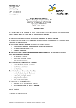
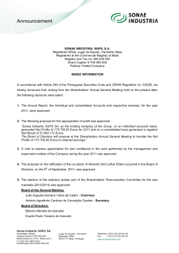
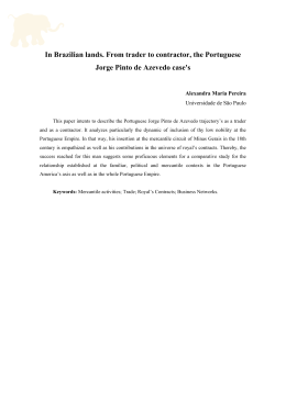
JOURNAL OF LAPAROENDOSCOPIC & ADVANCED SURGICAL TECHNIQUES
Volume &, Number &, 2009
ª Mary Ann Liebert, Inc.
DOI: 10.1089=lap.2009.0080
Full Report
Intraperitoneal Pressure and Volume of Gas Injected
as Effective Parameters of the Correct Position
of the Veress Needle During Creation of Pneumoperitoneum
João L.M.C. Azevedo, MD, PhD,1 Otavio C. Azevedo, MD, PhD,1 Albino A. Sorbello, MD, PhD,2
Otavio M. Becker, MD, PhD,1 Otavio Hypolito, MD, PhD,1 Dalmer Freire, MD, PhD,1
Susana Miyahira, MD,1 Afonso Guedes, Master, MD,1 and Glicia C. Azevedo , MD1
Abstract
Objective: The aim of this work was to establish reliable parameters of the correct position of the Veress needle
in the peritoneal cavity during creation of pneumoperitoneum.
Methods: The Veress needle was inserted into the peritoneal cavity of 100 selected patients, and a carbon-dioxide
flow rate of 1.2 L=min and a maximum pressure of 12 mm Hg were established. Intraperitoneal pressure (IP) and
the volume of gas injected (VG) were recorded at the beginning of insufflation and at every 20 seconds.
Correlations were established for pressure and volume in function of time. Values of IP and VG were predicted
at 1, 2, 3, and 4 minutes of insufflation, by applying the following formulas: IP ¼ 2.3083 þ 0.0266time þ
8.310ÿ5time2 ÿ 2.4410ÿ7time3; and VG ¼ 0.813 þ 0.0157time.
Results: A strong correlation was observed between IP and preestablished time points during creation of the
pneumoperitoneum, as well as between VG and preestablished time points during creation of the pneumoperitoneum, with a coefficient of determination of 0.8011 for IP and of 0.9604 for VG. The predicted values
were as follows: 1 minute ¼ 4.15; 2 minutes ¼ 6.27; 3 minutes ¼ 8.36; and 4 minutes ¼ 10.10 for IP (mm Hg); and
1 minute ¼ 1.12; 2 minutes ¼ 2.07; 3 minutes ¼ 3.01; and 4 minutes ¼ 3.95 for VG (L).
Conclusions: Values of IP and VG at given time points during insufflation for creation of the pneumoperitoneum, using the Veress needle, can be effective parameters to determine whether the needle is correctly
positioned in the peritoneal cavity.
Introduction
pressure level of over 20 mmHg. The position of the needle
was changed until normal pressure was reached. Approximately 2.5 liters of carbonic gas were insufflated into the patient’s abdomen for five minutes. The patient’s abdominal
wall was lifted and the first trocar was inserted. Vital signs
were normal at that moment. Four minutes later, the patient
was placed in the Trendelenburg position for the surgical
procedure. Vital signs were checked and the systolic pressure
could not be detected. No bleeding was observed upon inspection of the abdominal cavity, and resuscitation measures
were carried out after removal of the laparoscope. After several minutes, a needle was inserted into the patient’s abdomen
to check for bleeding. No blood was aspirated. The hypothesis
of an anaphylactic reaction in response to the drug used was
considered. In spite of the resuscitation measures, the patient
I
njury to the great vessels is the main cause of death
during laparoscopic procedures.1 Various reports of injury
to the great vessels caused by the Veress needle are found in the
literature.2 These cases illustrate the difficulty in correctly diagnosing this complication, which is mainly due to the retroperitoneal position of the vessels.3–11 The following report by
Peterson et al.12 is a good example to illustrate this situation:
A 38-year-old female was hospitalized to undergo laparoscopic tubal esterilization. The patient was submitted to
general anesthesia and placed supine. The abdominal wall
was lifted using surgical forceps. A small infraumbilical incision was made for the introduction of the Veress needle.
Immediately after needle insertion, the insufflator indicated a
1
Department of Surgery, Federal University of São Paulo, São Paulo, Brazil.
Department of Surgical Gastroenterology, Hospital do Servidor Publico Estadual, São Paulo, Brazil.
2
1
2
AZEVEDO ET AL.
died after two hours. Autopsy revealed a peritoneal cavity
filled with blood and extensive hemorrhage. A 3-mm injury to
the anterior and posterior walls of the abdominal aorta was
observed, just before its ramification.
The Veress needle is typically inserted through the abdominal midline, at the umbilicus. Albeit effective, insertion of the
Veress needle through the midline poses danger. All injuries
to the great vessels caused by the Veress needle reported in
the literature resulted from midline punctures in the umbilical
region.2 On the other hand, insertion of the Veress needle into
the left hypochondrium has been reported as safe and effective,13 and potential injuries are less severe.
Nevertheless, it is essential that the position of the needle
after insertion be determined as accurately as possible.
Needle-positioning tests prior to insufflation have been evaluated and considered adequate to guide surgeons with regard
to the correct positioning of the Veress needle for creation of
the pneumoperitoneum.14 These tests can avoid not only
iatrogenic injury, but also gas insufflation into the wrong
site, because surgeons intuitively know that intraperitoneal
pressure (IP) and the volume of gas injected (VG) into the
peritoneal cavity are predictable at certain time points during insufflation. In addition, high-pressure values and low
carbon-dioxide (CO2) volume in the beginning of insufflation
suggest incorrect positioning of the needle or lack of muscular
paralysis, whereas low-pressure values and increased CO2
volume over a long insufflation period suggest injury to hollow viscera or gas leakage. Pressure and volume values can,
therefore, be correlated with the position of the Veress needle
in the abdominal cavity. Studies must be carried out in order
to establish reliable, objective parameters to determine the
position of the needle in the peritoneal cavity during creation
of the pneumoperitoneum. Such studies might serve as
guidelines for laparoscopic surgeons. No research has yet
been carried out in humans, only in pigs.15 The aim of the
present study was to determine reliable predictive values for
Table 1. Descriptive Statistics of the Demographic
Data from Patients of the Sample
Parameters
Age (years)
mean (SD)
Minimum=maximum
Sex (n; %)
Female
Male
BMI (kg=m2)
Mean (SD)
Minimum=maximum
Height (m)
Mean (SD)
Minimum=maximum
Weight (kg)
Mean (SD)
Minimum=maximum
Intervention (n; %)
Cholecystectomy
Fundocardioplasty
Bilat ing herniorrhaphy
Unilat ing herniorrhaphy
FIG. 1. Schematic illustration showing Veress needle puncture in the left hypochondriac region, at the costal margin,
8 cm from the midline. Note the entry point of the needle and
the distance from the great vessels.
IP and VG at given time points during creation of the pneumoperitoneum when using a Veress needle.
Materials and Methods
This study was approved by the Research Ethics Committees of the Health Care Institute of the São Paulo Hospital for
State Civil Servants (protocol no. 045=03), and of the Federal
University of São Paulo, (under research protocol no.
1405=03). Table 1 shows the demographic data. A total of 100
patients were included in the present study. Such patients had
been scheduled to undergo laparoscopic procedures at the
Surgical Gastroenterology Service of São Paulo Hospital for
State Civil Servants. All patients were older than 18 years of
age and nonobese [body-mass index (BMI) lower than
30 kg=m2), with no history of peritonitis or abdominal surgery. A dose of 0.1 mg=kg of midazolam was administered to
patients 30 minutes before anesthesia. Anesthesia was induced
with doses of 2 mg=kg of propofol and of 0.5 mcg=kg
(n ¼ 100)
53.7 (13.1)
27–77
58
42
25.4 (2.4)
20.6–29.7
1.64 (0.09)
1.45–1.87
68.7 (9.8)
49.5–90.00
80
9
3
8
n, number of patients; SD, standard deviation; %, percentage; BMI,
body-mass index; bilat, bilateral; unilat, unilateral; ing, inguinal.
FIG. 2. Degree of dispersion of pressure in function of time
and respective regression curve. The regression model above
was statistically significant (P < 0.001), with a coefficient
of determination of 80.1% (R2 ¼ 0.8011), indicating good adjustment.
PARAMETERS OF THE POSITION OF THE VERESS NEEDLE
3
Results
FIG. 3. Degree of dispersion of volume in function of time
and respective regression line. The regression model above
was statistically significant (P < 0.001), with a coefficient of
determination of 96.04% (R2 ¼ 0.9604), indicating very good
adjustment.
A strong positive correlation between pressure and
given time points was observed during creation of the pneumoperitoneum (Fig. 2). The curve shows good adjustment,
with a coefficient of determination of 0.80 (pressure ¼
ÿ2E ÿ07time3 þ 8E ÿ 05time2 þ 0.0266time þ 2.3083). A
strong positive correlation between volume and given time
points was observed during creation of the pneumoperitoneum (Fig. 3). The curve shows good adjustment, with a
coefficient of determination of 0.96 (volume ¼ 0.0157time þ
0.1813).
Prediction of IP and VG (Table 2) yielded the following
results: 1 minute ¼ 4.15; 2 minutes ¼ 6.27; 3 minutes ¼ 8.36;
and 4 minutes ¼ 10.10 for pressure (mm Hg); and 1 minute ¼
1.12; 2 minutes ¼ 2.07; 3 minutes ¼ 3.01; and 4 minutes ¼ 3.95
for volume (L). Mean and standard deviation of measured
values were: 1 minute ¼ 4.34 1.48; 2 minutes ¼ 6.05 1.44; 3
minutes ¼ 8.44 1.47; and 4 minutes ¼ 10.43 1.48 for IP
(mm Hg); and 1 minute ¼ 1.11 0.14; 2 minutes ¼ 2.18 0.21;
3 minutes ¼ 3.13 0.27; and 4 minutes ¼ 3.87 0.40 for VG (L).
Discussion
of fentanyl. For curarization, a dose of 0.5 mg=kg of atracurium was used. Patients were submitted to general anesthesia
with orotracheal intubation and controlled mechanical ventilation. An orogastric tube was then inserted for aspiration of
the stomach contents.
After inserting a Veress needle through the left hypochondrium (Fig. 1), the following tests were performed to
determine whether the needle was in the peritoneal cavity11:
the aspiration test, the injection test, the recovery test, the
saline drop test, and the initial IP test.14 When all tests were
positive, insufflation continued. Variations in IP and VG
were recorded at every 20 seconds, until IP reached 12 mm Hg
(maximum IP). The total amount of time required for insufflation was also recorded.
The data collected were submitted to statistical analysis.
Qualitative variables were expressed as absolute and relative
frequencies. Quantitative variables were expressed as mean,
standard deviation, and minimum and maximum values. The
correlation coefficients between pressure and time and volume and time were tested, and polynomial regression models
were constructed to estimate volume and pressure in function
of time.12 The accuracy of the equations in predicting IP and
VG was given by the determination coefficient. The results
obtained by the equations were compared with the actual
values observed at 1, 2, 3, and 4 minutes after the beginning of
insufflation.
The aim of this prospective clinical trial was to establish
reliable parameters to guide surgeons through creation of the
pneumoperitoneum in selected patients, providing objective
data to determine the real position of the Veress needle. Predetermined time points during creation of the pneumoperitoneum were correlated with IP and VG. This was done in
order to establish the expected values for IP and VG (dependent variables) in function of time (independent variable).
This relation was mathematically expressed by an equation
that correlated the variables.16
The correlation coefficient is a pure number that indicates
whether correlation is perfect (¼1), strong (>0.75 and <1),
average (>0.5 and <0.75), weak (<0.5), or absent (¼0).12 In the
present study, a strong correlation was observed between IP
and time and VG and time. The coefficient of determination
indicates the accuracy of a predicted value of a variable (dependent) in the function of another variable (independent).
In the present study, equations for pressure and volume prediction devised from the regression curves (Figs. 2 and 3) were
tested, and a high determination coefficient was observed between pressure and time, as well as between volume and time.
In order to minimize the effects of surgical scars, peritoneal
adhesions, and obesity on the elasticity and complacency of
the abdominal wall (which could work as confounding factors
and influence the results), patients with a history of surgery
and peritonitis, as well as with BMI over 30, were not included
Table 2. Predicted Values of Pressure and Volume at Given Moments of Insufflation
Applying the Estimated Modelsa
Observation time in seconds
Mean
Mean
Mean
Mean
values
values
values
values
and standard deviation of measured pressure (mm Hg)
of estimated pressure (mm Hg)
and standard deviation of measured volume (L)
of estimated volume (L)
1 minute
2 minutes
3 minutes
4 minutes
4.34 (1.48)
4.15
1.11 (0.14)
1.12
6.05 (1.44)
6.27
2.18 (0.21)
2.07
8.44 (1.47)
8.36
3.13 (0.27)
3.01
10.43 (1.48)
10.10
3.87 (0.40)
3.95
a
(pressure ¼ 2.3083 þ 0.0266time þ 8.310ÿ5time2 ÿ 2.4410ÿ7time3; volume ¼ 0.813 þ 0.0157time). Mean values and standard
deviation of measured pressure and mean values of predicted pressure.
4
AZEVEDO ET AL.
in the present study. In addition, the flow rate was set at
1.2 L=min, which is known to be adequate for gradual creation
of the pneumoperitoneum (minimizing hyperreflexia of the
parasympathetic nervous system), and the expected values of
volume (0.2 L=s) were determined. The protocol for curarization was rigorously followed in order to avoid undesired
effects on IP values.
The present study allowed pressure and volume values to
be accurately predicted at key moments during insufflation in
a selected population (Table 2). Time points at 1, 2, 3, and 4
minutes were chosen to show the insufflation process from
beginning to end. The resulting algorithms might be entered
in an insufflator processor, as a safety device, helping the surgeon to identify the incorrect positioning of the Veress needle
during insufflation or even an inappropriate degree of abdominal muscle paralysis. Further studies, involving a random population and investigating the effects of BMI, sex, and
a history of abdominal surgery on pressure and volume curves,
are being undertaken.
6.
7.
8.
9.
10.
11.
12.
Conclusions
Values for IP and VG at certain time points during insufflation for creation of the pneumoperitoneum, using the
Veress needle in a selected population, are effective parameters to determine whether the needle is correctly positioned in
the peritoneal cavity.
13.
14.
Disclosure Statement
No competing financial interests exist.
15.
References
1. Rajesh V, Gupta JK. Laparoscopic entry techniques: Clinical
guideline, national survey, and medicolegal ramifications.
Surg Endosc 2008;22:2686–2697.
2. Azevedo JLMC, Azevedo OC, Miyahira SA, Miguel GPS,
Becker JOM, Hipólito OHM, Machado ACCG, Cardia W,
Aguiar GS, Godinho LS, Freire DF, Moreira CH, Almeida
CES. Injuries caused by Veress needle insertion for creation
of pneumoperitoneum: A systematic literature review. Surg
Endosc 2009;23:in press.
3. Baadsgaard SE, Bille S, Egeblad K. Major vascular injury
during gynecologic laparoscopy. Report of a case and review of
pubished cases. Acta Obstet Gynecol Scand 1989;68:283–285.
4. Bergqvist D, Bergqvist A. Vascular injuries during gynecologic surgery. Acta Obstet Gynecol Scand 1987;66:19–23.
5. Bonjer HJ, Hazebroek EJ, Kazemier G, Giuffrida MC, Meijer
WS, Lange JF. Open versus closed establishment of pneu-
16.
moperitoneum in laparoscopic surgery. Br J Surg 1997:84:
599–602.
Catarci M, Carlini M, Gentileschi P, Santoro E. Major and
minor injuries during the creation of pneumoperitoneum. A
multicenter study on 12,919 cases. Surg Endosc 2001;15:
566–569.
Chapron CM, Pierre F, Lacroix S, Querleu D, Lansac J, Dubuiosson JB. Major vascular injuries during gynecologic
laparoscopy. J Am Coll Surg 1997;185:461–465.
Fruhwirth J, Koch G, Mischinger HJ, Werkgartner G, Tesch
NP. Vascular complications in minimally invasive surgery.
Surg Laparosc Endosc 1997;7:251–254.
Hanney RM, Alle KM, Cregan PC. Major vascular injury and
laparoscopy. Aust N Z Surg 1995;65:533–535.
Roviaro GC, Varoli F, Saguatti L, Vergani C, Maciocco M,
Scarduelli A. Major vascular injuries in laparoscopic surgery.
Surg Endosc 2002;16:1192–1196.
Schafer M, Lauper M, Krahenbuhl L. Trocar and Veress
needle injuries during laparoscopy. Surg Endosc. 2001;15:
275–280.
Peterson HB, Greenspan JR, Ory HW. Death following
puncture of the aorta during laparoscopic sterilization. Obstet Gynecol 1982;59:133–134.
Azevedo OC, Azevedo JLMC, Sorbello AA, Miguel GPS,
Guindalini RSC, Godoy AC. Veress needle insertion in the
left hypochondrium in creation of the pneumoperitoneum.
Acta Cir Bras 2006;21:296–303.
Azevedo OC, Azevedo JLMC, Sorbello AA, Miguel GPS,
Wilson JL, Jr, Godoy AC. Evaluation of tests performed to
confirm the position of the Veress needle for creation of
pneumoperitoneum in selected patients: A prospective,
clinical trial. Acta Cir Bras 2006;21:385–391.
Azevedo JLMC, Guindalini RSC, Sorbello AA, Silva CEP,
Azevedo OC, Aguiar GS, Menezes FJC, Delorenzo A, Pasqualin RC, Kozu FO. Evaluation of the positioning of the tip
of the Veress needle during creation of closed pneumoperitoneum in pigs. Acta Cir Bras 2006;21:26–30.
Guyatt G, Walter S, Shannon H, Cook D, Jaeschke R, Heddle
N. Basic statistics for clinicians: 4. Correlation and regression. CMAJ 1995;152:497–504.
Address correspondence to:
João L.M.C. Azevedo, MD, PhD
Department of Surgery
Federal University of São Paulo
R. Joaquim Tavora, 550, Apt. 101-A
São Paulo 04015011
Brazil
E-mail: [email protected]
Baixar