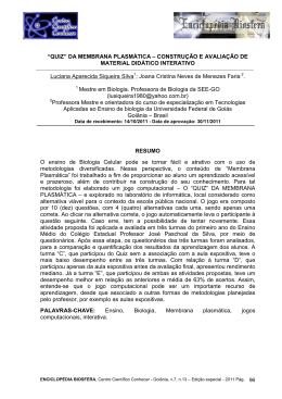
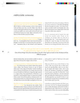
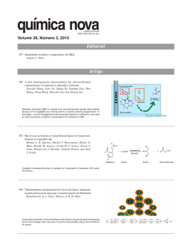
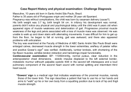
7FSTÎPJNQSFTTB WPMO4FUFNCSP0VUVCSP 3#0 %&4%& 253 ISSN 0034-7280 Revista Brasileira de (Versão impressa) ISSN 1982-8551 (Versão eletrônica) Oftalmologia PUBLICAÇÃO OFICIAL: SOCIEDADE BRASILEIRA DE OFTALMOLOGIA SOCIEDADE BRASILEIRA DE CATARATA E IMPLANTES INTRAOCULARES SOCIEDADE BRASILEIRA DE CIRURGIA REFRATIVA Sociedade Brasileira de Oftalmologia Indexada nas bases de dados: LILACS Literatura Latinoamericana em Ciências da Saúde SciELO Scientific Electronic Library OnLine WEB OF SCIENCE www.freemedicaljournals.com Disponível eletronicamente: www.sboportal.org.br Publicação bimestral Editor Chefe Newton Kara-Junior (SP) Editor Executivo Arlindo José Freire Portes (RJ) Co-editores André Luiz Land Curi (RJ) Arlindo José Freire Portes (RJ) Bruno Machado Fontes (RJ) Carlos Eduardo Leite Arieta (SP) Hamilton Moreira (PR) Liana Maria Vieira de Oliveira Ventura (PE) Marcony Rodrigues de Santhiago (RJ) Mario Martins dos Santos Motta (RJ) Maurício Maia (SP) Niro Kasahara (SP) Renato Ambrósio Jr. (RJ) Rodrigo Jorge (SP) Rodrigo Pessoa Cavalcanti Lira (PE) Silvana Artioli Schellini (SP) Walton Nosé (SP) Corpo Editorial Internacional Baruch D. Kuppermann - Califórnia - EUA Christopher Rapuano - Phyladelphia - EUA Curt Hartleben Martkin - Colina Roma - México Daniel Grigera - Olivos - Argentina Deepinder Kauer Dhaliwal - Pittsburg - EUA Felipe A. A. Medeiros - Califórnia - EUA Felix Gil Carrasco - México – México Fernando Arevalo - Riyadh - Arábia Saudita Francisco Rodríguez Alvira – Bogotá - Colombia Howard Fine - Eugene - EUA Jean Jacques De Laey - Ghent - Bélgica Kevin M. Miller - Califórnia - EUA Lawrence P. Chong - Califórnia - EUA Lihteh Wu – San José - Costa Rica Liliana Werner - Utah - EUA Miguel Burnier Jr. - Montreal - Canadá Pablo Cibils - Assunção - Paraguai Patricia Mitiko Santello Akaishi – Arábia Saudita Peter Laibson - Phyladelphia - EUA Steve Arshinoff - Toronto - Canadá Corpo Editorial Nacional A. Duarte - Rio de Janeiro - RJ Abelardo de Souza Couto - Rio de Janeiro- RJ Abrahão da Rocha Lucena - Fortaleza - CE Alexandre Augusto Cabral de Mello Ventura - Recife - PE Alexandre H. Principe de Oliveira – Salvador – BA Alexandre Seminoti Marcon – Porto Alegre - RS Ana Carolina Cabreira Vieira – Rio de Janeiro – RJ Ana Luisa Hofling de Lima - São Paulo - SP André Correa de Oliveira Romano – Americana - SP André Curi - Rio de Janeiro - RJ André Luis Freire Portes - Rio de Janeiro - RJ André Marcio Vieira Messias – Ribeirão Preto – SP Andrea Kara José Senra - São Paulo – SP Antonio Marcelo Barbante Casella - Londrina - PR Armando Stefano Crema- Rio de Janeiro- RJ Beatriz de Abreu Fiuza Gomes – Rio de Janeiro - RJ Bruna Vieira Ventura - Recife - PE Coordenação de Aperfeiçoamento de Pessoal de Nível Superior http://www.capes.gov.br Rev Bras Oftalmol, v. 73, n. 5, p. 253 - 316, Set./Out. 2014 Bruno Diniz – Goiânia - GO Carlos Augusto Moreira Jr.- Curitiba- PR Carlos Gabriel Figueiredo - São José do Rio Preto - SP Carlos Ramos de Souza Dias- São Paulo- SP Claudio do Carmo Chaves - Manaus - AM Cristiano Caixeta Umbelino - São Paulo - SP Daniel Lavinsky – Porto Alegre - RS David Leonardo Cruvinel Isaac – Goiania - GO Diego Tebaldi Q. Barbosa - São Paulo - SP Edmundo Frota De Almeida Sobrinho- Belém- PA Eduardo Buchele Rodrigues – Florianópolis - SC Eduardo Cunha de Souza – São Paulo - SP Eduardo Damasceno - Rio de Janeiro - RJ Eduardo Dib – Rio de Janeiro - RJ Eduardo Ferrari Marback- Salvador- BA Eliezer Benchimol - Rio de Janeiro - RJ Enzo Augusto Medeiros Fulco – Jundiaí - SP Eugenio Santana de Figueiredo – Juazeiro do Norte - CE Fábio Marquez Vaz – Ondina – BA Felipe Almeida - Ribeirão Preto - SP Fernando Cançado Trindade - Belo Horizonte- MG Fernando Marcondes Penha - Florianópolis - SC Fernando Oréfice- Belo Horizonte- MG Fernando Roberte Zanetti – Vitória - ES Flavio Rezende- Rio de Janeiro- RJ Francisco de Assis Cordeiro Barbosa - Recife - PE Frederico Valadares de Souza Pena – Rio de Janeiro - RJ Frederico Guerra - Niterói - RJ Giovanni N.U.I.Colombini- Rio de Janeiro- RJ Guilherme Herzog Neto- Rio de Janeiro- RJ Harley Biccas - Ribeirão Preto - SP Haroldo Vieira de Moraes Jr.- Rio de Janeiro- RJ Hélcio Bessa - Rio de Janeiro - RJ Helena Parente Solari - Niterói - RJ Heloisa Helena Abil Russ – Curitiba – PR Henderson Celestino de Almeida- Belo Horizonte- MG Hilton Arcoverde G. de Medeiros- Brasilia- DF Homero Gusmao de Almeida- Belo Horizonte- MG Italo Mundialino Marcon- Porto Alegre- RS Iuuki Takasaka – Santa Isabel - SP Ivan Maynart Tavares - São Paulo - SP Jaco Lavinsky - Porto Alegre - RS Jair Giampani Junior – Cuiabá - MT Jeffersons Augusto Santana Ribeiro - Ribeirão Preto - SP João Borges Fortes Filho- Porto Alegre- RS João Luiz Lobo Ferreira – Florianópolis – SC João Marcelo de Almeida G. Lyra - Maceió - AL João Orlando Ribeiro Goncalves- Teresina- PI Jorge Carlos Pessoa Rocha – Salvador – BA JorgeAlberto de Oliveira - Rio de Janeiro - RJ José Augusto Cardillo – Araraquara – SP José Beniz Neto - Goiania - GO José Ricardo Carvalho L. Rehder- São Paulo- SP Laurentino Biccas Neto- Vitória- ES Leonardo Akaishi - Brasília - DF Leonardo Provetti Cunha - SP Leticia Paccola - Ribeirão Preto - SP Liana Maria V. de O. Ventura- Recife- PE Luiz Alberto Molina - Rio de Janeiro - RJ Manuel Augusto Pereira Vilela- Porto Alegre- RS Marcelo Hatanaka – São Paulo – SP Marcelo Netto - São Paulo - SP Marcelo Palis Ventura- Niterói- RJ Marcio Bittar Nehemy - Belo Horizonte - MG Marco Antonio Bonini Filho - Campo Grande - MS Marco Antonio Guarino Tanure - Belo Horizonte - MG Marco Antonio Rey de Faria- Natal- RN Marcos Pereira de Ávila - Goiania - GO Maria de Lourdes Veronese Rodrigues- Ribeirão Preto- SP Maria Rosa Bet de Moraes Silva- Botucatu- SP Maria Vitória Moura Brasil - Rio de Janeiro - RJ Mário Genilhu Bomfim Pereira - Rio de Janeiro - RJ Mario Luiz Ribeiro Monteiro - São Paulo- SP Mário Martins dos Santos Motta- Rio de Janeiro- RJ Marlon Moraes Ibrahim – Franca - SP Mauricio Abujamra Nascimento – Campinas - SP Maurício Bastos Pereira - Rio de Janeiro - RJ Maurício Dela Paolera - São Paulo - SP Miguel Ângelo Padilha Velasco- Rio de Janeiro- RJ Miguel Hage Amaro - Belém - PA Milton Ruiz Alves- São Paulo- SP Moyses Eduardo Zadjdenweber - Rio de Janeiro - RJ Nassim da Silveira Calixto- Belo Horizonte- MG Nelson Alexandre Sabrosa - Rio de Janeiro – RJ Newton Kara-José - São Paulo - SP Newton Leitão de Andrade – Fortaleza – CE Núbia Vanessa dos Anjos Lima Henrique de Faria - Brasília-DF Octaviano Magalhães Júnior - Atibaia - SP Oswaldo Moura Brasil- Rio de Janeiro- RJ Otacílio de Oliveira Maia Júnior – Salvador - BA Patrick Frensel de Moraes Tzelikis – Brasília – DF Paulo Augusto de Arruda Mello Filho – São Paulo – SP Paulo Augusto de Arruda Mello- São Paulo- SP Paulo Schor - São Paulo - SP Pedro Carlos Carricondo – São Paulo – SP Pedro Duraes Serracarbassa – São Paulo – SP Priscilla de Almeida Jorge – Recife – PE Rafael Ernane Almeida Andrade - Itabuna – BA Raul N. G. Vianna - Niterói - RJ Remo Susanna Jr.- São Paulo- SP Renata Rezende - Rio de Janeiro - RJ Renato Ambrosio Jr.- Rio de Janeiro- RJ Renato Luiz Nahoum Curi- Niterói- RJ Richard Yudi Hida – São Paulo – SP Riuitiro Yamane - Niterói - RJ Roberto Lorens Marback - Salvador - BA Roberto Pinto Coelho – Ribeirão Preto – SP Rodrigo França de Espíndola – São Paulo – SP Rogerio Alves Costa- Araraquara- SP Rogerio de Almeida Torres - Curitiba - PR Rubens Belfort Neto – São Paulo – SP Rubens Camargo Siqueira- São José do Rio Preto- SP Sebastião Cronemberger So.- Belo Horizonte- MG Sérgio Henrique S. Meirelles- Rio de Janeiro- RJ Sérgio Kwitko - Porto Alegre - RS Sérgio Luis Gianotti Pimentel – São Paulo – SP Silvana Artioli Schellini - Botucatu- SP Suel Abujamra- São Paulo - SP Suzana Matayoshi - São Paulo - SP Tânia Mara Cunha Schaefer – Curitiba – PR Vitor Cerqueira - Rio de Janeiro - RJ Walter Yukihiko Takahashi – São Paulo – SP Walton Nose- São Paulo- SP Wener Passarinho Cella - Plano Piloto - DF Wesley Ribeiro Campos- Passos- MG Yoshifumi Yamane- Rio de Janeiro- RJ Redação: Rua São Salvador, 107 Laranjeiras CEP 22231-170 Rio de Janeiro - RJ Tel: (0xx21) 3235-9220 Fax: (0xx21) 2205-2240 Tiragem: 5.000 exemplares Edição:Bimestral Secretaria: Marcelo Diniz Editoração Eletrônica: Sociedade Brasileira de Oftalmologia Responsável: Marco Antonio Pinto DG 25341RJ Publicidade: Sociedade Brasileira de Oftalmologia Responsável: João Diniz [email protected] Contato publicitário: Westinghouse Carvalho Tel: (11)3726-6941 / 99274-0724 [email protected] Revisão: Eliana de Souza FENAJ-RP 15638/71/05 Normalização: Edna Terezinha Rother Assinatura Anual: R$420,00 ou US$280,00 Impressão: Gráfica Stamppa Associada a ABEC - Associação Brasileira de Editores Científicos ARTIGO ORIGINAL 279 Novel spatula and dissector for safer deep anterior lamellar keratoplasty Uso de espátula e dissector para otimização da ceratoplastia lamelar anterior profunda (DALK) Gustavo Bonfadini1,2,3, Eun Chul Kim1,4, Mauro Campos3, Albert S. Jun1 ABSTRACT Objective: We describe a novel spatula and dissector to facilitate the big-bubble technique in deep anterior lamellar keratoplasty (DALK). Methods: A 29-year-old man who was diagnosed with bilateral keratoconus underwent deep anterior lamellar keratoplasty (DALK). After 350µm partial thickness incision of the recipient cornea, the Bonfadini dissector was inserted at the deepest point in the peripheral incision and could be advanced to the center of the cornea safely because of its “semi-sharp” tip. After achieving the big-bubble (BB) separation of Descemet membrane (DM) from the overlying stroma, the anterior stromal disc was removed. Viscoelastic material was placed on the stromal bed to prevent uncontrolled collapse and perforation of DM during the paracentesis blade incision into the BB. We could detect the safe opening of the BB using the Bonfadini dissector by the leakage of air bubbles into the viscoelastic material. After injecting viscoelastic material into the BB space, we inserted the Bonfadini spatula into the big-bubble safely because of its curved profile and blunt edges. The groove along the length of the Bonfadini spatula enables safe and efficient incision or the residual stromal tissue using the pointed end of a sharp blade while protecting the underlying DM. After removal of posterior stroma, the donor button was sutured with 16 interrupted 10-0 nylon sutures. Results: This technique and the use of the Bonfadini spatula and dissector facilitate exposure of Descemet membrane. Conclusion: The smooth Bonfadini DALK spatula and dissector facilitate safe and efficient completion of DALK surgery. Keywords: Deep anterior lamellar keratoplasty; Corneal transplantation/methods; Keratoplasty; Bonfadini dissector; Bonfadini spatula R ESUMO Objetivo: Descrevemos o uso de novos instrumentais cirúrgicos para facilitar a técnica de “big-bubble” na ceratoplastia lamelar anterior profunda (DALK). Métodos: Paciente masculino, 29 anos, foi diagnosticado com ceratocone bilateral e submetido à ceratoplastia lamelar anterior profunda (DALK). Após incisão da córnea receptora numa profundidade de 350µm de espessura parcial, o dissector Bonfadini foi inserido no ponto mais profundo da incisão periférica e pode avançar para o centro da córnea com segurança devido à sua ponta semiafiada. Depois de realizar a “big-bubble” (BB) e atingir a separação da Membrana de Descemet (MD) do estroma sobrejacente, o disco corneano de estroma anterior foi removido. Um viscoelástico foi colocado sobre o leito do estroma remanescente para impedir o colapso não-controlado e perfuração da MD durante a incisão na BB com lâmina de paracentese. Verificamos segurança no rompimento do estroma remanescente com o auxílio do dissector Bonfadini, para liberação da bolha de ar da BB através do viscoelástico. Depois de injetar o viscoelástico no espaço da BB, inserimos a espátula Bonfadini neste espaço, o que demonstrou-se seguro devido ao formato curvo e das bordas arredondadas do instrumental. A chanfradura ao longo do comprimento da espátula Bonfadini permite a incisão pela ponta de uma lâmina afiada, protegendo assim a MD subjacente. Após a remoção do estroma posterior, o botão doador foi suturado com 16 pontos interrompidos de fio nylon 10.0. Resultados: Esta técnica e o uso da espátula Bonfadini e dissector facilitam a exposição de membrana de Descemet. Conclusão: A superfície lisa da espátula Bonfadini e dissector, facilitam a realização segura e eficiente da ceratoplastia lamelar anterior profunda (DALK). Descritores: Ceratoplastia lamelar anterior profunda; Transplante de córnea/métodos; Ceratoplastia; Dissector Bonfadini; Espátula Bonfadini Cornea & Anterior Segment Service, Wilmer Eye Institute, Johns Hopkins School of Medicine, Baltimore, Maryland, USA; Rio de Janeiro Eye Bank, Rio de Janeiro, RJ, Brazil; 3 Escola Paulista de Medicina, Universidade Federal de São Paulo, São Paulo, SP, Brazil; 4 College of Medicine, Catholic University of Korea, Seoul, Korea. 1 2 The authors have no public and private financial support, or financial interest Recebido para publicação em 25/02/2014 - Aceito para publicação em 26/04/2014 Rev Bras Oftalmol. 2014; 73 (5): 279-81 280 Bonfadini G, Kim EC, Campos M, Jun AS D INTRODUCTION eep anterior lamellar keratoplasty (DALK) has been proposed as an alternative to penetrating keratoplasty (PK) for the treatment of various corneal diseases not affecting the endothelium. DALK surgery removes the anterior layers of the cornea, cleaving the deep stroma from Descemet membrane (DM). The advantages of the DALK technique for corneal stromal diseases include absence of endothelial rejection, avoidance of potential open-sky intra-operative complications of PK, faster visual rehabilitation due to earlier suture removal, and a predicted longer graft survival because of the lower rate of endothelial cell loss (1). DALK is a time-consuming and technically demanding procedure. The most frequent intraoperative complication is perforation of DM while attempting to separate it from the overlying stroma during creation of the big-bubble (BB) (2). Sarnicola et al.(3) reported that a smooth spatula and cannula can facilitate a high percentage of successful DALK and make the procedure more reliable than compared to air injection with a needle. In this report, we describe a novel Bonfadini dissector and spatula for safely manipulating the big-bubble and removing posterior stroma. Surgical technique A 29-year-old man diagnosed with bilateral keratoconus 6 years ago, visited our service complaining of ocular pain and decreased vision in the right eye. He had worn rigid gas permeable (RGP) contact lenses for 16 years. His best spectacle corrected visual acuity OD was 20/80 and OS was 20/25. Slitlamp examination revealed marked corneal stromal scarring and epithelial punctuate erosions on the right central cornea. Hence, deep anterior lamellar keratoplasty (DALK) was completed uneventfully in the right eye. The operation was performed under sub-Tenon anesthesia by A.S.J; The technique described by Anwar et al.(4) was followed with the described modifications. A surgical marking pen was used to mark the center of the host cornea. An 8.5mm diameter trephine was used to lightly score the epithelium of the host cornea to outline the recipient bed. An astigmatic keratotomy blade was used to incise the recipient cornea to a depth of 350µm along the 8.5mm trephine mark. Through a small peripheral clear cornea paracentesis, the anterior chamber (AC) was filled with air, and approximately physiologic intraocular pressure confirmed. The Bonfadini dissector (Katena Products, USA; Figure 1A) was inserted at the deepest point in the peripheral groove and was advanced toward the center of the cornea. Once the Bonfadini dissector was approximately 1-2mm from the apex of the cone (Figure 1B), it was removed and the Fogla 27 gauge air injection cannula (Bausch & Lomb Storz Ophthalmic, USA), attached to a 5mL syringe filled with air was introduced into the corneal tunnel. Air was then injected into the stroma to achieve the formation of a big-bubble (figure 1C). The central anterior stromal disc was removed with an angled crescent knife, and then air was evacuated through the previously placed paracentesis. To enter into the big-bubble, we used the Ophthalmic Viscosurgical Device–Assisted Incision technique (5). A cohesive viscoelastic (Healon, Abbott Medical Optics) was placed on the stromal bed and a 1.0mm incision was then created with a paracentesis blade using only the tip of the blade with a “lifting” motion to prevent rapid collapse of the bubble which could result in Descemet membrane perforation. Entry into the big-bubble was confirmed by the appearance of a small bubble within the overlying viscoelastic material, which also served to prevent rapid egress of air from the big-bubble and rapid collapse leading to a higher probability of perforating the Descemet membrane (figure 1D). Entry into the big-bubble was confirmed by the appearance Rev Bras Oftalmol. 2014; 73 (5): 279-81 of a small bubble within the overlying viscoelastic material, which also served to prevent rapid egress of air from the big-bubble and rapid collapse leading to a higher probability of perforating the Descemet membrane (figure 1D). Viscoelastic material was injected into the pre-Descemet space to expand the potential space and separate the Descemet membrane from the overlying stromal tissue. The Bonfadini spatula (Katena Products, USA; figure 2A) was introduced into the pre-Descemet space. This instrument has blunt edges and a curved profile to minimize inadvertent damage to Descemet membrane. The groove along the length of the Bonfadini spatula serves as a guide to allow for rapid incision of the posterior stromal tissue using the sharp point of a paracentesis blade (figure 2B). The spatula serves to protect the DM from Figure 1: The Bonfadini dissector has a fine, rounded tip to enable stromal dissection while preventing inadvertent perforation of Descemet membrane (A); use of the Bonfadini dissector to create a deep tunnel toward the center of the cornea from a peripheral partial thickness groove incision (B); big-bubble formation by deep, intrastromal air injection with accompanying stromal opacification (C); opening of the big-bubble with a sharp blade was detected by air leaking into viscoelastic material placed on the posterior stromal bed (D) inadvertent perforation while incising the posterior stromal tissue. Once sufficient radial incisions in the posterior stroma were completed, we removed stroma with cornealscleral scissors along the peripheral partial thickness groove incision. The full-thickness donor graft was punched at 8.75mm diameter from the endothelial side, and the endothelium was stripped from the posterior surface using surgical spears. The donor button was then sutured into position with 16 interrupted 10-0 nylon sutures (figure 2C). The patient achieved uncorrected visual acuity of 20/125 and 20/60 with pinhole on day 1 postoperatively with a wellattached graft. At 3 months after DALK, his uncorrected visual acuity was 20/60 and best spectacle corrected visual acuity (1.25 + 1.5 x 30 degrees) was 20/25 OD. DISCUSSION Corneal transplantation is singular because it is habitually performed on persons with visual deficiency but with preserved life expectancy, mobility and social life. A graft not well succeeded may cause real blindness and permanent misery due to pain and discomfort (6). To obtain a satisfactory surgical result, there is a need of an appropriate patient selection and guidance about their eye problem, the proposed surgery, care and risk per and post operative as well as the visual rehabilitation perspective (7). 281 Novel spatula and dissector for safer deep anterior lamellar keratoplasty Figure 2: The groove along the Bonfadini spatula serves as a guide to protect Descemet membrane (DM) from inadvertent perforation when incising the posterior stroma with a blade (A); creation of wedgeshaped incisions of posterior stroma, using a blade guided along the groove of the Bonfadini spatula. The spatula serves to protect inadvertent perforation of DM (B); donor button was sutured into position with 16 interrupted 10-0 nylon sutures (C) Keratoconus is one of the main indications of keratoplasty in Brazil (8), and DALK is more cost-effective than penetrating keratoplasty (PK) (9). Prevention of immune-mediated graft rejection can be achieved through meticulous surgical technique such as lamellar surgery (10). DALK aims to remove and replace total or near-total corneal stroma while preserving host healthy endothelium. The advantages of DALK include reducing the risk of endothelial graft rejection, preservation of host endothelium with minimal surgical trauma, efficient visual rehabilitation relative to penetrating keratoplasty (PK), and also fewer intraoperative and postoperative complications including expulsive hemorrhage, anterior synechia, postoperative endophthalmitis, and glaucoma in comparison to PK. This procedure also requires less rigid criteria for donor corneal tissue selection that is often weighted toward donor endothelium in PK (11). Major disadvantages of anterior lamellar keratoplasty as compared to penetrating keratoplasty are the irregularity and sub-optimal optics of the corneal stromal bed which occur following manual lamellar dissection techniques (12). These issues are avoided in DALK. However, the challenge with DALK continues to be the learning curve for novice surgeons when trying to expose DM (descemetic DALK [dDALK]) versus dissection in a pre-Descemetic stromal plane (pre-Descemetic DALK [pdDALK]) (3). The dDALK procedure allows faster visual recovery than pdDALK(13). Thus, the goal of DALK is to expose the DM without damage and to achieve dDALK. Lamellar dissection of the stroma can be performed by a manual technique using a variety of instruments, including lamellar knives and dissectors (14). Using the Bonfadini dissector, we could create a deep tunnel near the corneal center prior to air injection. The Bonfadini dissector has a fine, rounded tip, which can dissect stromal tissue relatively easily while reducing the likelihood of penetrating Descemet membrane. Accessing the deep stroma for air injection may improve the success of achieving the big-bubble. The most serious complication during big-bubble DALK surgery is intraoperative perforation of Descemet membrane, which may require subsequent conversion to full-thickness penetrating keratoplasty (15). Perforation of Descemet membrane is more likely to occur as a result of direct needle trauma during initial air injection and dissection of the corneal stroma (16). As well, perforation of DM can occur while opening the big-bubble and dissecting remaining stroma over Descemet membrane. In the technique described here, viscoelastic material is placed on the stromal bed before opening the big-bubble (5). We can detect entry into the big-bubble (BB) by air leaking into the viscoelastic material. This sign allows a very controlled entry into the BB as it is readily and immediately visible and prevents rapid egress of air and collapse of the BB. Once the BB is accessed and further expanded by injection of viscoelastic into the BB, the design of the Bonfadini spatula allows it to be manipulated within the BB space with minimal chance of inadvertent trauma to DM. The groove along the length of the Bonfadini spatula also serves as a convenient guide to facilitate rapid incision of the posterior stroma into wedges, which can be excised at the periphery using standard corneal scissors. Thus, we present the novel Bonfadini dissector and spatula as aids for the successful completion of DALK surgery. In summary, the benefits of our proposed technique could be shown more conclusively in a case–control or prospective study with a larger number of patients to validate our findings. REFERENCES 1. 2. 3. 4. 5. 6. 7. 8. 9. 10. 11. 12. 13. 14. 15. 16. Shimmura S, Tsubota K. Deep anterior lamellar keratoplasty. Curr Opin Ophthalmol. 2006;17(4):349-55. Review. Michieletto P, Balestrazzi A, Balestrazzi A, Mazzotta C, Occhipinti I, Rossi T. Factors predicting unsuccessful big bubble deep lamellar anterior keratoplasty. Ophthalmologica. 2006;220(6):379-82. Sarnicola V, Toro P. Blunt cannula for descemetic deep anterior lamellar keratoplasty. Cornea. 2011;30(8):895-8. Anwar M, Teichmann KD. Big-bubble technique to bare Descemet’s membrane in anterior lamellar keratoplasty. J Cataract Refract Surg. 2002;28(3):398-403. Goshe J, Terry MA, Shamie N, Li J. Ophthalmic viscosurgical deviceassisted incision modification for the big-bubble technique in deep anterior lamellar keratoplasty. J Cataract Refract Surg. 2011;37(11):1923-7. Marcomini LA, Sobral RM, Seixas GO, Sousa SJ. [Corneal selection for transplants]. Rev Bras Oftalmol. 2011;70(6):430-6. Portuguese. Kara-Junior N, Mourad PC, Espíndola RF, AbilRuss HH. [Expectation and knowledge among patients with keratoplasty indication]. Rev Bras Oftalmol. 2011;70(4):230-4. Portuguese. Zeschau A, Balestrin IG, Stock RA, Bonamigo EL. [Indications of keratoplasty: a retrospective study in a University Hospital]. Rev Bras Oftalmol. 2013;72(5):316-20. Portuguese. Koo TS, Finkelstein E, Tan D, Mehta JS. Incremental cost-utility analysis of deep anterior lamellar keratoplasty compared with penetrating keratoplasty for the treatment of keratoconus.Am J Ophthalmol. 2011;152(1):40-47.e2. Costa DC, Kara-Joseì N. [Corneal transplant rejection]. Rev Bras Oftalmol. 2008; 67(5):255-63. Portuguese. Espandar L, Carlson AN. Lamellar Keratoplasty: A Literature Review. J Ophthalmol. 2013;2013:894319. Review. Parthasarathy A, Por YM, Tan DT. Use of a “small-bubble technique” to increase the success of Anwar’s “big-bubble technique” for deep lamellar keratoplasty with complete baring of Descemet’s membrane. Br J Ophthalmol. 2007;91(10):1369-73. Sarnicola V, Toro P, Gentile D, Hannush SB. Descemetic DALK and predescemetic DALK: outcomes in 236 cases of keratoconus. Cornea. 2010;29(1):53-9. Bonfadini G, Moreira H, Jun AS, Campos M, Kim EC, Arana E, et al. Modified femtosecond laser-assisted sutureless anterior lamellar keratoplasty. Cornea. 2013;32(4):533-7. Jhanji V, Sharma N, Vajpayee RB. Intraoperative perforation of Descemet’s membrane during “big bubble” deep anterior lamellar keratoplasty. Int Ophthalmol. 2010;30(3):291-5. Leccisotti A. Descemet’s membrane perforation during deep anterior lamellar keratoplasty: prognosis. J Cataract Refract Surg. 2007;33(5):825-9. Address reprint requests to: Albert S. Jun Cornea and External Disease Service Wilmer Smith Building 5011, The Johns Hopkins Medical Institutions 400 N. Wolfe Street, Baltimore, MD 21231 Rev Bras Oftalmol. 2014; 73 (5): 279-81
Baixar