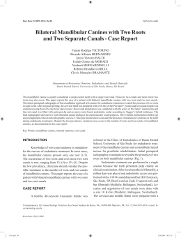
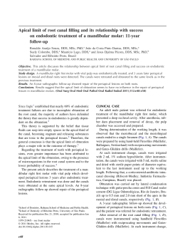
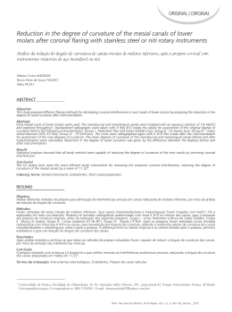
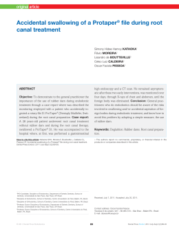
Aust Endod J 2007
C A S E R E P O RT
Mandibular molar with five root canals
Fernando Branco Barletta, PhD1,2; Sidney Ricardo Dotto, MSc1; Magda de Sousa Reis, MSc1;
Ronise Ferreira, PhD1; and Rosana Maria Coelho Travassos, PhD3
1 Universidade de Santa Cruz do Sul, Departamento de Enfermagern e Odontologia, Santa Cruz do Sul, RS, Brazil
2 Universidade Luterana do Brazil, Departamento de Odontologia Conservadora, Canoas, RS, Brazil
3 Faculdade de Odontologia de Pernambuco, Universidade de Pernambuco, Recife, PE, Brazil
Keywords
endodontically treated tooth, molar, pulp
chamber, radiography, root canals.
Correspondence
Dr Magda de Sousa Reis, Rua Egon Francisco
Knak, 129, Higienópolis, CEP 96825-320 –
Santa Cruz do Sul, RS - Brazil. Email:
[email protected]
doi:10.1111/j.1747-4477.2007.00089.x
Abstract
The purpose of this study was to demonstrate the importance of knowledge of
the internal anatomy of root canals for the success of endodontic treatment.
Lack of knowledge of anatomic variations and their characteristics in different
teeth has been pointed out as one of the main causes of endodontic therapy
failure. In this report, the authors describe the endodontic treatment of a
mandibular first molar with five root canals, evaluate the rate of occurrence of
this number of canals, and discuss the importance of their identification and
treatment.
Introduction
Knowledge of internal dental anatomy is fundamental to
the success of endodontic treatment.
Incomplete instrumentation, inadequate cleaning and
shaping, and the subsequent defective obturation of root
canals are the main causes of endodontic treatment
failure (1). Anatomical characteristics of the different
types of teeth and their possible variations are challenges
routinely faced by practitioners performing endodontic
treatment.
The correct access into the pulp chamber, which should
allow access to the orifices of the root canals and an
optimal view of the chamber floor, is a fundamental step
in endodontic therapy as it enables the identification of
any variation in the number and position of root canals
(2). Several studies have evaluated the degree of variation in the number of roots and root canals in mandibular
first molars.
Hess (3) conducted a study with 512 mandibular first
molars and reported that 0.3% of the teeth had only one,
17.7% had two, 78% had three, and 4% had four canals.
De Deus (4) studied 75 mandibular molars and found that
8% had two, 56% had three, and 36% had four canals.
According to Martinez-Berna and Badanelli (5), several in
vitro and in vivo studies have investigated the anatomical
configuration and the number of root canals of mandibular molars. The most important studies were those conducted by Hess (3), Okumura (6), Skidmore and Bjorndal
© 2007 The Authors
Journal compilation © 2007 Australian Society of Endodontology
(7), Pineda and Kuttler (8), and Hartwell and Bellizzi (9).
However, their studies did not report on any case of
mandibular molars with five canals.
Fabra-Campos (10) studied 145 mandibular first
molars and found that 2.75% of the teeth had five canals.
Martinez-Berna and Badanelli (5) conducted a clinical
investigation and found 29 teeth with five root canals in
a sample of 2362 mandibular permanent molars. Jacobsen et al. (11) found a substantial rate of occurrence of a
third mesial canal in mandibular first molars, and
reported that 12 out of the 100 molars studied had a third
mesial canal.
According to Ingle (12), one of the most important
causes of endodontic treatment failure is the incomplete
obturation of the root canal system. Therefore, the
correct location, instrumentation and obturation of all
canals are indispensable procedures. Similarly, Vertucci
(13) and De Grood and Cunningham (14) reported that
a considerable number of failures could be assigned to
anatomical variations, such as the presence of canals not
usually found.
This clinical case describes a mandibular first molar
with five canals, three distal and two mesial. The third
canal of the distal root is called mid-distal or simply
third distal canal. The authors review related literature,
describe the clinical case and make considerations about
how dentists should perform the examination of the
pulp chamber to achieve successful results in similar
cases.
1
Mandibular Molar with Five Root Canals
F. B. Barletta et al.
Case report
A 28-year-old patient presented with a complaint of
diffuse pain in the right mandibular first molar. Pulp
testing and percussion tests in the region revealed intense
and continuous pain and confirmed irreversible acute
pulpitis. Radiographs showed deep caries in the tooth and
no changes in the apical region. No anatomical abnormality was observed on the radiographs. Endodontic treatment was performed as described below.
First visit
Adequate anaesthesia was obtained and the chamber
accessed.
As the negotiation of canals began with a no. 10 file, a
third canal was found in the distal root between the
previously identified distolingual and distobuccal canals.
After the instrumentation of the cervical third of the
root canals, a dental operating microscope (8¥ magnification) was used to confirm the existence of the five root
canals.
The instrumentation of the initial 2/3 of the five visible
canals was performed using the hybrid crown-down
technique described by Marshall and Papin (15). An
apical locater (Endex, Osada, Japan) and a no. 15 file
were used to establish working length that was confirmed
radiographically.
The second phase of instrumentation, using the hybrid
step-back technique, was performed with a no. 30 file for
all canals.
Following cleaning, shaping and final irrigation with
EDTA for 3 min, the canals were dried with paper points
and an intracanal dressing with calcium hydroxide was
applied for 14 days (Figs 1–3).
Figure 2 Location of five canals.
Figure 3 Odontometry.
Second visit
In the second visit, the calcium hydroxide intracanal
dressing was removed, the master cone fit was checked,
and the root canals were dried with absorbing paper
points.
Root canals were obturated using the hybrid technique
described by Tagger (16). After obturation, glass ionomer
cement was used for the temporary sealing (Fig. 4).
Discussion
Figure 1 Initial radiograph.
2
Studies about the anatomy of root canals conducted by
Vande Voorde et al. (17), Badanelli and Martinez-Berna
© 2007 The Authors
Journal compilation © 2007 Australian Society of Endodontology
Mandibular Molar with Five Root Canals
F. B. Barletta et al.
excellent auxiliary clinical resources to locate root canals
after good coronal access.
References
Figure 4 Final radiograph of obturation of five canals.
(18) and Fabra-Campos (10) reinforced the importance of
an accurate clinical evaluation of a possible fourth or fifth
root canal to ensure success of endodontic treatment.
Martinez-Berna and Badanelli (5) drew attention to the
importance of investigating the existence of a fourth and
even a fifth root canal.
Several studies investigated the anatomy of root canal
systems and the anatomical variations found in the different types of teeth to provide information that might
improve the outcome of endodontic treatment. However,
few studies discussed the occurrence of a third distal canal
in the mandibular first molar. New technologies, such as
the dental operating microscope, offer great magnification and illumination of the operating field and substantially improves the visualisation of root canal orifices.
Carvalho and Zuolo (19) described the usefulness of
microscopes in the accurate location of root canal orifices,
which may substantially improve treatment outcomes.
Only Jacobsen et al. (11) found a substantial rate of
occurrence of a third mesial canal in mandibular first
molars: they reported that 12 out of the 100 molars
studied had a third mesial canal.
Clinical evaluations have shown a small but significant
number of mandibular molars with five canals (5,20).
The region between the distolingual and distobuccal
canals should be carefully examined in case of the possible occurrence of a fifth canal.
Conclusion
Knowledge of dental anatomy is fundamental for good
endodontic practice. Dental operating microscopes are
© 2007 The Authors
Journal compilation © 2007 Australian Society of Endodontology
1. Leonardo MR. Aspectos anatômicos da cavidade pulpar:
relações com o tratamento de canais radiculares. In:
Leonardo MR, Leal JM, eds. Endodontia: tratamento de
canais radiculares. 3rd ed. São Paulo: Panamericana;
1998. p. 191.
2. Balleri P, Gesi A, Ferrari M. Primer premolar superior
con tres raíces. J Endod Pract 1997; 3: 2.
3. Hess W. The anatomy of the root canals of the teeth of
the permanent dentition. Part 1. New York: Williams
Wood; 1925.
4. De Deus QD. Endodontia. 5th ed. Rio de Janeiro: Medsi;
1992.
5. Martinez-Berna A, Badanelli P. Investigación clinica de
molares inferiores con cinco conductos. Boletín de
Information Dental 1983; 43: 27–41.
6. Okumura T. Anatomy of the root canals. JADA 1927;
632–6.
7. Skidmore AE, Bjorndal AM. Root canal morphology of
the human mandibular first molar. Oral Surg 1971; 32:
778–84.
8. Pineda F, Kuttler Y. Mesiodistal and buccolingual roentgenographic investigation of 7,275 root canals. Oral Surg
Oral Med and Oral Path 1972; 33: 101–10.
9. Hartwell G, Bellizzi R. Clinical investigation of in vivo
endodontically treated mandibular and maxillary molars.
J Endod 1982; 8: 555–7.
10. Fabra-Campos CH. Unusual root anatomy of mandibular
first molars. J Endod 1985; 11: 568–72.
11. Jacobsen EL, Dick K, Bodell R. Mandibular first molars
with multiple mesial canals. J Endod 1994; 20:
610–13.
12. Ingle JI. Endodontics. 3rd ed. Philadelphia, PA:
Saunders; 1985.
13. Vertucci FJ. Root canal anatomy of the human permanent teeth. Oral Surg Oral Med Oral Pathol 1984; 58:
589–99.
14. De Grood ME, Cunningham CJ. Mandibular molar
with 5 canals: report of a case. J Endod 1997; 23:
60–2.
15. Marshall FJ, Papin J. A crown-down pressureless preparation root canal enlargement technique. Technique
manual. Portland: Oregon Health Sciences University;
1980.
16. Tagger M. Use of thermo-mechanical compactors as an
adjunct to lateral condensation. Quintessence Int 1984;
15: 27–30.
17. Vande Voorde HE, Odendahl D, Davis J. Molar 4th
canals: frequent cause of endodontic failure. III. Dent J
1975; 44: 779–86.
3
Mandibular Molar with Five Root Canals
18. Badanelli P, Martinez-Berna A. Obturacion de un molar
inferior con cinco conductos. In: Lasala A, ed. Endodoncia. Barcelona: Salvat S. A.; 1979. p. 407.
19. Carvalho MC, Zuolo ML. Orifice locating with a microscope. J Endod 2000; 26: 532–4.
4
F. B. Barletta et al.
20. Bueno CE, Cunha RS, Dotto SR, Ferreira R. Um molar
inferior com cinco canais – caso reportado. Rev Fac
Odontol Univ Passo Fundo 2002; 7: 51–3.
© 2007 The Authors
Journal compilation © 2007 Australian Society of Endodontology
Baixar