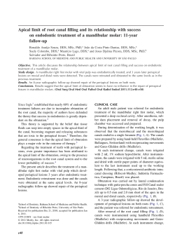
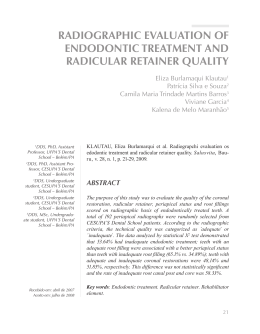
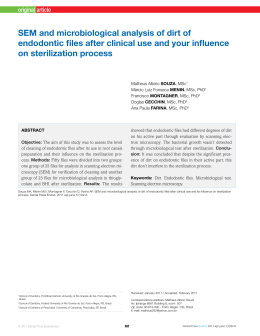
[ original article ] Healing of an extensive periapical lesion by means of conventional endodontic treatment
Introduction
The radicular cyst currently represents the most
common odontogenic cysts, this lesion predominates
in adults between the third and sixth decade of life.1-5
Also called peripheral cysts, are inflammatory cysts
of the jaws, formed in the dental apices, with necrotic
and infected pulps, thus they are considered as direct
consequences of apical granulomas.3,6 Its etiology
generally come from the root canal infections caused
by caries. It is known that the immune-inflammatory
process is the basis of formation of dental granulomas and radicular cysts.7
The radicular cyst represents a pathological cavity,
internally coated by epithelium and externally by a
fibrous that contains, inside a semi-fluid or fluid material.8,9 The formation of the radicular cyst has three
distinct phases: initial phase, the phase of cyst formation and growth stage. In the first phase the epithelial
rest of Malassez are continuously proliferate when
stimulated by chemical mediators released during the
inflammatory process, forming a epithelial net inside
the apical granuloma. In the last phase, the existing
micro cavity characterizes a cystic lesion that may be
microscopically diagnosed. The cystic growth occurs
slowly and continuously.5
Radiographically, the radicular cyst presents a picture which is radiolucent homogeneous density unilocular, circumscribed, round, oval, associated with an
intact root apex, a devitalized tooth with rupture of the
hard lamina at the apex,10 very familiar to granuloma,
as a circumscribed peripheral bone rarefaction may
present a radiopaque line delimiting the lesion.11
According to World Health Organization the radicular cyst is classified as an inflammatory odontogenic cyst and represents a major cause of bone
destruction of the jaws. Therefore and the frequent
incidence of these lesions in the clinics that provide
dental care, researches are developed in this area.12,13
The recommended treatment for periapical lesions,
with or without the involvement of periapex, has
been the root canal treatment. A discussion regarding
the efficacy of one and two-visit treatment of infected
root canals has been going on among scientists for
a long time.14 Currently strong scientific evidence
indicates regression of cystic lesions after conventional endodontic therapy with periodic changes of
the medication based on calcium hydroxide with
© 2012 Dental Press Endodontics
or without paramonochlorophenol camphor or 2%
chlorhexidine gel, with a very high success rate.15-20
The endodontic surgery should only be given
when the conventional endodontic treatment fails to
restore the integrity of the peripheral tissue.11,21
case report
The patient, age 49, female, Caucasian, ought dental endodontic referred by a dentist oral maxillofacial,
who found the presence of a radicular cyst between
teeth #33 and #34, near the region of the mental foramen. The patient complained of pain and mobility in
the region of tooth #34. Anamnesis was conducted,
clinical examination, radiographic examination and
testing of pulp vitality in teeth #33 and #34. Pulp necrosis and mobility was observed only on tooth 34. In
peripheral radiograph (Fig 1), the panoramic (Fig 2)
and tomography (Fig 3), observed the presence of radiolucent, unilocular, located in the anterior mandible
between the elements #33 and #34. By the clinical
and radiographic characteristic, the hypothetical diagnosis was inflammatory peripheral cyst associated
with the first pre-molar, tooth #34. The necropulpectomy endodontic treatment was performed using the
crown-apex technique. The channel was modeled irrigated with solution sodium hypochlorite 1%, the instrument memory was #45 K-file. After biomechanical
preparation complete was used as canal dressing calcium hydroxide paste, made with calcium hydroxide
pro-analysis and propylene glycol. The dressing with
calcium hydroxide paste was renewed on a monthly
basis for 10 months. The coronal sealing was carried
out with light-curing glass ionomer cement type IV
(Vitremer, 3M ESPE, U.S.A). After 10 months through
periapical radiographs was observed significant regression of lesions with significant new bone formation and no symptoms in the patient and performed
the root canal filling using thermoplastic technique using gutta-percha cones and Endofill cement with the
help of a thermo compactor 60 (Mc Spadden), after a
week was made final restoration of the tooth 34 with
composite resin (Filtek- 3M ESPE- U.S.A).
results
After one year of conventional endodontic treatment the patients without signs of recurrence, the
endodontic treatment provided a favorable clinical and
66
Dental Press Endod. 2012 Oct-Dec;2(4):65-9
Andrade ICGB, Silva R, Hochheim Neto R, Cristofolini MD
Figure 1. Periapical radiograph showing extensive periapical lesion of endodontic origin, suggestive of periapical cyst between teeth #33
and #34.
Figure 2. Panoramic radiograph showing extensive periapical lesion of endodontic origin,
suggestive of periapical cyst between teeth
#33 and #4.
periapical (Fig 4) and panoramic radiographic (Fig 5),
without pain symptoms with the signs of regression of
lesions, no need for additional surgery in the periapical
region. The case remains being accompanied.
Figure 3. Cone beam volumetric tomography
with shooting in small volumes the jaw teeth
#33 to #36.
discussion
In this case was observed a significant regression
of a cystic lesion associated with tooth #34 after conventional endodontic treatment associated with the
medication a paste of calcium hydroxide monthly renewed for 10 months.
The results of this study is in agreement with other
studies22-28 which observed regression of non-surgical
radicular cysts after conventional endodontic treatment, allowing tissue reactions and immunological
and inflammatory nature, consistent with the repair.
Was inferred that the endodontic treatment nonsurgical can be successfully implemented in a high
percentage of cases of radicular cysts and that its
success doesn’t depend on the nature of the lesion,
but appears to be influenced by individual variations
of host ’t immune response.22 Proper preparation of
biomechanics followed by calcium hydroxide medication periodically renewed represents a nonsurgical
approach to resolve extensive inflammatory peripheral lesions.28 The regression of the cystic lesion with
conservative treatment (based on successive changes
of dressings Ca(OH)2 basic, could occur due to collagen deposition generated by the healing process.
Such a deposit would compress the capillaries involved in nutrition of the epithelial cystic line, which
is degenerate being phagocytized by macrophages.29
Figure 4. Periapical radiograph after 1 year of endodontic obturation of
the tooth #34. Note advanced repair in the periapical region between
teeth #33 and #34.
Figure 5. Panoramic radiograph after 1 year root canal illing of the
tooth #34. Note advanced repair in the periapical region between teeth
#33 and #34.
© 2012 Dental Press Endodontics
67
Dental Press Endod. 2012 Oct-Dec;2(4):65-9
[ original article ] Healing of an extensive periapical lesion by means of conventional endodontic treatment
It has been shown that treatment with calcium hydroxide as an intracanal dressing in the presence of
large and chronic peripheral lesions can create and
environment more conducive to healing and start
bone repair. Calcium hydroxide is an effective intracanal antibacterial agent because of its high pH 12.5,
with bactericidal and bacteriostatic.20
In the literature, some authors believe that direct
contact with calcium hydroxide to the peripheral
tissue benefits the osteoinduction, others have suggested that calcium hydroxide in the apical region
has anti-inflammatory activity of neutralizing acidic
products, stimulates alkaline phosphatase and also
has antibacterial action.30 The bacterial activity of
various pastes of calcium hydroxide was confirmed
with different vehicle.28
The success rate of endodontic treatment has increased significantly, explained fact by the development of techniques and instruments used for modeling and root canal filling and also related to pathology installed.31-34
© 2012 Dental Press Endodontics
In this clinical case, as in other studies, because it is
an extensive peripheral lesion, in a region close to the
mental foramen with clinical and radiographic characteristics, suggestive of periapical cyst, Panoramic, periapical and tomography radiographs were performed.
Although the panoramic radiographs and periapical
acceptable reproduce details in the mesiodistal, the
observation in the bucco-lingual is inadequate, being
important a tomography that provides three-dimensional visualization of pathologic lesions and their relationship to important anatomic structures.35
conclusions
After one year of conventional endodontic treatment the patient is without signs of recurrence, the
endodontic treatment provided a favorable clinical
and radiographic response, without pain symptoms,
with evidence of regression of the lesion, with significant bone formation without the necessity for additional surgical in the periapical region. The case is
being accompanied.
68
Dental Press Endod. 2012 Oct-Dec;2(4):65-9
Andrade ICGB, Silva R, Hochheim Neto R, Cristofolini MD
references
1. Bystrom A, Happonen RP, Sjogren U, Sundqvist G. Healing of
periapical lesions of pulpless teeth after endodontic treatment
with controlled asepsis. Endod Dent Traumatol. 1987;3(2):58-63.
2. Shafer WG, Hine MK, Levy BM. Tratado de Patologia bucal. 4a ed.
Rio de Janeiro: Guanabara Koogan;1987.
3. Regezi JA, Sciubba JJ. Cistos da boca. In: Patologia bucal:
correlações clinicopatológicas. 3a ed. Rio de Janeiro: Guanabara
Koogan; 2000. p. 260-91.
4. Tommasi AF. Diagnóstico em patologia bucal. 3a ed. São Paulo:
Pancast; 2002.
5. Shear M. Cholesterol in dental cysts. Oral Sug Oral Med Oral
Pathol. 1963;16:1465-73.
6. Neville BW. Doenças da polpa e do periápice. In: Neville BW,
Damm DD, Allen CM, Bouquot JE. Patologia oral e maxilofacial.
Rio de Janeiro: Guanabara Koogan; 1998. cap. 3, p. 9.
7. Santos LCS, Ramos EAG, Meira TC, Figueiredo CRLV, Santos JN.
Etiopatogenia do cisto radicular. Parte I. Rev Ci Méd Biol.
2006;5(1):69-74.
8. Hisatomi M, Asaumi J, Konouchi H, Shigehara H, Yanagi Y, Kishi
K. MR imaging of epithelial cysts of the oral and maxillofacial
region. Eur J Radiol. 2003;48(2):178-82.
9. Gálvez-Gastélum FJ, Sandoval-Rodríguez AS; ArmendárizBorunda,J. El factor de crecimiento transformante ß como blanco
terapéutico. Salud Publica Méx. 2004;46(4):341-50.
10. Freitas A, Rosa JE, Souza IF. Radiologia odontológica. 5a ed. São
Paulo: Artes Médicas; 2000.
11. Azambuja TWF, Bercini F, Alano F. Cirurgia paraendodôntica:
revisão da literatura e apresentação de casos clínicos cirúrgicos.
Rev Fac Odontol Porto Alegre. 2006;47(1):24-9.
12. Kramer IR, Pindborg JJ, Shear M. The WHO histological typing
of odontogenic tumors; a commentary on the second edition.
Cancer. 1992;70:12.
13. Taylor AM,Camacho MEI, Franco MAD,Tejero MAT. Quistes
odontogénicos: análisis de 856 casos. Med Oral. 2002;7(2):89-96.
14. Pietrzycka K , Pawlicka H. Eficacy of treatment of teeth with
infected root canals as related to clinical procedure. J Stoma.
2011;64(3-4):174-86.
15. Lin LM, Ricucci D, Lin J, Rosenberg PA. Nonsurgical root canal
therapy of large cyst-like inlammatory periapical lesions and
inlammatory apical cysts. J Endod. 2009;35(5):607-15.
16. Oliveira, SHG, Iório LS, Maekawa LE, Camargo SEA, Camargo
CHR, Palo RM. Tratamento não-cirúrgico de patologias com
imagens radiográicas sugestivas de cisto radicular. Rev. Assoc
Paul Cir Dent. 2009;63(1).164-8.
17. Caliskan MK. Prognosis of large cyst-like periapical lesions
following nonsurgical root canal treatment: a clinical review. Int
Endod J. 2004;37(6):408-16.
18. Cerda-Cristerna BI, Silva-Herzog FD, Pozos-Guillén A, Sayão SA.
Reparo de reabsorção radicular externa associada com queratocisto
odontogênico: relato de caso. RSBO. 2009;6(1)100-3.
© 2012 Dental Press Endodontics
19. Soares J, Brito-Junior M, Silveira FF, Nunes E, Santos SM.
Favorable response of an extensive periapical lesion to root canal
treatment. J Oral Sci. 2008;50(1):107-11.
20. Kusgoz AK, Yildirim S, Gokalp A. Nonsurgical endodontic
treatments in molar teeth with large periapical lesions in children:
2-year follow-up. Oral Surg Oral Med Oral Pathol Oral Radiol
Endod. 2007;10(104):60-5.
21. Guimarães KB. Cirurgia parendodôntica com obturação
simultânea dos canais: relato de caso. Rev Ci Méd Biol.
2006;5(2):188-94.
22. Shah N. Nonsurgical management of periapical lesions:
a prospective study. Oral Surg Oral Med Oral Pathol.
1988;66(3):365-71.
23. Sjögren U, Hägglund B, Sundqvist G, Wing K. Factors affecting
the long term results of endodontic treatment. J Endod.
1990;16(10):498-504.
24. Murphy WK, Kaugars GE, Collett WK, Dodds RN. Healing of
periapical radiolucencies after nonsurgical endodontic therapy.
Oral Surg Oral Med Oral Pathol. 1991;71(5):620-4.
25. Nair PNR, Pajarola G, Schroeder HE. Types and incidence of
human periapical lesions obtained with extracted teeth. Oral Surg
Oral Med Oral Pathol Oral Radiol Endod. 1996;81(1):93-102.
26. Laszkiewicz J. Evaluation of calcium hydroxide in conservative
treatment of large periapical lesions. Int Endod J. 1998;31(3):203.
27. Oztan MD. Endodontic treatment of teeth associated with a large
periapical lesion. Int Endod J. 2002;35(1):73-8.
28. Soares J, Santos S, Silveira F, Nunes E. Nonsurgical treatment
of extensive cyst-like periapical lesion of endodontic origin. Int
Endod J. 2006;39(7)566-75.
29. Bender IB, Seltzer S, Soltanoff W. Endodontic success. A
reappraisal of criteria. 1. Oral Surg Oral Med Oral Pathol.
1966;22(6):780-9.
30. Souza FJ, Soares AJ, Vianna ME, Zaia AA, Ferraz CC, Gomes
PB. Antimicrobial effect and pH of chlorhexidine gel and calcium
hydroxide alone and associated with other materials. Braz Dent J.
2008;19(1):28-33.
31. Cunha PO. Avaliação radiográica dos tratamentos endodônticos
realizados por alunos de graduação da faculdade de odontologia
da UFAM [monograia]. Manaus (AM): UFAM; 2010.
32. Gil AC, et al. Revisão contemporânea da obturação
termoplastiicada, valendo-se da técnica de compactação
termomecânica. Rev Saúde. 2009;3(3):20-9.
33. Lopes HP, Siqueira JF Jr. Endodontia: biologia e técnica. 3a ed.
Rio de Janeiro: Guanabara Koogan; 2010.
34. Tsurumachi T, Honda K. A new cone beam computerized
tomography system for use in endodontic surgery. Int Endod J.
2007;40(3):224-32.
35. Huumonen S, Kvist T, Gröndahl K , Molander A. Diagnostic value
of computed tomography in retreatment of root illings in maxillary
molars. Int Endod J. 2006;39(10):827-33.
69
Dental Press Endod. 2012 Oct-Dec;2(4):65-9
Information for authors
— Dental Press Endodontics publishes original research
(e.g., clinical trials, basic science related to the biological aspects of endodontics, basic science related
to endodontic techniques and case reports). Review
articles only for invited authors. Authors of potential
review articles are encouraged to first contact the
editor during their preliminary development.
must be provided. This information is not made
available to the reviewers.
2. Abstract
— Preference is given to structured abstracts in English with 250 words or less.
— The structured abstracts must contain the following sections: INTRODUCTION: outlining the objectives of the study; METHODS, describing how
the study was conducted; RESULTS, describing the
primary results, and CONCLUSIONS, reporting the
authors’ conclusions based on the results, as well as
the clinical implications.
— Abstracts in English must be accompanied by 3 to 5
keywords, or descriptors, which must comply with
MeSH.
— Dental Press Endodontics uses the Publications Management System, an online system,
for the submission and evaluation of manuscripts. To submit manuscripts please visit:
www.dentalpressjournals.com.br/rdpendo
— Please send all other correspondence to:
Dental Press Endodontics
Av. Euclides da Cunha 1718, Zona 5
Zip Code: 87.015-180, Maringá/PR
Phone. (44) 3031-9818
E-mail: [email protected]
3. Text
— The text must be organized in the following sections: Introduction, Materials and Methods, Results,
Discussion, Conclusions, References and Figure
legends.
— Texts must contain no more than 4,000 words, including captions, abstract.
— Figures and tables must be submitted in separate
files (see below).
— Insert the Figure legends also in the text document
to help with the article layout.
— The statements and opinions expressed by the
author(s) do not necessarily reflect those of the
editor(s) or publisher, who do not assume any responsibility for said statements and opinions. Neither the editor(s) nor the publisher guarantee or
endorse any product or service advertised in this
publication or any claims made by their respective
manufacturers. Each reader must determine whether or not to act on the information contained in this
publication. The Dental Press Endodontics and its
sponsors are not liable for any damage arising from
the publication of erroneous information.
4. Figures
— Digital images must be in JPG or TIF, CMYK or
grayscale, at least 7 cm wide and 300 dpi resolution.
— Images must be submitted in separate files.
— In the event that a given illustration has been published previously, the legend must give full credit to
the original source.
— The author(s) must ascertain that all figures are cited in the text.
— To be submitted, all manuscripts must be original
and not published or submitted for publication elsewhere. Manuscripts are assessed by the editor and
consultants and are subject to editorial review. Authors must follow the guidelines below.
5. Charts
— Files containing the original versions of charts must
be submitted.
— It is not recommended that such charts be submitted only in bitmap image format (not editable).
— Drawings may be improved or redesigned by the
journal’s production department at the discretion of
the Editorial Board.
— All articles must be written in English.
GuIdelInes For submIssIon
oF manuscrIPTs
— Manuscritps must be submitted via www.dentalpressjournals.com.br/rdpendo. Articles must be organized as described below.
6. Tables
— Tables must be self-explanatory and should supplement, not duplicate the text.
— Must be numbered with Arabic numerals in the order they are mentioned in the text.
— A brief title must be provided for each table.
— In the event that a table has been published previously, a footnote must be included giving credit to
the original source.
1. Title Page
— Must comprise the title in English, an abstract and
keywords.
— Information about the authors must be provided on
a separate page, including authors’ full names, academic degrees, institutional affiliations and administrative positions. Furthermore, the corresponding
author’s name, address, phone numbers and e-mail
© 2012 Dental Press Endodontics
70
Dental Press Endod. 2012 Oct-Dec;2(4):70-2
Baixar