Universidade de São Paulo
Instituto de Biociências
Gustavo Monteiro Silva
Estudo e caracterização do processo de glutatiolação e
desglutatiolação da unidade 20S do proteassomo da levedura
Saccharomyces cerevisiae: Implicações na regulação do
metabolismo redox intracelular e na geração de peptídeos
São Paulo
2010
Universidade de São Paulo
Instituto de Biociências
Gustavo Monteiro Silva
Estudo e caracterização do processo de glutatiolação e
desglutatiolação da unidade 20S do proteassomo da levedura
Saccharomyces cerevisiae: Implicações na regulação do metabolismo
redox intracelular e na geração de peptídeos
Study and characterization of the S-glutathiolation and
deglutathiolation of the 20S proteasome core from the yeast
Saccharomyces cerevisiae: Implications for the intracellular redox
metabolism and peptide generation.
São Paulo
2010
Universidade de São Paulo
Instituto de Biociências
Gustavo Monteiro Silva
Estudo e caracterização do processo de glutatiolação e
desglutatiolação da unidade 20S do proteassomo da levedura
Saccharomyces cerevisiae: Implicações na regulação do metabolismo
redox intracelular e na geração de peptídeos
Study and characterization of the S-glutathiolation and
deglutathiolation of the 20S proteasome core from the yeast
Saccharomyces cerevisiae: Implications on the intracellular redox
metabolism and on peptide generation.
Tese apresentada ao Instituto de Biociências da Universidade de São Paulo,
para a obtenção de Título de Doutor em Ciências, na Área de Biologia-Genética.
Orientador:
Dr. Luis Eduardo Soares Netto
Co-orientador:
Dra. Marilene Demasi
Departamento de Genética e Biologia Evolutiva
Laboratório de Bioquímica e Biofísica
Instituto de Biociências
Universidade de São Paulo
Instituto Butantan
São Paulo
2010
Ficha Catalográfica
Silva, Gustavo Monteiro
“Estudo e caracterização do processo de glutatiolação e
desglutatiolação da unidade 20S do proteassomo da levedura
Saccharomyces cerevisiae: Implicações na regulação do metabolismo
redox intracelular e na geração de peptídeos.”
159 páginas
Tese (Doutorado) – Instituto de Biociências da Universidade de São
Paulo.
Departamento de Genética e Biologia Evolutiva
1.Proteassomo; 2. Glutatiolação; 3. Metabolismo Redox; 4. Proteólise
I. Universidade de São Paulo, Instituto de Biociências. Departamento
de Genética e Biologia Evolutiva.
Comissão Julgadora
Prof. Dr. Marcelo D. Gomes (FMRP-USP)
Prof. Dr. Fabio C. Gozzo (IQ-Unicamp)
Prof. Dr. Francisco R. M. Laurindo (FM-USP)
Prof. Dr. Mario H. Barros (ICB-USP)
Profa. Dra. Marilene Demasi (Instituto Butantan)
Presidente
À minha mãe,
A quem devo tudo.
“A morte da interpretação é o crer que há símbolos
que existem primariamente, originalmente,
realmente como marcas coerentes, pertinentes e
sistemáticas. A vida da interpretação, pelo
contrário, é o crer que não há mais do que
interpretações.”
Michel Foucault
Nietzsche, Freud e Marx/TheatrumPhilosoficum
“Tudo pode ser recriado, se acharmos que assim
deve ser...”
Jurandir Freire Costa
Sem Fraude Nem Favor (1998)
Agradecimentos
À Dra. Marilene Demasi, primeiramente pela oportunidade, orientação e confiança no meu trabalho.
Seu profissionalismo e sua dedicação à ciência deixaram aprendizados que carregarei por toda a vida.
Ao professor Dr. Luis Eduardo Soares Netto por seu apoio e conselhos durante todo este período.
Sua competência e afabilidade inspiram a todos que almejam tornarem-se grandes cientistas.
À FAPESP e ao CNPq pelo indispensável auxílio financeiro para realização deste projeto.
À Dra. Mari Sogayar pela disponibilização irrestrita de seu laboratório e ao Dr. Marcos Demasi por
toda paciência e atenção nos ensinamentos da arte dos géis 2D. A ajuda de vocês foi de outra
dimensão.
Ao Dr. Fábio Cesar Gozzo e aos demais integrantes do lab. Dalton. Em especial ao Luiz Fernando
Santos e ao Dr. Eduardo Pilau por serem tão prestativos e atenciosos, sempre dispostos a tirar
minhas infinitas dúvidas sobre espectrometria. A colaboração de vocês além de essencial foi muito
massa!
Ao Dr. Daniel Carvalho Pimenta e a Clécio Klitzke pela ajuda e discussões sobre Maldis e ToFs.
À Dra. Sylvia Carneiro e à Simone do laboratório de Biologia Celular do Instituto Butantan, por me
auxiliarem na visualização do grande proteassomo por microscopia eletrônica.
À Dra. Adriana Rios Lopes pelas diversas discussões sobre catálise e cinética enzimática.
Aos doutores Ana Marisa Chudzinski Tavassi, Ana Olivia de Souza e Ivo Lebrun, pela disponibilização
de equipamentos e materiais necessários a conclusão deste trabalho.
Ao Dr. Paolo Di Mascio e as técnicas de seu laboratório Fernanda Prado e Isaura Toma pela ajuda nos
ensaios iniciais de espectrometria de massa.
À Dra. Karen Discola, senhora Grx, por toda a ajuda e companhia agradável no lab, congressos e
afins. E ainda por ter me apresentado o Mr. Ray, que deixou minha tese muito mais linda!
Às alunas que participaram e darão prosseguimento a esse projeto. À Daniéélee Silva figuraça, que
implementou o 19S (apesar de multiplicar as concentrações do gel nativo) e instaurou a sessão
comédia do lab na hora do café e à Vanessa Simões (Oaa Boi!), pela companhia nos dias de Unicamp,
pelo trabalho nas férias e que apesar de só saber fazer contas com fósforos, revisou cuidadosamente
este manuscrito.
Ao agora Doutor e antes de tudo mais que irmão Zézinho Renato Cussiol, por além da Ohr, todos os
anos de convivência, amizade, abrigo, gargalhadas, discussões, shishas, trocadilhos, voltas ao mundo,
baladas... Lou, ainda temos que colocar nosso plano caipira em ação!
Aos amigos do Butantan por tornarem tão agradáveis os meus dias de trabalho. Aos nossos amigos
Bob e Gilberto ao mesmo tempo pelo bom humor e amizade; a Paty Gabi por sua simpatia matinal,
infinitas companhias de almoço e por manter o bom andamento do lab; ao Adrian (Vai
Adriaaanooo!), o filósofo tricolor mais prestativo que já conheci e ao Chiquinho, o aluno de ouro.
Sem esquecer os ex-técnicos Beatriz e Angelino que sempre colaboraram e ajudaram
incondicionalmente.
À Prof. Dra. Gisele Monteiro (maMÃÃE!) por todos os ensinamentos, paciência e horas infinitas de
risadas. Espelho-me e quero ser como você quando crescer, mas sem dedar os outros.
Ao Dr. Bruno Gato Horta e ao Thithi Alegria, pelas conversas, pelo companheirismo, amizade e
“carinho”. Amigos que quero ter por toda vida.
Aos amigos e colegas do IB-USP da turma antiga (Andressa, Aninha, Camila, Dani, Mirian, Prof.
Marcão, Rafael, Roberta, Six, Suzy, Telma e Viiiictor) e da nova (Aline, Daiane, Eduardo, Gabriel, Lucas
Mica 5’, Marcella ForestGump e Tati).
Aos amigos do lab. de Bioquímica e às funcionárias mais legais do Instituto Butantan, Patrícia,
Silvana, Toninha e Val, sempre dispostas a ajudar no que fosse preciso e por aquele cafezinho no
meio da tarde pra acordar!
À turma 00N da Bio que me proporcionou a companhia mais agradável que uma classe poderia dar
ao longo dessa década. E a todos os amigos que fiz ao longo da vida acadêmica. Em especial à Beka,
minha filhota querida. Adoro todos vocês e Paulo.
Às famílias JogaNoPagode e Biosal, times de futebol da Biologia USP, pelos campeonatos que
disputei, medalhas que ganhei, amigos que fiz e gols que perdi... Inesquecível!
Aos amigos de sempre, que me ensinaram o verdadeiro valor de uma amizade. Tranka, Bitch, Hector
Fenômeno, Bonga, Zura, Fê, Gus, Lê Gordinho e Celso. Apesar de o contato ter diminuído, o
sentimento se mantém inalterado e a consideração só aumenta.
Ao meu pai Carlos que apesar de não ser tão presente em minha vida, sei que torce muito pelo meu
sucesso.
À trinca de tias mais legais que uma pessoa pode ter. Tia Célia, Tia Maísa (dinha), e Tia Vera, que
junto com o Binho, Lauzinho e Tio Lau, viraram mais do que família. O apoio de vocês foi
fundamental desde o começo de tudo.
Ao irmão que eu sempre tive, mas que só agora ganhei. Lê, é bom sentir e saber que agora eu tenho
uma família (Valeu Max cunhadinha, deu um jeito no caboclo). E ao seu professor e amigo Ajarn
Petchpayao, que apesar de não me ensinar muay thai, cozinhou pratos deliciosos durante o período
dedicado à escrita desta tese.
À minha querida Bi, por todos esses anos de companheirismo, carinho, compreensão, apoio,
amadurecimento, ajuda, sorrisos do Chandler, gaPAAO... Galega, não seria o que sou hoje sem você.
Por último e neste caso, mais importante, à minha mama Marlene, exemplo de caráter e de mulher,
que, além da educação, lutou muito e me proporcionou a coisa mais importante que os pais
poderiam proporcionar: A liberdade de escolha de ser o que eu quisesse. Obrigado, mãe (Mas eu
ainda me lembro do avental...).
Sumário
Resumo
11
Abreviaturas e Siglas
13
I. Introdução
O proteassomo e a proteólise intracelular
Regulação da atividade catalítica do 20SPT
Modificações pós-traducionais do proteassomo 20S
Glutationa e glutatiolação de proteínas
O proteassomo 20S e a geração de peptídeos intracelulares
14
14
18
22
23
27
II. Objetivos
29
III. Materiais e Métodos
Reagentes
Crescimento celular, extração e purificação do 20SPT
Ensaio de atividade do proteassomo 20S
Redução e glutatiolação in vitro do proteassomo
Determinação de glutationa
Eletroforese bidimensional
Imunomarcação anti-GSH
Processamento dos spots para espectrometria de massas
Fingerprinting por MALDI-TOF
Detecção de S-glutatiolação por LC-MS/MS
Linhagens bacterianas
Clonagem do gene GRX2
Indução e expressão do gene GRX2
Purificação e ensaio de atividade enzimática da Grx2
Oxidação de proteínas
Análise da degradação de proteínas por SDS-PAGE
Determinação do perfil de fragmentação de proteínas por HPLC
Análise do proteassomo 20S por microscopia eletrônica
30
30
30
31
31
31
31
32
32
33
34
34
34
35
35
36
36
36
37
IV. Resultados
Glutatiolação da unidade catalítica do proteassomo
Desglutatiolação do proteassomo 20S
Modulação da proteólise e mecanismo de hidrólise
Dinâmica de abertura da câmara catalítica
38
38
54
55
71
V. Discussão
74
VI. Conclusões
84
VII. Referências Bibliográficas
86
VIII. Anexos
98
Resumo
O proteassomo é o componente do sistema Ubiquitina-Proteassomo (UPS),
responsável pela degradação de proteínas intracelulares marcadas com cauda de ubiquitina.
No entanto, a unidade catalítica do proteassomo (20SPT), destituída de unidades
regulatórias, é capaz de degradar proteínas de maneira ubiquitina-independente. Diversas
modificações pós-traducionais já foram descritas para o 20SPT, incluindo a S-glutatiolação.
De acordo com Demasi e col., (2003) o 20SPT da levedura Saccharomyces cerevisiae possui a
atividade tipo-quimiotripsina modulada por glutationa e o mecanismo de glutatiolação
implica na formação do intermediário ácido sulfênico. No presente trabalho, identificamos
por espectrometria de massas (MS/MS) um total de sete resíduos diferentes de cisteína
glutatiolados no 20SPT, sendo seis in vitro por incubação com GSH e três in vivo, extraído de
células crescidas até atingir fase estacionária tardia em meio rico. Analisando a estrutura 3D
do 20SPT, observou-se que os resíduos de cisteína glutatiolados não estão localizados na
entrada da câmara catalítica nem próximos aos sítios-ativos, indicando um mecanismo
alostérico da modulação da atividade proteassomal. O proteassomo glutatiolado extraído de
leveduras é capaz de degradar proteínas oxidadas de maneira mais eficiente que o
proteassomo reduzido por DTT, e ainda, esta degradação gera perfis peptídicos
diferenciados por utilizar distintamente as atividades sítio-especificas, como visualizado por
análises de HPLC e MS/MS. Por microscopia eletrônica verificamos a conformação aberta da
câmara catalítica do proteassomo glutatiolado, sendo esta imediatamente fechada pela
remoção da glutationa do 20SPT na presença de DTT. Caracterizamos ainda, enzimas
reponsáveis pela desglutatiolação do 20SPT, capazes de recuperar as atividades
proteassomais que haviam sido diminuídas pela glutatiolação: as oxidoredutases
glutarredoxina 2 e as tiorredoxinas citosólicas. O mecanismo ainda inclui a hidrólise dessas
oxidorredutases, fenômeno também verificado para diversas proteínas da suprafamília
tiorredoxina, provavelmente devido a propriedades estruturais desta família. A glutatiolação
do proteassomo apresenta-se como uma nova modificação pós-traducional de ocorrência
fisiológica dependente do estado redox celular. Esta modificação promove aumento da
atividade proteolítica, sugerindo uma função antioxidante atuante na remoção de proteínas
oxidadas durante desafios oxidativos.
11
Abstract
The proteasome is the protease of the Ubiquitin-Proteasome System (UPS)
responsible for the breakdown of intracellular ubiquitin-tagged proteins. However, the
catalytic particle of the proteasome (20SPT) is capable of hydrolyzing some substrates in an
ubiquitin-independent fashion. The S-glutathiolation of the 20SPT was described among
several post-translational modifications and according to Demasi et. al. (2003), the
chymotrypsin-like activity of proteasome from yeast Saccharomyces cerevisiae is regulated
by glutathione. The mechanism of S-glutathiolation is dependent on the formation of the
sulfenic acid intermediate in the cisteine residues of the 20SPT. In this present work, we
identified in vitro and in vivo, a total of seven different S-glutathiolated proteasomal cysteine
residues by mass spectrometry studies (MS/MS) and, by analyzing the 3D structure of the
20SPT, the modified cysteine residues are not located either on the entrance of the catalytic
core or near to the active sites, indicating an allosteric mechanism of proteasomal
modulation. During protein degradation, the natively S-glutathiolated 20SPT produces
different patterns of peptide products when compared to the DTT-reduced particle through
distinct site-specific cleavage of the protein substrates, as herein demonstrated by HPLC and
MS/MS analyses. Furthermore, by electron microscopy, we showed that the entrance of the
natively glutathiolated 20SPT is in the open conformation that immediately shifts to the
closed conformation in the presence of DTT. We have also characterized the deglutathiolase
role of the oxidoreductases Glutaredoxin 2 and Citosolic Thioredoxins 1 and 2 which recover
the partially inhibited 20SPT activities. The deglutathiolation mechanism also includes the
oxidoreductase degradation dependent on the 20SPT activation. The proteasome Sglutathiolation emerges as a new physiological post-translational modification correlated to
the cellular redox state.
Moreover, the S-glutathiolation of the 20SPT increases its
proteolytic activity suggesting an antioxidant role by removing oxidized proteins generated
during oxidative challenges.
12
Abreviaturas e Siglas
19SPT
Proteassomo 19S – unidade regulatória
20SPT
Proteassomo 20S – unidade catalítica
26SPT
Proteassomo 26S – unidade regulatória acoplada à catalítica
BSA
Albumina do soro bovino
BSAox
Albumina do soro bovino oxidada por H2O2
ChT-L
Atividade tipo-quimiotripsina do proteassomo
cTpx
Tiorredoxina peroxidase citosólica
Cys
Resíduo de cisteína
Cys-SOH
Resíduo de cisteína oxidado a ácido sulfênico
DTNB
5,5’-ditio-bis-(2-nitrobenzóico) ou reagente de Ellman
DTPA
Ácido dietileno triamino pentacético
DTT
1,4-Ditiotreitol
ESI
Ionização por Electrospray
GSH
Glutationa
GSSG
Glutationa oxidada
Glr
Glutationa redutase
Grx
Glutarredoxina
LC (HPLC/UPLC)
Cromatografia líquida (Alto/Ultra Desempenho)
MALDI
Ionização por dessorção a laser auxiliada por matriz
MS
Espectrometria de massas
Ohr
Proteína recombinante "Organic Hydroperoxide Resistance”
Ova
Ovalbumina
PA
Atividade pós-acídica do proteassomo
PDB
Banco de dados de estuturas de proteínas - Protein Data Bank
PiPs
Proteínas que interagem com o proteassomo
PT-YPD
Proteassomo nativamente glutatiolado extraído de YPD
PT-SG
Proteassomo glutatiolado in vitro
PT-SH
Proteassomo reduzido com DTT
PTM
Modificação pós-traducional
Q-TOF
Quadrupolo-Tempo de Vôo
T-L
Atividade tipo-tripsina do proteassomo
TFA
Ácido trifluoracético
Trr
Tiorredoxina redutase
Trx
Tiorredoxina
UPS
Sistema Ubiquitina-Proteassomo
13
I.
Introdução
O Proteassomo e a proteólise intracelular
As células eucarióticas possuem os mais diferentes mecanismos de regulação dos
processos fisiológicos. Uma enormidade de vias pode ser regulada a partir da transcrição de
genes ou da tradução de proteínas efetoras, entretanto, diversos eventos celulares são
modulados na esfera da degradação de proteínas pelos sistemas intracelulares de proteólise.
Além da regulação de processos celulares, a proteólise intracelular está envolvida no
controle de qualidade, removendo proteínas aberrantemente sintetizadas assim como as
que tenham sido danificadas durante o metabolismo. A proteólise também se faz
importante na manutenção do conteúdo de aminoácidos e na geração de peptídeos ativos.
Outro aspecto da proteólise engloba a degradação de proteínas que variam a concentração
ao longo do tempo, comumente relacionadas com vias regulatórias específicas. Entre os
eventos celulares em que ocorre a participação do proteassomo podemos citar o controle do
ciclo, divisão e diferenciação celular, controle da expressão gênica pela degradação de
fatores de transcrição, regulação de oncoproteínas, geração de peptídeos para a
apresentação antigênica, entre outros (Fanasaro e col., 2010; Gasparian e col., 2009; Chen e
col., 1998; Jariel-Encontre, 1995; Kloetzel, 2004). O primeiro sistema descrito de proteólise
intracelular foi o endossômico-lisossômico, no qual as proteínas endocitadas ou contidas em
vesículas citoplasmáticas eram degradadas pelas proteases existentes no interior do
lisossomo (de Duve e col., 1955; de Duve, 1983). Entretanto, esse sistema não era capaz de
responder as questões sobre a especificidade protéica, diferenças de meia-vida intracelular
e, principalmente, a observação de que algumas proteínas eram degradadas por processo
dependente de ATP.
Somente no final da década de 70 que se iniciou a caracterização de uma via
alternativa de degradação de proteínas que respondesse as questões apresentadas sobre a
especificidade dos substratos. Esta via dependente de ATP foi descrita primeiramente em
reticulócitos de coelho, eritrócitos imaturos, desprovidos de núcleo e organelas como os
lisossomos (Etlinger e Goldberg, 1977). Esta nova via proteolítica denominada Sistema
14
Ubiquitina-Proteassomo (UPS – Ubiquitin Proteasome System) é composta por uma série de
enzimas capaz de marcar proteínas, sinalizando-as para degradação. Esta marcação confere
a especificidade aos substratos que posteriormente serão reconhecidos por um complexo
protéico que promoverá a degradação destas proteínas (revisto por Nandi e col., 2006). A
proteína responsável pela marcação dos substratos para degradação foi denominada
ubiquitina, uma pequena proteína de 8,5 kDa, altamente conservada evolutivamente,
considerada ubíqua nos diversos reinos durante sua caracterização (Schlensinger e col.,
1975, Wilkinson e col., 1980). A ubiquitinação de uma proteína como sinalização para
degradação envolve uma cascata de reações que compreendem três enzimas. A E1 (enzima
ativadora da ubiquitina) que, a custa de ATP, ativa a molécula de ubiquitina, transferindo-a
para as E2’s (enzima conjugadora). As E2’s interagem com diversas E3’s (ubiquitina ligase),
complexando a ubiquitina direta ou indiretamente em lisinas N-terminais dos substratos
protéicos. Após a adição da primeira ubiquitina, novas moléculas de ubiquitina são inseridas
nas moléculas já existentes, formando uma cadeia de poli-ubiquitina que atuará como sinal
de reconhecimento para degradação (Revisto por Hershko e Ciechanover, 1998) (Fig. 1).
A via de ubiquitinação protéica foi descoberta praticamente uma década antes da
caracterização da protease responsável pela hidrólise das proteínas ubiquitinadas
(Ciechanover e col., 1978; Hershko e col., 1979; Hershko e col., 1983.). Em 1987, Hough e
colaboradores caracterizaram um complexo proteolítico de alto peso molecular encarregado
da degradação de proteínas poli-ubiquitinadas, que, por Arrigo e col. (1988), foi nomeado
Proteassomo (Proteasome).
Após a descrição no final da década de 80, muito se foi investido na caracterização
deste complexo de alto peso molecular e no seu envolvimento com este novo sistema
proteolítico intracelular. Em 1993, Jap e colaboradores conseguiram obter os primeiros
cristais para decifrar a estrutura tridimensional do proteassomo de arqueobactéria, que em
1995, pelo mesmo grupo, foi finalmente elucidada (Löwe e col. 1995). Estes trabalhos
abriram diversas frentes para a caracterização desta protease e a compreensão funcional
nos mais diversos organismos. Morimoto e col. (1995) caracterizaram a primeira estrutura
3D de um 20SPT de eucarioto e finalmente em 1997, foi resolvida a estrutura do 20SPT da
levedura Saccharomyces cerevisiae (Groll e col., 1997).
15
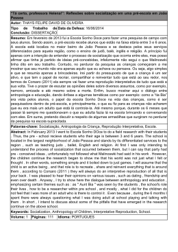
Figura 1. Via de ubiquitinação
protéica.
A molécula de ubiquitina (Ub) é
ativada a custa de ATP pela enzima
ativadora (E1) e transferida para a
enzima
conjugadora
(E2).
A
conjugadora transfere a ubiquitina
diretamente para resíduos de lisinas
(Lys) do substrato, associada à ligase
(E3 - Ring) ou transfere para a E3 da
classe HECT que reconhecerá o
substrato, ubiquitinando-o. Novas
moléculas
de
ubiquitina
são
adicionadas a partir de resíduos de
lisina e da glicina76 C-terminal das
moléculas subseqüentes. Proteínas
fusionadas a cadeias de poli-ubiquitina
baseadas na lisina 48 da ubiquitina
serão preferencialmente reconhecidas
para degradação pelo proteassomo
26S.
Estruturalmente, a unidade catalítica 20S do proteassomo (20SPT) é composta por
quatro anéis heptaméricos empilhados em forma de barril. Os dois anéis idênticos externos
alfa ( ) contêm sete subunidades distintas (de 1 a 7) formando um portão que controla o
acesso de substratos à câmara catalítica. Já cada anel interno beta ( ) também heptamérico,
possui três subunidades cataliticamente distintas, responsáveis pela hidrólise dos substratos
(Coux e col., 1996; Jung e col., 2009). Um estreito canal isola a célula dos sítios catalíticos
voltados para a face interna da câmara, evitando degradações não específicas. A estrutura
tridimensional do proteassomo 20S está demonstrada na figura 2, assim como a disposição
das subunidades nos anéis heptaméricos.
16
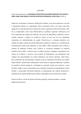
Figura 2. Estrutura tridimensional do proteassomo 20S - (A) Vista longitudinal da câmara catalítica
apresentando os quatro anéis na disposição simétrica ββ . (B) Vista frontal do proteassomo 20S
demonstrando centralmente a porta de entrada da câmara catalítica. (C) A vista frontal
apresentando a disposição de cada uma das sete subunidades constituintes do anel coloridas
distintamente. PDB Id: 1RYP. Imagens gráficas geradas pelo software Pymol (Delano Scientific).
As três subunidades catalíticas ( 1, 2 e 5) são expressas na forma de pré-proteínas
e, após seu autoprocessamento, possuem o resíduo de treonina 1 (Thr1) como sítio-ativo
(Groll e col., 1999). A hidrólise da ligação peptídica dos substratos ocorre pela ação da
hidroxila da cadeia lateral da Thr1 que age como nucleófilo, no entanto, devido à
características específicas do bolsão de cada sítio ativo, as três subunidades possuem
atividades diferentes entre si. A subunidade 1 possui atividade pós-acídica (PA), clivando
majoritariamente após resíduos ácidos como aspartato e glutamato; a subunidade 2 possui
atividade tipo-tripsina (T-L), clivando após resíduos de aminoácidos básicos como lisina e
arginina; e a subunidade 5 tida como a mais ativa e importante, possui atividade do tipoquimiotripsina (ChT-L), hidrolisando cadeias peptídicas após aminoácidos hidrofóbicos
(Arendt e Hochstrasser, 1997; Groll e col., 1999; Heinemeyer e col., 1997; Dick e col., 1998;
Nussbaum e col., 1998).
Em relação à localização subcelular, o proteassomo é abundante no núcleo e no
citoplasma de células eucarióticas, co-localizado a filamentos intermediários na membrana
do retículo endoplasmático e associado ao centrossomo (Rivett e col., 1992; Palmer e col.,
1996, Soza e col., 1997, Wigley e col., 1999). Estudos em levedura demonstraram altas
concentrações citoplasmáticas de proteassomo ao redor da rede de membranas formada
17
pelo envelope nuclear e pelo retículo endoplasmático (Wilkinson e col., 1998; Enenkel e col.,
1998).
Regulação da atividade catalítica do 20SPT
Juntamente à unidade catalítica central 20S (20SPT), complexos protéicos
regulatórios podem se associar por interação com as subunidades alfa, modulando a
atividade do proteassomo. Um importante complexo regulatório denominado proteassomo
19S (19SPT) ou PA700 é composto por 19 subunidades diferentes entre si, relacionado com o
reconhecimento de substratos poli-ubiquitinados, desdobramento das proteínas-alvo,
desubiquitinação do substrato, abertura da câmara catalítica e a translocação de substrato
para o interior do 20SPT (Glickman e col., 1998 e 1999; Navon e Goldberg, 2001; Smith e
col., 2006). A partícula regulatória 19SPT acoplada ao anel alfa da unidade catalítica formam
o proteassomo 26S, protease essencial do UPS, necessária para o reconhecimento e
degradação de proteínas poli-ubiquitinadas.
Outros complexos regulatórios descritos mais recentemente são o proteassomo 11S
ou PA26, envolvido na geração de peptídeos para a apresentação antigênica em associação
ao imunoproteassomo (Li e Rechsteiner, 2001; Hill e col., 2002.) e o PA200 envolvido na
estabilidade e no reparo de DNA (Ustrell e col., 2002; Blickwedehl e col., 2008). O
proteassomo 11S é formado também por anéis heptaméricos que estimulam a hidrólise de
peptídeos via estabilização da conformação aberta do 20SPT (Förster e col., 2003 e 2005).
Em levedura ainda não foi descrita a identificação do 11SPT, porém, outro sistema
regulatório do 20SPT é o Blm10, relacionado ao PA200 de mamíferos. O Blm10 é uma
proteína de aproximadamente 250 kDa sem função fisiológica conhecida, entretanto,
quando associada ao proteassomo, promove a estimulação da degradação de peptídeos,
mas não estimula a degradação de proteínas (Sadre-Bazzaz e col., 2010). Diversos complexos
regulatórios já foram caracterizados nos mais diferentes organismos como capazes de
estimular a abertura da câmara catalítica e propiciar uma maior degradação dos substratos.
O dinamismo da abertura da câmara catalítica devido ao acoplamento de complexos
regulatórios pode ser visualizado na figura 3.
18
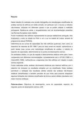
Figura 3. Efeito da abertura da câmara catalítica por complexos regulatórios. (A) Vista frontal das
estruturas tridimensionais demonstra em verde, a câmara catalítica do proteassomo 20S de
Saccharomyces cerevisiae (PDB Id: 1RYP) em sua conformação fechada e, em azul, o proteassomo de
Trypanosoma brucei (PDB Id: 1FNT) acoplado ao complexo regulatório PA26 em sua conformação
aberta. (B) Vista longitudinal evidenciando o complexo PA26 externamente ao 20SPT em azul.
Imagens gráficas geradas pelo software Pymol (Delano Scientific).
O sistema proteassomo é encontrado em todos os organismos eucarióticos, de
fungos a mamíferos. Diversas eubactérias apresentam um sistema tipo-proteassomo
denominado HslUV, no qual a protease HslV possui baixa atividade peptidásica e é regulada
pelos anéis ATPásicos do complexo HslU, assemelhando-se a ativação do 20SPT pelo anel
ATPásico do 19SPT. Com relação à arquitetura, a HslV é composta por dois anéis
hexaméricos e análises estruturais demonstraram analogia funcional com as subunidades
do 20SPT. Por outro lado, as arqueobactérias, evolutivamente mais próximas aos eucariotos,
apresentam um protótipo de proteassomo que também possui a organização heptamérica
, porém, cada anel é homo-oligomérico, possuindo somente uma isoforma de
subunidade alfa e uma de beta (Groll e Clausen, 2003).
Apesar da existência de diversas proteínas e complexos regulatórios, e de também
possuir arquitetura, função e regulação extremamente conservadas, as células de mamíferos
possuem mais de 30 % do proteassomo destituído de subunidades regulatórias, não sendo,
portanto, capaz de reconhecer e degradar proteínas poli-ubiquitinadas (Tanahashi e col.,
19
2000; Hendil e col., 1998). Em leveduras, a parcela correspondente ao 20SPT ainda é
altamente representativa, sendo ao redor de 20 a 25 % (Babbitt e col., 2005).
Comumente é relatado que o 20SPT somente é capaz de lentamente degradar
peptídeos, sendo ativo proteolíticamente apenas na presença de ativadores como SDS e
cardiolipina (Coux e col., 1996, Shibatani e Ward, 1995; Yamada e col., 1998; Ruiz de Mena e
col., 1993). Embora não sejam conhecidos os substratos degradados in vivo pelo 20SPT, a
grande parcela desse complexo de proteases desprovido de subunidades regulatórias deve
desempenhar uma importante função intracelular. Cada vez mais na literatura são descritos
exemplos de proteínas degradadas pelo proteassomo independente de ubiquitinação. Entre
essas proteínas podemos citar o fator de transcrição c-Jun (Jariel-Encontre e col., 1995), a
proteína ligadora de cálcio calmodulina (Tarcsa e col., 2000), troponina C (Benaroudj e col.,
2001), a oncoproteína p53 (Asher e col., 2002), a alfa-sinucleína (Tofaris e col., 2001) e a
ornitinia descarboxilase (Asher e col., 2005; Takeuchi e col., 2008). Essas proteínas, de
alguma forma ainda não bem identificada, devem interagir com as subunidades alfa do
proteassomo e adentrar a câmara catalítica para a degradação (Benaroudj e col., 2001;
Baugh e col., 2009). Mais ainda, Baugh e col. (2009) analisando lisados de células de
mamíferos, descreveram que o 20SPT é capaz de degradar especificamente, sem a
necessidade de ubiquitinação, mais de 20 % das proteínas celulares.
Apesar da função intracelular do 20SPT não estar bem caracterizada, uma importante
atividade atribuída ao 20PT é a de degradação de proteínas desnaturadas, com erros de
tradução ou dobramento, e também, das proteínas danificadas. Estas modificações incorrem
em perda de estrutura secundária e terciária, assim como em aumento de hidrofobicidade
superficial, fatores responsáveis pelo reconhecimento das proteínas-alvo pelo 20SPT (Pacifici
e Davies, 1990; Davies, 2001; Ferrington e col., 2001, Varshavsky, 2005, Jung e col., 2009).
Entre as proteínas danificadas, uma importante parcela corresponde às proteínas
danificadas oxidativamente. O processo de oxidação de proteínas é um fenômeno
constante, presente em todas as células. As proteínas podem ser oxidadas durante o ciclo
catalítico de enzimas, nos processos sinalizadores, e, principalmente, durante condições de
desafios e estresses oxidativos (Davies, 2000). O acúmulo de proteínas oxidadas tanto
devido ao aumento do processo oxidativo quanto ao decréscimo de sua remoção está
fortemente associado ao envelhecimento (Stadtman, 2006).
20
As condições não reguladas de oxidação levam ao dano protéico com possível
desestruturação e perda de função. Caso a remoção destas proteínas não ocorra
corretamente, seu acúmulo pode acarretar em grande citotoxicidade, levando a morte
celular (Costa e col., 2007; Dunlop e col., 2009). Shringarpure e col. (2003) demonstraram
que células contendo a enzima E1, ativadora de ubiquitina, termolábil foram capazes de
promover uma degradação de proteínas oxidadas de forma ATP-independente, degradação
esta bloqueável por inibidores específicos do proteassomo. Consistente com as propostas do
20SPT ser o responsável pela remoção de proteínas danificadas, Inai e Nishikimi, (2002)
descreveram uma linhagem de levedura deficiente em 26SPT, mais eficiente que a
respectiva linhagem selvagem na remoção de proteínas oxidadas. Mais ainda, o 26SPT
apresenta sua atividade peptidásica completamente abolida após o tratamento com 1 mM
de H2O2, enquanto o 20SPT não sofre nenhuma modificação de atividade ou estrutural
mesmo após o tratamento com 5 mM de H2O2, sendo capaz de manter sua atividade
catalítica durante desafios oxidativos (Reinheckel e col., 1998 e 2000). Esta inativação do
26SPT provavelmente se dá pela oxidação de resíduos específicos do 19SPT, responsável
pela proteólise ATP-dependente. A via de ubiquitinação (ativação e conjugação) também é
inativada durante desafios oxidativos, por mecanismos que provavelmente envolvem a
glutatiolação de resíduos específicos de cisteína dessas proteínas (Shang e Taylor, 1995;
Jahngen-Hodge e col., 1997). Esses dados reunidos reforçam a hipótese do 20SPT como
principal responsável pela degradação de proteínas oxidadas intracelularmente. Teoh e
Davies (2004) hipotetizaram ainda, que as proteínas oxidadas durante o metabolismo celular
e desafios oxidativos seriam responsáveis por parte dos peptídeos expostos na membrana
celular pelas proteínas codificadas a partir do complexo de histocompatibilidade do tipo I
(MHC-I); hipótese cunhada como PrOxI – Protein Oxidation and Immunoproteasome
Hypothesis. Estes dados indicam que apesar do proteassomo estar envolvido na regulação
de muitos eventos celulares e possuir diversos complexos regulatórios modulando seus
processos proteolíticos, o proteassomo 20S também deve necessitar de processos
regulatórios dentro da célula.
Dois interessantes processos de regulação da atividade do 20SPT são a expressão do
imunoproteassomo e do timoproteassomo. O imunoproteassomo tem suas subunidades
cataliticamente ativas ( 1,
2 e
5) substituídas por outras (LMP2, MECL-1 e LMP7;
respectivamente), expressas via regulação por interferon- (Tanaka, 1994; Rivett e Hearn,
21
2004; Kloetzel e Ossendorp, 2004; Strehl e col., 2005). A expressão do imunoproteassomo
está intimamente relacionada ao sistema imune e a geração de peptídeos específicos para a
apresentação antigênica. Recentemente, uma nova isoforma do proteassomo foi
identificada em células do timo. Denominado timoproteassomo, esta isoforma apresenta
substituição da subunidade hidrofóbica 5 por uma subunidade de característica hidrofílica
5t, capaz de produzir um conjunto de peptídeos próprios completamente diferente do
proteassomo tradicional. Este diferente perfil peptídico parece ser necessário na seleção de
linfócitos T CD8(+) (Murata e col., 2007 e 2008; Tomaru e col., 2009). Além da expressão de
proteassomos alternativos, outras modulações da atividade do 20SPT se dão no âmbito de
modificações pós-traducionais.
Modificações pós-traducionais do proteassomo 20S
Uma série de modificações pós-traducionais (PTMs) já foi descrita para o
proteassomo 20S de diversos organismos, porém, a maior parte das modificações
identificadas in vivo foi descrita em baixas concentrações, necessitando técnicas sensíveis
para a detecção (Zong e col., 2008; Kikuchi e col., 2010). Apesar de identificadas, muitas
destas modificações ainda não possuem função
fisiológica conhecida. Existem
interpretações e alguns dados indiretos de que essas modificações estejam correlacionadas
com a modulação da atividade proteolítica, manutenção da meia-vida da unidade 20S e no
processo de associação das subunidades para a montagem do 20SPT (processo de assembly).
Entre as modificações descritas para o proteassomo 20S está a acetilação N-terminal
de subunidades. Apesar da incerteza sobre a função desta PTM, a acetilação N-terminal de
proteínas tem sido descrita como uma modificação capaz de afetar a estabilidade, função e
degradação de proteínas (Polevoda e Sherman, 2000). As subunidades 2, 5, 7, 3, e 4
foram descritas como acetiladas em células de mamíferos (Gomes e col., 2006) e em
Saccharomyces cerevisiae, Kimura e col. (2000 e 2003) descreveram a acetilação do 26SPT
além das enzimas responsáveis por essa acetilação. Todas as subunidades alfa e as
subunidades 3 e 4 são acetiladas no N-terminal. A conservação em leveduras e mamíferos
da acetilação N-terminal destas duas subunidades , assim como as das subunidades 2, 5
e 7 indicam um possível papel funcional, no qual estes autores sugerem ser a associação
22
entre as subunidades do 20SPT. Além disso, a ausência de acetilação em linhagens mutantes
para a N- acetiltranferase 1 (Nat1) é capaz de aumentar a atividade tipo-quimiotripsina de
maneira significativa (Kimura e col., 2003).
Gomes e col. (2006) também identificaram a fosforilação da subunidade 7 do centro
catalítico. A fosforilação da subunidade 7 é mais bem estudada entre as PTM’s do 20SPT e
apesar de não ser essencial na montagem do 26SPT, a fosforilação desta subunidade parece
estar envolvida na estabilização do complexo. A fosforilação da subunidade 7 é regulada
negativamente por ação de interferon- , diminuindo a quantidade de 26S e aumentando os
níveis do imunoproteassomo (Bose e col., 2004). Iwafune e col. (2002, 2004) caracterizaram
em levedura a fosforilação das subunidades do 20SPT. As subunidades
2,
4 e
7
apresentaram-se em múltiplos spots em análise por eletroforese bidimensional sendo
reduzidas a um único spot após o tratamento com fosfatase-alcalina. Foram identificados no
proteassomo de levedura três resíduos de serinas (Ser258, Ser263, and Ser264) fosforilados
na subunidade 7 do 20SPT (Iwafune e col., 2004). O tratamento do proteassomo com a
fosfatase-alcalina aumentou ainda, o valor da constante cinética Km para a atividade tipoquimiotripsina, indicando a fosforilação como detentora de um papel regulatório da
atividade proteassomal (Iwafune e col., 2002).
Modificações
pós-traducionais
como
glicação
(adição
não
enzimática
de
carboidratos) e conjugação de 4-hidroxi-2-nonenal, um importante subproduto de
peroxidação lipídica, diminuem a atividade tipo-quimiotripsina do proteassomo. Em
humanos, estas informações se tornaram bastante importantes, pois, além de estarem
positivamente correlacionadas com o aumento da idade dos indivíduos, estão também
vinculadas a quadros patológicos (Carrard e col., 2003; Dahlmann, 2007).
Glutationa e glutatiolação de proteínas
Outra modificação pós-traducional sofrida pelo 20SPT é a adição de glutationa nos
resíduos de cisteína das subunidades constituintes. A glutationa (GSH) é um tri-peptídeo ( Glu-Cys-Gly) encontrado em algumas eubactérias e em todos os organismos eucarióticos,
incluindo vegetais e fungos. A alta concentração intracelular se encontra na ordem de 1 a 10
mM dependendo do tecido e da organela (Sies, 1999). A glutationa é responsável pela
23
manutenção da homeostase redox, sendo um dos principais tampões redox intracelulares.
Está envolvida no seqüestro de radicais livres e espécies reativas de oxigênio e nitrogênio,
servindo de equivalente redutor para diversas enzimas como glutationa peroxidase (Gpx) e
glutarredoxinas (Grx), além da ribonucleotídeo redutase envolvida na síntese de DNA (Pocsi
e col., 2004; Meyer e Hell, 2005, Avval e Holmgren, 2009). A razão entre a forma reduzida
(GSH) e a forma oxidada (GSSG) é um dos fatores determinantes do estado redox
intracelular (Jones e col., 2002). O metabolismo de GSSG envolve ainda sua redução pela
ação da enzima Glutationa Redutase (Glr1) que utiliza NADPH como equivalente redutor,
recuperando o conteúdo de GSH.
A adição de glutationa em proteínas, amplamente descrita na literatura mediante
desafios oxidativos, era considerada apenas um processo antioxidante no qual as sulfidrilas
protéicas seriam protegidas de hiperoxidações irreversíveis através da formação de
dissulfetos mistos com a glutationa. A célula, ao se recuperar do estresse, reduziria os
dissulfetos mistos por processos enzimáticos regenerando as sulfidrilas protéicas (Gilbert,
1995). Por sua vez, o conteúdo de glutationa intracelular também estaria protegido contra
oxidações durante o processo, já que o excesso de glutationa oxidada é exportado das
células (LeMoan e col., 2006). No retículo endoplasmático onde a razão GSH/GSSG não é
superior a três, mais da metade da glutationa presente está na forma de dissulfeto misto
com proteínas (Bass e col., 2004). Atualmente, a glutatiolação protéica é vista não somente
como um processo protetor dos resíduos de cisteína, mas também como uma importante
modificação pós-traducional, atuando de maneira regulatória sobre a atividade de diversas
enzimas (Giustarini e col., 2004; Ghezzi, 2005).
A formação do dissulfeto misto entre proteínas e glutationa pode ocorrer de diversas
maneiras. Os mecanismos mais frequentemente apontados estão demonstrados na figura 4.
A glutatiolação pode ocorrer pela reação direta do dissulfeto GSSG com Cys protéicas
reduzidas, mecanismo que supostamente ocorre quando a razão GSH/GSSG intracelular
diminui a níveis críticos (Fig.4, reação 1). No entanto, em trabalho recentemente publicado
por nosso grupo ficou demonstrada a glutatiolação da proteína Thimet oligopeptidase 24.15,
pelo mecanismo acima citado em concentrações fisiológicas de GSSG (Demasi e col., 2008).
Alternativamente, os resíduos de Cys protéicos no estado reduzido podem se oxidar através
de fontes endógenas ou exógenas de oxidantes como, por exemplo, pela ação de peróxidos,
levando a formação do intermediário ácido sulfênico (Fig. 4, reação 2). Apesar de poder ser
24
estabilizado em algumas proteínas, o ácido sulfênico é altamente reativo, sendo susceptível
a tiolações, incluindo a S-glutatiolação. (Claiborne e col., 2001; Netto e col., 2007 – revisão
apresentada como anexo VI)
Figura 4. Principais mecanismos de S-glutatiolação e desglutatiolação de proteínas - Resíduos de
cisteína protéicos podem se glutatiolar por diferentes vias. A cisteína reduzida (-SH) pode se
glutatiolar via reação com glutationa oxidada GSSG (reação 1). Já a glutatiolação por glutationa
reduzida (GSH) pode ocorrer após a oxidação da sulfidrila por espécies reativas do oxigênio (ROS)
formando intermediários não radicalares como o ácido sulfênico (-SOH, reação 2) ou intermediários
radicalares (-S , reação 3). No último caso, a adição de glutationa gera um intermediário radicalar
(PSSG•-) que pode decair a dissulfeto. A glutatiolação por GSH também pode ocorrer pela reação com
a cisteína na forma de nitrosotiol (-SNO, reação 4) ou ainda pela redução de ligações dissulfeto inter
ou intraprotéicas (reação 5). O dissulfeto misto com glutationa (-S-SG) é posteriormente reduzido por
tiol-dissulfeto oxidorredutases, como glutarredoxinas (Grx), que utiliza GSH como equivalente
redutor (reação 6), revertendo a sulfidrila a sua forma reduzida.
Baseado nos mecanismos de glutatiolação citados, a literatura tem destacado
diversos trabalhos envolvendo a glutatiolação protéica como modulador da atividade e
função de proteínas. Proteínas de diversas vias metabólicas e das mais diferentes funções
25
celulares são moduladas por glutatiolação. Exemplificando, a proteína quinase dependente
de cAMP em situações de estresse oxidativo tem sua atividade prontamente inibida pela
glutatiolação da Cys199, localizada próxima ao sítio-ativo (Humphries e col., 2002). A tirosina
hidroxilase, enzima passo-limitante da síntese de dopamina é inibida por glutatiolação,
sendo esta inibição completamente revertida por DTT ou por glutarredoxina. (Borges e col.,
2002). A proteína S100A1 da família de proteínas ligantes de cálcio do tipo EF-Hand, quando
glutatiolada, aumenta em 10 vezes a afinidade do loop-C a cálcio e quatro ordens de
grandeza as constantes de ligação do loop-N (Goch e col., 2005). Em trabalho recente, Ghezzi
e col. (2006) identificaram a glutatiolação in vivo da ciclofilina A de linfócitos T humano após
ativação mitogênica. Mais ainda, demonstraram por estudos de dicroísmo circular um
grande impacto na estrutura secundária da proteína glutatiolada. Acredita-se que estas
modificações estruturais promovidas pela glutatiolação estejam correlacionadas com a
modulação da atividade das proteínas descritas.
A descrição que o proteassomo sofre o processo de S-glutatiolação se deu
primeiramente em células de mamífero (epitélio hepático de ratos) incubadas com
inibidores irreversíveis e específicos deste complexo protéico como lactacistina, NLVS e lactona (Demasi e col., 2001). Nesse trabalho, foi verificado também que preparações de
20SPT purificadas de eritrócito humano e de eritroblasto de coelho possuíam atividade tipoquimiotripsina (ChT-L), porém não a tipo-tripsina (T-L), modulável pela incubação com GSH,
GSSG e cisteína. A modulação da atividade do proteassomo por GSH e GSSG in vitro, foi
interpretada como fenômeno conseqüente da glutatiolação do proteassomo 20S (Demasi e
col., 2001).
Descrevemos em Demasi e col. (2003 - Anexo IV) que o proteassomo de levedura
também responde a incubações com diversos reagentes tiólicos, inclusive GSH. O
mecanismo de glutatiolação do 20SPT inclui a formação do intermediário ácido sulfênico em
suas sulfidrilas e esta modificação causa diminuição da atividade tipo-quimiotripsina in vitro.
Células de levedura desafiadas com peróxido de hidrogênio revelaram a presença de
glutationa incorporada ao 20SPT, demonstrando a provável ocorrência do fenômeno in vivo,
dependente do estado redox celular. Em trabalho publicado em 2005 por Dixon e seus
colaboradores, foi descrito em escala proteômica a glutatiolação de diversas proteínas
mediante estresse oxidativo em células vegetais de Arabidopsis thaliana. Neste trabalho, as
células foram submetidas a estresse oxidativo por incubação na presença de terc26
butilhidroperóxido e utilizando glutationa biotinilada como isca, foi possível isolar e
identificar inúmeras proteínas glutatioladas, dentre as quais, diversas subunidades do 20SPT.
Niture e col. (2005) desenvolveram técnicas para mimetizar a S-glutatiolação e dessa
forma identificaram proteínas contendo cisteínas reativas purificadas por cromatografia de
afinidade à glutationa. Utilizando células de meduloblastoma humano, este grupo foi capaz
de identificar subunidades do proteassomo e ainda posteriormente utilizar subunidades do
20SPT como controle positivo de proteína nativamente glutatiolada. Trabalhos como o de
Zong e col. (2008) identificaram diversas PTM’s no proteassomo de camundongo, inclusive
modificações oxidativas e nitrosativas. O tratamento do 20SPT com 10 M paraquat levou
ao aumento da oxidação de sulfidrilas em diversas subunidades, assim como o aumentou
também o teor de carbonilas protéicas formadas. No entanto, todos os clássicos protocolos
de purificação do proteassomo utilizam redutores tiólicos em seus tampões, excluindo a
possibilidade da identificação da glutatiolação como modificação do 20SPT. Devido a este
fato, é restrito o número de publicações envolvendo a descrição de subunidades
glutatioladas do 20SPT. O estudo da glutatiolação do proteassomo tem se mostrado
interessante devido a sua conservação em diversos organismos (se estendendo de levedura
a mamíferos, passando por plantas) e também devido ao fato de ser uma modificação que
possui um importante papel funcional. Faz-se então crucial, entender a dinâmica de
redução, oxidação e glutatiolação dos resíduos de cisteína envolvidos na regulação da
atividade do proteassomo 20S.
O proteassomo 20S e a geração de peptídeos intracelulares
O proteassomo é o grande responsável pela geração de peptídeos intracelulares, os
quais não são destinados exclusivamente à manutenção do conteúdo intracelular de
aminoácidos para a síntese de novas proteínas. Em mamíferos, estes peptídeos podem
apresentar as mais diversas funções e uma destas funções envolve a apresentação destes
peptídeos como antígenos na superfície celular (Kloetzel, 2004). Outra função reconhecida é
a geração de peptídeos livres com papel bioativo no interior das células. Essas moléculas
podem atuar na sinalização celular, modulando atividades enzimáticas, regulando a
transcrição gênica, entre outras funções (Ferro e col., 2004 e Cunha e col., 2008). Além disso,
27
estes peptídeos ainda podem ser substratos de peptidases intracelulares, levando ao
surgimento de novos peptídeos e possivelmente novas funções celulares (Saric e col., 2004).
Tendo em vista a importância dos peptídeos funcionais, a geração de um novo conjunto
peptídico intracelular distinto dos perfis existentes em condições basais, representaria uma
interessante resposta às modificações metabólicas de diversas origens. Além da expressão
de proteassomos alternativos como o timo e o imunoproteassomo, capazes de clivar os
substratos de maneira diferenciada, já foram descritos mecanismos alternativos de
processamento polipeptídico pelo proteassomo entre os diversos fatores que podem alterar
a geração de peptídeos. Entre estes mecanismos, podemos citar a atividade endoproteolítica
do proteassomo, na qual a degradação ocorre a partir regiões desestruturadas na porção
interna da proteína, liberando fragmentos terminais intactos da proteína (Liu e col., 2003;
Qing e col., 2007). Outro mecanismo é a capacidade do proteassomo em realizar o “splicing
peptídico”, gerando peptídeos que contêm uma seqüência de aminoácidos não contígua a
seqüência primária da proteína. Durante a hidrólise, ocorre a remoção da porção
intermediária de um determinado peptídeo e por transpeptidação um novo fragmento é
formado (Vigneron e col., 2004; Warren e col., 2006; Dalet e col., 2010). Warren e col. (2006)
inclusive descreveram o “splicing reverso” no qual o peptídeo gerado foi unido na ordem
inversa à seqüência original da proteína.
A complexidade dos processos proteolíticos, a relação com o metabolismo redox e a
função intracelular do 20SPT ainda não estão completamente caracterizadas e tornam esse
sistema proteolítico um interessante alvo de estudos. Dessa forma, compreender o papel
fisiológico da glutatiolação como moduladora da atividade proteolítica do proteassomo pode
elucidar alguns mecanismos do processamento dos polipeptídeos, a especificidade dos
substratos hidrolizados e a função do conteúdo peptídico produzido, desencadeando as mais
diversas respostas intracelulares.
28
II. Objetivos
O objetivo geral deste projeto consistiu em caracterizar o processo de S-glutatiolação
e desglutatiolação da unidade catalítica do proteassomo 20S da levedura Saccharomyces
cerevisiae. Dentre os objetivos específicos, investigar a ocorrência in vivo desta modificação
e identificar as subunidades e os resíduos específicos de cisteína do 20SPT susceptíveis a Sglutatiolação. Mais ainda, estudar sistemas enzimáticos responsáveis pela desglutatiolação
do proteassomo, entender a maneira pela qual a glutatiolação modula a atividade catalítica
da protease, além de compreender as implicações fisiológicas deste fenômeno.
29
III. Materiais e Métodos
Reagentes - BSA,
-caseína, DTPA, DTNB, DTT, HED, GSH, Glr, MCA, NBD, NEM, e
Ovalbumina foram adquiridos junto a SIGMA-Aldrich (St. Louis, Missouri, EUA). Substratos
fluorogênicos (z-LLE-MCA, z-ARR-MCA e s-LLVY-MCA) foram comprados da Calbiochem (San
Diego, CA, EUA). Anticorpo anti-glutationa nos foi vendido pela Arbor Assays (Ann Arbor,
Michigan, EUA). Todos os reagentes utilizados para os ensaios de eletroforese bidimensional
eram da GE Healthcare (Chalfont St. Giles, Reino Unido) e a Tripsina Gold utilizada para a
digestão de proteínas para espectrometria de massas foi obtida da Promega Corporation
(Madison, Wisconsin, EUA). Todos os demais reagentes não citados eram de grau analítico.
Crescimento celular, extração e purificação do proteassomo 20S de levedura – Para a
extração do proteassomo 20S, utilizamos a linhagem RJD1144 (MATa his3_200 leu2-3,112,
lys2-801 trp1_63 ura3-52 PRE1
FH
::Ylplac211 URA3) de Saccharomyces cerevisiae derivada
da linhagem JD47-13C, que nos foi gentilmente cedida pelo Dr. Raymond Deshaies da
Divisão de Biologia, Caltech, Pasadena, CA-EUA. Esta linhagem dispõe de uma cauda de polihistidina e a seqüência do peptídeo FLAG (Asp-Tyr-Lys-Asp-Asp-Asp-Asp-Lys) fusionadas à
subunidade Pre1 ( 4) do proteassomo. As leveduras foram crescidas em meio rico padrão
YPD, contendo 4 % de glicose (1 % de extrato de levedura, 2 % de peptona e 4 % de glicose)
e coletadas após 60 horas de incubação a 30 °C sob agitação. As células foram
ressuspendidas em tampão Tris-HCl (50 mM Tris-HCl pH 7,5, 5 mM de MgCl2 e 20 mM KCl)
contendo 500 mM NaCl e 20 mM de imidazol. As células foram rompidas por agitação
vigorosa com pérolas de vidro a 4 °C e o extrato celular obtido foi centrifugado a 15000 rpm
por 45 minutos a 4 °C. O proteassomo contido no extrato protéico foi purificado em colunas
de afinidade a níquel HisTraptm FF (GE Healthcare) de acordo com o manual do fabricante. A
eluição do proteassomo foi realizada em gradiente linear de imidazol no sistema ÄKTA
purifier (GE Healthcare). Após a determinação da atividade proteolítica, as alíquotas ativas
foram concentradas em filtros Amicon Ultra YM-100 e dessalinizadas em colunas PD-10
Desalting (GE Healthcare) para Tampão Padrão (20 mM Tris-HCl pH 7,5, 5 mM de MgCl2 e 20
mM KCl).
30
Ensaio de atividade do proteassomo 20S - A atividade catalítica do proteassomo foi
determinada utilizando-se peptídeos fluorogênicos como substrato. Para cada atividade
proteolítica, um substrato diferente foi utilizado: O peptídeo s-LLVY-MCA foi o substrato
responsável pela quantificação da atividade tipo-quimiotripsina (ChT-L), o substrato z-LLEMCA foi o substrato para a atividade pós-acídica (PA) e os substratos z-ARR-MCA para a
determinação da atividade tipo-tripsina (T-L). Alíquotas de proteassomo (1-5
g) foram
incubadas com 25-100 µM dos substratos fluorogênicos em tampão padrão por 60 minutos
a 30 °C e então, a emissão de fluorescência foi capturada a 440 nm (excitação a 365 nm). A
quantidade de MCA (amino metilcumarina) liberada foi calculada a partir de uma curva
padrão de MCA livre.
Redução e glutatiolação in vitro do proteassomo – Para a redução, amostras do 20SPT
purificado foram tratadas com 300 mM de DTT por 16 h a 4 °C. A dessalinização para
remoção do DTT foi realizada em colunas PD-10 Desalting, com a coleta da proteína em
tampão padrão. As alíquotas recolhidas foram monitoradas espectrofotometricamente a
412 nm para excluir a presença de DTT residual mediante reação com 0,75 mM de DTNB.
Para a glutatiolação in vitro, o 20SPT foi tratado com 1-10 mM de glutationa reduzida (GSH)
por 30 min à temperatura ambiente. O excesso de glutationa foi removido por ciclos de
filtração e rediluição em microfiltros YM-100 (Amicon Ultra - Millipore).
Determinação de glutationa – O ensaio para a dosagem de glutationa foi realizado em
tampão 0,1 M fosfato de potássio pH 7,0 contendo 1 mM EDTA, seguido da adição de 0,2
mM NADPH, 80
M de DTNB e 120 mU de glutationa redutase. Os reagentes foram
aclimatados a 37 °C por 2 minutos e a reação foi iniciada com a adição da amostra,
acompanhando a absorbância do tionitrobenzonato (TNB) formado a 412 nm por 3 minutos.
A quantificação da glutationa foi realizada de acordo com uma curva padrão de glutationa
oxidada (GSSG).
Eletroforese bidimensional – Para a padronização da técnica, utilizamos primeiramente a
plataforma de focalização e eletroforese horizontal Multiphor II. Um volume de amostra de
50 a 150 g de proteína foi diluído na solução de reidratação (Uréia 8 M, 2 % CHAPS, 0,5 %
Pharmalytes, e 0,007 % azul de bromofenol) e aplicado no cassete de reidratação sob tiras
31
de 11 cm. As tiras foram reidratadas por 16 h à temperatura ambiente. As tiras reidratadas
foram retiradas do cassete e transferidas para o sistema de focalização isoelétrica. Após a
montagem do aparelho Multiphor II segundo o manual do fabricante, as tiras foram
focalizadas e mantidas à -80 C até o momento da corrida da segunda dimensão. Os géis
SDS-PAGE 12,5 % para a segunda dimensão foram corridos imediatamente após duas
rodadas de equilíbrio das tiras por 12,5 min em Tampão de Equilíbrio (50 mM Tris-HCl pH
6,8, 6 M uréia, 30 % glicerol e 1 % SDS). Para as amostras que seriam reduzidas e alquiladas
com iodoacetamida (IAA), adicionou-se 50 mM DTT na primeira rodada de equilíbrio e 120
mM de IAA na segunda. A detecção das proteínas foi realizada através de coloração com
Coomassie blue G-250 coloidal conforme descrita por Neuhoff e col. (1988). Utilizamos
posteriormente a plataforma IPGphor3 que possibilitou a focalização de tiras de até 24 cm e
para a segunda dimensão a cuba vertical DaltSix (ambos GE Healthcare), de acordo com o
manual do fabricante. Os gradientes de voltagem para a focalização isoelétrica seguiram os
protocolos fornecidos pelo software Ettan IPGphor 3 Control Software (GE Healthcare).
Imunomarcação anti-GSH - Para realização dos experimentos de Western Blotting, as
amostras protéicas foram aplicadas em gel de poliacrilamida 12,5 % (SDS-PAGE) e
submetidas à eletroforese. As amostras foram transferidas para membrana de nitrocelulose
de 0,22 µm (Hybond-ECL da GE Healthcare) por 2 h mantendo a corrente elétrica constante
em 125 mA. A imunomarcação foi realizada utilizando o aparelho SNAP I.d. (Protein
Detection System – Millipore) com anticorpo primário anti-GSH (Arbor Assay) diluído de 1:
333, conforme manual do fabricante. O ensaio utilizou anticorpo secundário anti-IgG de
camundongo ligado à peroxidase HRP (Horse Radish Peroxidase), as soluções de detecção e o
agente bloqueador, todos fornecidos pelo kit ECL™ Western Blotting Systems (GE
Healthcare). O sinal quimioluminescente liberado após incubação da membrana com as
soluções de revelação foi captado por sensibilização de filme radiológico.
Processamento dos spots para espectrometria de massas - Todos os procedimentos, desde
a excisão dos spots do gel até a digestão com tripsina, foram realizados em fluxo laminar
para se minimizar a contaminação com queratina. Os pedaços de gel contendo as proteínas
de interesse foram retirados, cortados em pedaços menores e transferidos para tubos de
polipropileno (LoBind – Eppendorf). Os spots excisados foram descorados por duas vezes em
32
tampão 100 mM bicarbonato de amônio pH 7,8 contendo 50 % acetonitrila (ACN) por 45
minutos a 37 °C. Os fragmentos de gel foram desidratados por cinco minutos à temperatura
ambiente com 100 % ACN e os géis foram secos no Speed-Vac por 15 minutos para remover
a ACN residual. Os fragmentos secos foram reidratados por uma hora a 4 °C em solução de
tripsina (Trypsin Gold – Promega) para espectrometria de massa a 20 g/ml em 40 mM de
bicarbonato de amônio contendo 10 % de ACN. Posteriormente, adicionou-se tampão de
digestão não contendo tripsina até cobrir os pedaços de gel e as amostras foram incubadas
por 18 h a 37 °C. Para a recuperação dos peptídeos, o gel foi incubado com 150 l de água
Milli-Q por 10 minutos sob agitação. O volume foi recolhido em tubo novo e os peptídeos
foram extraídos por duas incubações sucessivas de 60 min com 50 l de solução 50 % ACN e
5 % TFA, agitando-se ocasionalmente. As duas soluções contendo os peptídeos foram
reunidas e secas em Speed-Vac.
Fingerprinting por MALDI-TOF – A análise dos peptídeos foi feita no espectrômetro de
massas do tipo MALDI-TOF modelo Ettan (GE Healthcare) em parceria com Clécio Klitzke.
Após a digestão in-gel, os peptídeos foram resolubilizados em 10
l de 0,1 % TFA e as
amostras foram dessalinizadas em colunas C18 ZipTip (Millipore), de acordo com as
instruções do fabricante. Os peptídeos foram eluídos em duas etapas. Na primeira com 5 l
de 50 % ACN, 0,1 % TFA e na segunda com 5 l de 80 % ACN, 0,1 % TFA. As amostras foram
secas mais uma vez e ressuspendida em volume mínimo (1-2 l) para a aplicação nos slides
do espectrômetro. Utilizamos como matriz o ácido
-ciano-4-hidroxicinâmico (solução
saturada em ACN:água, 1:1 contendo 0,1 % de TFA). Os espectros foram obtidos no modo
positivo com uma voltagem de aceleração de 20 kV. Os espectros finais foram resultantes da
somatória de 200 espectros acumulados obtidos no modo reflectron. O equipamento foi
previamente calibrado com um padrão de peptídeos (Angio II [M+H] + 1046,54 e P14R [M+H]+
1533,86).
O
OH
CN
HO
Ácido -ciano-4-hidroxicinâmico
33
Detecção de S-glutatiolação por LC-MS/MS – As análises para identificação das subunidades
e resíduos glutatiolados foram realizadas em espectrômetro de massa Waters SYNAPT HDMS
(geometria Q-TOF) acoplado ao sistema nanoAcquity Waters UPLC. O sistema de UPLC foi
equipado com uma coluna de dessalinização Waters Symmetry C18 (20 mm х 180 id μm; 5
μm de tamanho de partícula), seguido por uma coluna analítica C18 Waters BEH130 (100
mm х 100 id μm; 1,7μm de tamanho de partícula). Após o processamento dos spots, as
amostras tripsinizadas foram ressuspendidas em 8 μl de água deionizada e injetadas no
sistema. Os peptídeos foram dessalinizados na coluna trap por 3 min com fluxo de 5 μl/ min
de 97:3 H2O:ACN com 0,1% (v/v) de acido fórmico. Em seguida, a eluição dos peptídeos da
coluna trap foi realizada direcionando-os para a coluna analítica BEH130 C18 (Waters)
utilizando um fluxo de 1,0 μl/ min com gradiente linear de 97:3 para 30:70 H2O:ACN, com
0,1 % (v/v) de acido fórmico durante um total de 60 min de análise. Os dados espectrais
foram processados por deconvolução utilizando o algoritmo (MaxEnt3) do software
MassLynx v.4.1. Após a corrida das amostras, os arquivos RAW foram processados utilizando
ProteinLynx Global Server versao 2.2 (Waters) e analisados utilizando o programa de busca
MASCOT versão 2.2 (MatrixScience Ltd.). Para cada subunidade previamente identificada,
utilizamos também listas preferenciais de inclusão contendo a relação m/z dos íons
glutatiolados (+305,1). Caso a relação m/z do íon precursor fosse detectada, este íon seria
preferencialmente selecionado para análise de MS/MS. As massas constituintes da lista de
inclusão foram adquiridas a partir da digestão in silico de cada subunidade do 20SPT com
tripsina, utilizando o software Protein Prospector (prospector.ucsf.edu).
Linhagens Bacterianas:
DH5 ; [endA1, hsdR17 (rk- mk+), supE44, thi-1, recA1, gyrA (Na 1r), relA1,
(lacZYA-
argF)U169 (m80lacZ M15)]
BL21(DE3); [F-, amp T, hsdSb (rB- mb-), gal, dcm] (DE3) (Novagen)
Clonagem do gene GRX2 - O grupo do Prof. José Á. Bárcena do Departamento de Bioquímica
e Biologia Molecular da Universidade de Córdoba, Espanha, gentilmente nos cedeu a
linhagem bacteriana transformada com plasmídio contendo o gene codificante da Grx2. O
protocolo de clonagem está descrito em Pedrajas e col. (2002). Suscintamente, o gene foi
34
amplificado por PCR a partir do DNA genômico de S. cerevisiae. Foram utilizados primers
específicos contendo sítios de restrição para NdeI e BamHI baseados no banco de dados
“Saccharomyces Genome Databank”. O produto amplificado por PCR foi clonado no vetor
pGEM-T Vector System (Promega), seqüênciado e subclonado no vetor de expressão pET15b (Novagen). A proteína recombinante foi fusionada no N-terminal a um peptídeo de 20
resíduos de aminoácidos contendo uma cauda de poli-histidina e um sítio de clivagem por
trombina. Bactérias E. coli da linhagem BL21(DE3) da Novagen foram transformadas para a
indução e a expressão do gene.
Indução e expressão do gene GRX2 – A linhagen bacteriana BL21(DE3) transformada com o
plasmideo pET15b-GRX2 foi inoculada em meio Luria–Bertani (LB – 1 % triptona, 0,5 %
extrato de levedura e 1 % NaCl) contendo 100 g/ml do antibiótico ampicilina utilizado
como marca de seleção. As células foram deixadas sob agitação a 37 °C em shaker orbital até
atingirem OD600= 0,8. A indução do gene foi realizada por 4 horas após a adição de 0,5 mM
de isopropil -D-tiogalactosídeo (IPTG) ao meio de cultura. As células foram então coletadas
e congeladas a -20 °C até o momento da purificação.
Purificação e ensaio de atividade enzimática da Grx2 – Para a purificação da Grx2, as
células foram ressuspendidas em tampão 50 mM Tris-HCl pH 7,5 contendo 150 mM NaCl e
1 mM do inibidor de protease fenil-metil-sulfonil-fluoreto (PMSF). O sonicador Unique
Ultrasonic foi utilizado para a ruptura celular e o extrato foi tratado com 1 % de sulfato de
estreptomicina por 20 minutos a 4 °C sob agitação. A amostra foi então centrifugada a
15000 rpm por 45 minutos a 4 °C com subseqüente filtração do sobrenadante em filtros de
45 m (Amicon - Millipore). A Grx2 foi purificada do extrato celular por cromatografia de
afinidade a cobalto em colunas contendo resina TALON® (Clontech) de acordo com o
manual do fabricante. A eluição da proteína ocorreu a partir de um gradiente de imidazol.
Posteriormente, a proteína foi dessalinizada para o tampão 20 mM Tris-HCl pH 7,5 em
colunas PD-10 Desalting (GE Healthcare). A atividade óxido-redutora da Grx2 foi
determinada a partir da redução de 0,5 mM de 2-hidroxietil dissulfeto (HED), utilizando 0,5
mM de GSH, na presença de 0,2 U/ml de glutationa redutase de levedura (SIGMA) e 0,4
mM de NADPH como equivalente redutor. O consumo do NADPH foi monitorado
35
espectrofotometricamente a 340 nm. O ensaio foi realizado no volume final de 1 ml de
tampão 0,1 M Tris-HCl pH 7,4 contendo 1 mM EDTA.
Oxidação de proteínas – As amostras de albumina foram oxidadas por 5 mM H2O2 durante
30 min à temperatura ambiente. Para prevenir oxidações inespecíficas e a geração de
radicais livres via reação de Fenton, as amostras foram previamente incubadas por 5 min
com o quelante de metais DTPA (100 µM). Após a oxidação, as amostras foram filtradas e
ressuspendidas três vezes para a remoção do excesso de peróxido. A ovalbumina (Ova) foi
oxidada na presença de 100 mM H2O2 e 1 % SDS por 2 h à temperatura ambiente.
Posteriormente, Ovaox foi incubada a 90 °C na presença de 50 mM DTT. Após o tratamento, a
Ova foi filtrada e rediluída extensivas vezes para a remoção dos reagentes utilizados. A
concentração protéica das amostras foi quantificada por Bradford (Biorad) e as proteínas
oxidadas foram submetidas aos demais ensaios.
Análise da degradação de proteínas por SDS-PAGE - Alíquotas das proteínas foram
incubadas na presença do 20SPT de levedura em tampão padrão a 30 °C. O tempo de
incubação variou dependendo da característica da proteína e do interesse do experimento.
Após a incubação, separamos o proteassomo do substrato e seus fragmentos por filtração
em microfiltros de 100 kDa (Amicon Ultra). Ao filtrado recuperado, adicionamos tampão de
amostra (10 % de glicerol, 1 % SDS, 0,02 % de azul de bromofenol em 100 mM de tampão
Tris-HCl pH 6,8). Quando as proteínas utilizadas como substrato não possuíam massa
molecular próxima às das subunidades do proteassomo (entre 20 e 32 kDa), o proteassomo
também foi aplicado no gel e suas bandas aparecem destacadas acima das demais proteínas.
As amostras foram aplicadas em gel de poliacrilamida (12,5 a 20 %) e submetidas à
eletroforese. Os géis foram posteriormente corados com Coomassie Blue ou Nitrato de
Prata.
Determinação do perfil de fragmentação de proteínas por HPLC – Para a análise do perfil de
fragmentação de proteínas pelo 20SPT, 150 a 300 g das proteínas substrato (Ovalbumina
oxidada e Grx2) foram incubados na presença de 30 µg de 20SPT por 16 h a 30 °C. As
amostras hidrolisadas foram separadas das frações protéicas por microfiltração em 10 kDa.
O material filtrado foi imediatamente analisado por HPLC (Shimadzu Corporation). As
36
separações foram realizadas em coluna C18 de fase reversa (ODS Hypersil 150 mm
comprimento, ID 4.6 mm, tamanho de partícula 5um - Thermo) com um volume máximo de
injeção de 1 ml em fluxo de 1 ml/min. Os tampões da fase móvel utilizados foram: Solução A
(0,1 % de TFA) e solução B (0,1 % de TFA, 90% de acetonitrila). A eluição dos peptídeos foi
monitorada a 214 nm (UV-VIS detector Shimadzu SPD-10A). Como padrão de corrida dos
produtos de Ovalbumina, 3 pmol do peptídeo sintético SIINFEKL (MW 963,1 Da) foram
injetados para identificação do tempo de retenção e o isolamento do pico. Os métodos de
corrida contendo os gradientes utilizados para cada substrato estão destacados abaixo.
SIINFEKL e Ovalbumina
Glutarredoxina 2
Tempo de corrida
Solução B
Tempo de corrida
Solução B
0 min
0%
0 min
0%
30 min
30 %
40 min
80 %
40 min
60 %
50 min
100 %
50 min
100 %
Análise do proteassomo 20S por microscopia eletrônica - Um volume de 12 µl de
proteassomo (0,1- 0,5 g/ l) foi aplicado sobre grades de cobre (300-400 meshs) recobertas
com filme de parlódio e carbono. Após 1 minuto, o excesso do líquido foi removido com
auxílio de papel de filtro e imediatamente adicionou-se 12 µl de solução aquosa de 2% ácido
fosfotúngstico pH 7,2. Novamente com o auxílio de um papel de filtro, removemos por
completo a solução contrastante após 10 segundos. As grades foram então examinadas no
Microscópio Eletrônico de Transmissão LEO 906E (Zeiss, Alemanha) do Laboratório de
Biologia Celular do Instituto Butantan com voltagem de aceleração de 100 kV. As imagens
foram capturadas por câmera integrada Megaview III utilizando o software iTEM - Universal
TEM Imaging Platform (Olympus Soft Imaging Solutions GMBh, Alemanha).
37
IV. Resultados
Glutatiolação da unidade catalítica do proteassomo
O proteassomo e a caracterização do processo de glutatiolação deste complexo têm
sido objeto de estudo do nosso grupo há quase uma década. Com a descrição do mecanismo
bioquímico de glutatiolação do proteassomo 20S (Demasi e col., 2003 – Anexo IV) passamos
a investigar as condições fisiológicas de glutatiolação do 20SPT, bem como o mecanismo
pelo qual esta modificação pós-traducional altera a atividade proteolítica do complexo. Para
compreender este mecanismo, fez-se essencial a caracterização de condições nas quais a
glutatiolação ocorra in vivo, assim como identificar os sítios de glutatiolação na unidade
catalítica 20S. O 20SPT foi purificado a partir de células de levedura (linhagem RJD1144)
mantidas sob nossa condição padrão (YPD, contendo 4 % de glicose por 60 h de incubação a
30 °C), com as células sendo coletadas entre a transição diáuxica e a fase estacionária de
crescimento. O 20SPT contendo cauda de poli-histidina foi purificado por cromatografia de
afinidade e eluído em gradiente linear de imidazol. A figura 5 apresenta o cromatograma de
uma purificação padrão do 20SPT monitorado espectrofotometricamente a 280 nm,
evidenciando o pico de eluição da proteína em aproximadamente 60 % de solução B. O
excesso de NaCl e imidazol utilizados durante a purificação foi removido por dessalinização
em colunas do tipo PD-10 Desalting (GE Healthcare) e a pureza da preparação foi analisada
em gel SDS-PAGE, evidenciando o padrão de distribuição de bandas das 14 diferentes
subunidades (Fig. 6).
Demasi e col. (2001, 2003) descreveram que a atividade tipo-quimiotripsina (ChT-L)
do 20SPT é modulada por glutationa reduzida (GSH) tanto em levedura quanto em células de
mamíferos. Diferentemente do proteassomo de mamífero, a atividade do 20SPT de levedura
não responde a incubações na presença de glutationa oxidada (GSSG). No 20SPT de
leveduras, a via de glutatiolação envolve a formação do intermediário ácido sulfênico nos
resíduos de cisteína da proteína, tornando-os susceptíveis a reagir com a forma reduzida da
glutationa (Demasi e col., 2003).
38
Figura 5. Cromatograma da purificação do proteassomo 20S – A purificação do proteassomo 20S
por cromatografia de afinidade a níquel foi realizada em colunas HisTrap FF (GE Healthcare) no
sistema de FPLC ÄKTA purifier (GE Healthcare). A cromatografia foi monitorada a 280 nm e o pico do
proteassomo eluído foi verificado a partir de 60 % do tampão B (tampão padrão contendo 400 mM
de imidazol). A presença de proteassomo foi posteriormente confirmada nas alíquotas por
determinação da atividade peptidásica.
Figura 6. Verificação da pureza do proteassomo 20S
por SDS-PAGE - A pureza das preparações do 20SPT
foi verificada em gel 12,5 % SDS-PAGE evidenciando
o perfil de separação das 14 subunidades. 20SPT,
representa 15 g do proteassomo 20S purificado e
MW, o padrão de peso molecular Bench Mark
Protein Ladder (Invitrogen) em gel corado com
Coomassie Brilliant Blue.
Para identificarmos a presença de glutationa ligada ao 20SPT, realizamos tratamentos
com concentrações crescentes de GSH. Após analisar a modulação da atividade catalítica,
liberaríamos a GSH associada ao proteassomo por redução do dissulfeto misto formado. O
excesso de GSH não reagido foi eliminado da preparação por ciclos de filtração e re-diluição.
Subseqüente a filtração do 20SPT, alíquotas foram retiradas para quantificar a atividade
39
tipo-quimiotripsina (ChT-L) e conjuntamente
determinar os níveis de glutationa
incorporados. Para a dosagem de GSH, as alíquotas do 20SPT foram desnaturadas por
fervura, reduzidas na presença de 100 mM de borohidreto de sódio (NaBH4) e após sua
eliminação, reagidas com 80 M de DTNB. As concentrações de GSH utilizadas e a atividade
ChT-L correspondente estão apresentadas na tabela 1. A inibição de aproximadamente 70 %
da atividade ChT-L foi alcançada em altas concentrações (10 mM) de GSH e foi interessante
notar que mesmo a amostra não incubada com GSH (purificada diretamente de YPD) possuía
considerável quantia de GSH, evidenciando a glutatiolação in vivo do proteassomo. Esses
dados foram corroborados por ensaios de imunomarcação utilizando anticorpo anti-GSH, no
qual o proteassomo purificado de YPD (PT-YPD) e o proteassomo tratado com 10 mM GSH
(PT-SG) foram imunomarcados contra GSH. A imunomarcação anti-GSH confirmou a
presença de glutationa no 20SPT extraído diretamente de YPD e um intenso aumento após a
incubação com GSH (Fig. 7). Para a realização do ensaio, a proteína aplicada foi quantificada
pelo ensaio de Bradford e confirmada com a coloração Ponceau-S (Fig. 7), demonstrando
que as diferenças apresentadas devem-se exclusivamente a quantidade de glutationa
incorporada ao proteassomo.
Tabela 1. Quantificação de GSH liberada do proteassomo e determinação da atividade
tipo-quimiotripsina (ChT-L) correspondente.
GSH
GSH liberada
Atividade Cht-L (%)
(nmol/mg PT20S)
Sem adição
8,5
100 ± 12
7,5 mM GSH
33 ± 10
89 ± 18
10 mM GSH
30,5
28 ± 11
Proteassomo purificado de células crescidas em meio rico contendo 4% de glicose foi incubado com
as concentrações referidas de glutationa e após a lavagem do excesso de GSH, foi feita a
determinação da atividade Cht-L do PT20S. As mesmas amostras foram reduzidas com 100 mM de
NaBH4 para liberação da glutationa e o teor de GSH liberado foi dosado conforme descrito em
Materiais e Métodos.
40
Figura 7. Identificação da glutatiolação do 20SPT por
imunomarcação - O proteassomo purificado (20 g) de
YPD (PT-YPD) foi incubado com 10 mM de GSH (PTGSH) e ambas as amostras foram imunomarcadas com
anticorpo anti-GSH conforme descrito em Materiais e
Métodos. A membrana corada com Ponceau-S
demonstra que não há discrepância na aplicação
protéica das duas amostras. O ensaio demonstrou a
glutatiolação do 20SPT in vivo intensificada pelo
tratamento in vitro com GSH.
A partir dos dados obtidos na figura 7, estabelecemos estes dois modelos para
investigar quais seriam as subunidades modificadas do 20SPT e quais os possíveis
mecanismos que propiciam a modulação da atividade catalítica: o proteassomo nativamente
glutatiolado extraído de YPD (PT-YPD) e o proteasomo extraído de YPD tratado com GSH in
vitro (PT-SG). Somente a imunomarcação a partir de gel SDS-PAGE não seria capaz de
fornecer resolução suficiente para identificarmos cada subunidade glutatiolada, já que as
massas moleculares das 14 subunidades do proteassomo variam somente em 10 kDa (Tabela
2). Padronizamos com o auxílio da Dra. Mari Sogayar e do Dr. Marcos Demasi do Instituto de
Química da USP, a separação das subunidades do proteassomo por eletroforese
bidimensional (2-DE). Esta técnica proporcionou o isolamento de cada proteína componente
do 20SPT tanto por sua massa molecular quanto por ponto isoelétrico (pI). De acordo com as
informações
obtidas
no
banco
de
dados
“Saccharomyces
Genome
Database”
(www.yeastgenome.org), definimos a utilização de tiras de ampla faixa de pH (3 a 10) já que
o ponto isoelétrico teórico das subunidades do proteassomo varia entre 4,5 e 7,5 (Tabela 2).
Buscamos inicialmente identificar as subunidades glutatioladas por análises de
migração diferencial dos spots modificados comparando a amostra purificada de YPD (PTYPD) em gel redutor e não redutor (baseado em Kimura e col., 2000). A adição do
tripeptídeo GSH que contém um glutamato conferiria um caráter ácido à proteína, reduzindo
seu ponto isoelétrico. No entando, em nossos ensaios, não foi possível verificarmos
nenhuma migração notável de spots entre as condições reduzidas e glutatioladas (Fig. 8, A e
B respectivamente).
41
Tabela 2. Descrição das subunidades do proteassomo 20S da levedura S. cerevisiae.
Subunidade
Gene - Locus
Massa
pI
Número aa
N-term
C-Term
ALFA1
SCL1 - YGL011C
28001
6,17
252
MSGAAAA
VAIAEQD
ALFA2
PRE8 - YML092C
27162
5,50
250
MTDRYSF
NDRLEAL
ALFA3
PRE9 - YGR135W
28714
4,91
258
MGSRRYD
EADEDMK
ALFA4
PRE6 - YOL038W
28439
7,37
254
MSGYDRA
KKKKSNH
ALFA5
PUP2 - YGR253C
28617
4,49
260
MFLTRSE
EADVEMS
ALFA6
PRE5 - YMR314W
25604
7,43
234
MFRNNYD
EAVAKYI
ALFA7
PRE10 - YOR362C
31536
4,90
288
MTSIGTG
EGDIHLE
Subunidade
Gene - Locus
Massa
pI
N term
C Term
BETA1
PRE3 - YJL001W
23547
5,73
215
MNGIQVD
PDEYEQL
BETA2
PUP1 - YOR157C
28268
6,60
261
MAGLSFD
EQVDITA
BETA3
PUP3 - YER094C
22605
4,87
205
MSDPSSI
YLKMRQD
BETA4
PRE1 - YER012W
22516
6,15
198
MDIILGI
VDDFQAQ
BETA5
PRE2 - YPR103W
31636
6,17
287
MQAIADS
SFNNVIG
BETA6
PRE7 - YBL041W
26871
5,99
241
MATIASE
FYELKRD
BETA7
PRE4 - YFR050C
29443
5,81
266
MNHDPFS
GYGTQKI
Número aa
A tabela descreve as 14 subunidades do proteassomo de levedura de acordo com o banco de dados
“Saccharomyces Genome Database” (www.yeastgenome.org). O nome do gene e o respectivo locus,
assim como a massa molecular e o ponto isoelétrico (pI) de cada subunidade estão identificados. As
seqüências C e N-terminal de cada proteína também estão destacadas, assim como o número de
aminoácidos (aa) que consititui cada cadeia.
Como não obtivemos sucesso na identificação das subunidades glutatioladas por
análise migracional dos spots, decidimos empregar a utilização do anticorpo anti-GSH para a
imunomarcação das subunidades isoladas por 2-DE, porém, necessitávamos previamente
identificar cada spot para correlacionar com a subunidade correspondente. Padronizamos
então, a melhoria das condições de separação das subunidades do 20SPT por 2-DE para a
identificação por fingerprinting dos peptídeos por espectrometria de massas. A utilização do
sistema de focalização isoelétrica IPGphor3 em cuba de eletroforese DaltSix (26 x 20 cm)
(ambos GE Healthcare) possibilitou aumentar o tamanho das tiras e o tamanho dos géis
utilizados, fato que permitiu não somente um aumento na resolução de separação dos
spots, como também aplicação de maior massa protéica.
42
Figura 8. Análise por eletroforese bidimensional (2-DE) do proteassomo 20S de levedura
Saccharomyces cerevisiae - Alíquotas (50 g) do 20SPT purificado de YPD (PT-YPD) foram submetidas
à eletroforese bidimensional conforme descrito em Materiais e Métodos na presença de DTT (A) ou
na ausência (B), evidenciando a separação das subunidades do proteassomo.
Devido à maior capacidade de aplicação protéica, conseguimos pela primeira vez
visualizar subunidades que apareciam menos intensas em nossos experimentos. Apesar da
equimolaridade das subunidades do 20SPT, trabalhos da literatura já apresentaram
dificuldades para a visualização de algumas subunidades, como a subunidade
subunidades
2 e
7 e as
5 (Chouduri e col., 2008; Kimura e col., 2000). A padronização e a
melhoria da resolução do perfil de separação variando inclusive a faixa de pH utilizada, pode
ser verificada na figura 9, onde a região ao redor do pH 6,0 assinalada em azul apresenta
uma grande separação dos spots, assim como a região de pH mais ácido assinalada em
vermelho. O aumento na quantidade de proteína aplicada a cada tira também foi um fator
importante, já que, como essas subunidades apresentaram-se melhor representadas nos
géis, elas puderam ser excisadas para a correta identificação por espectrometria de massa.
43
Figura 9. Incremento na resolução da separação das subunidades do 20SPT por 2-DE - Para melhor
isolar as subunidades do 20SPT nos ensaios de eletroforese bidimensional, modificamos diversos
parâmetros da focalização isolétrica, entre eles: (A), tiras de 11 cm, pH 3 a 10; (B) tiras de 18 cm, pH
3 a 10; (C) tiras de 18 cm, pH 4 a 7; até atingir uma separação ideal dos spots, na qual não haja
sobreposição. A região próxima ao pH 6,0 (assinalada em azul) possui diversos spots com pI e massa
molecular próximos, aparecem mais destacados em C, assim como os spots assinalados na região
mais ácida em vermelho, evidenciando o ganho em resolução.
Para a identificação das proteínas constituintes de cada spot, contamos com a
colaboração de Clécio Klitzke, então pesquisador do Centro de Toxinologia Aplicada do
Instituto Butantan. Cada spot excisado foi digerido in-gel com solução de tripsina e os
produtos peptídicos foram analisados em um espectrômetro de massas tipo Maldi-TOF
44
(Ettan - GE Healthcare). O aparelho foi calibrado com dois padrões peptídicos e o espectro
destes calibrantes está apresentado na figura 10. A partir da calibração do aparelho, cada
digestão tríptica foi adicionada ao slide do aparelho e recoberta com ácido
-ciano-4-
hidroxicinâmico utilizado como matriz. As análises foram realizadas até acumularem 200
espectros e a figura 11 apresenta um espectro resultante representativo da digestão de uma
das subunidades do 20SPT (α6). A lista de massas obtida a partir de cada espectro foi
adicionada ao programa de busca Mascot (Matrix Science) utilizando um algoritmo baseado
em MOWSE (MOlecular Weight Search algorithm) segundo o banco de dados da levedura
Saccharomices cerevisiae. Uma tabela contendo os respectivos scores (pontuações), índices
de cobertura e peptídeos identificados para cada subunidade encontra-se no Anexo I.
Figura 10. Espectro de MS representativo dos padrões externos de calibração - Previamente as
análises por Maldi-TOF-MS, o aparelho foi calibrado utilizando como padrão externo a angiotensina II
[M+H]+ 1046,54 e o peptídeo sintético P14R [M+H]+ 1533,86. O espectro demonstra a pureza dos
calibrantes utilizados.
45
*
*
*
*
*
*
*
*
*
*
*
Figura 11. Espectro de massas representativo da digestão da subunidade 6 do proteassomo 20S Após a separação das subunidades por eletroforese bidimensional, os spots foram excisados do gel e
digeridos com tripsina segundo Materiais e Métodos. Após a dessalinização e purificação dos
peptídeos em colunas Zip-Tip (Millipore), as amostras foram adicionadas aos slides do espectrômeto
e cobertos com matriz até a completa cristalização. As razões massa/carga destacadas (*)
correspondem aos peptídeos trípticos posteriormente identificados como componentes da
subunidade 6.
Com a identificação das subunidades por espectrometria de massas, na figura 12
apresentamos o perfil de separação do 20SPT por 2-DE. Alguns spots apresentam-se em
formas múltiplas principalmente devido a modificações pós-traducionais e a diferentes
estados de oxidação (metionina e cisteína) que promovem mudança de mobilidade por
alteração do ponto isoelétrico. Juntamente com as 14 subunidades do 20SPT, diversas
outras proteínas foram identificadas em nossos estudos. Estas proteínas poderiam estar copurificando com a fração proteassomal, contaminando nossas amostras, no entanto, a
utilização de 500 mM de NaCl durante todo o processo de purificação, filtrações em
membranas de poros de 100 kDa e dessalinização em colunas de exclusão molecular
deveriam reduzir essa possibilidade. Por outro lado, diversas proteínas denominadas PiPs
(Proteasome interacting Proteins) já foram identificadas como capazes de interagir com
26SPT (Verma e col., 2000; Scanlon e col., 2009; Bardag-Gorce e col., 2010).
46
Figura 12. Identificação das subunidades do proteassomo 20S de levedura por 2-DE - Uma alíquota
de 150 g de proteassomo foi submetido à eletroforese bidimensional em tiras de 18 cm com
gradiente de pH variando entre 4 e 7. Os spots corados com Coomassie coloidal foram excisados e
tratados segundo Matérias e Métodos. Os spots tiveram seu conteúdo protéico digerido com tripsina
e os peptídeos analisados por espectrometria de massas do tipo MALDI-TOF para a identificação
correta de cada subunidade.
Em análises por ESI-Q-TOF a partir de uma amostra de proteassomo purificado
digerido em solução com tripsina, caracterizamos diversas proteínas que poderiam estar
associadas, interagindo com a unidade catalítica 20S. Concluímos que parte destas proteínas
estaria associada ao proteassomo e não somente co-purificando pela identificação de
proteínas envolvidas no metabolismo de ubiquitina como Hbt1 e Bul1 Ubp3. Entre as
proteínas identificadas em nossos estudos, uma em especial possui ponto isoelétrico e
massa molecular semelhante às subunidades do proteassomo. Esta proteína foi identificada
como a nicotinamidase Pnc1 estando também representada na figura 12. Esta enzima
converte nicotinamida a ácido nicotínico como parte da via de recuperação de NAD(+) e
47
ainda é requerida para a extensão da longevidade de leveduras induzida por restrição
calórica (Ghislain e col., 2002; Anderson e col., 2003). A expressão de Pnc1 responde a todos
os estímulos que expandam o tempo de vida replicativo da levedura e provavelmente esteja
expressa em nossa condição devido à fase estacionária de crescimento na qual as células
foram submetidas. Outras proteínas identificadas que estão correlacionadas com a condição
de crescimento celular são a álcool desidrogenase 3 (Adh3) e a gliceraldeído-3-fosfato
desidrogenase (Tdh1). O gene nuclear ADH3 tem sua expressão reprimida na presença de
glicose e codifica para a isoforma da matriz mitocondrial da álcool desidrogenase, descrita
como envolvida na manutenção do balanço redox mitocondrial durante o crescimento
anaeróbico (Bakker e col., 2000 e 2001). Já a Tdh1 é responsável por catalisar a reação de
gliceraldeído-3-fosfato a 1,3-bisfosfoglicerato, gerando NADH na glicólise (Delgado e col.,
2001). Identificamos também a quinase responsável pela expressão de genes reprimidos por
glicose, a Snf1 (Sucrose Non Fermenting). Esta proteína tem um papel central durante a
desrepressão catabólica, fosforilando ativadores e repressores transcricionais necessários ao
crescimento em fontes de carbono alternativas (Celenza e Carlson, 1984; Wilson e col., 1996;
Sanz, 2003). Compreender o mecanismo pelo qual essas proteínas metabólicas estão
envolvidas com o proteassomo e em especial com a fase estacionária de crescimento
poderia facilitar o entendimento da função da protease durante transições metabólicas.
Investigar também as propriedades de proteínas de função desconhecida pela ótica do
proteassomo poderia fornecer informações sobre a função destas proteínas e sua relação
com o metabolismo e a degradação de proteínas. A lista completa das proteínas copurificadas e/ou que interagem com o 20SPT está apresentada no Anexo II.
Com a definição do perfil de separação das subunidades do 20SPT, passamos a
buscar a glutatiolação das subunidades comparando o perfil apresentado na figura 12 aos
ensaios de imunomarcação anti-GSH. Analisando o ensaio anti-GSH do PT-SG isolado por 2DE, uma subunidade nitidamente imunomarcada foi a subunidade α7, que possui ponto
isoelétrico e peso molecular muito distinto das demais (Fig. 13). Diversos spots
imunomarcados geram dúvidas quanto a sua real equivalência, principalmente devido à
presença de subunidades em spots múltiplos e devido ao fato de spots minoritários
desconhecidos não terem sido identificados.
48
Figura 13. Imunomarcação anti-GSH do 20SPT
isolado por eletroforese bidimensional - O
20SPT purificado e tratado com 10 mM de
GSH foi separado em tiras de 7 cm, com a
faixa de pH variando de 4 a 7. Após a
finalização da segunda dimensão, o gel foi
transferido para membrana de nitrocelulose e
submetido imunomarcação anti-GSH (Arbor
Assay) conforme descrito nos Materiais e
Métodos. Os spots circulados correspondem a
subunidade α7 identificada como glutatiolada.
Apesar da subunidade α7 estar glutatiolada, é notável que esta abordagem
subestimou o número de subunidades modificadas, uma vez que diversos outros spots
apresentam-se imunomarcados. Além disso, a subunidade α7 possui três resíduos de
cisteína e a correta identificação de qual resíduo estaria modificado seria importante para o
entendimento do mecanismo pelo qual a glutationa regula a atividade catalítica do
proteassomo. Para isso, uma nova abordagem foi utilizada: A partir do 2-DE, analisamos os
fragmentos trípticos por espectrometria de massa em um equipamento de alta sensibilidade
e resolução, que possibilita a análise seqüencial (MS/MS) e a atribuição de seqüencia dos
peptídeos encontrados. Contamos então com a colaboração do Dr. Fabio Gozzo do Instituto
de Química da Universidade Estadual de Campinas (Unicamp) e com o apoio do doutorando
Luiz Fernando Santos e do Dr. Eduardo Jorge Pilau de seu laboratório. Com a utilização de
um espectrômetro de massas de geometria Q-TOF (Synapt HDMS Ultima, Waters, Milford)
acoplado ao sistema nanoAcquity de UPLC foi possível analisarmos não somente a massa dos
peptídos trípticos do proteassomo 20S, mas também, os íons gerados após a fragmentação
do íon precursor na câmara de colisão, atribuindo seqüencia cada íon fragmento.
49
Para determinarmos se com esta técnica seria possível identificar e atribuir a
seqüencia os peptídeos glutatiolados, utilizamos primeiramente como uma abordagem mais
extensiva, o proteassomo glutatiolado in vitro na presença de 10 mM de GSH (PT-SG). Outro
fator importante foi a alquilação dos resíduos de cisteína livres com iodoacetoamida (IAA)
para impedir que estas sulfidrilas reduzam as Cys glutatioladas e evitar a formação de
ligações dissulfeto durante a preparação das amostras. Os spots selecionados tiveram suas
proteínas digeridas e os produtos peptídicos foram injetados via UPLC na coluna analítica de
fase reversa, sendo imediatamente ionizados na fonte electrospray. Os espectros resultantes
de cada corrida foram processados e analisados no software Mascot (Matrix Science). Alguns
peptídeos esperados pela digestão in silico tiveram sua corrente iônica traçada no software
MassLynx (Waters) para uma identificação mais acurada. Esta análise permitiu a
identificação de três diferentes subunidades glutatioladas (α5, α6 e α7) e um total de seis
resíduos de cisteína distribuídos entre elas.
A figura 14 apresenta o espectro de MS gerado contendo diversos peptídeos que
possuem o mesmo tempo de retenção, incluindo um dos peptídeos de interesse de 1752,8
Da. Este peptídeo possui uma diferença de massa de +305 Da (relação m/z 585,28 para o íon
com três cargas [M+3H]3+) quando comparado com a espécie não glutatiolada do mesmo
peptídeo (m/z 724,84 duplamente carregado [M+2H]2+, 1447,67 Da). O espectro
representativo de MS/MS de um dos peptídeos glutatiolados (Cys221 da subunidade α5),
exibe a atribuição de seqüência do peptídeo LDENNAQLSCITK e sua respectiva série de
fragmentação, demonstrando os íons detectados contendo a adição de glutationa (Fig. 15).
A identificação do peptídeo tríptico + 305,1 Da na análise de MS e a identificação dos íons
y4-13 e do íon b12 contendo a modificação por MS/MS mostram inequivocamente a
presença da glutationa incorporada à cisteína deste peptídeo. Como controle, apresentamos
na figura 16 o mesmo peptídeo LDENNAQLSCITK, extraído de uma amostra reduzida por
DTT.
50
Figura 14. Espectro de MS do peptideo glutatiolado m/z 585,28 [M+3H]3+ da subunidade α5 do
proteassomo 20S - Em (A) todos os íons detectados juntamente com o peptídeo de interesse (m/z
585) e em (B), o padrão de distribuição isotópico do íon 585,28 demonstrando estar triplamente
carregado.
51
585.27
100
86.10
a1
L
I
183.13
201.14
a2
%
248.18
y2
586.31
b5
657.34
b6
569.25
147.12
y1
340.15
411.19
323.11
727.34
1525.75 1624.80
y11
856.42
y5
969.50
y6
840.44
1097.54
y7
951.50
768.36
473.19
1396.72
y10
857.38
1168.60
y8
1039.58
1735.80
1640.76
y12
1507.74
1378.67
1607.80
b12
1282.59
y9
0
100
200
300
400
500
600
700
800
900
1000
1100
1200
1300
1400
1500
1600
1700
M/z
Figura 15. Espectro de MS/MS do peptídeo LDENNAQLSCITK S-glutatiolado - Espectro
representativo da atribuição de seqüência do peptídeo tríptico da subunidade α5 do 20SPT
contendo a Cys221 glutatiolada. A identificação de diversos íons da série y e b (anotados em
vermelho na série de fragmentação) comprovam a adição de + 305,1 Da no resíduo de cisteína (+103
Da), corroborando a presença de glutationa no proteassomo. A seqüência do peptídeo e a série de
fragmentação com os íons identificados estão assinalados na figura.
664.40
y6
100
792.45
y7
551.31
y5
%
863.49
y8
86.10
a1
L
I
1431.71
724.84
464.28
y4
248.17
229.13
b2
129.11
183.12
1220.63
y11
640.28
340.14 411.18
323.12
200
300
400
1335.69
y12
881.43
793.51
472.21
b4
898.47
b8
500
600
700
800
900
1202.64
1074.55
985.51
b9
0
100
1091.59
y10
977.55
y9
1000
1092.66
1183.57
1100
1318.65
1430.74
1414.69
1221.70
1200
1300
1400
M/z
Figura 16. Espectro de MS/MS do peptídeo LDENNAQLSCITK reduzido - Espectro representativo da
atribuição de seqüência do peptídeo tríptico da subunidade α5 do 20SPT contendo a Cys221
reduzida. A identificação de diversos íons da série y e b (anotados em vermelho na série de
fragmentação) comprovam a presença da cisteína em sua forma reduzida (+ 103 Da). A seqüência do
peptídeo e a série de fragmentação com os íons identificados estão assinalados na figura.
52
A partir destes resultados passamos a investigar a glutatiolação in vivo do 20SPT, com
ênfase nas subunidades glutatioladas já identificadas in vitro. Utilizando o proteassomo
purificado a partir de nossa condição padrão (PT-YPD), identificamos três resíduos de
cisteína glutatiolados sendo um deles diferente dos identificados previamente in vitro. Estes
resultados apontam que somente uma pequena parcela (3 de 36) dos resíduos de cisteína
foram susceptíveis a glutatiolação in vivo nas nossas condições, sugerindo ser altamente
específico, o processo de regulação redox do 20SPT. A tabela 3 apresenta uma compilação
de todos os resíduos de cisteína glutatiolados in vitro e in vivo, assim como as respectivas
subunidades, peptídeos e razões massa/carga (m/z) detectadas.
Tabela 3 – Resíduos de cisteína glutatiolados identificados por LC-MS/MS
Subunidade
Resíduo
Posição
Glutatiolado
Seqüência do Peptídeo
MS íon
+ GSH
MS/MS
íon + GSH
Delta
569,91
890,41
585,262
-0,03
-0,04
-0,02
PT-SG
Alfa 5
73-86
94-122
212-224
Cys76
Cys117
Cys221
R.HIGCAMSGLTADAR.S
K.LDENNAQLSCITK.Q
1707,72
3558,63
1753,77
Alfa 6
66-82
Cys66
K.CDEHMGLSLAGLAPDAR.V
2060,88
687,63
-0,02
Alfa 7
42-52
73-86
Cys42
Cys76
K.CNDGVVFAVEK.L
1485,63
1791,81
743,32
597,94
-0,01
-0,02
R.TAAVTHNLYYDEDINVESLTQSVCDLALR.F
R.HIGCVYSGLIPDGR.H
PT-YPD
Alfa 4
40-48
Cys41
K.NCVVLGCER.R
1353,66
677,83
+0,11
Alfa 5
73-86
206-224
Cys76
Cys221
R.HIGCAMSGLTADAR.S
1706,81
2513,10
569,94
1257,62
-0,09
-0,09
K.QVMEEKLDENNAQLSCITK.Q
O proteassomo nativamente glutatiolado purificado de YPD (PT-YPD) e o proteassomo glutatiolado in
vitro (PT-SG) tiveram suas subunidades isoladas por eletroforese bidimensional, digeridas com
tripsina e os produtos peptídicos analisados por espectrometria de massa ESI-Q-TOF-MS. A tabela
apresenta as subunidades e os resíduos de Cys glutatiolados do 20SPT.
53
Tomando por base os ensaios de imunomarcação, provavelmente outras
subunidades ainda possam ser identificadas como modificadas. Apesar das limitações de
ambas as técnicas que utilizamos, é interessante notar que somente as subunidades alfa do
proteassomo estão glutatioladas. Como a glutatiolação promove alteração da atividade do
proteassomo, nossos dados sugerem que os sítios catalíticos sejam regulados por alteração
em porções estruturalmente distantes.
Desglutatiolação do proteassomo 20S
Uma modificação pós-traducional para ser considerada regulatória, necessita uma
diversidade de fatores: ocorrência fisiológica, modulação das características da proteína,
especificidade de sítios e ser reversível. A ocorrência, a especificidade e modulação da
atividade catalítica do 20SPT foram demonstradas tanto in vitro quanto in vivo, restando
apenas ser demonstrada a reversibilidade do processo. Presume-se que as enzimas
responsáveis pela redução de dissulfetos mistos entre proteína e glutationa são as
oxidorredutases Glutarredoxinas (Grx) e Tiorredoxinas (Trx). Em levedura já foram descritas
oito isoformas de glutarredoxinas divididas em duas classes de acordo com o número de
cisteínas envolvidas no mecanismo catalítico (Luikenhuis e col., 1998; RodriguezManzaneque e col., 1999; Mesecke e col., 2008, Eckers e col., 2009). O mecanismo
monotiólico é responsável pela redução de dissulfetos mistos entre proteína e glutationa ou
tióis de baixo peso molecular enquanto o ditiólico pela redução de ligações dissulfeto em
proteínas (Fernandes e Holmgren, 2004). Entre as Grxs ditiólicas que estão relacionadas com
a desglutatiolação de proteínas, a Grx1 e a Grx2 possuem alta similaridade de seqüência
primária (76 %). A linhagem mutante do gene GRX1 é sensível a estresse oxidativo induzido
pelo radical ânion superóxido enquanto a linhagem Δgrx2 é sensível a estresse oxidativo
induzido por peróxido de hidrogênio (Luikenhuis e col., 1998).
De acordo com os dados de expressão protéica (Ghaemmaghami e col., 2003),
localização celular e prováveis vias metabólicas envolvidas, a Grx2 era a proteína mais
indicada a se buscar uma atividade desglutatioladora sobre o 20SPT. Além disso, Grx2 é duas
ordens de grandeza mais eficiente no mecanismo monotiólico do que Grx1 (Discola e col.,
2009). Com a obtenção do clone bacteriano de expressão da Grx2 de levedura, ensaios in
54
vitro foram realizados demonstrando a recuperação das atividades ChT-L e PA que se
apresentavam parcialmente inibidas. Além disso, a desglutatiolação pela Grx2 estava
correlacionada à ativação do 20SPT, o que ficou comprovado pela maior eficiência da Grx2
sobre o 20SPT ativado por cardiolipina. Interessantemente, o mecanismo envolve a posterior
degradação da oxidorredutase. Em nossos estudos cristalográficos para a elucidação da
estrutura tridimensional da Grx2 (Discola e col., 2005; trabalho apresentado como Anexo V)
foi possível determinar que todas as mensurações da proteína (18 x 30 x 40 Å) são maiores
ou se aproximam ao diâmetro da abertura da câmera catalítica do 20SPT (4 Å no estado
latente e 17 Å no estado ativado por 11S, por exemplo – Förster e Hill, 2003), no entanto,
apesar desta aparente incompatibilidade, a Grx2 é capaz de adentrar a câmara e ser
degradada. Resultados similares sobre a desglutatiolação do 20SPT foram obtidos com as
tiorredoxinas citosólicas. Este conjunto de dados sobre a desglutatiolação da câmara
catalítica do proteassomo 20S resultaram em um trabalho publicado sob o título de “Role of
glutaredoxin 2 and cytosolic thioredoxins in cysteinyl-based redox modification of the 20S
proteasome” (Silva e col., 2008) e está apresentado na forma de anexo (Anexo VII).
Modulação da proteólise e mecanismo de hidrólise
Após a demonstração da glutatiolação (Demasi e col., 2003, Anexo IV) e da
desglutatiolação do proteassomo (Silva e col., 2008, Anexo VII) passamos a investigar os
mecanismos pelo qual esta modificação pós-traducional regula a atividade proteolítica do
20SPT. Estudar a maneira pela qual a glutatiolação altera a proteólise realizada pelo 20SPT
era um dos objetivos específicos a serem abordados neste projeto. Já era de nosso
conhecimento que esta modificação pós-traducional causa modulação das atividades sítioespecíficas (utilizando peptídeos ligados a fluoróforos como substrato), mas nada ainda
havia sido demonstrado quanto ao efeito proteolítico. Iniciamos esses estudos a partir de
proteínas classicamente conhecidas por serem hidrolisadas pelo 20SPT de forma ubiquitinaindependente.
Devido ao fato do 20SPT extraído de nossa condição padrão (PT-YPD) estar
nativamente glutatiolado, como controle, reduzimos o proteassomo purificado com DTT e
este proteassomo será tratado daqui em diante como PT-SH. Para analisar o efeito da
55
glutatiolação sobre a degradação de proteínas, utilizamos caseína e a albumina bovina
oxidada como substratos degradáveis pelo 20SPT. Nestes primeiros ensaios acompanhamos
o perfil de degradação destas proteínas e o surgimento de fragmentos peptídicos por SDSPAGE. Para confirmar a degradação pelo proteassomo, as amostras ainda foram incubadas
na presença do ativador SDS (+SDS) demonstrando a integridade da protease e a
intensificação da hidrólise após o tratamento (Fig. 17). Estes ensaios demonstraram que o
PT-YPD é capaz de degradar os substratos de forma mais eficiente que o PT-SH.
Figura 17. Degradação diferencial de
proteínas pelo 20SPT glutatiolado e reduzido Amostras do proteassomo nativamente
glutatiolado (PT-YPD) e reduzido (PT-SH) foram
incubadas com Caseína e Albumina Bovina
Oxidada (BSAox) na presença e na ausência de
0,0125 % de SDS (+SDS). As incubações foram
realizadas
durante
15
e
30
min
respectivamente, evidenciando uma maior
eficiência de degradação do PT-YPD. P
representa a proteína controle na ausência de
proteassomo e MW, o padrão de peso
molecular.
Estes dados sugerem um aparente paradoxo no qual a presença de glutationa inibe
parcialmente as atividades sítio-específicas determinadas com fluoropeptídeos e, ainda
assim, é capaz de promover uma degradação mais intensa de substratos protéicos.
Entretanto, temos que levar em consideração que diversos fatores podem influenciar a
degradação de proteínas, não somente a atividade sítio-específica peptidásica aparente. Um
substrato protéico possui diversas características que o difere de um substrato peptídico ao
ser degradado por uma protease. Uma proteína substrato necessita interagir com a
protease, atingir e se acomodar no sítio ativo para que seus aminoácidos específicos sofram
o processo de hidrólise (Someya e Takeda, 2009; Patterson-Ward e col., 2009). Ressaltamos
56
que o 20SPT é uma protease e não uma peptidase, sendo mais relevantes fisiologicamente
os fenômenos destacados para a atividade proteolítica em detrimento à atividade
peptidásica. Os dados publicados previamente por nosso grupo (Demasi e col., 2003) sobre o
efeito da glutiolação na atividade catalítica do 20SPT envolviam basicamente a atividade
tipo-quimiotripsina (ChT-L), tida como a mais importante atividade proteassomal. Dados
crescentes na literatura têm reforçado a idéia de que as demais atividades (tipo-tripsina e
pós-acídica) e a interação entre elas são também de alta relevância quando analisamos a
proteólise. Somente com a inibição de múltiplos sítios é possível reduzir drasticamente a
degradação de proteínas-alvo (Kisselev e col., 2006). Além disso, as propriedades estruturais
da proteína-substrato e a composição de aminoácidos constituintes são fatores crucias para
determinar a importância relativa de cada atividade catalítica sobre degradação de proteínas
(Kisselev e col., 2006).
Tendo em mente os fatores apontados por Kisselev e os dados que demonstraram
que a Grx2 é degradável pelo 20SPT (Silva e col., 2008) decidimos verificar se o mesmo
fenômeno visualizado com Caseína e BSAox seria reprodutível com Grx2. Primeiramente
incubamos a Grx2 com o PT-SH e PT-YPD e verificamos a degradação em gel SDS-PAGE. O PTYPD também degradou a Grx2 de maneira mais eficiente que PT-SH, confirmando o efeito
anteriormente visualizado para as demais proteínas (Fig. 18).
Grx2
20SPT
C
PT-YPD
Figura 18. Degradação diferencial da Grx2 pelo
20SPT glutatiolado e reduzido - Amostras do
proteassomo nativamente glutatiolado (PT-YPD) e
reduzido
(PT-SH)
foram
incubadas
com
Glutarredoxina 2 (Grx2) na presença e na ausênciade
0,0125 % de SDS (+SDS) por 2 horas, demonstrando
uma maior taxa de degradação pelo PT-YPD. O
proteassomo (20SPT) não foi removido do ensaio
previamente a aplicação no gel evidenciando também
a equivalência da concentração de 20SPT.
PT-YPD PT-SH PT-SH
+ SDS
+SDS
A degradação diferencial de Grx2 pelo 20SPT foi posteriormente verificada também
por espectrometria de massas, ensaio no qual variando os tempos de incubação, vimos que
57
o PT-YPD é capaz de gerar um perfil peptídico muito mais rico que o PT-SH (Fig. 19). O pico
de relação massa/carga aproximada em 14 kDa corresponde à Grx2 íntegra monocarregada
e o pico de m/z de aproximadamente 7 kDa à proteína íntegra duplamente carregada.
(A) PT-SH 30 min
(B) PT-YPD 30 min
58
(C) PT-SH 120 min
(D) PT-YPD 120 min
59
Figura 19. Espectros de massa (Maldi-TOF) analisando o perfil de degradação de Grx2 pelo 20S em
diferentes estados redox - Os espectros foram obtidos em modo linear positivo empregando ácido
sinapínico como matriz. Amostras de 100 µg de Grx2 foram incubadas na presença do proteassomo
nativamente glutatiolado (PT-YPD) ou do proteassomo reduzido com DTT (PT-SH). Os espectros
representam a Grx2 incubada com (A) PT-SH por 30 min; (B) PT-YPD por 30 min; (C) PT-SH por 120
min e; (D) PT-YPD por 120 min. Posteriormente a incubação, as amostras foram filtradas para
remover o 20SPT e aplicadas no espectrômetro de acordo com Materiais e Métodos.
Um dado interessante foi o surgimento de um único peptídeo de grande massa
gerado a partir da degradação da Grx2 após 30 minutos de incubação com PT-YPD (Fig. 19b).
A perda de exatidão do MALDI-TOF (calibrado com padrões peptídicos) para grandes
relações
m/z
não
nos
permitiu
identificar
o
grande
peptídeo
corretamente
(aproximadamente 4850 kDa), porém, este peptídeo teve sua seqüência atribuída
posteriormente por análises do tipo MS/MS. Estas análises revelaram que este fragmento
correspondia à porção C-terminal da proteína de massa 4889,65 kDa, contendo 44 resíduos
de
aminoácidos,
cuja
seqüência
é
D.ALEEISGQKTVPNVYINGKHIGGNSDLETLKKNGKLAEILKPVFQ.
O proteassomo é conhecido por clivar proteínas em peptídeos que variam entre 4 a
25 resíduos de aminoácidos, com o padrão mais comum de 7 a 9 resíduos (Akopian e col.,
1997; Kisselev e col., al., 1998 e 1999a). Sendo assim, a geração de um peptídeo contendo 44
resíduos de aminoácidos não é usual em uma digestão proteassomal. Já foram descritos
mecanismos nos quais a proteína não necessariamente adentra linearmente a câmara
catalítica através do N ou C-terminal para ocorrer a degradação. Os estudos conduzidos por
Liu e col. (2003) descreveram a habilidade do proteassomo em clivar de forma
endoproteolítica determinados substratos. Alças desestruturadas flanqueadas por domínios
estruturados seriam capazes de adentrar a câmara catalítica na forma de grampo e atingir os
sítios-ativos, liberando os domínios estruturados intactos. Devido à grande massa do
peptídeo Grx265-109 e sendo este um dos primeiros a serem gerados pelo 20SPT, sugerimos
um mecanismo de degradação da Grx2 pelo 20SPT no qual a porção C-terminal da proteína
seria clivada e mantida do lado de fora da câmara. Esta clivagem inicial desestruturaria o
restante da proteína, levando a sua degradação por completo.
Apesar de estar contido em uma alfa hélice, o resíduo de aspartato (Asp64) clivado
pelo proteassomo para produzir o peptídeo Grx265-109 se localiza bem externamente na
60
estrutura 3D da Grx2 (assinalado em amarelo, Fig. 20), o que corroboraria nossa hipótese da
Grx2 interagir com o proteassomo e ser clivada, deixando este grande peptídeo externo à
câmara catalítica. Estes resultados indicam que este peptídeo não adentrou a câmara
catalítica, caso contrário, seria intensamente processado até liberação dos peptídeos
característicos de até 25 resíduos de aminoácidos gerados pelo 20SPT (Akopian e col., 1997;
Kisselev e col., al., 1998, 1999a). Mutar este resíduo de aspartato e investigar sua correlação
com a degradação da Grx2 pelo 20SPT forneceriam informações importantes sobre o
mecanismo de hidrólise da Grx2 pelo proteassomo.
Figura 20. Análise estrutural do sítio de
clivagem da Grx2 de Saccharomyces cerevisiae A estrutura tridimensional da Grx2 (PDB Id:
3D4M) foi gerada utilizando o software PyMOl
(DeLano Scientific), evidenciando em amarelo a
posição superficial à estrutura do sítio de
clivagem inicial (Asp64) da Grx2. Em vermelho
está demonstrado o grande peptídeo Grx265-109,
identificado por espectrometria de massa.
A Grx2 é uma oxidorredutase integrante da Suprafamília Trx, família de proteínas
que, apesar da diferença de seqüência primária e de função, compartilham grande
similaridade estrutral (Atkinson e Babbitt, 2009). Entre as proteínas dessa família
encontram-se oxidorredutases como as glutarredoxinas e tiorredoxinas, peroxidases
dependente de tiol como as peroxirredoxinas, tioltransferases como glutationa S-transferase
e até proteínas ligadoras de cálcio como calsequestrinas (Copley e col., 2004). Copley e col.
(2004) sugerem ainda que estas proteínas tenham evoluído a partir de um ancestral comum
que continha o dobramento tiorredoxina canônico, sofrendo posteriormente extensões e
inserções, porém, mantendo o mesmo núcleo estrutural. O enovelamento Tiorredoxina
(Trx), consiste em uma folha
-hélices na conformação
central, composta geralmente por quatro fitas, rodeada por
(Pan e Bardwell, 2006).
Considerando a semelhança estrutural e o mecanismo proposto de degradação da
Grx2, testamos a degradação de diversas proteínas disponíveis em nosso laboratório
61
possuidoras do dobramento tiorredoxina (Trx). Apesar de variar a intensidade de
degradação, todas as proteínas testadas foram degradadas pelo 20SPT. Incluímos neste
ensaio a Grx1 de Saccharomyces cerevisiae, que compartilha 64 % de identidade e 85 % de
similaridade com Grx2 (Fig. 21a), Tiorredoxina peroxidases citosólicas (cTpxs – Fig. 21b) e as
Tiorredoxina citosólicas (Trxs – Fig. 21c), que possuem um motivo Trx. Apesar da falta de
identidade de seqüência primária destas proteínas, todas possuem cisteínas ativas em sua
estrutura e esta poderia ser a chave da interação destas proteínas com o 20SPT. Entretanto,
o ensaio de degradação com a proteína Ohr (Organic Hydroperoxide Resistance protein)
gentilmente cedida pelo Dr. José Renato Cussiol trouxe uma importante resposta para as
nossas questões. Esta proteína clonada de Xylella fastidiosa, funcionalmente relacionada
com as tiorredoxina peroxidases, decompõe preferencialmente peróxidos orgânicos a custa
de redutores tiólicos (Cussiol e col., 2003 e 2010). Apesar de ser uma proteína pequena (17
kDa), o que poderia facilitar sua degradação e mesmo apresentando cisteínas reativas no
sítio-ativo, a Ohr possui uma nova estrutura tridimensional
não relacionada com o
dobramento tiorredoxina (Oliveira e col., 2006). Mesmo após seis horas de incubação com o
proteassomo, essa proteína se mostrou resistente a degradação pelo 20SPT (Fig. 21d). Estes
dados reforçaram a observação de que a estrutura tiorredoxina (Trx) e/ou alguma
particularidade desta estrutura terciária, tornariam as proteínas desta família susceptíveis à
degradação pelo 20SPT. A figura 22 destaca as características estruturais de algumas
proteínas que possuem o enovelamento Trx e da Ohr.
62
Figura 21. Ensaio de degradação de proteínas - Diversas proteínas contendo o dobramento Trx
foram incubadas na presença (+) do proteassomo e a degradação pôde ser visualizada tanto pela
diminuição da banda original quanto pelo surgimento de fragmentos. Em (A) temos a incubação com
a glutarredoxina 1 (Grx1), em (B) a incubação das tiorredoxina peroxidases citosólicas (cTpxI, II e III)
por 2h; (C) as tiorredoxinas citosólicas (Trx1 e Trx2) também por 2 h; e em (D) a proteína Ohr, não
sendo possível visualizar qualquer degradação mesmo após 6h de incubação do 20SPT com esta
proteína não relacionada a suprafamília Trx. MW representa o padrão de peso molecular.
Figura 22. Enovelamento de proteínas da suprafamília Tiorredoxina e Ohr - O enovelamento de
proteínas da família Trx consiste em uma folha beta central de 4 ou 5 fitas (amarelo) rodeada por alfa
hélices (em vermelho). As extensões de Gtt2 estão mostradas em lilás. O dímero funcional da
proteína Ohr em azul e preto não apresenta o enovelamento Trx. Tiorredoxina - Trx (PDB Id: 3F3Q);
Glutarredoxina 2 – Grx2 (PDB Id: 3D4M); Proteína Dissulfeto Isomerase – PDI (PDB Id: 3BOA),
Glutationa S-tranferase - GTT2 (PDB Id: 3ERF), Organic Hydroperoxide resistence protein – Ohr (PDB
Id: 1ZB8). Imagens gráficas geradas pelo software Pymol (Delano Scientific).
O 20SPT é capaz de degradar diversas proteínas independentemente de ubiquitinação
(Benaroudj e col., 2001; Jariel-Encontre e col., 2008; Baugh e col., 2009). Descrevemos nesse
trabalho uma nova classe de proteínas que são degradadas pelo 20SPT sem a necessidade de
complexos regulatórios ou ativadores. As proteínas da suprafamília Trx possuem
63
características estruturais que possibilitam o acesso dessas proteínas, mesmo estruturadas,
à câmara catalítica para degradação. As glutarredoxinas possuem por volta de 14 kDa e
volume maior que da abertura da câmara, porém, provavelmente interajam com as
subunidades alfa do proteassomo possibilitando o acesso ao sítios-ativos, como mostraram
os resultados acima. Apesar do grande peso molecular, a oligopeptidase Thimet EP24.15 que
possui aproximadamente 80 kDa, também é capaz de ser degradada pelo 20SPT de maneira
ubiquitina-independente (dados não mostrados). Estes dados reforçam que não somente
proteínas desnaturadas e danificadas, mas também famílias estruturais e inclusive proteínas
de alto peso molecular, são substratos do 20SPT. A correta identificação das estruturas e das
características específicas que possibilitam que estas proteínas sejam degradadas pode
contribuir para a compreensão (ou aumentar a lista) dos substratos in vivo do 20SPT.
Nossos resultados até então apontam para uma possível via de interação e
degradação da Grx2 e das proteínas da suprafamília Trx, porém ainda restava compreender
o mecanismo pelo qual a glutatiolação da unidade 20S do proteassomo alterava a atividade
proteolítica. Utilizando substratos peptídicos, a glutatiolação afeta tanto a atividade tipoquimiotripsina (ChT-L) quanto a pós-acídica (PA), porém sem grandes efeitos sobre a
atividade tipo-tripsina (T-L). Kisselev e col. (1999b) propuseram um interessante modelo de
degradação protéica denominado “Bite and Chew”, no qual sucintamente a atividade ChT-L
seria a responsável por primeiramente “morder” as proteínas-alvo, gerando peptídeos
capazes de ativar a PA e de inibir temporariamente a ChT-L. Esta etapa possibilita que as
atividades T-L e PA possam “mastigar” esses peptídeos previamente gerados, liberando os
produtos finais de digestão. Quando a clivagem pela atividade PA não é mais possível, a
atividade ChT-L é reativada, iniciando uma nova rodada de “mordidas e mastigadas”.
Na condição de glutatiolação do 20SPT, estando a atividade ChT-L parcialmente
inibida, os demais sítios poderiam estar mais acessíveis ao substrato, segundo o modelo,
clivando a proteína de maneira diferenciada. Esta seria uma das possibilidades de entender
o paradoxo da modulação da atividade proteassomal mediante glutatiolação. Para
solucionar esta questão, primeiramente analisamos por HPLC, o perfil dos peptídeos de Grx2
gerado por PT-SH e PT-YPD. Como existe uma diferença significativa entre as velocidades de
degradação do PT-YPD e do PT-SH (Figs. 17-19), decidimos identificar os peptídeos finais
gerados após incubação da Grx2 por 16 h com o 20SPT. Ao término deste período, as
proteínas íntegras foram separadas dos peptídeos utilizando microfiltros e o material filtrado
64
foi submetido à cromatografia de fase reversa em coluna C18 ODS Hypersil (Thermo). A
corrida foi acompanhada a 214 nm e o cromatograma obtido está apresentado na figura 23.
Este cromatograma evidenciou a presença de picos distintos entre as duas condições e,
mesmo os picos comuns, apresentaram-se em diferentes proporções, indicando que o
proteassomo glutatiolado é capaz de degradar as proteínas de maneira diferenciada.
Figura 23. Degradação diferencial da Grx2 pelo 20SPT - 300µg de Grx2 foram incubados na presença
de 30 µg do proteassomo nativamente glutatiolado (PT-YPD, azul) ou reduzido com DTT (PT-SH,
vermelho) por 16 h a 30 °C. Após a incubação, as amostras foram filtradas para remover as proteínas
íntegras e os peptídeos recuperados foram analisados por HPLC. A cromatografia em coluna de fase
reversa C18 foi monitorada a 214 nm, realizada segundo Materiais e Métodos.
As diferenças de degradação apresentadas nos motivaram a investigar quais seriam
as atividades que estariam envolvidas no fenômeno da geração de peptídeos diferenciados
dependente da glutatiolação do 20SPT. Para isso, atribuímos a seqüência por espectrometria
de massas dos peptídeos gerados nas duas condições estudadas para inferirmos quais
atividades catalíticas do proteassomo estariam envolvidas no processo. Neste ensaio, após a
degradação da Grx2, os peptídeos foram concentrados e injetados diretamente no
espectrômetro. Os cromatogramas com a contagem total de íons para PT-YPD e PT-SH está
apresentado na figura 24 (azul e vermelho, respectivamente) demonstrando a quantidade
de peptídeos detectados em nossos ensaios.
65
Figura 24. Cromatogramas expressando a contagem total de íons gerados pela degradação de Grx2
- Após a incubação de Grx2 com o proteassomo nativamente glutatiolado (PT-YPD; azul) e o
proteassomo reduzido com DTT (PT-SH; vermelho), os peptídeos gerados foram analisados por
espectrometria de massas do tipo ESI-Q-TOF-MS. O cromatograma apresenta a contagem total de
íons para cada condição demonstrando a grande geração de peptídeos a serem analisados.
Analisando todos os peptídeos gerados utilizando o software Mascot (Matrix
Science), verificamos que existe uma produção de aproximadamente 30 % de peptídeos
próprios a cada condição (PT-YPD e PT-SH). Outro dado interessante é que, apesar de grande
parte dos peptídeos serem comuns às duas condições, existe variação na proporção destes
íons, refletindo a diferença das intensidades dos picos vista por HPLC. Com a identificação
dos peptídeos, analisar os sítios de clivagem de cada peptídeo gerado corresponderia a
verificar a participação de cada atividade específica na degradação do substrato. A tabela 4
apresenta o número total de peptídeos que contém cada resíduo específico de aminoácido
imediatamente anterior ao sítio de clivagem (posição P1), para as duas condições. As
maiores diferenças entre os sítios de clivagem estão evidenciadas em azul na tabela 4. Estes
dados mostram que a clivagem das proteínas não é simplesmente mais eficiente pelo PTYPD, mas também ocorre de uma maneira diferenciada, gerando peptídeos distintos.
Importante ainda verificar que na clivagem pelo PT-YPD, o sítio T-L pareceu mais ativo, uma
vez que foi encontrada uma grande prevalência de peptídeos clivados em lisina (K). A Grx2 é
uma proteína rica em lisinas e isto é um fator importante devido ao fato da atividade T-L do
PT-YPD não estar comprometida pela glutatiolação, sendo um substrato facilmente
66
degradável. Uma tabela representativa das análises de MS/MS contendo os peptídeos
gerados a partir da Grx2 e as proporções relativas de cada fragmento produzido pelo
proteassomo nativamente glutatiolado e o reduzido está apresentada como o Anexo III.
Tabela 4. Sítios de clivagem de Grx2 reconhecidos pelo proteassomo 20S.
PT-SH
PT-YPD
K
13
24
L
16
18
A
5
4
V
17
4
N
4
8
Clivagem C-term
Q
T
M
S
3
1
1
1
3
1
4
4
F
1
3
H
2
0
D
5
3
E
2
1
I
6
2
G
0
2
Total
77
81
A Grx2 foi incubada por 16 h com o proteassomo reduzido por DTT (PT-SH) e o proteassomo
nativamente glutatiolado (PT-YPD). Os peptídeos gerados pelas duas condições tiveram suas
seqüências atribuídas por análises de MS/MS e os sítios de clivagem foram separados de acordo
com o resíduo de aminoácido reconhecido na posição P1. As maiores diferenças estão destacadas em
azul e a quantidade total de íons detectados está marcada em vermelho.
A geração diferencial de peptídeos já foi demonstrada ser importante em processos
celulares. Tal qual o timo e o imunoproteassomo são capazes de gerar um perfil peptídico
diferente do 20SPT padrão, a glutatiolação da unidade catalítica parece exercer uma função
semelhante ao modificar a degradação de proteínas pelo 20SPT. Decidimos então investigar
a geração diferenciada de peptídeos visando verificar a influência que esta modificação póstraducional poderia ter no sistema imune. Apesar da levedura não apresentar sistema
imunológico, Niedermann e col. (1997) estudaram o processamento de um fragmento de
ovalbumina (Ova239-281) pelo 20SPT de diversos organismos, importante por gerar um
epítopo imunodominante (SIINFEKL - Ova257-264). Este grupo descreveu que o processamento
de polipeptídeos pelo proteassomo parece ser altamente conservado ao longo da escala
evolutiva e possivelmente influenciou a evolução das moléculas do MHC de classe I. Nesse
trabalho foi caracterizado que a geração de peptídeos pelo 20SPT purificado a partir de
diferentes organismos (inclusive o proteassomo de leveduras e de drosófilas) seria
compatível com os sistemas de apresentação antigênica, de forma altamente comparável
aos peptídeos gerados pelo 20SPT de mamíferos. A geração de Ova257-264 pelo 20SPT
somente se torna mais optimizada para a apresentação antigênica na presença do ativador
PA28. Utilizamos o proteassomo de levedura como modelo passível de extrapolação para o
sistema imune de vertebrados devido a três principais fatores: (1) o 20SPT de Saccharomyces
67
cerevisiae tem a capacidade de gerar peptídeos de forma comparável ao 20SPT de
mamíferos; (2) a glutatiolação do proteassomo não é uma modificação pós-traducional
exclusiva de levedura, ocorrendo também em plantas e mamíferos (Dixon e col., 2005;
Niture e col., 2005; Demasi e col., 2001); (3) A glutatiolação modifica o perfil peptídico
gerado pelo 20SPT, o que poderia acarretar uma resposta imunológica diferencial.
Estes fatores nos levaram a investigar se a glutatiolação do 20SPT influenciaria na
degradação da ovalbumina (Ova), proteína importante por gerar peptídeos apresentáveis na
superfície celular pelas proteínas expressas pelo complexo MHC de classe I (Niedermann e
col., 1997; Bem-Shahar e col., 1997; Craiu e col., 1997). Entretanto, a ovalbumina não é
hidrolisada em sua forma nativa pelo 20SPT. Para a degradação de Ova padronizamos um
tratamento com o qual tornamos possível seu acesso à câmara catalítica do 20SPT.
Protocolos na literatura descrevem a degradação de Ova após oxidação na presença de
ácido fórmico (Michalek e col., 1996), porém, somente foi possível conseguir resultados
satisfatórios após oxidar Ova na presença de H2O2 e SDS. A degradação de ovalbumina
oxidada pôde ser visualizada em SDS-PAGE, como verificada na figura 25.
Figura 25. Degradação de Ovalbumina pelo 20SPT - Na
amostra sem tratamento verificamos as duas bandas
constituintes da Ovalbumina comercial, na qual, após a
oxidação por H2O2 (Ovaox – ver Materiais e Métodos) foi
possível visualizar o surgimento dos produtos de
hidrólise decorrente da incubação com o 20SPT (+).
Após a incubação com 25 g de Ovaox,o 20SPT foi
removido das amostras por filtração.
Com a padronização do tratamento que proporcionou a degradação de Ova,
verificamos o perfil dos peptídeos gerados por PT-YPD e PT-SH por HPLC, assim como feito
anteriormente para Grx2. A degradação de Ova gerou um perfil diferenciado de peptídeos
comparando as duas condições analisadas (Fig. 26). Esta diferença indica uma habilidade do
proteassomo glutatiolado em gerar conjuntos peptídicos distintos do PT-SH, não somente
para Grx2.
68
Além de verificar a diferença entre os perfis peptídicos obtidos, buscamos em
seguida, mensurar a geração do peptídeo imunocompetente SIINFEKL localizado entre a
posição 257 e 264 da seqüência da Ova. Apesar de o epítopo SIINFEKL ser no final do
processo exposto na superfície celular, segundo relatos na literatura, este peptídeo não é
gerado diretamente em grandes proporções durante a degradação de Ova, sendo que vários
pré-peptídeos com extensões N-terminal já foram descritos (Cascio e col., 2001; Goldberg e
col., 2002). Estes peptídeos provavelmente seriam clivados por peptidases antes de serem
expostos na superfície celular. Embora já esperássemos uma baixa quantidade deste
peptídeo em nossas amostras, pudemos rastreá-lo por HPLC após a aquisição do peptídeo
SIINFEKL sintético. Para tanto, padronizamos um método de corrida no qual fosse possível
isolar o peptídeo SIINFEKL dos demais peptídeos de Ova e posteriormente comparar os
tempos de retenção do pico com os produtos de digestão de Ova incubada com 20SPT
glutatiolado e reduzido.
Apesar da pequena intensidade do pico de SIINFEKL, somente foi possível visualizar o
pico correspondente nas amostras incubadas com PT-SH (Fig. 26b). Este resultado foi
confirmado em três experimentos independentes, corroborando a hipótese da degradação
diferencial de proteínas pelo proteassomo glutatiolado. Como a glutatiolação do 20SPT
poderia ainda promover uma degradação diferencial do próprio SIINFEKL o que seria
responsável pelas diferenças visualizadas na figura 26, como controle incubamos o peptídeo
sintético com o proteassomo nas mesmas condições. A figura 27 demonstra que ambos os
proteassomos modificam o peptídeo na mesma intensidade, produzindo um novo pico com
um ligeiro aumento no tempo de retenção, que supomos seja o mesmo pico identificado
com uma seta na figura 26. Dessa forma ainda podemos concluir que existe uma geração
diferencial de peptídeos entre as duas condições. Ensaios de espectrometria de massas
ainda serão conduzidos para confirmar e quantificar a geração de SIINFEKL (e deste
subproduto) nestas condições. Nossos dados sugerem que esta modificação possui uma
importante função celular, gerando conjuntos diferentes de peptídeos dependente do
estado redox da célula.
69
Figura 26. Degradação diferencial de ovalbumina pelo 20SPT - 300 g de Ova oxidada foram
incubados na presença do proteassomo nativamente glutatiolado (PT-YPD; azul) e do reduzido (PTSH; vermelho). Os produtos de digestão foram aplicados em HPLC utilizando coluna C18 de fase
reversa, eluídos por um gradiente de ACN. Em (A) temos o cromatograma completo contendo os
produtos de Ova gerados pelo PT-YPD e pelo PT-SH. Em cinza está representado o peptídeo sintético
SIINFEKL eluído aos 30 min de corrida. Em (B) o cromatograma destaca o tempo de retenção do
peptídeo SIINFEKL (cinza) demonstrando a presença do peptídeo SIINFEKL somente na amostra
hidrolisada pelo PT-SH (vermelho). Cromatograma representativo de três experimentos
independentes. A seta indica um possível peptídeo fruto da hidrólise do SIINFEKL pelo proteassomo.
70
Figura 27. Controle da hidrólise de SIINFEKL pelo proteassomo – O peptídeo sintético SIINFEKL (azul)
foi incubado em duplicata na presença do proteassomo nativamente glutatiolado (preto e verde) e
do reduzido (vermelho e rosa), e os produtos gerados foram aplicados em HPLC utilizando coluna
C18 de fase reversa, eluídos por um gradiente de ACN. Neste ensaio controle evidenciamos que
ambos os proteassomos modificam o peptídeo na mesma intensidade.
Dinâmica de abertura da câmara catalítica
Apesar de sua complexa arquitetura protéica, o 20SPT não apresenta uma estrutura
rígida. Trabalhos como o de Osmulski e Gaczynska, (2000) demonstraram o dinamismo da
estrutura 3D do proteassomo dependente da presença de substratos quando analisado por
microscopia de força atômica. Neste trabalho foi descrito que o 20SPT pode oscilar entre
duas conformações diferentes verificadas pela abertura e fechamento da câmara catalítica.
Decidimos investigar se a glutatiolação do proteassomo poderia acarretar em modificações
estruturais na abertura da câmara catalítica do 20SPT, fato este que estaria relacionado com
a maior eficiência e a diferença do perfil peptídico gerado na degradação de proteínas. Além
disso, os dados obtidos nos experimentos de espectrometria de massas apontaram somente
resíduos de cisteína glutatiolado nas subunidades alfa do 20SPT.
As subunidades alfa estão intimamente relacionadas com a abertura e fechamento
da câmara catalítica (Groll e col., 2000; Bajorek e col., 2003). Com a colaboração da Dra.
Sylvia Carneiro do Laboratório de Biologia Celular do Instituto Butantan, passamos a analisar
o 20SPT por microscopia eletrônica de transmissão. Diversos trabalhos (Cascio e col., 2001;
71
Nickell e col., 2009; Effantin e col., 2009) adotaram a microscopia eletrônica como técnica de
visualização do proteassomo 20S e de seus complexos regulatórios. Padronizamos o ensaio
para que pudéssemos verificar o 20SPT em sua vista frontal para análise da abertura da
câmara catalítica. Para confirmar a integridade do 20SPT, visualizamos a proteína também
em sua vista lateral apresentando o formato de barril esperado (Fig. 28).
Figura 28. Microscopia eletrônica do proteassomo 20S - Amostras do 20SPT foram analisadas em
microtelas recobertas com carbono contrastadas negativamente com 2 % de fosfotungstato de
potássio segundo Materiais e Métodos. No quadro superior à esquerda temos uma visão frontal do
proteassomo, evidenciando a abertura da câmara catalítica e no quadro inferior, uma vista
longitudinal da câmara catalítica.
A partir da padronização, foi possível visualizar as modificações de abertura da
câmara catalítica dependente do estado redox dos resíduos de cisteína. O ensaio mais
interessante está demonstrado na figura 29, no qual o PT-YPD foi purificado e analisado no
microscópio, apresentando uma alta porcentagem da câmara catalítica na sua conformação
aberta (Fig. 29a). Uma alíquota do mesmo PT-YPD foi tratada com 20 mM de DTT por 30 min
à temperatura ambiente e levada imediatamente ao microscópio. A redução do
proteassomo com DTT levou ao fechamento de diversas unidades, demonstrando o
interessante dinamismo desta estrutura (Fig. 29b). A porcentagem de estruturas abertas
após tratamento com DTT caiu de 85 % para 20 %, para um total de 500 moléculas
analizadas por condição. A plasticidade da estrutura do proteassomo, dependente do estado
redox de suas sulfidrilas, pode fornecer respostas esclarecedoras sobre o mecanismo de
72
modulação da atividade catalítica, que, juntamente com os ensaios proteolíticos, descrevem
mais um interessante modelo de regulação deste importante complexo protéico envolvido
nas mais diversas vias metabólicas celulares.
Figura 29. Microscopia eletrônica do proteassomo 20S - Amostras do 20SPT foram analisadas em
microtelas recobertas com carbono contrastadas negativamente com 2 % de fosfotungstato de
potássio segundo Materiais e Métodos. Em (A) está apresentado o PT-YPD evidenciando a maioria
(aproximadamente 85 %) das moléculas na conformação aberta. A mesma amostra foi tratada com
20 mM DTT por 30 min à temperatura ambiente e visualizada em (B), demonstrando o fechamento
das unidades catalíticas, restando ao redor de 20 % de unidades abertas. Em destaque uma imagem
ampliada de cada condição.
73
V. Discussão
O proteassomo é responsável pela degradação de mais de 80 % das proteínas
intracelulares. Por estar envolvido nas mais diversas vias metabólicas, um fino sistema de
regulação se faz necessário. Descrita nos mais diversos organismos, a S-glutatiolação surge
como mais uma modificação pós-traducional capaz de regular a atividade proteassomal
(Dixon e col., 2005; Niture e col., 2005; Demasi e col., 2001 e 2003; Silva e col., 2008). Neste
trabalho caracterizamos duas condições experimentais nas quais detectamos a glutatiolação
do proteassomo por imunomarcação anti-GSH e por análises de MS/MS. Uma condição in
vivo, na qual o proteassomo foi purificado a partir de células crescidas até fase estacionária
em meio rico (PT-YPD) e outra condição na qual o proteassomo foi incubado in vitro com
GSH (PT-SG). Entre os objetivos deste projeto estava a identificação das subunidades
glutatioladas do proteassomo para a compreensão do mecanismo de modulação de sua
atividade catalítica.
Com as técnicas de imunomarcação anti-GSH e análises por LC-MS/MS, identificamos
diversas subunidades e resíduos de cisteína glutatiolados in vitro e in vivo. Entre as 14
subunidades do 20SPT, três delas não possuem resíduos de cisteína em sua seqüência
primária ( 2, 3 e 7). As demais subunidades contêm entre um e cinco resíduos de cisteína
por subunidade num total de 36 Cys diferentes, representando 72 resíduos por molécula de
proteassomo. Apesar do alto número de resíduos de cisteína, apenas sete foram
identificadas contendo a adição de glutationa, somando nossas duas condições
experimentais. O mais interessante é que a modificação ocorreu somente nas subunidades
do 20SPT.
A subunidade
3 é tida como a subunidade mais importante nos processos de
abertura e fechamento da câmara catalítica. E ainda, diversas interações de van der Waals e
ligações de hidrogênio se fazem necessárias entre as subunidades
2,
3 e
4 para a
manutenção da conformação fechada do 20SPT (Whitby e col., 2000). A deleção de nove
resíduos de aminoácidos da cauda N-terminal da subunidade 3 promove a desestruturação
das caudas das demais subunidades levando à abertura da camara catalítica, o que acarreta
uma velocidade de hidrólise de peptídeos comparável com a do 20SPT ativado por SDS (Fig.
29; extraída de Groll e col., 2000). A subunidade 3 não possui resíduos de cisteínas que
74
pudessem ser modificados diretamente por glutationa, porém, nada exclui a possibilidade da
desestruturação das caudas N-terminal das subunidades alfa ser desencadeada por
modificações em outras subunidades que não
3. Ou ainda, que a glutatiolação possa
promover um mecanismo diferenciado de abertura devido a modificações em outras
subunidades.
Segundo Förster e col. (2005) somente a desestruturaçao desta cauda não é
suficiente para a ativação do 20SPT. Um mecanismo ordenado da estabilização da forma
aberta também se faz necessário. Os mesmos autores em estudos com ativadores que
promovem a abertura sugerem que a manutenção da forma aberta se dá pela interação com
diversos resíduos das subunidades alfa distantes da entrada da câmara. Um segundo
trabalho do mesmo grupo (Bajorek e col., 2003) mostrou que a deleção da cauda N-terminal
da subunidade
7 não era suficiente para promover a abertura e a ativação do 20SPT,
porém, associada à deleção da cauda da 3, observou-se um efeito sinergístico na abertura
provavelmento pelo fato dessas duas subunidades estarem diametralmente opostas na
estrutura 3D (Fig. 30). Uma das hipóteses seria que a glutatiolação da subunidade
7
poderia ter um importante papel na regulação da atividade do 20SPT, principalmente por
interagir com
3, desencadeando um processo no qual participariam posteriormente as
demais subunidades ou ainda mantendo a estabilidade da câmara na conformação aberta.
Figura 30. Abertura da câmara catalítica do 20SPT por deleção da cauda N-terminal da subunidade
3. A deleção de nove resíduos N-term (GSRRYDSRT) da subunidade 3 foi suficiente para promover
a abertura da câmara catalítica do 20SPT da linhagem 3 N em comparação com a estrutura da
linhagem selvagem (WT). Extraído de Groll e col. (2000).
75
A localização das cisteínas glutatioladas na estrutura 3D do proteassomo não condiz
com nenhum mecanismo de regulação direta, não estando presente na entrada da câmara
catalítica nem próximo aos sítios-ativos (Fig. 31). Diversos trabalhos demonstram regulações
alostéricas da atividade catalítica do proteassomo, nos quais os mais diversos ligantes
interagem em sítios distantes das treoninas catalíticas modulando a atividade proteolítica do
20S (Jankowska e col., 2009; Gaczynska e col., 2003; Kisselev e col., 2002 e 2003). Apesar da
identificação de sete resíduos glutatioláveis, não podemos descartar que outros resíduos
ainda possam estar glutatiolados no 20SPT, já que cada técnica utilizada apresenta
limitações.
Figura 31. Localização das cisteína glutatioladas no 20SPT. Todos os resíduos de cisteína
identificados como glutatiolados nas subunidades alfa do 20SPT estão destacados nas imagens. Em
amarelo é possível verificar a posição das sulfidrilas. Para facilitar a visualização, apenas um anel alfa
está sendo demonstrado na figura. Em (A) temos a vista frontal do 20SPT e em (B) e (C) uma vista
lateral. Em (C) temos ainda a representação da estrutura de superfície do anel alfa do proteassomo,
evidenciando em amarelo o tiol exposto ao solvente da cisteína 221 da subunidade 5. PDB Id: 1RYP.
Imagens gráficas geradas pelo software Pymol (Delano Scientific).
Os ensaios por imunomarcação dependem da correta correlação entre o spot
imunomarcado e o perfil de separação das subunidades do 20SPT. A única subunidade
indubitavelmente glutatiolada, identificada por imunomarcação, foi a subunidade 7, devido
principalmente a seu ponto isoelétrico e massa molecular altamente distinta das demais. Os
demais spots imunomarcados na figura 13 foram de dificil correspondência com o perfil
previamente obtido, principalmente devido à presença de múltiplos spots e a
76
heterogeneidade da população do 20SPT. Assinalar uma destas subunidades como
modificada poderia acarrretar em uma série de erros. Como exemplo, podemos citar a
subunidade α6 que foi encontrada glutatiolada somente como um spot minoritário de pI
mais ácido, co-migrante com o spot da subunidade β6. Isso nos levaria erroneamente a
considerar o spot de β6 glutatiolado por imunomarcação, quando na realidade o spot
imunomarcado provavelmente era uma forma minoritária de α6. Espectrometria de massas
foi uma importante ferramenta para a confirmação da presença da glutationa modificando
as subunidades imunoidentificadas.
Os ensaios de espectrometria de massas confirmaram a glutatiolação da subunidade
7 assim como das subunidades
4,
5 e
6. Embora a análise por espectrometria de
massas tenha sido capaz de identificar diversos resíduos glutatiolados, esta técnica também
conta com suas limitações. Não são muito bem descritas as características de ionização de
peptídeos glutatiolados e nem a manutenção da integridade desta modificação peptídica no
processo de fragmentação dos peptídeos na câmara de colisão. Diversos trabalhos que
caracterizaram a glutatiolação de proteínas na literatura apenas identificaram a modificação
por MS ou utilizando glutationa biotinilada, identificaram por fingerprinting as proteínas
imunoprecipitadas, porém sem a necessidade da ionização de peptídeos contendo
glutationa. Embora a análise de S-glutatiolação por MS/MS não seja das mais simples por
envolver inúmeras variáveis, alguns trabalhos atuais já demonstram a glutatiolação de
proteínas por MS/MS (Regazzoni e col., 2009; Palmieri e col., 2010; Kambe e col., 2010).
Anteriormente aos ensaios com o 20SPT, testamos a manutenção da integridade da
modificação e a capacidade de ionização de peptídeos modificados, analisando a
glutatiolação da oligopeptidase Thimet EP. 24.15. Esta enzima possui 15 resíduos de cisteína
em sua seqüência primária e foi caracterizada como glutatiolável na presença de GSSG
(Demasi e col., 2008). A superexpressão desta proteína em bactérias e a obtenção de grande
massa protéica facilitaram as condições experimentais. Embora nem todos os resíduos de
cisteína tenham sido identificados em nossas análises (provavelmente devido a formações
de dissulfetos intra e intermoleculares), foi possível confirmar que a glutationa poderia se
manter intacta durante a fragmentação na câmara de colisão, possibilitando a atribuição de
seqüência do peptídeo contendo o resíduo de cisteína modificado por MS/MS (dados não
mostrados). Entre as dificuldades metodológicas encontradas, vale a pena destacar a
necessidade de índices de cobertura próximos a 100 % para identificar todos os peptídeos
77
possíveis, a intensidade e capacidade de ionização de cada peptídeo, a separação dos
peptídeos por UPLC para a seleção dos íons para MS/MS e o tamanho dos fragmentos
gerados pela digestão com tripsina. Apesar da alta capacidade das análises por
espectrometria de massas de identificar a glutatiolação, cabe ainda destacar que o número
de spots imunomarcados com o anticorpo anti-GSH é muito maior que o de subunidades
identificadas no total, indicando que nossos estudos ainda podem estar subestimando as
subunidades e Cys glutatioladas.
Durante nossas análises, diversos peptídeos foram encontrados não somente
glutatiolados, mas também reduzidos na mesma amostra (dados não mostrados). Este dado
indica que a glutatiolação não ocorre na totalidade das moléculas do 20SPT intracelular. A
heterogeneidade da população do 20SPT pode ser explicada devido à sublocalização celular
e à ausência de sincronia de ciclo celular das células de levedura, fator que pode alterar a
capacidade redutora da célula (Aon e col., 2007; Lloyd e col., 2003).
Analisando a localização dos resíduos de cisteína glutatiolados, apenas a Cys221
contida na subunidade 5 apresenta a sulfidrila totalmente exposta ao solvente (Fig. 31 e
32). Dados estruturais reforçam que esta Cys possui volume teórico suficiente para abrigar
uma molécula de glutationa. Este resíduo é o mais acessível à glutationa, o que está de
acordo com a maior quantidade detectada deste íon tanto in vitro quanto in vivo.
É intrigante que mesmo os demais resíduos, apesar de não tão expostos ao solvente,
também se apresentem glutatiolados em nossas análises. Uma possível hipótese é de que a
glutatiolação possa ocorrer de forma seqüencial no 20SPT. A cisteína 221 da subunidade 5,
devido a sua alta exposição ao solvente, seria a primeira a se modificar e a glutatiolação
deste resíduo acarretaria em mudanças estruturais que exporiam os demais resíduos de
cisteína tornando-os também susceptíveis a glutatiolação. Dois mecanismos surgiriam para a
regulação da atividade catalítica do 20SPT. A glutatiolação da 5Cys221 poderia atuar como
um gatilho, no qual ao ser ativado causaria mudanças estruturais que alterariam a atividade
catalítica do proteassomo com as demais glutatiolações sendo conseqüências deste disparo.
Outra hipótese é que a glutatiolação da
5Cys221 atuaria como um sensor redox do
proteassomo iniciando as modificações estruturais até a glutationa alcançar os resíduos
responsáveis pela modulação da atividade catalítica.
78
Figura 32. Imagem tridimensional destacando a localização da Cisteína 221 da subunidade 5 do
proteassomo - A figura representa a localização da Cys221 da subunidade α5 passível de
glutatiolação. A acessibilidade do tiol (amarelo) ao solvente indica a facilidade deste resíduo em
reagir com GSH, além disso, a análise estrutural demonstra volume suficiente para abrigar uma
molécula de glutationa. PDB Id: 1RYP. Imagens gráficas geradas pelo software Pymol (Delano
Scientific).
O proteassomo da arqueobactéria M. thermophila possui somente 14 resíduos de
cisteína e não responde a incubação com GSH mesmo na concentração de 10 mM (Demasi e
col., 2003). A pressão evolutiva sobre as subunidades do 20SPT pode ter selecionado
resíduos de cisteína específicos que atuem como gatilhos ou sensores, respondendo a
diferentes condições redox em eucariotos. Mutações sítio-dirigidas para os resíduos de
cisteína glutatiolados são as novas propostas para a continuidade deste projeto,
possivelmente gerando um proteassomo insensível a glutationa.
Por outro lado, a inserção de cisteínas especificamente posicionadas no 20SPT de
levedura (analisando a localização das Cys glutatioladas por GSSG no proteassomo de
mamíferos), poderia levar aos efeitos ativadores e a resposta a GSSG vistas para o
proteassomo de mamíferos (Demasi e col., 2001). Entender este fino mecanismo de
regulação e a importância de cada Cys no processo de modulação da atividade catalítica
daria uma importante contribuição para a caracterização do processo. Estas Cys poderiam se
tornar alvos de inibidores ou ativadores específicamente desenhados para o proteassomo,
importante alvo para tratamentos de tumores e doenças neurodegenerativas (Montagut e
col., 2006; Matsuda e Tanaka; 2009; Petrucelli e Dawson, 2004).
79
Outro fato interessante é que dois dos peptídeos identificados como glutatiolados
em duas diferentes subunidades (α5 e α7) compartilham identidade de seqüência primária
(DRHIGC). Uma busca nos bancos de dados de proteínas glutatioladas e análises estruturais
dos resíduos de aminoácidos envolvidos na estabilização da molécula de glutationa nas
cisteínas de proteínas poderão gerar informações importantes, estabelecendo novos
motivos protéicos correlacionados a modificações pós-traducionais específicas, dependente
do estado redox intracelular.
A glutatiolação de proteínas não é somente uma modificação pós-traducional que
ocorre em resposta a desafios oxidativos. Para ser caracterizada como uma modificação
regulatória, além de ocorrer em condições fisiológicas, o processo modulatório necessita ser
reversível. Caracterizamos neste trabalho dois sistemas enzimáticos capazes de
desglutatiolar o proteassomo (Anexo VII; Silva e col., 2008). Os sistemas glutarredoxina e
tiorredoxina são conhecidos por serem extremamente redundantes dentro das células e,
interessantemente, o sistema glutarredoxina foi caracterizado por reduzir a ribonucleotídeo
redutase em linhagem deficientes do sistema tiorredoxina (Holmgren, 1976). Verificamos in
vitro que a desglutatiolação da unidade catalítica do proteassomo recupera as atividades
sitio-específicas parcialmente inibidas (ChT-L e PA) e o mecanismo de desglutatiolaçao é
dependente da ativação do 20SPT, o que acarreta em grande proteólise das oxidorredutases.
Cabe agora, investigar o papel destas oxidoredutases in vivo na desglutatiolação do
proteassomo, analisando o nível de glutatiolação e a atividade do 20SPT em linhagens
mutantes para glutarredoxinas e tiorredoxinas ou ainda de glutationa redutases e
tiorredoxina redutases. Estudos sendo conduzidos em nosso laboratório investigam os
mecanismos de desglutatiolação do 20SPT pela Grx1 e Grx2 de levedura. A utilização de Grx
mutante para a cisteína de resolução poderá proporcionar a formação de uma ligação
dissulfeto estável entre a Grx e as cisteínas do proteassomo, indicando por imunomarcação
em 2-DE ou espectrometria de massas as cisteínas glutatioláveis passíveis de
desglutatiolação pelas Grxs. Embora ambas Grx possuam alta similaridade de seqüência
primária, a Grx2 possui atividade específica 15 vezes maior que Grx1. As características
estruturais de cada Grx refletem na diferença da atividade catalítica relatada (Discola e col.,
2009). Estas diferenças estruturais podem determinar a especificidade dos substratos das
Grx e analisar o efeito sobre o proteassomo como possível substrato endógeno pode
comprovar esta hipótese.
80
Outro dado importante obtido neste trabalho foi que a glutatiolação do 20SPT é
capaz de alterar a atividade proteolítica promovendo uma hidrólise diferenciada dos
substratos. Foi possível verificar que o PT-YPD é capaz de degradar diversas proteínas de
forma mais eficiente que o PT-SH e ainda, analisando o perfil peptídico gerado pelo PT-YPD e
pelo PT-SH, foi possível concluir que essas diferenças não se dão somente na velocidade de
catálise, mas também na participação diferencial de cada sítio catalítico. Os dados de HPLC e
espectrometria de massas de degradação protéica (Figs. 23 e 26; tabela 4; Anexo III)
demonstraram que cada 20SPT gerou uma parcela de aproximadamente 30 % de peptídeos
únicos, e que, mesmo entre os peptídeos comuns às duas condições, as proporções variaram
enormemente. É importante frisar que após o longo período de incubação dessas proteínas
com o 20SPT (16 h) podemos afirmar com certa confiabilidade que as diferenças
apresentadas são devido a diferenças de clivagem e não por estarmos analisando diferenças
intermediárias do mesmo processamento proteolítico. Dados não mostrados revelaram que
nas condições trabalhadas, após 6 horas de incubação com Grx2, não foi possível verificar
diferença significativa de proteólise. Mais ainda, identificando os íons produzidos após esta
longa incubação (Anexo III), não foi possível identificar uma geração significante de grandes
peptídeos como o Grx265-109, estando de acordo com o padrão de peptídeos gerados pelo
proteassomo. Ressaltamos que os resultados poderiam ser ainda mais contrastantes se
estivéssemos analisando uma população homogênea de proteassomo glutatiolado.
A importância das análises qualitativas realizadas é que mesmo uma proporção de
20SPT glutatiolado contida na amostra PT-YPD foi capaz de gerar peptídeos de forma distinta
e estes peptídeos puderam ser detectados, evidenciando sítios preferenciais de clivagem.
Análises quantitativas ainda não puderam ser realizadas já que a fração não glutatiolada do
PT-YPD pode estar suprimindo a real atividade do 20SPT modificado. Estudos preliminares
ainda não foram conclusivos sobre a diferença dos parâmetros cinéticos para cada sítio-ativo
entre as duas condições. Utilizando substratos fluoropeptídicos específicos para cada
atividade, buscaremos a obtenção de parâmetros que envolvam velocidade máxima de
catálise, constantes de afinidade pelo substrato e de eficiência catalítica. Apesar disso, a
glutatiolação pode não afetar nenhum destes parâmetros para a atividade peptidásica e
somente estar relacionada com a degradação de proteínas, contudo, acreditamos que estas
diferenças serão evidenciadas quando padronizarmos condições nas quais a glutatiolação
esteja presente em todas as moléculas do proteassomo. Esta homogeneidade do 20SPT
81
evidenciará todas as diferenças (proteolíticas e peptidásicas) entre a forma reduzida e
glutatiolada.
Pretendemos estudar condições de estresse oxidativo, assim como realizado por
Dixon e col., (2005), na tentativa de homogeneizar a população do proteassomo. Ensaios
neste sentido também poderão ser realizados in vitro, glutatiolando moderadamente o
proteassomo para a determinação dos parâmetros cinéticos, já que um intenso tratamento
com GSH aboliria por completo a atividade proteassomal. A presença intracelular de uma
população heterogênea do proteassomo 20S muito provavelmente está associada às
diferenças redox entre citoplasma e núcleo onde o proteassomo é abundante e, acreditamos
que essas diferenças também determinam a eficiência proteolítica e especificidade de
substratos.
Embora os estudos que visaram demonstrar a modulação dos sítios-ativos específicos
por glutationa ainda não serem conclusivos, investigamos se a diferença de degradação
poderia estar correlacionada com modificações estruturais na câmara catalítica. Diversos
trabalhos publicados pela dupla Osmulski e Gaczynska (2000, 2002 e 2008) haviam
demonstrado o dinamismo da estrutura tridimensional do proteassomo 20S por microscopia
de força atômica. Visualizando nossas amostras por microscopia eletrônica de transmissão,
foi possível correlacionar um interessante efeito de fechamento da câmara catalítica na
presença de DTT (Fig. 29). Este fenômeno pode ser juntamente com os mecanismos
alostéricos de regulação, um dos responsáveis pela degradação diferencial de proteínas
obtida em nossos experimentos. A glutatiolação, por promover a abertura da câmara,
poderia retirar o 20SPT do seu estado de latência, acarretando numa maior degradação de
proteínas oxidadas. Esta abertura permitiria um maior aporte de substrato aos sítios
catalíticos intensificando a degradação destas proteínas. Mais ainda, uma série de
experimentos que envolvem a estrutura do 20SPT está sendo realizada em nosso
laboratório, na qual além de verificar o dinamismo da estrutura 3D do proteassomo, envolve
a participação do proteassomo 19S. A hipótese é que o efeito da glutatiolação e
conseqüente abertura da câmara catalítica (modificações dependentes do estado redox
intracelular) poderiam promover diferenças de cinética de acoplamento e desacoplamento
entre a unidade catalítica e a regulatória. Especulamos ainda, baseando-nos em ensaios
preliminares, que a forma glutatiolada do 20SPT encontra-se desacoplada de unidades
regulatórias, o que permitiria uma degradação mais eficiente das proteínas oxidadas.
82
O conjunto de dados obtidos até o momento reforça uma importante atividade do
20SPT sobre a degradação de proteínas por uma via independente de poli-ubiquitinação,
estando em desacordo com as descrições sobre sua latência (Coux e col., 1996; Groll e col.,
2000). A glutatiolação promove sensíveis modificações de atividade e estrutura do
proteassomo, sendo um processo regulado por resíduos específicos de cisteína. Demasi e
col. (2003) descreveram o efeito do tratamento das células de levedura com H2O2 na
atividade proteassomal, indicando que esta modificação tende a ocorrer dependente do
balanço redox celular. Coincidentemente, o 20SPT glutatiolado é capaz de degradar de
forma mais eficiente as proteínas-substrato, incluindo proteínas oxidadas cuja ocorrência em
condições de estresse oxidativo está aumentada. Esta via de regulação é condizente com o
fato do 26SPT e a enzima ativadora de ubiquitina (E1) serem mais sensíveis, e estarem
inativadas, em condições de estresse oxidativo em comparação ao 20SPT (Reinheckel e col.,
1998 e 2000). Portanto, não seria possível creditar ao 26SPT inativo, a degradação das
proteínas oxidadas. O 20SPT destituído de subunidades regulatórias representa uma
importante porcentagem do proteassomo total intracelular. Sendo mais resistente aos
desafios oxidativos, o 20SPT desponta como principal candidato a degradação de proteínas
oxidadas intracelularmente, e ainda, a glutatiolação de resíduos específicos de cisteína
possuiria importante papel nesta transição funcional.
Embora muito ainda se tenha a explorar neste vasto campo que se abre em relação à
modificação da atividade catalítica do proteassomo, a glutatiolação do 20SPT surge como
uma modificação pós-traducional capaz de combater o excesso de proteínas danificadas
oxidativamente, sugerindo uma função antioxidante para o 20SPT. Cessando o desafio
oxidativo, as oxidorredutases da família Grx e Trx seriam capazes de desglutatiolar o 20SPT,
recobrando sua atividade catalítica basal, na qual acoplado ao 19SPT atuaria de forma
regulada na seleção de substratos para degradação via sistema ubiquitina-proteassomo.
83
VI. Conclusões
A caracterização do processo de glutatiolação e desglutatiolação da unidade catalítica
20S do proteassomo da levedura Saccharomyces cerevisiae foram os principais objetivos
durante a execução deste projeto. Dentre as principais conclusões extraídas podemos citar
que:
Foi demonstrado que o proteassomo 20S purificado de leveduras crescidas até
atingir fase estacionária apresenta-se nativamente glutatiolado.
Após o isolamento das 14 subunidades componentes do 20SPT por eletroforese
bidimensional, identificamos in vitro e in vivo, as subunidades e os resíduos de
cisteína glutatiolados por análises de imunomarcação anti-GSH e por estudos de
espectrometria de massa.
Considerando as duas condições estudadas, apenas sete dos 36 resíduos de
cisteína da câmara catalítica apresentaram-se modificados por glutationa,
sugerindo um mecanismo específico da regulação da atividade proteassomal.
Os resíduos de cisteína identificados como glutatiolados concentram-se
exclusivamente nas subunidades alfa do 20SPT. Analisando a disposição das Cys
glutatioladas na estrutura 3D da protease, verificamos que estes resíduos não se
localizam próximos a entrada da câmara catalítica, nem próximos aos sítiosativos, indicando um mecanismo alostérico de regulação.
A glutatiolação da câmara catalítica do 20SPT promove uma hidrólise mais
eficiente das proteínas degradáveis independentemente de ubiquitinação. Mais
ainda, a presença de glutationa altera o perfil peptídico gerado pela forma
reduzida do proteassomo, por utilizar as atividades sítio-específicas de maneira
diferenciada.
A geração diferencial de peptídeos pode ter uma grande relevância em diversos
eventos celulares, inclusive no sistema imune. Somente a forma reduzida do
proteassomo foi capaz de gerar o epítopo imunodominante SIINFEKL a partir da
degradação de ovalbumina. Este dado é interessante visto que, apesar da
levedura não possuir sistema imune, as vias de processamento polipeptídico são
altamente conservadas na escala evolutiva e o proteassomo de mamíferos
também possui sua atividade modulada por glutationa.
Um dos possíveis mecanismos pelo qual a glutatiolação altera a atividade do
proteassomo pode estar correlacionado com a dinâmica de abertura e
fechamento da câmara catalítica. Análises da vista frontal da unidade catalítica
demonstraram que o proteassomo nativamente glutatiolado encontra-se em sua
conformação aberta, sendo imediatamente fechado na presença do redutor DTT.
84
Caracterizamos ainda uma via de desglutatiolação do 20SPT capaz de recuperar
as atividades comprometidas pela presença de glutationa demonstrando a
reversibilidade desta modificação. A desglutatiolação ocorre enzimaticamente
pela ação da glutarredoxina 2, com resultados similares para as tiorredoxinas
citosólicas. O mecanismo ainda envolve a degradação das oxidoredutases
provavelmente devido a propriedades estruturais desta família de proteínas.
Os dados obtidos até então sugerem um interessante papel antioxidante do
20SPT durante desafios oxidativos. Nestas condições ocorreria a glutatiolação da
unidade catalítica, modulando a atividade proteassomal, removendo mais
eficientemente as proteínas oxidadas geradas durante o desafio.
Este trabalho abre uma série de perspectivas para o entendimento da
modulação da atividade proteassomal por glutatiolação e para o envolvimento
desta modificação pós-traducional com o metabolismo redox intracelular.
Devido à conservação desta protease e do mecanismo de glutatiolação nos mais
diversos organismos eucarióticos, estes estudos tornam-se cada vez mais
importantes para a compreensão da função intracelular do proteassomo 20S.
Compreender o papel de cada resíduo glutatiolável e investigar o mecanismo
alostérico de modulação da atividade são os mais novos objetivos deste grupo.
85
VII. Referências Bibliográficas
Akopian, T.N.; Kisselev, A.F. e Goldberg, A.L. (1997) “Processive degradation of proteins and other catalytic
properties of the proteasome from Thermoplasma acidophilum.” J Biol Chem., 272: 1791-8.
Anderson, R.M.; Bitterman, K.J.; Wood, J.G.; Medvedik, O. e Sinclair, D.A. (2003) “Nicotinamide and PNC1
govern lifespan extension by calorie restriction in Saccharomyces cerevisiae.” Nature, 423: 181-5.
Aon, M.A.; Cortassa, S.; Lemar, K.M.; Hayes, A.J. e Lloyd, D. (2007) “Single and cell population respiratory
oscillations in yeast: a 2-photon scanning laser microscopy study.” FEBS Lett., 581: 8-14.
Arendt, C.S. e Hochstrasser, M. (1997). “Identification of the yeast 20S proteasome catalytic centers and
subunit interactions required for active-site formation.” Proc Natl Acad Sci USA, 94: 7156–7161.
Arrigo, A.P.; Tanaka, K.; Goldberg, A.L. e, Welch, W.J. (1988) “Identity of the 19S 'prosome' particle with the
large multifunctional protease complex of mammalian cells (the proteasome).” Nature, 331: 192-4.
Asher, G.; Bercovich, Z.; Tsvetkov, P.; Shaul, Y. e Kahana, C. “20S proteasomal degradation of ornithine
decarboxylase is regulated by NQO1.” Mol Cell., 17: 645-55.
Asher, G.; Lotem, J.; Sachs, L.; Kahana, C. e Shaul, Y. (2002) “Mdm-2 and ubiquitin-independent p53
proteasomal degradation regulated by NQO1.” Proc Natl Acad Sci USA, 99: 13125-30.
Atkinson, H.J. e Babbitt, P.C. (2009) “An atlas of the thioredoxin fold class reveals the complexity of functionenabling adaptations.” PLoS Comput Biol., 5:e1000541.
Avval, F.Z. e Holmgren, A. (2009) “Molecular mechanisms of thioredoxin and glutaredoxin as hydrogen donors
for Mammalian s phase ribonucleotide reductase.” J Biol Chem., 284: 8233-40.
Babbitt, S.E.; Kiss, A.; Deffenbaugh, A.E.; Chang, Y.H.; Bailly, E.; Erdjument-Bromage, H.; Tempst, P.; Buranda, T.;
Sklar, L.A.; Baumler, J.; Gogol, E.; Skowyra, D. (2005) “ATP hydrolysis-dependent disassembly of the 26S
proteasome is part of the catalytic cycle.” Cell, 121: 553-65.
Bajorek, M.; Finley, D. e Glickman, M.H. (2003) “Proteasome disassembly and downregulation is correlated
with viability during stationary phase.” Curr Biol., 13: 1140-4.
Bakker, B.M.; Bro, C.; Kötter, P.; Luttik, M.A.; van Dijken, J.P. e Pronk, J.T. (2000) “The mitochondrial alcohol
dehydrogenase Adh3p is involved in a redox shuttle in Saccharomyces cerevisiae.” J Bacteriol., 182: 4730-7.
Bakker, B.M.; Overkamp, K.M.; van Maris, A.J.; Kötter, P.; Luttik, M.A.; van Dijken, J.P. e Pronk, J.T. (2001)
“Stoichiometry and compartmentation of NADH metabolism in Saccharomyces cerevisiae.” FEMS Microbiol
Rev., 25: 15-37.
Bardag-Gorce F. (2010) “Effects of ethanol on the proteasome interacting proteins.” World J Gastroenterol., 16:
1349-57.
Bass, R.; Ruddock, L.W.; Klappa, P. e Freedman, R.B. (2004) “A Major Fraction of Endoplasmic Reticulumlocated Glutathione Is Present as Mixed Disulfides with Protein” J. Biol. Chem., 279: 5257-62.
Baugh, J.M.; Viktorova, E.G. e Pilipenko, E.V. (2009) “Proteasomes can degrade a significant proportion of
cellular proteins independent of ubiquitination.” J Mol Biol., 386: 814-27.
86
Benaroudj, N.; Tarcsa, E.; Cascio, P. e Goldberg, A.L. (2001) “The unfolding of substrates and ubiquitinindependent protein degradation by proteasomes.” Biochimie. 83: 311-8.
Ben-Shahar, S.; Cassouto, B.; Novak, L.; Porgador, A. e Reiss, Y. (1997) “Production of a specific major
histocompatibility complex class I-restricted epitope by ubiquitin-dependent degradation of modified
ovalbumin in lymphocyte lysate.” J Biol Chem., 272: 21060-6.
Bernassola, F.; Karin, M.; Ciechanover, A. e Melino, G. (2008) “The HECT family of E3 ubiquitin ligases: multiple
players in cancer development.” Cancer Cell, 14: 10-21.
Blickwedehl, J.; Agarwal, M.; Seong, C.; Pandita, R.K.; Melendy, T.; Sung, P.; Pandita, T.K. e Bangia, N. (2008)
“Role for proteasome activator PA200 and postglutamyl proteasome activity in genomic stability.” Proc Natl
Acad Sci USA, 105: 16165-70.
Borges, C.R.; Geddes, T.; Watson, J.T. e Kuhn, D.M. (2002) “Dopamine biosynthesis is regulated by Sglutathionylation. Potential mechanism of tyrosine hydroxylast inhibition during oxidative stress.” J. Biol.
Chem., 277: 48295-302.
Bose, S.; Stratford, F.L.; Broadfoot, K.I.; Mason, G.G. e Rivett, A.J. (2004) “Phosphorylation of 20S proteasome
alpha subunit C8 (alpha7) stabilizes the 26S proteasome and plays a role in the regulation of proteasome
complexes by gamma-interferon.” Biochem J., 378: 177-84.
Carrard, G.; Dieu, M.; Raes, M.; Toussaint, O. e Friguet, B. (2003) “Impact of ageing on proteasome structure
and function in human lymphocytes.” Int J Biochem Cell Biol., 35: 728-39.
Cascio, P.; Hilton, C.; Kisselev, A.F.; Rock, K.L. e Goldberg, A.L. (2001) “26S proteasomes and
immunoproteasomes produce mainly N-extended versions of an antigenic peptide.” EMBO J., 20: 2357-66.
Celenza, J.L. e Carlson, M. (1984) “Cloning and genetic mapping of SNF1, a gene required for expression of
glucose-repressible genes in Saccharomyces cerevisiae.” Mol Cell Biol., 4: 49-53.
Chasapis, C.T. e Spyroulias, G.A. (2009) “RING finger E(3) ubiquitin ligases: structure and drug discovery.” Curr
Pharm Des., 15: 3716-31.
Chen E, Hrdlickova, R.; Nehyba, J.; Longo, D.L.; Bose, H.R. Jr e Li, C.C. (1998) “Degradation of proto-oncoprotein
c-Rel by the ubiquitin-proteasome pathway.” J Biol Chem., 273: 35201-7.
Chouduri, A.U.; Tokumoto, T.; Dohra, H.; Ushimaru, T. e Yamada, S. (2008) “Functional and biochemical
characterization of the 20S proteasome in a yeast temperature-sensitive mutant, rpt6-1.” BMC Biochem., 9: 20.
Ciechanover, A e col., (1982) “Covalent affinity’ purification of ubiquitin-activating enzyme.” J Biol Chem., 257:
2537-42.
Ciechanover, A.; Hod, Y. e Hershko, A. (1978) “A heat-stable polypeptide component of an ATP-dependent
proteolytic system from reticulocytes” Biochem Biophys Res Commun., 81: 1100-05.
Claiborne, A.;, Mallett, T.C.; Yeh, J.I.; Luba, J. e Parsonage, D. (2001) “Structural, redox, and mechanistic
parameters for cysteine-sulfenic acid function in catalysis and regulation.” Adv Protein Chem., 58: 215-76.
Costa, V.; Quintanilha, A. e Moradas-Ferreira, P. (2007) “Protein oxidation, repair mechanisms and proteolysis
in Saccharomyces cerevisiae.” IUBMB Life. 59: 293-8.
Coux, O.; Tanaka, K. e Goldberg, A.L. (1996) “Structure and functions of the 20S and 26S proteasomes.” Annu.
Rev. Biochem., 65: 801-47.
87
Craiu, A., Akopian, T.; Goldberg, A. e Rock, K.L. (1997) “Two distinct proteolytic processes in the generation of a
major histocompatibility complex class I-presented peptide.” Proc Natl Acad Sci USA., 94: 10850-5.
Cunha, F.M.; Berti, D.A.; Ferreira, Z.S.; Klitzke, C.F.; Markus, R.P. e Ferro, E.S. (2008) “Intracellular peptides as
natural regulators of cell signaling.” J Biol Chem., 283: 24448-59.
Cussiol, J.R., Alegria, T.G.; Szweda, L.I. e Netto, L.E. (2010) “Ohr (organic hydroperoxide resistance protein)
possesses a previously undescribed activity: Lipoyl-dependent peroxidase.” J Biol Chem., 285: 21943-50.
Cussiol, J.R., Alves, S.V.; de Oliveira, M.A. e Netto, L.E. (2003) “Organic hydroperoxide resistance gene encodes
a thiol-dependent peroxidase.” J Biol Chem., 278: 11570-8
Dahlmann, B. (2007) “Role of proteasomes in disease.” BMC Biochem., 8: Suppl 1:S3.
Dalet, A.; Vigneron, N.; Stroobant, V.; Hanada, K. e Van den Eynde, B.J. (2010) “Splicing of distant peptide
fragments occurs in the proteasome by transpeptidation and produces the spliced antigenic peptide derived
from fibroblast growth factor-5.” J Immunol., 184: 3016-24.
Dalle-Donne, I., Rossi, R.; Colombo, G.; Giustarini, D. e Milzani, A. (2009) “Protein S-glutathionylation: a
regulatory device from bacteria to humans.” Trends Biochem Sci., 34: 85-96.
Davies, K.J. (2001) “Degradation of oxidized proteins by the 20S proteasome.” Biochimie, 83: 301-10.
de Duve, C. (1983) “Lysosomes revisited.” Eur J Biochem., 137: 391-7
de Duve, C.; Pressman, B.C.; Gianetto, R.; Wattiaux, R. e Appelmans, F. (1955) “Tissue fractionation studies. 6.
Intracellular distribution patterns of enzymes in rat-liver tissue.” Biochem J., 60: 604-17
Delano, W. L. (2002) “The Pymol Molecular Graphics System” Delano Scientific, San Carlos, CA., USA (http:/
www.pymol.org)
Delgado, M.L.; O'Connor, J.E.; Azorín, I.; Renau-Piqueras, J.; Gil, M.L. e Gozalbo, D. (2001) “The glyceraldehyde3-phosphate dehydrogenase polypeptides encoded by the Saccharomyces cerevisiae TDH1, TDH2 and TDH3
genes are also cell wall proteins.” Microbiology, 147: 411-7.
DeMarini, D.J.; Papa, F.R.; Swaminathan, S.; Ursic, D.; Rasmussen, T.P.; Culbertson, M.R. e Hochstrasser, M.
(1995) “The yeast SEN3 gene encodes a regulatory subunit of the 26S proteasome complex required for
ubiquitin-dependent protein degradation in vivo.” Mol Cell Biol., 15: 6311-21.
Demasi, M.; Shringarpure, R. e Davies, K.J. (2001) “Glutathiolation of the proteasome is enhanced by
proteolytic inhibitors.” Arch Biochem Biophys., 389: 254-63.
Demasi, M.; Silva, G.M. e Netto, L.E. (2003) “20 S proteasome from Saccharomyces cerevisiae is responsive to
redox modifications and is S-glutathionylated.” J Biol Chem., 278: 679-85.
Demasi, M.; Piassa Filho, G.M.; Castro, L.M.; Ferreira, J.C.; Rioli, V. e Ferro, E.S. (2008) “Oligomerization of the
cysteinyl-rich oligopeptidase EP24.15 is triggered by S-glutathionylation.” Free Radic Biol Med., 44: 1180-90.
Deshaies, R.J. e Joazeiro, C.A. (2009) “RING domain E3 ubiquitin ligases.” Annu Rev Biochem., 78: 399-434.
Dick, T.P.; Nussbaum, A.K.; Deeg, M.; Heinemeyer, W.; Groll, M.; Schirle, M.; Keilholz, W.; Stevanovid, S.; Wolf,
D.H.; Huber, R.; Rammensee, H.G. e Schild, H. (1998) “Contribution of proteasomal beta-subunits to the
cleavage of peptide substrates analyzed with yeast mutants.” J Biol Chem., 273: 25637-46.
88
Discola, K.F.; Oliveira, M.A.; Silva, G.M.; Barcena, J.A.; Porras, P.; Padilla, A.; Netto, L.E. e Guimarães, B.G. (2005)
“Crystallization and preliminary X-ray crystallographic studies of glutaredoxin 2 from Saccharomyces cerevisiae
in different oxidation states.” Acta Crystallogr Sect F Struct Biol Cryst Commun., 61: 445-7.
Discola, K.F.; de Oliveira, M.A.; Cussiol, J.R.R.; Monteiro, G.; Bárcena, J.A.; Porras, P.; Padilla, C.A.; Guimarães,
B.G. e Netto, L.E. (2009) “Structural aspects of the distinct biochemical properties of glutaredoxin 1 and
glutaredoxin 2 from Saccharomyces cerevisiae.” J Mol Biol., 385: 889-901.
Dixon, D.P.; Skipsey, M.; Grundy, N.M. e Edwards, R. (2005) “Stress-Induced Protein S-Glutathionylation in
Arabidopsis” Plant Physiol., 138: 2233-44.
Dunlop, R.A.; Brunk, U.T. e Rodgers, K.J. (2009) “Oxidized proteins: mechanisms of removal and consequences
of accumulation.” IUBMB Life, 61: 522-7.
Eckers, E.; Bien, M.; Stroobant, V.; Herrmann, J.M. e Deponte, M. (2009) “Biochemical characterization of
dithiol glutaredoxin 8 from Saccharomyces cerevisiae: the catalytic redox mechanism redux.” Biochemistry, 48:
1410-23.
Effantin, G.; Rosenzweig, R.; Glickman, M.H. e Steven, A.C. (2009) “Electron microscopic evidence in support of
alpha-solenoid models of proteasomal subunits Rpn1 and Rpn2.” J Mol Biol., 386: 1204-11.
Elsasser, S.; Gali, R.R.; Schwickart, M.; Larsen, C.N.; Leggett, D.S.; Müller, B.; Feng, M.T.; Tübing, F.; Dittmar,
G.A. e Finley, D. (2002) “Proteasome subunit Rpn1 binds ubiquitin-like protein domains.” Nat Cell Biol., 4: 72530.
Etlinger, J.D. e Goldberg, A.L. (1977) “A soluble ATP-dependent proteolytic system responsible for the
degradation of abnormal proteins in reticulocytes.” Proc Natl Acad Sci USA. 74: 54-8.
Fasanaro, P; Capogrossi, M.C. e Martelli, F. (2010) “Regulation of the endothelial cell cycle by the ubiquitinproteasome system.” Cardiovasc Res., 85: 272-80.
Fernandes, A.P. & Holmgren, A. (2004) “Glutaredoxins: glutathione-dependent redox enzymes with functions
far beyond a simple thioredoxin backup system.” Antiox Redox Signal., 6: 63–74.
Ferrington, D.A.; Sun, H.; Murray, K.K.; Costa, J.; Williams, T.D.; Bigelow, D.J. e Squier, T.C. (2001) “Selective
degradation of oxidized calmodulin by the 20 S proteasome.” J Biol Chem., 276: 937-43.
Ferro, E.S.; Hyslop, S. e Camargo, A.C. (2004) “Intracellullar peptides as putative natural regulators of protein
interactions.” J Neurochem., 91: 769-77
Förster, A.; Masters, E.I.; Whitby, F.G.; Robinson, H. e Hill, C.P. (2005) “The 1.9 A structure of a proteasome-11S
activator complex and implications for proteasome-PAN/PA700 interactions.” Mol Cell, 18: 589-99.
Förster, A.; Whitby, F.G. e Hill, C.P. (2003) “The pore of activated 20S proteasomes has an ordered 7-fold
symmetric conformation.” EMBO J., 22: 4356-64.
Gaczynska, M. e Osmulski, P.A. (2008) “Atomic force microscopy as a tool to study the proteasome
assemblies.” Methods Cell Biol., 90: 39-60.
Gaczynska, M.; Osmulski, P.A.; Gao, Y.; Post, M.J. e Simons, M. (2003) “Proline- and arginine-rich peptides
constitute a novel class of allosteric inhibitors of proteasome activity.” Biochemistry, 42: 8663-70.
Gasparian, A.V.; Guryanova, O.A.; Chebotaev, D.V.; Shishkin, A.A.; Yemelyanov, A.Y. e Budunova, I.V. (2009)
“Targeting transcription factor NFkappaB: comparative analysis of proteasome and IKK inhibitors.” Cell Cycle, 8:
1559-66
89
Ghaemmaghami, S.; Huh, W.K.; Bower, K.; Howson, R.W.; Belle, A.; Dephoure, N.; O'Shea, E.K. e Weissman, J.S.
(2003) “Global analysis of protein expression in yeast.” Nature, 425: 737-41.
Ghezzi, P.; Casagrande, S.; Massignan, T.; Basso, M.; Bellacchio, E.; Mollica, L.; Biasini, E.; Tonelli, R.; Eberini, I.;
Gianazza, E.; Dai, W.W.; Fratelli, M.; Salmona, M.; Sherry, B. e Bonetto, V. (2006) “Redox regulation of
cyclophilin A by glutathionylation.” Proteomics, 6: 817-25.
Ghislain, M.; Talla, E. e François, J.M. (2002) “Identification and functional analysis of the Saccharomyces
cerevisiae nicotinamidase gene, PNC1.” Yeast, 19: 215-24.
Gilbert, H.F. (1995) “Thiol/disulfide exchange equilibria and disulfide bond stability” Methods Enzymol., 251: 828.
Giustarini, D.; Rossi, R.; Milzani, A.; Colombo, R. e Dalle-Donne, I. (2004) “S-glutathionylation: from redox
regulation of protein functions to human diseases.” J. Cell. Mol. Med., 8: 201-12.
Glickman, M.H.; Rubin, D.M.; Fried, V.A. e Finley, D. (1998) “The regulatory particle of the Saccharomyces
cerevisiae proteasome.” Mol Cell Biol., 18: 3149-62.
Glickman, M.H.; Rubin, D.M.; Fu, H.; Larsen, C.N.; Coux, O.; Wefes, I.; Pfeifer, G.; Cjeka, Z.; Vierstra, R.;
Baumeister, W.; Fried, V. e Finley, D. (1999) “Functional analysis of the proteasome regulatory particle.”Mol
Biol Rep., 26: 21-8.
Goch, G.; Vdovenko, S.; Kozłowska, H. e Bierzyñski, A. (2005) “Affinity of S100A1 protein for calcium increases
dramatically upon glutathionylation.”FEBS J., 272: 2557-65.
Goldberg, A.L.; Cascio, P., Saric, T. e Rock, K.L. (2002) “The importance of the proteasome and subsequent
proteolytic steps in the generation of antigenic peptides.” Mol Immunol., 39: 147-64.
Gomes, A.V.; Zong.,C.; Edmondson, R.D.; Li, X.; Stefani, E.; Zhang, J.; Jones, R.C.; Thyparambil, S.; Wang, G.W.;
Qiao, X., Bardag-Gorce, F. e Ping P. (2006) “Mapping the murine cardiac 26S proteasome complexes” Circ Res.,
99: 362-71.
Groll, M.; Ditzel, L.; Lowe, J.; Stock, D.; Bochtler, M.; Bartunik, H. e Huber, R. (1997) “Structure of 20S
proteasome from yeast at 2.4 A° resolution.” Nature, 386: 463-71.
Groll, M.; Heinemeyer, W.; Jäger, S.; Ullrich, T.; Bochtler, M.; Wolf, D.H. e Huber, R. (1999) “The catalytic sites
of 20S proteasomes and their role in subunit maturation: a mutational and crystallographic study.” Proc Natl
Acad Sci U S A, 96: 10976-83.
Groll, M.; Bajorek, M.; Köhler, A.; Moroder, L.; Rubin, D.M.; Huber, R.; Glickman, M.H. e Finley, D. (2000) “A
gated channel into the proteasome core particle.” Nat Struct Biol., 7: 1062-7.
Groll, M. e Clausen, T. (2003) “Molecular shredders: how proteasomes fulfill their role.” Curr Opin Struct Biol.,
13: 665-73.
Guerrero, C.; Milenkovic, T.; Przulj, N.; Kaiser, P. e Huang, L. (2008) “Characterization of the proteasome
interaction network using a QTAX-based tag-team strategy and protein interaction network analysis.” Proc Natl
Acad Sci U S A, 105: 13333-8.
Heinemeyer, W.; Fischer, M.; Krimmer, T.; Stachon, U. e Wolf, D.H. (1997) “The active sites of the eukaryotic 20
S proteasome and their involvement in subunit precursor processing.” J Biol Chem., 272: 25200-9.
Hershko, A e col.,(1983) “Components of ubiquitin-protein ligase system: resolution, affinity purification and
role in protein breakdown.” J Biol Chem., 258: 8206–8214.
90
Hershko, A. e Ciechanover, A. (1998) “The ubiquitin system.” Annu Rev Biochem., 67: 425-79.
Hershko, A.; Ciechanover, A. e Rose, I.A. (1979) “Resolution of the ATP-dependent proteolytic system from
reticulocytes: a component that interacts with ATP.” Proc Natl Acad Sci USA, 76: 3107-10
Hill, C.P.; Masters, E.I.; Whitby, F.G. (2002) “The 11S regulators of 20S proteasome activity.” Curr Top Microbiol
Immunol., 268: 73-89.
Holmgren, A. (1976) “Hydrogen donor system for Escherichia coli ribonucleoside-diphosphate reductase
dependent upon glutathione.” Proc Natl Acad Sci U S A. 73: 2275-9.
Hough, R.; Pratt, G. e Rechsteiner, M. (1987) “Purification of two high molecular weight proteases from rabbit
reticulocyte lysate.” J Biol Chem., 262: 8303-13.
Humphries, K.M.; Juliano, C. e Taylor, S.S. (2002) “Regulation of cAMP-dependent protein kinase activity by
glutathionylation.” J Biol Chem., 277: 43505-11.
Husnjak, K.; Elsasser, S.; Zhang, N.; Chen, X.; Randles, L.; Shi, Y.; Hofmann, K.; Walters, K.J., Finley, D. e Dikic, I.
(2008) “Proteasome subunit Rpn13 is a novel ubiquitin receptor.” Nature, 453: 481-8.
Inai, Y. e Nishikimi, M. (2002) “Increased degradation of oxidized proteins in yeast defective in 26 S proteasome
assembly.”Arch Biochem Biophys., 404: 279-84.
Iwafune, Y.; Kawasaki, H. e Hirano, H. (2002) “Electrophoretic analysis of phosphorylation of the yeast 20S
proteasome.” Electrophoresis, 23: 329-38.
Iwafune, Y.; Kawasaki, H. e Hirano, H. (2004) “Identification of three phosphorylation sites in the alpha7 subunit
of the yeast 20S proteasome in vivo using mass spectrometry.”Arch Biochem Biophys., 431: 9-15.
Jahngen-Hodge, J.; Obin, M.S.; Gong, X.; Shang, F.; Nowell, T.R. Jr.; Gong, J.; Abasi, H.; Blumberg, J. e Taylor, A.
(1997) “Regulation of ubiquitin-conjugating enzymes by glutathione following oxidative stress.” J. Biol. Chem.,
272: 28218–26.
Jankowska, E.; Gaczynska, M.; Osmulski, P.; Sikorska, E.; Rostankowski, R.; Madabhushi, S.; TokminaLukaszewska, M. e Kasprzykowski, F. (2010) “Potential allosteric modulators of the proteasome activity.”
Biopolymers, 93: 481-95.
Jap, B.; Pühler, G.; Lücke, H.; Typke, D.; Löwe, J.; Stock, D.; Huber, R. e Baumeister, W. (1993) “Preliminary X-ray
crystallographic study of the proteasome from Thermoplasma acidophilum.” J Mol Biol., 234: 881-4.
Jariel-Encontre, I.; Bossis, G.; Piechaczyk, M. (2008) “Ubiquitin-independent degradation of proteins by the
proteasome.” Biochim Biophys Acta., 1786: 153-77.
Jariel-Encontre, I.; Pariat, M.; Martin, F.; Carillo, S.; Salvat, C. e Piechaczyk, M. (1995) “Ubiquitinylation is not an
absolute requirement for degradation of c-Jun protein by the 26 S proteasome.” J Biol Chem., 270: 11623-7.
Jones, D.P. (2002) “Redox potential of GSH/GSSG couple: assay and biological significance.” Methods Enzymol.,
348: 93–112.
Jung, T.; Catalgol, B. e Grune, T. " (2009) “The proteasomal system.” Mol Aspects Med., 30: 191-296.
Kambe, T.; Song, T.; Takata, T.; Hatano, N.; Miyamoto, Y.; Nozaki, N.; Naito, Y.; Tokumitsu, H. e Watanabe, Y.
(2010) “Inactivation of Ca2+/calmodulin-dependent protein kinase I by S-glutathionylation of the active-site
cysteine residue.” FEBS Lett., 584: 2478-84.
Kee, Y. e Huibregtse, J.M. (2007) “Regulation of catalytic activities of HECT ubiquitin ligases.” Biochem Biophys
Res Commun., 354: 329-33.
91
Kikuchi, J.; Iwafune, Y.; Akiyama, T.; Okayama, A.; Nakamura, H.; Arakawa, N.; Kimura, Y. e Hirano, H. (2010)
“Co- and post-translational modifications of the 26S proteasome in yeast.”Proteomics, 2010. No prelo.
Kimura, Y.; Saeki, Y.; Yokosawa, H.; Polevoda, B.; Sherman, F. e Hirano, H. (2003) “N-Terminal modifications of
the 19S regulatory particle subunits of the yeast proteasome.” Arch Biochem Biophys., 409: 341-8.
Kimura, Y.; Takaoka, M.; Tanaka, S.; Sassa, H.; Tanaka, K.; Polevoda, B.; Sherman, F. e Hirano, H. (2000)
“N(alpha)-acetylation and proteolytic activity of the yeast 20 S proteasome.” J Biol Chem., 275: 4635-9.
Kisselev, A.F.; Akopian, T.N. e Goldberg, A.L. (1998) “Range of sizes of peptide products generated during
degradation of different proteins by archaeal proteasomes.” J Biol Chem., 273: 1982-9.
Kisselev, A.F.; Akopian, T.N.; Woo, K.M. e Goldberg, A.L. (1999a) “The sizes of peptides generated from protein
by mammalian 26 and 20 S proteasomes. Implications for understanding the degradative mechanism and
antigen presentation.”J Biol Chem., 274: 3363-71.
Kisselev, A.F.; Akopian, T.N.; Castillo, V. e Goldberg, A.L. (1999b) “Proteasome active sites allosterically regulate
each other, suggesting a cyclical bite-chew mechanism for protein breakdown.” Mol Cell, 4: 395-402.
Kisselev, A.F.; Callard, A. e Goldberg, A.L. (2006) “Importance of the different proteolytic sites of the
proteasome and the efficacy of inhibitors varies with the protein substrate.” J Biol Chem., 281: 8582-90.
Kisselev, A.F.; Kaganovich, D. e Goldberg, A.L. (2002) “Binding of hydrophobic peptides to several non-catalytic
sites promotes peptide hydrolysis by all active sites of 20 S proteasomes. Evidence for peptide-induced channel
opening in the alpha-rings.” J Biol Chem., 277: 22260-70.
Kisselev, A.F.; Garcia-Calvo, M.; Overkleeft, H.S.; Peterson, E.; Pennington, M.W.; Ploegh, H.L.; Thornberry, N.A.
e Goldberg, A.L. (2003) “The caspase-like sites of proteasomes, their substrate specificity, new inhibitors and
substrates, and allosteric interactions with the trypsin-like sites.” J Biol Chem., 278: 35869-77.
Kloetzel, P.M. (2004) “The proteasome and MHC class I antigen processing.” Biochim Biophys Acta, 1695: 22533.
Kloetzel, P.M. e Ossendorp, F. (2004) “Proteasome and peptidase function in MHC-class-I-mediated antigen
presentation.” Curr Opin Immunol., 16: 76-81.
Le Moan, N.; Clement, G.; Le Maout, S.; Tacnet, F. e Toledano M.B. (2006) “The Saccharomyces cerevisiae
proteome of oxidized protein thiols: contrasted functions for the thioredoxin and glutathione pathways”
J.Biol.Chem., 81: 10420-30.
Li, J. e Rechsteiner, M. (2001) “Molecular dissection of the 11S REG (PA28) proteasome activators.” Biochimie,
83: 373-83.
Li, W. e Ye, Y. (2008) “Polyubiquitin chains: functions, structures, and mechanisms.” Cell Mol Life Sci., 65: 2397406.
Liu, C.W.; Corboy, M.J.; DeMartino, G.N. e Thomas, P.J. (2003) “Endoproteolytic activity of the proteasome.”
Science, 299: 408-11.
Lloyd, D.; Lemar, K.M.; Salgado, L.E.; Gould, T.M. e Murray, D.B. (2003) “Respiratory oscillations in yeast:
mitochondrial reactive oxygen species, apoptosis and time; a hypothesis.” FEMS Yeast Res., 3: 333-9.
Löwe, J.; Stock, D.; Jap, B.; Zwickl, P.; Baumeister, W. e Huber, R. (1995) “Crystal structure of the 20S
proteasome from the archaeon T. acidophilum at 3.4 A resolution.” Science, 268: 533-9.
92
Luikenhuis, S.; Perrone, G.; Dawes, I.W. e Grant, C.M. (1998) “The yeast Saccharomyces cerevisiae contains two
glutaredoxin genes that are required for protection against reactive oxygen species.” Mol. Biol. Cell., 9: 108191.
Matsuda, N. e Tanaka, K. (2010) “Does impairment of the ubiquitin-proteasome system or the autophagylysosome pathway predispose individuals to neurodegenerative disorders such as Parkinson's disease?” J
Alzheimers Dis., 19: 1-9.
Mesecke, N.; Mittler, S.; Eckers, E.; Herrmann, J.M. e Deponte, M. (2008) “Two novel monothiol glutaredoxins
from Saccharomyces cerevisiae provide further insight into iron-sulfur cluster binding, oligomerization, and
enzymatic activity of glutaredoxins.” Biochemistry., 47: 1452-63.
Meyer, A.J. e Hell, R. (2005) “Glutathione homeostasis and redox-regulation by sulfhydryl groups” Photosynth
Res., 86: 435-57.
Michalek, M.T.; Grant, E.P. e Rock, K.L. (1996) “Chemical denaturation and modification of ovalbumin alters its
dependence on ubiquitin conjugation for class I antigen presentation.” J Immunol., 157: 617-24.
Montagut, C.; Rovira, A. e Albanell, J. (2006) “The proteasome: a novel target for anticancer therapy.” Clin
Transl Oncol., 8: 313-7.
Morimoto, Y.; Mizushima, T.; Yagi, A.; Tanahashi, N.; Tanaka, K.; Ichihara, A. e Tsukihara, T. (1995) “Ordered
structure of the crystallized bovine 20S proteasome.” J Biochem., 117: 471-4.
Murata, S.; Sasaki, K.; Kishimoto, T.; Niwa, S.; Hayashi, H.; Takahama, Y. e Tanaka, K. (2007) “Regulation of CD8+
T cell development by thymus-specific proteasomes.” Science, 316: 1349-53.
Murata, S.; Takahama, Y. e Tanaka, K. (2008) “Thymoproteasome: probable role in generating positively
selecting peptides.” Curr Opin Immunol., 20: 192-6.
Nandi, D.; Tahiliani, P.; Kumar, A. e Chandu, D. (2006) “The Ubiquitin-proteasome system.”J Biosci., 31: 137-55.
Navon, A. e Goldberg, A.L. (2001) “Proteins are unfolded on the surface of the ATPase ring before transport
into the proteasome.” Mol Cell, 8: 1339-49.
Neuhoff, V.; Arold, N.; Taube, D. e Ehrhardt, W. (1988) “Improved staining of proteins in polyacrylamide gels
including isoelectric focusing gels with clear background at nanogram sensitivity using Coomassie Brilliant Blue
G-250 and R-250.” Electrophoresis, 9: 255-62.
Nickell, S.; Beck, F.; Scheres, S.H.; Korinek, A.; Förster, F.; Lasker, K.; Mihalache, O.; Sun, N.; Nagy, I.; Sali, A.;
Plitzko, J.M.; Carazo, J.M.; Mann, M. e Baumeister, W. (2009) “Insights into the molecular architecture of the
26S proteasome.” Proc Natl Acad Sci U S A., 106: 11943-7.
Niedermann, G.; Grimm, R.; Geier, E.; Maurer, M.; Realini, C.; Gartmann, C.; Soll, J.; Omura, S.; Rechsteiner,
M.C.; Baumeister, W. e Eichmann, K. (1997) “Potential immunocompetence of proteolytic fragments produced
by proteasomes before evolution of the vertebrate immune system.” J Exp Med., 186: 209-20.
Niture, S.K.; Velu, C.S.; Bailey, N.I. e Srivenugopal, K.S. (2005) “S-thiolation mimicry: quantitative and kinetic
analysis of redox status of protein cysteines by glutathione-affinity chromatography.” Arch Biochem Biophys.,
444: 174-84.
Nussbaum, A.K.; Dick, T.P.; Keilholz, W.; Schirle, M.; Stevanovid, S.; Dietz, K.; Heinemeyer, W.; Groll, M.; Wolf,
D.H.; Huber, R.; Rammensee, H.G. e Schild, H. (1998) “Cleavage motifs of the yeast 20S proteasome beta
subunits deduced from digests of enolase 1.” Proc Natl Acad Sci U S A, 95: 12504-9.
93
Oliveira, M.A.; Guimarães, B.G.; Cussiol, J.R.; Medrano, F.J.; Gozzo, F.C. e Netto, L.E. (2006) “Structural insights
into enzyme-substrate interaction and characterization of enzymatic intermediates of organic hydroperoxide
resistance protein from Xylella fastidiosa.” J Mol Biol., 359: 433-45.
Osmulski, P.A. e Gaczynska, M. (2000) “Atomic force microscopy reveals two conformations of the 20 S
proteasome from fission yeast.” J Biol Chem., 275: 13171-4.
Osmulski, P.A. e Gaczynska, M. (2002) “Nanoenzymology of the 20S proteasome: proteasomal actions are
controlled by the allosteric transition.” Biochemistry., 41: 7047-53.
Pacifici, R.E. e Davies, K.J. (1990) “Protein degradation as an index of oxidative stress.” Methods Enzymol., 186:
485-502.
Palmer, A.; Rivett, A.J.; Thomson, S.; Hendil, K.B.; Butcher, G.W.; Fuertes, G. e Knecht, E. (1996)
“Subpopulations of proteasomes in rat liver nuclei, microsomes and cytosol.” Biochem J., 316: 401–7.
Palmieri, M.C.; Lindermayr, C.; Bauwe, H.; Steinhauser, C. e Durner, J. (2010) “Regulation of plant glycine
decarboxylase by s-nitrosylation and glutathionylation.” Plant Physiol., 152: 1514-28.
Pan, J.L. e Bardwell, J.C. (2006) “The origami of thioredoxin-like folds.” Protein Sci., 15: 2217-27.
Patterson-Ward, J.; Tedesco, J.; Hudak, J.; Fishovitz, J.; Becker, J.; Frase, H.; McNamara, K. e Lee, I. (2009)
“Utilization of synthetic peptides to evaluate the importance of substrate interaction at the proteolytic site of
Escherichia coli Lon protease.” Biochim Biophys Acta, 1794: 1355-63.
Pedrajas, J.R.; Porras, P.; Martinez-Galisteo, E.; Padilla, C.A.; Miranda-Vizuete, A. e Barcena, J.A. (2002) “Two
isoforms of Saccharomyces cerevisiae glutaredoxin 2 are expressed in vivo and localize to different subcellular
compartments.” Biochem. J., 364: 617-23.
Petrucelli, L. e Dawson, T.M. (2004) “Mechanism of neurodegenerative disease: role of the ubiquitin
proteasome system.” Ann Med., 36: 315-20.
Pineda-Molina, E.; Klatt, P.; Vázquez, J.; Marina, A.; García de Lacoba, M.; Pérez-Sala, D. e Lamas, S. (2001)
“Glutathionylation of the p50 subunit of NF-kappaB: a mechanism for redox-induced inhibition of DNA
binding.”Biochemistry, 40: 14134-42.
Pocsi, I.; Prade, R.A. e Penninckx, M.J. (2004) “Glutathione, altruistic metabolite in fungi.” Adv. Microb. Physiol.,
49: 1-76.
Polevoda, B. e Sherman, F. (2000) “Nalpha -terminal acetylation of eukaryotic proteins.” J. Biol. Chem., 275:
36479-82.
Qing, G.; Qu, Z. e Xiao, G. (2007) “Endoproteolytic processing of C-terminally truncated NF-kappaB2 precursors
at kappaB-containing promoters.” Proc Natl Acad Sci U S A, 104: 5324-9.
Regazzoni, L; Panusa, A.; Yeum, K.J.; Carini, M. e Aldini, G. (2009) “Hemoglobin glutathionylation can occur
through cysteine sulfenic acid intermediate: electrospray ionization LTQ-Orbitrap hybrid mass spectrometry
studies.” J Chromatogr B Analyt Technol Biomed Life Sci., 877: 3456-61.
Reinheckel, T.; Sitte, N.; Ullrich, O.; Kuckelkorn, U.; Davies, K.J. e Grune, T. (1998) “Comparative resistance of
the 20S and 26S proteasome to oxidative stress.” Biochem J., 335: 637-42.
Reinheckel, T.; Ullrich, O.; Sitte, N. e Grune, T. (2000) “Differential impairment of 20S and 26S proteasome
activities in human hematopoietic K562 cells during oxidative stress.” Arch Biochem Biophys., 377: 65-8.
94
Rivett, A.J.; Palmer, A. e Knecht, E. (1992) “Electron microscopic localization of the multicatalytic proteinase
complex in rat liver and in cultured cells.” J Histochem Cytochem., 40: 1165-72.
Rivett, A.J. e Hearn, A.R. (2004) “Proteasome function in antigen presentation: immunoproteasome complexes,
Peptide production, and interactions with viral proteins.” Curr Protein Pept Sci., 5: 153-61.
Rodriguez-Manzaneque, M.T.; Ros, J.; Cabiscol, E.; Sorribas, A. e Herrero, E. (1999) “Grx5 glutaredoxin plays a
central role in protection against protein oxidative damage in Saccharomyces cerevisiae.” Mol. Cell. Biol., 19:
8180-90.
Ruiz de Mena, I.; Mahillo, E.; Arribas, J. e Castano, J.G. (1993) “Kinetic mechanism of activation by cardiolipin
(diphosphatidylglycerol) of the rat liver multicatalytic proteinase.” Biochem J., 296: 93-7.
Sadre-Bazzaz, K.; Whitby, F.G.; Robinson, H.; Formosa, T. e Hill, C.P. (2010) “Structure of a Blm10 complex
reveals common mechanisms for proteasome binding and gate opening.” Mol Cell, 37: 728-35.
Sanz, P. (2003) “Snf1 protein kinase: a key player in the response to cellular stress in yeast.” Biochem Soc
Trans., 31: 178-81.
Saric, T.; Graef. C,I. e Goldberg, A.L. (2004) “Pathway for degradation of peptides generated by proteasomes: a
key role for thimet oligopeptidase and other metallopeptidases.” J Biol Chem., 279: 46723-32.
Scanlon, T.C.; Gottlieb, B.; Durcan, T.M.; Fon, E.A.; Beitel, L.K. e Trifiro, M.A. (2009) “Isolation of human
proteasomes and putative proteasome-interacting proteins using a novel affinity chromatography method.”
Exp Cell Res., 315: 176-189.
Schlesinger, D.H.; Goldstein, G. e Niall, H.D. (1975) “The complete amino acid sequence of ubiquitin, an
adenylate cyclase stimulating polypeptide probably universal in living cells.” Biochemistry, 14: 2214-8.
Shang, F. e Taylor, A. (1995) “Oxidative stress and recovery from oxidative stress are associated with altered
ubiquitin conjugating and proteolytic activities in bovine lens epithelial cells.” Biochem. J., 307: 297–303.
Sharon, M.; Taverner, T.; Ambroggio, X.I.; Deshaies, R.J. e Robinson, C.V. (2006) “Structural organization of the
19S proteasome lid: insights from MS of intact complexes.” PLoS Biol., 4:e287.
Shibatani, T. e Ward, W.F. (1995) “Sodium dodecyl sulfate (SDS) activation of the 20S proteasome in rat liver.”
Arch Biochem Biophys., 321: 160-6.
Shringarpure, R.; Grune, T.; Mehlhase, J. e Davies, K.J. (2003) “Ubiquitin conjugation is not required for the
degradation of oxidized proteins by proteasome.” J Biol Chem., 278: 311-8.
Sies, H., (1999). “Glutathione and its role in cellular functions.” Free Radic Biol Med., 27: 916–921.
Sigismund, S.; Polo, S. e Di Fiore, P.P. (2004) “Signaling through monoubiquitination.” Curr Top Microbiol
Immunol., 286: 149-85.
Silva, G.M.; Netto, L.E.; Discola, K.F.; Piassa-Filho, G.M.; Pimenta, D.C.; Bárcena, J.A. e Demasi, M. (2008) “Role
of glutaredoxin 2 and cytosolic thioredoxins in cysteinyl-based redox modification of the 20S proteasome.”
FEBS J., 275: 2942-55.
Smith, D.M.; Benaroudj, N. e Goldberg, A. (2006) “Proteasomes and their associated ATPases: a destructive
combination.” J Struct Biol., 156: 72-83.
Someya, Y. e Takeda, N. (2009) “Insights into the enzyme-substrate interaction in the norovirus 3C-like
protease.” J Biochem., 146: 509-21.
95
Soza, A.; Knuehl, C.; Groettrup, M.; Henklein, P.; Tanaka, K. e Kloetzel, P.M. (1997) “Expression and subcellular
localization of mouse 20S proteasome activator complex PA28.” FEBS Lett., 413: 27-34.
Stadtman, E.R. (2006) “Protein oxidation and aging.” Free Radic Res., 40: 1250-8.
Strehl, B.; Seifert, U.; Krüger, E.; Heink, S.; Kuckelkorn, U. e Kloetzel, P.M. (2005) “Interferon-gamma, the
functional plasticity of the ubiquitin-proteasome system, and MHC class I antigen processing.” Immunol Rev.,
207: 19-30.
Takeuchi, J.; Chen, H.; Hoyt, M.A. e Coffino, P. (2008) “Structural elements of the ubiquitin-independent
proteasome degron of ornithine decarboxylase.” Biochem J., 410: 401-7.
Tanaka, K. (1994) “Role of proteasomes modified by interferon-gamma in antigen processing.” J Leukoc Biol.,
56: 571-5.
Tarcsa, E.; Szymanska, G.; Lecker, S.; O'Connor, C.M. e Goldberg, A.L. (2000) “Ca2+-free calmodulin and
calmodulin damaged by in vitro aging are selectively degraded by 26 S proteasomes without ubiquitination.” J
Biol Chem., 275: 20295-301.
Tenno, T.; Fujiwara, K.; Tochio, H.; Iwai, K.; Morita, E.H.; Hayashi, H.; Murata, S.; Hiroaki, H.; Sato, M.; Tanaka, K.
e Shirakawa, M. (2004) “Structural basis for distinct roles of Lys63- and Lys48-linked polyubiquitin chains.”
Genes Cells, 9: 865-75.
Teoh, C.Y. e Davies, K.J. (2004) “Potential roles of protein oxidation and the immunoproteasome in MHC class I
antigen presentation: the 'PrOxI' hypothesis.” Arch Biochem Biophys., 423: 88-96.
Tofaris, G.K.; Layfield, R. e Spillantini, M.G. (2001) “alpha-synuclein metabolism and aggregation is linked to
ubiquitin-independent degradation by the proteasome.” FEBS Lett., 509: 22-6.
Tomaru, U.; Ishizu, A.; Murata, S.; Miyatake, Y.; Suzuki, S.; Takahashi, S.; Kazamaki, T.; Ohara, J.; Baba, T.;
Iwasaki, S.; Fugo, K.; Otsuka, N.; Tanaka, K. e Kasahara, M. (2009) “Exclusive expression of proteasome subunit
{beta}5t in the human thymic cortex.” Blood, 113: 5186-91.
Ustrell, V.; Hoffman, L.; Pratt, G. e Rechsteiner, M. (2002) “PA200, a nuclear proteasome activator involved in
DNA repair.” EMBO J., 21: 3516-25.
Varshavsky, A. (2005) “Regulated protein degradation.” Trends Biochem. Sci., 30: 283-6.
Verma, R.; Chen, S.; Feldman, R.; Schieltz, D.; Yates, J.; Dohmen, J. e Deshaies, R.J. (2000) “Proteasomal
proteomics: identification of nucleotide-sensitive proteasome-interacting proteins by mass spectrometric
analysis of affinity-purified proteasomes.” Mol Biol Cell, 11: 3425-39.
Verma, R.; Aravind, L.; Oania, R.; McDonald, W.H.; Yates, J.R.3rd; Koonin, E.V. e Deshaies, R.J. (2002) “Role of
Rpn11 metalloprotease in deubiquitination and degradation by the 26S proteasome.” Science, 298: 611-5.
Vigneron, N.; Stroobant, V.; Chapiro, J.; Ooms, A.; Degiovanni, G.; Morel, S.; van der Bruggen, P.; Boon, T. e Van
den Eynde, B.J. (2004) “An antigenic peptide produced by peptide splicing in the proteasome.” Science, 304:
587-90.
Warren, E.H.; Vigneron, N.J.; Gavin, M.A.; Coulie, P.G.; Stroobant, V.; Dalet, A.; Tykodi, S.S.; Xuereb, S.M.; Mito,
J.K.; Riddell, S.R. e Van den Eynde, B.J. (2006) “An antigen produced by splicing of noncontiguous peptides in
the reverse order.” Science, 313: 1444-7.
Whitby, F.G.; Masters, E.I.; Kramer, L.; Knowlton, J. R.; Yao, Y.; Wang, C.C. e Hill, C.P. (2000) “Structural basis for
the activation of 20S proteasomes by 11S regulators” Nature, 408: 115-20.
96
Wigley, W.C.; Fabunmi, R.P.; Lee, M.G.; Marino, C.R.; Muallem, S.; DeMartino, G.N. e Thomas, P.J. (1999)
“Dynamic association of proteasomal machinery with the centrosome.” J Cell Biol., 145: 481–490.
Wilkinson, K.D.; Urban, M.K. e Haas, A.L. (1980) “Ubiquitin is the ATP-dependent proteolysis factor I of rabbit
reticulocytes.” J Biol Chem., 255: 7529-32.
Wilkinson, C.R.; Wallace, M.; Morphew, M.; Perry, P.; Allshire, R.; Javerzat, J.P.; McIntosh, J.R. e Gordon, C.
(1998) “Localization of the 26S proteasome during mitosis and meiosis in fission yeast.” EMBO J., 17: 6465-76.
Wilson, W.A.; Hawley, S.A. e Hardie, D.G. (1996) “Glucose repression/derepression in budding yeast: SNF1
protein kinase is activated by phosphorylation under derepressing conditions, and this correlates with a high
AMP:ATP ratio.” Curr Biol., 6: 1426-34.
Yamada, S.; Sato, K.; Yamada, J.; Yasutomi, M.; Tokumoto, T. e Ishikawa, K. (1998) “Activation of the 20S
Proteasome of Xenopus Oocytes by SDS: Evidence for the Substrate-Induced Conformational Change
Characteristic of Trypsin-Like Peptidase.” Zoolog Sci., 15: 353-7.
Zong, C.; Young, G.W.; Wang, Y.; Lu, H.; Deng, N.; Drews, O. e Ping, P. (2008) “Two-dimensional electrophoresisbased characterization of post-translational modifications of mammalian 20S proteasome complexes.”
Proteomics, 8: 5025-37.
97
Anexo I.
Identificação das subunidades do proteassomo 20S por MALDI-TOF.
Subunidade 1 Score 68 Cobertura 42 %
Posição
m/z
15-24
34-46
49-57
63-77
78-91
99-105
121-131
175-187
231-244
233-244
1156,61
1401,63
992,36
1727,95
1439,80
793,26
1324,75
1601,78
1698,90
1455,63
Posição
m/z
5-17
18-29
64-83
99-108
100-108
1421,78
1291,80
2111,24
1223,78
1067,63
Posição
m/z
6-18
10-18
51-65
73-92
93-100
101-113
114-119
144-161
162-181
182-195
200-210
200-217
211-217
218-228
229-241
248-258
1528,76
1007,54
1679,81
1982,16
986,51
1560,87
731,46
2099,01
2082,01
1575,77
1229,63
2060,12
849,50
1208,65
1570,98
1341,68
Posição
m/z
5-27
65-81
87-95
96-109
110-117
118-139
2590,48
1816,93
1053,59
1648,88
920,50
2325,30
M. Teórica
1155,61
1400,62
991,35
1726,94
1438,79
792,25
1323,74
1600,78
1697,89
1454,62
M.
Calculada
1155,60
1400,74
991,50
1726,90
1438,76
792,38
1323,66
1600,78
1697,83
1454,70
Delta
0,00
-0,11
-0,15
0,05
0,03
-0,12
0,08
-0,01
0,06
-0,08
Miss
0
0
0
0
0
0
0
0
1
0
Seqüencia
R.HITIFSPEGR.L
K.ATNQTNINSLAVR.G
K.DCTVVISQK.K
K.LLDPTTVSYIFCISR.T
R.TIGMVVNGPIPDAR.N
K.AEAAEFR.Y
R.MANLSQIYTQR.A
K.QQEITTNLENHFK.K
K.DKFFTLSAENIEER.L
K.FFTLSAENIEER.L
Subunidade 2 Score 62 Cobertura 22 %
M.Teórica
1420,77
1290,79
2110,23
1222,77
1066,63
M.
Calculada
1420,69
1290,72
2110,04
1222,63
1066,53
Delta
0,09
0,08
0,19
0,13
0,08
Miss
0
0
0
1
0
Seqüencia
R.YSFSLTTFSPSGK.L
K.LGQIDYALTAVK.Q
K.VSLLTPDIGAVYSGMGPDYR.V
K.RIYGEYPPTK.L
R.IYGEYPPTK.L
Subunidade 3 Score 106 Cobertura 69 %
M. Teórica
1527,75
1006,53
1678,81
1981,15
985,50
1559,87
730,46
2098,00
2081,01
1574,76
1228,62
2059,12
848,49
1207,64
1569,97
1340,67
M.
Calculada
1527,73
1006,51
1678,86
1981,12
985,53
1559,82
730,43
2097,94
2081,05
1574,79
1228,56
2059,02
848,48
1207,58
1569,95
1340,46
Delta
0,02
0,02
-0,06
0,03
-0,03
0,05
0,02
0,06
-0,04
-0,02
0,06
0,09
0,02
0,06
0,02
0,22
Miss
1
0
1
0
0
0
1
0
0
1
0
1
0
1
1
0
Seqüencia
R.YDSRTTIFSPEGR.L
R.TTIFSPEGR.L
R.KVTSTLLEQDTSTEK.L
K.IAVAVAGLTADAEILINTAR.I
R.IHAQNYLK.T
K.TYNEDIPVEILVR.R
R.RLSDIK.Q
R.YGYQLYTSNPSGNYTGWK.A
K.AISVGANTSAAQTLLQMDYK.D
K.DDMKVDDAIELALK.T
K.TTDSSALTYDR.L
K.TTDSSALTYDRLEFATIR.K
R.LEFATIR.K
R.KGANDGEVYQK.I
K.IFKPQEIKDILVK.T
K.DEDEEADEDMK*
Subunidade 4 Score 74 Cobertura 46 %
M. Teórica
2589,47
1815,92
1052,58
1647,87
919,49
2324,29
M.
Calculada
2589,36
1815,91
1052,55
1647,87
919,49
2324,19
Delta
0,11
0,01
0,04
0,00
0,01
0,10
Anexo I - 98
Miss
1
0
1
0
0
0
Seqüencia
R.ALSIFSPDGHIFQVEYALEAVKR.G
K.IDSHVVLSFSGLNADSR.I
K.ARVEAQSHR.L
R.LTLEDPVTVEYLTR.Y
R.YVAGVQQR.Y
R.YTQSGGVRPFGVSTLIAGFDPR.D
145-164
2244,16
Posição
m/z
1-12
13-22
23-34
46-55
55-68
69-74
75-88
89-95
125-134
173-196
232-237
249-262
1461,73
1036,52
1439,75
1085,58
1545,77
744,34
1402,63
859,28
1038,48
2498,40
781,20
1493,54
Posição
m/z
3-17
18-29
30-38
39-50
65-81
82-88
89-100
118-138
139-163
191-201
191-207
218-231
1643,71
1423,71
916,43
1308,72
1755,82
864,43
1458,64
2271,16
2588,33
1130,73
2785,62
1526,68
Posição
m/z
63-68
69-82
101-111
175-186
229-241
729,40
1486,79
1197,68
1330,71
1447,77
Posição
m/z
7-16
17-26
27-33
81-102
103-118
105-118
162-171
172-182
1002,45
1067,51
832,33
244,19
1698,82
1456,69
1033,48
1161,55
2243,16
2243,09
0,07
0
K.LYQTEPSGIYSSWSAQTIGR.N
Subunidade 5 Score 91 Cobertura 52 %
M. Teórica
1460,72
1035,51
1438,74
1084,57
1544,76
743,33
1401,62
858,28
1037,48
2497,39
780,19
1492,53
M.
Calculada
1460,67
1035,50
1438,77
1084,62
1544,80
743,42
1401,65
858,40
1037,44
2497,24
780,37
1492,59
Delta
0,05
0,01
-0,03
-0,05
-0,05
-0,08
-0,03
-0,12
0,04
0,14
-0,18
-0,06
Miss
1
0
0
1
1
0
0
0
0
0
0
0
Seqüencia
-.GSMFLTRSEYDR.G
R.GVSTFSPEGR.L
R.LFQVEYSLEAIK.L
K.EGVVLGVEKR.A
K.RATSPLLESDSIEK.I
K.IVEIDR.H
R.HIGCAMSGLTADAR.S
R.SMIEHAR.T *
R.FGEGASGEER.L
K.AIGSGSEGAQAELLNEWHSSLTLK.E
K.IYDNEK.T
K.EAAESPEEADVEMS.-
Subunidade 6 Score 114 Cobertura 73 %
M. Teórica
1642,70
1422,70
915,43
1307,71
1754,82
863,43
1457,63
2270,15
2587,32
1129,72
2784,62
1525,67
M.
Calculada
1642,72
1422,78
915,51
1307,77
1754,81
863,49
1457,67
2270,17
2587,30
1129,61
2784,49
1525,67
Delta
-0,02
-0,07
-0,09
-0,05
0,01
-0,06
-0,04
-0,01
0,02
0,11
0,13
-0,06
Miss
0
0
0
1
0
0
0
0
0
0
1
0
Seqüencia
R.NNYDGDTVTFSPTGR.L
R.LFQVEYALEAIK.Q
K.QGSVTVGLR.S
R.SNTHAVLVALKR.N
K.CDEHMGLSLAGLAPDAR.V
R.VLSNYLR.Q
R.QQCNYSSLVFNR.K
K.NTQSYGGRPYGVGLLIIGYDK.S
K.SGAHLLEFQPSGNVTELYGTAIGAR.S
K.AGVEAISQSLR.D
K.AGVEAISQSLRDESLTVDNLSIAIVGK.
K.DTPFTIYDGEAVAK.Y
Subunidade 7 Score 60 Cobertura 22 %
M. Teórica
728,40
1485,78
1189,67
1329,71
1446,76
M.
Calculada
728,42
1485,74
1196,66
1329,68
1446,74
Delta
-0,02
0,04
0,01
0,03
0,03
Miss
0
0
0
0
0
Seqüencia
K.IQVVDR.H
R.HIGCVYSGLIPDGR.H
K.TPIPIPAFADR.L
K.LVDHHPEGLSAR.E
K.GDLLQEAIDFAQK.E
Subunidade 1 Score 66 Cobertura 44 %
M. Teórica
1001,44
1066,50
831,32
2443,18
1697,81
1455,68
1032,48
1160,54
M.
Calculada
1001,51
1066,54
831,48
2443,16
1697,91
1455,77
1032,50
1160,62
Delta
-0,07
-0,04
-0,16
0,03
-0,10
-0,00
-0,02
-0,08
Anexo I - 99
Miss
0
0
1
1
1
0
0
0
Seqüencia
K.DGVILGADSR.T
R.TTTGAYIANR.V
R.VTDKLTR.V
K.ELCYENKDNLTAGIIVAGYDDK.N
K.NKGEVYTIPLGGSVHK.L
K.GEVYTIPLGGSVHK.L
K.WDGSSGGVIR.M
R.MVVLTAAGVER.L *
Subunidade 2 Score 74 Cobertura 14 %
Posição
m/z
8-19
183-188
189-196
202-207
1276,73
766,36
976,56
797,42
Posição
m/z
M. Teórica
1275,72
765,36
975,55
796,41
M.
Calculada
1275,66
765,37
975,51
796,42
Delta
0,06
-0,01
0,04
-0,01
Miss
0
0
0
1
Seqüencia
K.FNNGVVIAADTR.S
K.DAEYLR.N
R.NYLTPNVR.E
K.SYKFPR.G
Subunidade 3 Score 59 Cobertura 24 %
28-41
80-97
98-114
99-114
1493,83
2070,17
1808,08
1651,91
M. Teórica
1492,82
2069,16
1807,07
1650,91
M.
Calculada
1492,79
2069,03
1806,98
1650,88
Delta
0,04
0,13
0,09
0,03
Miss
1
0
1
0
Seqüencia
R.LGSQSLGVSNKFEK.I
R.AIEPETFTQLVSSSLYER.R
R.RFGPYFVGPVVAGINSK.S
R.FGPYFVGPVVAGINSK.S
Subunidade 4 Score 87 Cobertura 29 %
Posição
m/z
71-85
96-109
96-110
112-125
171-177
183-190
1726,61
1621,63
1749,74
1681,64
924,33
915,42
Posição
m/z
1-7
8-19
82-91
92-106
107-121
108-121
159-167
160-167
809,43
1261,70
1240,68
1517,68
1700,87
1572,78
994,63
838,47
Posição
m/z
29-38
39-66
74-83
84-91
92-100
101-119
102-119
120-133
138-157
186-194
195-209
1182,66
2962,13
1389,47
831,48
1085,66
2241,34
2084,87
1546,84
2063,89
1013,54
1634,80
M. Teórica
1725,60
1620,63
1748,73
1680,63
923,33
914,42
M.
Calculada
1725,82
1620,86
1748,96
1680,87
923,44
914,52
Delta
-0,22
-0,24
-0,23
-0,24
-0,11
-0,10
Miss
0
0
1
0
1
1
Seqüencia
R.EDYELSPQAVSSFVR.Q
R.RPYQVNVLIGGYDK.K
R.RPYQVNVLIGGYDKK.K
K.NKPELYQIDYLGTK.V
K.RMPMDFK.G
K.IVDKDGIR.Q
Subunidade 5 Score 89 Cobertura 32 %
M. Teórica
808,42
1260,69
1239,68
1516,67
1699,87
1571,77
993,62
837,46
M.
Calculada
808,44
1260,68
1239,69
1516,68
1699,81
1571,71
993,58
837,48
Delta
-0,02
0,01
-0,02
-0,01
0,06
0,06
0,03
-0,02
Miss
0
0
0
0
1
0
1
0
Seqüencia
-.TTTLAFR.F
R.FQGGIIVAVDSR.A
K.ILSNLVYQYK.G
K.GAGLSMGTMICGYTR.K
R.KEGPTIYYVDSDGTR.L
K.EGPTIYYVDSDGTR.L
K.RSILAAAHR.D
R.SILAAAHR.D
Subunidade 6 Score 63 Cobertura 50 %
M. Teórica
1181,65
2961,12
1388,47
830,47
1084,65
2240,34
2083,86
1545,83
2062,88
1012,53
1633,79
M.
Calculada
1181,57
2961,35
1388,63
830,46
1084,60
2240,13
2084,03
1545,71
2063,05
1012,45
1633,94
Delta
0,09
-0,23
-0,16
0,01
0,05
0,21
-0,17
0,12
-0,17
0,09
-0,15
Anexo I - 100
Miss
0
1
1
0
0
1
0
0
0
0
0
Seqüencia
R.NITDYSINSR.Y
R.YEPKVFDCGDNIVMSANGFAADGDALVK.R
K.WYHFDHNDKK.L
K.LSINSAAR.N
R.NIQHLLYGK.R
K.RFFPYYVHTIIAGLDEDGK.G
R.FFPYYVHTIIAGLDEDGK.G
K.GAVYSFDPVGSYER.E
R.AGGAAASLIMPFLDNQVNFK.N
R.DSFTSATER.H
R.HIQVGDGLEILIVTK.D
Subunidade 7 Score 86 Cobertura 47 %
Posição
m/z
1-15
16-35
42-65
107-128
168-182
168-182
183-187
194-202
1589,68
2110,92
2581,13
2443,02
1631,73
1647,71
713,36
1020,47
M. Teórica
1588,67
2109,91
2580,12
2442,01
1630,72
1646,71
712,35
1019,46
M.
Calculada
1588,85
2110,07
2580,29
2442,25
1630,83
1646,83
712,39
1019,57
Delta
-0,18
-0,16
-0,16
-0,24
-0,12
-0,12
-0,04
-0,10
Miss
0
0
0
0
0
0
0
0
Seqüencia
-.TQQPIVTGTSVISMK.Y
K.YDNGVIIAADNLGSYGSLLR.F
R.LIPVGDNTVVGISGDISDMQHIER.L *
K.MNPLWNAIIVAGVQSNGDQFLR.Y
K.TTVQVAEEAIVNAMR.V
K.TTVQVAEEAIVNAMR.V *
R.VLYYR.D
R.NFSLAIIDK.N
Score de proteína maior que 56 foi significativa (p < 0,05) para o algoritmo MOWSE Score do software Mascot
(Matrix Science), (*) representa o resíduo de metionina oxidado a forma de metionina sulfóxido, m/z é a relação
massa/carga obtida, Miss significa a quantidade sítios de digestão ignorados pela tripsina, M. Teórico é a massa
teórica esperada e M. Calculada corresponde a massa calculada a partir de nossas análises.
Anexo I - 101
Anexo II.
Proteínas identificadas por espectrometria de massas ESI-Q-ToF que interagem com o proteassomo (PiPs).
O proteassomo 20S íntegro foi digerido em solução e aplicado diretamente no espectrômetro. Destacadas estão todas as subunidades do proteassomo 20S de levedura.
Gene
Score
Massa
Matches
Seq.
emPAI
1
YGL037C
2937
24978
107 (80)
10 (9)
4.12
2
YDL223C
1227
113548
46 (32)
22 (14)
0.49
3
YMR314W
874
25588
39 (31)
10 (10)
2.40
4
YGL011C
786
27983
55 (34)
15 (11)
3.83
5
YOR362C
643
31517
23 (17)
3 (3)
0.35
6
YGR135W
469
28697
30 (20)
7 (6)
0.93
7
YOL038W
454
28422
17 (11)
7 (4)
0.56
8
YPR103W
446
31617
47 (27)
5 (5)
0.65
9
YFR050C
333
29425
14 (12)
5 (5)
0.71
10
YMR083W
314
40344
33 (16)
8 (7)
0.88
11
YOL139C
310
24239
18 (14)
4 (4)
1.17
12
YJL001W
236
23533
27 (14)
10 (8)
1.90
13
YNL154C
213
62041
10 (5)
5 (2)
0.11
14
YER094C
161
22590
14 (8)
7 (4)
0.74
Descrição
PNC1 SGDID:S000003005, Chr VII from 427953-427303, reverse complement, Verified ORF, "Nicotinamidase
that converts nicotinamide to nicotinic acid as part of the NAD(+) salvage pathway, required for life span
extension by calorie restriction”.
BT1 SGDID:S000002382, Chr IV from 60406-57266, reverse complement, Verified ORF, "Substrate of the Hub1p
ubiquitin-like protein that localizes to the shmoo tip (mating projection)”.
PRE5 SGDID:S000004931, Chr XIII from 901708-902412, Verified ORF, "Alpha 6 subunit of the 20S proteasome".
SCL1 SGDID:S000002979, Chr VII from 475252-474494, reverse complement, Verified ORF, "Alpha 1 subunit of
the 20S proteasome involved in the degradation of ubiquitinated substrates”.
PRE10 SGDID:S000005889, Chr XV from 1018744-1017878, reverse complement, Verified ORF, "Alpha 7 subunit
of the 20S proteasome".
PRE9 SGDID:S000003367, Chr VII from 761397-762173, Verified ORF, "Alpha 3 subunit of the 20S proteasome,
the only nonessential 20S subunit; may be replaced by the alpha 4 subunit (Pre6p) under stress conditions to
create a more active proteasomal”.
PRE6 SGDID:S000005398, Chr XV from 255336-256100, Verified ORF, "Alpha 4 subunit of the 20S proteasome;
may replace alpha 3 subunit (Pre9p) under stress conditions to create a more active proteasomal isoform”.
PRE2 SGDID:S000006307, Chr XVI from 732347-733210, Verified ORF, "Beta 5 subunit of the 20S proteasome,
responsible for the chymotryptic activity of the proteasome".
PRE4 SGDID:S000001946, Chr VI from 249853-249053, reverse complement, Verified ORF, "Beta 7 subunit of the
20S proteasome".
ADH3 SGDID:S000004688, Chr XIII from 434787-435914, Verified ORF, "Mitochondrial alcohol dehydrogenase
isozyme III; involved in the shuttling of mitochondrial NADH to the cytosol under anaerobic conditions and
ethanol production".
CDC33 SGDID:S000005499, Chr XV from 61024-60383, reverse complement, Verified ORF, "Cytoplasmic mRNA
cap binding protein and translation initiation factor eIF4E”.
PRE3 SGDID:S000003538, Chr X from 435156-435220,435337-435919, Verified ORF, "Beta 1 subunit of the 20S
proteasome, responsible for cleavage after acidic residues in peptides".
YCK2 SGDID:S000005098, Chr XIV from 345278-343638, reverse complement, Verified ORF, "Palmitoylated
plasma membrane-bound casein kinase I isoform”.
PUP3 SGDID:S000000896, Chr V from 349342-348725, reverse complement, Verified ORF, "Beta 3 subunit of the
20S proteasome involved in ubiquitin-dependent catabolism; human homolog is subunit C10".
Anexo II - 102
Gene
Score
Massa
Matches
Seq.
emPAI
15
YGR253C
129
28599
11 (7)
3 (3)
0.39
16
YGR267C
114
27751
9 (5)
4 (2)
0.25
17
YBL041W
111
26854
12 (6)
5 (4)
0.60
18
YML050W
109
35954
4 (4)
1 (1)
0.19
19
YER012W
102
22503
13 (5)
5 (2)
0.32
20
YJL052W
98
35728
15 (5)
7 (3)
0.30
21
YMR242C
92
20424
11 (6)
5 (2)
0.36
22
YML092C
89
27145
16 (7)
5 (2)
0.26
23
YOL087C
84
125304
11 (5)
10 (5)
0.14
24
YGR010W
66
44881
12 (3)
7 (2)
0.24
25
YLR328W
64
45830
12 (2)
6 (1)
0.15
26
YOR276W
55
18212
4 (1)
2 (1)
0.19
27
YDL237W
54
44355
9 (2)
4 (1)
0.07
28
YPL216W
52
128052
5 (2)
3 (1)
0.03
29
YDR481C
52
62965
5 (2)
5 (2)
0.11
30
YOR157C
47
28250
6 (3)
2 (2)
0.25
31
YMR275C
45
109107
11 (1)
6 (1)
0.03
Descrição
PUP2 SGDID:S000003485, Chr VII from 999145-998363, reverse complement, Verified ORF, "Alpha 5 subunit of
the 20S proteasome involved in ubiquitin-dependent catabolism; human homolog is subunit zeta".
FOL2 SGDID:S000003499, Chr VII from 1025741-1025010, reverse complement, Verified ORF, "GTPcyclohydrolase I, catalyzes the first step in the folic acid biosynthetic pathway".
PRE7 SGDID:S000000137, Chr II from 141250-141975, Verified ORF, "Beta 6 subunit of the 20S proteasome".
AIM32 SGDID:S000004514, Chr XIII from 173139-174074, Verified ORF, "Putative protein of unknown function;
null mutant is viable and displays elevated frequency of mitochondrial genome loss".
PRE1 SGDID:S000000814, Chr V from 177834-178430, Verified ORF, "Beta 4 subunit of the 20S proteasome;
localizes to the nucleus throughout the cell cycle".
TDH1 SGDID:S000003588, Chr X from 338265-339263, Verified ORF, "Glyceraldehyde-3-phosphate
dehydrogenase, isozyme 1, involved in glycolysis and gluconeogenesis; tetramer that catalyzes the reaction of
glyceraldehyde-3-phosphate to 1,3 bis-phosphoglycerate”.
RPL20A SGDID:S000004855, Chr XIII from 754219-754219,753741-753224, reverse complement, Verified ORF,
"Protein component of the large (60S) ribosomal subunit, nearly identical to Rpl20Bp and has similarity to rat
L18a ribosomal protein".
PRE8 SGDID:S000004557, Chr XIII from 86739-85987, reverse complement, Verified ORF, "Alpha 2 subunit of the
20S proteasome".
YOL087C SGDID:S000005447, Chr XV from 158637-155287, reverse complement, Uncharacterized ORF,
"Putative protein of unknown function”.
NMA2 SGDID:S000003242, Chr VII from 511549-512736, Verified ORF, "Nicotinic acid mononucleotide
adenylyltransferase, involved in de novo and salvage synthesis of NAD(+)".
NMA1 SGDID:S000004320, Chr XII from 784913-786118, Verified ORF, "Nicotinic acid mononucleotide
adenylyltransferase, involved in pathways of NAD biosynthesis, including the de novo, NAD(+) salvage, and
nicotinamide riboside salvage pathways".
CAF20 SGDID:S000005802, Chr XV from 841332-841817, Verified ORF, ""Phosphoprotein of the mRNA capbinding complex involved in translational control, repressor of cap-dependent translation initiation, competes
with eIF4G for binding to eIF4E".
AIM6 SGDID:S000002396, Chr IV from 30657-31829, Verified ORF, "Putative protein of unknown function,
required for respiratory growth”.
YPL216W SGDID:S000006137, Chr XVI from 143820-147128, Uncharacterized ORF, "Putative protein of unknown
function; YPL216W is not an essential gene".
PHO8 SGDID:S000002889, Chr IV from 1420243-1418543, reverse complement, Verified ORF, "Repressible
alkaline phosphatase, a glycoprotein localized to the vacuole regulated by levels of inorganic phosphate”.
PUP1 SGDID:S000005683, Chr XV from 631752-630967, reverse complement, Verified ORF, "Beta 2 subunit of
the 20S proteasome; endopeptidase with trypsin-like activity that cleaves after basic residues”.
BUL1 SGDID:S000004888, Chr XIII from 818580-815650, reverse complement, Verified ORF, "Ubiquitin-binding
component of the Rsp5p E3-ubiquitin ligase complex, functional homolog of Bul2p”.
Anexo II - 103
Gene
Score
Massa
Matches
Seq.
emPAI
32
YJL020C
38
128218
5 (1)
3 (1)
0.03
33
YML035C
36
93244
2 (1)
2 (1)
0.04
34
YKL129C
36
142419
7 (2)
5 (2)
0.05
35
YHR097C
31
40639
3 (1)
2 (1)
0.08
36
YDR213W
31
100277
2 (1)
2 (1)
0.03
37
YKL035W
30
55953
2 (1)
2 (1)
0.06
38
YMR301C
30
77473
4 (1)
2 (1)
0.04
39
YLR371W
29
152501
2 (1)
2 (1)
0.02
40
YDR430C
28
112110
10 (1)
4 (1)
0.03
41
YEL036C
26
58146
3 (1)
3 (1)
0.06
42
YOR233W
26
90033
7 (1)
2 (1)
0.04
43
YGR173W
26
40980
28 (1)
2 (1)
0.08
44
YKL091C
26
36050
1 (1)
1 (1)
0.09
45
YER151C
25
101854
1 (1)
1 (1)
0.03
46
YLR397C
25
84695
1 (1)
1 (1)
0.04
47
YMR306W
21
207349
7 (1)
3 (1)
0.02
Descrição
BBC1 SGDID:S000003557, Chr X from 402404-398931, reverse complement, Verified ORF, "Protein possibly
involved in assembly of actin patches; interacts with an actin assembly factor Las17p and with the SH3 domains
of Type I myosins Myo3p and Myo5p”.
AMD1 SGDID:S000004498, Chr XIII from 208860-206428, reverse complement, Verified ORF, "AMP deaminase,
tetrameric enzyme that catalyzes the deamination of AMP to form IMP and ammonia”.
MYO3 SGDID:S000001612, Chr XI from 200163-196348, reverse complement, Verified ORF, "One of two type I
myosins; localizes to actin cortical patches”.
YHR097C SGDID:S000001139, Chr VIII from 298612-298488,298363-297388, reverse complement,
Uncharacterized ORF, "Putative protein of unknown function”.
UPC2 SGDID:S000002621, Chr IV from 889749-892490, Verified ORF, ""Sterol regulatory element binding
protein, induces transcription of sterol biosynthetic genes and of DAN/TIR gene products; Ecm22p homolog”.
UGP1 SGDID:S000001518, Chr XI from 369534-371033, Verified ORF, "UDP-glucose pyrophosphorylase
(UGPase), catalyses the reversible formation of UDP-Glc from glucose 1-phosphate and UTP”.
ATM1 SGDID:S000004916, Chr XIII from 869626-867554, reverse complement, Verified ORF, "Mitochondrial
inner membrane ATP-binding cassette (ABC) transporter, exports mitochondrially synthesized precursors of
iron-sulfur (Fe/S) clusters to the cytosol".
ROM2 SGDID:S000004363, Chr XII from 862713-866783, Verified ORF, "GDP/GTP exchange protein (GEP) for
Rho1p and Rho2p”’
CYM1 SGDID:S000002838, Chr IV from 1328463-1325494, reverse complement, Verified ORF, "Lysine-specific
metalloprotease of the mitochondrial intermembrane space, member of the pitrilysin family”.
ANP1 SGDID:S000000762, Chr V from 84552-83050, reverse complement, Verified ORF, "Subunit of the alpha1,6 mannosyltransferase complex; type II membrane protein; has a role in retention of glycosyltransferases in
the Golgi”.
KIN4 SGDID:S000005759, Chr XV from 775847-778249, Verified ORF, "Serine/threonine protein kinase that
inhibits the mitotic exit network (MEN) when the spindle position checkpoint is activated”.
RBG2 SGDID:S000003405, Chr VII from 843859-844965, Uncharacterized ORF, "Protein with similarity to
mammalian developmentally regulated GTP-binding protein".
YKL091C SGDID:S000001574, Chr XI from 270294-269362, reverse complement, Verified ORF, "Putative
homolog of Sec14p, which is a phosphatidylinositol/ phosphatidylcholine transfer protein involved in lipid
metabolism; localizes to the nucleus".
UBP3 SGDID:S000000953, Chr V from 472419-469681, reverse complement, Verified ORF, ""Ubiquitin-specific
protease that interacts with Bre5p to co-regulate anterograde and retrograde transport between endoplasmic
reticulum and Golgi compartments”.
AFG2 SGDID:S000004389, Chr XII from 914892-912550, reverse complement, Verified ORF, "ATPase of the
CDC48/PAS1/SEC18 (AAA) family, forms a hexameric complex; may be involved in degradation of aberrant
mRNAs".
FKS3 SGDID:S000004923, Chr XIII from 881158-886515, Verified ORF, "Protein involved in spore wall assembly,
has similarity to 1,3-beta-D-glucan synthase catalytic subunits Fks1p and Gsc2p”.
Anexo II - 104
Gene
Score
Massa
Matches
Seq.
emPAI
48
YDL055C
21
0
2 (1)
2 (1)
0.08
49
YLR442C
20
0
7 (1)
5 (1)
0.03
50
YDR205W
20
80526
1 (1)
1 (1)
0.04
51
YML109W
19
105432
27 (1)
3 (1)
0.03
52
YJR121W
18
54760
4 (1)
2 (1)
0.06
53
YNL101W
18
0
3 (1)
1 (1)
0.04
54
YDL160C
16
57508
3 (1)
3 (1)
0.06
Descrição
PSA1 SGDID:S000002213, Chr IV from 356759-355674, reverse complement, Verified ORF, "GDP-mannose
pyrophosphorylase (mannose-1-phosphate guanyltransferase), synthesizes GDP-mannose from GTP and
mannose-1-phosphate in cell wall biosynthesis”.
SIR3 SGDID:S000004434, Chr XII from 1022248-1019312, reverse complement, Verified ORF, "Silencing protein
that interacts with Sir2p and Sir4p, and histone H3 and H4 tails, to establish a transcriptionally silent chromatin
state”.
MSC2 SGDID:S000002613, Chr IV from 859344-861518, Verified ORF, "Member of the cation diffusion facilitator
family, localizes to the endoplasmic reticulum and nucleus”.
ZDS2 SGDID:S000004577, Chr XIII from 51640-54468, Verified ORF, "Protein that interacts with silencing
proteins at the telomere, involved in transcriptional silencing”.
ATP2 SGDID:S000003882, Chr X from 647597-649132, Verified ORF, "Beta subunit of the F1 sector of
mitochondrial F1F0 ATP synthase, which is a large, evolutionarily conserved enzyme complex required for ATP
synthesis".
AVT4 SGDID:S000005045, Chr XIV from 435001-437142, Verified ORF, "Vacuolar transporter, exports large
neutral amino acids from the vacuole; member of a family of seven S. cerevisiae genes (AVT1-7) related to
vesicular GABA-glycine transporters".
DHH1 SGDID:S000002319, Chr IV from 171931-170411, reverse complement, Verified ORF, "Cytoplasmic
DExD/H-box helicase, stimulates mRNA decapping, coordinates distinct steps in mRNA function and decay’.
Anexo II - 105
Anexo III.
Peptídeos de Grx2 gerados pelo 20SPT identificados por espectrometria de massas
ESI-Q-ToF.
A Grx2 foi incubada com o proteassomo nativamente glutatiolado PT-YPD e com o proteassomo
reduzido PT-SH por 16 h. A porcentagem relativa de cada peptídeo identificado está identificada na
tabela. Destacado em azul estão os peptídeos gerados preferencialmente pelo PT-YPD e em laranja os
peptídeos gerados majoritariamente pelo PT-SH. Em vermelho estão assinalados os peptídeos
identificados somente em uma das condições.
% íons
Posição
Peptídeo
36-45
37-45
37-51
VSQETVAHVK
SQETVAHVK
SQETVAHVKDLIGQK
0,16
0,96
0,16
0,24
1,91
0,48
37-56
37-57
38-45
39-45
40-45
40-56
42-54
42-55
SQETVAHVKDLIGQKEVFVA
SQETVAHVKDLIGQKEVFVAA
QETVAHVK
ETVAHVK
TVAHVK
TVAHVKDLIGQKEVFVA
AHVKDLIGQKEVF
AHVKDLIGQKEVFV
0,16
0,00
0,16
0,32
0,48
0,16
0,00
0,00
0,24
0,48
0,48
0,24
0,48
0,00
0,24
0,24
42-56
43-51
43-54
43-56
44-55
44-56
44-57
AHVKDLIGQKEVFVA
HVKDLIGQK
HVKDLIGQKEVF
HVKDLIGQKEVFVA
VKDLIGQKEVFV
VKDLIGQKEVFVA
VKDLIGQKEVFVAA
0,32
0,00
0,16
0,48
0,00
0,16
0,00
0,48
0,24
0,24
0,24
0,24
0,24
0,24
45-51
45-54
45-55
45-56
45-57
46-54
46-55
46-56
KDLIGQK
KDLIGQKEVF
KDLIGQKEVFV
KDLIGQKEVFVA
KDLIGQKEVFVAA
DLIGQKEVF
DLIGQKEVFV
DLIGQKEVFVA
0,00
0,16
0,16
0,32
0,16
0,00
0,16
9,49
0,24
0,24
0,24
0,48
0,24
0,48
0,48
3,58
46-57
46-58
47-54
47-55
47-56
48-56
48-58
DLIGQKEVFVAA
DLIGQKEVFVAAk
LIGQKEVF
LIGQKEVFV
LIGQKEVFVA
IGQKEVFVA
IGQKEVFVAAK
1,61
0,32
0,16
0,00
0,16
0,16
0,16
0,48
0,00
0,24
0,24
0,48
0,00
0,00
49-58
GQKEVFVAAK
0,16
0,00
PT-YPD PT-SH
Anexo III - 106
Posição
Peptídeo
52-56
53-57
57-68
EVFVA
VFVAA
AKTYCPYCKATL
3,86
0,32
0,16
3,58
0,24
0,24
57-69
58-68
66-71
AKTYCPYCKATLS
KTYCPYCKATL
ATLSTL
0,16
0,16
0,16
0,00
0,24
0,00
66-75
66-81
66-83
68-75
69-75
69-81
69-82
ATLSTLFQEL
ATLSTLFQELNVPKSK
ATLSTLFQELNVPKSKAL
LSTLFQEL
STLFQEL
STLFQELNVPKSK
STLFQELNVPKSKA
0,16
0,64
0,16
0,80
2,73
0,48
0,00
0,00
0,00
0,00
0,24
3,10
0,48
0,48
69-83
69-85
69-98
STLFQELNVPKSKAL
STLFQELNVPKSKALVL
STLFQELNVPKSKALVLELDEMSNGSEIQD
0,00
0,32
0,00
0,24
0,48
0,24
70-81
70-83
70-85
72-81
72-82
TLFQELNVPKSK
TLFQELNVPKSKAL
TLFQELNVPKSKALVL
FQELNVPKSK
FQELNVPKSKA
0,16
0,16
0,16
0,64
0,16
0,00
0,00
0,00
0,24
0,48
72-83
72-85
72-88
72-90
72-98
73-85
73-90
FQELNVPKSKAL
FQELNVPKSKALVL
FQELNVPKSKALVLELD
FQELNVPKSKALVLELDEM
FQELNVPKSKALVLELDEMSNGSEIQD
QELNVPKSKALVL
QELNVPKSKALVLELDEM
0,16
0,16
0,16
0,16
0,16
0,32
0,16
0,48
0,00
0,00
0,24
0,24
0,48
0,00
76-81
76-83
76-90
76-98
76-107
82-86
82-88
82-89
NVPKSK
NVPKSKAL
NVPKSKALVLELDEM
NVPKSKALVLELDEMSNGSEIQD
NVPKSKALVLELDEMSNGSEIQDALEEISGQK
ALVLE
ALVLELD
ALVLELDE
0,00
0,00
0,16
0,16
0,16
0,96
0,32
0,00
0,24
0,24
0,24
0,24
0,00
0,24
0,24
0,24
82-98
82-107
82-115
82-117
83-88
84-88
84-98
ALVLELDEMSNGSEIQD
ALVLELDEMSNGSEIQDALEEISGQK
ALVLELDEMSNGSEIQDALEEISGQKTVPNVYIN
ALVLELDEMSNGSEIQDALEEISGQKTVPNVYINGK
LVLELD
VLELD
VLELDEMSNGSEIQD
8,84
0,96
0,32
0,16
0,16
0,16
0,16
4,77
0,00
0,00
0,00
0,00
0,00
0,24
84-106
84-107
84-109
VLELDEMSNGSEIQDALEEISGQ
VLELDEMSNGSEIQDALEEISGQK
VLELDEMSNGSEIQDALEEISGQKTV
0,32
1,13
0,32
0,00
0,00
0,00
PT-YPD PT-SH
Anexo III - 107
Posição
Peptídeo
84-115
84-117
VLELDEMSNGSEIQDALEEISGQKTVPNVYIN
VLELDEMSNGSEIQDALEEISGQKTVPNVYINGK
0,16
0,16
0,00
0,00
86-106
86-107
86-109
ELDEMSNGSEIQDALEEISGQ
ELDEMSNGSEIQDALEEISGQK
ELDEMSNGSEIQDALEEISGQKTV
0,48
2,89
0,48
0,00
0,72
1,19
86-111
86-115
87-107
87-109
87-115
88-107
89-107
ELDEMSNGSEIQDALEEISGQKTVPN
ELDEMSNGSEIQDALEEISGQKTVPNVYIN
LDEMSNGSEIQDALEEISGQK
DEMSNGSEIQDALEEISGQK
LDEMSNGSEIQDALEEISGQKTVPNVYIN
DEMSNGSEIQDALEEISGQKTV
EMSNGSEIQDALEEISGQK
0,16
0,16
0,16
0,16
0,16
0,00
0,16
0,00
0,00
0,24
0,24
0,00
0,24
0,24
89-109
90-107
90-109
EMSNGSEIQDALEEISGQKTV
MSNGSEIQDALEEISGQK
MSNGSEIQDALEEISGQKTV
0,16
0,16
0,16
0,24
0,24
0,24
91-106
91-107
91-109
91-111
91-113
SNGSEIQDALEEISGQ
SNGSEIQDALEEISGQK
SNGSEIQDALEEISGQKTV
SNGSEIQDALEEISGQKTVPN
SNGSEIQDALEEISGQKTVPNVY
0,16
4,66
2,41
0,16
0,16
0,00
2,39
2,63
0,24
0,24
91-114
91-115
91-117
93-107
93-109
94-107
95-107
SNGSEIQDALEEISGQKTVPNVYI
SNGSEIQDALEEISGQKTVPNVYIN
SNGSEIQDALEEISGQKTVPNVYINGK
SEIQDALEEISGQK
GSEIQDALEEISGQKTV
SEIQDALEEISGQK
EIQDALEEISGQK
0,16
1,45
0,16
0,16
0,00
0,16
0,16
0,00
0,95
0,24
0,24
0,24
0,00
0,00
95-109
95-115
97-109
98-109
99-103
99-106
99-109
99-111
EIQDALEEISGQKTV
EIQDALEEISGQKTVPNVYIN
QDALEEISGQKTV
DALEEISGQKTV
ALEEI
ALEEISGQ
ALEEISGQKTV
ALEEISGQKTVPN
0,16
0,16
0,00
0,00
0,00
0,00
1,29
0,16
0,24
0,00
0,24
0,72
0,72
0,24
6,21
0,24
99-113
99-114
99-115
99-117
99-128
100-109
101-109
ALEEISGQKTVPNVY
ALEEISGQKTVPNVYI
ALEEISGQKTVPNVYIN
ALEEISGQKTVPNVYINGK
ALEEISGQKTVPNVYINGKHIGGNSDLETL
LEEISGQKTV
EEISGQKTV
0,32
0,16
5,47
0,16
0,00
0,16
0,16
0,72
0,24
3,58
0,00
0,24
0,00
0,24
101-115
102-109
102-115
EEISGQKTVPNVYIN
EISGQKTV
EISGQKTVPNVYIN
0,16
0,00
0,16
0,24
0,24
0,24
PT-YPD PT-SH
Anexo III - 108
Posição
Peptídeo
104-113
104-115
SGQKTVPNVY
SGQKTVPNVYIN
0,00
0,16
0,24
0,48
107-115
108-113
108-115
KTVPNVYIN
TVPNVY
TVPNVYIN
0,16
0,00
0,00
0,48
0,24
0,24
110-114
110-115
110-117
110-124
114-128
115-128
115-143
PNVYI
PNVYIN
PNVYINGK
PNVYINGKHIGGNSD
INGKHIGGNSDLETL
NGKHIGGNSDLETL
NGKHIGGNSDLETLKKNGKLAEILKPVFQ
0,00
0,32
0,16
0,00
0,00
0,00
0,00
0,24
0,24
0,00
0,24
0,24
0,24
0,24
116-128
116-131
116-137
GKHIGGNSDLETL
GKHIGGNSDLETLKKN
GKHIGGNSDLETLKKNGKLAEI
1,45
0,00
0,16
3,82
0,24
0,24
116-138
116-139
116-143
117-128
118-128
GKHIGGNSDLETLKKNGKLAEIL
GKHIGGNSDLETLKKNGKLAEILK
GKHIGGNSDLETLKKNGKLAEILKPVFQ
KHIGGNSDLETL
HIGGNSDLETL
0,16
0,32
8,68
0,16
0,16
0,00
0,24
1,67
0,48
0,24
118-131
118-134
118-135
118-136
118-139
118-143
120-143
HIGGNSDLETLKKN
HIGGNSDLETLKKNGKL
HIGGNSDLETLKKNGKLA
HIGGNSDLETLKKNGKLAE
HIGGNSDLETLKKNGKLAEILK
HIGGNSDLETLKKNGKLAEILKPVFQ
GGNSDLETLKKNGKLAEILKPVFQ
0,00
0,00
0,00
0,16
0,16
1,77
0,16
0,24
0,24
0,24
0,24
0,00
1,19
0,24
121-143
125-137
125-143
126-143
129-137
129-142
129-143
130-143
GNSDLETLKKNGKLAEILKPVFQ
LETLKKNGKLAEI
LETLKKNGKLAEILKPVFQ
ETLKKNGKLAEILKPVFQ
KKNGKLAEI
KKNGKLAEILKPVF
KKNGKLAEILKPVFQ
KNGKLAEILKPVFQ
0,16
0,32
0,32
0,32
0,00
0,16
4,18
0,00
0,00
0,24
0,48
0,72
0,48
0,00
4,77
0,48
131-143
132-139
132-143
133-143
134-143
135-143
136-143
NGKLAEILKPVFQ
GKLAEILK
GKLAEILKPVFQ
KLAEILKPVFQ
LAEILKPVFQ
AEILKPVFQ
EILKPVFQ
0,48
0,16
7,23
0,48
3,54
0,16
0,16
1,43
0,00
8,83
0,72
5,25
3,34
0,95
137-143
138-143
ILKPVFQ
LKPVFQ
Total
0,16
0,16
100 %
0,48
1,67
100 %
PT-YPD PT-SH
Anexo III - 109
Anexo IV.
J Biol Chem. 2003 Jan 3;278(1):679-85. Epub 2002 Oct 29.
20 S proteasome from Saccharomyces cerevisiae is responsive to
redox modifications and is S-glutathionylated.
Demasi M, Silva GM, Netto LE.
Departmento de Biologia, Instituto de Biociências, Universidade de São Paulo, Rua do Matão, 277 São
Paulo, São Paulo 05508-900, Brazil. [email protected]
Abstract
The 20 S proteasome core purified from Saccharomyces cerevisiae is inhibited by reduced glutathione
(GSH), cysteine (Cys), or the GSH precursor gamma-glutamylcysteine. Chymotrypsin-like activity was
more affected by GSH than trypsin-like activity, whereas the peptidylglutamyl-hydrolyzing activity
(caspase-like) was not inhibited by GSH. Cys-sulfenic acid formation in the 20 S core was demonstrated
by spectral characterization of the Cys-S(O)-4-nitrobenzo-2-oxa-1,3-diazole adduct, indicating that 20 S
proteasome Cys residues might react with reduced sulfhydryls (GSH, Cys, and gamma-glutamylcysteine)
through the oxidized Cys-sulfenic acid form. S-Glutahionylation of the 20 S core was demonstrated in vitro
by GSH-biotin incorporation and by decreased alkylation with monobromobimane. Compounds such as Nethylmaleimide (-S-sulfhydril H alkylating), dimedone (-SO sulfenic acid H reactant), or 7-chloro-4nitrobenzo-2-oxa-1,3-diazole (either -SH or -SOH reactant) highly inhibited proteasomal chymotrypsin-like
activity. In vivo experiments revealed that 20 S proteasome extracted from H(2)O(2)-treated cells showed
decreased chymotrypsin-like activity accompanied by S-glutathionylation as demonstrated by GSH release
from the 20 S core after reduction with NaBH(4). Moreover, cells pretreated with H(2)O(2) showed
decreased reductive capacity assessed by determination of the GSH/oxidized glutathione ratio and
increased protein carbonyl levels. The present results indicate that at the physiological level the yeast 20 S
proteasome is regulated by its sulfhydryl content, thereby coupling intracellular redox signaling to
proteasome-mediated proteolysis.
PMID: 12409293 [PubMed - indexed for MEDLINE]Free Article
110
THE JOURNAL OF BIOLOGICAL CHEMISTRY
© 2003 by The American Society for Biochemistry and Molecular Biology, Inc.
Vol. 278, No. 1, Issue of January 3, pp. 679 –685, 2003
Printed in U.S.A.
20 S Proteasome from Saccharomyces cerevisiae Is Responsive to
Redox Modifications and Is S-Glutathionylated*
Received for publication, September 10, 2002, and in revised form, October 24, 2002
Published, JBC Papers in Press, October 29, 2002, DOI 10.1074/jbc.M209282200
Marilene Demasi‡, Gustavo Monteiro Silva, and Luis Eduardo Soares Netto
From the Departmento de Biologia, Instituto de Biociências, Universidade de São Paulo, Rua do Matão, 277 São Paulo,
São Paulo 05508-900, Brazil
The 20 S proteasome core purified from Saccharomyces cerevisiae is inhibited by reduced glutathione (GSH),
cysteine (Cys), or the GSH precursor ␥-glutamylcysteine. Chymotrypsin-like activity was more affected by
GSH than trypsin-like activity, whereas the peptidylglutamyl-hydrolyzing activity (caspase-like) was not inhibited by GSH. Cys-sulfenic acid formation in the 20 S core
was demonstrated by spectral characterization of the
Cys-S(O)-4-nitrobenzo-2-oxa-1,3-diazole adduct, indicating that 20 S proteasome Cys residues might react with
reduced sulfhydryls (GSH, Cys, and ␥-glutamylcysteine)
through the oxidized Cys-sulfenic acid form. S-Glutahionylation of the 20 S core was demonstrated in vitro by
GSH-biotin incorporation and by decreased alkylation
with monobromobimane. Compounds such as N-ethylmaleimide (-S-sulfhydril H alkylating), dimedone (-SO
sulfenic acid H reactant), or 7-chloro-4-nitrobenzo-2oxa-1,3-diazole (either -SH or -SOH reactant) highly inhibited proteasomal chymotrypsin-like activity. In vivo
experiments revealed that 20 S proteasome extracted
from H2O2-treated cells showed decreased chymotrypsin-like activity accompanied by S-glutathionylation as
demonstrated by GSH release from the 20 S core after
reduction with NaBH4. Moreover, cells pretreated with
H2O2 showed decreased reductive capacity assessed by
determination of the GSH/oxidized glutathione ratio
and increased protein carbonyl levels. The present results indicate that at the physiological level the yeast 20
S proteasome is regulated by its sulfhydryl content,
thereby coupling intracellular redox signaling to proteasome-mediated proteolysis.
belief that this event would take place solely when intracellular
GSSG1 levels increased upon oxidative stress, with the formation of mixed disulfides between protein Cys-SH residues and
GSSG (7, 8), to the present and more complete understanding
of protein sulfhydryl chemistry and evidence showing formation of protein-Cys-SOH derivatives (9) that are prone to Sglutathionylation by reduced GSH (10).
Either individual enzyme activity or global cellular responses can be rapidly controlled by the oxidation of proteinCys-SH residues (reviewed in Ref. 6) generating Cys-SOH,
Cys-SO2H, and Cys-SO3H acid forms (11), where Cys-SOH is
susceptible to S-thionylation and reversibly reduced to Cys-SH
(11–14). Cys-SOH formation and S-glutathionylation during
enzyme catalysis and redox signaling are novel cofactors in the
context of redox regulation (14).
In a recent publication (15) it was demonstrated that the
reduced and oxidized forms of GSH modulate the chymotrypsin-like activity of purified 20 S proteasome extracted from
mammalian cells. In the present report we show that the activity of the 20 S proteasome purified from the yeast Saccharomyces cerevisiae is also sensitive to GSH, though in a different way from that observed in the mammalian proteasome. The
20 S proteasome extracted from yeast is inhibited by reduced
GSH and S-glutathionylated in vitro, as well as in vivo, when
cells are submitted to oxidative challenge. Considering that the
proteasome plays important role in cell signaling regulation by
hydrolysis of many proteins involved in cascade events of the
cellular regulatory pathways, it is not surprising that its activity may be regulated by its Cys-SH residues redox status.
MATERIALS AND METHODS
The proteasome is an essential proteolytic complex in eukaryotic cells where it is responsible for the degradation of
many cellular proteins. It plays an important role in cell-cycle
regulation, cell signaling, including apoptosis, and elimination
of abnormal proteins generated by mutation (1, 2) and oxidative damage (3–5).
In recent years, many publications have reported the reversible S-glutathionylation of a discrete number of proteins (6).
Protein S-glutathionylation seems to play an essential role in
redox regulation. This form of regulation has direct effects on
both enzyme activity and the ability of transcription and replication factors to bind DNA targets. The mechanism of protein
glutathionylation has evolved in recent years from the strict
* This work was supported by Fundação de Amparo à Pesquisa do
Estado de São Paulo. The costs of publication of this article were
defrayed in part by the payment of page charges. This article must
therefore be hereby marked “advertisement” in accordance with 18
U.S.C. Section 1734 solely to indicate this fact.
‡ To whom correspondence should be addressed. Tel.: 55-11-30917589; Fax: 55-11-3091-7553; E-mail: [email protected].
This paper is available on line at http://www.jbc.org
Chemicals and Reagents—Diethylenetriaminepentaacetic acid
(DTPA), dimedone (5,5-dimethyl-1,3-cyclo-hexanedione), dinitrophenylhydrazine, dithionitrobenzoic acid (DTNB), N-ethylmaleimide (NEM),
fluorogenic substrates succinyl-Leu-Leu-Val-Tyr-MCA (s-LLVY-MCA)
and t-butoxycarbonyl-Gly-Lys-Arg-MCA, ␥-glutamylcysteine (GC), and
streptavidin immobilized on 4% beaded agarose were purchased from
Sigma. The fluorogenic substrate carbobenzoxy-Leu-Leu-Glu-MCA, the
proteasome inhibitors lactacystin and tri-leucine vinyl sulfone, and
monobromobimane (mBrB) were purchased from Calbiochem. 7-Chloro4-nitrobenzo-2-oxa-1,3-diazole (NBD) was purchased from Aldrich. All
other reagents used were of analytical grade, and the water was purified with the Milli-Q system.
Yeast Strains and Growth—S. cerevisiae BY4741 strain (MATa his3⌬1
leu2⌬0 met15⌬0 ura3⌬0) was obtained from Euroscarf, Frankfurt,
1
The abbreviations used are: GSSG, oxidized glutathione; Cys-SH,
reduced cysteine; Cys-SOH, Cys-sulfenic acid; Cys-SO2H, Cys-sulfinic
acid; Cys-SO3H, Cys-sulfonic acid; DTNB, dithionitrobenzoic acid;
DTPA, diethylenetriaminepentaacetic acid; DTT, dithiothreitol; GC,
␥-glutamylcysteine; GSH, reduced glutathione; mBrB, monobromobimane; MCA, 4-methylcoumarin-7-amide; NBD, 7-chloro-4-nitrobenzo2-oxa-1,3-diazole; NEM, N-ethylmaleimide; s-LLVY-MCA, succinylLeu-Leu-Val-Tyr-MCA; HPLC, high pressure liquid chromatography;
ANOVA, analysis of variance.
679
680
Yeast 20 S Proteasome S-Glutathionylation
Germany, and RJD1144 (MATa his3⌬200 leu2-3,112, lys2-801 trp1⌬63
ura3-52 PRE1FH::Ylplac211 URA3) derived from strain JD47-13C was
kindly donated by Dr. Raymond Deshaies, Division of Biology, Caltech,
Pasadena, CA. This strain has the 20 S proteasome PRE1 subunit
tagged with the FLAG peptide sequence and a polyhistidine tail (16).
Cells were cultured in YPD medium (2% glucose, 2% peptone, and 1%
yeast extract) or as otherwise specified at 30 °C with reciprocal shaking.
Log phase cells were harvested at the optical density of the culture at
600 nm (A600) of 0.6 – 0.8. Stationary phase cells were harvested at A600
higher than 1.5.
Extraction and Purification of the 20 S Proteasome—Yeast cells were
disrupted according to the protocol described in Ref. 16. The 20 S
proteasome from untagged strain was purified by sequential chromatographs on DEAE-Sepharose, SephacrylTM S-400 column (Amersham
Biosciences), and a monoQ column in an HPLC system. The purification
was performed according to a method described previously (17), except
that gel filtration was used instead of a sucrose gradient, and the
monoQ column was used in the HPLC instead of the fast protein liquid
chromatography system. The 20 S proteasome core from the RJD1144
strain with a FLAGHis6-tagged PRE1 subunit was purified by affinity
chromatography or, when specified, by immunoprecipitation. Affinity
chromatography was performed using a HiTrapTM chelating HP column
(Amersham Biosciences) attached to a P1 peristaltic pump (Amersham
Biosciences) according to the manufacturer’s protocol. The proteasome
was eluted from the column with 300 mM imidazole. Active fractions
from all steps of all preparations were assayed by the degradation of the
fluorogenic peptide s-LLVY-MCA and confirmed by inhibition with
tri-leucine vinyl sulfone. Final enriched fractions were pooled, concentrated, and rediluted twice in a Centricon CM-30 apparatus (Amicon).
Purification by immunoprecipitation was performed according to the
protocol described in Ref. 16 with the anti-FLAG威 M2 affinity gel
freezer-safe antibody (Sigma) immobilized on agarose beads. The 20 S
proteasome was eluted from the agarose beads, when specified, by
incubation with the FLAG® peptide (Sigma) according to the manufacturer’s protocol. The final preparations were analyzed by SDS-PAGE
and by electrophoresis on a 5% non-denaturing polyacrylamide gel
according to the protocol described in Ref. 17. The three different 20 S
proteasome preparations described here were used for the assays. The
results shown below (in vivo and in vitro) refer to the preparations
obtained from strains containing FLAGHis6-tagged PRE1; however, we
did not observe any difference compared with preparations from strains
containing untagged PRE1 (data not shown).
Hydrolysis Assay of Fluorogenic Peptides—20 S proteasome (0.5–3
g) or cell extracts (400 –500 g protein/ml) were incubated at 37 °C in
10 mM Tris/HCl buffer, pH 7.8, containing 20 mM KCl and 5 mM MgCl2,
here referred to as standard buffer. Incubation was started by the
addition of 25–50 M of the peptides (other additions are specified in the
legends to the figures). The reaction was stopped by adding 4 volumes
of 0.1 M sodium borate, pH 9, containing 7.5% ethanol. Fluorescence
emission was recorded at 430 nm (excitation at 365 nm). MCA liberated
from the substrates was calculated from a standard curve of free MCA.
Oxidation of 20 S Proteasome with H2O2—When specified, purified
20 S proteasome preparations were treated with H2O2. Proteasome
concentration in these experiments was typically 50 g or as otherwise
specified, and the preparation was incubated for 30 min at room temperature in the presence of 5 mM H2O2 with or without 100 M DTPA in
standard buffer. After incubation, excess H2O2 was removed by three
cycles of centrifugation and redilution with standard buffer through
Microcon CM-10 filters (Amicon). Aliquots of the 20 S proteasome were
taken for further incubation or for hydrolysis assay after determination
of protein concentration.
Reduction of 20 S Proteasome with NaBH4—After isolation of the 20
S core with the anti-FLAG antibody, as described above, the agarose
beads were washed three times according to the manufacturer’s protocol. NaBH4 reduction was performed with the 20 S proteasome bound to
the anti-FLAG-agarose bead complex incubated in the presence of 20
mM NaBH4 buffered in 50 mM Tris/HCl, pH 8.6, for 40 min at 37 °C.
After incubation, the suspension was centrifuged, the supernatant was
utilized for GSH determination (described below), and the beads were
washed twice with standard buffer. The proteolysis assay, when specified, was performed with the 20 S proteasome still bound to the beads.
GSH Determination—Intracellular GSH was extracted by lysing the
cell pellets in 1 volume of glass beads and 2 volumes of 3.5% sulfosalicylic acid. The suspension was vortexed for 20 min at 4 °C in a multifold vortex and centrifuged at maximum speed in a microcentrifuge.
This procedure was repeated twice, and the supernatants were combined. Total GSH, as well as GSSG, was assayed according to a protocol
described previously (15). The determination was performed by reaction
with DTNB in the presence of glutathione reductase and NADPH.
Samples for GSSG measurement were incubated previously for 1 h with
NEM after adjusting the pH to 7. GSH released from 20 S proteasome
was detected as follows: 20 S proteasome isolated by immunoprecipitation was reacted with NaBH4 as described above. The supernatant
recovered was filtered through a Microcon CM-10 filter. The filtrate was
acidified to remove remaining NaBH4 (reaction with NaBH4 was performed at pH 8.6), and the pH was adjusted to 7 prior to the addition of
DTNB, glutathione reductase, and NADPH. GSH detection was increased in the presence of glutathione reductase and NADPH comparing to detection in the presence of DTNB only. GSH concentration was
calculated from standard curves of GSSG. Intracellular reduced GSH
was determined by the difference between total GSH and GSSG.
GSH-Biotin Preparation—GSH-biotin derivative was achieved as described in Ref. 18. GSH was reacted with sulfo-NHS-biotin (EZ-LinkTM
NHS-LC-Biotin; Pierce) at 1/1 molar ratio in 50 mM NaHCO3, pH 8.5,
for 1 h at room temperature. The reaction was stopped by the addition
of 5⫻ excess NH4CO3 compared with initial biotin concentration.
Proteasome S-Modification by the GSH-Biotin Derivative—H2O2treated or untreated 20 S proteasome (450 g) was incubated with 5 mM
GSH-biotin for 20 min at room temperature. Excess GSH-biotin was
removed by filtration through a Microcon CM-10 filter followed by
redilution three times in standard buffer. After washing, protein was
diluted in standard buffer, mixed with streptavidin-agarose beads (100
l/mg protein), and incubated for 60 min at 4 °C. The beads were
washed by four cycles of centrifugation/redilution in standard buffer
containing 150 mM NaCl, and the remaining protein was eluted from
the beads by incubation for 30 min in standard buffer containing 0.1%
SDS and 10 mM DTT, followed by centrifugation at maximum speed for
15 min. The supernatants were submitted to 12.5% SDS-PAGE after
determination of protein concentration.
Cell Viability—Aliquots of the cell suspension were diluted to an A600
of 0.2 immediately after the treatments, followed by a further 5,000⫻
redilution. Aliquots of 100 l of the final suspension were spread on
YPD-agar plates (five per sample) and incubated at 30 °C for 48 h. After
incubation, colonies were counted manually. Cell viability assays were
repeated at least five times.
Assay for the Protein Carbonyl Group—Carbonyl group formation in
the proteins was determined by derivatization with dinitrophenylhydrazine and quantified by spectrophotometry as described in Ref. 19.
Protein Determination—Protein concentration was determined with
Bradford威 reagent (Bio-Rad).
RESULTS
Yeast 20 S Proteasomal Activity Is Inhibited by Sulfhydryl
Compounds—Purified 20 S proteasome core isolated from S.
cerevisiae was assayed for hydrolysis with the s-LLVY-MCA
substrate in the presence of GSH, GSSG, and Cys (Fig. 1).
GSSG had a slight effect on 20 S proteasomal activity at any
concentration tested. However, GSH or Cys strongly inhibited
the proteasomal activity at 5 mM concentration. The GSH precursor GC also inhibited chymotrypsin-like activity in a pattern similar to that obtained by Cys treatment (results not
shown). The trypsin-like activity determined by the hydrolysis
of the fluorogenic substrate t-butoxycarbonyl-Leu-Lys-ArgMCA was 50% decreased in the presence of 10 mM GSH (results
not shown). The trypsin-like specific activity (mol MCA/min/
mg) represented less than 5% of the chymotrypsin-like activity
measured under the experimental conditions described here
(the same protocols were used for both determinations). The
peptidylglutamyl-hydrolizing activity assayed by the hydrolysis
of the fluorogenic peptide carbobenzoxy-Leu-Leu-Glu-MCA was
not affected by any of the sulfhydryl compounds tested (GSH, GC,
or Cys) at any concentration or by GSSG (result not shown).
One question raised by these results was how the reduced
form of GSH and other thiols tested (Cys and GC), but not
GSSG, inhibited 20 S proteasomal activity. Our working hypothesis was that Cys-SH residues in the 20 S proteasome
structure are probably oxidized to the Cys-SOH form, susceptible to S-glutathionylation by GSH, as described elsewhere (6,
20), but not by GSSG, according to the reaction,
GSH ⫹ proteasome⫺S⫺OH 3 proteasome ⫺SSG ⫹ H2O
Yeast 20 S Proteasome S-Glutathionylation
681
FIG. 1. Chymotrypsin-like activity of the 20 S proteasome extracted from yeast determined in the presence of GSH, GSSG,
and Cys. The 20 S proteasome (1–3 g/ml) isolated from S. cerevisiae,
as described under “Materials and Methods,” was preincubated in
standard buffer for 10 min at room temperature in the presence of 0 –5
mM Cys, GSH, or GSSG, followed by a further 1-h incubation at 37 °C
with 10 M s-LLVY-MCA. Results are expressed as means ⫾ S.D. of
four independent experiments.
To test this hypothesis, purified 20 S proteasome preparations
were incubated in the presence of 5 mM H2O2 and 100 M
DTPA. The iron chelator DTPA was used to prevent the Fenton
reaction and consequently the generation of the very reactive
hydroxyl radical, which may produce nonspecific protein oxidation, in addition to iron-catalyzed thiol oxidation and generation of several protein-sulfur derivatives (21). Thus, in the
absence of iron or another transition metal, sulfhydryl oxidation to Cys-SOH may prevail, though further oxidation of CysSOH to Cys-SO2H and Cys-SO3H is expected (11). According to
our results, 20 S proteasome treatment with H2O2 in the presence of DTPA decreased chymotrypsin-like activity to 80% of
the original level (Fig. 2A), whereas when H2O2 treatment was
performed in the absence of DTPA the activity was reduced to
60% (data not shown). When 20 S proteasome was pretreated
with H2O2 plus DTPA the chymotrypsin-like activity was much
more affected by GSH (Fig. 2A). In fact, after H2O2/DTPA
pretreatment, inhibition by GSH occurred at concentrations as
low as 0.01 mM (35% inhibition), and GSH at 1 mM promoted
stronger inhibition (60%; see Fig. 2A) than that verified without pretreatment (Fig. 1). In contrast, GSSG did not affect 20 S
proteasome activity (Fig. 2A). These results are in agreement
with our hypothesis in the reaction shown above.
In contrast to H2O2, DTT treatment enhanced proteasomal
activity by about 15–20% (Fig. 2B). Moreover, it sensitized the
proteasome to GSH incorporation after H2O2 treatment. When
20 S proteasome samples were treated with 10 mM DTT, followed by incubation with H2O2/DTPA, the chymotrypsin-like
activity was even more sensitive to GSH when compared with
samples not preincubated with DTT (Fig. 2A). In this condition,
proteasomal activity was decreased to 50 and 5% in the presence of 0.5 or 1 mM GSH, respectively (Fig. 2B), whereas when
the same GSH concentrations were employed without DTT
pretreatment, chymotrypsin-like activity was decreased only to
60 and 40%, respectively (Fig. 2A). Cys-SOH already present in
20 S proteasome was probably reduced to Cys-SH by DTT, with
the consequent prevention of hyperoxidation of Cys-SOH to
Cys-SO2H or Cys-SO3H by H2O2 treatment.
As expected according to our hypothesis described in the
reaction shown above, when the 20 S core was pretreated only
FIG. 2. A, H2O2/DTPA pretreatment of 20 S proteasome sensitizes
chymotrypsin-like activity to GSH inhibition. 20 S proteasome was
pretreated with H2O2 in the presence of DTPA, as described under
“Materials and Methods.” Aliquots of H2O2-treated samples (1–3 g)
were taken and incubated with GSH or GSSG, as described in the
legend for Fig. 1, followed by a hydrolysis assay with s-LLVY-MCA. B,
DTT preincubation followed by oxidation with H2O2/DTPA increases 20
S core sensitization to GSH inhibition. 20 S proteasome (100 g) was
preincubated for 30 min at room temperature in standard buffer in the
presence of 10 mM DTT. After incubation, DTT was washed out by
filtration through Microcon CM-10 filters. Protein recovered from the
filter was treated with H2O2/DTPA, as described under “Materials and
Methods.” Aliquots of H2O2-treated samples (1–3 g) were taken and
incubated with GSH or GSSG, as per the legend for Fig. 1, followed by
a hydrolysis assay with s-LLVY-MCA. Results are represented as percentage of control samples (no treatment) set as 100 and are expressed
as means ⫾ S.D. of four independent experiments.
with DTT, no alteration by GSH was observed in its activity
(Fig. 2B). Also, proteasome reduced by DTT was not inhibited
by GSSG (result not shown) as could be expected, because
GSSG is able to react with sulfhydryl groups to form mixed
disulfides (7, 8). Reduced Cys-20 S might be prevented from
reacting with GSSG by structural constraints imposed by
nearby groups.
Proteasomal Activity Is Inhibited by Cys-SH and Cys-SOH
Reactants—To demonstrate that modification of Cys-20 S core
residues is responsible for the inhibition of chymotrypsin-like
activity, we next tested this activity in the presence of Cys-SH
and Cys-SOH reactants, such as NBD, dimedone, and NEM
(Table I). We observed that chymotrypsin-like activity was
inhibited 40 and 30% by 50 M NBD and 1 mM NEM, respectively. NEM alkylates Cys-SH whereas NBD is incorporated
into both Cys-SH and Cys-SOH. This might be, at least in part,
the reason why NBD is more potent than NEM in terms of
chymotrypsin-like activity inhibition. After inhibition by NBD,
20 S proteasomal activity was recovered by incubation with 5
mM DTT (result not shown). Because DTT treatment leads to
NBD release (9), this result indicates that inhibition was promoted by Cys conjugation.
The specific Cys-SOH reagent dimedone (9) produced low
inhibition when compared with the former reagents. Dimedone
682
Yeast 20 S Proteasome S-Glutathionylation
TABLE I
Effect of Cys-SH and Cys-SOH reactants on proteasomal activity
The 20 S proteasome (1–3 g/ml) was preincubated, as per Fig. 1, for 30 min at room temperature in the presence of NBD, NEM, or dimedone,
at the indicated concentrations, followed by a further 1-h incubation at 37 °C with 10 M s-LLVY-MCA. H2O2/DTPA-treated samples, prior to the
hydrolysis assay or dimedone incubation, samples were treated with 5 mM H2O2 and 100 M DTPA, as described under “Materials and Methods,”
followed by incubation with dimedone and the assay with the fluorogenic substrate. The s-LLVY-MCA hydrolysis assay is described under
“Materials and Methods.” Results are means ⫾ S.D. of three independent experiments.
Proteasome incubation
MCA release
mol ⫻ min⫺1 ⫻ mg⫺1
Control (no addition)
H2O2/DTPA-treated samples
NBD
Dimedone
H2O2/DTPA-treated samples plus 10 mM dimedone
NEM
a
50 M
1 mM
5 mM
10 mM
1 mM
5 mM
10 mM
25.4 ⫾ 1.4
20.3 ⫾ 1.5a
15.7 ⫾ 0.8a
24.1 ⫾ 1.1
22.6 ⫾ 1.8
19.6 ⫾ 1.8a
11.6 ⫾ 1.0a
17.6 ⫾ 1.0a
10.4 ⫾ 0.8a
7.6 ⫾ 0.6a
p ⱕ 0.0001 (ANOVA).
promoted 23% inhibition only at 10 mM concentration. On the
other hand, when purified 20 S proteasome preparations were
pretreated with H2O2/DTPA prior to incubation with dimedone, we observed increased proteolytic inhibition compared
with the inhibition observed in the absence of H2O2/DTPA
pretreatment followed by dimedone incubation (results shown
in italics in Table I). Upon H2O2 pretreatment, 10 mM dimedone decreased chymotrypsin-like activity to 50% of that observed in H2O2 control samples.
Taken together, the results reported thus far indicate that
any group located in Cys residues of the 20 S core decreases its
hydrolytic activity, at least the chymotrypsin-like activity,
which is considered the strongest of its activities (1). It seems
that Cys residues in the 20 S proteasome must be reduced as
much as possible to allow maximum activity. Nevertheless,
these residues appear to oxidize easily to Cys-SOH.
Cys-SOH Formation and S-Glutathionylation—To demonstrate that Cys residues in the 20 S proteasome structure are
oxidized to Cys-SOH, 20 S proteasome preparations were incubated with NBD. This compound reacts with Cys-SH, as well as
with Cys-SOH. NBD adducts of Cys-SOH and Cys-SH can be
distinguished by their spectra (9). The proteasome-Cys-S(O)NBD adduct was generated by 20 S proteasome pretreatment
with H2O2/DTPA followed by NBD incubation (Fig. 3, dashed
line spectrum). This adduct showed maximum absorbance at
345 nm whereas the purified 20 S core not oxidized by H2O2
yielded the NBD adduct with a maximum absorbance at 420
nm (Fig. 3, solid line spectrum).
We also reacted denatured 20 S proteasome preparations
with the –SH reactant mBrB (22). Denatured 20 S proteasome
samples were preincubated with DTT or GSH. After incubation, DTT and GSH were removed, and the samples were
treated overnight with 150 M mBrB. Proteasome-Cys-bimane
conjugates were detected by fluorescence emission recorded
after removal of mBrB. DTT-reduced samples showed fluorescence emission at least twice as high as control samples,
whereas GSH-treated samples showed reduced fluorescence,
probably because 20 S proteasome mixed disulfides could not
conjugate with mBrB (Fig. 4). This result indicates that GSH
does not play the role of a reducing compound as DTT, but
probably GSH was incorporated into the protein structure by
S-conjugation.
To demonstrate effectively that the effect of GSH on proteasomal activity is because of S-glutathionylation, 20 S proteasome preparations were incubated with the GSH-biotin derivative according to the protocol described in the literature (18).
This method allows the direct determination of protein S-glutathionylation, because biotinylated GSH-proteasome com-
FIG. 3. NBD-modified 20 S proteasome UV-visible spectra. The
Cys-S(O)-NBD conjugate (dashed line) was generated by incubating
denatured 20 S proteasome (40 g) in standard buffer with H2O2/DTPA,
as described under “Materials and Methods,” followed by incubation
with 100 M NBD for 30 min. All reagents were washed out by filtration
through Microcon CM-10 filters prior to absorbance measurements. The
Cys-S-NBD conjugate (solid line) was generated by incubating denatured 20 S proteasome (40 g) in standard buffer with 100 M NBD.
Proteasome was denatured by preincubation in 5 M guanidine solution
buffered in 50 mM Tris/HCl, pH 7.5. Spectra were recorded with a
Hitachi spectrophotometer.
plexes can be isolated by the streptavidin affinity procedure. 20
S proteasome pretreatment with H2O2 increased GSH incorporation (Fig. 5). The protein concentration determined after
protein elution from the streptavidin-agarose beads was 4-fold
higher in samples pretreated with H2O2. Protein recovered
from control and H2O2-treated samples after elution from the
streptavidin beads was 10 and 40.5 g, respectively (the
amount of protein reacted with GSH-biotin was the same in
both samples, i.e. 300 g). This result is direct proof that 20 S
proteasome is susceptible to S-glutathionylation by means of
GSH addition to Cys-SOH. Because a significant amount of
S-glutathionylated 20 S proteasome was detected in the control
sample, part of its Cys residues were probably already oxidized
to Cys-SOH (Fig. 5). It should be emphasized that the reaction
with GSH-biotin was performed under non-denaturing conditions. The preparation was brought to denaturing conditions
only after incubation with and removal of GSH-biotin to displace the 20 S core from the streptavidin-agarose beads. Thus
the results described in Fig. 5 do not allow us to predict how
many or which subunits of the 20 S core were S-glutathiony-
Yeast 20 S Proteasome S-Glutathionylation
683
TABLE II
Effect of Cys modification on Archaeon proteasome activity
Archaeon proteasome was purchased from ICN Biomedical Research
Products. 25–50 g/ml was preincubated for 10 or 30 min at room
temperature in the presence of GSH or NEM, respectively, at the
indicated concentrations, followed by a further 1-h incubation at 37 °C
with 10 M s-LLVY-MCA. Hydrolysis determination was described
under “Materials and Methods.” Results are means ⫾ S.D. of three
independent experiments.
s-LLVY-MCA hydrolysis
No addition
GSH
NEM
a
FIG. 4. mBrB incorporation into the 20 S core after DTT and
GSH preincubation. 20 S proteasome (10 g) was incubated in standard buffer for 30 min with DTT or for 10 min with GSH at room
temperature at the indicated concentrations. GSH or DTT was removed, and protein samples were washed in standard buffer by filtration. The Cys-mBrB conjugate was generated by overnight incubation
with 150 M mBrB at 4 °C in the dark. After incubation, 20 S proteasome was precipitated with 20% trichloroacetic acid followed by washing three times with 10% trichloroacetic acid. The protein was dissolved
in 5 M guanidine buffered in 0.1 M Tris/HCl, pH 7.5. Fluorescence
emission was recorded at 476 nm (excitation at 400 nm). Results are
expressed as means ⫾ S.D. of three independent experiments. *, p ⬍
0.000025. **, p at least 0.001 (ANOVA).
FIG. 5. 20 S proteasome S-glutathionylation by GSH-biotin.
Glutathionylated 20 S proteasome was purified from control (300 g)
and H2O2-treated (300 g) samples after incubation with GSH-biotin
followed by SDS-PAGE. The complete experimental procedure is described under “Materials and Methods.” The gel shown is representative of three independent experiments. The left lane refers to 5 g of a
standard preparation of 20 S proteasome, and the middle and right
lanes refer to the GSH-biotin-incubated control (10 g) and to H2O2treated samples (40.5 g), respectively.
lated. We tried to denature the proteasome just after incubation with GSH-biotin and prior to the streptavidin incubation
to determine approximately how many and which subunits
would be S-glutathionylated. However, under these conditions,
the proteasome concentration of control and H2O2-treated samples recovered from the agarose beads was not sufficient to be
detected on the gel by Coomassie Blue staining. This is an
indication that perhaps very few or even only one subunit
might be S-glutathionylated.
Comparative Catalytic Regulation by GSH among 20 S Proteasome Structures—Chymotrypsin-like activity of 20 S proteasome from Methanosarcina thermophila, an Archaeon bacterium, was assayed in the presence of GSH and NEM for
comparison to its counterpart, the yeast 20 S proteasome. The
Archaeon proteasome was not affected by GSH and was inhibited slightly by NEM (Table II). In the case of mammalian 20 S
mM
MCA mol ⫻ min⫺1 ⫻ mg⫺1
5
10
1
5
10
8.8 ⫾ 0.5
9.0 ⫾ 0.5
10.2 ⫾ 0.9
8.7 ⫾ 0.6
7.5 ⫾ 0.7
6.2 ⫾ 0.4a
p ⱕ 0.001 (ANOVA).
proteasome, both GSH and GSSG at micromolar concentrations activated the chymotrypsin-like activity, whereas it was
inhibited by millimolar concentrations (15). Moreover, 20 S
mammalian proteasomal activity was inhibited strongly by
NEM. On the other hand, the yeast 20 S core is inhibited only
by reduced GSH (Fig. 1) and is inhibited strongly by NEM
(Table I) whereas the Archaeon proteasome is not affected by
any GSH form and is only affected slightly by NEM (Table II).
These results indicate a possible role of Cys residues inside
the 20 S core controlling its catalytic activity. Comparing Cys
residue content among archaeon, yeast, and mammalian (human) 20 S proteasomes, 28, 74, and 100 (108 total Cys) reduced
Cys residues are found, respectively. Most likely the differential effect of GSH addition on proteasome activity is related to
non-conserved Cys residues. Our belief is that the 20 S proteasome core has evolved as an important sensor of cellular redox
status by modification in its catalytic activity brought about by
changes in its Cys residues, promoting the cellular response to
redox alterations.
In Vivo 20 S Proteasome Glutathionylation—Our next approach was to search for in vivo 20 S proteasome S-glutathionylation. Our hypothesis was that proteasome would be sensitive to intracellular reductive capacity, being more active as the
cell reductive environment is maximized. This might be an
additional way for cells to trigger the signaling response to
oxidative alterations. To test this hypothesis, we treated cells
with increasing H2O2 concentrations. After treatment, the hydrolysis of the fluorogenic substrate s-LLVY-MCA was determined in preparations of 20 S proteasome isolated from the cell
extract with the anti-FLAG system and in the proteasome-free
cell extract obtained after 20 S proteasome extraction. The 20
S core-anti-FLAG antibody complex bound to agarose beads
was assayed for the hydrolysis of the fluorogenic peptide sLLVY-MCA before and after incubation in the presence of 20
mM NaBH4 to reduce the S-S bridges formed by mixed disulfides or to reduce Cys-SOH to Cys-SH. NaBH4 can also reduce
carbonyl groups or Schiff bases formed by the oxidation of some
specific amino acid residues to alcohol derivatives (23). Our
purpose in using NaBH4 was to reduce oxidized Cys residues
inside the 20 S core, thereby recovering proteasome sulfhydryls
and promoting GSH release from the core. GSH was assayed in
the supernatant recovered after reduction with NaBH4, as
described under “Materials and Methods.”
The reductive cellular capacity was evaluated by the determination of the GSH/GSSG ratio. We also determined cell
viability and the formation of carbonyl protein as a marker of
oxidative stress (Table III).
H2O2 treatment leads to a significant increase in the formation of protein carbonyl when added to the culture medium at
684
Yeast 20 S Proteasome S-Glutathionylation
TABLE III
Proteasomal activity and redox parameters upon H2O2 cell treatment
Cells were grown in YPD medium to A600 of 0.6 – 0.8 followed by a further 1-h incubation with H2O2 added to the medium at the final
concentrations shown. After incubation, aliquots were taken for cell viability assay. The remaining cells were harvested by centrifugation and
washed twice with water. Cell extract preparation, 20 S proteasome purification by immunoprecipitation, reduction with NaBH4, and measurements of proteolysis, total and oxidized glutathione, GSH released from 20 S proteasome, and carbonyl proteins are described under “Materials and
Methods.” ND, not determined.
H 2 O2
No addition
0.1
0.5
1
mM
Cell viability, no. of colonies
s-LLVY-MCA hydrolysis, %
20 S proteasome fractionb,c
Before NaBH4 reduction
After NaBH4 reduction
Proteasome-free cell extractb,d
GSH, nmol/mge
GSH/GSSG
Protein carbonyl formation, nmol/mgf
62 ⫾ 2
62 ⫾ 2
28 ⫾ 2a
19 ⫾ 1a
100 ⫾ 6
110 ⫾ 7
100 ⫾ 4
Not detected
54 ⫾ 3
20 ⫾ 2
102 ⫾ 3
ND
128 ⫾ 9a
ND
66 ⫾ 2a
24 ⫾ 2
50 ⫾ 3a
70 ⫾ 4a
160 ⫾ 7a
1.40 ⫾ 0.05
37 ⫾ 9a
27 ⫾ 1g
36 ⫾ 2a
ND
165 ⫾ 8a
ND
29 ⫾ 1a
ND
p ⱕ 0.00027 (ANOVA).
Results are expressed as percentage of control samples (No addition), set as 100.
c
20 S proteasome was purified from the cell extract by immunoprecipitation and assayed for proteolysis before and after reduction with NaBH4.
d
Proteolysis was determined in the cell extract, free of the 20 S proteasome particle, recovered after incubation two times with anti-FLAG
antibody.
e
GSH was released from samples of 20 S proteasome after reduction with NaBH4, as described in footnote c.
f
The protein fraction from cell extracts was precipitated with 20% trichloroacetic acid. All results are means ⫾ S.D. of four to five independent
experiments.
g
p ⫽ 0.027 (ANOVA).
a
b
a concentration of at least 0.5 mM (Table III). At this concentration, H2O2-promoted oxidative stress was accompanied by
decreased reductive capacity of the cell, as evaluated by the
GSH/GSSG ratio, which dropped to 68.5% compared with that
found in control cells. Under the same conditions, proteasomal
activity decreased 50% before incubation with NaBH4 and was
partially recovered after reduction with NaBH4 to 70% of control samples (Table III). Hydrolysis of the fluorogenic peptide
s-LLVY-MCA, determined in the proteasome-free extract, increased by 60%. Reports in the literature (24 –26) show that
mammalian cells under oxidative stress show increased proteolysis. The cited authors assume that increased proteolysis is
because of increased proteasomal activity such as that seen in
vitro when oxidized substrates show increased hydrolysis levels by purified 20 S proteasome preparations from mammalian
cells (3). However, our results show that yeast 20 S proteasome
is highly affected by oxidative stress whereas hydrolysis not
dependent on proteasome is increased (Table III).
The reason for the partial recovery of proteasome activity
after NaBH4 treatment (Table III) is probably the reduction of
Cys-SOH or Cys-SSG to Cys-SH. However, other oxidative
processes are responsive to reduction by NaBH4, e.g. formation
of carbonyl and Schiff bases (19), but in this case the original
amino acid structures are not regenerated as expected for CysSOH and Cys-SSG. GSH release from the 20 S core after
reduction with NaBH4 (Table III) is strong evidence that proteasomal activity might be regulated by S-glutathionylation
under intracellular oxidative conditions, and consequently decreased reductive capacity, here attested by a reduced GSH/
GSSG ratio upon H2O2 cell treatment.
Taken together, results obtained in vivo after cell treatment
with H2O2 indicate that loss of reductive cellular capacity,
according to GSH/GSSG ratios and protein carbonyl levels, is
associated with loss of 20 S proteasomal activity and with its
S-glutathionylation. These data are an indication of redox modulation of 20 S proteasome activity.
DISCUSSION
It is becoming increasingly apparent that many oxidantsensitive proteins are S-glutathionylated in response to intracellular redox status (6, 18, 27) (reviewed in Ref. 28). Cys-SOH
is an intermediate form involved in redox regulation and catalysis by protein sulfhydryl groups (reviewed in 6). Distinct
fates for protein Cys-SOH have been considered (11, 14)
whereby S-glutathionylation would be one of the mechanisms
modulating protein activity. Protein S-glutathionylation is a
reversible process, and there is considerable evidence that GSH
release is controlled enzymatically, probably by glutaredoxin
(29).
In this manuscript we describe the mechanism by which 20
S proteasome is glutathionylated in vitro. Taken together,
those results indicate that during moderate oxidative stress
Cys residues inside the 20 S proteasome core might be oxidized
to Cys-SOH, reversibly protected by S-glutathionylation, and
most likely deglutathionylated after cellular recovery from oxidative stress.
From the data in Table III we calculated that one to two
molar GSH was released per mol of 20 S proteasome, considering that the molecular mass of the yeast protease is ⬃700
kDa. GSH was not detected in 20 S proteasome samples obtained from control cells, probably because of the sensitivity of
the assay utilized for GSH detection. The low GSH concentration released from the 20 S core is a suggestion that S-glutathionylation of 20 S proteasome at the physiological level is
highly specific; probably very few Cys residues in the core are
prone to S-glutathionylation during metabolic processes. Our
hypothesis is that S-glutathionylation of the 20 S proteasome is
coupled to the oxidation of sulfhydryls, according to the reaction shown above.
Comparing the results described here about the effect of
GSH and GSSG on proteasomal activity from yeast (see Figs. 1
and 2) and M. thermophila (Table II) to earlier results (15)
obtained for mammalian 20 S proteasome, it is clear that the 20
S proteasome homologues respond differentially to changes in
redox conditions. In this regard, it is important to observe that
the mammalian counterpart has either long- or short-lived
proteins as substrates, and its localization inside cells is widespread; it is found in the cytoplasm and nucleus (30). On the
other hand, the yeast proteasome is associated with the degradation of short-lived proteins and localized mainly inside the
cell nucleus, and perhaps not more than 20% occurs in the
Yeast 20 S Proteasome S-Glutathionylation
cytoplasm associated with the nuclear endoplasm reticulum
network (31).
It is interesting to point out that GSH distribution inside
yeast cells is still not clear. Although it has been demonstrated
already that GSH is distributed inside the mammalian nucleus
at concentrations as high as in the cytoplasm (32), its presence
inside the yeast nucleus is not clear, neither is the distribution
of glutarredoxin isoforms inside yeast cells clear thus far. A
mammalian nuclear isoform was described already (33, 34)
whereas information on yeast nuclear isoforms is still lacking.
Because glutaredoxin activity is coupled to direct GSH consumption, its presence inside the nucleus would be strong
evidence of GSH distribution in the nucleus. Considering proteasome distribution and its role in the yeast cell, as discussed
above, elucidation of GSH distribution inside yeast cells is an
important matter to corroborate the findings discussed here.
Another important difference suggested in the literature (30,
31) is that yeast proteasome is always capped by the 19 S
regulatory unit in contrast to the finding that its counterpart,
the mammalian 20 S proteasome, is found in 3– 4-fold excess
over the 19 S regulatory unit. In our opinion, even if it is true
that the yeast proteasome is always capped with the 19 S
regulator, this does not rule out the possibility that the catalytic unit represented by 20 S proteasome is regulated independently of either protein ubiquitinylation or substrate recognition by the 26 S proteasome. Our results indicated that redox
regulation by glutathionylation is important in vivo (Table III).
Other examples of redox regulation and S-glutathionylation
have been described for other metabolic processes (27, 35, 36).
Our results lead us to speculate that 20 S proteasomal activity
can be modulated by S-glutathionylation through the increased
presence of oxidants, which would be used in signaling processes. Besides, transient 20 S proteasome inhibition would
decrease the hydrolysis of proteins responsible for redox signaling, e.g. AP-1-like factors. AP-1-like proteins are sensors of
the redox state of the cell (37) and, as already demonstrated in
mammalian cells, are degraded by the proteasome (38). The
metabolic advantage of protein S-glutathionylation in redox
signaling is the prevention of irreversible oxidation of the Cys
thiol group to Cys-SO2H or Cys-SO3H, permitting protein reactivation by reduction. Such mechanism might work in parallel to the main mechanism controlling proteasome-mediated
proteolysis, i.e. ubiquitinylation. The relationship between proteasome glutathionylation and ubiquitinylation of substrates
remains to be established.
REFERENCES
1. Coux, O., Tanaka, K., and Goldberg, A. F. (1996) Annu. Rev. Biochem. 65,
801– 847
2. Bochtler, M., Ditzel, L., Groll, M., Hatmann, C., and Huber, R. (1999) Ann.
685
Rev. Biophys. Biomol. Struct. 28, 295–317
3. Giulivi, C., Pacifici, R. E., and Davies, K. J. A. (1994) Arch. Biochem. Biophys.
311, 329 –341
4. Berlett, B. S., and Stadtman E. R. (1997) J. Biol. Chem. 272, 20313–20316
5. Ullrich, O., Reinheckel, T., Sitte, N., Hass, R., Grune, T., and Davies, K. J. A.
(1999) Proc. Natl. Acad. Sci. U. S. A. 96, 6223– 6228
6. Claiborne, A., Mallett, T. C., Yeh, J. I., Luba, J., and Parsonage, D. (2001) Adv.
Prot. Chem. 58, 215–276
7. Gilbert, H. F. (1995) Methods Enzymol. 251, 8 –28
8. Thomas, J. A., Poland, B., and Honzatko, R. (1995) Arch. Biochem. Biophys.
319, 1–9
9. Ellis, H. R., and Poole, L. B. (1997) Biochemistry 36, 15013–15018
10. Barrett, W. C., DeGnore, J. P., Keng, Y. F., Zhang, Z. Y., Yim, M. B., and
Chock, P. B. (1999) J. Biol. Chem. 274, 34543–34546
11. Yang, K. S., Kang, S. W., Woo, H. A., Hwang, S. C., Chae, H. Z., Kim, K., and
Rhee, S. G. (2002) J. Biol. Chem. 277, 38029 –38036
12. Benitez, L. V., and Allison, W. S. (1973) Arch. Biochem. Biophys. 159, 89 –96
13. Allison, W. S. (1976) Acc. Chem. Res. 9, 293–299
14. Claiborne, A., Yeh, J. I., Mallett, T. C., Luba, J., Crane, E. J., Charrier, V., and
Parsonage, D. (1999) Biochemistry 38, 15407–15416
15. Demasi, M., Shringarpure, R., and Davies, K. J. A. (2001) Arch. Biochem.
Biophys. 389, 254 –263
16. Verma, R., Chen, S., Feldman, R., Schieltz, D., Yates, J., Dohmen, J., and
Deshaies, R. J. (2000) Mol. Biol. Cell 11, 3425–3439
17. Hough, R., Pratt, G., and Rechsteiner, M. (1987) J. Biol. Chem. 262,
8303– 8313
18. Sullivan, D. M., Wehr, N. B., Fergusson, M. M., Levine, R. L., and Finkel, T.
(2000) Biochemistry 39, 11121–11128
19. Levine, R. L., Williams, J. A., Stadtman, E. R., and Shacter, E. (1994) Methods
Enzymol. 233, 346 –357
20. Radi, R., Beckman, J. S., Bush, K. M., and Freeman, B. A. (1991) J. Biol. Chem.
266, 4244 –5250
21. Netto, L. E. S., and Stadtman, E. R. (1996) Arch. Biochem. Biophys. 333,
233–242
22. Kosower, E. M., and Kosower, N. S. (1995) Methods Enzymol. 251, 133–148
23. Stadtman, E. R. (1993) Annu. Rev. Biochem. 62, 797– 821
24. Grune, T., Reinheckel, T., and Davies, K. J. A. (1995) J. Biol. Chem. 270,
2344 –2351
25. Grune, T., Reinheckel, T., and Davies, K. J. A. (1996) J. Biol. Chem. 271,
15504 –15509
26. Reinheckel, T., Sitte, N., Ullrich, O., Kuckelkorn, U., Davies, K. J. A., and
Grune, T. (1998) Biochem. J. 335, 637– 642
27. Fratelli, M., Demol, H., Puype, M., Casagrande, S., Eberin, I., Salmona,
M., Bonetto, V., Mengozzi, M., Duffieux, F., Miclet, E., Bachi, A.,
Vandekerckhove, J., Gianazza, E., and Ghezzi, P. (2002) Proc. Natl. Acad.
Sci. U. S. A. 99, 3505–3510
28. Cotgreave, I. A., and Gerdes, R. G. (1998) Bioch. Biophys. Res. Comm. 242, 1–9
29. Cotgreave, I. A., and Gerdes, R, G., Schuppe-Koistinen, I., and Lind, C. (2002)
Methods Enzymol. 348, 175–182
30. Brooks, P., Fuertes, G., Murray, R. Z., Bose, S., Knecht, E., Rechsteiner, M. C.,
Hendil, K. B., Tanaka, K., Dyson, J., and Rivett, J. (2000) Biochem. J. 346,
155–161
31. Russell, S. J., Steger, K. A., and Johnston, A. S. (1999) J. Biol. Chem. 274,
21943–21952
32. Bellomo, G., Palladini, G., and Vairetti, M. (1997) Microsc. Res. Tech. 36,
243–252
33. Bandyopadhyay, S., Starke, D. W., Mieyal, J. J., and Gronostaiski, R. M.
(1998) J. Biol. Chem. 273, 392–397
34. Lundberg, M., Johansson, C., Chandra, J., Enoksson, M., Jacobsson, C., Ljung,
J., Johansson, M., and Holmgren, A. (2001) J. Biol. Chem. 276,
26269 –26275
35. Nulton-Persson, A. C., and Szweda, L. I. (2001) J. Biol. Chem. 276,
23357–23361
36. Nulton-Persson, A. C., and Szweda, L. I. (2002) Free Radic. Biol. Med. 33, S90
37. Toone, W. M., Morgan, B. A., and Jones, N. (2001) Oncogene 20, 2336 –2346
38. Acquaviva, C., Brockly, F., Ferrara, P., Bossis, G., Salvat, C., Jariel-Encontre,
I., and Piechaczyk, M. (2001) Oncogene 20, 7563–7572
Anexo V.
Acta Crystallogr Sect F Struct Biol Cryst Commun. 2005 Apr 1;61(Pt 4):445-7. Epub 2005 Apr 1.
Crystallization and preliminary X-ray crystallographic studies of
glutaredoxin 2 from Saccharomyces cerevisiae in different oxidation
states.
Discola KF, Oliveira MA, Silva GM, Barcena JA, Porras P, Padilla A, Netto LE, Guimarães BG.
Departamento de Biologia, Instituto de Biociências, Universidade de São Paulo, Rua do Matão 277, São
Paulo, SP, Brazil.
Abstract
Glutaredoxins are small (9-12 kDa) heat-stable proteins that are highly conserved throughout evolution;
the glutaredoxin active site (Cys-Pro-Tyr-Cys) is conserved in most species. Five glutaredoxin genes have
been identified in Saccharomyces cerevisiae; however, Grx2 is responsible for the majority of
oxidoreductase activity in the cell, suggesting that its primary function may be the detoxification of mixed
disulfides generated by reactive oxygen species (ROS). Recombinant Grx2 was expressed in Escherichia
coli as a 6xHis-tagged fusion protein and purified by nickel-affinity chromatography. Prior to crystallization
trials, the enzyme was submitted to various treatments with reducing agents and peroxides. Crystals
suitable for X-ray diffraction experiments were obtained from untreated protein and protein oxidized with tbutyl hydroperoxide (10 mM). Complete data sets were collected to resolutions 2.15 and 2.05 A for
untreated and oxidized Grx2, respectively, using a synchrotron-radiation source. The crystals belong to
space group P4(1)2(1)2, with similar unit-cell parameters.
PMID: 16511065 [PubMed - indexed for MEDLINE]PMCID: PMC1952414
118
crystallization communications
Acta Crystallographica Section F
Structural Biology
and Crystallization
Communications
ISSN 1744-3091
Karen Fulan Discola,a
Marcos Antonio de Oliveira,a
Gustavo Monteiro Silva,a
José Antonio Barcena,b
Pablo Porras,b Alicia Padilla,b
Luis Eduardo Soares Nettoa and
Beatriz Gomes Guimarãesc*
a
Departamento de Biologia, Instituto de
Biociências, Universidade de São Paulo, Rua do
Matão 277, São Paulo, SP, Brazil,
b
Departamento de Bioquı́mica y Biologı́a
Molecular, Campus de Rabanales, Edificio
‘Severo Ochoa’, Universidad de Córdoba,
14071 Córdoba, Spain, and cLaboratório
Nacional de Luz Sı́ncrotron, CP 6192,
CEP 13084-971, Campinas, SP, Brazil
Crystallization and preliminary X-ray
crystallographic studies of glutaredoxin 2 from
Saccharomyces cerevisiae in different oxidation
states
Glutaredoxins are small (9–12 kDa) heat-stable proteins that are highly
conserved throughout evolution; the glutaredoxin active site (Cys-Pro-TyrCys) is conserved in most species. Five glutaredoxin genes have been identified
in Saccharomyces cerevisiae; however, Grx2 is responsible for the majority of
oxidoreductase activity in the cell, suggesting that its primary function may be
the detoxification of mixed disulfides generated by reactive oxygen species
(ROS). Recombinant Grx2 was expressed in Escherichia coli as a 6His-tagged
fusion protein and purified by nickel-affinity chromatography. Prior to
crystallization trials, the enzyme was submitted to various treatments with
reducing agents and peroxides. Crystals suitable for X-ray diffraction
experiments were obtained from untreated protein and protein oxidized with
t-butyl hydroperoxide (10 mM). Complete data sets were collected to
resolutions 2.15 and 2.05 Å for untreated and oxidized Grx2, respectively, using
a synchrotron-radiation source. The crystals belong to space group P41212, with
similar unit-cell parameters.
1. Introduction
Correspondence e-mail: [email protected]
Received 25 February 2005
Accepted 17 March 2005
Online 1 April 2005
# 2005 International Union of Crystallography
All rights reserved
Acta Cryst. (2005). F61, 445–447
Glutaredoxins (Grx) are small (9–12 kDa) heat-stable proteins with
at least one cysteine in their active site that are highly conserved
throughout evolution (reviewed by Holmgren, 1989). The ubiquitous
distribution of glutaredoxins is probably related to the fact that these
proteins are involved in many cellular processes, including protein
folding, regulation of protein activity, reduction of dehydroascorbate,
repair of oxidatively damaged proteins and sulfur metabolism
(Holmgren, 1989; Rietsch & Beckwith, 1998).
There are five glutaredoxin genes (GRX1–5) in the yeast
Saccharomyces cerevisiae. The Grx1 and Grx2 isoforms are dithiol
proteins with a Cys-Pro-Tyr-Cys motif in their active site, whereas
Grx3, Grx4 and Grx5 are monothiol proteins that contain Cys-GlyPhe-Ser in their active site (Rodrı́guez-Manzaneque et al., 1999; Bellı́
et al., 2002). The importance of glutaredoxin is related to the fact that
these proteins possess the ability to reduce disulfide bonds
(Holmgren, 1989). Changes in the thiol–disulfide redox status of
proteins are important not only for the reactivation of enzymes and
for protein folding and stability, but also for the control of protein
function (Ritz & Beckwith, 2001). In vitro studies have shown that
both Grx1 and Grx2 can reduce mixed disulfides. However, despite
the homology between Grx2 and Grx1 (64% identity and 85%
similarity), Grx2 is responsible for the majority of oxidoreductase
activity in the cell (Luikenhuis et al., 1998; Grant, 2001). Grx2
performs the reversible disulfide-bond exchange reaction between its
active site and the active site of the substrate protein via a monothiol
mechanism, initiated by the nucleophilic N-terminal cysteine residue
(Luikenhuis et al., 1998; Collinson & Grant, 2003; Ritz & Beckwith,
2001).
It was recently proposed that Grx2 also possesses peroxidase and
glutathione S-transferase activities (Collinson et al., 2002; Collinson
& Grant, 2003). Accordingly, the resistance of yeast to oxidative
stress induced by hydroperoxides is affected by changes in the levels
of glutaredoxins among other factors (Collinson et al., 2002). Yeast
strains with deletions in either the GRX1 or GRX2 genes are sensitive to hydroperoxides; however, the GRX2 null mutant has higher
hydrogen peroxide sensitivity, showing that Grx2 is probably the
doi:10.1107/S1744309105008730
445
crystallization communications
main glutaredoxin involved in the detoxification of this oxidant in the
cell (Collinson et al., 2002; Wheeler & Grant, 2004). Both dithiolic
glutaredoxins are capable of reducing hydroperoxides directly in a
catalytic manner: their active sites act as an electron-transfer site and
undergo a reversible oxidation of two vicinal protein thiol groups to a
disulfide bridge (Collinson et al., 2002). The resulting oxidized
glutaredoxin is reduced by two GSH molecules and the resulting
GSSG is reduced back by NADPH in a reaction catalyzed by
glutathione reductase (Jordan et al., 1997). Preliminary results from
our group have indicated that Grx2 is also involved in another
process: the deglutathionylation of the 20S proteasome. The converse
process of deglutathionylation, glutathionylation, occurs avoiding the
irreversible oxidation of the proteasome (Demasi et al., 2003).
Recently, two different isoforms of Grx2 have been identified, one
with a molecular weight of 15.9 kDa and the other of 11.9 kDa. Both
isoforms are localized in the mitochondria, but the shorter isoform is
also detected in the cytosolic fraction (Pedrajas et al., 2002). The
multitude of Grx2 functions can probably be related to the fact that
this protein possesses these two isoforms.
At present, three-dimensional structures of glutaredoxins from
only four sources have been determined: Escherichia coli Grx1
(Sodano et al., 1991), Grx2 (Xia et al., 2001) and Grx3 (Nordstrand et
al., 1999), bacteriophage T4 Grx (Eklund et al., 1992), Sus scrofa Grx
(Katti et al., 1995) and Homo sapiens Grx (Sun et al., 1998). These
proteins share the same thioredoxin overall fold, despite variations in
amino-acid sequences (Stefankova et al., 2005). The glutaredoxin 2
from E. coli, the only Grx2 of known three-dimensional structure, is
an atypical glutaredoxin with a molecular weight of 24 kDa,
compared with the typical molecular weight of 9–12 kDa for other
known glutaredoxins (Xia et al., 2001). In this work, we present
preliminary X-ray crystal analysis aiming towards the three-dimensional structure determination of the shorter S. cerevisiae Grx2
isoform, which may be helpful in comprehension of the molecular
mechanisms by which Grx2 operates.
2. Methods
2.1. Cloning
The 432 bp GRX2 gene was PCR amplified from S. cerevisiae
genomic DNA of strain W303 using primers which, in addition to the
open reading frame, contained cloning adaptors (NdeI and BamHI
restriction sites) to favour the gene transfer to expression plasmids.
The PCR product and the expression vector pET15b were first
digested with NdeI and then with BamHI. The fragments generated
by NdeI/BamHI digestion containing the GRX2 gene and pET15b
were extracted from agarose gel by the Rapid Gel Extraction Concert
kit (Invitrogen). After purification, the GRX2 gene was ligated to the
digested pET15b expression vector. The cloned gene sequence was
confirmed by automated DNA sequencing and the resulting pET15b/
GRX2 was used to transform E. coli BL21 (DE3).
2.2. Expression and purification
E. coli BL21 (DE3) strain harbouring the pET15b/GRX2 plasmid
was grown (50 ml) overnight in LB medium containing 100 mg ml1
ampicillin at 310 K and transferred to 1 l fresh LB/amp medium and
cultured further at 310 K until the OD600 reached 0.6–0.8. Expression
was induced with 0.5 mM isopropyl--d-thiogalactopyranoside and
the cells were harvested after 4 h incubation at 310 K.
After cell lysis, the protein was purified by cobalt-affinity chromatography with an imidazole gradient (Talon metal-affinity resin
from Clontech). We have obtained approximately 50 mg pure protein
from 1 l cell culture. Protein purity was confirmed by SDS–PAGE and
the purified protein was concentrated to 10 mg ml1 in 5 mM Tris–
HCl pH 7.5.
2.3. Crystallization
Figure 1
Crystals of Grx2 from S. cerevisiae. (a) Untreated protein, (b) oxidized protein.
Both Grx2 crystals were grown using 30% PEG 4000, 0.1 M sodium acetate pH 4.6
and 0.2 M ammonium acetate.
446
Discola et al.
Glutaredoxin 2
Crystallization trials were executed using the hanging-drop vapourdiffusion method. Prior to crystallization, enzyme samples were
submitted to treatment for 1 h at 310 K with hydrogen peroxide,
t-butyl hydroperoxide, diamide and DTT. Initial screening was
performed at 293 K using the commercially available Crystal Screen
and Crystal Screen II kits from Hampton Research. The drops were
prepared by mixing equal volumes (2 ml) of protein solution and
reservoir solution. Promising crystals were identified in four conditions of the Crystal Screen kit: condition Nos. 10, 28, 32 and 47. The
conditions were optimized by variation of precipitant or salt
concentrations and pH. Crystals suitable for X-ray diffraction
experiments were obtained from the untreated protein and from
protein after oxidation with 10 mM t-butyl hydroperoxide. In both
cases, the optimal condition was obtained with a reservoir solution
consisting of 30% PEG 4000, 0.1 M sodium acetate pH 4.6 and 0.2 M
ammonium acetate. The crystals grown from the untreated sample
reached dimensions of 0.10 0.15 0.9 mm after one week
(Fig. 1a), while crystals grown from the oxidized protein reached
0.10 0.15 0.8 mm in about the same time (Fig. 1b).
Acta Cryst. (2005). F61, 445–447
crystallization communications
Table 1
Data-collection parameters and crystallographic data statistics.
Values in parentheses refer to the highest resolution shell.
Wavelength (Å)
Temperature (K)
Space group
Unit-cell parameters (Å)
Resolution range (Å)
Total reflections
Unique reflections
Completeness (%)
Multiplicity
Rsym† (%)
Average I/(I)
† Rsym =
P
hkl
P
i
jIhkl;i hIihkl j=
Untreated protein
Oxidized protein
1.431
110
P41212
a = b = 47.66,
c = 95.00
42.60–2.15 (2.27–2.15)
68434
6439
100.0 (100.0)
10.6 (10.7)
10.3 (31.2)
23.5 (7.3)
1.431
110
P41212
a = b = 47.63,
c = 94.59
42.56–2.05 (2.16–2.05)
90136
7295
99.8 (99.3)
12.3 (11.2)
8.0 (29.4)
28.9 (7.8)
P
transferase from S. scrofa (Katti et al., 1995; PDB code 1kte; 36%
sequence identity). Previous attempts to solve the structure using the
1kte coordinates as the search model for molecular replacement had
failed. Currently, structure refinement is in progress.
We expect that knowledge of the three-dimensional structure of
Grx2 in different redox states will contribute to the understanding of
the catalytic mechanism of the enzyme, which is involved in several
biological processes.
This work was supported by grant 01/07539-5, the Structural
Molecular Biology Network (SMOLBnet), from the Fundação de
Amparo à Pesquisa do Estado de São Paulo (FAPESP), by the
Brazilian Synchrotron Light Laboratory (LNLS) under proposal
D03B-1795 and by Spanish MCyT, grant BFI2002-00755.
jhIihkl j.
2.4. Data collection and processing
References
The crystals, cryoprotected by a solution consisting of 30% PEG
4000, 0.1 M sodium acetate pH 4.6, 0.2 M ammonium acetate and
20% glycerol, were cooled to 110 K in a nitrogen-gas stream and
X-ray diffraction data were collected using synchrotron radiation at
the protein crystallography beamline D03B of the Laboratório
Nacional de Luz Sı́ncrotron (LNLS), Campinas, Brazil. LNLS D03B
is a monochromatic beamline with maximum photon flux between 1.3
and 1.6 Å. The wavelength of the incident X-ray was set to 1.431 Å. A
MAR CCD detector was used to record the oscillation data with
’ = 1 . Data sets were processed using the programs MOSFLM
(Leslie, 1992) and SCALA (Evans, 1993) from the CCP4 package
(Collaborative Computational Project, Number 4, 1994).
Bellı́, G., Polaina, J., Tamarit, J., Torre, M. A., Rodrı́guez-Manzaneque, M. T.,
Ros, J. & Herrero, E. (2002). J. Biol. Chem. 277, 37590–37596.
Claude, J. B., Suhre, K., Notredame, C., Claverie, J. M. & Abergel, C. (2004).
Nucleic Acid Res. 32, W606–W609.
Collaborative Computational Project, Number 4 (1994). Acta Cryst. D50, 760–
763.
Collinson, E. J. & Grant, C. M. (2003). J. Biol. Chem. 278, 22492–22497.
Collinson, E. J., Wheeler, G. L., Garrido, E. O., Avery, A. M., Avery, S. V. &
Grant, C. M. (2002). J. Biol. Chem. 277, 16712–16717.
Demasi, M., Silva, G. M. & Netto, L. E. S. (2003). J. Biol. Chem. 278, 679–685.
Eklund, H., Ingelman, M., Söderberg, B. O., Uhlin, T., Nordlund, P., Nikkola,
M., Sonnerstam, U., Joelson, T. & Petratos, K. (1992). J. Mol. Biol. 228, 596–
618.
Evans, P. R. (1993). Proceedings of the CCP4 Study Weekend. Data Collection
and Processing, edited by N. Isaacs, L. Sawyer & S. Bailey, pp. 114–122.
Warrington: Daresbury Laboratory.
Grant, C. M. (2001). Mol. Microbiol. 39, 533–541.
Holmgren, A. (1989). J. Biol. Chem. 264, 13963–13966.
Jordan, A., Aslund, F., Pontis, E., Reichard, P. & Holmgren, A. (1997). J. Biol.
Chem. 272, 18044–18050.
Katti, S. K., Robbins, A. H., Yang, Y. & Wells, W. W. (1995). Protein Sci. 4,
1998–2005.
Leslie, A. G. W. (1992). Jnt CCP4/ESF–EACBM Newsl. Protein Crystallogr.
26.
Luikenhuis, S., Perrone, G., Dawes, I. W. & Grant, C. M. (1998). Mol. Biol.
Cell, 9, 1081–1091.
Matthews, B. W. (1968). J. Mol. Biol. 33, 491–497.
Navaza, J. (2001). Acta Cryst. D57, 1367–1372.
Nordstrand, K., Aslund, F., Holmgren, A., Otting, G. & Berndt, K. D. (1999). J.
Mol. Biol. 286, 541–552.
Pedrajas, J. R., Porras, P., Martinez-Galisteo, E., Padilla, C. A., MirandaVizuete, A. & Bárcena, J. A. (2002). Biochem. J. 364, 617–623.
Rietsch, A. & Beckwith, J. (1998). Annu. Rev. Genet. 32, 163–184.
Ritz, D. & Beckwith, J. (2001). Annu. Rev. Microbiol. 55, 21–48.
Rodrı́guez-Manzaneque, M. T., Ros, J., Cabiscol, E., Sorribas, A. & Herrero, E.
(1999). Mol. Cell. Biol. 19, 8180–8190.
Sodano, P., Xia, T. H., Bushweller, J. H., Bjornberg, O., Holmgren, A., Billeter,
M. & Wulthrich, K. (1991). J. Mol. Biol. 221, 1311–1324.
Stefankova, P., Maderova, J., Barak, I., Kollarova, M. & Otwinowski, Z. (2005).
Acta Cryst. F61, 164–168.
Sun, C., Berardi, M. J. & Bushweller, J. H. (1998). J. Mol. Biol. 280, 687–701.
Wheeler, G. L. & Grant, C. M. (2004). Physiol. Plant. 120, 12–20.
Xia, B., Vlamis-Gardikas, A., Holmgren, A., Wright, P. E. & Dyson, H. J.
(2001). J. Mol. Biol. 310, 907–918.
3. Results and discussion
Glutaredoxin 2 from S. cerevisiae was submitted to crystallization
trials after treatment with hydrogen peroxide, t-butyl hydroperoxide,
diamide and DTT. Microcrystals were obtained from all samples, but
to date crystals suitable for X-ray diffraction experiments only grew
from the untreated protein and the sample after oxidation with
10 mM t-butyl hydroperoxide.
A crystal of untreated Grx2 diffracted to 2.15 Å resolution and
belonged to space group P41212, with unit-cell parameters a = b = 47.66,
c = 95.00 Å. The oxidized Grx2 crystal diffracted to 2.05 Å resolution
and also belongs to space group P41212, with similar unit-cell parameters. Table 1 summarizes the data-collection statistics. In order to
estimate the number of molecules in the asymmetric unit, the
Matthews coefficient VM was calculated (Matthews, 1968). Both
crystals presented one molecule per asymmetric unit with
VM = 2.3 Å3 Da1 and solvent contents of 45.8 and 45.0% for the
native and oxidized protein, respectively.
Application of the molecular-replacement method using the
program AMoRe (Navaza, 2001) indicated a probable solution when
a polyalanine theoretical model constructed with the program
MODELLER (Claude et al., 2004) was used as the search model. The
theoretical model was built based on the coordinates of a thiol-
Acta Cryst. (2005). F61, 445–447
Discola et al.
Glutaredoxin 2
447
Anexo VI.
Comp Biochem Physiol C Toxicol Pharmacol. 2007 Jul-Aug;146(1-2):180-93. Epub 2006 Sep 6.
Reactive cysteine in proteins: protein folding, antioxidant defense,
redox signaling and more.
Netto LE, de Oliveira MA, Monteiro G, Demasi AP, Cussiol JR, Discola KF, Demasi M, Silva GM, Alves
SV, Faria VG, Horta BB.
Departamento de Genética e Biologia Evolutiva, Instituto de Biociências, Universidade de São Paulo, São
Paulo-SP, Brazil. [email protected]
Abstract
Cysteine plays structural roles in proteins and can also participate in electron transfer reactions, when
some structural folds provide appropriated environments for stabilization of its sulfhydryl group in the
anionic form, called thiolate (RS(-)). In contrast, sulfhydryl group of free cysteine has a relatively high pK(a)
(8,5) and as a consequence is relatively inert for redox reaction in physiological conditions. Thiolate is
considerable more powerful as nucleophilic agent than its protonated form, therefore, reactive cysteine are
present mainly in its anionic form in proteins. In this review, we describe several processes in which
reactive cysteine in proteins take part, showing a high degree of redox chemistry versatility.
PMID: 17045551 [PubMed - indexed for MEDLINE]
122
Comparative Biochemistry and Physiology, Part C 146 (2007) 180 – 193
www.elsevier.com/locate/cbpc
Review
Reactive cysteine in proteins: Protein folding, antioxidant
defense, redox signaling and more☆
Luis Eduardo Soares Netto a,⁎, Marcos Antonio de Oliveira a , Gisele Monteiro a ,
Ana Paula Dias Demasi a , José Renato Rosa Cussiol a , Karen Fulan Discola a , Marilene Demasi b ,
Gustavo Monteiro Silva a , Simone Vidigal Alves a , Victor Genu Faria a , Bruno Brasil Horta a
a
Departamento de Genética e Biologia Evolutiva, Instituto de Biociências, Universidade de São Paulo, São Paulo—SP, Brazil
b
Laboratório de Bioquímica e Biofísica, Instituto Butantan, São Paulo-SP, Brazil
Received 1 May 2006; received in revised form 13 July 2006; accepted 31 July 2006
Available online 6 September 2006
Abstract
Cysteine plays structural roles in proteins and can also participate in electron transfer reactions, when some structural folds provide appropriated
environments for stabilization of its sulfhydryl group in the anionic form, called thiolate (RS−). In contrast, sulfhydryl group of free cysteine has a
relatively high pKa (8,5) and as a consequence is relatively inert for redox reaction in physiological conditions. Thiolate is considerable more
powerful as nucleophilic agent than its protonated form, therefore, reactive cysteine are present mainly in its anionic form in proteins. In this review,
we describe several processes in which reactive cysteine in proteins take part, showing a high degree of redox chemistry versatility.
© 2006 Elsevier Inc. All rights reserved.
Keywords: Antioxidant; Cysteine; Disulfide; Peroxide; Electron transfer; Signaling; Thiols; Thiolate
Contents
1.
2.
Introduction . . . . . . . . . . . . .
Roles of reactive cysteine in biology
2.1. Reduction of disulfide bonds .
2.2. Formation of disulfide bonds.
2.3. Protein S-glutathionylation . .
2.4. Antioxidant defense . . . . .
2.5. Redox signalling . . . . . . .
3. Conclusions . . . . . . . . . . . . .
Acknowledgements . . . . . . . . . . . .
References . . . . . . . . . . . . . . . .
.
.
.
.
.
.
.
.
.
.
.
.
.
.
.
.
.
.
.
.
.
.
.
.
.
.
.
.
.
.
.
.
.
.
.
.
.
.
.
.
.
.
.
.
.
.
.
.
.
.
.
.
.
.
.
.
.
.
.
.
.
.
.
.
.
.
.
.
.
.
.
.
.
.
.
.
.
.
.
.
.
.
.
.
.
.
.
.
.
.
.
.
.
.
.
.
.
.
.
.
.
.
.
.
.
.
.
.
.
.
.
.
.
.
.
.
.
.
.
.
.
.
.
.
.
.
.
.
.
.
☆
This paper is part of the 4th special issue of CBP dedicated to The Face of
Latin American Comparative Biochemistry and Physiology organized by
Marcelo Hermes-Lima (Brazil) and co-edited by Carlos Navas (Brazil), Rene
Beleboni (Brazil), Rodrigo Stabeli (Brazil), Tania Zenteno-Savín (Mexico) and
the editors of CBP. This issue is dedicated to the memory of two exceptional
men, Peter L. Lutz, one of the pioneers of comparative and integrative
physiology, and Cicero Lima, journalist, science lover and Hermes-Lima's dad.
⁎ Corresponding author. Tel.: +55 11 30917589; fax: +55 11 30917553.
E-mail address: [email protected] (L.E.S. Netto).
1532-0456/$ - see front matter © 2006 Elsevier Inc. All rights reserved.
doi:10.1016/j.cbpc.2006.07.014
.
.
.
.
.
.
.
.
.
.
.
.
.
.
.
.
.
.
.
.
.
.
.
.
.
.
.
.
.
.
.
.
.
.
.
.
.
.
.
.
.
.
.
.
.
.
.
.
.
.
.
.
.
.
.
.
.
.
.
.
.
.
.
.
.
.
.
.
.
.
.
.
.
.
.
.
.
.
.
.
.
.
.
.
.
.
.
.
.
.
.
.
.
.
.
.
.
.
.
.
.
.
.
.
.
.
.
.
.
.
.
.
.
.
.
.
.
.
.
.
.
.
.
.
.
.
.
.
.
.
.
.
.
.
.
.
.
.
.
.
.
.
.
.
.
.
.
.
.
.
.
.
.
.
.
.
.
.
.
.
.
.
.
.
.
.
.
.
.
.
.
.
.
.
.
.
.
.
.
.
.
.
.
.
.
.
.
.
.
.
.
.
.
.
.
.
.
.
.
.
.
.
.
.
.
.
.
.
.
.
.
.
.
.
.
.
.
.
.
.
.
.
.
.
.
.
.
.
.
.
.
.
.
.
.
.
.
.
.
.
.
.
.
.
.
.
.
.
.
.
.
.
.
.
.
.
.
.
.
.
.
.
.
.
.
.
.
.
.
.
.
.
.
.
.
.
.
.
.
.
.
.
.
.
.
.
.
.
.
.
.
.
.
.
.
.
.
.
.
.
.
.
.
.
.
.
.
.
.
.
.
.
.
.
.
.
.
.
.
.
.
.
.
.
.
.
.
.
.
.
.
.
.
.
.
.
.
.
.
.
.
.
.
.
.
.
.
.
.
.
.
.
.
.
.
.
.
.
.
.
180
181
181
183
183
184
186
190
190
190
1. Introduction
Proteins are the final products of gene expression and are
responsible to execute the biological information contained in
the nucleotide sequences of their respective genes. A classical
dogma in biology is that the genetic information presented in
the nucleotide sequence of DNA is expressed in a two-stage
L.E.S. Netto et al. / Comparative Biochemistry and Physiology, Part C 146 (2007) 180–193
process: transcription and translation. Proteins need to adopt
proper tertiary and quaternary structures to perform their biological functions. Because most proteins spontaneously fold
into their native conformation under physiological conditions,
the central dogma also implies that protein's primary structure
dictates its tertiary structure.
Our interest is on proteins that have the ability to participate
in electron transfer reactions. Most proteins rely on organic and
on inorganic redox cofactors (NAD+, FAD, heme, Cu, Fe and
other transition metals) for redox activity. In contrast, for other
proteins, amino acids, mainly cysteines, are responsible for this
property. Free cysteine possesses low reactivity to undergo
redox transitions (Wood et al., 2003b). However, protein
folding can generate environments in which cysteine residues
are reactive. The reactivity of a sulfhydryl group is related to its
pKa, since its deprotonated form (thiolate = RS−) is more
nucleophilic and reacts faster with oxidants than the protonated
form (R-SH). The sulfhydryl groups of most cysteines (linked to
a polypeptide backbone or free cysteine) possess low reactivity,
which is related to the fact that their pKa is around 8,5 (Benesch
and Benesch, 1955). In contrast, some redox proteins possess a
reactive cysteine that is stabilized in the thiolate form by a basic
residue, in most cases a lysine or an arginine residue (Copley
et al., 2004). In conclusion, reactive cysteines in proteins are
kept in a reactive form (thiolate = RS−) by structural interactions
with other amino acids. These reactive cysteines residues are
compounds with very versatile redox chemistry because its
sulfur atom can undergo redox transitions into any oxidation
state between + 6 and − 2 (Jacob et al., 2003). Several proteins
took advantage of this versatility to perform various biological
functions as will be discussed below.
2. Roles of reactive cysteine in biology
2.1. Reduction of disulfide bonds
Many proteins with reactive cysteines are involved in
controlling thiol/disulfide exchange reactions (Fig. 1A), a
central theme in biology. Thiol/disulfide-exchange reactions
are nucleophilic substitutions. A thiol or thiolate (RSH or RS− )
acts as a nucleophilic agent on a disulfide bond (RS–SR). These
reactions are, for example, used to form and reversibly destroy
structural disulfides in proteins and peptides, to regulate enzyme
activity and to maintain cellular redox balance. The rate of this
reaction is dependent on the pKa of the sulfhydryl compound
that is the nucleophilic agent. The lower the pKa, higher is the
amount of deprotonated form of sulfhydryl group (thiolate) and
faster are the reactions at physiological pH. The tri-peptide
glutathione (γ-Glu–Cys–Gly, GSH) is the most abundant thiol
in cells and is vital for the maintanance of the intracellular redox
balance, among other functions (reviewed by Jacob et al.,
2003). Glutathione is almost completely protonated at physiological pH because its pKa is 9,2 (Jung et al., 1972) which
disfavor its reaction with disulfides. However, it should be also
considered that glutathione levels in cells are very high, which
should favor disulfide reduction, since rate of a reaction depend
also on the concentration of substrates.
181
Besides glutathione, thiol proteins such as thioredoxin,
glutaredoxin (also known as thioltransferase) and protein
disulfide isomerase are also involved in the regulation of the
intracellular redox balance and, therefore, they are also known
as thiol/disulfide oxido-reductases. Thioredoxin appears to be a
very ancient protein since it is widespread among all the living
organisms. These small proteins (12–13 kDa) possess disulfide
reductase activity endowed by two vicinal cysteines present in a
CXXC motif (typically CGPC), which are used to reduce target
proteins that are recognized by other domains of thioredoxin
polypeptide. The reduction of target proteins results in a
disulfide bridge between the two cysteines from the thioredoxin
CXXC motif, which is then reduced by thioredoxin reductase
that utilizes reducing equivalents from NADPH. Some of the
target proteins of thioredoxin include ribonucleotide reductase
(important for DNA synthesis), methionine sulfoxide reductase,
peroxiredoxins and transcription factors such as p53 and NF-kB
(reviewed by Powis and Montfort, 2001).
Since thioredoxin plays multiple roles, it was surprising to
observe that deletion of their genes in Escherichia coli resulted
in a viable bacteria, capable to synthesize desoxyribonucleotides, among other processes. Holmgren (1976) showed that
glutaredoxin was the backup for thioredoxin in the reduction of
ribonucleotides. Like thioredoxin, glutaredoxin possess a
CXXC motif in their active site (typically CPYC) and most of
them are low molecular weight proteins (12–13 kDa). Glutaredoxin can also catalyze the reduction of disulfide bond in target
proteins like thioredoxin through thiol/disulfide exchange
reactions (Fig. 1A).
Furthermore, glutaredoxin also catalyzes the reduction of
mixed disulfides with glutathione in a process that only the Nterminal cysteine thiolate participates (reviewed by Fernandes
and Holmgren, 2004). Interestingly, some glutaredoxin isoforms possess only the N-terminal cysteine and are only capable
to reduce mixed disulfides with glutathione. Thioredoxins
reduce a wider range of disulfides in proteins than glutaredoxins, but cannot reduce mixed disulfides with glutathione. The
disulfide form of glutaredoxin is reduced by glutathione, which
is then reduced by NADPH in a reaction catalyzed by glutathione reductase.
The thioredoxin system (NAPDH + thioredoxin reductase +
thioredoxin) and the glutathione system (NADPH + glutathione
reductase + glutathione) are the major thiol dependent redox
pathways present in the cells. Glutathione systems may or may
not contain glutaredoxin, depending on the process considered.
Several enzymes and other effectors can be reduced by both
systems but many processes are reduced by either thioredoxin or
by glutathione system. Interestingly, in platyhelminths, the
thioredoxin and glutathione systems are linked in only one
pathway. This worm possesses a seleno-cysteine containing
enzyme named thioredoxin-glutathione reductase, which possess thioredoxin reductase, glutathione reductase and glutaredoxin activities (Sun et al., 2001).
The two electron redox potential of the cysteine/cystine
couple in thiol/disulfide oxido-reductases is influenced by
several factors. Thioredoxin and glutaredoxin are strong reducing agents and therefore possess a very negative redox
182
L.E.S. Netto et al. / Comparative Biochemistry and Physiology, Part C 146 (2007) 180–193
Fig. 1. Nucleophilic substitutions and reactive cysteines. (A) Thiol-disulfide exchange reaction. This reaction is faster when the sulfhydryl group is deprotonated
(thiolate). Therefore, rate of this reaction is given by V = k[Rn − S−] [RSSRlg]. The same rationale applies for the other reactions depicted here. (B) Peroxide reduction,
resulting in a sulfenic acid derivative (Cys-SOH) and an alcohol corresponding to the peroxide. The sulfenic acid derivative can have different outcomes depending on
the kind of peroxiredoxin considered (see Fig. 4) and on the environment where it is located. (C) Sulfoxide reduction, resulting in methionine regeneration and sulfenic
acid formation in methionine sulfoxide reductase, which is reduced back to its sulfhydryl from by thioredoxin (reviewed by Weissbach et al., 2002). Abbreviations: lg =
leaving group; n = nucleophilic agent.
potential. In contrast, protein disulfide isomerases (PDI) and
DbsAs (bacterial PDI counterparts) possess much lower
negative potentials (Table 1). Therefore reduction potential of
the best oxidant (DsbA) is 146 mV higher than the best reductant
(thioredoxin), corresponding to a ratio of 105 in thermodynamic
stability of the dithiol/disulfide equilibrium (Xiao et al., 2005).
This diversity in potentials reflects the biological roles of these
thiol proteins. Thioredoxins and glutaredoxins preferentially
reduce disulfide bridges, whereas protein disulfide isomerase
and DbsAs preferentially generate disulfide bonds in proteins
(Jacob et al., 2003).
It is interestingly to observe that in spite of these great
differences in redox properties all these oxido-reductases share
several similarities: (1) Sγ atom is mostly deprotonated in
N-terminal cysteine residue of the CXXC motif at physiological
conditions. Therefore, the most N-terminal cysteine is more
Table 1
Properties of Thiol/disulfide oxido-reductases
Oxido-reductase
Motif in
active site
Redox Potential
(Eo, mV)
pKa
Thioredoxin
Glutaredoxin
Tryparedoxin
Protein Disulfide
Isomerase (PDI)
DbsA
Cys–Gly–Pro–Cys
Cys–Pro–Tyr–Cys
Cys–Pro–Pro–Cys
Cys–Gly–His–Cys
− 270a
− 198 to − 233b
− 249c
− 127d
6.3–7.5f
3.5–3.8g
7.2h
3.5–6.7I
Cys–Pro–His–Cys
− 125e
3.5j
— Miranda-Vizuete et al. (1997), Nishinaka et al. (2001). b — Aslund et al.
(1997). c — Reckenfelderbaumer et al. (2002). d — Lundstrom and Holmgren
(1993). e — Collet and Bardwell (2002). f — Holmgren (1972), Kallis and
Holmgren (1980), Reutimann et al. (1981), Dyson et al. (1997), Li et al. (1993),
Chivers et al. (1997), Dillet et al. (1998), Vohnik et al. (1998). g — Gan et al.
(1990), Yang and Wells (1991), Mieyal et al. (1991), Jao et al. (2006).
h
— Reckenfelderbaumer and Krauth-Siegel (2002); i — Darby and Creighton
(1995), Hawkins and Freedman (1991); j — Nelson and Creighton (1994),
Grauschopf et al. (1995).
a
nucleophilic and more exposed than the second cysteine. The
most C-terminal cysteine is usually protonated and more buried
in the polypeptide chain; (2) global fold of five stranded β-sheet
flanked by four helices, the so-called thioredoxin fold (Fig. 2A);
(3) the active site that contains the CXXC motif is located
on a surface loop at the end of strand β2 and followed by a long
α-helix (Fig. 2A).
Several reasons have been raised to explain this variation of
the reducing capabilities, such as the composition of the amino
acids in the CXXC motif (Table 1), network of charged amino
acids and structural factors, such as the dipole property of the
α-helix where the buried cysteine is located (reviewed by
Carvalho et al., 2006). These different redox properties among
thiol/disulfide oxido-reductases appear not to be related with the
stability of the disulfide bonds, since their lengths are very
similar among these proteins (reviewed by Carvalho et al.,
2006). In any case, for all of these enzymes, the pKa of the
reactive cysteine is considerably lower than the pKa of free
cysteine (Table 1), but the mechanism by which the thiolate is
stabilized varies.
The stabilization of the thiolate anion in thioredoxin is
relatively well characterized and was taken as an example for
thiol/disulfide oxido-reductases. It depends on: (i) a network of
charged residues, especially on specific aspartate and lysine
residues (Fig. 2B); (ii) dipole character of the α-helix where the
C-terminal cysteine is located and (iii) hydrogen bonding
between the reactive and the C-terminal cysteine residues
(reviewed by Carvalho et al., 2006).
Interestingly, mutations of Asp26 and Lys57 of thioredoxin
affect only the pKa of the active site thiol, but not the structure
of the protein (Dyson et al., 1997). For the other oxido-reductases the network of charged residues is different and
involves Glu 30 for PDI (PDB ID = 1MEK), Glu 24 for DsbA
(PDB ID = 1A23) and Glu30, Asg26 and Lys27 for glutaredoxin
(PDB ID = 1KTE).
L.E.S. Netto et al. / Comparative Biochemistry and Physiology, Part C 146 (2007) 180–193
Fig. 2. Structural characteristics of thiol/disulfide oxido-reductases. Thioredoxin
from Escherichia coli (PDB ID = 1XOB) was chosen as a model to describe
several features common to thiol/disulfide oxido-reductases. (A) General view
of thioredoxin fold: β-sheet composed of five strands (yellow) flanked by four
α-helixes (red). Both main and side chains of the two cysteine residues
belonging to the CXXC motif are showed (gray). The reactive cysteine (Cys 32)
is the most exposed one. (B) View of the active site, showing the network of
amino acids involved in the stabilization of reactive cysteine in the thiolate form.
Residues involved in the network of charged amino acids are represented with
colors and with dots representing their electronic densities (Asp 26 — magenta,
Cys 32 — green, Cys 35 — cyan, Lys 57 — magenta). Pro76 (yellow) is not
involved in the network of charged residues that stabilize the thiolate form of
reactive cysteine, but its main and side chains are shown here because this
residue plays a central role in the recognition of thioredoxin substrates. Figures
were generated by the Pymol software (www.pymol.org). (For interpretation of
the reference to colour in this figure legend, the reader is referred to the web
version of this article.)
We are interested in the functional and structural characterization of proteins belonging to thioredoxin and glutathione
systems form the yeast Saccharomyces cerevisiae. In this
regard, we solved the structure of thioredoxin reductase I
(Oliveira et al., 2005) and preliminary data indicated that the
two thioredoxin systems are not completely redundant.
2.2. Formation of disulfide bonds
The formation of disulfide bonds stabilizes the most active
conformation of proteins that will be secreted. One would
183
expect that PDI and DsbA would be more suitable to catalyze
the formation of disulfide bond given their reduction potential
(Table 1). In fact, the search for the enzymatic catalyst of
oxidative folding led to isolation of PDI (Goldberger et al.,
1963). PDI can catalyze the formation, reduction or isomerization of disulfide bonds depending on the redox conditions of
the assay and on the nature of the substrate protein. However,
the environment in which PDI is found (endoplasmic reticulum), favors only the formation and isomerization of disulfide
bonds. Both, the formation of disulfide bond and isomerase
activities occur by thiol/disulfide exchange reactions (Fig. 1A).
Like thioredoxins and glutaredoxins, the ability of PDI to
catalyze thiol/disulfide exchange reactions is given by a CXXC
motif (typically CGHC) among other factors (Aslund et al.,
1997). When the cysteines in the active site are present in the
disulfide form, PDI can directly oxidize thiol groups of target
proteins into disulfide bridges (dithiol oxidase activity). In
contrast, the isomerase activity of PDI relies on the dithiol
(reduced) configuration state of the active site cysteines,
suitable for disulfide reshuffling (reviewed by Frand et al.,
2000).
In spite of the different redox properties among all these
oxido-reductases, they can catalyze both reduction and formation of disulfide bonds in vitro, depending on the experimental conditions. In fact, Grx1 from E. coli is even more
efficient than PDI to catalyze disulfide bond formation (Xiao
et al., 2005), indicating that kinetic parameters should also to be
taken into account. Therefore, it is important to consider the
environment in which the thiol oxido-reductase is located to
analyze its function. In eukaryotic cells, protein disulfide bond
formation takes place within the lumen of the endoplasmic
reticulum. Proteins that will be secreted to the extracellular
space are processed inside this organelle. The redox state of the
endoplasmic reticulum is more oxidizing than that of cytosol, a
difference that favors the formation of disulfide bonds, which is
important to maintain the structure of the exported protein in the
harsh extracellular environment. The major redox buffer in the
cytosol as well as in the lumen of ER is the couple GSH/GSSG.
However, GSH/GSSG ratios are quite different: 1:1 to 3:1 for
the lumen of endoplasmic reticulum and 30:1 to 100:1 for the
cytosol and mitochondrial matrix. Therefore, the ability of
thioredoxin and glutaredoxin to catalyze reduction of disulfide
bond in protein and of PDI to catalyze the reverse process is
consequence of several factors such as redox potentials of
vicinal sulfhydryl groups in these proteins and redox balance of
the environment.
2.3. Protein S-glutathionylation
Another thiol/disulfide exchange process that deserves
special consideration here is S-glutathionylanion of cysteine
residues in proteins. In resting state, levels of S-glutathionylated
proteins in cells are around 1%, but upon oxidative stress a
significant increase is observed. Therefore, initially, the meaning
of the S-glutathionylation was thought to be the protection of
cysteine residues against overoxidation to sulfinic (RSO2H) or
sulfonic (RSO3H) acids, which can lead to protein inactivation
184
L.E.S. Netto et al. / Comparative Biochemistry and Physiology, Part C 146 (2007) 180–193
(Thomas et al., 1995). Later, it was shown that for some
enzymes, protein S-glutathionylation affects enzyme activities,
suggesting a regulatory role for this process (Chrestensen et al.,
2000; Davis et al., 1997; Demasi et al., 2003). If S-glutathionylation is in fact a regulatory event, it is expected the occurrence
of proteins capable to catalyze the addition and removal of
glutathione from target proteins. Glutaredoxins, especially those
containing only one cysteine in their active site, have been most
frequently implied as dethiolases (Molina et al., 2004). The yeast
S. cerevisiae has five glutaredoxins, three monothiolic and two
dithiolic, distributed in different compartments and performing
similar, but not completely redundant roles (Wheeler and Grant,
2004). We have recently solved the crystal structure of glutaredoxin 2 (Discola et al., 2005) and unpublished results have
demonstrated its role on the removal of GSH from S-glutathionylated 20S proteasome extracted from yeast cells. We hope
that with the elucidation of glutaredoxin 2 structure it will be
possible to obtain insights into the mechanisms by which this
thiol/disulfide oxido-reductase act as a dethiolase in the yeast S.
cerevisiae.
2.4. Antioxidant defense
Proteins with reactive cysteine considered so far, catalyze
thiol/disulfide exchange reactions. In contrast, thiol-dependent
peroxidases have evolved the ability to cleave a peroxide bond
that is a more difficult process than the reduction of a disulfide
bond (Fig. 1B). Copley et al. (2004) elegantly hypothesized that
peroxiredoxins, a class of thiol-dependent peroxidases, present
Fig. 3. Structure of peroxiredoxin active site. Reactive cysteine in peroxiredoxins
corresponds to the C-terminal cysteine of CXXC motifs in thioredoxins (Copley et
al., 2004). Therefore, they are located in an α-helix as the C-terminal cysteine of
thioredoxin is. Structure of human peroxiredoxin 5 (PDB ID = 1HD2) is shown as
an example. Electronic density of arginine residue (Arg127) involved in the
stabilization of the thiolate is represented with dots, as well as reactive cysteine
(Cys47). Thiolate function (RS−) of Cys 47 is brown. Main and side chains of a
threonine residue (Thr44 that corresponds to the N-terminal cysteine in
thioredoxin) that plays a role in stabilization of thiolate is shown in green, as
well as the chains of Pro 40 (yellow) that is involved in protection of peroxiredoxin
from overoxidation. Finally, main and side chains of histidine 51 (purple), forming
a salt bridge with Arg 127 (cyan) is also shown here. Figure was generated by the
Pymol software (www.pymol.org). (For interpretation of the reference to colour in
this figure legend, the reader is referred to the web version of this article.)
several amino acids substitutions from the more ancient thiol/
disulfide oxido-reductases, which make them capable to reduce
OO bonds through a reactive cysteine.
Both hydrogen and organic hydroperoxides can be decomposed by peroxiredoxins and in most of cases they utilize
reductive equivalents from thioredoxins (Netto et al., 1996).
Therefore, the majority of peroxiredoxins are also called
thioredoxin peroxidases. Recently, it was shown that some
peroxiredoxins can also decompose peroxynitrite (Bryk et al.,
2000; Dubuisson et al., 2004; Trujillo et al., 2004; Wong et al.,
2002). These reactions catalyzed by peroxiredoxins have been
implied in both peroxide detoxification and cellular signaling as
will be discussed below. Like other thiol/disulfide oxidoreductases, peroxiredoxins are widespread in nature and are
found in several cell compartments such as cytosol, mitochondria, nucleus and chloroplast (Rhee et al., 2005a).
As described for the thiol/disulfide oxido-reductases, the high
reactivity of the active site cysteine in peroxiredoxins is related
to the fact that the thiol group of this residue possesses very low
pKa. In the case of peroxiredoxins, the presence of a guanidine
group from a fully conserved arginine residue (Wood et al.,
2003b) is a key factor for the stabilization of the thiolate.
Interestingly, the reactive cysteine from peroxiredoxins is
homologous to the C-terminal cysteine of the CXXC motif in
oxido-reductases, which is not the most nucleophilic. The
reactive cysteine (the most N-terminal and most solvent exposed
seen in Fig. 2) in oxido-reductases was replaced by a threonine
residue in peroxiredoxins and the other cysteine acquired high
nucleophilicity due to several structural features and amino acids
interactions, such as the hydrogen bonding with an arginine
residue mentioned above (Fig. 3).
Other peroxide-removing enzymes evolved other strategies
to decompose peroxides. Catalase and mammalian glutathione
peroxidase utilize heme or seleno-cysteine to decompose peroxides, whereas peroxiredoxins have a very reactive cysteine in
their active site. Initially, these differences in the active sites was
thought to reflect the fact that peroxiredoxins would have
moderate catalytic efficiency (∼ 105 M- 1 s− 1), (Hofmann et al.,
2002) when compared with catalases (∼ 106 M− 1 s− 1) (Hillar
et al., 2000) and glutathione peroxidases (∼ 108 M− 1 s− 1)
(Hofmann et al., 2002). Recently, however, some reports have
described higher rate constants (106–107 M− 1 s− 1) for the
reaction of reduced peroxiredoxins with different kinds of
peroxides (Akerman and Muller, 2005; Baker and Poole, 2003;
Dubuisson et al., 2004; Parsonage et al., 2005). In any case, it is
important to emphasize that peroxiredoxins are abundant in
aerobic cells. For example: (i) peroxiredoxins are among the ten
most abundant proteins in E. coli (Link et al., 1997); (ii) peroxiredoxins are the second or third most abundant protein in
erythrocytes (Moore et al., 1991) and (iii) compose 0.1–0.8% of
the soluble proteins in other mammalian cells (Chae et al., 1999).
Furthermore, it was demonstrated that peroxiredoxin, but not
catalase, was responsible for protection of bacteria against
endogenously generated hydrogen peroxide (Costa Seaver and
Imlay, 2001).
There are several kinds of peroxiredoxins and several
classifications were proposed based on different criteria.
L.E.S. Netto et al. / Comparative Biochemistry and Physiology, Part C 146 (2007) 180–193
Generally, every aerobic cell possesses several different kinds of
peroxiredoxins. The most frequently used criteria for classification is the presence or absence of additional conserved
cysteines (Wood et al., 2003b). Peroxiredoxins that contain two
conserved cysteines are called 2-Cys Prx, whereas those that
possess only one conserved cysteine are referred as 1-Cys Prx.
In both cases, the reactive cysteine attacks the hydroperoxide
and is oxidized to sulfenic acid (Cys-SOH), while the corresponding alcohol is released (Fig. 4). Because the reactive
cysteine is the one that directly interact with peroxides it is
called peroxidatic cysteine and is located at the N-terminal part
of the protein. Three peroxiredoxin classes can be recognized
based on the next step of the catalytic cycle (1-Cys Prx; typical
2-Cys Prx and atypical 2-Cys Prx). The 1-Cys Prx presents the
simplest mechanism: they are oxidized to a stable sulfenic acid
and then reduced back by a reductant. The biological electron
donors of most 1-Cys Prx are still unknown. One exception is
the 1-Cys Prx from yeast, whose electron donor is mitochondrial thioredoxin (Pedrajas et al., 2000). Furthermore, mammalian 1-Cys Prx can form heterodimer complexes with
185
Glutathione S-transferase π, being capable to accept electrons
from glutathione (Ralat et al., 2006).
The enzymatic mechanism of 2-Cys Prx differs from the 1Cys Prx's mechanism because these proteins have a second
conserved cysteine, also called resolving cysteine, which is also
involved in the catalytic cycle. The sulfenic acid formed in the
peroxidatic cysteine reacts with the resolving cysteine of other
protein, generating an intermolecular disulfide bridge. In the
case of atypical 2-Cys Prx, the resolving cysteine belongs to the
same polypeptide backbone of the peroxidatic cysteine,
therefore an intramolecular disulfide bond is generated. For
the majority of the typical and atypical 2-Cys Prx proteins,
disulfide bonds are reduced by thioredoxins (Fig. 4).
Alternatively, peroxiredoxins can be classified according to
their amino acid sequence, which is very variable among five
different groups (Trivelli et al., 2003). In spite of the fact that
peroxiredoxins groups share very low amino acid sequence
similarity, they have residues that are very conserved among all
members (Wood et al., 2003b): (1) A proline that limits solvent
and peroxide access in the active site and therefore probably
Fig. 4. Catalytic mechanism of Prxs. As described in Fig. 1B reduction of peroxides by reactive cysteines generated a sulfenic acid derivative in all kinds of
peroxiredoxins. (A) In 1-Cys peroxiredoxins the sulfenic acid derivative is stabilized by the polypeptide backbone and is directly reduced by a thiol reductant. (B) In
typical 2-Cys peroxiredoxins, the sulfenic acid interacts with another thiol group from other subunit, generating an intermolecular disulfide bond, which is then
reduced by a biological substrate, in most cases thioredoxin. (C) In atypical 2-Cys peroxiredoxins, the catalytical mechanism is very similar to 2-Cys typical, with the
exception that an intramolecular disulfide bond is formed.
186
L.E.S. Netto et al. / Comparative Biochemistry and Physiology, Part C 146 (2007) 180–193
shields the cysteine sulfenic acid from overoxidation; (2) an
arginine residue that is involved in the stabilization of
peroxidatic cysteine in the thiolate form and (3) an threonine
residue that also interacts with the sulfur atom of peroxidatic
cysteine (Fig. 3). Besides these similarities, all peroxiredoxins
possess a common structural feature: the thioredoxin fold, which
was described before. Interestingly, all other thiol/disulfide
oxido-reductases presented here (thioredoxin, glutaredoxin and
protein disulfide isomerase) also possess the thioredoxin fold
(Fig. 2A). Differently than the thiol/disulfide oxido-reductases,
peroxiredoxins contain central insertions, N-terminal and
C-terminal expansions to the thioredoxin fold that are different
for different groups of these thiol dependent peroxidases. Due to
these structural similarities and through a motif analysis, it was
proposed that all these thiol proteins might have a common
ancestor (Copley et al., 2004).
The yeast S. cerevisiae, which has been used as model for
higher eukaryotes, possesses five peroxiredoxins belonging to
four different sub-groups (Park et al., 2000). Our studies have
demonstrated that although all five yeast peroxiredoxins have
the same biochemical activity (thioredoxin dependent peroxidase); their cellular functions are not completely redundant. For
example, cytosolic thioredoxin peroxidase I (Tsa1/YML028W)
is specifically important for the defense of yeast with dysfunctional mitochondria (Demasi et al., 2001; Demasi et al.,
2006), whereas mitochondrial thioredoxin peroxidase I (PrxI/
YBL064C) is more important in conditions where yeast obtain
ATP preferentially by respiration (Monteiro et al., 2002;
Monteiro and Netto, 2004). Finally, cytosolic thioredoxin
peroxidase II (cTPxII/Tsa2/YDR453C) appears to be an important backup for cTPxI for the defense against organic
peroxides, independently of the functional state of mitochondria
(Munhoz and Netto, 2004). Interestingly, mitochondria are
protected not only by the mitochondrial isoform (PrxI/
YBL064C) but also by cytosolic isoforms and in cooperation
with mitochondrial pool of glutathione against Ca2+ induced
stress (Monteiro et al., 2004). This partial redundancy observed
among yeast peroxiredoxins probably parallels the roles that
these peroxidases play in mammalian cells.
Recently, a new kind of peroxidase that also operates through
a reactive cysteine was described (Lesniak et al., 2002; Cussiol
et al., 2003). Initially, it was demonstrated that the deletion of
genes encoding these peroxidases rendered Xanthomonas
campestris specifically sensitive to organic peroxides, but not
to hydrogen peroxide (Mongkolsuk et al., 1998). Therefore, this
gene was named organic hydroperoxide resistance (Ohr) and
was later shown to be exclusively present in bacteria, most of
them pathogenic. Interestingly, only dithiols support the
peroxidase activity of Ohr and it is considerably more efficient
in the removal of organic peroxides than in the decomposition
of hydrogen peroxide (Cussiol et al., 2003). It was noteworthy
to observe that differently than other thiol-dependent peroxidases (glutathione peroxidases and peroxiredoxins), Ohr does
not possess the thioredoxin fold. Instead, Ohr is a dimer
composed of two six-strand β-sheet and two central α-helixes
(Lesniak et al., 2002; Meunier-Jamin et al., 2004; Oliveira et al.,
2006). Contrary to the other thiol/disulfide oxido-reductases
Fig. 5. Ohr structure with hidden cysteines residues. Overall view of Xylella
fastidiosa quartenary structure (PDB = 1ZB9). Contrary to peroxiredoxins and
thiol/disulfide oxido-reductases, reactive cysteine (Cys61 in pink) is buried in
the polypeptide backbone (two β-sheet composed of six strands). The side chain
of Arg 19 (magenta) that is involved in the stabilization of thiolate form of Cys
61 and Glu51 (red) that forms a salt bridge with Arg19 are also shown in dark
color. Cys 125 (in yellow), involved in the formation of an intramolecular
disulfide bond, is also represented with black color. Figure was generated by the
Pymol software (www.pymol.org). (For interpretation of the reference to colour
in this figure legend, the reader is referred to the web version of this article.)
and peroxidases described so far, the reactive cysteine is located
in a very hydrophobic environment (Fig. 5). Due to these
differences and because Ohr are exclusively present in bacteria,
these peroxidases might represent interesting targets for drug
design.
Finally, antioxidant proteins also make use of reactive
cysteine to repair oxidative damage. Methionine sulfoxide
reductase has a reactive cysteine capable to cleave an S_O
bond, also by a nucleophilic substitution mechanism (Weissbach et al., 2002), (Fig. 1C).
2.5. Redox signalling
Since reactive cysteines can decompose peroxides yielding
products that can be reduced back to the sulfhydryl form,
several proteins containing this kind of residues are in principle
adapted to participate in redox signaling mediated by hydrogen
peroxide. Although hydrogen peroxide has been classically
associated with oxidative stress, there is a growing amount of
evidences about the role of this mild oxidant as a cell messenger
(Rhee et al., 2005b). Hydrogen peroxide can cross membranes
and is relatively stable, two features suitable for a cell
messenger in analogy to nitric oxide (Stone, 2004). This idea
was strengthened by the discovery that non-phagocytic cells
also possess NADPH oxidase, a source for hydrogen peroxide
(Bokoch and Knaus, 2003). In fact, there are numerous reports
about the effect of hydrogen peroxide in terms of both cellular
responses and signaling pathways activated (reviewed by Stone,
2004).
The best characterized mediator of peroxide induced stress is
OxyR, a transcription activator found only in bacteria. Genes
regulated by OxyR includes enzymes involved in peroxide
L.E.S. Netto et al. / Comparative Biochemistry and Physiology, Part C 146 (2007) 180–193
decomposition (catalase, peroxiredoxin, thioredoxin, glutathione reductase and glutaredoxin) and cell signaling (small RNA
molecule). The mechanism by which OxyR senses H2O2
involves a reactive cysteine that once again is stabilized in the
thiolate form by a conserved arginine among other amino acids
(Choi et al., 2001). The oxidation of this cysteine generates a
disulfide bond that causes a conformational change in the
protein. Both the oxidized and reduced forms of OxyR can
bind DNA, but only the oxidized form is capable to recognize
specific elements in the promoters of target genes and activate
their transcription (Fig. 6A). The reactive cysteine of OxyR
possesses a relatively high rate constant (2 × 10⁵ M− 1 s− 1, see
Aslund et al., 1999) and can activate transcription when intracellular concentrations of hydrogen peroxide are as little as
100 nM (Costa Seaver and Imlay, 2001). The activation of
OxyR is reversed by reduction of reactive cysteine by GSH and
glutaredoxin 1 (Fig. 6).
Response of bacteria to oxidative stress is mediated by other
transcriptional regulators besides OxyR. OhrR is a transcriptional repressor that is also capable to sense peroxides through a
reactive cysteine (Mongkolsuk and Helmann, 2002). The only
one known target of OhrR so far described is Ohr that is
specifically induced by organic peroxides, the preferable
substrate of this dithiol-dependent peroxidase. Therefore, Ohr/
OhrR is a pathway specifically involved in the oxidative stress
response to organic, but not to hydrogen peroxide (Klomsiri
et al., 2005). In vitro, reduced OhrR binds tightly to its target
DNA and therefore blocks the transcription of ohr (Fuangthong
et al., 2001). Oxidation of a conserved and reactive cysteine in
OhrR by peroxides leads to derepression of ohr transcription,
which is reversed by a reducing agent such as DTT. Differently
than OxyR, OhrR is oxidized to a sulfenic acid (CysSOH)
instead of a disulfide (Fuangthong and Helmann, 2002).
Another transcriptional repressor of bacteria that was implied
in peroxide sensing in bacteria through reactive cysteines is
PerR (Mongkolsuk and Helmann, 2002). PerR belongs to a
family of transcriptional regulators that are dimeric proteins and
187
that contain two metal sites per monomer. One binds zinc and
appears to play mainly structural roles, whereas the second site
can bind both iron and manganese and has a regulatory role.
PerR complexed with either Mn+2 or Fe+2 can bind DNA and
repress transcription of its target genes such as catalase and
peroxiredoxin. However, only when PerR is complexed with
Fe+2 there is derepression of gene expression and lack of DNA
binding ability (Herbig and Helmann, 2001). Because DNA
binding of PerR is restored by thiol reductants and because PerR
has a CXXC motif, it was proposed that peroxide sensing might
involve a reactive cysteine being oxidized to a disulfide bond.
Very recently, however, the same group has shown that PerR
senses hydrogen peroxide by a Fenton-like reaction mediated
by Fe+2 complexed with histidines. This process provokes
oxidation of histidine residues (His37 and His91) to 2-oxohistidines. This is the first description of a metal catalyzed
protein oxidation process involved with redox signaling (Lee
and Helmann, 2006).
Besides transcriptional regulators, bacteria also possess a
chaperone (Hsp33), whose activity is redox regulated through
reduction/oxidation cycles that involve a reactive cysteine
(Janda et al., 2004). In this case, cysteines residues in the
reduced state can bind zinc but after oxidation to disulfide
bonds, Hsp33 loses this ability but acquires high affinity for
unfolded proteins (chaperone holdase activity). Thioredoxin (or
glutaredoxin) can then reduce the reactive cysteine of Hsp33,
restoring its ability to bind zinc. This ensures that proteins with
transient exposed hydrophobic surfaces do not form insoluble
aggregates. Upon return to non stress conditions other chaperone systems are available to interact with the partially
unfolded proteins released by Hsp33 (reviewed by Winter
and Jakob, 2004). Interestingly, Hsp33 appears to be active in
severe oxidative stress, condition in which other chaperones are
inactive (Winter et al., 2005).
Another level of regulation was possible in eukaryotes with
the appearance of cellular compartments. In fact, the control of a
transcriptional regulator's activity by regulated nuclear
Fig. 6. Redox regulation by OxyR. Each OxyR subunit is represented here by an elliptical symbol. The darker symbols represent the reduced tetramer and the lighter
the oxidized (disulfide) tetramer that assumes different conformations. Only the oxidized formed is capable to recognize specific sequences (elements) repeated four
times in the promoters of targets genes and as a consequence stimulate their transcription.
188
L.E.S. Netto et al. / Comparative Biochemistry and Physiology, Part C 146 (2007) 180–193
accumulation is a common theme in biology. Therefore, the
higher the amount of a transcriptional regulator in the nucleus,
the higher is its activity (repression or induction of gene
expression). The best characterized mechanism of a redox
signaling process in an eukaryotic cell through a reactive
cysteine is that mediated by Yap1 (reviewed by Paget and
Buttner, 2003).Yap1 belongs to the AP-1 family of proteins that
includes the proto-oncogenes Jun and Fos, all of them
possessing a Leu zipper involved in the dimerization of these
proteins (Fig. 7A). Yap1 also possesses a nuclear export signal
(NES) that in basal conditions is recognized by Cmr1 that then
transport this transcriptional activator from the nucleus to the
cytosol (Fig. 7B i). Therefore, in basal conditions Yap1 is
preferentially located in the cytosol, does not interact with target
promoters and consequently does not induce gene expression.
Upon oxidation, Yap1 cysteine residues are oxidized and NES
adopt a different conformation, not recognizable by Crm1.
Therefore, Yap1 accumulates in the nucleus, being capable to
physically interact with target promoters.
Yap1 can be oxidized into two products: (1) a disulfide
between cysteines residues of the C-terminal cysteine rich
domain (Fig. 7B ii) or (2) a disulfide between one cysteine
of the N-terminal and the other of the C-terminal rich domain
(Fig. 7B v). Mode (1) of Yap1 oxidation is the simplest and is
mediated by thiol oxidizing agents such as diamide (Fig. 7B ii).
The mode (2) is a pathway that involves other proteins besides Yap1. In this case, the oxidant is a peroxide molecule
that is sensed by a protein, homologous to the seleniumdependent glutathione peroxidase (Gpx3/Orp1) from mammalian cells (Delaunay et al., 2002). Gpx3/Orp1 is oxidized to a
sulfenic acid derivative (Fig. 7B iii), which condenses with a
reactive cysteine of Yap1, generating a mixed disulfide bond
(Fig. 7B iv). Finally a thiolate group from the N-terminal
cysteine rich domain attacks the mixed disulfide, generating an
intra-molecular disulfide bond in Yap1, which is not recognized
by Crm1 and accumulates in the nucleus (Fig. 7B v). Besides
Yap1, other transcriptional regulators are involved in the response of yeast to oxidative stress which is a very complex
phenomenon. As an example, the regulation of mitochondrial
thioredoxin peroxidase I involves Hap1 (YLR256W), Msn2/4
(YMR037C/YKL062W) and Yap1 among other regulators
(Monteiro et al., 2002; Monteiro and Netto, 2004).
The mechanisms by which hydrogen peroxide is sensed in
mammalian cells are much more controversial. Much attention
Fig. 7. Yap1 activation by nuclear accumulation dependent on oxidation. (A) Yap1 domains. Leu-ZIP = leucine rich domain; N-CRD = N-terminal cysteine rich
domain; C-CRD = C-terminal cysteine rich domain. NES= Nuclear Export Signal. (B) The names of cellular compartments with capitol letters indicate the location
where Yap1 accumulates. The arrow represents exportation of Yap1 out of the nucleus and the symbols of arrows with an axis represent inhibition of this process by
oxidation of Yap1 cysteines. (i) Yap1 in the ground state is reduced and, therefore, its NES is recognized by Cmr1, leading to its exportation out of the nucleus.
(ii) Thiols oxidizing agents, such as diamide, oxidize thiolate groups of the C-CRD, provoking inhibition of its exportation. After consumption of the oxidant,
thioredoxin can reduce Yap1 back to the reduced state (i). (iii) Gpx3/Orp1 is oxidized by peroxide, generating a sulfenic acid derivative. (iv) Sulfenic acid form of
Gpx3/Orp1 condenses with a thiolate group from C-CRD generating a mixed disulfide bridge, which is attacked by a thiolate from N-CRD, generating an intramolecular disulfide bridge between cysteines of different domains (v). After consumption of the peroxide this disulfide can be reduced back to the ground state (i).
L.E.S. Netto et al. / Comparative Biochemistry and Physiology, Part C 146 (2007) 180–193
Fig. 8. Nrf2 activation by nuclear accumulation dependent on oxidation of
Keap1. (A) Under basal conditions, Keap1 is in the reduced state and sequester
Nrf2 in the cytoplasm. Keap1 is also connected to the cell cytoskeleton. In this
condition, Keap1 also induces ubiquitination of Nfr2 that is then degraded by
proteasome. (B) Under oxidative stress, thiolate groups of keap1 are oxidized,
leading to Nrf2 release that can then accumulate in the nucleus and activate
transcription in the target genes.
is given to protein tyrosine phosphatases (PTP) as biological
sensors of hydrogen peroxide. Hydrogen peroxide can react
with cysteines from the active site of PTP, generating sulfenic
acids, which was proposed to be a redox regulatory event (Lee
et al., 1998). Later, two groups have shown independently that
sulfenic acids in PTP are converted to sulfenyl-amide by
reaction of sulfenic acids with backbone amide of a serine
residue (Salmeen et al., 2003; Van Montfort et al., 2003). The
sulfenyl-amide form of PTP is inactive; therefore this process
should provoke an increase in the levels of tyrosine phosphorylation. As a consequence, PTP targets such as MAP kinases
should be phosphorylated in higher levels. Besides sulfenylamides, reactive cysteines were also found in the sulfinate
(RSO2−) and sulfonate (RSO3−) forms in the crystal structure of
PTP, when these proteins were treated with large excess of
hydrogen peroxide (Van Montfort et al., 2003). Contrary to the
sulfinate and sulfonate forms, sulfenyl-amides can be reduced
back by classical reductants such as DTT and thioredoxin
(Salmeen et al., 2003; Van Montfort et al., 2003). Therefore,
because their formation is reversible, sulfenyl-amides were
proposed as an important step in the redox signaling by PTPs.
However, redox regulation by PTP is controversial, mainly
because the reaction of these phosphatases with hydrogen
peroxide is slow (reaction constant is around 10 M− 1 s− 1),
(Stone, 2004). Considering that intracellular concentration of
hydrogen peroxide is in between 1 to 700 nM and that the levels
of glutathione are around 1–10 mM, a target for redox
regulation should react faster with this mild oxidant than PTP
does. In fact, as mentioned before, one biological sensor of
hydrogen peroxide in bacteria, the transcriptional factor OxyR
189
possesses a reaction constant of 2 × 10⁵ M− 1 s− 1 (Aslund et al.,
1999). OxyR, like other thiol proteins mentioned here, possesses a very reactive cysteine, which is deprotonated at
physiological pH. Therefore, the hydrogen peroxide sensor in
mammalian cells should be in principle a protein that possesses
a reaction constant with hydrogen peroxide in this range.
Peroxiredoxins are good candidates as biological redox
sensors in mammalian cell, since their reaction constants with
hydrogen peroxide are around 105 M− 1 s− 1 or even higher
(Akerman and Muller, 2005; Baker and Poole, 2003; Parsonage
et al., 2005). In fact, there are many suggestions that peroxiredoxins could be the biological sensors of hydrogen peroxide
(Wood et al., 2003a,b). In this regard, it was shown that bacterial
2-Cys Prx are one hundred times more resistant to hydrogen
peroxide inactivation than some of their counterparts in
eukaryotic cells (Wood et al., 2003a). In both cases, the
inactivation by hydrogen peroxide occurs due to oxidation of
sulfenic acid (Cys-SOH) in the reactive cysteine to sulfinic acid
(Cys-SO2H). Interestingly, the all 2-Cys Prx that are sensitive to
inactivation possess two common motifs: GGLG and YF (Wood
et al., 2003a). Therefore, it seems very probable that the high
sensitivity of these peroxiredoxins to peroxide inactivation it is
not a limitation in the mechanism of catalysis, but instead a
property that was selected during evolution of eukaryotes (Wood
et al., 2003a).
In support to this hypothesis, it was shown that sulfinic acids
in 2-Cys Prx are reduced in vivo in sensitive 2-Cys Prx (Woo
et al., 2003). This was a quite surprising result, since it is well
established that sulfinic acids in peroxiredoxins and in any other
protein are not reducible in vitro by classical reducing agents,
such as DTT and thioredoxin. The enzymatic system responsible
to regenerate sulfhydryl groups from sulfinic acids in 2-Cys Prx
was first identified in the yeast S. cerevisiae and was named
sulfiredoxin (Biteau et al., 2003). Sulfiredoxin is a low molecular weight protein (13 kDa) that possesses homologues in
higher eukaryotes including human, but its physiological role
was unknown. The proposed mechanism of catalysis involves
phosphotransferase and thiol transferase activities through a
reactive cysteine and it is dependent on ATP. The basis of this
mechanism was confirmed by biochemical and crystallographic
studies (Jonsson et al., 2005). Biteau et al. (2003) suggested that
sulfinic acid formation in 2-Cys Prx could represent an
additional level of redox regulation for peroxiredoxins. The
sulfiredoxin homologue in mammalian cells was also identified
and in this case it was shown that sulfinic acid regeneration is
exclusive for 2-Cys Prx (Chang et al., 2004; Woo et al., 2005).
Recently, Budanov et al. (2004) have shown that another class of
proteins can also reduce sulfinic acids specifically of mammalian peroxiredoxins. Like sulfiredoxins, sestrin possesses a
conserved cysteine that is responsible for the catalytic mechanism. Moreover, the reduction is also dependent on ATP. However, sestrins do not share homology with sulfiredoxins. Sestrin
expression is regulated by p53 indicating that this process
possesses high physiological relevance.
The importance of sulfinic acids generated in the peroxidatic
cysteines of 2-Cys Prx was further strengthened by the
observation that peroxiredoxins from yeast also possess
190
L.E.S. Netto et al. / Comparative Biochemistry and Physiology, Part C 146 (2007) 180–193
chaperone activity. Interestingly, the chaperone activity is independent of peroxidatic and resolving cysteines. Both peroxides
and high temperatures induce chaperone activity, which is
dependent on the oligomerization of peroxiredoxin polypeptides. Remarkably, under oxidative and thermal stresses these
protein form very high molecular weight complexes that can be
visualized by electron microscopy. Therefore, peroxidatic
cysteines of these yeast peroxiredoxins are important not only
for the decomposition of peroxides but also to induce protein
oligomerization and consequently chaperone activity. In fact,
sulfinic acid formation was suggested as a trigger event for the
formation of a superchaperone that possesses a molecular weight
of more than 1000 kDa (Jang et al., 2004). This dual chaperone/
peroxidase activities of yeast 2-Cys Prx was implied with the
observation that it specifically protects cells with dysfunctional
mitochondria from peroxide insult (Demasi et al., 2006).
Recently, the chaperone activity was also described for
peroxiredoxins from mammals and bacteria (Moon et al.,
2005; Chuang et al., 2006).
The versatility of peroxiredoxin function can be further
demonstrated by the observation that addition of single amino
acid (Phe) close to the reactive cysteine converts a bacterial
peroxiredoxin into a disulfide reductase (Ritz et al., 2001). This
appears to be a relevant phenomenon, since bacteria lacking
both thioredoxin reductase and glutathione reductase are viable
only if cells possess peroxiredoxin with disulfide reductase
activity (Ritz et al., 2001).
Recently, a novel redox mechanism for regulation of gene
expression was demonstrated in mammals (Venugopal and
Jaiswal, 1996; Itoh et al., 1997). As the redox regulation of Yap1
activity, the regulation of Nfr2, also a leucine zipper transcriptional activator, involves control of its nuclear localization.
However, differently than Yap1, Nfr2 is not directly redox
regulated, but instead reactive cysteines of a cytoplasmatic
anchor (Keap1) are susceptible to oxidation by peroxides and
electrophiles. Under basal conditions, Nfr2 is sequestered from
the nuclei by Keap1 through non-covalent interactions (Itoh
et al., 1999). Because Keap1 is bound to actin, Nrf2 is also
connected to the cell cytoskeleton (reviewed by Motohashi and
Yamamoto, 2004). These protein–protein interactions also
induced ubiquitination of Nfr2 and consequently proteolytic
digestion by proteasome (Fig. 8A). When mammalian cells are
exposed to peroxides and electrophiles, reactive cysteines of
Keap1 are oxidized to disulfide bonds, it suffers a conformational change and consequently Nfr2 is released and accumulates in the nucleus, being capable to recognize its target
promoters (Fig. 8B).
3. Conclusions
The majority of the cysteine residues in proteins play no role
in electron transfer reaction, because their pKa make them
appear mainly in the protonated form in physiological
conditions. In contrast, some protein foldings create environments in which the deprotonated form of cysteine (RS− =
thiolate) is stabilized, being susceptible to oxidation. Thiol/
disulfide reactions are the most frequently considered, but thiol/
sulfenic acid reactions have also been implicated in some
biological processes a long time ago. Recently, thiol/sulfinic
redox chemistry has received attention in terms of redox
signaling. Therefore, the versatile redox chemistry of thiolate in
proteins has served to various biological roles as described in
this review and should be a promising research field.
Acknowledgements
This work is supported by grants from Fundação de Amparo
à Pesquisa do Estado de São Paulo (FAPESP); Conselho
Nacional de Pesquisa e Tecnologia (CNPq), as part of the Instituto do Milênio Redoxoma and by the Brazilian Synchrotron
Light Laboratory (LNLS) under proposals D03B-1689 and
MAS-3149.
References
Akerman, S.E., Muller, S., 2005. Peroxiredoxin-linked detoxification of
hydroperoxides in Toxoplasma gondii. J. Biol. Chem. 280, 564–570.
Aslund, F., Berndt, K.D., Holmgren, A., 1997. Redox potentials of
glutaredoxins and other thiol-disulfide oxidoreductases of the thioredoxin
superfamily determined by direct protein–protein redox equilibria. J. Biol.
Chem. 272, 30780–30786.
Aslund, F., Zheng, M., Beckwith, J., Storz, G., 1999. Regulation of the OxyR
transcription factor by hydrogen peroxide and the cellular thiol-disulfide
status. Proc. Natl. Acad. Sci. U. S. A. 96, 6161–6165.
Baker, L.M., Poole, L.B., 2003. Catalytic mechanism of thiol peroxidase from
Escherichia coli. Sulfenic acid formation and overoxidation of essential
Cys61. J. Biol. Chem. 278, 9203–9211.
Benesch, R.E., Benesch, R., 1955. The acid strength of the-SH group in cysteine
and related compounds. J. Am. Chem. Soc. 77, 5877–5881.
Biteau, B., Labarre, J., Toledano, M.B., 2003. ATP-dependent reduction of
cysteine–sulphinic acid by Saccharomyces cerevisiae sulphiredoxin. Nature
425, 980–984.
Bokoch, G.M., Knaus, U.G., 2003. NADPH oxidases: not just for leukocytes
anymore! Trends Biochem. Sci. 28, 502–508.
Bryk, R., Griffin, P., Nathan, C., 2000. Peroxinytrite reductase activity of
bacterial peroxiredoxins. Nature 407, 211–215.
Budanov, A.V., Sablina, A.A., Feinstein, E., Koonin, E.V., Chumakov, P.M.,
2004. Regeneration of peroxiredoxins by p53 — regulated sestrins,
homologs of bacterial AhpD. Science 304, 596–600.
Carvalho, A.P., Fernandes, P.A., Ramos, M.J., 2006. Similarities and differences
in the thioredoxin superfamily. Prog. Biophys. Mol. Biol. 91, 229–248.
Chae, H.Z., Kim, H.J., Kang, S.W., Rhee, S.G., 1999. Characterization of three
isoforms of mammalian peroxiredoxin that reduce peroxides in the presence
of thioredoxin. Diabetes Res. Clin. Pract. 45, 101–112.
Chang, T.-S., Jeong, W., Woo, H.A., Lee, S.M., Park, S., Rhee, S.G., 2004.
Characterization of mammalian sulfiredoxin and its reactivation of
hyperoxiidized peroxiredoxin through reduction of cysteine sulfinic acid
in the active site cysteine. J. Biol. Chem. 279, 50994–51001.
Chivers, P.T., Prehoda, K.E., Volkman, B.F., Kim, B.M., Markley, J.L., Raines,
R.T., 1997. Microscopic pKa values of Escherichia coli thioredoxin.
Biochemistry 36, 14985–14991.
Chrestensen, C.A., Starke, D.W., Mieyal, J.J., 2000. Acute Cadmium exposure
inactivate thioltransferase (glutaredoxin), inhibits intracellular reduction of
protein-glutathionyl-mixed disulfides, and initiates apoptosis. J. Biol. Chem.
295, 26556–26565.
Choi, H., Kim, S., Mukhopadhyay, P., Cho, S., Woo, J., Storz, G., Ryu, S., 2001.
Structural basis of the redox switch in the OxyR transcription factor. Cell
105, 103–113.
Chuang, M.H., Wu, M.S., Lo, W.L., Lin, J.T., Wong, C.H., Chiou, S.H., 2006.
The antioxidant protein alkylhydroperoxide reductase of Helicobacter
pylori switches from a peroxide reductase to a molecular chaperone
function. Proc. Natl. Acad. Sci. U. S. A. 103, 2552–2557.
L.E.S. Netto et al. / Comparative Biochemistry and Physiology, Part C 146 (2007) 180–193
Collet, J.F., Bardwell, J.C., 2002. Oxidative protein folding in bacteria. Mol.
Microbiol. 44, 1–8.
Copley, S.D., Novak, W.R.P., Babbitt, P.C., 2004. Divergence of function in the
thioredoxin fold suprafamily: evidence for evolution of peroxiredoxins from
a thioredoxin-like ancestor. Biochemistry 43, 13981–13995.
Costa Seaver, L., Imlay, J.A., 2001. Alkyl hydroperoxide reductase is the
primary scavenger of endogenous hydrogen peroxide in Escherichia coli.
J. Bacteriol. 183, 7173–7181.
Cussiol, J.R., Alves, S.V., Oliveira, M.A., Netto, L.E.S., 2003. Organic
hydroperoxide resistance gene encodes a thiol-dependent peroxidase. J. Biol.
Chem. 180, 2636–2643.
Darby, N.J., Creighton, T.E., 1995. Characterization of the active site cysteine
residues of the thioredoxin-like domains of protein disulfide isomerase.
Biochemistry 34, 16770–16780.
Davis, D.A., Newcomb, F.M., Strke, D.W., Ott, D.E., Mieyal, J.J., Yarchoan, R.,
1997. Thioltransferase (glutaredoxin) is detected within HIV-1 and can regulate
the activity of glutathionylated HIV-1 protease in vitro. J. Biol. Chem. 272,
25935–25940.
Delaunay, A., Pflieger, D., Barrault, M.B., Vinh, J., Toledano, M.B., 2002. A
thiol peroxidase is an H2O2 receptor and redox-transducer in gene activation.
Cell 111, 471–481.
Demasi, A.P.D., Pereira, G.A.G., Netto, L.E.S., 2001. Cytosolic thioredoxin
peroxidase I is essential for the antioxidant defense of yeast with
dysfunctional mitochondria. FEBS Lett. 509, 430–434.
Demasi, M., Silva, G.M., Netto, L.E.S., 2003. 20S proteasome from Saccharomyces cerevisiae is responsive to redox modifications and is S-glutathionylated. J. Biol. Chem. 278, 679–685.
Demasi, A.P.D., Pereira, G.A.G., Netto, L.E.S., 2006. Yeast oxidative stress
response: influences of cytosolic thioredoxin peroxidase I and of the
mitochondrial functional state. FEBS J. 273, 805–816.
Dillet, V., Dyson, H.J., Bashford, D., 1998. Calculations of electrostatic
interactions and pKas in the active site of Escherichia coli thioredoxin.
Biochemistry 37, 10298–10306.
Discola, K.F., Oliveira, M.A., Silva, G.M., Barcena, J.A., Porras, P., Padilla, A.,
Netto, L.E.S., Guimarães, B.G., 2005. Crystallization and preliminary X-ray
diffraction analysis of glutaredoxin 2 from Saccharomyces cerevisiae in
different oxidation states. Acta Crystallogr., F F61, 445–447.
Dubuisson, M., Vander-Stricht, D., Clippe, A., Etienne, F., Nauser, T., Kissner,
R., Koppenol, W.H., Rees, J.F., Knoops, B., 2004. Human peroxiredoxin 5 is
a peroxynitrite reductase. FEBS Lett. 571, 161–165.
Dyson, H.J., Jeng, M.F., Tennant, L.L., Slaby, I., Lindell, M., Cui, D.S., Kuprin,
S., Holmgren, A., 1997. Effects of buried charged groups on cysteine thiol
ionization and reactivity in Escherichia coli thioredoxin: structural and
functional characterization of mutants of Asp 26 and Lys 57. Biochemistry
36, 2622–2636.
Fernandes, A.P., Holmgren, A., 2004. Glutaredoxins: glutathione-dependent
redox enzymes with functions far beyond a simple thioredoxin backup
system. Antioxid. Redox Signal. 6, 63–74.
Frand, A.R., Cuozzo, J.W., Kaiser, C.A., 2000. Pathways for protein disulphide
bond formation. Trends Cell Biol. 10, 203–210.
Fuangthong, M., Helmann, J.D., 2002. The OhrR repressor senses organic
hydroperoxides by reversible formation of a cysteine–sulfenic acid
derivative. Proc. Natl. Acad. Sci. U. S. A. 99, 6690–6695.
Fuangthong, M., Atichartpongkul, S., Mongkolsuk, S., Helmann, J.D., 2001.
OhrR is a repressor of ohrA, a key organic hydroperoxide resistance determinant in Bacillus subtilis. J. Bacteriol. 183, 4134–4141.
Gan, Z.R., Sardana, M.K., Jacobs, J.W., Polokoff, M.A., 1990. Yeast
thioltransferase — the active site cysteines display differential reactivity.
Arch. Biochem. Biophys. 282, 110–115.
Goldberger, R.F., Epstein, C.J., Anfinsen, C.B., 1963. Acceleration of
reactivation of reduced bovine pancreatic ribonuclease by a microsomal
system from rat liver. J. Biol. Chem. 238, 628–635.
Grauschopf, U., Winther, J.R., Korber, P., Zander, T., Dallinger, P., Bardwell,
J.C.A., 1995. Why is DsbA such an oxidizing disulfide catalyst? Cell 83,
947–955.
Hawkins, H.C., Freedman, R.B., 1991. The reactivities and ionization properties
of the active-site dithiol groups of mammalian protein disulphide–isomerase.
Biochem. J. 275 (Pt 2), 335–339.
191
Herbig, A.F., Helmann, J.D., 2001. Roles of metal ions and hydrogen peroxide
in modulating the interaction of the Bacillus subtilis PerR peroxide regulon
repressor with operator DNA. Mol. Microbiol. 41, 849–859.
Hillar, A., Peters, B., Pauls, R., Loboda, A., Zhang, H., Mauk, A.G., Loewen,
P.C., 2000. Modulation of the activities of catalase–peroxidase HPI of
Escherichia coli by site-directed mutagenesis. Biochemistry 39, 5868–5875.
Hofmann, B., Hecht, H.J., Flohe, L., 2002. Peroxiredoxins. Biol. Chem. 383,
347–364.
Holmgren, A., 1972. Tryptophan fluorescence study of conformational
transitions of the oxidized and reduced form of thioredoxin. J. Biol.
Chem. 247, 1992–1998.
Holmgren, A., 1976. Hydrogen donor system for Escherichia coli ribonucleoside–diphosphate reductase dependent upon glutathione. Proc. Natl. Acad.
Sci. U. S. A. 73, 2275–2279.
Itoh, K., Chiba, T., Takahashi, S., Ishii, T., Igarashi, K., Katoh, Y., Oyake, T.,
Hayashi, N., Satoh, K., Hatayama, I., Yamamoto, M., Nabeshima, Y., 1997.
An Nrf2/small Maf heterodimer mediates the induction of phase II
detoxifying enzyme genes through antioxidant response elements. Biochem.
Biophys. Res. Commun. 236, 313–322.
Itoh, K., Wakabayashi, N., Katoh, Y., Ishii, T., Igarashi, K., Engel, J.D.,
Yamamoto, M., 1999. Keap1 represses nuclear activation of antioxidant
responsive elements by Nrf2 through binding to the amino-terminal Neh2
domain. Genes Dev. 13, 76–86.
Jacob, C., Giles, G.I., Giles, N.M., Sies, H., 2003. Sulfur and selenium: the role
of oxidation state in protein structure and function. Angew. Chem., Int. Ed.
42, 4742–4758.
Janda, I., Devedjiev, Y., Derewenda, U., Dauter, Z., Bielnicki, J., Cooper, D.R.,
Graf, P.C., Joachimiak, A., Jakob, U., Derewenda, Z.S., 2004. The crystal
structure of the reduced, Zn2+-bound form of the B. subtilis Hsp33
chaperone and its implications for the activation mechanism. Structure 12,
1901–1907.
Jang, H.H., Lee, K.O., Chi, Y.H., Jung, B.G., Park, S.W., Park, J.H., Lee, J.R.,
Lee, S.S., Moon, J.C., Yun, J.W., Choi, Y.K., Kim, W.Y., Kang, J.S.,
Cheong, G.-W., Yun, D.-J., Rhee, S.G., Cho, M.J., Lee, S.Y., 2004. Two
enzymes in one: two yeast peroxiedoxins display oxidative stress-dependent
switching from a peroxidase to a molecular chaperone. Cell 117, 625–635.
Jao, S.C., English Ospina, S.M., Berdis, A.J., Starke, D.W., Post, C.B., Mieyal,
J.J., 2006. Computational and mutational analysis of human glutaredoxin
(thioltransferase): probing the molecular basis of the low pKa of cysteine 22
and its role in catalysis. Biochemistry 45, 4785–4796.
Jonsson, T.J., Murray, M.S., Johnson, L.C., Poole, L.B., Lowther, W.T., 2005.
Structural basis for the retroreduction of inactivated peroxiredoxins by
human sulfiredoxin. Biochemistry 44, 8634–8642.
Jung, G., Breitmaier, E., Voelter, W., 1972. Dissociation equilibrium of
glutathione. A Fourier transform-13C-NMR spectroscopic study of pHdependence and of charge densities. Eur. J. Biochem. 24, 438–445.
Kallis, G.B., Holmgren, A., 1980. Differential reactivity of the functional
sulfhydryl groups of cysteine-32 and cysteine-35 present in the reduced form
of thioredoxin from Escherichia coli. J. Biol. Chem. 255, 10261–10265.
Klomsiri, C., Panmanee, W., Dharmsthiti, S., Vattanaviboon, P., Mongkolsuk,
S., 2005. Novel roles of ohrR-ohr in Xanthomonas sensing, metabolism, and
physiological adaptive response to lipid hydroperoxide. J. Bacteriol. 187,
3277–3281.
Lee, J.W., Helmann, J.D., 2006. The PerR transcription factor senses H2O2 by
metal-catalysed histidine oxidation. Nature 440, 363–367.
Lee, S.R., Kwon, K.S., Kim, S.R., Rhee, S.G., 1998. Reversible inactivation of
protein tyrosine phosphatase 1B in A431 cells stimulated with epidermal
growth factor. J. Biol. Chem. 273, 15366–15372.
Lesniak, J., Barton, W.A., Nikolov, D.B., 2002. Structural and functional
characterization of the Pseudomonas hydroperoxide resistance protein Ohr.
EMBO J. 21, 6649–6659.
Li, H., Hanson, C., Fuchs, J.A., Woodward, C., Thomas Jr., G.J., 1993.
Determination of the pKa values of active-center cysteines, cysteines-32 and
-35, in Escherichia coli thioredoxin by Raman spectroscopy. Biochemistry
32, 5800–5808.
Link, A.J., Robison, K., Church, G.M., 1997. Comparing the predicted and
observed properties of proteins encoded in the genome of Escherichia coli
K-12. Electrophoresis 18, 1259–1313.
192
L.E.S. Netto et al. / Comparative Biochemistry and Physiology, Part C 146 (2007) 180–193
Lundstrom, J., Holmgren, A., 1993. Determination of the reduction-oxidation
potential of the thioredoxin-like domains of protein disulfide–isomerase
from the equilibrium with glutathione and thioredoxin. Biochemistry 32,
6649–6655.
Meunier-Jamin, C., Kapp, U., Leonard, G.A., McSweeney, S., 2004. The
structure of organic hydroperoxide resistance protein from Deinococcus
radiodurans: do conformational changes facilitate recycling of the redox
disulphide? J. Biol. Chem. 279, 25830–25837.
Mieyal, J.J., Starke, D.W., Gravina, S.A., Hocevar, B.A., 1991. Thioltransferase
in human red blood cells: kinetics and equilibrium. Biochemistry 30,
8883–8891.
Miranda-Vizuete, A., Damdimopoulos, A.E., Gustafsson, J., Spyrou, G., 1997.
Cloning, expression, and characterization of a novel Escherichia coli
thioredoxin. J. Biol. Chem. 272, 30841–30847.
Molina, M.M., Belli, G., Torre, M.A., Rodriguez-Manzaneque, M.T., Herrero,
E., 2004. Nuclear monothiol glutaredoxins of Saccharomyces cerevisiae can
function as mitochondrial glutaredoxins. J. Biol. Chem. 279, 51923–51930.
Mongkolsuk, S., Helmann, J.D., 2002. Regulation of inducible peroxide stress
responses. Mol. Microbiol. 45, 9–15.
Mongkolsuk, S., Praituan, W., Loprasert, S., Fuangthong, M., Chamnongpol, S.,
1998. Identification and characterization of a new organic hydroperoxide
resistance (ohr) gene with a novel pattern of oxidative stress regulation from
Xanthomonas campestris pv. phaseoli. J Bacteriol. 180, 2636–2643.
Monteiro, G., Netto, L.E.S., 2004. Glucose repression of PRX1 expression is
mediated by Tor1p and Ras2p through inhibition of Msn2/4p in Saccharomyces cerevisiae. FEMS Microbiol. Lett. 241, 221–228.
Monteiro, G., Pereira, G.A.G., Netto, L.E.S., 2002. Regulation of mitochondrial
thioredoxin peroxidase I expression by two different pathways: one dependent
on cAMP and the other on heme. Free Radic. Biol. Med. 32, 278–288.
Monteiro, G., Kowaltowski, A.J., Barros, M.H., Netto, L.E.S., 2004. Glutathione
and thioredoxin peroxidases mediate susceptibility of yeast mitochondria to
Ca(2+)-induced damage. Arch. Biochem. Biophys. 425, 14–24.
Moon, J.C., Hah, Y.S., Kim, W.Y., Jung, B.G., Jang, H.H., Lee, J.R., Kim, S.Y.,
Lee, Y.M., Jeon, M.G., Kim, C.W., Cho, M.J., Lee, S.Y., 2005. Oxidative
stress-dependent structural and functional switching of a human 2-Cys
peroxiredoxin isotype II that enhances HeLa cell resistance to H2O2induced cell death. J. Biol. Chem. 280, 28775–28784.
Moore, R.B., Mankad, M.V., Shriver, S.K., Mankad, V.N., Plishker, G.A., 1991.
Reconstitution of Ca(2+)-dependent K+ transport in erythrocyte membrane
vesicles requires a cytoplasmatic protein. J. Biol. Chem. 266, 18964–18968.
Motohashi, H., Yamamoto, M., 2004. Nrf2-Keap1 defines a physiologically
important stress response mechanism. Trends Mol. Med. 10, 549–557.
Munhoz, D.C., Netto, L.E.S., 2004. Cytosolic thioredoxin peroxidase I and II
are important defenses of yeast against organic hydroperoxide insult:
catalases and peroxiredoxins cooperate in the decomposition of H2O2 by
yeast. J. Biol. Chem. 279, 35219–35227.
Nelson, J.W., Creighton, T.E., 1994. Reactivity and ionization of the active site
cysteine residues of DsbA, a protein required for disulfide bond formation in
vivo. Biochemistry 33, 5974–5983.
Netto, L.E.S., Chae, H.Z., Kang, S.W., Rhee, S.G., Stadtman, E.R., 1996.
Removal of hydrogen peroxide by thiol-specific antioxidant enzyme (TSA)
is involved with its antioxidant properties. TSA possesses thiol peroxidase
activity. J. Biol. Chem. 271, 15315–15321.
Nishinaka, Y., Masutani, H., Nakamura, H., Yodoi, J., 2001. Regulatory roles of
thioredoxin in oxidative stress-induced cellular responses. Redox Rep. 6,
289–295.
Oliveira, M.A., Discola, K.F., Alves, S.V., Barbosa, J.A.R.G., Medrano, F.J.,
Netto, L.E.S., Guimarães, B.G., 2005. Crystallization and preliminary X-ray
diffraction analysis of NADPH-dependent thioredoxin reductase from Saccharomyces cerevisiae. Acta Crystallogr., Sect. F Struct. Biol. Cryst.
Commun. 61, 387–390.
Oliveira, M.A., Guimarães, B.G., Cussiol, J.R.R., Medrano, F.J., Gozzo,
F.C., Netto, L.E.S., 2006. Strutural insights into enzyme-substrate
interaction and characterization of enzymatic intermediates of organic
hydroperoxide resistence protein from Xylella fastidiosa. J. Mol. Biol.
359, 433–445.
Paget, M.S., Buttner, M.J., 2003. Thiol-based regulatory switches. Annu. Rev.
Genet. 37, 91–121.
Park, S.G., Cha, M.K., Jeong, W., Kim, I.H., 2000. Distinct physiological
functions of thiol peroxidase isoenzymes in Saccharomyces cerevisiae.
J. Biol. Chem. 275, 5723–5732.
Parsonage, D., Youngblood, D.S., Sarma, G.N., Wood, Z.A., Karplus, P.A.,
Poole, L.B., 2005. Analysis of the link between enzymatic activity and
oligomeric state in AhpC, a bacterial peroxiredoxin. Biochemistry 44,
10583–10592.
Pedrajas, J.R., Miranda-Vizuete, A., Javanmardy, N., Gustfsson, J.A., Spyrou,
G., 2000. Mitochondrial of Saccharomyces cerevisiae contain oneconserved cysteine type peroxiredoxin with thioredoxin peroxidase activity.
J. Biol. Chem. 26, 16296–16301.
Powis, G., Montfort, W.R., 2001. Properties and biological activities of
thioredoxins. Annu. Rev. Biophys. Biomol. Struct. 30, 421–455.
Ralat, L.A., Manevich, Y., Fisher, A.B., Colman, R.F., 2006. Direct evidence for
the formation of a complex between 1-cysteine peroxiredoxin and glutathione S-transferase pi with activity changes in both enzymes. Biochemistry
45, 360–372.
Reckenfelderbaumer, N., Krauth-Siegel, R.L., 2002. Catalytic properties, thiol
pK value, and redox potential of Trypanosoma brucei tryparedoxin. J. Biol.
Chem. 277, 17548–17555.
Reutimann, H., Straub, B., Luisi, P.L., Holmgren, A., 1981. A conformational
study of thioredoxin and its tryptic fragments. J. Biol. Chem. 256, 6796–6803.
Rhee, S.G., Chae, H.Z., Kim, K., 2005a. Peroxiredoxins: a historical overview
and speculative preview of novel mechanisms and emerging concepts in cell
signaling. Free Radic. Biol. Med. 38, 1543–1552.
Rhee, S.G., Kang, S.W., Jeong, W., Chang, T.S., Yang, K.S., Woo, H.A., 2005b.
Intracellular messenger function of hydrogen peroxide and its regulation by
peroxiredoxins. Curr. Opin. Cell Biol. 17, 183–189.
Ritz, D., Lim, J., Reynolds, C.M., Poole, L.B., Beckwith, J., 2001. Conversion
of a peroxiredoxin into a disulfide reductase by a triplet repeat expansion.
Science 294, 158–160.
Salmeen, A., Andersen, J.N., Myers, M.P., Meng, T.C., Hinks, J.A., Tonks,
N.K., Barford, D., 2003. Redox regulation of protein tyrosine phosphatase 1B
involves a sulphenyl-amide intermediate. Nature 423, 769–773.
Stone, J.R., 2004. An assessment of proposed mechanisms for sensing
hydrogen peroxide in mammalian systems. Arch. Biochem. Biophys. 422,
119–124.
Sun, Q.A., Kirnarsky, L., Sherman, S., Gladyshev, V.N., 2001. Selenoprotein
oxidoreductase with specificity for thioredoxin and glutathione systems.
Proc. Natl. Acad. Sci. U. S. A. 98, 3673–3678.
Thomas, J.A., Poland, B., Honzatko, R., 1995. Protein sulfhydryls and their role
in the antioxidant function of protein S-thiolation. Arch. Biochem. Biophys.
319, 1–9.
Trivelli, X., Krimm, I., Ebel, C., Verdoucq, L., Prouzet-Mauleon, V., Chartier,
Y., Tsan, P., Lauquin, G., Meyer, Y., Lancelin, J.M., 2003. Characterization
of yeast peroxiredoxin ahp1 in its reduced and overoxidized inactive forms
using NMR. Biochemistry 42, 14139–14149.
Trujillo, M., Budde, H., Pineyro, M.D., Stehr, M., Robello, C., Flohe, L., Radi,
R., 2004. Trypanosoma brucei and Trypanosoma cruzi tryparedoxin
peroxidases catalytically detoxify peroxynitrite via oxidation of fast reacting
thiols. J. Biol. Chem. 279, 34175–34182.
Van Montfort, R.L., Congreve, M., Tisi, D., Carr, R., Jhoti, H., 2003. Oxidation
state of the active-site cysteine in protein tyrosine phosphatase 1B. Nature
423, 773–777.
Venugopal, R., Jaiswal, A.K., 1996. Nrf1 and Nrf2 positively and c-Fos and
Fra1 negatively regulate the human antioxidant response element-mediated
expression of NAD(P)H:quinone oxidoreductase1 gene. Proc. Natl. Acad.
Sci. U. S. A. 93, 14960–14965.
Vohnik, S., Hanson, C., Tuma, R., Fuchs, J.A., Woodward, C., Thomas Jr., G.J.,
1998. Conformation, stability, and active-site cysteine titrations of Escherichia coli D26A thioredoxin probed by Raman spectroscopy. Protein Sci. 7,
193–200.
Weissbach, H., Etienne, F., Hoshi, T., Heinemann, S.H., Lowther, W.T.,
Matthews, B., St John, G., Nathan, C., Brot, N., 2002. Peptide methionine
sulfoxide reductase: structure, mechanism of action, and biological function.
Arch. Biochem. Biophys. 397, 172–178.
Wheeler, G.L., Grant, C.M., 2004. Regulation of redox homeostasis in the yeast
Saccharomyces cerevisiae. Physiol. Plant. 120, 12–20.
L.E.S. Netto et al. / Comparative Biochemistry and Physiology, Part C 146 (2007) 180–193
Winter, J., Jakob, U., 2004. Beyond transcription—new mechanisms for the
regulation of molecular chaperones. Crit. Rev. Biochem Mol Biol. 39,
297–317.
Winter, J., Linke, K., Jatzek, A., Jakob, U., 2005. Severe oxidative stress causes
inactivation of DnaK and activation of the redox-regulated chaperone
Hsp33. Mol. Cell 17, 381–392.
Wong, C.M., Zhou, Y., Ng, R.W., Kung, H.F., Jin, D.Y., 2002. Cooperation of
yeast peroxiredoxins Tsa1p and Tsa2p in the cellular defense against
oxidative and nitrosative stress. J. Biol. Chem. 277, 5385–5394.
Woo, H.A., Chae, H.Z., Hwang, S.C., Yang, K.S., Kang, S.W., Kim, K., Rhee,
S.G., 2003. Reversing the inactivation of peroxiredoxins caused by cysteine
sulfinic acid formation. Science 300, 653–656.
Woo, H.A., Jeong, W., Chang, T.-S., Park, K.J., Park, S.J., Yang, J.S., Rhee, S.G.,
2005. Reduction of cysteine sulfinicacid by sulfiredoxin is specific to 2-Cys
peroxiredoxins. J. Biol. Chem. 280, 325–328.
193
Wood, Z.A., Poole, L.B., Karplus, P.A., 2003a. Peroxiredoxin evolution and the
regulation of hydrogen peroxide signaling. Science 300, 650–653.
Wood, Z.A., Schroder, E., Harris, J.R., Poole, L.B., 2003b. Structure, mechanism
and regulation of peroxiredoxins. Trends Biochem. Sci. 28, 32–40.
Xiao, R., Lundstrom-Ljung, J., Holmgren, A., Gilbert, H.F., 2005. Catalysis of
thiol/disulfide exchange. Glutaredoxin 1 and protein-disulfide isomerase use
different mechanisms to enhance oxidase and reductase activities. J. Biol.
Chem. 280, 21099–21106.
Yang, Y.F., Wells, W.W., 1991. Identification and characterization of the
functional amino acids at the active center of pig liver thioltransferase by
site-directed mutagenesis. J. Biol. Chem. 266, 12759–12765.
Anexo VII.
FEBS J. 2008 Jun;275(11):2942-55. Epub 2008 Apr 23.
Role of glutaredoxin 2 and cytosolic thioredoxins in cysteinyl-based
redox modification of the 20S proteasome.
Silva GM, Netto LE, Discola KF, Piassa-Filho GM, Pimenta DC, Bárcena JA, Demasi M.
Instituto Butantan, Laboratório de Bioquímica e Biofísica, São Paulo, Brazil, and Departamento de
Genética e Biologia Evolutiva, Instituto de Biociências, Universidade de São Paulo, Brazil.
Abstract
The yeast 20S proteasome is subject to sulfhydryl redox alterations, such as the oxidation of cysteine
residues (Cys-SH) into cysteine sulfenic acid (Cys-SOH), followed by S-glutathionylation (Cys-S-SG).
Proteasome S-glutathionylation promotes partial loss of chymotrypsin-like activity and post-acidic cleavage
without alteration of the trypsin-like proteasomal activity. Here we show that the 20S proteasome purified
from stationary-phase cells was natively S-glutathionylated. Moreover, recombinant glutaredoxin 2
removes glutathione from natively or in vitro S-glutathionylated 20S proteasome, allowing the recovery of
chymotrypsin-like activity and post-acidic cleavage. Glutaredoxin 2 deglutathionylase activity was
dependent on its entry into the core particle, as demonstrated by stimulating S-glutathionylated
proteasome opening. Under these conditions, deglutathionylation of the 20S proteasome and glutaredoxin
2 degradation were increased when compared to non-stimulated samples. Glutaredoxin 2 fragmentation
by the 20S proteasome was evaluated by SDS-PAGE and mass spectrometry, and S-glutathionylation
was evaluated by either western blot analyses with anti-glutathione IgG or by spectrophotometry with the
thiol reactant 7-chloro-4-nitrobenzo-2-oxa-1,3-diazole. It was also observed in vivo that glutaredoxin 2 was
ubiquitinated in cellular extracts of yeast cells grown in glucose-containing medium. Other cytoplasmic
oxido-reductases, namely thioredoxins 1 and 2, were also active in 20S proteasome deglutathionylation by
a similar mechanism. These results indicate for the first time that 20S proteasome cysteinyl redox
modification is a regulated mechanism coupled to enzymatic deglutathionylase activity.
PMID: 18435761 [PubMed - indexed for MEDLINE]
137
Role of glutaredoxin 2 and cytosolic thioredoxins in
cysteinyl-based redox modification of the 20S proteasome
Gustavo M. Silva1,2, Luis E.S. Netto2, Karen F. Discola2, Gilberto M. Piassa-Filho1,
Daniel C. Pimenta1, José A. Bárcena3 and Marilene Demasi1
1 Instituto Butantan, Laboratório de Bioquı́mica e Biofı́sica, São Paulo, Brazil
2 Departamento de Genética e Biologia Evolutiva, Instituto de Biociências, Universidade de São Paulo, Brazil
3 Departamento de Bioquı́mica y Biologı́a Molecular, Universidad de Córdoba, Spain
Keywords
20S proteasome; deglutathionylation;
glutaredoxin; S-glutathionylation;
thioredoxins
Correspondence
M. Demasi, Instituto Butantan, Laboratório
de Bioquı́mica e Biofı́sica, Avenida Vital
Brasil, 1500, 05503 900 São Paulo, Brazil
Fax: +55 11 3726 7222 ext. 2018
Tel: +55 11 3726 7222 ext. 2101
E-mail: [email protected]
(Received 8 December 2007, revised 31
March 2008, accepted 3 April 2008)
doi:10.1111/j.1742-4658.2008.06441.x
The yeast 20S proteasome is subject to sulfhydryl redox alterations, such as
the oxidation of cysteine residues (Cys-SH) into cysteine sulfenic acid (CysSOH), followed by S-glutathionylation (Cys-S-SG). Proteasome S-glutathionylation promotes partial loss of chymotrypsin-like activity and
post-acidic cleavage without alteration of the trypsin-like proteasomal
activity. Here we show that the 20S proteasome purified from stationaryphase cells was natively S-glutathionylated. Moreover, recombinant glutaredoxin 2 removes glutathione from natively or in vitro S-glutathionylated
20S proteasome, allowing the recovery of chymotrypsin-like activity and
post-acidic cleavage. Glutaredoxin 2 deglutathionylase activity was dependent on its entry into the core particle, as demonstrated by stimulating
S-glutathionylated proteasome opening. Under these conditions, deglutathionylation of the 20S proteasome and glutaredoxin 2 degradation were
increased when compared to non-stimulated samples. Glutaredoxin 2 fragmentation by the 20S proteasome was evaluated by SDS–PAGE and mass
spectrometry, and S-glutathionylation was evaluated by either western blot
analyses with anti-glutathione IgG or by spectrophotometry with the thiol
reactant 7-chloro-4-nitrobenzo-2-oxa-1,3-diazole. It was also observed
in vivo that glutaredoxin 2 was ubiquitinated in cellular extracts of yeast
cells grown in glucose-containing medium. Other cytoplasmic oxido-reductases, namely thioredoxins 1 and 2, were also active in 20S proteasome
deglutathionylation by a similar mechanism. These results indicate for the
first time that 20S proteasome cysteinyl redox modification is a regulated
mechanism coupled to enzymatic deglutathionylase activity.
Oxidation of protein cysteine residues into sulfenic
acid (Cys-SOH) and the subsequent S-glutathionylation of these residues during enzyme catalysis and
redox signaling have been increasingly accepted as
commonly occurring events in redox regulation [1–9].
This reversible mechanism is believed to play a regulatory role in enzyme catalysis and binding of transcription factors to DNA targets, among other processes.
The first step in protein-Cys-SH oxidation generates
Cys-SOH, which is prone to S-glutathionylation by
Abbreviations
20S PT, 20S proteasome core; AMC, 7-amido-4-methylcoumarin; CDL, cardiolipin; Cys-SOH, cysteine sulfenic acid; GR, glutathione
reductase; Grx2, recombinant glutaredoxin 2; Grx2C30S, mutant glutaredoxin 2; GSH, glutathione; HED, hydroxyethyldisulfide; NBD,
7-chloro-4-nitrobenzo-2-oxa-1,3-diazole; n-PT, natively S-glutathionylated 20S proteasome; PT-SG, in vitro S-glutathionylated 20S proteasome;
PT-SH, dithiotreitol-treated 20S proteasome; RS, reductive system for Grx2 containing 2 mM NADPH, 0.3 UÆmL)1 GR and 0.5 mM GSH;
s-LLVY-AMC, succinyl-Leu-Leu-Val-Tyr-AMC; Trr1, recombinant thioredoxin reductase 1; z-ARR-AMC, carbobenzoxy-Ala-Arg-Arg-AMC;
z-LLE-AMC, carbobenzoxy-Leu-Leu-Glu-AMC.
2942
FEBS Journal 275 (2008) 2942–2955 ª 2008 The Authors Journal compilation ª 2008 FEBS
G. M. Silva et al.
sulfhydryls, e.g. glutathione (GSH); otherwise, the oxidation continues to further generate the cysteine sulfinic (Cys-SO2H) and cysteine sulfonic (Cys-SO3) acid
forms [5,10]. Glutaredoxins [9,11,12], as well as thioredoxins [13], are postulated to be directly responsible
for deglutathionylation in yeast cells. The first function
assigned to glutaredoxins was the reduction of intramolecular disulfide bonds in the ribonucleotide reductase of thioredoxin-deleted Escherichia coli strains [14].
Since then, biochemical and genetic approaches have
provided evidence for a protective role of glutaredoxins under oxidative conditions and during redox signaling, e.g. GSH-dependent reduction of protein-mixed
disulfides by means of its so-called deglutathionylase
activity in various eukaryotic cells [9,11,12,15–17].
Yeast possesses two dithiolic (Grx1 and Grx2) and
five monothiolic glutaredoxins. These isoforms differ
in their location and response to oxidative stress,
among other factors [9,11,18–22]. Evidence indicates
that Grx2 is the main glutathione-dependent oxidoreductase in yeast, whereas Grx1 and Grx5 may be
required during certain stress conditions or after the
formation of particular mixed disulfide substrates
[11,12].
We have shown previously that yeast Cys-20S proteasomal residues are S-glutathionylated in vitro by
reduced glutathione if previously oxidized to Cys-SOH
[8]. Moreover, this mechanism was shown to be
responsible for a decrease in proteasomal chymotrypsin-like activity. Here, we show that the 20S proteasome core purified from stationary-phase cells is also
S-glutathionylated under basal conditions, and that
Grx2 was able to dethiolate the 20S core. Another
interesting finding is that the resulting deglutathionylation process restores proteasomal chymotrypsin-like
activity and post-acidic cleavage concomitant with
Grx2 degradation by the 20S particle. We also show
that cytoplasmic thioredoxins 1 and 2 play similar
roles. Both isoforms were able to deglutathionylate the
20S core, allowing rescue of proteasomal activities.
Results
20S proteasome is natively S-glutathionylated
We demonstrated previously that the 20S proteasome
core (PT) is S-glutathionylated when cells are challenged with H2O2 [8]. We began the present investigation by verifying whether the 20S PT is also natively
S-glutathionylated. Remarkably, the 20S core purified
from cells grown to stationary phase in glucoseenriched medium was natively S-glutathionylated, as
assessed by western blotting using anti-GSH (Fig. 1A,
Cysteinyl-based modification of the 20S proteasome
A
B
Fig. 1. Anti-GSH blotting of 20S proteasome preparations. After
proteasome purification, samples (30 lg) were dissolved in gel
loading buffer containing 10 mM N-ethylmaleimide and applied to
SDS–PAGE. (A) Representative blots of natively (n-PT) and in vitro
S-glutathionylated (PT-SG) proteasomal preparations. (B) 20S proteasome preparations obtained from cells grown to stationary
phase in glycerol ⁄ ethanol- (Gly) or glucose-containing (Glu) media.
DTT, sample of the n-PT preparation treated with 300 mM dithiothreitol. Anti-FLAG, loading control performed as described in
Experimental procedures on the same membranes utilized for antiGSH blotting.
n-PT). By comparing the in vitro proteasome S-glutathionylation (PT-SG) to that observed in preparations obtained from cells grown to stationary phase
(n-PT), we observed that the 20S particle was not fully
S-glutathionylated in vivo when compared to the
in vitro process (Fig. 1A). The in vitro assay results
indicated that the potential for S-glutathionylation of
20S proteasome subunits is much higher than that
observed inside cells (Fig. 1A). Moreover, the 20S core
purified from cells grown to stationary phase in glucose-containing medium was more greatly S-glutathionylated when compared to preparations obtained
FEBS Journal 275 (2008) 2942–2955 ª 2008 The Authors Journal compilation ª 2008 FEBS
2943
Cysteinyl-based modification of the 20S proteasome
G. M. Silva et al.
from cells grown in glycerol ⁄ ethanol-containing medium (Fig. 1B, lanes Glu and Gly, respectively). As a
control, samples purified from cells grown in glucose
were treated with 10 mm dithiothreitol dithiothreitol
before loading onto the gel utilized for the immunoblot assay (Fig. 1B, lane dithiothreitol). After dithiothreitol treatment, 20S proteasome S-glutathionylated
bands were completely absent. The purified 20S PT
SDS ⁄ PAGE profile is shown in supplementary Fig. S1
(lane 2).
As shown previously [23] and confirmed in our laboratory, intracellular reductive ability is higher when
yeast cells are grown in glycerol ⁄ ethanol-enriched medium (data not shown). Glucose is known to repress
expression of genes related to antioxidant defenses and
mitochondrial biogenesis [24,25], but glycerol ⁄ ethanol
growth conditions only support respiratory growth
and maintain antioxidant defenses at increased levels
[23]. Together with increased antioxidant parameters,
we found that the chymotrypsin-like activity of purified 20S proteasome obtained from cells grown in glycerol ⁄ ethanol was five times that of preparations
obtained from cells grown in glucose-containing medium, with no alteration of 20S proteasome levels (data
not shown). These results suggest that proteasomal
activity might be modulated according to intracellular
redox modifications.
20S proteasome deglutathionylation by Grx2
The observation that the 20S core purified from stationary-phase cells was already S-glutathionylated,
together with our data showing that S-glutathionylation of the 20S core particle varies according to the
metabolic conditions of yeast cells (Fig. 2 and Demasi
M & Silva GM unpublished results), provide strong
evidences that this redox alteration plays an important
physiological role. Our next goal was to identify an
enzymatic mechanism that is able to modulate the proteasomal activity by redox modifications, e.g. deglutathionylation. Based on reports in the literature, Grx2
is one of the enzymes responsible for GSH-dependent
deglutathionylase activity in yeast cells [11], and, in
addition, Grx2 co-localizes with the proteasome in the
cytosol. Thus, recombinant Grx2 was evaluated for its
ability to deglutathionylate PT-SG obtained through a
multi-step procedure as described in Experimental procedures. Preparations from each step (oxidized, in vitro
S-glutathionylated and Grx2-treated samples) were
reacted with 7-chloro-4-nitrobenzo-2-oxa-1,3-diazole
(NBD), a sulfhydryl and sulfenic acid reagent [1], and
the formation of Cys-S-NBD and Cys-S(O)-NBD adducts or their disappearance was followed by spectral
2944
A
B
Fig. 2. Recombinant Grx2 deglutathionylase activity on S-glutathionylated 20S PT. (A) Assay with the sulfhydryl and sulfenic acid
reactant 7-chloro-4-nitrobenzo-2-oxa-1,3-diazole (NBD). The CysS(O)–NBD conjugate (solid line) or NBD-reacted S-glutathionylated
20S core (dotted line) were generated by reaction of 100 lM NBD
with H2O2- or GSH-treated proteasome preparations (described in
Experimental procedures) denatured using 5 M guanidine. The CysS–NBD conjugate (dashed line) was generated by incubation of
S-glutathionylated 20S PT with Grx2 in the presence of the RS
(2 mM NADPH, 0.3 UÆmL)1 GR and 0.5 mM GSH), followed by reaction with NBD. Excess NBD was removed by filtration as described
previously [8]. Spectra were recorded as indicated. (B) Anti-GSH
blotting. The in vitro S-glutathionylated 20S PT was prepared as
described in Experimental procedures. Samples (20 lg PT-SG) were
incubated for 30 min at 37 C under the indicated conditions in a
final volume of 40 lL and applied to 12.5% SDS–PAGE for immunoblot analysis. RS, sample incubated in the presence of 0.5 mM
GSH, 2 mM NADPH and 0.3 UÆmL)1 GR without Grx2; PT-SG, sample incubated without the RS or Grx2; Grx2-incubated, samples
incubated in the presence of the RS plus Grx2 at the indicated
concentrations. Anti-FLAG, loading control performed as described
in Experimental procedures on the same membranes utilized for
anti-GSH blotting.
measurement. When the 20S core was oxidized with
H2O2, sulfenic acid was formed (Fig. 2A, solid line).
However, the sulfenic form of the 20S core cysteine
residues completely disappeared when H2O2-oxidized
20S preparations were treated with GSH (Fig. 2A,
FEBS Journal 275 (2008) 2942–2955 ª 2008 The Authors Journal compilation ª 2008 FEBS
G. M. Silva et al.
dotted line). This result is consistent with the idea that,
under these conditions, cysteine residues of the 20S
core are protected from NBD modification by S-glutathionylation. The S-glutathionylated 20S core was
reduced to Cys-SH after incubation with recombinant
Grx2 (Fig. 2A, dashed line), indicating that this thiol
disulfide oxido-reductase is capable of removing GSH
residues from the core. Similar S-glutathionylated 20S
PT samples were also analyzed by immunoblot with
anti-GSH IgG (Fig. 2B). PT-SG was incubated with
two concentrations of recombinant Grx2 in the presence of the GSH-dependent reductive system, as
described in Experimental procedures. As seen in
Fig. 2B, S-glutathionylated bands of the 20S core
(PT-SG) significantly decreased after incubation in the
presence of Grx2 (Grx2-incubated), and incubation
with 10 lg Grx2 increased proteasome deglutathionylation when compared to the incubation with 5 lg
Grx2. The molar ratios of PT : Grx2 were 1 : 10 and
1 : 20, respectively. To evaluate the effect of the GSHdependent reductive system on deglutathionylation,
proteasomal preparations were incubated in standard
buffer containing the reductive system but not Grx2
(Fig. 2B, RS). The reductive system had no effect on
20S PT deglutathionylation.
Taken together, the results shown in Fig. 2 provide
direct evidence that Grx2 is capable of partly deglutathionylating the 20S proteasome.
Grx2 increases chymotrypsin-like activity and
post-acidic cleavage of the S-glutathionylated
20S proteasome
To demonstrate to what extent S-glutathionylation
interferes with proteasomal activity, site-specific activities were determined using n-PT and in vitro S-glutathionylated PT-SG and PT-SH preparations (Fig. 3).
Chymotrypsin-like proteasomal activities from n-PT
and PT-SG preparations were 62% and 45% of that
observed in the PT-SH preparation, respectively,
whereas the post-acidic cleavage in the n-PT and PTSG preparations was 50% and 35%, respectively, of
that in PT-SH preparations (Fig. 3; samples indicated
by )). As observed previously [8], the trypsin-like
activity was not modified by any redox modification of
the core. The results shown in Fig. 3 (samples indicated by )) demonstrate that proteasomal activities are
inversely correlated to the extent of S-glutathionylation.
As discussed above, chymotrypsin-like activity and
post-acidic cleavage were decreased by S-glutathionylation. Next, our goal was to verify whether reduction of
S-glutathionylated proteasome by Grx2 would increase
Cysteinyl-based modification of the 20S proteasome
Fig. 3. Effect of Grx2 on proteasomal hydrolytic activities. To test
for the recovery of proteasomal chymotrypsin-like activity and postacidic cleavage after pre-incubation with Grx2, the indicated proteasomal preparations (50 lgÆ200 lL)1) were immobilized on antiFLAG affinity gel as described previously [8]. Grx2 (1 lg) plus the
GSH-dependent reductive system (RS) were mixed with immobilized proteasome preparations, and the samples were incubated for
30 min at 37 C with shaking. After incubation, control ()) and
Grx2-incubated samples (+) were washed three times by centrifugation (8000 g · 15 mins at room temperature) and redilution with
standard buffer through Microcon YM-100 filters. Final immobilized
proteasome preparations were transferred to 96-well plates in
100 lL standard buffer. Indicated substrates (ChT-L, chymotrypsinlike; T-L, trypsin-like; PA, post-acidic) were added to a final concentration of 50 lM. Hydrolysis was followed for 45 min at 37 C, and
fluorescence (440 nm; excitation 365 nm) was recorded every
5 min. All results are means ± SD and are expressed as nmol AMC
released per lg proteasome per min. Asterisk indicate a P value of
< 0.0003 (ANOVA) compared to PT-SH samples.
modified proteasomal activities to the levels of the
PT-SH preparation. As expected, Grx2 pre-incubation
with S-glutathionylated forms of the 20S proteasome
(n-PT and PT-SG) resulted in increased chymotrypsinlike activity and post-acidic cleavage (Fig. 3; samples
indicated by +). The activities in the PT-SH preparation did not change after incubation with Grx2. If the
dithiothreitol-reduced proteasomal activity (PT-SH) is
taken as the maximum attainable (100%), chymotrypsin-like activity for n-PT was 63% recovered after
incubation with Grx2, whereas the recovery was 48%
for PT-SG. Post-acidic cleavage for the PT-SG and
n-PT preparations was totally recovered after incubation
with Grx2. Again, trypsin-like proteasomal activity
was not modified by any of the treatments performed
here. Taken together, the results presented so far indicate that S-glutathionylation and Grx2 modulate postacidic cleavage and chymotrypsin-like activity by
modifying the redox state of proteasomal cysteine
residues.
Similar experiments to those described above were
performed using cytosolic thioredoxins, and they also
FEBS Journal 275 (2008) 2942–2955 ª 2008 The Authors Journal compilation ª 2008 FEBS
2945
Cysteinyl-based modification of the 20S proteasome
G. M. Silva et al.
Fig. 4. Deglutathionylation of 20S proteasome preparations by
recombinant Trx1 and Trx2. (A) n-PT preparations (20 lg) were
mixed with Trx1 (3 lg) plus 2 mM NADPH and 0.5 lg Trr1 and incubated at 37 C for 15 or 30 min (lanes indicated by 15 and 30,
respectively) or kept on ice (lane indicated by 0). Samples were
analyzed by western blotting with anti-GSH as described in Fig. 1.
St, control n-PT preparation incubated for 30 min at 37 C in the
absence of Trx1. Anti-20SPT, loading control performed with the
same membranes utilized for anti-GSH blotting. (B) PT-SH, PT-SOH
(PT-SH after treatment with hydrogen peroxide) and PT-SG preparations were generated as described in Experimental procedures. The
Cys-S–NBD (solid line), Cys-S(O)–NBD (dashed line) conjugates and
the NBD-reacted S-glutathionylated 20S core (dashed ⁄ dotted line)
were generated from 100 lg PT-SOH or PT-SG preparations. The
Cys-S–NBD conjugate (dotted line) was obtained after incubation of
PT-SG (100 lg) with Trx1 or Trx2 (1 lg) in the presence of 2 mM
NADPH and 0.5 lg Trr1 per 100 lL (final concentration), followed
by dilution in 5 M guanidine and reaction with NBD. Results shown
are representative of three independent experiments. (C) Effect of
Trx1 and Trx2 on the recovery of chymotrypsin-like proteasomal
activity. One microgram of PT-SH, PT-SOH or PT-SG, as indicated,
was assayed for hydrolysis of the fluorogenic peptide s-LLVY-AMC
(10 lM), as described in Experimental procedures. PT-SG samples
(50 lg) were incubated for 30 min in the presence of Trx1 (1 lg) or
Trx2 (1 lg) plus 2 mM NADPH and 0.5 lg Trr1 per 100 lL. Aliquots
(1 lg) of Trx1- and Trx2-treated PT-SG were removed for the
hydrolytic assay. The results shown are means ± SD and represent
six independent experiments. Asterisks indicate P values of
< 0.000012 (ANOVA) compared to PT-SG samples.
A
B
C
exhibited deglutathionylase activity towards 20S PT as
evaluated by both anti-GSH probing and NBD assay
of similar proteasome preparations (Fig. 4A,B, respectively). An immunoblot analysis performed after
2946
incubation of n-PT preparations with Trx1 revealed
that the time course of proteasomal deglutathionylation was as short as 15 min, and 30 min incubation
did not change the extension of deglutathionylation
when these blots (Fig. 4A, 15 and 30) were compared
to the control sample of n-PT (Fig. 4A, St).
Figure 4B shows results obtained for an NBD
assay performed with both Trx1 and Trx2. The molar
ratio between thioredoxins and the in vitro S-glutathionylated core (PT-SG) was 10 : 1. As shown in
Fig. 4B, incubation of PT-SG (Fig. 4B, Cys-S-SG)
with either Trx1 or Trx2 promoted the appearance of
the reduced Cys-S–NBD adduct. However, formation
of proteasomal intraprotein sulfur bonds is expected
during treatment with H2O2, as in vitro S-glutathionylation of proteasomal preparations occurs through
formation of cysteine sulfenic acid, as described in
supplementary material Doc. S1. To rule out the possibility that the Cys-S–NBD adduct formed after
incubation of S-glutathionylated proteasome preparations with thioredoxins was formed by reduction of
sulfur bonds instead of deglutathionylation, proteasome preparations were incubated with Trx1 just after
treatment with H2O2 (molar ratio 20S PT : Trx1 of
1 : 20), followed by reaction with NBD. The results
did not indicate formation of the Cys–NBD adduct
FEBS Journal 275 (2008) 2942–2955 ª 2008 The Authors Journal compilation ª 2008 FEBS
G. M. Silva et al.
(data not shown). The proteasome concentration in
the assays was five times the concentration utilized in
the experiments shown in Fig. 4B. Thus, we concluded from this set of experiments that formation of
the Cys–NBD adduct after incubation of PT-SG
preparations with thioredoxins (as shown in Fig. 4B)
most likely occurred through deglutathionylation.
Next we performed assays to test whether thioredoxins could recover the hydrolytic activity of S-glutathionylated proteasome preparations. Recovery of the
chymotrypsin-like activity of the in vitro S-glutathionylated core (PT-SG) by Trx1 and Trx2 was very similar
(Fig. 4C). The chymotrypsin-like activity of PT-SG
preparations compared to that obtained from dithiothreitol-reduced preparations (PT-SH) was 71% and
77% after incubation with Trx1 and Trx2, respectively.
These results were very close to those obtained with
Grx2 (63%), as described above.
Cysteinyl-based modification of the 20S proteasome
A
B
Mechanism of deglutathionylation
One question raised during the experiments described
above was whether the oxido-reductases exerted their
effects by reducing only mixed disulfides located on
the surface of the 20S core particle, or whether they
were also able to enter the latent 20S PT to reduce
cysteine residues inside the catalytic chamber. By analyzing structural features of yeast 20S PT from the
Protein Data Bank (PDB identification 1RYP), we
determined that only a few cysteine residues among
the total of 72 are exposed to the environment: 10 solvent-accessible cysteines were determined to be present
on the surface, with some of them being totally
exposed and others slightly buried but still solventaccessible. All of the other cysteine residues are either
buried in the skeletal structure or exposed to the internal catalytic chamber environment. Therefore, we
investigated whether Grx2 enters the core particle.
Assuming that Grx2 must be at least partially
degraded to reach inside the proteasome, we first evaluated Grx2 degradation using SDS–PAGE (Fig. 5A).
Degradation of Grx2 was achieved by incubating n-PT
with Grx2 in standard buffer for 2 h (Fig. 5A, lane 2)
or by proteasomal stimulation with 0.0125% SDS
(Fig. 5A, lane 4). As a control, proteasomal preparations were heated to 100 C (Fig. 5A, lane 3) prior to
incubation with Grx2 and compared to standard Grx2
incubated in standard buffer lacking proteasome
(Fig. 5A, lane 1); no proteolysis was seen. Degradation
by the proteasome was determined by the decreased
intensity of Grx2 bands as evaluated by measurement
of optical density. When incubated in standard buffer,
n-PT was able to degrade about 70% of Grx2
C
Fig. 5. Degradation of Grx2, Trx1 and Trx2 by n-PT preparations.
(A) Grx2 (5 lg) was incubated in the presence of 2.5 lg n-PT for
2 h at 37 C and afterwards applied to 20% SDS–PAGE. Lane 1
represents standard Grx2 (ST-Grx2) incubated in standard buffer
without n-PT, and lanes 2–4 represent of Grx2 incubation in the
presence of n-PT in standard buffer (Tris), heated at 100 C or activated by 0.0125% SDS before addition of Grx2. M, molecular mass
markers. (B) Optical density measurement of Grx2 bands. Grx2
bands shown in (A) were quantified using IMAGEQUANT software. Values are means ± SD from three independent experiments. The
results are expressed as a percentage of the ST-Grx2 band, which
was set as 100. (C) Trx1 and Trx2 aliquots (5 and 10 lg, respectively) were incubated with 2.5 lg 20SPT (+) in standard buffer for
30 min at 37 C. After incubation, samples were applied to 20%
SDS–PAGE. ()), Trx1 and Trx2 samples incubated under the same
conditions in the absence of natively S-glutathionylated 20S PT. M,
molecular mass markers.
(Fig. 5B). It is well established that 20S PT is activated
by SDS at low concentrations [26]. When 0.0125%
SDS was added to the buffer (Fig. 5A, lane 4), Grx2
FEBS Journal 275 (2008) 2942–2955 ª 2008 The Authors Journal compilation ª 2008 FEBS
2947
Cysteinyl-based modification of the 20S proteasome
G. M. Silva et al.
A
B
C
Fig. 6. Stimulation of Grx2-dependent proteasome deglutathionylation by cardiolipin. (A) Increased degradation of Grx2 in the presence of cardiolipin (CDL). 20% SDS–PAGE representative of n-PT
preparations (2.5 lg) incubated for 2 h at 37 C in standard buffer
with Grx2 (5 lg). Lane 1, purified Grx2 incubated without n-PT;
lane 2, Grx2 plus n-PT; lane 3, Grx2 plus CDL-activated n-PT (preincubation in the presence of 1.75 lg CDL per lg n-PT for 5 min
at 37 C). (B) Optical density quantification of Grx2 bands. Values
are means ± SD for three independent experiments represented
in (A). The results are expressed as a percentage of the ST-Grx2
band, which was set as 100%. (C) Anti-GSH immunoblot. N-PT
(20 lg) samples were incubated with Grx2 in a final volume of
40 lL (10 lg; +Grx2) in the presence or absence of CDL (Grx2+
CDL) for the indicated durations. N-PT, 20S PT preparation incubated under the same conditions without Grx2 or CDL. Anti-FLAG,
loading control performed as described in Experimental procedures on the same membranes utilized for anti-GSH blotting.
2948
degradation was increased to 98% when compared to
the standard band for Grx2. The same results were
obtained with the other deglutathionylases assayed,
Trx1 and Trx2. As shown in Fig. 6C, both Trx1 and
Trx2 were degraded by the proteasome (molar ratios
for n-PT : Trx1 and n-PT : Trx2 were 1 : 10 and
1 : 20, respectively).
To evaluate whether Grx2 degradation was a
non-specific process, Grx2, commercially available
cytochrome c, recombinant peroxidase Ohr (organic
hydroperoxide resistance protein), ovalbumin and
bovine casein at similar concentrations were incubated
with n-PT (supplementary Fig. S2). We selected cytochrome c because of its well-known resistance to degradation by the latent form of the 20S particle [27,28],
and because its molecular mass (12 kDa) is close to
that of recombinant Grx2 (14.1 kDa), eliminating the
possibility of size- or protein diameter-specific degradation. The organic hydroperoxide resistance protein Ohr
(17 kDa) was tested because of its cysteinyl-based
active site [29,30]. Ovalbumin is a larger protein
(44 kDa) that known to be degraded in vitro by 20S PT
only when denatured [31,32]. Moreover, we compared
the degradation of all proteins with that of casein,
which has a low secondary structure content and is easily hydrolyzed by the 20S core. After incubation and
prior to application to SDS–PAGE, n-PT was removed
by filtration. The only two proteins degraded by 20S
PT were Grx2 and casein (supplementary Fig S2), indicating a specific proteolytic process, probably correlated to the structural characteristics of Grx2 and its
interaction with 20S PT. All of the other proteins tested
here were resistant to degradation, in agreement with
the view that the latent form of the 20S PT recognizes
specific features in target proteins. These results gave
further support to the notion that Grx2 deglutathionylase activity plays a regulatory role in 20S PT activities.
We next analyzed Grx2 fragmentation using mass
spectrometry, by incubating Grx2 in standard buffer
for 30 min or 2 h in the presence of n-PT. After incubation, standard Grx2 and fragments recovered by filtering the incubation mixture through 100 kDa cut-off
micro filters were processed for MS analysis, as
described in Experimental procedures. Grx2 degradation by the core, as shown by SDS–PAGE (Fig. 5A),
was confirmed by the MS analysis (Table 1 and supplementary Fig. S3). As expected, Grx2 fragmentation
by 20S PT was increased after 2 h incubation compared to the 30 min incubation (supplementary
Fig. S3B,C, respectively). MS analysis of purified
recombinant Grx2 not incubated with the proteasome
confirmed the high degree of purity and absence of
FEBS Journal 275 (2008) 2942–2955 ª 2008 The Authors Journal compilation ª 2008 FEBS
G. M. Silva et al.
Cysteinyl-based modification of the 20S proteasome
Table 1. Peptides derived from in vitro degradation of Grx2 by the 20S proteasome and identified by mass spectrometry. Samples were
prepared as described in Experimental procedures. Results shown were obtained as described for supplementary Fig. S3.
Peaka
Residues
Parent ion mass
Peptide sequence
a
b
c
d
e
f
g
22–65
47–103
33–72
41–75
33–65
45–75
66–93
4898.75
6255.05
4445.30
3887.68
3703.95
3517.84
3032.25
VSQETVAHVKDLIGQKEVFVAAKTY CPYC KATLSTLFQELNVPK
47
CPYC51KATLSTLFQELNVPKSKALVLELDEMSNGSEIQDALEEISGQKTVPNVYINGK
47
51
LIGQKEVFVAAKTY CPYC KATLSTLFQELNVPKSKALVLE
47
51
VAAKTY CPYC KATLSTLFQELNVPKSKALVLELDE
47
51
LIGQKEVFVAAKTY CPYC KATLSTLFQELNVPK
47
51
TY CPYC KATLSTLFQELNVPKSKALVLELDE
SKALVLELDEMSNGSEIQDALEEISGQK
a
±
±
±
±
±
±
±
0.19
0.32
0.31
0.20
0.65
0.42
0.29
47
51
Peaks shown in supplementary Fig. S3.
any fragmentation after 2 h incubation in standard
buffer at 37 C (supplementary Fig. S3A). As shown
in supplementary Fig. S3B, after 30 min incubation
with the proteasome, a 4898 kDa Grx2 fragment was
generated (Table 1). Although Grx2 fragmentation was
greatly increased after the 2 h incubation when compared to the 30 min incubation (supplementary
Fig. S3C and Table 1), the 4898 kDa peptide remained
intact. It is noteworthy that almost all the fragments
detected after the 2 h incubation, possess the active site
(47CPYC51; Table 1). Most probably, these N-terminal fragments are correctly structured and retain oxido-reductase activity as the CPYC domain appears in
the inner core of most of them.
To corroborate the results shown above, we tested
whether deglutathionylation by Grx2 is increased when
its entry into the catalytic chamber is stimulated. Cardiolipin is a well-established proteasome activator that
is capable of stimulating 20S core particle entry [33].
Our hypothesis was that cardiolipin would have a synergistic effect on Grx2-dependent deglutathionylation
by increasing Grx2 core entry. Therefore, after incubation of 20S PT with cardiolipin and Grx2, samples
were analyzed by SDS–PAGE (Fig. 6A,B) and western
blot using antibody against GSH (Fig. 6C), in parallel
with proteasomal activity measurement in order to
confirm catalytic recovery (Table 2).
It was found that activation of the 20S core by cardiolipin increased Grx2 degradation by 30% according
to optical density measurements when compared to its
degradation by the 20S PT but not stimulated by cardiolipin (Fig. 6A, lanes 3 and 2, respectively, and
Fig. 6B). In parallel, deglutathionylation by Grx2
(evaluated by anti-GSH blotting analysis) in the presence of cardiolipin was greatly enhanced (Fig. 6C). It
is noteworthy that, with increasing incubation time,
the effect of cardiolipin was much more pronounced
when compared to proteasome samples solely incubated with Grx2 for the same duration of incubation
(Fig. 6C). These results strongly suggest that protea-
Table 2. Effect of Grx2 on chymotrypsin-like activity and postacidic cleavage of the natively and in vitro S-glutathionylated 20S
PT pre-incubated with cardiolipin. Natively (n-PT) and in vitro (PTSG) S-glutathionylated proteasome preparations in 20 mM Tris ⁄ HCl,
pH 7.5 (20 lgÆ100 lL)1) were pre-incubated for 5 min with cardiolipin (1.75 lgÆ1 lg)1 proteasome) followed by addition of Grx2 plus
the RS. After 30 min at 37 C, samples were filtered through YM100 microfilters and washed three times with standard buffer. Proteasome recovered on the microfilter membrane was incubated
(1 lgÆ100 lL)1) with the indicated substrates (each at 50 lM). Fluorescence emission (440 nm; excitation 365 nm) was determined
after 45 min incubation at 37 C. All results are means ± SD and
are expressed as nmol AMC released per lg proteasome per min.
As controls, n-PT preparations were incubated in standard buffer in
the absence of Grx2 or pre-treatment with cardiolipin (CDL), or preincubated with CDL in the absence of Grx2. Asterisks indicate a P
value < 0.00034 compared to same proteasomal samples incubated in the presence of Grx2 without CDL (ANOVA).
n-PT
Pre-incubated with CDL
n-PT ⁄ Grx2
+ CDL
PT-SG ⁄ Grx2
+ CDL
Chymotrypsin-like
(s-LLVY-AMC)
Post-acidic
(z-LLE-AMC)
28
30
40
50
37
61
14 ± 1.1
15.5 ± 0.9
19 ± 0.7
30.5 ± 1.5*
18 ± 1.0
36 ± 3.5*
±
±
±
±
±
±
2
1.8
1.5
4*
2.5
4.5*
some deglutathionylation is dependent on Grx2 entry
into the catalytic chamber. The results shown in
Table 2 confirm the cardiolipin stimulatory effect on
20S PT deglutathionylation, showing increased chymotrypsin-like activity and post-acidic proteasomal cleavage after simultaneous incubation of proteasome
preparations with cardiolipin and Grx2. The results
obtained showed 25% and 65% increased chymotrypsin-like activity and 61% and 100% increased postacidic cleavage of n-PT and PT-SG preparations,
respectively, when compared to samples incubated
solely in the presence of Grx2. In all of the experiments described, after a 30 min pre-incubation with
20S core particle, Grx2 and cardiolipin were removed
FEBS Journal 275 (2008) 2942–2955 ª 2008 The Authors Journal compilation ª 2008 FEBS
2949
Cysteinyl-based modification of the 20S proteasome
G. M. Silva et al.
by cycles of filtration and re-dilution, as described in
the legend to Table 2, immediately prior to hydrolytic
activity measurement. This procedure ensured that the
increased post-acidic cleavage and chymotrypsin-like
activity observed after 20S PT incubation with Grx2 in
the presence of cardiolipin were due to increased deglutathionylation rather than cardiolipin-dependent
proteasomal-stimulated activity, as previously reported
when 20S PT activity was determined during incubation with cardiolipin [33]. To control the cardiolipin
washing procedure, proteasomal catalytic activity was
determined with samples not incubated with Grx2.
Under these conditions, proteasomal activity was not
increased after washing cardiolipin from the reaction
mixture when compared to proteasomal activity determined in samples of untreated 20S PT (Table 2). Our
conclusion from this set of experiments was that cardiolipin-stimulated Grx2 entry into the core increased
20S PT deglutathionylation. These results suggest that
cysteine residues located inside the core are critical for
redox regulation through S-glutathionylation.
Glutaredoxins with two cysteines in the active site
possess two activities: mono- and dithiolic [9]. Therefore, we performed experiments with the Grx2C30S
mutant, which lacks the C-terminal cysteine residue
and retains only monothiolic activity. Grx2C30S activity determined using hydroxyethyldisulfide (HED) as a
substrate, as described in the Experimental procedures,
was 70% of that with the wild-type protein (data not
shown). Monothiolic Grx2C30S was also able to
deglutathionylate n-PT, although to a lesser extent
than wild-type Grx2 (supplementary Fig. S4, C30S and
WT, respectively). The active C30S mutant was also
degraded by the 20S PT (data not shown). Therefore,
monothiolic glutaredoxins should be considered as
potential proteasomal deglutathionylases.
Grx2 is ubiquitinated in vivo
To determine whether Grx2 ubiquitination takes place
at the physiological level, we next analyzed the presence of Grx2–ubiquitin complexes in crude cellular
extract from yeast grown to stationary phase in glucose-enriched medium. During ubiquitination, up to
six molecules of ubiquitin (8.5 kDa) can be added to
form a polyubiquitin chain. We performed the experiments by immunoprecipitating Grx2 from the crude
cellular extracts, followed by anti-ubiquitin and antiGrx2 western blotting analyses (Fig. 7). Blotting with
anti-Grx2 serum under reducing conditions showed the
short (11.9 kDa) and long (15.9 kDa) forms of Grx2
(Fig 7, anti-Grx2). The band at 20 kDa is compatible
with the size of mono-ubiquitinated short Grx2 iso2950
Fig. 7. In vivo Grx2 ubiquitination. Grx2 was immunoprecipitated
with anti-Grx2 from crude a cellular extract of yeast cells grown to
stationary phase in glucose-enriched medium, followed by blotting
with anti-Grx2 (Anti-Grx2) or anti-ubiquitin (Anti-ub) as indicated.
Immunoprecipitated samples were treated with 100 mM dithiothreitol prior to western blotting analyses. The molecular masses shown
were deduced from a molecular mass standard ladder (Kaleidoscope; GE Biosciences, Piscataway, NJ, USA) by overlapping the
membrane and overexposed blotted films (data not shown). LC and
HC, light and heavy chains of IgG immunoglobulin.
forms (cytosolic and mitochondrial matrix) [34], as the
same band was seen in the anti-ubiquitin blot (Fig. 7,
anti-Ub). Blotting of the same samples with antiubiquitin revealed the presence of higher molecular
mass complexes (above 50 kDa), compatible with polyubiquitinated Grx2 isoforms (Fig. 7, anti-Ub). These
bands were not visualized in the anti-Grx2 blotting,
most probably because they represent poly-ubiquitinated isoforms with a low concentration of Grx2. These
results are the first demonstration that Grx2 is ubiquitinated in vivo.
Discussion
Sulfhydryl groups play a critical role in the function of
many proteins, including enzymes, transcription factors
and membrane proteins [35]. In a previous report, we
concluded that oxidative stress induced proteasome
glutathionylation and loss of chymotrypsin-like activity
[8]. Now, we show that the S-glutathionylation and deglutathionylation processes represent biological redox
regulation of 20S PT under basal conditions. We also
showed the existence of regulatory mechanisms (best
characterized in the case of Grx2) that are able
to deglutathionylate the core particle, leading to
FEBS Journal 275 (2008) 2942–2955 ª 2008 The Authors Journal compilation ª 2008 FEBS
G. M. Silva et al.
concomitant recovery of proteolytic activities. Our
data show that two cytosolic thioredoxins also have
the same effects on the 20S particle (Fig. 4). Furthermore, in principle, monothiolic glutaredoxins might
also dethiolate the core, based on the ability of mutant
Grx2C30S to perform this activity (supplementary
Fig. S4). The existence of multiple pathways to dethiolate 20S PT may represent a highly tuned process to
regulate this protease complex.
The data present in Figs 5 and 6 indicate that either
Grx2, Trx1 and Trx2 must enter the latent 20S core to
deglutathionylate proteasomal cysteine residues and
recover proteasomal activities (Figs 3 and 4C). Moreover, as Grx2 entry into the 20S core particle
increased, deglutathionylation and recovery of proteasomal activities were significantly improved (Fig. 6C
and Table 2). Therefore, a question to be raised is
whether these oxido-reductases undergo catalytic cycles
during proteasomal deglutathionylation since they are
degraded by the core. We do not have a definitive
answer so far. Based on the results obtained by mass
spectrometry analysis, a considerable proportion of
Grx2 was not cleaved even after 2 h incubation (supplementary Fig. S3C). Furthermore, as noted above, it
is possible that the 4898 kDa peptide detected after
30 min incubation that contains the conserved CXXC
motif retains dethiolase activity. Nevertheless, the central point addressed here is that Grx2 is involved in
redox regulation of the proteasome, either by an enzymatic or chemical reaction. The details of this process
will be further investigated.
As already demonstrated in mammals, some proteins
are able to enter the 20S core particle, whereas, for
others, only partial structural loss or the existence of
poorly structured domains allow free entry [36,37].
Crystallographic modeling shows that the molecular
architecture of Grx2 consists of a four-stranded, mixed
b-sheet and five a-helices. The b-sheet forms the central
core of the protein, with helices 1 and 3 located on one
side of the sheet and helices 2, 4 and 5 located on the
other side [38] (Discola KF & Netto LES, unpublished
results). Most probably, a specific interaction of particular domains of these oxido-reductases stimulates 20S PT
opening to allow their entry. Additionally, glutaredoxins
and thioredoxins share a common fold, the so-called
thioredoxin fold [39], and isoforms of both oxido-reductase families (Grx2, Trx1 and Trx2) are able to deglutathionylate the 20S PT. The recognition of structural
features in Grx2, Trx1 and Trx2 by 20S PT indicates
that the deglutathionylase activity reported here represents a relevant signaling event. We are presently investigating whether that common feature is related to their
easy entry into the latent 20S particle.
Cysteinyl-based modification of the 20S proteasome
According to our data, Grx2 is ubiquitinated inside
cells (Fig. 7). Although Grx2 degraded by the 20S PT
in vitro, the present findings show that degradation of
Grx2 might be controlled by ubiquitination at the
physiological level. Reports in the literature raise the
possibility that proteins that can freely enter the 20S
PT can be degraded by both ubiquitin-dependent and independent processes [37].
Experimental procedures
Materials
Anti-FLAG IgG, cardiolipin (CDL), dithionitrobenzoic acid,
diethylenetriaminepentaacetic acid, dithiothreitol, N-ethylmaleimide, GSH, glutathione reductase (GR), NaBH4 and
Tris(2-carboxy-ethyl) phosphine hydrochloride were purchased from Sigma (St Louis, MO, USA). Anti-20S PT
serum, cytochrome c from equine heart and the fluorogenic
substrates carbobenzoxy-Leu-Leu-Glu-AMC (z-LLE-AMC),
carbobenzoxy-Ala-Arg-Arg-AMC (z-ARR-AMC) and succinyl-Leu-Leu-Val-Tyr-AMC (s-LLVY-AMC) were obtained
from Calbiochem (Darmstadt, Germany). Molecular mass
markers for SDS–PAGE and Protein A–Sepharose 4B Fast
Flow were obtained from Amersham Biosciences (Piscataway, NJ, USA). NBD and HED were purchased from
Aldrich (St. Louis, MO, USA). AMC (7-amido-4-methylcoumarin) was purchased from Fluka (Buchs Switzerland).
Anti-GSH serum was obtained from Invitrogen (Carlsbad,
CA, USA). Anti-ubiquitin monoclonal serum was purchased
from Santa Cruz Biotechnology (Santa Cruz, CA, USA).
Bradford protein assay reagent was purchased from Bio-Rad
(Hercules, CA, USA). Sinapinic acid (matrix) and myoglobin
(MS standard) were part of the ProteoMass kit (Sigma).
Yeast strain and growth
Saccharomyces cerevisiae RJD1144 (MATa his3D200 leu23,112 lys2-801 trp1D63 ura3-52 PRE1FH::Ylplac211 URA3)
derived from strain JD47-13C was kindly donated by
R. Deshaies (Division of Biology, Caltech, Pasadena, CA,
USA). In this strain, the 20S proteasome Pre1 subunit is
tagged with the FLAG peptide sequence and a polyhistidine tail, which allows single-step purification [40]. Cells
were cultured in glucose-enriched YPD medium (4%
glucose, 1% yeast extract and 2% peptone) at 30 C with
reciprocal shaking, and harvested after 60 h incubation.
Extraction and purification of the 20S
proteasome
The 20S PT was purified by nickel-affinity chromatography
or by immunoprecipitation with anti-FLAG M2 affinity
gel freezer-safe (Sigma) as described previously [8].
FEBS Journal 275 (2008) 2942–2955 ª 2008 The Authors Journal compilation ª 2008 FEBS
2951
Cysteinyl-based modification of the 20S proteasome
G. M. Silva et al.
Non-tagged 20S PT was purified by conventional multi-step
chromatography as described previously [8]. 20S proteasome preparations obtained by affinity chromatography
were utilized in all experiments. Preparations obtained by
conventional chromatography were utilized as controls for
the tagged 20S particle. The purity of 20S PT preparations
was confirmed by SDS–PAGE and non-denaturing PAGE
as described previously [8].
Proteasome activity determination by hydrolysis
of fluorogenic peptides
trifugation at 8000 g for 15 mins at room temperature, and
re-dilution through Microcon filters. The S-glutathionylated
core used for the assays described here was either the
natively S-glutathionylated proteasome(n-PT) purified from
cells grown to stationary phase in glucose-enriched medium
or the in vitro S-glutathionylated core (PT-SG), as described
above. After determination of protein concentration, aliquots of PT-SH, n-PT or PT-SG preparations were used for
further incubation, immunoblot analyses, SDS–PAGE and
hydrolytic assays.
Cloning and expression of yeast GRX2
Fluorogenic peptides (AMC, 7-amido-4-methylcoumarin as
the fluorescent probe) were utilized for determination of
proteasomal activity, as described elsewhere [41]. s-LLVYAMC was utilized as a standard peptide to assess the chymotrypsin-like activity of the core, z-LLE-AMC for the
post-acidic cleavage and z-ARR-AMC for the trypsin-like
activity [41]. 20S PT (0.5–3 lg) was incubated at 37 C in
20 mm Tris ⁄ HCl buffer, pH 7.5, herein referred to as standard buffer. Incubation was started by the addition of
10–50 lm of peptide. Fluorescence emission was recorded
at 440 nm (excitation at 365 nm). The amount of AMC
released from the substrates was calculated using a standard curve of free AMC.
Cloning of the yeast GRX2, its expression in E. coli, and
Grx2 purification have been described previously [20]. The
recombinant protein is tagged with an N-terminal polyhistidine sequence. Purified Grx2 was analyzed by SDS–PAGE.
Grx2 activity was determined spectrophotometrically by
measuring the reduction of 0.5 mm HED in the presence of
0.5 mm GSH, 0.1 mm NADPH and 0.3 UÆmL)1 GR at
37 C, and following the disappearance of NADPH at
340 nm. All of the experiments with Grx2 were controlled
by assaying non-tagged protein (thrombin-treated Grx2).
No difference between tagged and non-tagged Grx2 was
observed.
Reduction, oxidation and S-glutathionylation of
the 20S proteasome
Cloning, expression and purification of yeast
thioredoxin reductase 1 (Trr1)
Preparations of purified 20S PT (500–1000 lg) extracted
from cells grown in glucose-containing medium were incubated overnight at 4 C in 20 mm Tris buffer, pH 7.5, containing
300 mm
dithiothreitol.
Then,
proteasome
preparations were passed through a HiTrap desalting column to remove dithiothreitol, according to the manufacturer’s protocol (Amersham Biosciences). Eluted protein
fractions were tested for the presence of dithiothreitol by
reaction with dithionitrobenzoic acid. Enriched protein fractions, identified by reactivity to Bradford reagent and for
which dithiothreitol reactivity was decreased, were selected
for further use. Combined fractions were filtered and concentrated through Microcon YM-100 filters (Millipore, Billerica, MA, USA). These preparations are referred to here as
dithiothreitol-reduced 20S proteasome (PT-SH). Aliquots of
these preparations were oxidized by incubation in standard
buffer in the presence of 5 mm H2O2 and 100 lm diethylenetriaminepentaacetic acid for 30 min at room temperature.
After incubation, excess H2O2 was removed by two cycles of
centrifugation at 8000 g for 15 mins at room temperature,
and re-dilution through Microcon YM-100 filters. S-glutathionylated 20S core (PT-SG) was obtained by incubation of
oxidized 20S proteasome (PT-SOH) aliquots (100 lg) at
room temperature for 20 min in the presence of 5–10 mm
GSH. Afterwards, GSH was removed by four cycles of cen-
Cloning of the yeast TRR1, its expression in E. coli, and
Trr1 purification have been described previously [42]. The
recombinant protein was tagged with an N-terminal polyhistidine sequence.
2952
Cloning, expression and purification of yeast Trx1
and Trx2
The trx1 and trx2 genes were amplified by PCR from yeast
genomic DNA (Research Genetics, Invitrogen), as described
previously [43]. PCR products were cloned into the NdeI
and SpeI restriction sites of pET17b expression vector
(Novagen, Darmstadt, Germany). E. coli BL21 (DE3) cells
were transformed with pET17b ⁄ trx1 or pET17b ⁄ trx2 vectors. Protein purification was performed as described previously [43].
Incubation of S-glutathionylated 20S proteasome
with Grx2 and thioredoxins
S-glutathionylated 20S PT (20–50 lg), obtained either by
growing cells to stationary phase in 4% glucose (n-PT) or
by in vitro S-glutathionylation (PT-SG), was incubated at
37 C for 15–120 min in the presence of Grx2 (5–15 lg) in
FEBS Journal 275 (2008) 2942–2955 ª 2008 The Authors Journal compilation ª 2008 FEBS
G. M. Silva et al.
0.1 mL standard buffer containing 0.5 mm GSH, 2 mm
NADPH and 0.3 UÆmL)1 GR, herein referred to as the
reductive system (RS). Incubation with thioredoxins was
performed under the same conditions with standard buffer
containing 2 mm NADPH and 0.5 lg Trr1 per 100 lL (final
concentration). When specified, 1.75 lg CDL per lg 20S PT
was added to the mixture. After incubation, samples were
filtered by four cycles of centrifugation at 8000 g for 15
mins at room temperature, and re-dilution through YM-100
Microcon filters. Control samples were incubated in the
presence of all reagents except Grx2, Trx1, Trx2 or CDL.
SDS–PAGE analysis of proteins
SDS–PAGE was performed as described previously [44].
Protein preparations, after incubation under the indicated
conditions (described in figure legends), were mixed with
gel loading buffer (60 mm Tris ⁄ HCl, pH 6.8, containing
25% glycerol, 2% SDS and 0.1% bromophenol blue) and
frozen until applied to the gel. Gels were stained either by
Coomassie brilliant blue or by the silver staining method,
as described previously [44].
Cysteinyl-based modification of the 20S proteasome
horseradish peroxidase-conjugated secondary antibodies
and protein signals were detected using enhanced chemiluminescence western blotting detection reagents (Amersham
Biosciences). Proteasome samples analyzed using anti-GSH
serum were mixed with gel loading buffer containing
10 mm N-ethylmaleimide. As a control, samples of PT-SH
were run in parallel to all experiments shown and no background signal was observed. The loading control was evaluated by anti-FLAG blotting, as follows: the same
membranes utilized for anti-GSH blotting were incubated
overnight with 10 mm Tris(2-carboxy-ethyl) phosphine
hydrochloride in NaCl ⁄ Pi (100 mm Tris, pH 7.5 containing
200 mm NaCl). Next, membranes were washed five times
with NaCl ⁄ Pi followed by the anti-FLAG blotting. Dilutions of antibodies were as follows: 1 : 200 (anti-ubiquitin),
1 : 1000 (anti-GSH and anti-20S PT) and 1 : 2000 (antiGrx2 and anti-FLAG).
Optical density measurements
When specified, optical density measurements were performed using imagequant software from Molecular
Dynamics (Sunnyvale, CA, USA).
Immunoprecipitation with anti-Grx2 serum
Grx2 immunoprecipitation from yeast cell lysates was performed as follows: pellets (150–200 mg) of yeast cells were
disrupted by vortexing cells mixed with 1 volume of glass
beads and 2 volumes of standard buffer, containing 500 mm
NaCl plus 1 lL protease inhibitor cocktail (Calbiochem) per
20 mg cellular pellet. After 10 cycles of 2 min vortexing and
1 min resting on ice, the cell lysate was centrifuged at
15 000 g for 40 min. The supernatant was used for immunoprecipitation. After pre-clearing the cell lysate (1 mg protein) in 200 lL of pre-treated Protein A–Sepharose, the
supernatant obtained after centrifugation was used for incubation with anti-Grx2 serum (1 : 50 dilution). Incubation
was performed for 1 h in a cold room at 8C on a rotary
shaker and transferred to 200 lL Protein A–Sepharose for a
further 15 h incubation. Samples were then centrifuged at
10 000 g, and the Protein A–Sepharose beads were washed
five times with 500 lL Tris ⁄ NaCl buffer. After removing the
final supernatant, 100 lL SDS–PAGE sample buffer was
added to the immunoprecipitate, and samples were heated
for 10 min at 100 C. After centrifugation, at 15 000 g for
10 mins at room temperature samples were applied to
12.5% SDS–PAGE. Grx2 immunoprecipitates were immunoblotted with anti-Grx2 and anti-ubiquitin serum.
Immunoassays
Immunoblotting was performed using the ECL western
blotting system (Amersham Biosciences) according to the
manufacturer’s protocol. Membranes were incubated with
Acknowledgements
This work was supported by FAPESP (Fundação de
Amparo à Pesquisa do Estado de São Paulo) and the
Redoxome Network of CNPq (Conselho Nacional de
Pesquisa). This manuscript was revised by the American Journal Experts (http://www.journalexperts.com).
References
1 Ellis HR & Poole LB (1997) Novel application of
7-chloro-4-nitrobenzo-2-oxa-1,3-diazole to identify
cysteine sulfenic acid in the AhpC component of alkyl
hydroperoxide reductase. Biochemistry 36, 15013–15018.
2 Barrett WC, DeGnore JP, Keng YF, Zhang ZY, Yim
MB & Chock PB (1999) Roles of superoxide radical
anion in signal transduction mediated by reversible regulation of protein-tyrosine phosphatase 1B. J Biol Chem
274, 34543–34546.
3 Claiborne A, Yeh JI, Mallett TC, Luba J, Crane EJ,
Charrier V & Parsonage D (1999) Protein-sulfenic acids:
diverse roles for an unlikely player in enzyme catalysis
and redox regulation. Biochemistry 38, 15407–15416.
4 Claiborne A, Mallett TC, Yeh JI, Luba J & Parsonage
D (2001) Structural, redox, and mechanistic parameters
for cysteine-sulfenic acid function in catalysis and regulation. Adv Prot Chem 58, 215–276.
5 Yang KS, Kang SW, Woo HA, Hwang SC, Chae HZ,
Kim K & Rhee SG (2001) Inactivation of human
FEBS Journal 275 (2008) 2942–2955 ª 2008 The Authors Journal compilation ª 2008 FEBS
2953
Cysteinyl-based modification of the 20S proteasome
6
7
8
9
10
11
12
13
14
15
16
17
18
G. M. Silva et al.
peroxiredoxin I during catalysis as the result of the oxidation of the catalytic site cysteine to cysteine-sulfinic
acid. J Biol Chem 277, 38029–38036.
Nulton-Persson AC & Szweda LI (2000) Modulation of
mitochondrial function by hydrogen peroxide. J Biol
Chem 276, 23357–23361.
Nulton-Persson AC, Starke DW, Mieyal JJ & Szweda
LI (2003) Reversible inactivation of alpha-ketoglutarate
dehydrogenase in response to alterations in the mitochondrial glutathione status. Biochemistry 42, 4235–
4242.
Demasi M, Silva GM & Netto LE (2003) 20S proteasome from saccharomyces cerevisiae is responsive to
redox modifications and is S-glutathionylated. J Biol
Chem 278, 679–685.
Fernandes AP & Holmgren A (2004) Glutaredoxins:
glutathione-dependent redox enzymes with functions far
beyond a simple thioredoxin backup system. Antiox
Redox Signal 6, 63–74.
Georgiou G & Masip L (2003) An overoxidation journey with a return ticket. Science 300, 592–594.
Luikenhuis S, Perrone G, Dawes IW & Grant CM
(1998) The yeast Saccharomyces cerevisiae contains two
glutaredoxin genes that are required for protection
against reactive oxygen species. Mol Biol Cell 9, 1081–
1091.
Shenton D, Perrone G, Quinn KA, Dawes IW & Grant
CM (2002) Regulation of protein S-glutathionylation by
glutaredoxin 5 in the yeast Saccharomyces cerevisiae.
J Biol Chem 277, 16853–16859.
Garrido EO & Grant CM (2002) Role of thioredoxins
in the response of Saccharomyces cerevisiae to oxidative
stress induced by hydroperoxides. Mol Microbiol 43,
993–1003.
Holmgren A (1976) Hydrogen donor system for Escherichia coli ribonucleoside-diphosphate reductase dependent upon glutathione. Proc Natl Acad Sci USA 73,
2275–2279.
Jung CH & Thomas JA (1996) S-glutathiolated hepatocyte proteins and insulin disulfides as substrates for
reduction by glutaredoxin, thioredoxin, protein disulfide
isomerase, and glutathione. Arch Biochem Biophys 335,
61–72.
Davis DA, Newcomb FM, Starke DW, Ott DE, Mieyal
JJ & Yarchoan R (1997) Thioltransferase (glutaredoxin)
is detected within HIV-1 and can regulate the activity
of glutathionylated HIV-1 protease in vitro. J Biol
Chem 272, 25935–25940.
Bandyopadhyay S, Starke DW, Mieyal JJ & Gronostajski RM (1998) Thioltransferase (glutaredoxin) reactivates the DNA-binding activity of oxidation-inactivated
nuclear factor I. J Biol Chem 273, 392–397.
Rodrı́guez-Manzaneque MT, Ros J, Cabiscol E, Sorribas A & Herrero E (1999) Grx5 glutaredoxin plays a
central role in protection against protein oxidative
2954
19
20
21
22
23
24
25
26
27
28
29
30
damage in Saccharomyces cerevisiae. Mol Cel Biol 19,
8180–8190.
Gan ZR (1992) Cloning and sequencing of a gene
encoding yeast thioltransferase. Biochem Biophys Res
Commun 187, 949–955.
Pedrajas JR, Porras P, Martı́nez-Galisteo E, Padilla
CA, Miranda-Vizuete A & Bárcena JA (2002) Two
isoforms of Saccharomyces cerevisiae glutaredoxin 2 are
expressed in vivo and localize to different subcellular
compartments. Biochem J 364, 617–623.
Lee JH, Kim K, Park EH, Ahn K & Lim CJ (2007)
Expression, characterization and regulation of a Saccharomyces cerevisiae monothiol glutaredoxin (Grx6) gene
in Schizosaccharomyces pombe. Mol Cells 24, 316–322.
Mesecke N, Mittler S, Eckers E, Herrmann JM &
Deponte M (2008) Two novel monothiol glutaredoxins
from Saccharomyces cerevisiae provide further insight
into iron–sulfur cluster binding, oligomerization, and
enzymatic activity of glutaredoxins. Biochemistry 47,
1452–1463.
Jamieson DJ (1998) Oxidative stress responses of the
yeast Saccharomyces cerevisiae. Yeast 14, 1511–1527.
Moradas-Ferreira P & Costa V (2000) Adaptive
response of the yeast Saccharomyces cerevisiae to reactive oxygen species: defences, damage and death. Redox
Rep 5, 277–285.
Maris AF, Assumpcao AL, Bonatto D, Brendel M &
Henriques JA (2001) Diauxic shift-induced stress resistance against hydroperoxides in Saccharomyces cerevisiae is not an adaptive stress response and does not
depend on functional mitochondria. Curr Genet 39,
137–149.
Reshetnyak YK, Kitson RP, Lu M & Goldfarb RH
(2004) Conformational and enzymatic changes of 20S
proteasome of rat natural killer cells induced by
mono- and divalent cátions. J Struct Biol 145, 263–
271.
Huffman HA, Sadeghi M, Seemuller E, Baumeister W
& Dunn MF (2003) Proteasome-cytochrome c interactions: a model system for investigation of proteasome
host–guest interactions. Biochemistry 42, 8679–8686.
Sharon M, Felderer WS, Rockel B, Baumeister W &
Robinson CV (2006) 20S proteasomes have the potential to keep substrates in store for continual degradation. J Biol Chem 281, 9569–9575.
Cussiol JR, Alves SV, de Oliveira MA & Netto LE
(2003) Organic hydroperoxide resistance gene encodes a
thiol-dependent peroxidase. J Biol Chem 278, 11570–
11578.
Oliveira MA, Guimarães BG, Cussiol JR, Medrano FJ,
Gozzo FC & Netto LE (2006) Structural insights into
enzyme–substrate interaction and characterization of
enzymatic intermediates of organic hydroperoxide resistance protein from Xylella fastidiosa. J Mol Biol 359,
433–445.
FEBS Journal 275 (2008) 2942–2955 ª 2008 The Authors Journal compilation ª 2008 FEBS
G. M. Silva et al.
31 Cascio P, Hilton C, Kisselev AF, Rock KL & Goldberg
AL (2001) 26S proteasomes and immunoproteasomes
produce mainly N-extended versions of an antigenic
peptide. EMBO J 20, 2357–2366.
32 Kisselev AF, Callard A & Goldberg AL (2006) Importance of the different proteolytic sites of the proteasome
and the efficacy of inhibitors varies with the protein
substrate. J Biol Chem 281, 8582–8590.
33 Ruiz de Mena I, Mahillo E, Arribas J & Castaño PG
(1993) Kinetic mechanism of activation by cardiolipin
(diphosphatidylglycerol) of the rat liver multicatalytic
proteinase. Biochem J 296, 93–98.
34 Porras P, Padilla CA, Krayl M, Voos W & Bárcena JA
(2006) One single in-frame AUG codon is responsible
for a diversity of subcellular localizations of glutaredoxin 2 in Saccharomyces cerevisiae. J Biol Chem 281,
16551–16562.
35 Netto LE, Oliveira MA, Monteiro G, Demasi AP, Cussiol JR, Discola KF, Demasi M, Silva GM, Alves SV,
Faria VG et al. (2006) Reactive cysteine in proteins:
protein folding, antioxidant defense, redox signaling
and more. Comp Biochem Physiol C Toxicol Pharmacol
146, 180–193.
36 Forster A & Hill CP (2003) Proteasome degradation:
enter the substrate. Trends Cell Biol 13, 550–553.
37 Orlowski M & Wilk S (2003) Ubiquitin-independent
proteolytic functions of the proteasome. Arch Biochem
Biophys 415, 1–5.
38 Discola KF, Oliveira MA, Silva GM, Barcena JA, Porras P, Padilla A, Netto LE & Guimarães BG (2005)
Crystallization and preliminary X-ray crystallographic
studies of glutaredoxin 2 from Saccharomyces cerevisiae
in different oxidation states. Acta Crystallogr F Struct
Biol Cryst Commun 61, 445–447.
39 Copley SD, Novak WR & Babbitt PC (2004) Divergence of function in the thioredoxin fold suprafamily:
evidence for evolution of peroxiredoxins from a thioredoxin-like ancestor. Biochemistry 43, 13981–13995.
40 Verma R, Chen S, Feldman R, Schieltz D, Yates J,
Dohmen J & Deshaies RJ (2000) Proteasomal proteomics: identification of nucleotide-sensitive proteasomeinteracting proteins by mass spectrometric analysis of
affinity-purified proteasomes. Mol Biol Cell 11, 3425–
3439.
Cysteinyl-based modification of the 20S proteasome
41 Gaczynska M & Osmulski PA (2005) Characterization
of noncompetitive regulators of proteasome activity.
Methods Enzymol 398, 425–438.
42 Oliveira MA, Discola KF, Alves SV, Barbosa JA,
Medrano FJ, Netto LE & Guimarães BG (2005) Crystallization and preliminary X-ray diffraction analysis of
NADPH-dependent thioredoxin reductase I from Saccharomyces cerevisiae. Acta Crystallogr F Struct Biol
Cryst Commun 61, 387–390.
43 Jeong JS, Kwon SJ, Kang SW, Rhee SG & Kim K
(1999) Purification and characterization of a second
type thioredoxin peroxidase (type II TPx) from Saccharomyces cerevisiae. Biochemistry 38, 776–783.
44 Bollag DM, Rozycki MD & Edelstein ST (1996) Protein
Methods, 2nd edn. Wiley-Liss Publishers, New York.
Supplementary material
The following supplementary material is available
online:
Fig. S1. SDS ⁄ PAGE profile of 20S PT preparations.
12.5% SDS–PAGE of 20S PT stained with Coomassie
brilliant blue (lane 2). Lane 1 contains molecular mass
markers.
Fig. S2. In vitro degradation of standard proteins by
the 20S PT. Five micrograms of each protein, except
casein (10 lg), were incubated for 2 h at 37 C in standard buffer with 2.5 lg n-PT.
Fig. S3. MALDI-TOF analyses of Grx2 after incubation with natively S-glutathionylated 20S PT.
Fig. S4. Comparative 20S PT deglutathionylation by
Grx2C30S and wild-type Grx2.
Doc. S1. MALDI-TOF analyses of Grx2.
This material is available as part of the online article
from http://www.blackwell-synergy.com
Please note: Blackwell Publishing are not responsible
for the content or functionality of any supplementary
materials supplied by the authors. Any queries (other
than missing material) should be directed to the corresponding author for the article.
FEBS Journal 275 (2008) 2942–2955 ª 2008 The Authors Journal compilation ª 2008 FEBS
2955
SUPPLEMENTAL DATA
FIGURE S1
12.5% SDS-PAGE pattern of 20S PT stained with Coomassie Brilliant Blue is shown (lane 2)
and a molecular weight marker (lane 1).
FIGURE S2
Five micrograms of each protein, except casein (10 μg), were incubated for 2 h at 37°C in
standard buffer with 2.5 μg n-PT (+, even samples). The odd bands (-) correspond to standard
protein samples incubated at the same conditions without n-PT. After incubation, n-PT was
removed by filtration prior to application to a 15% SDS-PAGE gel. Protein samples were Grx2,
Cytochrome C (CytC), Organic hydroperoxide resistance protein (Ohr), Ovalbumin (Ova) and
Casein (Cas).
FIGURE S3
MALDI-TOF analyses of Grx2 after incubation with natively S-glutathionylated 20S PT.
All incubations were performed in standard buffer at 37oC. Molar ratio (n-PT:Grx2) was 1:20.
After incubation, proteasome was removed by filtration, and Grx2 samples were utilized for
MALDI-TOF analyses as described in Materials and Methods. (A) purified Grx2 incubated for
2h in the absence of proteasome and in the presence of n-PT (B) for 30 minutes or (C) 2 hours
incubation. The fragments generated during incubation (ordered by letters) are shown in Table 1.
FIGURE S4
Comparative 20S PT deglutathionylation by Grx2C30S and wild type Grx2. 20 g n-PT was
incubated (final volume of 40 L standard buffer containing 2 mM NADPH, 0.3 U/ml GR and
0.5 mM GSH) in the presence of 10 g Grx2C30S (C30S) or 10 g wild type Grx2 (WT) for 30
minutes. C, n-PT samples incubated under the same conditions in the absence of the oxidoreductases. Anti-FLAG, loading control performed as described in Materials and Methods on the
same membranes utilized for the anti-GSH blotting.
Doc. S1. MALDI-TOF analyses of Grx2
Prior to incubation with the 20S PT, Grx2 samples were filtered through YM-30 Microcon filters,
and the protein recovered on the membrane was diluted into standard buffer. After incubation in
the presence of the 20S core, the samples were filtered through 100 kDa cutoff microfilters to
separate the proteasome from intact and fragmented Grx2. Total Grx2 obtained in the filtrate was
used for MS determination. Samples were desalted through reverse-phase C18 Zip-Tips
(Millipore), according to the manufacturer’s protocol. MS analyses were performed on an Ettan
MALDI/ToF-Pro instrument (Amersham Biosciences) under linear mode using sinapinic acid as
the matrix. Briefly, 1 µl Zip-Tip processed samples were mixed with 1 µL saturated matrix
solution (50% ACN containing 0.1% TFA) and 0.4 µl were deposited onto the sample slide and
allowed to dry. Data acquisition and processing were performed using Ettan MALDI software.
External calibration was performed with myoglobin
Cloning and expression of Grx2-C30S
Site-directed mutagenesis was employed to mutate the Grx2 cysteine 30 to a serine
residue. The GRX2 gene was PCR-amplified from S. cerevisiae genomic DNA (strain W303).
Forward and reverse primers (mutated sequence is underlined), respectively, were
5’cgcgatccatatgatggtatcccaggaaacagttgctcacgtaaaggatctgattggccaaaaggaagtgtttgttgcagcaaagacatac
tgcccttacagcaaagctactttg3’, containing the NdeI restriction site, and
5’cgcaagcttggatccctattgaaataccggcttc3’, containing the BamHI restriction site. The gene sequence
was confirmed with an automated DNA sequencer. The E. coli BL21 (DE3) strain was
transformed with the resulting pET15b/grx2-C30S construct. Expression was induced with 1 mM
isopropyl-beta-D-thiogalactopyranoside (IPTG) for 3 hours. The protein was purified by cobaltaffinity chromatography, and purity grade was confirmed by SDS-PAGE.
Baixar