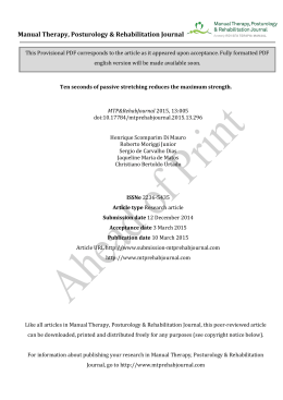
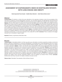
679 Original Article Effect of a muscle stretching program using the Global Postural Re-education method on respiratory muscle strength and thoracoabdominal mobility of sedentary young males* Marlene Aparecida Moreno1, Aparecida Maria Catai2, Rosana Macher Teodori3, Bruno Luis Amoroso Borges1, Marcelo de Castro Cesar4, Ester da Silva3 Abstract Objective: To evaluate the effect that respiratory muscle stretching using the global postural reeducation (GPR) method has on respiratory muscle strength, thoracic expansion and abdominal mobility in sedentary young males. Methods: This was a randomized study involving 20 sedentary volunteers, aged 22.7 ± 2.5 years, divided into two groups of 10: a control group, composed of subjects not performing any exercises, and a group of subjects submitted to the GPR method. The protocol consisted of a program to stretch the respiratory muscles with participants in the ‘open-arm, open hip joint angle’ position, which was regularly performed twice a week for 8 weeks, totaling 16 sessions. The two groups were submitted to measurements of maximal inspiratory pressure, maximal expiratory pressure, thoracic expansion and abdominal mobility, prior to and after the intervention period. Results: The initial and final values for maximal respiratory pressures, thoracic expansion and abdominal mobility for the control group showed no significant differences (p > 0.05). However, for the GPR group, all values increased after the intervention (p < 0.05). Conclusions: Respiratory muscle stretching using the GPR method was efficient in promoting an increase in maximal respiratory pressures, thoracic expansion and abdominal mobility, suggesting that it could be used as a physiotherapy resource to develop respiratory muscle strength, thoracic expansion and abdominal mobility. Keywords: Posture; Muscle Strength; Respiratory Muscles; Muscle Stretching Exercises. * Study conducted in the Laboratory for Cardiovascular Therapy Research and Functional Testing at the Faculdade de Ciências da Saúde – FACIS, School of Health Sciences – of the Universidade Metodista de Piracicaba – UNIMEP, Methodist University of Piracicaba – Piracicaba, Brazil. 1. Professor of Physical Therapy at the Universidade Metodista de Piracicaba – UNIMEP, Methodist University of Piracicaba – Piracicaba, Brazil. 2. Professor in the Graduate Program in Physical Therapy at the Universidade Federal de São Carlos – UFSCar, Federal University of São Carlos – São Carlos, Brazil. 3. Professor in the Graduate Program in Physical Therapy at the Universidade Metodista de Piracicaba – UNIMEP, Methodist University of Piracicaba – Piracicaba, Brazil. 4. Professor in the Graduate Program in Physical Education at the Universidade Metodista de Piracicaba – UNIMEP, Methodist University of Piracicaba – Piracicaba, Brazil. Correspondence to: Marlene Aparecida Moreno. Rua Santa Cruz, 990, Bairro Alto, CEP 13419-030, Piracicaba, SP, Brasil. Tel 55 19 3433-0743. E-mail: [email protected] Submitted: 28 November 2006. Accepted, after review: 14 March 2007. J Bras Pneumol. 2007;33(6):679-686 680 Moreno MA, Catai AM, Teodori RM, Borges BLA, Cesar MC, Silva E Introduction Maintaining respiratory muscle function is of vital importance for the respiratory system, just as the heart muscle is for the circulatory system. These muscles are fundamental in the maintenance of respiratory mechanics, and, under physiopathological conditions, muscle force is altered, which is reflected as a reduction in the respiratory pressures.(1,2) When a muscle loses its normal flexibility, the length-tension relationship is altered, preventing the muscle from reaching sufficient peak tension, which evolves to muscle weakness and retraction.(3) This muscle shortening can result from various factors, such as incorrect postural alignment, immobilization of the muscle, muscle weakness and aging. In the literature, there is little data regarding the effect of respiratory muscle stretching, probably because the respiratory muscle group is functionally complex, and there are therefore no specific techniques. Muscle stretching is a resource that is widely used in rehabilitation programs as well as in sports, since it can prevent injuries and increase flexibility.(4) In themselves, muscle fibers are incapable of lengthening, which requires an external force applied to the muscle. Diverse methods and techniques have been described with the objective of promoting muscle stretching, the static method being the most widely used to increase flexibility and achieve relaxation. This method involves the use of exercises that can be performed in isolation or in a general fashion, involving various segments simultaneously. During static stretching, the tension created in the muscle groups is of low intensity, allowing the patient to remain comfortable while achieving efficacy in the treatment.(5) The active muscle stretching method, first described in 1987,(6) stretches the antigravity muscles (internal rotators and respiratory muscles) as a group and is based on an understanding of the postural muscle groups. Consequently, it is known as global postural re-education (GPR). This method is widely employed and has often been used as in the practice of physical therapy to achieve postural alterations, principally in individuals presenting spinal column disorders. Therefore, there is little documentation of its effect on the respiratory system. J Bras Pneumol. 2007;33(6):679-686 Despite the fact that its benefits have been demonstrated in clinical practice, scientific evidence is fundamental for its validation as a treatment alternative, since there are few data regarding its effects, principally on respiratory function. In view of the muscle re-education that the GPR method can provide, this study had the objective of analyzing the effect of respiratory muscle stretching on respiratory muscle force, as well as on thoracic expansion and abdominal mobility, in young men with a sedentary lifestyle. Methods The study was approved by the Ethics in Research Committee of the Institution (protocol 03/05). The objectives, as well as the experimental procedures, were explained in detail to the volunteers, all of whom gave written informed consent. The sample size calculation was performed using the GraphPad StatMate program, version 1.01i, with a 95% confidence interval and a power of 80%. The study included 20 male volunteers with a sedentary lifestyle and presenting low aerobic capacity, according to the American Heart Association system of classification, which specifies a maximal oxygen uptake of 30.2 ± 4.3 mL/kg/min. All of the volunteers were nonsmokers and presented similar anthropometric characteristics (Table 1). According to previous clinical evaluations, none of these individuals had any history of musculoskeletal, cardiovascular or respiratory disease. Each of the volunteers was submitted to a complete physical therapy evaluation, including the postural evaluation recommended by Souchard and Ollier,(7) which requires that the shortening of a given muscle group be determined qualitatively. This evaluation includes Table 1 - Comparison between the volunteers in the control group and those in the global postural re-education group, in mean and standard deviation, in terms of age and anthropometric data. Control (n = 10) GPR (n = 10) Age (years) 23.4 ± 2.7 22.9 ± 2.0 Body mass (kg) 81.1 ± 7.3 80.0 ± 4.3 Height (cm) 177.7 ± 6.1 176.4 ± 5.7 BMI (kg/m2) 25.6 ± 1.2 25.1 ± 1.9 There were no statistically significant differences for any of the variables studied (p > 0.05 for all); GPR: global postural re-education; and BMI: body mass index. Effect of a muscle stretching program using the Global Postural Re-education method on respiratory muscle strength and thoracoabdominal mobility of sedentary young males the following elements: ‘Photograph’, ‘Interview’, ‘Examination of retractions’, ‘Re-education’ and ‘Result’. In our study sample, we included only those volunteers who presented the following characteristics: ‘Photograph’ - anterior projection of the head, lumbar hyperlordosis and anterior tilting of the pelvis when the volunteer was instructed to remain standing with arms outstretched; ‘Interview’ - absence of pain; ‘Examination of retractions’ - anterior projection of the head, rounded shoulders with medial rotation of the humerus, dorsal kyphosis, lumbar hyperlordosis, anterior tilting of the pelvis, valgus knee, flat feet and valgus heel; ‘Re-education’ - increased compensatory movements during prolonged expiration; ‘Result’ - opting for a posture that is open from the angle of the hip with the arms outstretched (open-arm, open hip joint angle). The prediction equations for normal values of maximal inspiratory pressure (MIP) and maximal expiratory pressure (MEP) used in our study were those described by Neder et al.(8) All of the volunteers presented MIP values below that predicted for the population studied (139.4 cmH2O, with a lower limit of 112.1 cmH2O). The MEP values were within the limits of normality (146.9 cmH2O, with a lower limit of 121.3 cmH2O). The volunteers were randomly divided into two groups of 10 (randomization performed by numeric table): those in the control group did not participate in the stretching protocol, whereas those in the study group were submitted to the GPR method intervention. During the study period, there were no losses. Before and after the intervention period, all of the volunteers were submitted to an evaluation of maximal respiratory pressures and to measurement of thoracic expansion and abdominal mobility. The volunteers were familiarized with all of the procedures prior to the beginning of the experiment. In order to avoid any bias in the results, all of the measurements were taken by a researcher who was blinded as to which group a given volunteer belonged. The respiratory pressures were measured with a vacuum manometer (±300 cmH2O; GER-AR, São Paulo, SP, Brazil). The device was precalibrated using a mercury column. All measurements were made by the same researcher using homogeneous verbal commands 681 while the volunteers were seated and wearing nose clips to keep their nostrils closed. The MIP was measured during exertion starting from the residual volume, whereas the MEP was measured starting from the total lung capacity. Each volunteer performed five maximal inspiration maneuvers and five maximal expiration maneuvers, each sustained for at least 2 s, with a variation between values of ≤10%, the highest value being recorded for later evaluation.(8-10) In the evaluation of thoracic expansion and abdominal mobility, a metric tape was used in order to measure the thoracic (axillary and xiphoid) and abdominal circumferences. During the measurements, the volunteer was instructed to perform a maximum expiration followed by a maximum inspiration. The difference between these measurements furnished information regarding the degree of expansibility and retraction of the movements.(11-13) To ensure reliability, the measurements were performed in triplicate at each level, the highest value being recorded for later evaluation. The intervention protocol consisted of a program of respiratory muscle stretching using the GPR method in the ‘open-arm, open hip joint angle’ posture, performed twice a week for 8 weeks, totaling 16 sessions. Prior to the postural exercises, each volunteer was placed in the supine position and submitted to a diaphragmatic relaxation maneuver that consists of applying gentle, sliding pressure with the fingertips, working bilaterally from the xiphocostal angle to the lower ribs. This maneuver made it possible to relax the diaphragm in preparation for the stretching.(14) The volunteer was then positioned with his arms at an angle of approximately 45 degrees to the body, forearms downs, palms up, legs spread, hips flexed, knees bent and the soles of the feet together (Figure 1). Pressure was applied to the back in order to align the dorsal and cervical curves of the spinal column, whereas sacral pressure was used in order to straighten the lumbar spine. The volunteer was instructed to the spread his hips from the initial position, maintaining the soles of the feet together, in alignment with the body axis. The physical therapist used verbal commands and manual contact in order to maintain the alignment and make the necessary postural corrections, with the objective of optimizing the stretching and J Bras Pneumol. 2007;33(6):679-686 682 Moreno MA, Catai AM, Teodori RM, Borges BLA, Cesar MC, Silva E Figure 1 - Illustration of the initial position of the ‘open-arm, open hip joint angle’ posture used in global postural re-education. Figure 2 - Illustration of the final position of the ‘open-arm, open hip joint angle’ posture used in global postural re-education. discouraging compensatory movements. The volunteer was instructed to the inhale calmly and the exhale slowly, lowering the ribcage and extending the abdomen as much as possible in order to stretch the respiratory muscles, while the physical therapist assisted in maintaining the axial lengthening. During the postural exercise, the arms remained abducted, with progressive stretching of the pectoral muscles, thereby avoiding compensatory movements. Likewise, the legs remained in apposition and advanced caudally, principally in order to stretch the iliac psoas muscle, keeping the lumbar curve in contact with the surface used for support (Figure 2). The same postural exercise was performed in all of the sessions, in each of which the physical therapist advanced the posture to the limit for each volunteer, promoting the progressive stretching of the muscle groups involved in the postural exercise during the treatment. The posture was maintained for 30 min. For the statistical analysis, the Statistica for Windows program, Release 5.1. (StatSoft, Inc., Tulsa, OK, USA) was used. The Kolmogorov-Smirnov test was used in order to determine the normality of the distribution of the data and to reject the null hypothesis for all of the variables studied. In the analysis of the significance, nonparametric tests were used, the Wilcoxon test for paired samples and the Mann-Whitney test for unpaired samples. Values of p < 0.05 were considered statistically significant. Results J Bras Pneumol. 2007;33(6):679-686 Table 2 shows that, in the control group, the pre-intervention absolute and relative values of MIP and MEP in relation to the predicted values did not differ significantly from those obtained after the intervention. For the GPR group, these values were significantly higher after the intervention. In the intergroup evaluation, the values were found to be comparable in the pre-intervention period, whereas, in the post-intervention period, the GPR group presented significantly higher values after the last of the 16 sessions. Table 3 presents the measurements of thoracic expansion and abdominal mobility. As can be seen, there were no significant differences between the pre- and post-intervention values for the control group, whereas all of the post-intervention values for the GPR group were significantly different from the corresponding pre-intervention values and from the post-intervention control group values. In the intergroup evaluation, we found that the two groups presented comparable pre-intervention values, whereas the GPR group presented significantly higher post-intervention values. Discussion The results of the present study demonstrate that physical therapy intervention using an 8-week program of GPR in young, healthy, yet sedentary, volunteers significantly increased the maximal Effect of a muscle stretching program using the Global Postural Re-education method on respiratory muscle strength and thoracoabdominal mobility of sedentary young males 683 Table 2 - Comparison between the volunteers in the control group (n = 10) and those in the global postural re-education group (n = 10) in terms of maximal inspiratory pressure and maximal expiratory pressure, before and after the intervention period. MIPa MEPa Before After Before Control (cmH2O) 104.5 ± 12.12 102.5 ± 11.84 132 ± 11.35 131 ± 11.25 GPR (cmH2O) 105.5 ± 11.16 146.5 ± 14.91b,c 136 ± 17.12 186.5 ± 25.17b,c d Control (%) 74.6 ± 8.6 73.2 ± 8.4 90.1 ± 7.7 90.6 ± 6.6 GPR (%)d 76.3 ± 9.8 105.2 ± 10.7b,c 92.1 ± 11.6 126.3 ± 10.6b,c GPR: global postural re-education; MIP: maximal inspiratory pressure; and MEP: maximal expiratory pressure; aMean ± standard deviation; bp = 0.002 GPR before vs. GPR after; cp = 0.0001 control after vs. GPR after; and dPercentage difference in relation to predicted values. respiratory pressures, as well as thoracic expansion and abdominal mobility. To date, few studies have shown improvements in the respiratory function of individuals submitted to this type of stretching. In addition to the GPR method, other physical activities have been shown to have an effect on respiratory function. In one study,(13) the author observed an increase in the inspiratory and expiratory muscle force, as well as in the thoracic mobility, of young, sedentary women after a 4-week program using proprioceptive neuromuscular facilitation techniques. Our results with GPR-guided respiratory muscle stretching corroborate those findings. Yoga also focuses on stretching and respiratory function, and breath control is crucial in its practice, as was demonstrated in a study that demonstrated a modification in the MIP after 3 months of regular practice of the activity.(15) In general, alterations in the respiratory mechanics result from excessive shortening of the respiratory musculature. The principal causes of such shortening are psychoneural factors (stress), an increase of the volume of the visceral mass, inappropriate posture, respiratory disease, muscle weakness and aging.(6) All of the postural exercises involved in the GPR method permit respiratory muscle stretching. However, one author stated that the ‘open hip joint angle’ and ‘standing open hip joint angle’ positions allow greater stability of the diaphragmatic insertions are are ideal for stretching the muscles of the diaphragm, the sternocleidomastoid muscle, scalene muscles, intercostal muscles and dorsal muscles, as well as the major and minor pectoral muscles.(6) The increased flexibility of the diaphragm is made possible through the fixation of its insertions, together with its eccentric contraction. Therefore, special attention has been given to the utilization of the GPR method in the stretching of the respiratory musculature.(14) When the muscle is immobilized, its immobilization is due to modifications of the contractile proteins and of the metabolism of the mitochondria, resulting in a reduction in the number of sarcomeres and an increase in the deposition of connective tissue,(16) leading to muscle shortening and limited articular mobility. The stretching of a muscle fiber promotes a serial increase in the number of sarcomeres.(17,18) Therefore, the increased muscle force in function of the stretching might be attributable to better interaction between the filaments of actin and myosin, by virtue of the increase in the functional length of the muscle. Despite the fact that the respiratory musculature cannot be immobilized, its constant contraction promotes a particular posture during inspiration,(14) restricting the mobility of the chest cavity. When the length of the muscle fiber is chronically altered, the number of sarcomeres adjusts to compensate for this change.(19) Although the extent of this adaptation is unknown, it is thought that such changes in muscle length will be reflected in the functional capacity of the muscle. In one study, it was reported that the generation of tension in the skeletal muscle, as determined by evaluating the length-tension relationship, is directly correlated with the degree to which actin and myosin filaments are superimposed, less superimposition of these filaments in the muscle at rest translating to greater capacity of the muscle of generate tension.(20) Studies using animal models, in which it is possible to analyze the muscle fibers, have shown J Bras Pneumol. 2007;33(6):679-686 684 Moreno MA, Catai AM, Teodori RM, Borges BLA, Cesar MC, Silva E Table 3 - Comparison between the volunteers in the control group and those in the global postural re-education group in terms of the circumference values obtained at the axillary, xiphoid and abdominal level, before and after the intervention period. Control (n = 10) GPR (n = 10) Beforea Aftera Beforea Aftera Axillary (cm) 6.2 ± 0.2 6.2 ± 0.2 6.1 ± 0.3 7.1 ± 0.2b,c Xiphoid (cm) 5.1 ± 0.2 5.2 ± 0.2 5.2 ± 0.2 6.9 ± 0.3b,c Abdominal (cm) 5.6 ± 0.4 5.6 ± 0.4 5.6 ± 0.4 7.1 ± 0.3b,c GPR: global postural re-education; aMean ± standard deviation; bp = 0.002 GPR before vs. GPR after; and cp = 0.0001 control after vs. GPR after. that performing stretching exercises once a week in shortened muscles is sufficient to reduce muscle atrophy.(21) Some authors have reported that, in normal muscles submitted to stretching exercises three times a week, there is a serial increase in the number of sarcomeres and in the cross-sectional area of the muscle fibers.(18) Other authors have stated that the length of the sarcomere is regulated by the duration of the tension to which the muscle is submitted(22); prolonged periods of stretching can lead to an adaptive muscular process that is more efficacious than that observed after short periods of stretching. In this aspect, in our study, the overall duration of the treatment, as well as the duration of each session of stretching, can promote an increase in the length of the sarcomeres and contractions that are more efficacious, as evidenced by the increase in the maximal respiratory pressures, in thoracic expansion and in abdominal mobility. The postural exercises used in the present study did indeed promote such an alteration in the interaction between the filaments of actin and myosin and, consequently, improved the contractile capacity of the respiratory muscle group. Another aspect relevant is the possible serial increase of the number of sarcomeres, which might have promoted the increase in the contractile capacity of this muscle group. Some authors have reported that the maximum pressure generated by a muscle reflects its strength.(1) Therefore, in the present study, we attempted to apply stretching techniques designed to improve the length-tension relationship of the muscle fibers, thereby also improving the performance of the respiratory muscle group. The maximal inspiratory and expiratory pressures (in absolute values and in values relative J Bras Pneumol. 2007;33(6):679-686 to those predicted), as well as the measurements of thoracic expansion and abdominal mobility, observed in the present study show that respiratory muscle stretching, performed in 16 sessions of 30 min each, was beneficial to the participants in that it resulted in greater respiratory muscle contractile force, as well as increasing thoracic expansion and abdominal mobility. Other authors report that, after a single session of intervention using the GPR method in 20 healthy young women, there were statistically significant increases in MIP and thoracic expansion in the axillary region. These gains were attributed to the increased respiratory muscle force and thoracic mobility resulting from the stretching, which involved the respiratory muscle group as part of the global posture.(23) Since assuming the ‘open-arm, open hip joint angle’ position requires considerable contraction of the expiratory muscles, the increase in expiratory muscle force observed in our results corroborates those of another study demonstrating that the better muscle performance can be explained by the improved ability to coordinate the activity of this muscle group, which can be characterized as motor learning.(24) As previously mentioned, the GPR method presents certain advantages in relation to other types of stretching due to the fact that GPR maintains the musculature in extension for a prolonged period. In addition, GPR affects the muscle groups in an integrated fashion, facilitating adaptations that promote improvements in flexibility and strength. There have been few studies showing the benefits of the GPR method, especially in terms of its effect on the respiratory musculature. However, the data from those that have been conducted unanimously indicate that GPR increases muscle length, as well as improving range of movement and flexibility, Effect of a muscle stretching program using the Global Postural Re-education method on respiratory muscle strength and thoracoabdominal mobility of sedentary young males consequently improving the contractile capacity of the affected muscles. In another study,(25) a 4-month GPR protocol involving stretching and flexibility exercises was applied in patients with ankylosing spondylitis. The authors found that the use of this protocol increased the range of movement and flexibility of the spinal column to a greater extent than did that of conventional physical therapy. Our research group focused on studying the influence of the GPR method on the respiratory musculature, as well as on thoracic expansion and abdominal mobility, in order to facilitate future studies involving individuals that present respiratory dysfunction. According to some authors,(1) the increase in lung volume results in significant shortening of the respiratory muscles. This increase is primarily seen in obstructive respiratory diseases, a classic example being chronic obstructive pulmonary disease (COPD). In COPD, the clinical profile and the repercussions for the overall health status of the patient are influenced by systemic manifestations and require a treatment strategy that addresses all of the components of the disease.(26) The morphofunctional characteristics of individuals with COPD are different from those presented by the volunteers evaluated in the present study. Nevertheless, we believe that, since COPD patients typically present mechanical dysfunction, they could benefit from the method proposed in terms of the alterations in the respiratory muscles. In patients with obstructive lung diseases, adequate muscle length would increases the efficacy of the respiratory muscles, promoting better respiratory mechanics. However, further investigation is necessary in order to test this hypothesis. It must be borne in mind that, in COPD, in addition to the impairment of the respiratory mechanics, there are other systemic manifestations that require care and specific treatment involving medical supervision. Therefore, muscle stretching should be proposed as only one part of a program of pulmonary rehabilitation. Within this context, the results of the present study show that the ‘open-arm, open hip joint angle’ postural exercise included in the GPR method was efficient in promoting an increase in the maximal respiratory pressures, as well as greater thoracic 685 expansion and abdominal mobility, suggesting that it can be used as a physical therapy resource for increasing respiratory muscle force, thoracic expansion and abdominal mobility in other situations. Despite the fact that our study reports data related to healthy individuals, all of the volunteers presented maximal respiratory pressures below the lower limit for the population studied and, after the intervention, there was a significant increase in those values. Therefore, the results obtained suggest that the exercises proposed can be of therapeutic importance in the treatment of respiratory muscle alterations. Further studies are needed in order to evaluate the effect that such stretching exercises have on the respiratory musculature of individuals with COPD, whose diaphragm muscles present a mechanical disadvantage in virtue of the hyperinflation caused by the disease. References 1. Derenne JP, Macklem PT, Roussos C. The respiratory muscles: mechanics, control, and pathophysiology. Am Rev Respir Dis. 1978;118(1):119-33. 2. Rochester DF, Braun NM. Determinants of maximal inspiratory pressure in chronic obstructive pulmonary disease. Am Rev Respir Dis. 1985;132(1):42-7. 3. Gossman MR, Sahrmann SA, Rose SJ. Review of lengthassociated changes in muscle. Experimental evidence and clinical implications. Phys Ther. 1982;62(12):1799-808. 4. Kubo K, Kanehisa H, Kawakami Y, Fukunaga T. Influence of static stretching on viscoelastic properties of human tendon structures in vivo. J Appl Physiol. 2001;90(2):520-7. 5. Kisner C, Colby LA. Stretching. In: Kisner C, Colby LA, editors. Therapeutic exercise: foundations and techniques. Philadelphia: F.A. Davis Company; 2002. p. 121-53. 6. Souchard PE. Reeducação postural global: método do campo fechado. São Paulo: Ícone, 1987. p. 91-104. 7. Souchard PE, Ollier M. As famílias de posturas – As posturas. In: Souchard PE, Ollier M, editors. As escolioses: seu tratamento fisioterapêutico e ortopédico. São Paulo: É Realizações; 2001. p. 145-66. 8. Neder JA, Andreoni S, Lerario MC, Nery LE. Reference values for lung function tests. II. Maximal respiratory pressures and voluntary ventilation. Braz J Med Biol Res. 1999;32(6):719-27. 9. Black LF, Hyatt RE. Maximal respiratory pressures: normal values and relationship to age and sex. Am Rev Respir Dis. 1969;99(5):696-702. 10. Souza RB. Pressões respiratórias estáticas máximas. J Pneumol. 2002;28(Supl 3):S155-S65. 11. Paulin E, Brunetto AF, Carvalho CRF. Efeitos de programa de exercícios físicos direcionado ao aumento da mobilidade torácica em pacientes portadores de doença pulmonar obstrutiva crônica. J Pneumol. 2003;29(5):287-94. 12. Silva FB, Sampaio LMM, Carrascosa AC. Avaliação fisioterapêutica dos sistemas mastigatórios e respiratório de J Bras Pneumol. 2007;33(6):679-686 686 Moreno MA, Catai AM, Teodori RM, Borges BLA, Cesar MC, Silva E um portador de síndrome otodental: um estudo de caso. Rev Bras Fisioter. 2006;10(1):133-6. 13. Moreno MA. Padrões de facilitação neuromuscular proprioceptiva e seu efeito na capacidade respiratória [dissertação]. Campinas: Universidade Estadual de Campinas; 2000. 14. Souchard PE. Respiração. São Paulo: Summus; 1989. p. 100-2. 15. Godoy DV, Bringhenti RL, Severa A, Gasperi R, Poli LV. Ioga versus atividade aeróbia: efeitos sobre provas espirométricas e pressão inspiratória máxima. J Bras Pneumol. 2006;32(2):130-5. 16. Williams PE, Goldspink G. Changes in sarcomere length and physiological properties in immobilized muscle. J Anat. 1978;127(Pt 3):459-68. 17. Shah SB, Peters D, Jordan KA, Milner DJ, Fridén J, Capetanaki Y, et al. Sarcomere number regulation maintained after immobilization in desmin-null mouse skeletal muscle. J Exp Biol. 2001;204(Pt 10):1703-10. 18. Coutinho EL, Gomes AR, Franca CN, Oishi J, Salvini TF. Effect of passive stretching on the immobilized soleus muscle fiber morphology. Braz J Med Biol Res. 2004;37(12):1853-61. 19. Lieber RL. Skeletal muscle response to injury. In: Lieber RL, editor. Skeletal muscle structure, function and plasticity: the physiological basis of rehabilitation. Baltimore: Lippincott Williams & Wilkins; 2002. p. 287-346. J Bras Pneumol. 2007;33(6):679-686 20. Lieber RL, Bodine-Fowler SC. Skeletal muscle mechanics: implications for rehabilitation. Phys Ther. 1993; 73(12):844-56. 21. Gomes AR, Coutinho EL, Franca CN, Polonio J, Salvini TF. Effect of one stretch a week applied to the immobilized soleus muscle on rat muscle fiber morphology. Braz J Med Biol Res. 2004;37(10):1473-80. 22. Herring SW, Grimm AF, Grimm BR. Regulation of sarcomere number in skeletal muscle: a comparison of hypotheses. Muscle Nerve. 1984;7(2):161-73. 23. Teodori RM, Moreno MA, Fiori Junior JF, Oliveira ACS. Alongamento da musculatura inspiratória por intermédio da reeducação postural global (RPG). Rev Bras Fisioter. 2003;7(1):25-30. 24. Jones DA, Rutherford OM, Parker DF. Physiological changes in skeletal muscle as a result of strength training. Q J Exp Physiol. 1989;74(3):233-56. 25. Fernández-de-Las-Peñas C, Alonso-Blanco C, MoralesCabezas M, Miangolarra-Page JC. Two exercise interventions for the management of patients with ankylosing spondylitis: a randomized controlled trial. Am J Phys Med Rehabil. 2005;84(6):407-19. 26. Dourado VZ, Tanni SE, Vale SA, Faganello MM, Sanchez FF, Godoy I. Manifestações sistêmicas na doença pulmonar obstrutiva crônica. J Bras Pneumol. 2006;32(2):161-71.
Baixar