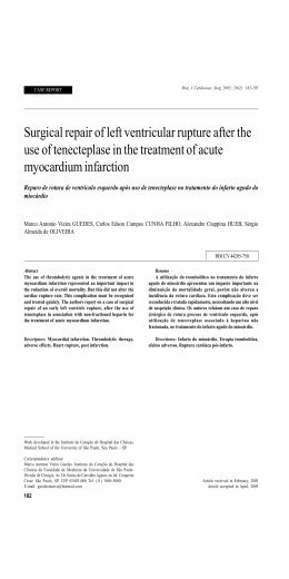
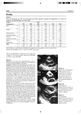
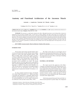
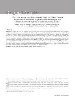
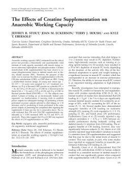
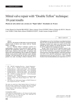
+Model
ARTICLE IN PRESS
REPC-231; No. of Pages 4
Rev Port Cardiol. 2013;xxx(xx):xxx---xxx
Revista Portuguesa de
Cardiologia
Portuguese Journal of Cardiology
www.revportcardiol.org
CASE REPORT
Ischemic rupture of the anterolateral papillary muscle
Catarina Vieira a,∗ , António Gaspar a , Miguel Álvares Pereira a , Nuno Salomé a ,
Jorge Almeida b , Mário Jorge Amorim b
a
b
Department of Cardiology, Hospital de Braga, Braga, Portugal
Department of Cardiothoracic Surgery, Hospital de São João, Oporto, Portugal
Received 25 May 2012; accepted 4 June 2012
KEYWORDS
Papillary muscle;
Rupture;
Surgical
reimplantation;
Echocardiography
PALAVRAS-CHAVE
Músculo papilar;
Rutura;
Reimplantação
cirúrgica;
Ecocardiograma
∗
Abstract We describe the case of a 59-year-old man who presented with chest pain and
ST-segment elevation in the inferior leads, R > S in V1 and ST depression in the anterior leads due
to proximal occlusion of the first obtuse marginal. Primary coronary angioplasty and stenting of
this artery were performed. Twelve hours later the patient became hemodynamically unstable
and severe mitral regurgitation due to rupture of one of the heads of the anterolateral papillary
muscle was diagnosed. Emergency surgery was performed (papillary muscle head reimplantation, mitral annuloplasty with a rigid ring, tricuspid annuloplasty and coronary artery bypass
grafting). On surgical inspection, it was observed that the detached muscle head had become
trapped in the left ventricle by a secondary cord attached to the other head.
This case is unusual in presenting two uncommon features of ischemic papillary muscle:
rupture of the anterolateral muscle in myocardial infarction involving the inferoposterior walls,
and the fact that the ruptured muscle head did not prolapse because it had become trapped in
the left ventricle by secondary cord attachment.
© 2012 Sociedade Portuguesa de Cardiologia. Published by Elsevier España, S.L. All rights
reserved.
Rutura isquémica do músculo papilar ântero-lateral
Resumo Apresentamos um caso de um homem, de 59 anos de idade, admitido no Serviço de
Urgência por dor torácica e elevação ST nas derivações inferiores, r > s em V1 e depressão ST nas
derivações precordiais, devido a oclusão da primeira obtusa marginal. Foi realizada angioplastia
primária dessa lesão com implantação de stent revestido. Doze horas depois, o doente desenvolve instabilidade hemodinâmica, tendo sido diagnosticada uma insuficiência mitral grave por
rutura de uma das cabeças do músculo papilar ântero-lateral. O doente foi submetido a cirurgia emergente (reimplantação do músculo papilar ântero-lateral, anuloplastia mitral com anel
rígido, anuloplastia tricúspide e revascularização miocárdica). A inspeção cirúrgica revelou que
a cabeça muscular rompida se mantinha presa ao ventrículo esquerdo por uma corda secundária
que a ligava à outra cabeça do músculo papilar ântero-lateral.
Corresponding author.
E-mail address: [email protected] (C. Vieira).
0870-2551/$ – see front matter © 2012 Sociedade Portuguesa de Cardiologia. Published by Elsevier España, S.L. All rights reserved.
http://dx.doi.org/10.1016/j.repc.2012.06.014
Please cite this article in press as: Vieira C, et al. Ischemic rupture of the anterolateral papillary muscle. Rev Port Cardiol.
2013. http://dx.doi.org/10.1016/j.repc.2012.06.014
+Model
REPC-231; No. of Pages 4
ARTICLE IN PRESS
2
C. Vieira et al.
Este caso tem a particularidade de apresentar 2 características incomuns da isquemia do músculo papilar: rutura do músculo ântero-lateral num enfarte póstero-inferior e o facto de a
cabeça de músculo não prolapsar para a aurícula esquerda, por se encontrar presa ao ventrículo
esquerdo por uma corda secundária.
© 2012 Sociedade Portuguesa de Cardiologia. Publicado por Elsevier España, S.L. Todos os
direitos reservados.
Introduction
Papillary muscle rupture is an infrequent but often
catastrophic mechanical complication of acute myocardial
infarction (MI) accounting for approximately 5% of early
deaths.1 The condition may result in acute mitral regurgitation, acute pulmonary edema, and/or cardiogenic shock.2
Despite high operative mortality, surgery is indicated as survival of medically treated patients is very low.3
We report an uncommon presentation of an anterolateral
papillary muscle rupture successfully repaired by surgical
reimplantation of one of the papillary muscle heads.
(Figure 4) had become trapped in the left ventricle by a
secondary cord attached to the other head. Papillary muscle
head reimplantation, mitral annuloplasty with a rigid ring,
tricuspid annuloplasty with a Sovering ring and double coronary artery bypass grafting (CABG) (left internal mammary
artery to first obtuse marginal and right internal thoracic
artery to anterior descending artery) were performed.
The postoperative period was complicated by nosocomial pneumonia but the patient recovered fully and
was discharged home on the 18th day. Transthoracic
echocardiography performed 15 days after surgery showed
preserved global left ventricular function and mild mitral
regurgitation.
Case report
Discussion
A 59-year-old man was admitted to the emergency room
with acute chest pain. The 12-lead ECG (Figure 1) showed ST
elevation in the inferior leads, R > S in V1 and ST depression
in the anterior leads. The patient was hemodynamically stable and cardiac and pulmonary auscultations were normal.
Coronary angiography showed obstruction of the proximal segment of the first obtuse marginal and significant
stenosis (80%) of the proximal segment of the anterior
descending artery. Primary coronary angioplasty of the first
obtuse marginal and implantation of a drug-eluting stent
were performed. Twelve hours later the patient suddenly
became hypotensive and tachycardic. Physical examination revealed a new 3/6 holosystolic murmur at the apex
and significant bilateral rales. Transthoracic echocardiography showed severe mitral regurgitation due to posterior
mitral leaflet prolapse and an image of a mass in the
left ventricle, which did not prolapse totally into the
left atrium during systole, suggestive of an anterolateral
papillary muscle head. Left ventricular size and function
were relatively normal but the posterior and inferior walls
were hypokinetic. Moderate tricuspid regurgitation was
noticed and pulmonary artery systolic pressure was estimated at 60 mmHg.
The patient became oliguric, with deterioration of renal
function, and so an intra-aortic balloon was placed and he
was transferred to a cardiothoracic department for emergency surgery.
Intraoperative 2D/3D transesophageal echocardiography
confirmed rupture of one of the heads of the anterolateral
papillary muscle, leading to extensive prolapse of the P1
and P2 segments and causing severe mitral regurgitation
(Figures 2 and 3A and B; videos 1, 2 and 3). On surgical
inspection, it was observed that the detached muscle head
The posteromedial papillary muscle is supplied with blood
from the posterior descending artery, while the anterolateral papillary muscle has a dual blood supply, from the
left anterior descending and left circumflex arteries.4---6 This
difference in blood supply explains why rupture of the posteromedial papillary muscle is 6---12 times more common.
On the other hand, rupture of the anterolateral papillary
muscle is usually associated with anterolateral MI, rarely
occurring in the context of inferior MI, as happened in the
case presented.7
Transthoracic echocardiography is useful in the diagnosis
of papillary muscle rupture, with a sensitivity of 65---85%.8
A flail segment of the mitral valve and a severed papillary muscle or chorda can frequently be seen moving freely
within the left ventricular cavity. However, in some cases,
transthoracic echocardiography is not informative and transesophageal echocardiography is required to establish the
diagnosis; this is most likely to occur when the ruptured head
does not prolapse into the left atrium, as was observed in
our patient, a feature reported in up to 35% of cases.9
Emergency surgical intervention remains the treatment
of choice for papillary muscle rupture. Surgery involves significant operative mortality (18%), but there are recent
trends for lower operative risk, particularly with associated
CABG (8.7%).10 Long-term outcome after surgery is the same
as that of similar MI without papillary muscle rupture.10
These encouraging observations emphasize the importance
of prompt diagnosis and an aggressive therapeutic approach
for patients suffering papillary muscle rupture after MI.
This case is unusual in presenting two uncommon features
of ischemic papillary muscle: rupture of the anterolateral
muscle in MI involving the inferoposterior walls, and the fact
that the ruptured muscle head did not prolapse because it
Please cite this article in press as: Vieira C, et al. Ischemic rupture of the anterolateral papillary muscle. Rev Port Cardiol.
2013. http://dx.doi.org/10.1016/j.repc.2012.06.014
+Model
ARTICLE IN PRESS
REPC-231; No. of Pages 4
Ischemic rupture of the anterolateral papillary muscle
3
Figure 1 Electrocardiogram on admission demonstrating slight ST-segment elevation in the inferior leads, R > S in V1 and
ST-segment depression in the anterior leads.
Figure 2 Two-dimensional transesophageal echocardiogram showing severe mitral regurgitation caused by posterior leaflet prolapse. LA: left atrium; LV: left ventricle.
A
B
Ao
LA
LAA
LV
Figure 3 Three-dimensional transesophageal echocardiography. (A) Full volume cropped image showing the detached head of the
anterior papillary muscle inside the LV (arrow) causing severe prolapse of the posterior leaflet; (B) 3D Zoom of the mitral valve
showing prolapse of the P1 and P2 segments. LA: left atrium; LV: left ventricle; Ao: aortic valve; LAA: left atrial appendage.
Please cite this article in press as: Vieira C, et al. Ischemic rupture of the anterolateral papillary muscle. Rev Port Cardiol.
2013. http://dx.doi.org/10.1016/j.repc.2012.06.014
+Model
REPC-231; No. of Pages 4
ARTICLE IN PRESS
4
C. Vieira et al.
Right to privacy and informed consent. The authors have
obtained the written informed consent of the patients or
subjects mentioned in the article. The corresponding author
is in possession of this document.
Conflicts of interest
The authors have no conflicts of interest to declare.
Appendix A. Supplementary data
Supplementary data associated with this article can
be found, in the online version, at doi:10.1016/j.repc.
2012.06.014.
References
Figure 4 Intraoperative view showing the forceps holding
the detached muscle head. The two primary cords connecting
the muscle head to the edge of the posterior leaflet are clearly
seen.
had become trapped in the left ventricle by a cord attachment.
Ethical disclosures
Protection of human and animal subjects. The authors
declare that no experiments were performed on humans or
animals for this study.
Confidentiality of data. The authors declare that they have
followed the protocols of their work center on the publication of patient data and that all the patients included in the
study received sufficient information and gave their written
informed consent to participate in the study.
1. Nishimura RA, Schaff HV, Shub C, Gersh BJ, Edwards WD, Tajik
AJ. Papillary muscle rupture complicating acute myocardial
infarction: analysis of 17 patients. Am J Cardiol. 1983;51:373---7.
2. Sanders RJ, Neubuerger KT, Ravin A. Rupture of papillary muscles: occurrence of rupture of the posterior muscle in posterior
myocardial infarction. Dis Chest. 1957;31:316---23.
3. Kishon Y, Oh JK, Schaff HV, Mullany CJ, Tajik AJ, Gersh BJ. Mitral
valve operation in postinfarction rupture of a papillary muscle:
immediate results and long-term follow-up of 22 patients. Mayo
Clin Proc. 1992;67:1023.
4. Reeder GS. Identification and treatment of complications of
myocardial infarction. Mayo Clin Proc. 1995;70:880.
5. Lavie CJ, Gersh BJ. Mechanical and electrical complications of
acute myocardial infarction. Mayo Clin Proc. 1990;65:709.
6. James TN. Anatomy of the coronary arteries in health and disease. Circulation. 1965;32:1020.
7. Voci P, Bilotta F, Caretta Q, Mercanti C, Marino B. Papillary
muscle perfusion pattern. A hypothesis for ischemic papillary
muscle dysfunction. Circulation. 1995;91:1714---8.
8. Czarnecki A, Thakrar A, Fang T, et al. Acute severe mitral
regurgitation: consideration of papillary muscle architecture.
Cardiovasc Ultrasound. 2008;6:5.
9. Moursi MH, Bhatnagar SK, Vilacosta I, San Roman JA, Espinal
MA, Nanda NC. Transesophageal echocardiographic assessment
of papillary muscle rupture. Circulation. 1996;94:1003.
10. Russo A, Suri RM, Grigioni F, et al. Clinical outcome after surgical correction of mitral regurgitation due to papillary muscle
rupture. Circulation. 2008;118:1528---34.
Please cite this article in press as: Vieira C, et al. Ischemic rupture of the anterolateral papillary muscle. Rev Port Cardiol.
2013. http://dx.doi.org/10.1016/j.repc.2012.06.014
Baixar