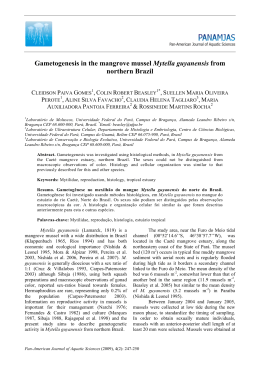
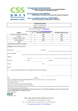
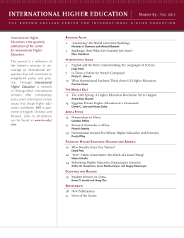
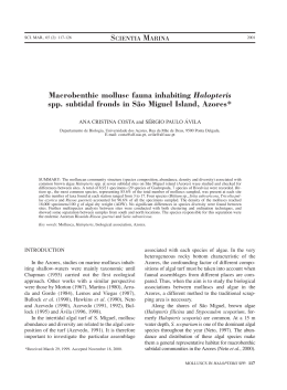
Journal of Invertebrate Pathology 103 (2010) 43–47
Contents lists available at ScienceDirect
Journal of Invertebrate Pathology
journal homepage: www.elsevier.com/locate/jip
Parasites of three commercially exploited bivalve mollusc species
of the estuarine region of the Cachoeira river (Ilhéus, Bahia, Brazil)
Guisla Boehs a,*, Antonio Villalba b, Liliane Oliveira Ceuta a, Joaldo Rocha Luz a
a
b
Universidade Estadual de Santa Cruz-UESC, Departamento de Ciências Biológicas, Rodovia Ilhéus-Itabuna, km 16, 45.650-900, Ilhéus, Bahia, Brazil
Centro de Investigacións Mariñas, Aptdo. 13, Vilanova de Arousa 36620, Spain
a r t i c l e
i n f o
Article history:
Received 23 April 2009
Accepted 14 October 2009
Available online 19 October 2009
Keywords:
Mytella guyanensis
Anomalocardia brasiliana
Iphigenia brasiliana
Parasite
Rickettsia-like organisms
Nematopsis
Bucephalidae
Tylocephalum
Brazilian molluscs
Bahia
a b s t r a c t
This paper reports the parasites found in three commercially exploited bivalve molluscs (Mytella guyanensis, Anomalocardia brasiliana and Iphigenia brasiliana) of an estuarine region of Ilhéus, south of Bahia,
Brazil (14°480 230 0 S; 39°020 470 0 W). Samples of 20 individuals of each species were collected fortnightly
from August 2005 to August 2006. A total of 1480 individuals was collected and processed by standard
histologic techniques; the histologic sections were stained with Harris haematoxylin and eosin and
examined with light microscope. The water temperature in the study area varied from 24 to 30.5 °C
and the salinity from 0 to 23 ppt. Remarkable differences were found in the parasitic community between
the three mollusc species involved in the study, which occupied different habitats in the estuarine region
of the Cachoeira river. The following parasites were found: intracellular rickettsia-like colonies in digestive epithelia; intracellular gregarine Nematopsis sp. in gills, mantle, gonad, digestive gland and foot muscle; sporocysts of a Bucephalidae trematode in gonads, mantle, gills, digestive gland and foot;
unidentified digenetic metacercariae in digestive gland and gonad; metacestodes of Tylocephalum sp.
in connective tissue in the digestive gland and in gonad; and an unidentified metazoan in mantle and
intestinal lumen. No significant temporal variation in the prevalence of any parasite was detected, which
could be due to the narrow temperature range of the region and the absence of patterns of salinity and
rainfall variation through the year. The infestation by sporocyst was the only pathological threat detected
for the studied populations because of its potential for host castration. The low infection intensity and/or
prevalence of the other parasites and the lack of obvious lesions suggest that there is no other serious
pathological risk for the studied mollusc populations.
Ó 2009 Elsevier Inc. All rights reserved.
1. Introduction
The study of parasites and diseases affecting molluscs with economic interest is important both for the management of natural
stock and for aquaculture. It could even be helpful for the sanitary
evaluation for human consumption. According to Kinne (1983), the
main biologic agents causing diseases in marine bivalve molluscs
involve viruses, bacteria, fungi, protists, digenean trematodes,
polychaetes and copepods. The populations of the bivalve molluscs, Mytella guyanensis (Lamarck, 1819) (Mytilidae), Anomalocardia brasiliana (Gmelin, 1791) (Veneridae), and Iphigenia brasiliana
(Lamarck, 1818) (Donacidae), are exploited as a source of food
and for marketing in the estuarine area of the Cachoeira river
and other areas of the Brazilian coast. The ‘‘sururu” M. guyanensis
inhabits intertidal mangrove areas, attaching to mangrove roots
with the byssus; it is distributed from Mexico to Peru in the Pacific
coast and from Venezuela to Brazil in the Atlantic coast (Rios,
* Corresponding author. Fax: +55 73 3680 5226.
E-mail address: [email protected] (G. Boehs).
0022-2011/$ - see front matter Ó 2009 Elsevier Inc. All rights reserved.
doi:10.1016/j.jip.2009.10.008
1994). The Carib pointed – venus or ‘‘berbigão” A. brasiliana inhabits intertidal and subtidal shoals of unexposed areas (Narchi, 1974)
of West Indies, Brazil and Uruguay coasts (Rios, 1994). The giant
false coquina or ‘‘tarioba” I. brasiliana inhabits sand and sandy
mud shallow areas of Florida, West Indies, Surinam, Brazil and Uruguay coasts (Rios, 1994). The three mollusc species are potential
resources for mariculture.
Studies on mollusc parasites and diseases are scarce in Brazil;
nevertheless, this type of studies has been encouraged due to the
recent expansion of cultivation systems (Sabry et al., 2007). There
are some reports on Crassostrea rhizophorae (Nascimento et al.,
1986; Sabry and Magalhães, 2005; Sabry et al., 2007; Boehs
et al., 2009), Crassostrea gigas (Sabry and Magalhães, 2005), Perna
perna (Umiji et al., 1976; Lima et al., 2001; da Silva et al., 2002;
Galvão et al., 2006), M. guyanensis (Azevedo and Matos, 1999; Pinto
and Boehs, 2008), and A. brasiliana (Narchi, 1966; Araújo and
Rocha-Barreira, 2004; Boehs and Magalhães, 2004). Among them,
only the reports by Nascimento et al. (1986), Pinto and Boehs
(2008), and Boehs et al. (2009) correspond to the coast of the state
of Bahia.
44
G. Boehs et al. / Journal of Invertebrate Pathology 103 (2010) 43–47
The aim of the present study was to characterize the parasitic
organisms of three economically important mollusc species (M.
guyanensis, A. brasiliana and I. brasiliana) in the estuarine region
of the Cachoeira river, located in south of Bahia (Brazil). The estuary is moderately polluted due to domestic wastes and heavy metals derived from agriculture activities in the region (Schiavetti
et al., 2002). That compromises fishery and consumption of local
shellfish.
which is irregularly scattered through the year, without a seasonal
pattern in the region. A seasonal pattern of temperature variation
was neither observed.
Microscopic examination of histologic sections disclosed parasitization by prokaryotic, protozoan and metazoan organisms. No
significant temporal variation in prevalence was detected for any
parasite (p > 0.05) (Table 1). No period with significantly elevated
parasite intensity was identified.
Intracellular colonies of rickettsia-like organisms were observed
in epithelial cells of the digestive gland of M. guyanensis (Fig. 1),
with low prevalence (Table 1). Colony diameter varied from 7 to
24 lm (mean ± SD = 14.5 ± 3.6; n = 44). The intensity of infection
was low (2–16 colonies per histologic section), and most frequently, only 1 colony per digestive tubule was observed. Host
cells showed hypertrophy, but there was no obvious defense
response.
Intrahemocytic oocysts of gregarines of the genus Nematopsis
Schneider, 1892 (Apicomplexa: Eugregarinida) enclosing a single
sporozoite were observed in M. guyanensis (Fig. 2) and A. brasiliana. The prevalence in the former host was much higher than in
the latter (Table 1). In the case of M. guyanensis, the most frequent tissue location was gills, followed by mantle and digestive
gland, and less frequently in the foot muscle. The number of oocysts per phagocyte in M. guyanensis varied from 1 to 20, with
1–3 being the most frequent condition. Oocyst length varied
from 3 to 10 lm (mean ± SD = 5 ± 1.3; n = 46). In few cases with
higher infection intensity, branchial and mantle morphology was
altered. In the case of A. brasiliana, Nematopsis sp. oocysts were
only observed in the foot muscle with 1 oocyst/phagocyte. Oocyst length varied from 13 to 19 lm (mean ± SD = 16 ± 2.8;
n = 5), and the infection was light without lesions or host
reaction.
Sporocysts of a Bucephalid species (Digenea) enclosing germ
balls and developing cercariae were observed in M. guyanensis
(Fig. 3) and A. brasiliana. Sporocysts showed the same apparent
morphology in both host species, but the prevalence was higher
in the latter host (Table 1). This infestation in the two host species
occurred in specimens whose size was above the sample mean.
Two specimens of M. guyanensis showed the mantle with granular
texture and orange color, which was interpreted as a macroscopic
sign of the infestation. The most frequent parasite location was the
gonad (100% of the cases); sporocysts were also found in the digestive gland and the gills and less frequently in the foot muscle. This
2. Materials and methods
Sampling was performed fortnightly, from August 2005 to August 2006, at the confluence of the Cachoeira, Santana and Fundão
rivers (14°480 2300 S; 39°020 4700 W), in the estuarine region of Cachoeira river (Ilhéus, Bahia). Twenty individuals of each species
were collected by hand, M. guyanensis from the mangrove, A. brasiliana from a shoal close to the mangrove (intertidal and upper subtidal), and I. brasiliana from the river bottom (subtidal). Sampling
was carried out during low tide, when the intertidal area was
uncovered. Water temperature and salinity were recorded while
sampling, with a standard mercury thermometer and a handheld
refractometer, respectively. The specimens in the samples were
measured in their longest axis (dorso-ventral axis for M. guyanensis
and antero-posterior axis for A. brasiliana and I. brasiliana), and
then shucked, examined for macroscopic anomalies, fixed in
Davidson’s solution (Shaw and Battle, 1957), dehydrated in an ethanol series, embedded in paraffin, sectioned (7 lm thick) and
stained with Harris haematoxylin and eosin (H&E). Histologic sections were examined by light microscopy. Parasite prevalence (No.
of infected individuals/No. of examined individuals), and distribution in the host were evaluated for each host species. Kruskal-Wallis test was used to check parasitization temporal trends
(significance limit: a = 0.05). Infection intensity was evaluated by
counting the number of parasites per histologic section when possible or by gross estimation without counting.
3. Results
Water temperature varied from 24 to 30.5 °C (mean ± SD =
27.2 ± 1.9; N = 25) and salinity from 0 to 23 ppt (mean ± SD =
13.7 ± 7.5; N = 25). The width of the salinity range was due both
to daily variations coupled to the tide cycle and to the rainfall,
Table 1
Host size, parasite prevalences (%) and significance (p values) of the Kruskal–Wallis tests performed to check prevalence temporal variation.
Months
Mollusc host species
M. guyanensis
N = 480, height range: 34.5–74.9 mm
Mean ± SD: 55.4 ± 6.7 mm
RLOs
August
September
October
November
December
January
February
March
April
May
June
July
August
Mean prevalence
(p) Kruskal–Wallis
Ntps
Ts
5
2.5
5
–
45
65
65
95
70
75
77.5
62.5
97.5
87.5
88.3
42.5
–
0.83
0.97
72.57
0.18
2.5
5
2.5
A. brasiliana
N = 500, length range: 22.3–40.0 mm
Mean ± SD: 32.06 ± 2.8 mm
UM
2.5
2.5
2.5
2.5
5
Ntps
2.5
2.5
2.5
–
–
2.08
0.58
0.21
0.99
0.58
0.99
Ts
15
2.5
2.5
12.5
2.5
7.5
17.5
10
10
1.67
10
5
7.43
0.14
UMc
2.5
2.5
2.5
0.58
0.99
Tyl
5
12.5
7.5
12.5
2.5
5
12.5
10
5
15
13.3
2.5
7.94
0.24
I. brasiliana
N = 500, length range: 45.7–74.8 mm
Mean ± SD: 60.2 ± 4.4 mm
UM
UMc
Tyl
UM
5
5
2.5
2.5
2.5
2.5
0.96
0.57
0.19
0.99
2.5
0.76
0.99
0.19
0.99
RLOs = rickettsia-like organisms; Ntps = Nematopsis sp.; Ts = trematode sporocysts; UMc = unidentified metacercariae; Tyl = Tylocephalum sp.; UM = unidentified Metazoa.
G. Boehs et al. / Journal of Invertebrate Pathology 103 (2010) 43–47
Fig. 1. Intracellular colony of Rickettsia-like organisms (arrow) in the epithelium of
a digestive tubule of M. guyanensis. Bar. = 20 lm.
Fig. 2. Oocysts of the gregarine Nematopsis sp. (arrow) in the digestive gland of M.
guyanensis. Bar. = 20 lm.
parasitization was associated with gonad follicle destruction. Host
reaction was limited to a few cases of focal hemocytic infiltration
surrounding dead or degenerating sporocysts.
Unidentified digenean metacercariae were observed in the connective tissue of the digestive gland in A. brasiliana and the gonad
of I. brasiliana; host reaction involved encapsulation of the metacercariae, which were surrounded by several layers of fibers and
hemocytes (Figs. 4 and 5). Tegumental spines were visible in some
cases (Fig. 4). The prevalence was very low for both host species
(Table 1).
Metacestodes of the genus Tylocephalum Linton, 1890 (Tetragonocephalidae) were found in the connective tissue of A. brasiliana
(Fig. 6) and of I. brasiliana. The prevalence was much higher in the
former host (Table 1). Only one metacestode per section was observed, except in three specimens of A. brasiliana in which two parasites were seen. The metacestodes were located in the connective
tissue, mostly in or close to the digestive gland but some of them in
the gonad area. The parasites were surrounded by a thick multilay-
45
Fig. 3. Trematode sporocysts (S) containing germ balls (small arrows) and cercariae
(large arrows) of a Bucephalid species in the gonad of M. guyanensis showing, in
some cases, a bifid and broad tail base (arrow head). Bar. = 20 lm.
Fig. 4. Digenean metacercaria () encysted in the connective tissue of the digestive
gland of A. brasiliana, showing tegumental spines (arrow head), cyst wall and slight
haemocytic host response (arrow). Bar. = 20 lm.
ered fibrous capsule including orange fibroblast-like cells and
hemocytes. Some images of metacestode destruction and reabsorption were observed involving loss of capsule, parasite disintegration, hemocytic infiltration and phagocytosis of parasite
residues.
Some degraded metazoan organisms occurring in the mantle of
M. guyanensis and in the intestinal lumen of the other two bivalve
mollusc species could not be identified (Table 1).
4. Discussion
Remarkable differences were found in the parasitic community
between the three mollusc species involved in the study, which
occupied different habitats in the estuarine region of the Cachoeira
river. Very few cases of parasitization were observed in I. brasiliana.
A protozoan, Nematopsis sp., was the most prevalent parasite in M.
guyanensis and a metazoan, trematode sporocysts, was the most
prevalent parasite in A. brasiliana. Rickettsia-like organisms (RLOs)
were detected only in few specimens of just one of the three
46
G. Boehs et al. / Journal of Invertebrate Pathology 103 (2010) 43–47
Fig. 5. Digenean metacercaria () in female gonad (FG) of I. brasiliana, showing
ventral sucker (V) and the capsule (C). Bar. = 20 lm.
Fig. 6. Tylocephalum metacestode () in the connective tissue of the digestive gland
of a male of A. brasiliana, showing fibrous capsule (FC) with hemocytic response and
myzorhynchus (M) of metacestode. Bar. = 20 lm.
species. The occurrence of RLOs in epithelial cells of the gills and
digestive diverticula is widespread among molluscs (Bower et al.,
1994). Like the present study, RLOs do not cause important lesions
in the host except for host cell hypertrophy, and do not elicit obvious defense response (Figueras et al., 1991; Carballal et al., 2001).
Nevertheless, some histopathologic effects have been reported,
such as disruption of those infected digestive tubules containing
the larger colonies (Cremonte et al., 2005a) in Pitar rostrata, or
hypertrophy and lysis of host gill epithelial cells and inflammatory
reaction in Venerupis rhomboides, which could be associated with
host death (Villalba et al., 1999).
The highest prevalence recorded in this study corresponded to
Nematopsis sp. Gregarines of the genus Nematopsis had been reported in the bivalve molluscs from Brazilian coasts, C. rhizophorae
(Nascimento et al., 1986; Sabry and Magalhães, 2005; Sabry et al.,
2007), M. guyanensis (Azevedo and Matos, 1999; Pinto and Boehs,
2008) and P. perna (Lima et al., 2001), and the gastropod mollusc
Nerita ascencionis (Azevedo and Padovan, 2004). As observed in this
study, these gregarines cause focal hemocytic infiltration at most,
without obvious pathogenic effects (Bower et al., 1994); their most
frequent tissue locations are gills and mantle (Carballal et al., 2001;
Winstead et al., 2004; Sabry and Magalhães, 2005; Sabry et al.,
2007). Life cycle of these parasites is completed in the intestine
of marine arthropods (Bower et al., 1994). According to observations during sampling, crustaceans were much more abundant in
the mangrove area than in the other two sampled areas; the difference in crustacean abundance could contribute to explain the significant differences in the Nematopsis sp. prevalence between M.
guyanensis and A. brasiliana. Further studies of the parasites of
crustaceans in those areas should be performed to assess the
hypothesis.
The bucephalids are a group of digenean trematodes with complex life cycles in which bivalve molluscs are intermediate hosts
harboring larval stages and some finfish species are known definitive hosts. Bucephalid cercariae occurring in bivalve molluscs have
typical gasterostome organization and a bifid tail with a very short
and broad tail base (Paperna, 1995). The morphology of the sporocysts and cercariae observed in this study resembled that of
Bucephalids. The macroscopic evidence of this infestation in two
specimens of M. guyanensis is consistent with the characteristic
pigmentation referred as ‘‘orange sickness” (Cole 1935, in Lasiak,
1993) observed in mussels infested by bucephalid trematodes.
The occurrence of this infestation in molluscs with larger size (older) agrees with reports by Lasiak (1993) in the mussel P. perna and
Villalba et al. (1997) in the mussel Mytilus galloprovincialis, who
observed higher prevalence of bucephalid infestation in the larger/older individuals; older individuals are exposed longer to
infection and filter higher volumes of water than younger/smaller
individuals. The gonad follicle destruction associated with this
infestation in the study is well documented (Lauckner, 1983)
and, probably, is due to both mechanical damage and physiologic
decomposition of tissues (Winstead et al., 2004). According to
Bower et al. (1994), the sporocysts reduce glycogen content (energy reserves) of the tissues and efficiency of the circulatory system, resulting in disturbance to gametogenesis and castration.
The very limited host reaction observed in this study agrees with
observations reported by Winstead et al. (2004) in Crassostrea virginica. Hemocyte infiltration was reported in specimens of Protothaca antiqua with early infections by sporocysts containing germinal
balls inside but without developing cercariae (Cremonte et al.,
2005b). In the mussel P. perna, hemocytic infiltration was observed
in very heavily infected animals (da Silva et al., 2002), but such
reaction seems to be uncommon in molluscs acting as first intermediate hosts of digenetic trematodes (Cremonte et al., 2005b).
Digenean trematodes have been reported in A. brasiliana (Narchi,
1966; Araújo and Rocha-Barreira, 2004; Boehs and Magalhães,
2004) from the Brazilian coasts; the first two reports corresponded
to bucephalids. Bucephalid trematodes have also been reported in
other Brazilian mollusc species not involved in this study, P. perna
(Umiji et al., 1976; Lima et al., 2001; da Silva et al., 2002; Galvão
et al., 2006) and C. rhizophorae (Nascimento et al., 1986). All those
reports referred to host castration.
Trematode metacercariae were also found with no obvious host
damage. The digenean metacercariae usually do not cause host
castration (Lauckner, 1983) or other significant lesions to bivalve
molluscs (Bower et al., 1994; Carballal et al., 2001; Winstead
et al., 2004); however, shell lesions or disturbance in shell growth
could occur in molluscs harboring numerous metacercariae
(Lauckner, 1983; Cremonte et al., 2005b). The tegumental spines
observed in this study were reported in previous studies (Paperna,
1995; Cremonte et al., 2005a).
Larval cestodes corresponding to genus Tylocephalum were observed in this study. The adults of the cestodes utilizing bivalve
molluscs as intermediate hosts are parasites of elasmobranchs;
therefore larval cestode infestations in bivalves are most common
in tropical and subtropical waters where elasmobranchs constitute
an important proportion of the vertebrate fauna (Lauckner, 1983).
Previous records of cestodes in molluscs from Brazilian coasts
G. Boehs et al. / Journal of Invertebrate Pathology 103 (2010) 43–47
involve the following hosts: C. rhizophorae (Nascimento et al.,
1986; Sabry et al., 2007), C. gigas (Sabry and Magalhães, 2005)
and A. brasiliana (Boehs and Magalhães, 2004). The histologic location of cestode larvae in this study agrees with previous reports in
bivalve molluscs (Cheng and Rifkin, 1968; Lauckner, 1983; Boehs
and Magalhães, 2004; Sabry and Magalhães, 2005; Sabry et al.,
2007). The organ where invasion starts is believed to be the mantle
cavity rather than from alimentary tract (Cheng and Rifkin, 1968).
The capsule surrounding the cestode larvae was similar to that described in clams Tapes semidecussata infested by metacestodes of
Tylocephalum sp. (Cheng and Rifkin, 1968). The lack of obvious lesions in the host agrees with previous reports on cestode larvae
infestation of bivalves (Nascimento et al., 1986; Winstead et al.,
2004; Sabry and Magalhães, 2005; Sabry et al., 2007).
The lack of significant prevalence temporal variation for any
parasite likely is a consequence of the narrow temperature range
and the absence of patterns of salinity and rainfall variation
through the year. Nematopsis and trematodes are parasites with
complex life cycles. The differences in their prevalence between
hosts in this study could be due to differences in host-parasite
specificity but also to differences in the connections between those
mollusc hosts and the other hosts in their respective life cycles, because there were differences in the mollusc habitats (including tide
height). That was the case of crustaceans, which were more abundant in mangrove areas. In conclusion, the infestation by sporocysts was the only pathological threat detected for the studied
bivalve populations because of its potential to reduce host fecundity. The low infection intensity and/or prevalence of the other
parasites and the lack of obvious lesions suggest that there is no
other serious pathological risk for the studied bivalve mollusc.
Acknowledgments
This study was partially supported by the Coordenação de Aperfeiçoamento de Pessoal de Nível Superior (CAPES, Brazil), Conselho
Nacional de Desenvolvimento Científico e Tecnológico (CNPq, Brazil) and by the Fundación Carolina (Spain).
References
Araújo, M.L.R., Rocha-Barreira, C.A., 2004. Occurrence of Bucephalus sp. (Trematoda:
Bucephalidae) in Anomalocardia brasiliana (Gmelin, 1791) (Mollusca:
Veneridae) from Canto da Barra Beach, Fortim, Ceará State, Brazil. Arq. Ciên.
Mar. 37, 35–38.
Azevedo, C., Matos, E., 1999. Description of Nematopsis mytella n.sp. (Apicomplexa),
parasite of the mussel Mytella guyanensis (Mytilidae) from the Amazon Estuary
and description of its oocysts. Eur. J. Protistol. 35, 427–433.
Azevedo, C., Padovan, I., 2004. Nematopsis gigas n. sp. (Apicomplexa), a parasite of
Nerita ascencionis (Gastropoda, Neritidae) from Brazil. J. Eukaryot. Microbiol. 51,
214–219.
Boehs, G., Magalhães, A.R.M., 2004. Simbiontes associados com Anomalocardia
brasiliana (Gmelin) (Mollusca, Bivalvia, Veneridae) na Ilha de Santa Catarina e
região continental adjacente, Santa Catarina. Brasil. Rev. Bras. Zool. 21, 865–
869.
Boehs, G., Lenz, T.M., Villalba, A., 2009. Xenomas in Crassostrea rhizophorae
(Ostreidae) from Camamu Bay, Bahia, Brazil. Braz. J. Biol. 69, 457–458.
Bower, S.M., McGladdery, S.E., Price, I.M., 1994. Synopsis of infectious diseases and
parasites of commercially exploited shellfish. Annu. Rev. Fish Dis. 4, 1–199.
47
Carballal, M.J., Iglesias, D., Santamarina, J., Ferro-Soto, B., Villalba, A., 2001. Parasites
and pathologic conditions of the cockle Cerastoderma edule populations of the
coast of Galicia (NW Spain). J. Invertebr. Pathol. 78, 87–97.
Cheng, T.C., Rifkin, E., 1968. The occurrence and resorption of Tylocephalum
metacestodes in the clam Tapes semidecussata. J. Invertebr. Pathol. 10, 65–69.
Cremonte, F., Balseiro, P., Figueras, A., 2005a. Occurrence of Perkinsus olseni
(Protozoa: Apicomplexa) and other parasites in the venerid commercial clam
Pitar rostrata from Uruguay, southwestern Atlantic coast. Dis. Aquat. Org. 64,
85–90.
Cremonte, F., Figueras, A., Burreson, E.M., 2005b. A histopathological survey of some
commercially exploited bivalve molluscs in northern Patagonia, Argentina.
Aquaculture 249, 23–33.
da Silva, P.M., Magalhães, A.R.M., Barracco, M.A., 2002. Effects of Bucephalus sp.
(Trematoda: Bucephalidae) on Perna perna mussels from a culture station in
Ratones Grande Island, Brazil. J. Invertebr. Pathol. 79, 154–162.
Figueras, A.J., Jardon, C.F., Caldas, J.R., 1991. Diseases and parasites of mussels
(Mytilus edulis, Linneaus, 1758) from two sites on the east coast of the United
States. J. Shellfish Res. 10, 89–94.
Galvão, M.S.N., Henriques, M.B., Pereira, O.M., Marques, H.L.A., 2006. Ciclo
reprodutivo e infestação parasitária de mexilhões Perna perna (Linnaeus,
1758). B. Inst. Pesca 32, 59–71.
Kinne, O., 1983. Diseases of Marine Animals. Biologische Anstalt Helgoland,
Hamburg.
Lasiak, T.A., 1993. Bucephalid trematode infections in the brown mussel Perna perna
(Bivalvia: Mytilidae). S. Afr. J. Mar. Sci. 13, 127–134.
Lauckner, G., 1983. Diseases of Mollusca: Bivalvia. In: Kinne, O. (Ed.), Diseases of
Marine Animals. Biologische Anstalt Helgoland, Hamburg, pp. 477–879.
Lima, F.C., Abreu, M.G., Mesquita, E.F.M., 2001. Monitoramento histopatológico de
mexilhão Perna perna da Lagoa de Itaipu, Niterói. RJ. Arq. Bras. Med. Vet.
Zootecnol. 53, 203–206.
Narchi, W., 1966. Encontro de Bucephalopsis haimeana (Lacaze-Duthiers) no Brasil.
Ciência e Cultura 18, 22–24.
Narchi, W., 1974. Aspectos ecológicos e adaptativos de alguns bivalves do litoral
paulista. Papéis Avulsos Zool. 27, 235–262.
Nascimento, I.A., Smith, D.H., Kern II, F., Pereira, S.A., 1986. Pathological findings in
Crassostrea rhizophorae from Todos os Santos Bay, Bahia, Brazil. J. Invertebr.
Pathol. 47, 340–349.
Paperna, I., 1995. Digenea (Phylum Platyhelminthes). In: Woo, P.T.K. (Ed.), Fish
Diseases and Disorders. Protozoan and Metazoan Infections, Vol. 1. University
Press, Cambridge, pp. 329–389.
Pinto, T.R., Boehs, G., 2008. Nematopsis sp. (Apicomplexa: Eugregarinida) em Mytella
guyanensis (Lamarck, 1819) (Bivalvia: Mytilidae) da região estuarina do Rio
Cachoeira, Ilhéus, Bahia, Brasil. Braz. J. Vet. Res. Anim. Sci. 45, 95–100.
Rios, E.C., 1994. Seashells of Brazil. Fundação da Universidade do Rio Grande, Rio
Grande.
Sabry, R.C., Magalhães, A.R., 2005. Parasitas em ostras de cultivo (Crassostrea
rhizophorae e Crassostrea gigas) da Ponta do Sambaqui, Florianópolis, SC. Arq.
Bras. Med. Vet. Zootecnol. 57, 194–203.
Sabry, R.C., Gesteira, T.C.V., Boehs, G., 2007. First record of parasitism in the
mangrove oyster Crassostrea rhizophorae (Bivalvia: Ostreidae) at Jaguaribe River
estuary – Ceará, Brazil. Braz. J. Biol. 67, 755–758.
Schiavetti, A., Schilling, A.C., Oliveira, H.T., 2002. Caracterização Sócio-ambiental da
Bacia Hidrográfica do Rio Cachoeira, Sul da Bahia, Brasil. In: Schiavetti, A.,
Camargo, A.F.M. (Eds.), Conceitos de Bacias Hidrográficas: Teoria e Aplicações.
Editus, Ilhéus, pp. 141–161.
Shaw, B.L., Battle, H.I., 1957. The gross and microscopic anatomy of the
digestive tract of the oyster Crassostrea virginica (Gmelin). Can. J. Zool. 35,
325–347.
Umiji, S., Lunetta, J.E., Leonel, R.M.V., 1976. Infestation of the mussel Perna perna by
digenetic trematodes of the Bucephalidae family, gen. Bucephalus. Anais Acad.
Bras. Cienc. 47, 115–117.
Villalba, A., Mourelle, S.G., Carballal, M.J., López, C., 1997. Symbionts and diseases of
farmed mussels Mytilus galloprovincialis throughout the culture process in the
Rías of Galicia (NW Spain). Dis. Aquat. Org. 31, 127–139.
Villalba, A., Carballal, M.J., López, C., Cabada, A., Corral, L., Azevedo, C., 1999.
Branchial rickettsia-like infection associated with clam Venerupis rhomboides
mortality. Dis. Aquat. Org. 36, 53–60.
Winstead, J.T., Volety, A.K., Tolley, S.G., 2004. Parasitic and symbiotic fauna in
oysters (Crassostrea virginica) collected from the Caloosahatchee River and
estuary in Florida. J. Shellfish Res. 23, 831–840.
Download