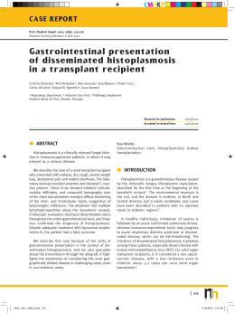
CMYKP
NEPHROPATHOLOGY QUIZ
Port J Nephrol Hypert 2013; 27(4): 309-312
Advance Access publication 12 December 2013
A clinical case to solve...
Helena Viana1, Fernando Teixeira e Costa2, Teresa Santos3, Fernanda Carvalho1
1
Renal Morphology Laboratory, Nephrology Department, Hospital Curry Cabral. Lisboa, Portugal.
Nephrology Department, Hospital Cuf Descobertas. Lisboa, Portugal.
3 Nephrology Department, Instituto Português de Oncologia. Porto, Portugal.
2
CLINICAL PRESENTATION
A 49-year-old Caucasian man was admitted in
Intensive Care Unit for resistant hypertension, with
need of labetalol intravenous perfusion.
His personal history was irrelevant, with previous
normal arterial pressure. He travelled to Mozambique
six week before and takes mefloquin for malaria
prophylaxis.
Three week before admission, he presented low
fever, odynophagia, rhinorrhoea and myalgia. He
was not seen by a doctor and self-medicated with
Ilvico N® (acetaminophen, brompheniramine and
caffeine) for three days, without improvement. He
had been then observed in an emergency room and
medicated with levofloxacin for seven days, without
amelioration. Ceftriaxone was prescribed after that.
Two days after ceftriaxone beginning, he was admitted to hospital with severe hypertension.
serum creatinine was 6.8 mg/dl. Urinalyses showed
haemoglobinuria without leucocyturia. Proteinuria
quantification was 0.3 g/day.
Renal echography revealed bilateral enlarged
kidneys (right-140mm, left-141mm) with normal contours, increased parenchymal thickness and corticomedular differentiation increment were related.
Kidney lithiasis and urinary tract dilatation were
excluded. The renal doppler was normal, excluding
venous thrombosis.
In the third hospitalization day, a kidney percutaneous biopsy was done to clarify the acute renal
failure.
Only one histological specimen was collected and
sent in formaldehyde to our laboratory. No frozen
fragment was available for immunofluorescence. The
fragment was paraffin-embedded for optical and
immunofluorescence observation.
On Intensive Care Unit admission, he presented
normal temperature, resistant hypertension, oliguria,
polypnoea, peripheral oedema, pleural and pericardium effusion.
Immunofluorescence examination of the paraffin
fragment was negative for interstitial or glomerular
deposition of IgG, IgA, IgM, C3, C4, C1q, albumin
and fibrinogen.
Haemoglobin was 10.3 g/dl without leukocytosis
or eosinophilia. C-reactive protein was 5 mg/dl and
The main optical microscopy aspects are demonstrated in the following figures.
309
Nefro - 27-4 - MIOLO.indd 309
18-12-2013 11:19:53
CMYKP
Helena Viana, Fernando Teixeira e Costa, Teresa Santos, Fernanda Carvalho
Figure 1
Figure 4
Masson trichrome green (x100)
Masson trichrome green (x200)
Figure 2
Figure 5
Periodic acid-Schiff (x400)
Masson trichrome green (x400)
Figure 3
Figure 6
Haematoxylin-eosin (x400)
Masson trichrome green (x400)
310
Nefro - 27-4 - MIOLO.indd 310
Port J Nephrol Hypert 2013; 27(4): 309-312
18-12-2013 11:19:54
CMYKP
A clinical case to solve...
HISTOLOGICAL DIAGNOSIS
Figure 1 reveals extensive mononuclear inflammatory infiltrate, fibro-oedematous interstitium and focal
acute tubular necrosis.
Figure 2 demonstrates a normal glomerulus and
the presence of some lymphocytes in tubules- tubulitis. All the seventeen glomeruli and the blood vessels of this biopsy were normal.
In Fig. 3 we can observe the presence of interstitial
inflammatory infiltrate of mononuclear cells and some
eosinophils. A normal glomerulus is presented. An
isolated multinucleated giant cell is visible.
In Fig. 4 we can see two non-caseous granulomas
associated with multinucleated giant cells. This
biopsy presents a total of nine granulomas.
Figure 5 shows a perivascular granuloma consisting of epithelioid histiocytes and lymphocytes.
Figure 6 presents an isolated multinucleated giant
cell and interstitial fibro-oedema. These cells were
scattered diffusely in the interstitium.
In summary, optical microscopy demonstrates normal glomeruli and blood vessels; acute inflammatory
infiltrate, with predominance of lymphocytes and some
eosinophils; fibro-oedematous interstitium, focal acute
tubular necrosis; focal tubulitis; nine non-caseous
granulomas with multinucleated giant cells.
Immunofluorescence examination and the Ziehl-Nielson
stain were negative.
The histological diagnosis was granulomatous
acute interstitial nephritis.
HCV and HIV were negative. Thoracic scan presented
small bilateral pleural effusion and small pericardium
effusion; absence of axillary, hilar, mediastinal adenomegalies and normal pulmonary parenchyma. Bronchoscopy was normal and no BAAR were identified
in direct examination of broncho-alveolar specimen.
Solving the case
DISCUSSION
Granulomatous interstitial nephritis (GIN) is a rare
histological diagnosis, representing 0.5%-0.9% of
total renal biopsies.1,2
The main causes of granulomatous interstitial
nephritis, according to the review of 40 cases by
Javaud et al.1 are sarcoidosis (50%), drugs (17.5%),
tuberculosis (7.55%), Wegener’s granulomatosis (5%),
leprosy (2.5%), Mycobacterium avium infection (2.5%),
Crohn’s disease (2.5%) and idiopathic (12.5%). In the
review (n = 18) of Joss et al.2, TINU and drugs are
responsible in the same percentage (11%) for GIN
after idiopathic cases (50%) and sarcoidosis (28%).
Almost all drug classes have been implicated in
acute interstitial nephritis: antibiotics (penicillins,
cephalosporins, sulfonamides, vancomycin, rifampicin, tetracyclines, erythromycin and most others, if
not all); NSAIDS; diuretics (thiazides, furosemide,
triamterene); antiviral (acyclovir, foscarnet, indinavir);
and miscellaneous drugs (phenytoin, allopurinol,
cimetidine, captopril, lithium, valproate, warfarin,
interferon, lamotrigine).
The presence of granulomas is most often reported
after the use of ampicillin, methicillin, penicillin,
rifampicin, furosemide, allopurinol and phenytoin3.
COMPLEMENTARY EVALUATION
From the literature review, we found a case of
levofloxacin inducing GIN4.
The urine was negative for BAAR direct examination and for bacterial culture.
The mainstay of therapy for drug-induced AIN is
timely discontinuation of the causative agent.
The serum angiotensin-converting enzyme (ACE)
concentration was normal. Antineutrophil cytoplasmic
(ANCA) and anti-glomerular basement membrane (antiGBM) antibodies were negative. Serology for HBV,
Anatomo-clinical diagnosis
Drug-induced acute interstitial nephritis.
Port J Nephrol Hypert 2013; 27(4): 309-312
Nefro - 27-4 - MIOLO.indd 311
311
18-12-2013 11:20:00
CMYKP
Helena Viana, Fernando Teixeira e Costa, Teresa Santos, Fernanda Carvalho
Treatment and evolution
The patient’s creatinine started downloading on
the fourth day. After histological diagnosis, fifth day
after admission, three consecutive 1 g daily pulse of
methylprednisolone were administered, with rapid
improvement of the kidney function. No oral corticoids were prescribed. Currently, tree months after
acute renal failure, the patient’s serum creatinine is
1.2 mg/dl, with a normal urinalysis.
References
1. Javaud N, Belenfant X, Stirnemann J, et al. Renal granulomatoses: a retrospective study
of 40 cases and review of the literature. Medicine (Baltimore) 2007;86(3):170-180
2. Joss N, Morris S, Young B, Geddes C. Granulomatous interstitial nephritis. Clin J Am
Soc Nephrol 2007;2(2):222-230
3. Rossert J. Drug-induced acute interstitial nephritis. Kidney Int 2001;60(2):804-817
4. Ramalakshmi S, Bastacky S, Johnson JP. Levofloxacin-induced granulomatous intersti-
tial nephritis. Am J Kidney Dis 2003 Feb;41(2)
Correspondence to:
Dr.ª Helena Viana
Department of Nephrology, Hospital Curry Cabral
Rua da Beneficência, n.º 8, 1069-166
Lisboa, Portugal
E-mail: [email protected]
312
Nefro - 27-4 - MIOLO.indd 312
Port J Nephrol Hypert 2013; 27(4): 309-312
18-12-2013 11:20:02
Download