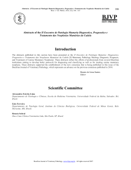
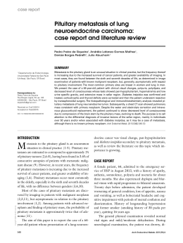
International Section
Arch. Esp. Urol., 61, 5 (658-662), 2008
LUNG METASTASES OF LOW GRADE PHYLLODES TUMOR OF THE PROSTATE:
HISTOPATHOLOGIC CONFIRMATION
David Parada, Gustavo Ugas1, Karla Peña, Luis Caricote1 and Nelson Mujica1.
Servicio de Anatomía Patológica. Hospital Vargas. Caracas. Venezuela.
Urólogos1. Centro Médico El Valle. Isla de Margarita. Venezuela.
Summary.- OBJECTIVE: Phyllodes tumor of the prostate is a rare neoplasm. We report the clinical, histopatho-logical, and immunohistochemical features of a low
grade phyllodes tumor of the prostate with lung metastases.
METHODS: A 68-year-old man presented acute urinary
obstruction. Suprapubic prostatectomy was performed,
and one month later he developed acute urinary obstruction, shortness of breath and cough.
RESULTS: Microscopically, the prostatic tumor showed
elongated ducts and cellular stroma. Lung biopsy was
composed of stromal cells with enlarged, hyperchromatic nuclei.
CONCLUSION: This case represents a typical low grade prostatic phyllodes tumor to lung. Grading of these
neoplasm is not always of value to predict patient prognosis.
Keywords: Phyllodes tumor. Prostate. Malignant.
Metastases. Lung.
Resumen.- OBJETIVO: El tumor phyllodes de la prostata es una rara neoplasia. Nosotros reportamos los
hallazgos clínicos, histopatológicos e inmunohistoquímicos de un tumor phyllodes de la prostata con metastasis
pulmonar.
MÉTODOS: Un paciente de 68 años sufrió de obstrucción urinaria aguda. Se practicó una prostatectomía
suprapúbica, un mes después desarrollo obstrucción
urinaria, dificultad respiratoria y tos.
RESULTADOS: Microscópicamente, el tumor prostático
mostró ductos elongados y estroma celular. La biopsia
pulmonar estaba compueta de células estromales con
núcleos hipercromáticos, alargados.
CONCLUSIÓN: Este caso representa un típico phyllodes prostático de bajo grado metastizando al pulmón.
La gradación de stas neoplasias no es siempre de valor
para predecir el pronóstico.
Correspondence
Palabras clave: Tumor Phyllodes. Próstata. Maligno. Metástasis. Pulmón.
David Parada
Servicio de Patología
Hospital Vargas
San Francisquito a Monte Carmelo
Esquina El Recodo, San José
Apdo. 1010 Caracas. (Venezuela).
[email protected]
Accepted for publication: 19th April, 2007.
INTRODUCTION
Phyllodes tumor of the prostate is an extremely rare neoplasm that resembles the well-known phyllodes tumor of the breast, with a distinctive biphasic
pattern of hyperplastic epithelium lined cysts, leaf-like
intraluminal projections, compressed and elongated
slit-like epithelial lined spaces, and variable cellular
spindle cell stroma with cytological atypia (1-3). It has
LUNG METASTASES OF LOW GRADE PHYLLODES TUMOR OF THE PROSTATE: HISTOPATHOLOGIC CONFIRMATION.
been reported as liable to early recurrence, with an
occasional metastatic potential (1,3,4).
In this report we describe a 68-year-old man
who developed low grade phyllodes tumor of the
prostate that metastasized to the lung. To the best of
our knowledge this is the second case in which such
an evolution is documented. We also briefly review
the literature.
CASE REPORT
A 68-year-old man complained of acute urinary obstruction. He had suffered from a diminished urinary
659
stream and frequency during 6 months. Digital rectal
examination revealed a benign feeling prostate. The
rest of the physical examination was unremarkable.
Prostate specific antigen (PSA) level was 1.1 ng/ml
(normal: < 4.0 ng/ml). An abdominal ultrasonography confirmed prostatic enlargement. Cystoscopy
revealed a normal urethra. The bladder base was
elevated at the bladder neck but the mucosa was normal. Clinical diagnosis was benign prostatic hyperplasia (BPH) grade III. A suprapubic prostatectomy
was performed, with a histological diagnosis of benign prostatic hyperplasia One month later the patient had a new episode of acute urinary obstruction.
An abdomino-pelvic CT showed a 12 cm irregular
tumor, protruding into the bladder (Figure 1A) and
bilateral ureterohydronephrosis. A TURP gave a histological diagnosis of myofibroblastic inflammatory
tumor. Additionally, the patient developed shortness
of breath and cough. A chest roentgenogram and
a CT revealed a peripheral lesion at the right lung
(Figure 1B). A histological analysis done through an
open lung biopsy was consistent with an inflammatory process and atypical mesenchimal proliferation.
After the histological conclusion, the patient received
chemotherapy treatment. The patient died due to cardiac arrest.
PATHOLOGIC FINDINGS
FIGURE 1. A, Abdominal Computerized Tomography.
The tumor (White arrows) is located centrally and
arising into the urinary bladder (after first resection).
B, Thoracic Computerized Tomography.
A right posterior tumor is observed. This lesion was
interpreted as metastatic.
Paraffin-embedded tissue and histological
preparations were available (courtesy of Cristina
Pastrán, MD, of Porlamar, Nva. Esparta, Venezuela).
Sections of paraffin-embedded tissue were stained for
conventional histological study with haematoxylineosin. Immunohistochemical studies were performed
using the labeled LSAB® method (DAKO®, Carpintería, USA). Monoclonal/polyclonal antibodies
were used at dilutions suggested by the manufacturer (between 1:50 and 1:200). Antibodies were directed against smooth muscle actin, CD34, CD117,
S-100 protein, vimentin, estrogen and progesterone
receptors, myoglobin D1, broad-spectrum cytokeratin
AE1/AE3, high molecular weight keratin (34βE12),
epithelia membrane antigen (EMA), prostatic specific
antigen (PSA), p53 protein and Ki-67 proliferating
nuclear antigen. Appropriate positive controls were
prepared for each. Sections processed without primary antibodies were employed as negative controls.
Scoring was based on the percentage of positive tumor cells.
Microscopic examination of the prostatic
biopsy showed complete replacement by a biphasic
tumor. The tumor showed elongated ducts and cellular
stroma, resembling a phyllodes tumor of the breast (Figure 2 A,B). The epithelial cells lining the ducts were
660
D. Parada, G. Ugas, K. Peña y cols.
FIGURE 2. Microscopic findings. A, Cleft-like spaces lined by epithelial cells. (H-E 40x). B, Elongated ducts and
cellular stroma, resembling phyllodes tumor of the breast. Neoplastic cells showing no clear boundary between the
tumor and epithelial cells (H-E 100x, detail). C, Cuboidal, epithelial cell lining of the ducts with oval nuclei and
inconspicuous nucleoli (H-E 400x). D. Epithelial cells showing strong reactivity to prostatic specific antigen (DAB,
400x). E, Metastatic lung biopsy showing stromal neoplasia (H-E 40x). F, Stromal cells with enlarged, hyperchromatic nuclei intermixed with bland, spindled nuclei. These cells had moderate amount of eosinophilic cytoplasm (H-E
100x). [insert 100x].
LUNG METASTASES OF LOW GRADE PHYLLODES TUMOR OF THE PROSTATE: HISTOPATHOLOGIC CONFIRMATION.
cuboidal to columnar with oval nuclei and inconspicuous nucleoli (Figure 2C). No atypia was observed.
The stroma cells were variable with dense cellular
areas with hyperchromatic ovoid cells that condensed the glands forming loose and myxomatous areas
(Figure 2 A,B,C). Few mitosis (1-2/ 10 HPF (400X))
were seen. Perineural, lymphatic or vascular invasion
was not found.
An immunohistochemical study of stromal
cells showed focal positivity for CD34 and smooth
muscle actin. Epithelial cells were positive to PSA
and EMA (Figure 2D). Stromal cells failed to stain
for estrogen receptor, progesterone receptor, CD117,
desmin, myoglobin D1, S100 protein and cytokeratin
AE1/AE3. p53 immunostaining was negative in epithelial and stromal components. Immunoreactivity to
Ki-67 (proliferanting nuclear antigen) showed about
45% of stromal tumor cells with nuclear positivity.
According to Bostwick’s et al. (1) grading, the tumor
was interpreted as low grade phyllodes tumor of the
prostate.
Histological material obtained from the lung
biopsy was also examined. The tumor was exclusively
formed by stromal cells with enlarged, hyperchromatic nuclei intermixed with bland, spindled nuclei (Figure 2 E,F). Nucleoli were not visible and mitoses were
not found. These cells had a moderate amount of eosinophilic cytoplasm and in some areas they resembled
smooth muscle cells (Figure 2 E,F). Immunohistochemical results were similar to the prostatic tumor, with focal CD34 and smooth muscle actin reactivity. Normal
pulmonary tissue was not seen. The final diagnosis
was metastatic low grade sarcoma, consistent with
metastatic prostatic phyllodes tumor.
DISCUSSION
Phyllodes tumor of the prostate is a rare lesion, analogous to epithelial-stromal lesions of the
breast, of unknown histogenesis (1). Like their breast
homonymous, phyllodes tumors of the prostate vary
in their histological appearance and clinical behavior. Patients typically consult due to urinary obstruction, hematuria and dysuria. These symptoms were
referred by our patient, with progression to acute
urinary obstruction. Additionally, the serum PSA level
was normal.
Although most reported prostate cases show
a benign clinical course, some have been reported to
be malignant (1,5-7). Five histological features have
been proposed for correlating the clinical outcome:
stromal cellurarity, cytological atypia, mitotic figures
(in 10 high power field), stromal/epithelia ratio, and
661
necrosis (1). Our case was interpreted as low grade,
considering its low stromal cellularity, mild cytologic
atypia, less than 2 mitosis in 10 HPF, mild stromal/
epithelial ratio, and absence of necrosis. Our findings
are consistent with those showing low grade tumors
with local recurrence and presence of metastases.
Additionally, phyllodes tumor of the prostate should
be considered a neoplasm rather than atypical hyperplasia due to early recurrences and metastatic
spread.
The histology of the metastatic deposits has
not been well documented. There is only one report
of a fine needle aspiration biopsy of a prostatic phyllodes tumor that presented a bone metastasis that
showed malignant spindle cells, consistent with leyomiosarcoma (8).
In our case it was possible to compare the
histological findings in a primary prostatic lesion with
a lung metastasis. In this last one the tumor was exclusively composed by mesenchimal cells with similar
characteristics to those observed in the prostatic lesion
(mild stromal cellularity and cytologic atypia, low mitotic index (less than 2 mitosis/10 HPF, and no necrosis). To the best of our knowledge this is the first case
in the English literature with histological confirmation
of a prostatic phyllodes tumor with lung metastases,
showing that the stromal component was similar in
both cases. Sarcoma frequently emerges from primary or metastatic lesions. In our patient, there was no
transformation to high grade sarcoma. On the other
hand, similar stromal findings were observed and this
probably suggests that a change from a low grade
to high grade sarcoma is not necessary to produce
metastatic disease. Because there are so few of these
tumors, it has been suggested that all cases should
be reported in the literature, to help to evaluate the
relationship between histological findings and clinical
behavior.
CONCLUSION
Our case represents a typical phyllodes tumor of the prostate with lung metastases. These tumors should be considered as a neoplasm rather than
an atypical hyperplasia due to recurrences, as well as
the potential for extraprostatic and metastatic spread.
The stromal component has a metastatic potential and
grading is not always of value for predicting patient
outcome.
662
REFERENCES AND RECOMENDED READINGS
(*of special interest, **of outstanding interest)
**1. BOSTWICK, D.; HOSSAIN, D.; QIAN J. et al.:
“Phyllodes tumor of the prostate: Long-term follow-up study of 23 cases.” J. Urol., 172: 894,
2004.
*2. SHIRAISHI, K.; MORI, J.; EGUCHI, S. et al.:
“Phyllodes tumor of the prostate: Recurrent obstructive symptom and stromal proliferative activity,” Int. J. Urol., 11: 801, 2004.
3. SCHAPMANS, S.; VAN LEUVEN, L.; CORTVRIEND, J. et al.: “Phyllodes tumor of the prostate. A case report and review of the literature.”
Eur. Urol., 38: 649, 2000.
**4. HERAWI, M.; EPSTEIN, J.: “Specialized stromal
tumors of the prostate: A clinicopathologic study of
50 cases,” Am. J. Surg. Pathol., 30: 694, 2006.
5. KIM, H.; LEE, J.; NAM, J. et al.: “Malignant phyllodes tumor of the prostate.” Pathol. Int., 49: 1105,
1999.
6. WATANABE, M.; YAMADA, Y.; KATO, H. et al.:
“Malignant phyllodes tumor of the prostate: retrospective review of specimens obtained by sequential transurethral resection”. Pathol. Int., 52: 777,
2002.
7. YAMAMOTO, S.; ITO, T.; MIKI M. et al.: “Malignant phyllodes tumor of the prostate.” Int. J.
Urol., 7: 378, 2000.
*8. YUM, M.; MILLER, J.; AGRAWAL, J.: “Leiomyosarcoma arising in atypical fibromuscular hyperplasia (phyllodes tumor) of the prostate with distant
metastasis.” Cancer., 68: 910,1990.
Download