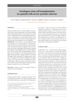
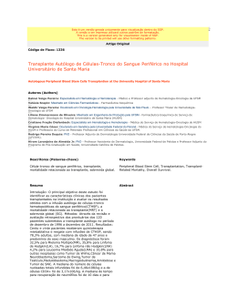
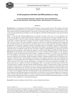
CMYKP
CASE REPORT
Port J Nephrol Hypert 2012; 26(3): 213-216
Advance Access publication 8 June 2012
Gastrointestinal presentation
of disseminated histoplasmosis
in a transplant recipient
Cristina Outerelo1, Rita Nortadas2, Rita Gouveia1, Ana Mateus1, Pedro Cruz1,
Carlos Oliveira1, Raquel B. Ilgenfritz3, Aura Ramos1
1
Nephrology Department, 2 Intensive Care Unit, 3 Pathology Department.
Hospital Garcia de Orta, Almada, Portugal.
Received for publication:
Accepted in revised form:
ABSTRACT
Histoplasmosis is a clinically relevant fungal infection in immunosuppressed patients, in whom it may
present as a serious disease.
We describe the case of a renal transplant recipient
who presented with malaise, dry cough, severe weight
loss, abdominal pain and watery diarrhoea. The laboratory workup revealed anaemia and elevated C reactive protein. Chest X-ray showed bilateral reticulonodular infiltrates, and computed tomography scan
of the chest and abdomen revealed diffuse thickening
of the inter- and intralobular septs, suggestive of
lymphangitic infiltration. The abdomen had multiple
lymphadenopathies along the mesenteric vessels.
Endoscopic evaluation disclosed disseminated ulcers
throughout the entire gastrointestinal tract, and biopsies confirmed the diagnosis of histoplasmosis.
Despite adequate treatment with liposomal amphotericin B, the patient had a fatal outcome.
We describe this case because of the rarity of
gastrointestinal presentation in the context of disseminated histoplasmosis, and we also speculate
about the transmission through the allograft. It highlights the importance of considering this once geographically limited disease in challenging cases, even
in non-endemic areas.
12/03/2012
24/05/2012
Key-Words:
Gastrointestinal tract; histoplasmosis; kidney
transplantation.
INTRODUCTION
Histoplasmosis is a granulomatous disease caused
by the dimorphic fungus Histoplasma capsulatum,
described for the first time at the beginning of the
twentieth century1. The environmental reservoir is
the soil, and the disease is endemic in North and
Central America, but it exists worldwide, and cases
have been described in patients with no reported
travel to endemic regions2.
In healthy individuals, inhalation of spores is
followed by an acute self-limited pulmonary illness,
whereas immunocompromised hosts may progress
to acute respiratory distress syndrome or disseminated disease, which can be life-threatening. The
incidence of disseminated histoplasmosis is greatest
among these patients, especially those infected with
human immunodeficiency virus (HIV). For solid organ
transplant recipients, it is considered a rare opportunistic disease, with a low incidence even in
endemic areas: 4.1 cases per 1000 solid organ
transplants3.
213
Nefro - 26-3 - MIOLO.indd 213
17-09-2012 17:57:38
CMYKP
Cristina Outerelo, Rita Nortadas, Rita Gouveia, Ana Mateus, Pedro Cruz, Carlos Oliveira, Raquel B. Ilgenfritz, Aura Ramos
CASE REPORT
A 46-year-old Portuguese Caucasian female with
chronic kidney disease of unknown aetiology received
a cadaveric kidney transplant six years before admission. The donor was a 59-year-old female who died
of intracranial haemorrhage. Our patient had a history of obesity, arterial hypertension, dyslipidaemia,
deep venous thrombosis, oesophagitis and laparoscopic cholecystectomy for vesicular lithiasis. She
had always lived in Portugal and had never travelled
abroad during her entire life.
Immunosuppression regimen consisted of tacrolimus 0.5mg twice daily (trough level of 5-6ng/dL),
mycophenolate mophetil (MMF) 250mg twice daily
and prednisone 5mg daily. She had chronic allograft
dysfunction, with stable creatinine values of 3mg/
dL, and there were no episodes of rejection.
She presented with a three-month history of general malaise, dry cough and severe weight loss of
20 kilograms. More recently she reported abdominal
pain and watery diarrhoea. She denied fever or other
symptoms.
On admission, she was prostrated, pale, dehydrated and afebrile. Her weight was 68 kilograms,
blood pressure 138/86 mmHg, pulse 78 beats per
minute and respiratory rate 24 breaths per minute.
Physical examination revealed clear lungs, normal
cardiac sounds and non-tender abdomen with no
palpable masses. There were no mucocutaneous
lesions or palpable lymphadenopathies.
Figure 1
Chest CT scan showing diffuse thickening of interlobular septs.
infiltration (Fig.1). An abdominal CT showed multiple
lymphadenopathies along the mesenteric vessels.
Gastric and colonic endoscopic evaluations were
performed, and multiple ulcers with raised borders were
observed in the duodenum, colon and terminal ileum.
Biopsies taken from these ulcers revealed extensive
inflammatory infiltrate, and numerous intra- and extracellular microorganisms, forming yeasts, in a diffuse
histiocytosis pattern. Periodic acid-Schiff (PAS) and Grocott stains confirmed the presence of Histoplasma species (Fig.2). Serology tests were not available.
The laboratory evaluation on admission revealed
haemoglobin 9 g/dL, white blood cell count 5.7×10⁹/L
(91% neutrophils, 5.3% lymphocytes), platelet count
155×109/L, blood urea 116 mg/dL, creatinine 3 mg/
dL and C reactive protein 7.3 mg/dL. Urinalysis
showed leucocyturia, and urine culture identified
Escherichia coli, treated with ceftriaxone.
Serologic testing for HIV, hepatitis B, hepatitis C
and leishmania were all negative, as were blood and
stool cultures. Serum polymerase chain reaction (PCR)
for cytomegalovirus and Epstein-Barr virus were negative. Chest X-ray revealed bilateral reticulonodular
infiltrates, and computed tomography (CT) scan of
the chest disclosed diffuse thickening of the interand intralobular septs, suggestive of lymphangitic
214
Nefro - 26-3 - MIOLO.indd 214
Figure 2
Colonic biopsy specimen showing intra- and extracellular organisms characteristic of
Histoplasma capsulatum (Grocott stain 40×).
Port J Nephrol Hypert 2012; 26(3): 213-216
17-09-2012 17:57:39
CMYKP
Gastrointestinal presentation of disseminated histoplasmosis in a transplant recipient
The patient was started on liposomal amphotericin
B (3 mg/kg/day), but on the 4th day of directed
therapy, she maintained anorexia, diffuse abdominal
pain and abundant watery diarrhoea. By that time,
progressive reduction of urinary output and worsening dyspnoea was seen. Physical and radiology
examination of the lungs revealed the presence of
a bilateral reticulonodular infiltrate, with a consolidation confined to the lower left lobe. She was hypotensive and hypoxaemic, so that tracheal intubation and
mechanical ventilation was needed, and she was
transferred to the Intensive Care Unit (ICU). Considering disseminated infection, immunosuppression was
reduced: tacrolimus was targeted to a level of 3ng/
dL, MMF was stopped, and steroid dose was left
unchanged. She started renal replacement therapy
with continuous venovenous haemodiafiltration
(CVVHDF), due to oliguric acute kidney injury. Despite
adequate fluid administration, vasopressors were
required to restore haemodynamics. On ICU admission, it was considered that despite the diagnosis
of histoplasmosis, we could not exclude hospitalacquired pneumonia, and so empiric broad spectrum
antibiotic therapy with vancomycin, meropenem and
gentamicin was started. Liposomal amphotericin B
was continued. As she remained oliguric, dialysis
dependent and there was no improvement in her
clinical condition, immunosuppression was stopped.
Blood, urine and bronchoalveolar secretions cultures
were negative for common pathogens.
During the ICU stay, she was persistently febrile,
and haemodynamically unstable. Weaning from
mechanical ventilation was never successful. By the
17th day of empiric antibiotic therapy with meropenem
and vancomycin (gentamicin was only administered
for 5 days), as there were no isolates and the patient
was not improving, antibiotics were stopped, except
for amphotericin, and microbiologic tests repeated
– urine, blood, and brochoalveolar secretions obtained
through protected specimen brush. All the samples
were once again negative.
The patient ultimately died of multiple organ failure on the 42nd day of hospital stay, the 27th in the
ICU.
To our current knowledge, the receptor of the
other kidney, who was transplanted in another unit,
is doing well and has not had any symptoms that
could be related to this disease.
DISCUSSION
Disseminated histoplasmosis among solid
organ transplant recipients may occur as a primary
infection, reactivation of latent infection, or
donor-transmitted infection3. In immunocompromised hosts, disseminated disease may progress
to septic shock, acute respiratory distress syndrome and multiorgan failure1, as occurred with
our patient.
In this report, we could not confirm evidence of
Histoplasma capsulatum in the lung, despite persistent pulmonary imaging abnormalities. Bronchoalveolar lavage could have been useful for confirmation of lung involvement, although finding of
the fungus in organic smears is extremely difficult,
even using special staining. Blood culture specific
for Histoplasma growth is not routinely performed
in our centre, but all the cultures sampled – blood,
urine, and pulmonary secretions obtained with
protected specimen brush – were persistently negative, ruling out involvement of other infectious
agents.
Gastrointestinal tract involvement is common during disseminated disease (70-90%), but often remains
asymptomatic4. Gastrointestinal symptoms have been
described more frequently in disseminated disease
affecting HIV-infected patients5.
The presence of extracellular organisms in histologic examination, as in our patient, is rare and represents a marker of severe disease4. This could explain
the adverse outcome despite antifungal therapy,
which has accounted for an important decrease in
the mortality attributed to this infection. The majority
of Histoplasma-related infections occur in the early
post-transplant period, when higher doses of immunosuppression are used. In our case report, the
patient had a five-year history of transplantation and
her immunosuppression regimen at the time and in
the past was not aggressive, which makes this case
even rarer.
In 1988, the first case of disseminated histoplasmosis in Britain was reported6, in a renal transplant
recipient who presented with a cecal perforation.
Since then, few case reports of transplant recipients
with symptomatic gastrointestinal involvement have
been described7.
Port J Nephrol Hypert 2012; 26(3): 213-216
Nefro - 26-3 - MIOLO.indd 215
215
17-09-2012 17:57:42
CMYKP
Cristina Outerelo, Rita Nortadas, Rita Gouveia, Ana Mateus, Pedro Cruz, Carlos Oliveira, Raquel B. Ilgenfritz, Aura Ramos
Some reports seem to relate this disease in renal
recipients with latent infection transmitted via the
donor8,9. This is a very unlikely hypothesis, although
no obvious epidemiologic context was found in the
recipient. Transmission might have occurred through
an infected close contact, but as a self-limited mild
respiratory illness in healthy individuals, it is difficult
to identify it. Also, this is an extremely rare disease
in Portugal, with only a few cases described, and
that explains why this diagnosis was not suspected
on initial presentation.
References
1. Kauffman CA. Histoplasmosis: a clinical and laboratory update. Clin Microbiol Rev
2007;50:115-132
2. Lo MM, Mo JQ, Dixon BP, et al. Disseminated histoplasmosis associated with
hemophagocytic lymphohistiocytosis in kidney transplant recipients. Am J Transpl
2010;10:687-691
3. Cuellar-Rodriguez J, Avery RK, Lard M, et al. Histoplasmosis in solid organ transplant
recipients: 10 Years of Experience at a Large Transplant Center in an Endemic Area.
Clin Infect Dis 2009;49:710-716
4. Kahi CJ, Wheat LJ, Allen MD, et al. Gastrointestinal histoplasmosis. Am J Gastroen-
terol 2005;100:220-231
5. Suh KN, Anekthananon T, Mariuz PR. Gastrointestinal histoplasmosis in patients with
Its rarity and nonspecific symptoms make the
diagnosis and treatment difficult and often delayed,
even though the prognosis appears to be good in
the majority of cases described3. Our patient had a
fatal outcome, which we relate to an aggressive form
of disease with a long-time course.
AIDS: case report and review. Clin Infect Dis 2001;32:483-491
6. Brett MT, Kwan JT, Bending MR. Caecal perforation in a renal transplant patient with
disseminated histoplasmosis. J Clin Pathol 1988;41:992-95
7. Zainudin BM, Kassim F, Annuar NM, et al. Disseminated histoplasmosis presenting with
ileal perforation in a renal transplant recipient. J Trop Med Hyg 1992;95:276-279
8. Limaye AP, Connolly PA, Sagar M, et al. Transmission of Histoplasma capsulatum by
organ transplantation. N Engl J Med 2000;343:1163-1166
This clinical report highlights the importance of
recognising uncommon pathogens, especially as
international travelling increases and immunosuppression becomes more potent. Healthcare providers
must be aware of this and expect to encounter oncerare and geographically limited diseases.
Conflict of interest. None declared.
216
Nefro - 26-3 - MIOLO.indd 216
9. Wong SY, Allen, DM. Transmission of disseminated histoplasmosis via cadaveric renal
transplantation: case report. Clin Infect Dis 1992;14:232-234
Correspondence to:
Dr Cristina Outerelo
Nephrology Department, Hospital Garcia de Orta
Av. Torrado da Silva, Pragal
2801-951 Almada, Portugal
E-mail: [email protected]
Port J Nephrol Hypert 2012; 26(3): 213-216
17-09-2012 17:57:43
Download