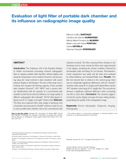
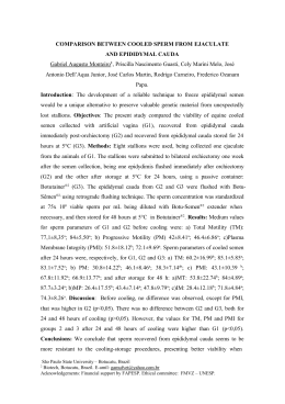
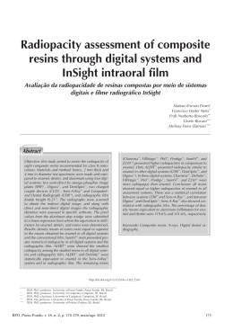
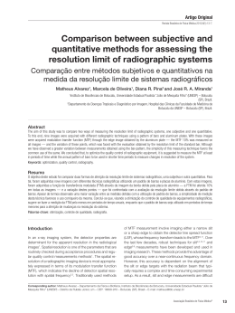
Artigo Original
Revista Brasileira de Física Médica. 2011;5(2):119-22.
Optimization of pediatric chest radiographic
images using optical densities ratio
Otimização de imagens radiográficas de tórax pediátrico
utilizando a razão de densidades ópticas
Rafael T. F. Souza1, Diana R. Pina2, Sérgio B. Duarte3 and José R. A. Miranda1
Instituto de Biociências de Botucatu, Universidade Estadual Paulista
“Júlio de Mesquita Filho” (UNESP) – Botucatu (SP), Brazil.
2
Departamento de Doenças Tropicais e Diagnóstico por Imagem,
Hospital das Clínicas da Faculdade de Medicina de Botucatu (UNESP) – Botucatu (SP), Brazil.
3
Centro Brasileiro de Pesquisas Físicas (CBPF/MCT) – Rio de Janeiro (RJ), Brazil.
1
Abstract
The aim of this study is the optimization of radiographic images for the pediatric patients in the age range between 0 and 1 years old, through
Optical Density Ratio (ODR), considering that pediatric patients are overexposed to radiation in the repeated attempts to obtain radiographic images
considered of good quality. The optimization of radiographic techniques was carried out with the RAP-PEPP (Realistic Analytical Phantom coupled to
homogeneous Phantom Equivalent to Pediatric Patient) phantom in two incubators and one cradle. The data show that the clinical routine radiographic
techniques generate low-quality images at up to 18.8% when evaluated by the ODRs, and increases in doses up to 60% when compared to the
optimized techniques doses.
Keywords: optimization, phantom, dosimetry, image quality, pediatrics.
Resumo
O objetivo deste estudo é a otimização das imagens radiográficas para pacientes pediátricos na faixa etária de 0 a 1 anos de idade, através da
razão de densidade óptica, considerando que pacientes pediátricos são superexpostos à radiação nas repetidas tentativas de se obter imagens
radiográficas consideradas de boa qualidade. A otimização das técnicas radiográficas foi realizada com o fantoma RAP-PEPP (fantoma analítico
realístico acoplado a um fantoma homogêneo equivalente ao paciente pediátrico), em duas incubadoras e um berço. Os dados mostram que as
técnicas radiográficas de rotina clínica criam imagens de qualidade inferior em até 18,8%, quando avaliadas por razões de densidade óptica, e
aumentam em doses de até 60% ao serem comparadas às doses das técnicas otimizadas.
Palavras-chave: otimização, fantoma, dosimetria, qualidade da imagem, pediatria.
Introduction
The process of optimization of pediatric radiographic images is part of an effective quality control program, which
must be implemented in every institution that makes use
of ionizing radiation1,2.
This process have a great importance in the radiological protection context, keeping in view that pediatric
patients are subjected to a lot of X-rays, depending on
the conditions under which they are, as weight (between
750 g and 2000 g), gestational age (between 6 and 9
months), respiratory problems and other intrinsic conditions of the newborn3.
Moreover, these patients are overexposed to radiation
in the repeated attempts to obtain radiographic images
considered of good quality3. It is important to point out that
radiation exposure during the first 10 years of life presents
a risk attributed to the lifetime (for radiation-induced biological effects, deterministic and stochastic) 3 to 4 times higher when compared with exposures between the ages of
30 and 40 years and up to 7 times higher when compared
with adults aged over 50 years.
Thus, it is extremely necessary the standardization of
radiographic techniques to conduct pediatric tests in order
to obtain good quality images with doses as low as reasonably achievable4,5.
In this context, the objective of this study is the optimization of radiographic images inserted in pediatric patients aged between 0 and 1 year old by Optical Density
Ratio (ODR), following the ALARA (As low as reasonable
Corresponding author: Rafael Toledo Fernandes de Souza – Departamento de Física e Biofísica, Instituto de Biociências de Botucatu, Universidade Estadual
Paulista “Júlio de Mesquita Filho” – Distrito de Rubião Júnior, s/n – CEP: 18608-970 – Botucatu (SP), Brasil – E-mail: [email protected]
Associação Brasileira de Física Médica®
119
Souza RTF, Pina DR, Velo AF, Alvarez M, Duarte SB, Miranda JRA
achievable), which prioritizes the acquisition of radiographic images considered of good quality with doses as low
as reasonably achievable.
Methodology
The process of validation of radiographic techniques (combinations of kVp and mAs) seeks to find the combination to
be optimum, which is able to generate radiographic images of good quality. This process was accomplished with
the RAP (Realistic Analytical Phantom) aid, that is aimed at
assessing the quality of radiographic images6.
The RAP, as illustrated in Figure 1, is a phantom consisting of realistic and analytical structures, which follows the
recommendations of ICRU (International Commissions on
Radiation Units and Measurements) Reports 44 and 487,8.
The analytical structures found in RAP are test objects that
allow the quantification of the image quality produced by
different kVp and mAs combinations.
This phantom consists of a PMMA (polymethylmethacrylate) plate with dimensions of 30cmx30cmx5 cm, in
which the test objects are inserted: (A) five acrylic steps
plus air gaps, simulating cavities; (B) five acrylic steps plus
PVC, simulating bone structures; (C) four nylon spherical
segments simulating tumours; (D) three nylon cylinders simulating fat tissue; (E) six aluminium spheres to determine
the degree of visualization of the bone tissue boundaries;
(F) four groups of human organic micro-calcification, simulating cortical bone grain with different sizes; (G) one resolution grid (0.1 mm Pb Nr 1000943 LP/mm; Nuc. Assoc.
Carle Plate, N.Y-07-538); (H) 1/2 human thoracic vertebra;
(I) two steel spheres separated by 0.80 cm for magnification analysis; and (J) one tin wire to determine the coincidence of light and radiation fields6.
The process of optimizing radiographic techniques was
carried out with the RAP-PEPP phantom, which is formed
by coupling the RAP to the PEPP (homogeneous Phantom
Equivalent to Pediatric Patient), as shown in Figure 2.
Figure 2 (A) illustrates the adaptation of the RAP to the
PEPP. Table 1 shows the PEPP structure in the AnteriorPosterior (AP) projection. In this adaptation, the lower pair
of PEPP was replaced by the RAP, which has the same
equivalence in attenuation of the part that was replaced.
The configuration of the posterior pair, referring to PEPP,
was maintained original (thicknesses of PMMA and aluminum) for the simulation of the pediatric patient chest.
Figure 2 (B) illustrates the RAP-PEPP coupled structure.
Quantitative analysis is performed using the parameter called Optical Density Ratio (ODR), which is calculated through the ratio between the optical densities of two
distinct regions and interest in the images obtained with
Figure 2. (A and B) Illustration of the RAP-PEPP structure used
to obtain the radiographic techniques based on the quality of the
acquired images.
Table 1. Thicknesses of PEPP material simulators, PMMA and
aluminum (Al) in the Anterior-Posterior projection.
Figure 1. Schematic view of structures in the RAP.
120
Revista Brasileira de Física Médica. 2011;5(2):119-22.
Top pair
PMMA (mm)
Al (mm)
35.00
0.42
Lower pair
PMMA (mm)
Al (mm)
35.00
0.83
Optimization of pediatric chest radiographic images using optical densities ratio
the RAP-PEPP. For this study, we selected values of optical density (OD) for test objects designated by (A) and (B),
represented schematically in Figure 1. The test object (A)
represents the overlap of soft tissue and bone, which consists of steps with different amounts of PMMA and PVC.
The test object (B) represents the overlap of soft tissue
and air, simulating the interior of the chest, which consists
of steps with different amounts of PMMA and air9. For this
study, the steps with the greatest amount of PVC (test
4,4
Used equipments
Incubator FANEM Vision
Incubator Isolette
Cradle FANEM AQ 50
ODR - Soft tissue and bone
4,2
4,0
3,8
3,6
3,4
3,2
3,0
2,8
2,6
2,4
2,2
2,0
Chi^2
4,20882E-4
4,69281E-4
4,63976E-4
50
R^2
0,9994
0,9992
0,9989
55
Results
60
65
70
75
80
kVp
Figure 3. Optical Density Ratio measurements between soft
tissue and bone as a function of the voltage applied to X-ray
tube (kVp) were evaluated, respectively, for the incubators FANEM Vision (black), Isolette (dark gray) and Cradle FANEM AQ
50 (light gray).
2,5
2,3
Figures 3 and 4 present the results of quantification of the
radiographic images quality by optical density ratio measurements in radiographic images with the RAP-PEPP
phantom. The ODRs were evaluated between soft tissues
and bone, and between soft tissue and air, for the incubators FANEM Vision and Isolette, and to the Cradle FANEM
AQ 50, presented, respectively, in black, dark gray and
light gray.
Table 2. Combinations between the voltage (kVp) and load
(mAs) applied to X-ray tube, absorbed entrance surface doses
of the RAP-PEPP (ESD) given in μGy, and the optical density
ratio (ODR) obtained between bone and soft tissue (Bone-soft),
and between the soft tissue and air (Air-soft), evaluated for optimized radiographic techniques in this study (Ot.) and techniques
commonly used in the HCFMB-UNESP clinical routine (Rot.).
Used equipments
Incubator FANEM Vision
Incubator Isolette
Cradle FANEM AQ 50
2,4
ODR - Soft tissue and air
object A), air (test object B) and soft tissue (between test
objects A and B) were chosen to simulate, respectively, the
bone tissue, air inside the lungs and soft tissue that exist in
the chest of a pediatric patient.
The ODRs were measured in two incubators, one manufactured by FANEM Vision and the other model manufactured by Isolette, and a cradle manufactured by FANEM
(AQ 50 model), using a x-ray equipment manufactured by
GE VMX Plus model, which has constant potential10. These
devices belong to the Hospital das Clínicas da Faculdade
de Medicina de Botucatu (UNESP-HCFMB).
It was further quantified the dose absorbed in the entrance surface. This was estimated from of the amount of
exposure measured in air, on the phantom (RAP-PEPP) in
the center of the useful beam of X-rays, using an electrometer (model 9015) and an ionization chamber (model 10X5-6), both manufactured by Radical Corporation.
Measurements were made taking into account the backscattered radiation.
The association of the radiographic image with better
quality and the absorbed dose in the entrance surface was
considered as a figure of merit (FOM).
2,2
2,1
2,0
1,9
1,8
1,7
1,6
Chi^2
2,45094E-5
3,41642E-4
2,13143E-5
50
55
Incubator
FANEMVision
R^2
0,99948
0,99979
0,99948
60
65
70
75
Ot.
Rot.
80
kVp
Figure 4. Optical Density Ratio measurements between soft
tissue and air as a function of the voltage applied to X-ray tube
(kVp) were evaluated, respectively, for the incubators FANEM Vision (black), Isolette (dark gray) and Cradle FANEM AQ 50 (light
gray).
Incubator
Isolette
Cradle
FANEMAQ 50
Ot.
Rot.
Ot.
Rot.
kVp
mAs
ESD(μGy)
55
60
66
71
50
60
57
65
50
55
60
2.50
1.25
2.50
1.00
3.20
1.00
3.20
2.00
2.00
2.50
2.00
52.71
82.57
131.6
66.83
74.01
42.17
112.2
84.32
65.25
81.56
84.35
ODR
Bone-soft Air-soft
3.05
1.95
2.70
1.84
2.41
1.73
2.19
1.69
4.00
2.30
3.20
2.05
3.22
1.93
3.05
1.84
4.28
2.34
3.95
2.17
3.78
2.02
Revista Brasileira de Física Médica. 2011;5(2):119-22.
121
Souza RTF, Pina DR, Velo AF, Alvarez M, Duarte SB, Miranda JRA
The recommended optimal radiographic techniques
were then compared with those normally applied in clinical
routine of HCFMB-UNESP. These techniques were applied
in the RAP-PEPP in order to quantify the image quality
parameter using ODR. Table 2 presents the radiographic
techniques obtained in this study and the techniques normally applied in clinical practice, which are obtained empirically and may vary by a factor of 10 between different
diagnostic services specialist in pediatric patients11.
Discussion and conclusions
The data presented in Table 2 show that radiographic techniques (combinations of kVp and mAs) used in HCFMBUNESP routine clinical generate radiographic images with
quality reduced by up to 18.8% and 13.7% when evaluated by the ODRs, respectively, between the soft tissues
and bone, and between soft tissue and air. These reductions are due to higher effective energy of x-ray beam
used, compared to the energies used in radiographic techniques optimized. It is worth emphasizing that the voltages applied to the optimized radiographic techniques have
values below those recommended by the Commission of
European Communities (between 60 and 65 kV)12.
The doses used in clinical routine show also increased by 60% when compared to the doses applied by the
optimized techniques. These last are still, mostly, below
80μGy, a value recommended by the Commission of
European Communities on the exams for children12.
The radiographic optimum techniques obtained using
the RAP-PEPP has provided the acquisition of the figures
of merit. The results of the image quality quantification
showed that the figures of merit do not have the highest
ODRs, but are obtained with voltages applied to the X-ray
tube between 50 and 60 kVp and load in a range between 1-3 mAs.
Absorbed entrance surface doses show linear behavior with the load applied to the X-ray tube and quadratic
with the voltage applied to the tube10,13. Thus, the radiographic techniques used in routine provide this increase
in absorbed dose due to the higher values of voltage and
load applied to the tube.
The optimizing of the chest radiographic images quality
performed for pediatric patients in this study is a contribution to better quality images when compared with those
obtained by clinical routine and have lower radiation doses, thus following the 3D principle (diagnostic, dose and
dollar), which prioritizes the acquisition of quality images,
122
Revista Brasileira de Física Médica. 2011;5(2):119-22.
providing a safe medical diagnosis, with radiation doses as
low as reasonable achievable, consequently encouraging
the lowest cost to the institution for the execution of radiographic examinations2.
Acknowledgment
We would like to express our gratitude to FAPESP for financial support.
References
1.
2.
3.
4.
5.
6.
7.
8.
9.
10.
11.
12.
13.
Brasil. Ministério da Saúde. Secretaria de Vigilância Sanitária. Portaria
Federal n° 453, de 1º de junho de 1998. Brasília: Diário Oficial da União,
Poder Executivo; 1º de junho de 1998.
Mohamadain KE, da Rosa LA, Azevedo AC, Guebel MR, Boechat MC,
Habani F. Dose evaluation for paediatric chest x-ray examinations in
Brazil and Sudan: low doses and reliable examinations can be achieved in
developing countries. Phys Med Biol. 2004;49(6):1017-31.
Armpilia CI, Fife IA, Croasdale PL. Radiation dose quantities and risk in
neonates in a special care baby unit. Br J Radiol. 2002;75(895):590–5.
Punwani S, Zhang J, Davies W, Greenhalgh R, Humphries P. Paediatric
CT: the effects of increasing image noise on pulmonary nodule detection.
Pediatr Radiol. 2008;38(2):192-201.
Vano E, Ubeda C, Leyton F, Miranda P. Radiation dose and image quality for
paediatric interventional cardiology. Phys Med Biol. 2008;53(15):4049–62.
Pina DR, Duarte SB, Ghilardi Netto T, Trad CS, Brochi MA, de Oliveira
SC. Optimization of standard patient radiographic images for chest,
skull and pelvis exams in conventional X-ray equipment. Phys Med Biol.
2004;49(14):N215-26.
International Commission on Radiation Units and Measurements. Tissue
Substitutes in Radiation Dosimetry and Measurement. ICRU Report
44. Bethesda, MD: International Commission on Radiation Units and
Measurements; 1989.
International Commission on Radiation Units and Measurements. Phantoms
and Computational Models in Therapy, Diagnosis and Protection. ICRU
Report 48. Bethesda, MD: International Commission on Radiation Units
and Measurements; 1992.
Pina DR, Duarte SB, Ghilardi Netto T, Morceli J, Carbi EDO, Souza RTF, et
al. Controle de qualidade e dosimetria em equipamentos de tomografia
computadorizada. Radiol Bras. 2009;42(3):171-7.
Curry TS, Dowdey JE, Murry RC. Christensen’s physics of diagnostic
radiology. Philadelphia: Lee & Febiger; 1990.
Lima AA, Carvalho ACP, Azevedo ACP. Avaliação dos padrões de dose em
radiologia pediátrica. Radiol Bras. 2004;37(4):279-82.
Commission of the European Communities. European guidelines and
quality criteria for diagnostic radiographic images. Report EUR 16260EN.
Bruxelas: European Communities/Union, 1996.
Johns HE, Cunningham JR. The Physics of Radiology. Illinois: Charles C
Thomas Publisher; 1983.
Download