.Joao
======~~
R.m. na,tal!~lnli"na"'=~:===;
EDlTDR5
Compu/a/ional Vision and MedicaI Image Process ing - Tavares & Na/aI Jorge (eds)
© 2072 Taylor & Francis Group, London, ISBN 978-D-415-68395· 7
volume ilIumination, in which lhe deplh-of-focus
is dctcrmincd by lhe charnctcristics af objective
uscd. As a conscquencc, despi te bcing ccntered
aI lhe mid-plane af lhe channcl whcrc lhe ccll
vc10city is lhe highesl. cells a I diffcrent : -planes
5
.a• ...
~
lii _.11
~
~
c
...
are a1so caplurcd. In this case, wc bclicvc Iha l the
black tells are nOI truly Jocalcd aI lhe mid-planc
....
ilnd thcrcrorc its vclocity is slighlly lowcr Ihan
lha! af lhe grcy cclls. Following Ihese prclimi nary
rcsults. furlher invcsligation 00 lhe ccll velocities
u i
and deformarian index in various regiaos Df lhe
microchanncls undcr diffcrcnt
will bc pcrforrncd.
now
conditions
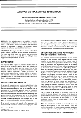
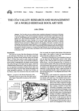
Figure 4. Compari~on af ueformruio n inde:o; ai dirrcrco! now mies in difTcrcnt regioos.
ACKNOIVLEDGEMENTS
~ o· o.oJ
O.
lo)
Ib)
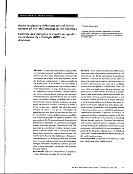
Figure S. (a) Original imllgc conlaining RI1Cs with Y.lr·
ious intcnsitics: I . low (blllck). 2. jn t~rmctl ia lc (gn:y) and
] . high (while), lInd (b) Corresponding binary imngc.
Wc thank Dr. Mntsuki for hclp with blood s<l mplc
colleclion. Additionnlly, we acknowledgc lhe finan ciaI support provided by 2007 GlobalCOE Program
"Global Nano-BME Education nnd Rcscarch
Network", Japao. \VC are a1so thankful lo FCf
(Portugal) and COMPETE ror fin anciai support
through projects PT DC/SAU·OED/ IOB728/200B,
PTDC/SAU·BEBf105650/2008 and PTDC/EME·
MFE1099 109/2008.
An automatic method to lrack Red Blood Cells in microchannels
D . Pinho
P(lI)'tccllllic InslillllL' (I[ Bragal/fa. ESTiGIIPn,
c. Stu. Apt/ltmiu.
Bmganço. PlJrlllXlIl
F. Gayubo
Flllldaciôn CARTIF, DÍI'i.ritlll de Ru!Jvlica J' Vi.MII Artificial. ParqllL' 1i'f/wlóg icn tlc B(lccill(l. l"alllldolid. SI1(1;/1
A.
Isabel
Po!ytcc!lIl;c !I/.rt;/rllc o[ /1mxulI{:(I. ESTiGII PB. C Sw. Apolol/;a. Bragal/ça. Purl llga!
ALGORlTMI. J\/i"ho Ullirerslt): Cumpll.r de A=lIrêm. G/limurik.r. I'orlllgal
R. Lima
POIYI('clmic II/sliture o[ Bmgm/çll. ESTiGIIPB. C. Sta. Ap%l/ia. Bragal/ra. Portugal
CEFT, FEUJ~ R. Dr. Roherto FrülJ'. Port(l, p(lr/llgal
ABSTRACT: Imnge analysis is extremely importnnt to obtain crucial info rmati on about the blood
phenomena in microcirculalion. T he currenl study proposcs an automatic method for segmentat ion and
tracking Retl D\ood Cclls (RDCs) flowi ng Ih rough a 100 pm G lass capillary. T hc origin:1I im ngcs \Vere
obtained by menns Df a coofocal systcrn and Ihen processed in Matlab using the Imagc Processing Toolbox. The automalic mensuremcnls obtained w ith lhe proposcd a Uloma lic method are compnrcd with a
manua l trac king method using a plugin fmm ImageJ.
REFERENCES
.
~--------
~ ~ ...<9 ...oP ...oP ...-E' ...~
(b)
-f -f #~.f>~.s>,f.f
Dlstance In X ..Is (11m)
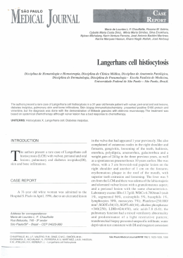
Figure 6. (a) Trncking of RDCs with dilTercnl inlcmily
leveis for vclodly rnensurements. (b) Axial \'c!ocity profiles of the lo\\' and intermcdiale inlensity RDCs along
the eenterline aI Q '" 9.4$ ].tUmin.
220
AbramofT, M. , Magclhacs, P., Ram, S., 2004. Imil!!!:
processing wi th image. 1. Diophotonics lnt. 11 ,36-42.
Dobbe, J.G.G., H ardeman, M.R., Streekslrn. G.1.,
Strackcc, 1., Ince, C., Grimbergen, C.A., 2002.
Analyzing red blood cell-dcformnbility distributions.
BloO<! Cc!ls, MoI. Dis. 28, 373-384.
Fujiwara, H .. Ishikawa, T. Lima, R.. Mat5uki, N.,
lmai, Y., Kaji, li., Nishizawa. M., Yamaguchi, T.,
2009. Rcd blood ccll motions in a high hcma!ocn!
blood nowing through n stcnoscd micro·channcl.
1. Biomceh. 42, 838- 843.
Guegucn, r.'f., Bidet,1.M., Durand, F., Driss, F., 1oITtc, A.,
Gene!et, n., 19R4. I'iltr.ttion pn:ssun: aml n:lI blood ecl!
defonnabili!y: cvaluntion af a new device: erythromctn:.
Biorhcology Suppl. 1,26 1- 265.
Mokkn, ECh., Kedaria, M., Henny, OI.P., Uardcm:m. M.R,
Gdb, A.W., 1992. The clinicai importance of cryth·
troeyte defonnability. a hemorrheological pammeter,
Ano. I-Icmatol. 64, 11 ] - 122.
Oliveim, M.S.N., Ahl:S, M.A., Pinbo, ET., McKin1cy, G. H.,
2007. Viscous now thro ugh microfabricated hyperbolic
contraetions. E,<p. Auids. 4], 4]7-451.
Shelby, 1.P., Whilc, 1., Ganesan, K., R:lIhod, P.K.,
Chiu, D.T., 200]. A micronuidic modcl for single·cell
caplllary obslruclion by Pln~mocli u m falciparuminfectcd crythroeytcs. PNAS. 100, 14618-14622.
Shin, 5. , Ku, Y., Park, MS , Suh, 1.5., 2004. Measurcmcnt
of red ccll dcfonnnbility nnd wholc blood viscosity
using lascr-dilfraclion slit rheomclcr. Korea-Australia
Rheol. 1. 16,85-90.
INTRODUCTION
The study of the red btood cclts (RBCs) nowing in
microvessds and microchanncls is vcry important
10 provide a better understanding on thc blood
rhcological propcrtics and disordcrs in microvesSeis [l-5]. In this kind or study, thc image nnalysis
is an cssentiat part to obtain crucial inrormation
about the blood rheology. Howcver, mosl of the
data analysis proeedures have becn execu tcd manually [1 - 3] whieh is an extremely time consuming task espcciaJty with a largc amounl or dala.
Additi onally, manual tracking methods can also
introduee user errors into the data. Hcnce, iI is
importa0! to dcvelop image analysis mcthods able
lo gct the data automatically. The ma in purpose
or this work is to dcvclop an appronch Olble 10
lrack the RBCs with x a nd y coordimll cs nutomaticruly. To uccomplish it wc tesled rille rin!!.
segmentalÍon und fealure e:'IIractio n fun ctioos
available in MatLub.
Olympus) combined with a Confocal Scan nin g
Uni! (CSU22; Yokogawa), a Diodc-Pumped
Solid-State (DPSS) laser ( Lase r Qua mum) with
an excitat ion wavelCngth Df 53 2 nm and a highspced camera (Phantom v7.1 ; Vi sion Rcscarch)
(Fig. 1). The glass capi ltary was p laced o n lhe
stagc or the invcrted microscopc and by usin g
a syringe pump (KD Scientific) n pressuredrivcn now was kept constant (Rc - 0.008).
2 MATERIALS ANO METHODS
2. I Experimemal Jct-/lP
The conrocnl micro-PIV system uscd in Ihis
study consists or an inverted microscope (lX71 ;
221
Figure I.
Experimcntal sct-up.
More detailed info rmation abo ul lhis syslcm cOln
bc found elscwherc (I].
2.2
Im{/~e alZa/y~·is
The laser beam was iIIuminated from below the
microscopc stage through a dry 40x objeclive lens
with a Numericnl Apcrturc (NA) egual to 0.9. The
confocal imagcs were capturcd in middle of the
cnpillary \Vi th a reso lution Df 640 x 480 pixel ai
a rale Df 100 frames/s with an cxposure time Df
9.4 ms. Two image analyscs mcthod s \Vere u sed
in Ihis sludy: melhod I (manual approach) and
mcthod 2 (<lnlomalic approach).
2.2.1 Methad I
A manual tracking plugin (MTrackJ) [6] of an
imagc analysis software (ImageJ, NIH) [7] was
used to Irack individual RBC. By using MTrackJ
plugin, lhe bright ccntroid of lhe sclccted RBC was
aUlomalicaUy computcd through successivc images
for an inlerval of lime of 10 ms. Aficr obtaining
x and J' positions, lhe dala \\'crc exporled fo r lhe
dctermihation of cach individual RBC trajeclo ry.
2.2.2
A'fetflOrI 2
Ali fromes \\'ere loaded and pre-processed using
Matlab [8]. The region of inlerest was Ihen cropped
from lhe images with lhe function imcmp. The
median function , IIIcdfi/t2, with one mask 5 x 5 pixel,
FigUTl! 2. The n:giun of interest (above) and the image
filtercd by usin g the median funclion lIIt'dfilf2.
was applicd lo climinalC mOSI of lhc noise nnd 10
enhancc lhe nowing objccl. In Fig. 2 wc can scc lhe
result of lhcse processing steps. In lhe nexl slep, lhe
images are subjccI to a ~egmenlalion filt er, SolJcl.
With Ihis segmenlation it is possiblc la separalc
RBCs from lhe baekground. i.e. differentiate the
arca of inlerest (the RBCs) fram the not·intcrest arca
(background image). TItis i5 possible using a tlm!,~" ·
oldmethod, wherc a derinition of one or more values
of separotion is cnough lO divide lhe image ioto one
o r more regions. n lC functio n itemtire tltresf/OltI was
applied for lhe segucncc o f ali lhe images.
The objecls are defined wi lh the Sobel filter (see
Fig. 3), which shows only the edge of lhe objecls.
The Sobel computes an upproximution of the gru·
dient of the image intensity. AI each pixel poiol
in lhe imagc, the result o f the Sohel operator i5
eilher lhe corresponding gradient vector ar the
na rm of this veclor.
3 RESULTS AND DlSCUSSION
Aft er the segmen tatian processing, lhe RBCs were
tracked and seis of data (x and J' positions) were
obtained with lhe Matlab function (Fig. 4), stored
in the image proccssing toolbox, regionprops.
This funclion mensures a set of propcrties (nrca,
centroid, elc.) for eaeh conncctcd component
(RBC) in lhe binary image.
In lhe Fig. 5 we cun see lhe tracking of (wo
RBCs, in a seguenee of successive images. w1th an
inlcrvaJ Df 4 frames.
Ali of Ihese image processes, prcsenled in Ihis
work, are placed in an applicatio n, RBC Dato
Tracking, built in MotLab, in which ali lhe stcps
can be done automutically.
Fig. 6 shows a gual it ative comparison betwecn
method I (manual) and method 2 (aulomotic). Thc
trajectorics obtaincd from the proposcd uutomotic
method loaks more smooth when compared with
manual method.
Some devialiom are observed between both
rnethods. This may bc duc to the inaccuracy in
man ual track ing, espccially for dctcnnination o f
the ceOler of lhe RDes, because the aulomatic
method is more sensitive, evcn in the presente of
small changes in the centroid.
.-
U .
oo--__~--_
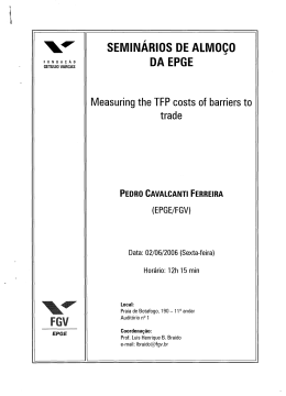
Figure 7.
222
d~til
extraction.
_ <'' ''''"_ ___ ",,"',.,...•
"
,
-- -----------' ,
.
Vclucity of Iwo cdls by Ilsing both mcthoc.l .....
F ig. 7 shows lhe vclocity of ccll 1 and ccU 2 c<ll cu laled by data obtained froro bolh mcthod~. Thc
rcsults show good Ilgreemen l hetwcen lhe IWO
mcthods.
Figule 5. RBCs Irllcking a nd data e~ lra ction in
sc:quence of 4 lo 4 fra mes.
4 CONCWS IONS
.l
Celll
/1\
r, ;, "
t, ......' '" ",
:'.!A.!l\A~,
~
\. '..
... .
\,t......
,
:: I
Although lhe a utomalic mcthod prcsemed in this
sludy i5 a promising wny lo lrack lhe n owing R BCs.
udditional image Ilnalysis nccds lo be pcrformed.
Hence, delailed quantitative measurements of the
RBC trajectories are currently under way <lnd will
be prescnled in due time.
In fUlure work we ure planning lo explore more
techniques to oblain quantilative measurcmcnls
of the RBC trajectorie.~, and more image analysis
strategies need 10 be pcrformcd.
ACKNOWLEDGEMENTS
~
ri'
.
.~
'"
~
~
.n
~
The <luthors acknowledge the financiai supporl
providcd
by:
PTDC/SAU-BEB/10872R/2008,
PTDOSAU-BEB1l05650/2008 and PTDOEME•
M FEl099 I09/2008 from lhe FCT (Scicnce and Tcchnology Foundalion) and COMPETE, Port ugal.
no
,~
.)
CI1I2
RE FERENCES
fll Lima R.
Ish ikawa T, Imai Y. Takcda M .
Wnda S and Yamaguchi T. Measurcmcnl Df
individual rcd bloml ccll motions under high
hemutocrit conditions using a confocal micro.
PTV syslem. Alllm/s o/ Bi(}merliml EI/gillcering
37,1546-1 559,2009.
[2] Fujiwara H. Ishikawa T, Lima R, et aI. Rcd
blood cell motion~ in high-hcmatocril blood
flowing through 1\ stcnosed microchannel.
!
.
JO/frllol o/ Biomedltllu"t-$ 42 , 838-
,~
RnCs trad:ing i1nd
- --( .. , • ." .... , ..
.. . ~ ~J',._.,_:.._.-...." ~-::~--'~!--"'
~
-0..:P-~/1
O' ~
843, 2009 .
[31 Suzuki Y, Tatcishi N, Soulani M and Maeda N.
b)
Figure 4.
_ "Il "",~
•
".
FigUTl! 3. Re.~uh Df the iler.ttive IIIf"t'slmltl mClhod a nd
the fjllcr Sohe/.
VelDdty
..."
Ficure 6. Comparison (lf the manual (a) and a UlOmalic
(b) met hods.
223
Deformmion of CrythrocYle.c; in miclOvesscls and
g1asscapillaries:crrCClsofcrythrocytedcrormability.
Microdmdntioll J, 49- 51. 1996.
Computational Vision and Medicallmage Processing - Tavares & Natal Jorge (eds)
2012 Taylor & Francis Group, London, ISBN 978-0-415-68395-1
ce
(4) Pries A, Secomb T, et 011. Resistance lo blood
now in microvessels in vivo. CirclIlatioll
Research 75, 904-915, 1994.
[5) Pinho D, Cl aI. Rcd blood cells motion in a gluss
microchannc\ , Numcrical Analysis and Applied
Mathematics, Vol. 1281 : 963-966, 2010.
[6} Mcijcring E, Small und Danuser G. Tracking
in molecular bioimaging, IEEE Sigllal Process.
Mag. 23: 46-53, 2006.
(7) AbramoIT M, Magclhaes P and Ram S. Image
processing with imageJ, Bfoplmtrmic.f l11f.
11: 36-42, 2004.
[8] Stcvcn L. Eddins. Rarael C Gonzalc:z, Richarcl E
Woods, DigitallmageProcessing Using Matlab,
2002.
Speech articulation assessment using dynamic Magnetic Resonance
Imaging techniques
S.R. Ventura
School of A Ilictllfra//h Scic"cc~ POr/o Po~rlecllllic Ill.fti/lIlc. V N Gaia. Portllgal
M.J.M. Vasconcelos & D.R. Freitas
Fa cult), of ElIgillcerillg. Vllil'er.fit}' uf Por/o. Por/ o. Porlll!:fI!
I.M . Ramos
Ror/i%!;)' SCrI'ice, St. 10/11/ flmpital alld Frtcult)' li! Ml'llicilll'. Unilw.tit,l' (lf Porlll. Porto. Portl/gal
João Manuel R.S. Tavares
Facult)' Df EIIgillecrillK. Ullil'ersi!)' of Por/o. Por/(J. Porlllgal
ARSTRACf: Magnetic Rcso nancc lmaging (MRI) has been succel'isrully applied on rea.l·timc ílnalysis
of lhe articulmors tluring speech production alcog the whole vocal trael, wilh gootl signal-to·lloisc ratio
and without ionizing efTccts. Deeause sp!.."\.'Ch dynamic evenls need a minimíll sampling mIe, an improvement 011 the tempo ral resolulion cr MRI systems is demanded. Qur aim is to describe 11 dynumic MRI
Icrhniq ue lo aequire a nd aSl'i ess the main artieulatory events during the production of some Europcan
Portuguese utterances. Hence, novel pcrccptions ror dynamic MRI techniquc using li 3.0 Tesla System are
prescnled in order to study lhe shape or the vocal trael during speech produclion.
Kf!)'ll'ords:
image analysis, medicai imaging, specch production, dynamic teehniqucs
INTRODUCTION
I.'
Spef!ch productioll mwl)'.fi.\· (/lId clwltellge.f
The speech production mcchanism is íl complclt
human motor aClivity thal is able to achieve voiec
modulation and produce spcceh based mainly in
lhe articuJators' movements. Thc orgu ns involved,
mostly formed of sofi lissues., such as lhe longue,
lhe lips. lhe velum and the phnrynll, assume
c:~lremely important roles during spcech produc·
tion. In ract, lhese organs togelher with some
bOIlCS, i.e. the palalc and lhe jaw. modiry lhe reso·
nanee cavilies and lhe shape of lhe vocal Irael in
order to producc the sounds.
The huma n vocal tract's shape (Fig. I) is dif·
rerent among subjccls and prescnls n non-rcgulnr
conlour defined by lhe air-sofl IÍssues' boundaries.
This tube cxtcnds rrom lhe lips 10 the glotti.'i, and is
formed by rour maio structurcs: lhe oral cavilY, lhe
nasal cuvity, lhe velum and lhe pharynx.
The tonguc is the most impo rtanl articulalor,
mainly bccausc il is the Jargcst one, and perrorm!>
a \Vide range or sl ow and rast movemeol.~ duriog
specrh produclion.
224
225
Figu re I. The shape of tI\I: vocal trael durins lhe pro·
duclion or {[] vowcl in an ;rnagc ncquim.l bya 3.0 Tesla
MR sySlern.
Many npproachcs havc becn uscd to Imck a nd
observe lhe movements of lhe a rtieulalors. in particular ar the longue, but mOSl of thcm employ
sensors (c.g. elcctromagnetic articulography) or lhe
dircct contact with lhe tongue and the palale (c.g.
clectropalalography).
Magllelic Resonanee Imagi ng (MR!) has bcen
successCully applied on rcal-lime analysis a f the
Baixar