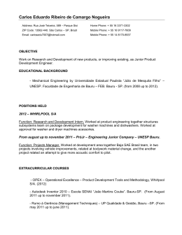
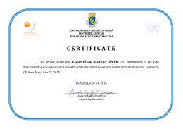
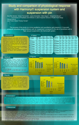
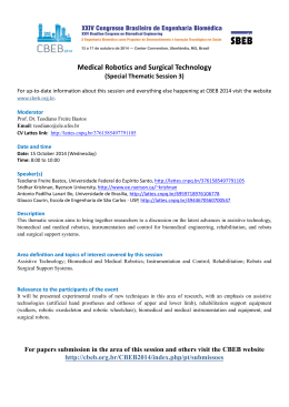
USE
OF TITANIUM SCREWS FOR
DENTURES OR SURGICAL GUIDE
FIXATION AFTER SULCOPLASTY
Luis Eduardo Marques Padovan1
Paulo Domingos Ribeiro Júnior1
Department of Dentistry/Center for Biological
Sciences – University of
the Sacred Heart
1
PADOVAN, Luis Eduardo Mar ques; RIBEIRO JÚNIOR, Paulo Domingos. Use
of titanium scre ws for dentures or sur gical guide f ixation after sulcoplasty .
Salusvita, Bauru, v. 21, n. 3, p. 119-128, 2002
ABSTRACT
It is presented an alternative procedure to the f ixation of prostheses
and surgical guides to the maxilla, avoiding the use of maxillary suspensions. Titaniun screws are used in this tec hnique fixing the prostheses in the anterior w all of the maxillary sinus. In this method the
inconvenience of performing suspensions is eliminated, reducing surgical time, making possible to carry it out under local anesthesia, minimizing the risk of inf ections, promoting a more stable fixation and
more comfortable post-operative period for the patient.
KEY WORDS: Rigid Fixation; Screws; Sulcoplasty; Pre-prosthetic
surgery.
INTRODUCTION
Received on: July 8, 2002
Accepted on: December 3, 2003
The prosthetic rehabilitation of totaly edentulous patients may
pose some difficulties, which are mostly related to the insuf ficient
height of the prosthetic area. The absence of prosthesis or the use of
in adequately adapted prosthetic devices increase the absor ption of
the alveolar crest (CARVALHO, 1980; ZANINI, 1990). Moreo ver,
the bone structures undergo a continuous reshaping process and the
reabsorption phase may be enhanced b y local and/or systemic pathologies (CARVALHO, 1980).
The insufficient height of the prosthetic area ma y be corrected through surgery in the soft tissues, namely the vestibular fornix
119
deepening or vestibuloplasty (PETERSON et al., 1999). Such procedures aim to increase the height of the prosthetic area (ZANINI,
1990). To indicate surgery it is necessary to evaluate the bone height
of the alveolar crest, what can be done through X-ra ys. According
to Peterson et al., (1999) 15 mm is the minimal height of the mandibular bone necessar y to obtain some predicab le success in the
procedure.
The earliest technique for sulcoplasty w as described by Kazanjian in 1924. From then on, many techniques have been developed with the same objective (CLARK, 1953; KETHEY; GAMBLE,
1978; OBWEGESER, 1959; ARRUDA, 1965). Such techniques
aim to obtain healing through reepithelization since there is healing
by second intention. Other techniques use mucosa or skin to co ver
the row area (ARRUDA, 1965; YRASTORZA, 1976).
To be successful these procedures need to maintain the depth
of the vestibular sulcus during the post operati ve period. If tissues
are not kept within the desirab le limits and there is healing b y second intention it is possib le to have a loss of 60% of the deepened
area (GREGORY, 1982; PETERSON et al., 1999).
For a very long time now authors have been seeking a method
to keep the vestibular flap in a more apical region, mainly during the
early post operative period, aiming to obtain less scar contraction
and, thus, a better predictability for the f inal result.
With this pur pose, many techniques have been used, such as
the simple suture of the v estibular flap in the deepest re gions close
to the periosteum, transf ixing suture through the soft tissue of the
submentonian region with a capitel, transfixing suture through a latex tube aiming to k eep the flap in a more apical position, use of
prosthesis or f ixed / non-f ixed surgical guides by mandibular cerclage and/or maxillar y suspension (CARVALHO, 1980; ZANINI,
1990) and the isolated use of screws to anchor tissues in a more apical position (DYM; CERBONE, 1991).
Recently, its was proposed by Nary Filho et al. (1994) the positioning of prosthetic devices in the maxilla in cases of facial fractures and/or ortognatic surgery through the transfixation of such devices or surgical guides with titanium scre ws bolted in the lateral
and anterior maxillary wall.
The objective of this study is to sho w the viability of the use
of titanium screw to f ix prosthesis or sur gical guides to the bone
crest following sulcoplasty aiming to obtain a more stab le deepening with less discomfort to the patient.
120
PADOVAN, Luis Eduardo
Marques; RIBEIRO
JÚNIOR, Paulo Domingos.
Use of titanium screws for
dentures or surgical guide
fixation after sulcoplasty.
Salusvita, Bauru, v. 21,
n. 3, p. 119-128, 2003
PADOVAN, Luis Eduardo
Marques; RIBEIRO
JÚNIOR, Paulo Domingos.
Use of titanium screws for
dentures or surgical guide
fixation after sulcoplasty.
Salusvita, Bauru, v. 21,
n. 3, p. 119-128, 2003
CASE REPORT
J. M., male, 45 years old, attended the Clinic of Bucomaxilofacial Traumatology of the University of the Sacred Heart, reporting
failure of previous dentistry treatment, when several total superior
prosthesis were made. Lack of retention and the instability of the
prosthesis were the main reasons for discontinuing its use.
An intraoral exam revealed healthy oral mucosa lining both
superior and inferior crests. It w as noted that the patient had lo w
muscular insertion in the upper crest leading to a def iciency in
height (FIGURE 1). The inferior arch was partially dented allowing
the use of a removable partial prosthesis. By palpation it w as identified that the upper crest showed a reasonable height.
To support the clinical diagnosis some X-ra y were used; orthopantomography (FIGURE 2) and cephalometry of the lateral aspect of the face were used to confirm the presence of bone tissue in
the upper crest. These X-ray views revealed enough upper crest
bone tissue to sustain the prosthetic device.
In association with the prosthesis technician the plan of treat ment was established including the pre-prosthetic surgery for vestibular fornix deepening and later construction of a total superior and
a partial inferior removable prosthesis.
The proposed treatment was superior sulcoplasty and maintenance of tissue b y means of a sur gical gutter f ixed with four titanium screws to the lateral and anterior wall of the maxilla since the
patient had already lost the pre viously constructed prosthesis. Fol-
FIGURE 1 - Pré operative view showing low muscular insertion.
121
PADOVAN, Luis Eduardo
Marques; RIBEIRO
JÚNIOR, Paulo Domingos.
Use of titanium screws for
dentures or surgical guide
fixation after sulcoplasty.
Salusvita, Bauru, v. 21,
n. 3, p. 119-128, 2003
FIGURE 2 - Panoramic X-ray take showing the maxillar y alveolar bone crest
height.
lowing the anamnesis and the analysis of routine laboratorial exams
the superior arch of the patient w as molded to permit the construction of the surgical guide in acrylic resin.
After antisepsis and preparation of the sur gical field the area
was anesthetized with mepi vacaine 2% with v asoconstrictor
1.100.000 by infiltrative regional block of the superior posterior ,
medium superior and anterior alveolar nerves and by infiltrative terminal anesthesia of all the alveolar process.
The selected surgical technique was that described b y Clark
(1953) with a mucosa incision in the rim of the alv
eolar crest
through the vestibular area from one tuber to the other . Then, with
a scalpel or scissor with b lunt points the mucous flaps w as undermined preserving the periosteum adhered to the v estibular crest.
The undermining was extended to the apical regional ticker muscular fibers that were making difficult the placement of the flap in a
more superior position or up to the point that the crest showed a sufficient height to the construction of a suitable total prosthesis.
After undermining the flap w as sutured (pol yglactine 910,
4/0) with continuous sutures bringing near the free v estibule muscosa to the muscular region and to the periosteum of the deep region
of the vestibule (FIGURE 3).
The edges of the pre viously constructed surgical guide were
far from the innermost part of the new alveolar sulcus (FIGURE 4)
and, thus, to maintain tissues in the desirable position the guide had
to be relined, which initially was done with a specif ic resin for rebasing of template of total rigid prosthesis after pol
ymerization
(Kooliner)1, applied directly in the mouth of the patient. In this step,
the guide was removed and replaced till the f inal polymerization of
the material. After that, a new relining was done with a resilient resin (Coesoft)2 allowing a better conditioning of tissues during the repair process (FIGURE 5).
122
Kooliner, rebasing resin.
Manufacturer: GC America
Inc
1
Coesoft, resilient resin for
postoperative prosthesis. Manufacturer: GC America inc
2
PADOVAN, Luis Eduardo
Marques; RIBEIRO
JÚNIOR, Paulo Domingos.
Use of titanium screws for
dentures or surgical guide
fixation after sulcoplasty.
Salusvita, Bauru, v. 21,
n. 3, p. 119-128, 2002
FIGURE 3 - Trans operative view showing the vestibular height after Clark’s sulcoplasty.
After removing the residual resin, the guide was put again on
the maxillary crest in order to allo w the perforation with a drill
transfixing the guide and the maxilla bilaterally in the region of the
canine and zigomatic pillar. The screws used for the f ixation of the
surgical guide measured 2.0 mm x 12 mm being the perforation obtained with a 1.5 mm drill, belonging to the same fixation system,
mounted in a lo w rotation motor under continuous ir rigation with
FIGURE 4 - The picture sho ws the sur gical guide pre viously made in a cast
model and the gain in vestibular height.
123
PADOVAN, Luis Eduardo
Marques; RIBEIRO
JÚNIOR, Paulo Domingos.
Use of titanium screws for
dentures or surgical guide
fixation after sulcoplasty.
Salusvita, Bauru, v. 21,
n. 3, p. 119-128, 2003
FIGURE 5 - The surgical guide being reshaped to aid in the suppor t of undermined tissues.
saline. The desirable depth was obtained when the drill reached the
inner part of the maxillary sinus. A screw was placed after each perforation. Two screws were used at each side to obtain an optimum
stability of the guide (FIGURE 6). After the procedure a micropore
dressing was applied on the upper lip to control edema, bleeding
and maintain the area immobile.
The patient received ampicilin, 500 mg e very 6 hours, for 7
days; dipirone 40 drops every 6 hours, for 2 days and mouth washes
with chlorhexidine 0.12 every 12 hours for 21 da ys. Postoperative
control was done weekly including oral higienization, br ushing of
mucosa and the external surface of the guide.
After 21 days the screws and the surgical guide was removed
after terminal infiltration anesthesia of the scre w region (FIGURE
7). At this moment it was observed the beginning of the reepithelization process in the area, which was kept raw and protected by the
reshaped guide. After another reshape with resilient resin the guide
was kept till the installation of the def initive total prosthesis, which
construction started one month after the sulcoplasty . The patient
was followed-up and he did not repor t any complaint regarding the
treatment (FIGURE 8) or the stability of the prosthesis.
124
PADOVAN, Luis Eduardo
Marques; RIBEIRO
JÚNIOR, Paulo Domingos.
Use of titanium screws for
dentures or surgical guide
fixation after sulcoplasty.
Salusvita, Bauru, v. 21,
n. 3, p. 119-128, 2003
FIGURE 6 - Fixation of the sur gical guide in the maxilla through scre ws for
internal rigid fixation.
DISCUSSION
Since the introduction of rigid inter nal fixation, that is, osteosynthesis with screws and plaques, the use of intermaxillary fixation was restrict to a few cases and, when used, for a short period
(CROFTS et al., 1990). Ho wever, the inter maxillary block in the
transoperatory period was always necessary. Taking this into consi-
FIGURE 7 - 21st day post operati ve. The surgical guide and sutures w
removed. Note the alveolar crest undergoing reepithelization.
ere
125
PADOVAN, Luis Eduardo
Marques; RIBEIRO
JÚNIOR, Paulo Domingos.
Use of titanium screws for
dentures or surgical guide
fixation after sulcoplasty.
Salusvita, Bauru, v. 21,
n. 3, p. 119-128, 2002
FIGURE 8 - 6th month post operative. Note the alv eolar crest vestibular height
that was obtained.
deration and in the dif ficulty of these procedures to be car ried out
in edentulous patients with total prosthesis, Shetty et al. in 1987,
proposed a variation of the intermaxyllary block. In these cases they
proposed the use of “minihooks” similar to screws fixed in the maxillary bone and in the mandible. According to them, such “hooks”
allowed an efficient intermaxillary block during the trans-operative
period protecting surgeons form possible accidents. With the same
objective and considering the potential of exposure of health professional to punctures and wounds, which may be caused by traditional
techniques of inter maxillary block, through odontosynthesis,
Erich’s plaques and suspensions, Arthur and Berardo (1989) used titanium screws fixed in the basal maxillary and mandibular bone of
toothed patients. The screws, externally connected by a steel wire,
promoted the intermaxillary block. The authors emphasized the importance of this technique in the treatment of f acial fractures in
high-risk patients.
Dym and Cerbone (1991) used scre ws to maintain tissues after sulcoplasty. They avoided the use o guides and or patient’ s own
prosthesis and used only the sutures and two screws at each side to
the maxilla to maintain tissues. They report some success of this
procedure with a partial loss of the depth of the sulcus. The inconvenient of this technique is that, in some areas, tissues are not adequately maintained in the desirab le position, which could be attained only with a great number of screws. Besides that, the crown of
the crew was covered by soft tissue, at the moment of its remo val,
due to the action of muscles in the region. The discomfort of pa-
126
PADOVAN, Luis Eduardo
Marques; RIBEIRO
JÚNIOR, Paulo Domingos.
Use of titanium screws for
dentures or surgical guide
fixation after sulcoplasty.
Salusvita, Bauru, v. 21,
n. 3, p. 119-128, 2002
tients due to the tissues remaining ra w and not protected should be
borne in mind.
Nary Filho et al. (1994) used titanium screws to maintain total prosthesis on the crest or sur gical guides in the conser vative
treatment of mandibular fractures in edented patients that w ere in
need for stabilization b y intermaxillary block. The good results
opened way for the use of this technique in other situations, such
as partial resection of mandib le, in which the f ixation of the total
prosthesis aimed the oclusal orientation during mandibular reconstruction, or even for postoperative physical therapy. The technique
was also used in sulcoplasty with skin g raft, in which the surgical
quid was kept on place through scre ws, as repor ted in this study .
While removing the f ixation it w as noted the adv antages of this
technique as compared to the maxillary suspension. It is more comfortable since the screws are unbolted with a simple anesthetic in filtration in the area. There was no complication, such as bucosinusal communication or infectious process. It is e ven a more aseptic
procedure since it pre vents an element from being e xposed to the
oral cavity to go through tissues as happens with the remo val of
maxillary suspensions.
CONCLUSION
The authors believe that the fixation of guides and or prosthesis with screws may be done safel y and that the dissemination of
this method among dentists ma y increase conf idence with sulcoplasty since it is a simple and safe procedure, with adequate fixation
of prosthesis or surgical guide allowing more favorable results in attaining sulcus depth and comfort to the patient.
BIBLIOGRAPHIC REFERENCES
1. ARTHUR, G.; BERARDO, N. A simplified technique of maxillomandibular
fixation. J. oral Maxillofac. Surg., v. 47, p. 1234, 1989.
2. ARRUDA, J. V. Uma Técnica Cirúrgica para aprofundamento dos sulcos vestibulares e linguais nos desdentados totais. Bol. Fax. Farm. Odont. Piracicaba, v. 18, p. 1-25, 1965.
3. CARVALHO, A. C. P. Revista da A.P.C.D. Reg. Araç., v. 1, n. 1, p. 18-23 , 1980.
4. CLARK H. B. Deepening of the labial sulcus by mucosal Flap advancement,
Report of a case. J. Oral Surg, v. 11, n. 2, p. 165-168, 1953.
127
5. CROFTS, C. E. et al. A comparative in vitro study of f ixation of mandibular
fractures with paraskeletal clamps or scre w plates. J. oral Maxillofac Surg., v.
48, p. 461-466, 1999.
6. DYM, H. ; CERBONE, T. Bone screws as an aid in vestibuloplasty procedures. J. oral Maxillofac Surg., v. 49, p. 1132-1133, 1991.
7. GREGORI, C. Cirurgia Buco Dento Alveolar. 1. ed. São P aulo: SARVIER,
1996.
8. GREGORY, O. J. Surgical procedures to prepare the month for prosthetic replacement a review. Aust Dent J., v. 27, n. 141, p. 209-216, Aug, 1982.
9. KAZANJIAN, N. M. Sur gical operations related to satisf atory dentures.
Dent. Cosmos, v. 66, p. 367, 1924.
10. KETHLEY ; GAMBLE. The lops witch; a modification of Kazanjian’s labial
vestibuloplasty. J. Oral Surg, v. 36, n. 9, p. 701-705, 1978.
11. NARY FILHO, H. et al. Utilização de Parafusos de Titânio para Fixação de
Próteses ou Guias Cirírgicos com método alternativo às Suspensões Maxilares em Cirurgia Buco-Maxilo-Facial. Rev. Fac. Odont. Bauru, v. 2., n. 4,
p. 67-72, 1994.
12. OBWEGESER, M. Die Subnwk ose Vestibulumplastik. Dtsch Zahraerztla,
v. 14, p. 629, 1959.
13. PETERSON et al. Cirurgia Oral e Maxilo Facial Contemporânea. 2. ed. Rio
de Janeiro: Guanabara Yoogansa, 1999.
14. SHETTY, V. et al. Maxillomandib ular fixation with minihooks: A clinical
evaluation. Oral Surg Oral Med Oral Pathol., v. 64, p. 677-679, 1987.
15. YRASTORZA, J. A. Vestibuloplasty with skin g rafting. J. Oral Surg., v. 34,
p. 29-33, 1976.
16. ZANINI, S. A. Cirurgia e Traumatologia BucoMaxiloFacial. 1. ed. Rio de
Janeiro: Revinter, 1990.
128
PADOVAN, Luis Eduardo
Marques; RIBEIRO
JÚNIOR, Paulo Domingos.
Use of titanium screws for
dentures or surgical guide
fixation after sulcoplasty.
Salusvita, Bauru, v. 21,
n. 3, p. 119-128, 2003
Baixar