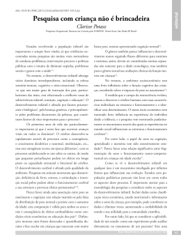
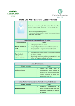
ISSN 1413-3555 Tutorial 471 Tutorial for writing systematic reviews for the Brazilian Journal of Physical Therapy (BJPT) Marisa C. Mancini, Jefferson R. Cardoso, Rosana F. Sampaio, Lucíola C. M. Costa, Cristina M. N. Cabral, Leonardo O. P. Costa Systematic Review 481 Static body postural misalignment in individuals with temporomandibular disorders: a systematic review Thaís C. Chaves, Aline M. Turci, Carina F. Pinheiro, Letícia M. Sousa, Débora B. Grossi 502 Walking training associated with virtual reality-based training increases walking speed of individuals with chronic stroke: systematic review with meta-analysis Juliana M. Rodrigues-Baroni, Lucas R. Nascimento, Louise Ada, Luci F. Teixeira-Salmela Brazilian Journal of Physical Therapy 2014 Nov-Dec; 18(6) ISSN 1413-3555 Original Articles 513 Relationship between the climbing up and climbing down stairs domain scores on the FES-DMD, the score on the Vignos Scale, age and timed performance of functional activities in boys with Duchenne muscular dystrophy Lilian A. Y. Fernandes, Fátima A. Caromano, Silvana M. B. Assis, Michele E. Hukuda, Mariana C. Voos, Eduardo V. Carvalho 521 Muscular performance characterization in athletes: a new perspective on isokinetic variables Giovanna M. Amaral, Hellen V. R. Marinho, Juliana M. Ocarino, Paula L. P. Silva, Thales R. de Souza, Sérgio T. Fonseca 530 Characteristics and associated factors with sports injuries among children and adolescents Franciele M. Vanderlei, Luiz C. M. Vanderlei, Fabio N. Bastos, Jayme Netto Júnior, Carlos M. Pastre 538 Can the adapted arcometer be used to assess the vertebral column in children? Juliana A. Sedrez, Cláudia T. Candotti, Fernanda S. Medeiros, Mariana T. Marques, Maria I. Z. Rosa, Jefferson F. Loss 544 Breathing exercises: inϐluence on breathing patterns and thoracoabdominal motion in healthy subjects Danielle S. R. Vieira, Liliane P. S. Mendes, Nathália S. Elmiro, Marcelo Velloso, Raquel R. Britto, Verônica F. Parreira 553 Application of positive airway pressure in restoring pulmonary function and thoracic mobility in the postoperative period of bariatric surgery: a randomized clinical trial Patrícia Brigatto, Jéssica C. Carbinatto, Carolina M. Costa, Maria I. L. Montebelo, Irineu Rasera-Júnior, Eli M. Pazzianotto-Forti 563 Functional priorities reported by parents of children with cerebral palsy: contribution to the pediatric rehabilitation process Marina B. Brandão, Rachel H. S. Oliveira, Marisa C. Mancini 572 Ground reaction forces during level ground walking with body weight unloading Ana M. F. Barela, Paulo B. de Freitas, Melissa L. Celestino, Marcela R. Camargo, José A. Barela 2014 Nov-Dec; 18(6) Editorial Rules 2014 Nov-Dec; 18(6) ASSOCIAÇÃO BRASILEEIR IRA DE PESQUISA E PÓS-GRADUAÇÃO ÇÃO EM FISIOTERAPIA FINANCIAL SUPPORT EDITORS Débora Bevilaqua Grossi – Universidade de São Paulo - Ribeirão Preto, SP, Brazil Sérgio Teixeira Fonseca – Universidade Federal de Minas Gerais - Belo Horizonte, MG, Brazil ADMINISTRATIVE EDITOR Aparecida Maria Catai – Universidade Federal de São Carlos - São Carlos, SP, Brazil INTERNATIONAL EDITOR David J. Magee – University of Alberta - Canada LIBRARIAN AND GENERAL COORDINATOR Dormélia Pereira Cazella – FAI/ Universidade Federal de São Carlos - São Carlos, SP, Brazil SPECIALIST EDITORS Ana Beatriz de Oliveira - Universidade Federal de São Carlos - São Carlos, SP, Brazil Ana Cláudia Mattiello-Sverzut – Universidade de São Paulo - Ribeirão Preto, SP, Brazil Anamaria Siriani de Oliveira – Universidade de São Paulo - Ribeirão Preto, SP, Brazil Anielle Cristhine de Medeiros Takahashi – Universidade Federal de São Carlos - São Carlos, SP, Brazil Audrey Borghi e Silva – Universidade Federal de São Carlos - São Carlos, SP, Brazil Christina Danielli Coelho de Morais Faria - Universidade Federal de Minas Gerais - Belo Horizonte, MG, Brazil Elaine Caldeira de Oliveira Guirro – Universidade de São Paulo - Ribeirão Preto, SP, Brazil Francisco Albuquerque Sendin - Universidad de Salamanca – Spain Helenice Jane Cote Gil Coury – Universidade Federal de São Carlos - São Carlos, SP, Brazil Hugo Celso Dutra de Souza - Universidade de São Paulo - Ribeirão Preto, SP, Brazil Isabel Camargo Neves Sacco – Universidade de São Paulo - São Paulo, SP, Brazil João Luiz Quagliotti Durigan - Universidade de Brasília – Brasília, DF, Brazil Leani Souza Máximo Pereira – Universidade Federal de Minas Gerais - Belo Horizonte, MG, Brazil Leonardo Oliveira Pena Costa – Universidade Cidade de São Paulo - São Paulo, SP, Brazil Luci Fuscaldi Teixeira-Salmela – Universidade Federal de Minas Gerais - Belo Horizonte, MG, Brazil Marisa Cotta Mancini – Universidade Federal de Minas Gerais - Belo Horizonte, MG, Brazil Nivaldo Antonio Parizotto – Universidade Federal de São Carlos - São Carlos, SP, Brazil Patrícia Driusso – Universidade Federal de São Carlos - São Carlos, SP, Brazil Paula Lanna Pereira da Silva – Universidade Federal de Minas Gerais - Belo Horizonte, MG, Brazil Paula Rezende Camargo – Universidade Federal de São Carlos - São Carlos, SP, Brazil Pedro Dal Lago – Universidade Federal de Ciências da Saúde de Porto Alegre - Porto Alegre, RS, Brazil Rosana Ferreira Sampaio – Universidade Federal de Minas Gerais - Belo Horizonte, MG, Brazil Stela Márcia Mattiello – Universidade Federal de São Carlos - São Carlos, SP, Brazil Tatiana de Oliveira Sato – Universidade Federal de São Carlos - São Carlos, SP, Brazil Thiago Luiz de Russo - Universidade Federal de São Carlos - São Carlos, SP, Brazil Verônica Franco Parreira – Universidade Federal de Minas Gerais - Belo Horizonte, MG, Brazil BRAZILIAN EDITORIAL BOARD Ada Clarice Gastaldi - Universidade de São Paulo - Ribeirão Preto, SP Amélia Pasqual Marques – Universidade de São Paulo - São Paulo, SP Ana Cláudia Muniz Rennó – Universidade Federal de São Paulo - Santos, SP André Luiz Felix Rodacki – Universidade Federal do Paraná- Curitiba, PR Anna Raquel Silveira Gomes – Universidade Federal do Paraná - Matinhos, PR Armèle Dornelas de Andrade – Universidade Federal do Pernambuco - Recife, PE Carlos Marcelo Pastre – Universidade Estadual Paulista - Presidente Prudente, SP Celso Ricardo Fernandes de Carvalho – Universidade de São Paulo - São Paulo, SP Cláudia Santos Oliveira – Universidade Nove de Julho - São Paulo, SP Cristiane Shinohara Moriguchi – Universidade Federal de São Carlos - São Carlos, SP Cristina Maria Nunes Cabral – Universidade Cidade de São Paulo - São Paulo, SP Daniela Cristina Carvalho de Abreu – Universidade de São Paulo - Ribeirão Preto, SP Dirceu Costa – Universidade Nove de Julho - São Paulo, SP Ester da Silva – Universidade Federal de São Carlos - São Carlos, SP Fábio de Oliveira Pitta – Universidade Estadual de Londrina - Londrina, PR Fábio Viadanna Serrão – Universidade Federal de São Carlos - São Carlos, SP Fátima Valéria Rodrigues de Paula – Universidade Federal de Minas Gerais - Belo Horizonte, MG Guilherme Augusto de Freitas Fregonezi – Universidade Federal do Rio Grande do Norte - Natal, RN Jefferson Rosa Cardoso – Universidade Estadual de Londrina - Londrina, PR João Carlos Ferrari Corrêa – Universidade Nove de Julho - São Paulo, SP José Angelo Barela – Universidade Cruzeiro do Sul - São Paulo, SP Josimari Melo de Santana – Universidade Federal de Sergipe - Aracajú, SE Juliana de Melo Ocarino – Universidade Federal de Minas Gerais - Belo Horizonte, MG Lucíola da Cunha Menezes Costa – Universidade Cidade de São Paulo - São Paulo, SP Luis Vicente Franco de Oliveira – Universidade Nove de Julho - São Paulo, SP Luiz Carlos Marques Vanderlei – Universidade Estadual Paulista - Presidente Prudente, SP Luzia Iara Pfeifer – Universidade de São Paulo - Ribeirão Preto, SP Marco Aurélio Vaz – Universidade Federal do Rio Grande do Sul - Porto Alegre, RS Naomi Kondo Nakagawa – Universidade de São Paulo - São Paulo, SP Nelci Adriana Cicuto Ferreira Rocha – Universidade Federal de São Carlos - São Carlos, SP Paulo de Tarso Camillo de Carvalho – Universidade Nove de Julho - São Paulo, SP Raquel Rodrigues Britto – Universidade Federal de Minas Gerais - Belo Horizonte, MG Renata Noce Kirkwood – Universidade Federal de Minas Gerais - Belo Horizonte, MG Ricardo Oliveira Guerra – Universidade Federal do Rio Grande do Norte - Natal, RN Richard Eloin Liebano – Universidade Cidade de São Paulo - São Paulo, SP Rinaldo Roberto de Jesus Guirro – Universidade de São Paulo - Ribeirão Preto, SP Rosana Mattioli – Universidade Federal de São Carlos - São Carlos, SP Rosimeire Simprini Padula – Universidade Cidade de São Paulo - São Paulo, SP Sara Lúcia Silveira de Menezes – Centro Universitário Augusto Motta - Rio de Janeiro, RJ Simone Dal Corso – Universidade Federal do Rio Grande do Sul - Porto Alegre, RS Stella Maris Michaelsen – Universidade do Estado de Santa Catarina - Florianópolis, SC Tania de Fátima Salvini – Universidade Federal de São Carlos - São Carlos, SP Thaís Cristina Chaves – Universidade de São Paulo - Ribeirão Preto, SP INTERNATIONAL EDITORIAL BOARD Alan M. Jette – Boston University School of Public Health - USA Chukuka S. Enwemeka – University of Wisconsin - USA Edgar Ramos Vieira – Florida International University - USA Gert-Ake Hansson – Lund University - SWEDEN Janet Carr – University of Sydney - AUSTRALIA Kenneth G. Holt – Boston University - USA LaDora V. Thompson – University of Minnesota - USA Liisa Laakso – Grifϔith University - AUSTRALIA Linda Fetters – University of Southern California - USA Paula M. Ludewig – University of Minnesota - USA Rik Gosselink – Katholieke Universiteit Leuven - BELGIUM Rob Herbert – The George Institute for International Health - AUSTRALIA Sandra Olney – Queen’s University - CANADA Braz J Phys Ther. 2014 Nov-Dec; 18(6):471-579 The Brazilian Journal of Physical Therapy is published by the Associação Brasileira de Pesquisa e PósGraduação em Fisioterapia – ABRAPG-Ft (Brazilian Association for Research and Graduate Studies in Physical Therapy). Published since 1996, the Brazilian Journal of Physical Therapy adopts a peer review process. Each article is only published after it is accepted by the reviewers, who are maintained anonymous during the process. The editors accept no responsibility for damage to people or property, which may have been caused by the use of ideas, techniques or procedures described in the material published by this journal. The submission of articles presupposes that these articles, with the exception of extended summaries, have not been previously published elsewhere, nor submitted to any other publication. The abbreviated title of the journal is Braz J. Phys. Ther. and this must be used in references, footnotes and bibliographic legends. The Brazilian Journal of Physical Therapy is freely accessible at the homepage on the web: http://www.scielo.br/rb is. Mission To publish original research articles on topics related to the areas of physical therapy and rehabilitation sciences, including clinical, basic or applied studies on the assessment, prevention, and treatment of movement disorders. Indexed in Cataloguing Card Brazilian Journal of Physical Therapy / Associação Brasileira de Pesquisa e Pós-Graduação em Fisioterapia. v. 1, n. 1 (1996). – São Carlos: Editora Cubo, 1996v. 18, n. 6 (Nov/Dec 2014). Bimonthly Continued Revista Brasileira de Fisioterapia ISSN 1413-3555 1. Physical Therapy. 2. Studies. I. Associação Brasileira de Pesquisa e Pós-Graduação em Fisioterapia. Librarian: Dormélia Pereira Cazella (CRB 8/4334) Contact Address Brazilian Journal of Physical Therapy Rod. Washington Luís, Km 235, Caixa Postal 676, CEP 13565-905 São Carlos, SP - Brasil +55(16) 3351-8755 [email protected] www.rbf-bjpt.org.br Technical and Administrative Support Ana Paula de Luca Leonor A. Saidel Aizza Raquel Mariane da Silveira Desktop Publishing and Editorial Consulting Printed in acid free paper. No part of this publication can be reproduced or transmitted by any media, be it electronic, mechanical or photocopy, without the express authorization of the editors. summary Tutorial 471 Tutorial for writing systematic reviews for the Brazilian Journal of Physical Therapy (BJPT) Marisa C. Mancini, Jefferson R. Cardoso, Rosana F. Sampaio, Lucíola C. M. Costa, Cristina M. N. Cabral, Leonardo O. P. Costa Systematic Review 481 Static body postural misalignment in individuals with temporomandibular disorders: a systematic review Thaís C. Chaves, Aline M. Turci, Carina F. Pinheiro, Letícia M. Sousa, Débora B. Grossi 502 Walking training associated with virtual reality-based training increases walking speed of individuals with chronic stroke: systematic review with meta-analysis Juliana M. Rodrigues-Baroni, Lucas R. Nascimento, Louise Ada, Luci F. Teixeira-Salmela Original Articles 513 Relationship between the climbing up and climbing down stairs domain scores on the FESDMD, the score on the Vignos Scale, age and timed performance of functional activities in boys with Duchenne muscular dystrophy Lilian A. Y. Fernandes, Fátima A. Caromano, Silvana M. B. Assis, Michele E. Hukuda, Mariana C. Voos, Eduardo V. Carvalho 521 Muscular performance characterization in athletes: a new perspective on isokinetic variables Giovanna M. Amaral, Hellen V. R. Marinho, Juliana M. Ocarino, Paula L. P. Silva, Thales R. de Souza, Sérgio T. Fonseca 530 Characteristics and associated factors with sports injuries among children and adolescents Franciele M. Vanderlei, Luiz C. M. Vanderlei, Fabio N. Bastos, Jayme Netto Júnior, Carlos M. Pastre 538 Can the adapted arcometer be used to assess the vertebral column in children? Juliana A. Sedrez, Cláudia T. Candotti, Fernanda S. Medeiros, Mariana T. Marques, Maria I. Z. Rosa, Jefferson F. Loss 544 Breathing exercises: inϐluence on breathing patterns and thoracoabdominal motion in healthy subjects Danielle S. R. Vieira, Liliane P. S. Mendes, Nathália S. Elmiro, Marcelo Velloso, Raquel R. Britto, Verônica F. Parreira 553 Application of positive airway pressure in restoring pulmonary function and thoracic mobility in the postoperative period of bariatric surgery: a randomized clinical trial Patrícia Brigatto, Jéssica C. Carbinatto, Carolina M. Costa, Maria I. L. Montebelo, Irineu Rasera-Júnior, Eli M. Pazzianotto-Forti 563 Functional priorities reported by parents of children with cerebral palsy: contribution to the pediatric rehabilitation process Marina B. Brandão, Rachel H. S. Oliveira, Marisa C. Mancini 572 Ground reaction forces during level ground walking with body weight unloading Ana M. F. Barela, Paulo B. de Freitas, Melissa L. Celestino, Marcela R. Camargo, José A. Barela Editorial Rules tutorial Tutorial for writing systematic reviews for the Brazilian Journal of Physical Therapy (BJPT) Tutorial para elaboração de revisões sistemáticas para o Brazilian Journal of Physical Therapy (BJPT) Marisa C. Mancini1, Jefferson R. Cardoso2, Rosana F. Sampaio3, Lucíola C. M. Costa4, Cristina M. N. Cabral4, Leonardo O. P. Costa4,5 ABSTRACT | Systematic reviews aim to summarize all evidence using very rigorous methods in order to address a specific research question with less bias as possible. Systematic reviews are widely used in the field of physical therapy, however not all reviews have good quality. This tutorial aims to guide authors of the Brazilian Journal of Physical Therapy on how systematic reviews should be conducted and reported in order to be accepted for publication. It is expected that this tutorial will help authors of systematic reviews as well as journal editors and reviewers on how to conduct, report, critically appraise and interpret this type of study design. Keywords: rehabilitation; physical therapy; literature review; systematic review; meta analysis. HOW TO CITE THIS ARTICLE Mancini MC, Cardoso JR, Sampaio RF, Costa LCM, Cabral CMN, Costa LOP. Tutorial for writing systematic reviews for the Brazilian Journal of Physical Therapy (BJPT). Braz J Phys Ther. 2014 Nov-Dec; 18(6):471-480. http://dx.doi.org/10.1590/bjpt-rbf.2014.0077 RESUMO | Revisões sistemáticas têm como objetivo sumarizar toda a evidência disponível, através de métodos rigorosos, para responder a uma pergunta de pesquisa específica com o mínimo de viés possível. Revisões sistemáticas são amplamente utilizadas na fisioterapia, porém nem todas as revisões possuem boa qualidade. Esse tutorial tem como objetivo guiar os autores do Brazilian Journal of Physical Therapy sobre como revisões sistemáticas deveriam ser conduzidas e descritas para que sejam aceitas para publicação. Espera-se que esse tutorial irá auxiliar autores de revisões sistemáticas, assim como editores e revisores de periódicos em como conduzir, descrever, fazer análise crítica e interpretar esse tipo de delineamento de pesquisa. Palavras-chave: reabilitação; fisioterapia; revisão da literatura; revisões sistemáticas; metanálise. Introdução Uma revisão de literatura pode ser definida como síntese e análise da informação com foco nas contribuições científicas dos estudos publicados1. Tais estudos não se restringem simplesmente a citações bibliográficas e servem para estabelecer novas conclusões1. Esse conceito não é novo, há sínteses do conhecimento que remontam ao início do século passado1,2. Na década de 60, estudos já integravam resultados de pesquisas e apontavam novas evidências científicas, principalmente nas ciências sociais, educação e psicologia. O reconhecimento da importância da aplicação da melhor informação científica disponível no campo da saúde trouxe a necessidade de se ancorar a prática clínica em evidências e, consequentemente, um aumento gradativo da demanda por esse tipo de informação2. Evidência refere-se ao conjunto de informações utilizadas para confirmar ou negar uma teoria ou hipótese científica e é produzida por um processo sistemático de investigação1. Apesar da reconhecida característica cumulativa da ciência, os métodos estatísticos para sintetizar evidências só foram desenvolvidos no século XX. Concomitante a esses avanços, os cientistas reconheceram que organizar e avaliar a informação científica acumulada ultrapassa a simples escolha do método. Tem sido destacada, desde então, a Programa de Pós-graduação em Ciências da Reabilitação, Departamento de Terapia Ocupacional, Escola de Educação Física, Fisioterapia e Terapia Ocupacional (EEFFTO), Universidade Federal de Minas Gerais (UFMG), Belo Horizonte, MG, Brasil 2 Laboratório de Biomecânica e Epidemiologia Clínica, Grupo PAIFIT, Universidade Estadual de Londrina (UEL), Londrina, PR, Brasil 3 Programa de Pós-graduação em Ciências da Reabilitação, Departamento de Fisioterapia, EEFFTO, UFMG, Belo Horizonte, MG, Brasil 4 Programa de Mestrado e Doutorado em Fisioterapia, Universidade Cidade de São Paulo (UNICID), São Paulo, SP, Brasil 5 Musculoskeletal Division, The George Institute for Global Health, Sydney, NSW, Australia Received: 12/02/2014 Revised: 12/04/2014 Accepted: 12/05/2014 1 http://dx.doi.org/10.1590/bjpt-rbf.2014.0077 Braz J Phys Ther. 2014 Nov-Dec; 18(6):471-480 471 Mancini MC, Cardoso JR, Sampaio RF, Costa LCM, Cabral CMN, Costa LOP necessidade de maior rigor metodológico para garantir a validade dos estudos de revisão, assim como se exige para os estudos considerados primários3. Há consenso de que a síntese do conhecimento é fundamental para o avanço da prática, da pesquisa e para a implementação de políticas de saúde. No entanto, sintetizar o conhecimento de forma clara e precisa exige habilidades e competências específicas do pesquisador. Investigar e selecionar todos os estudos relevantes, avaliar a sua qualidade e sintetizar dados são alguns dos desafios presentes na elaboração desse tipo de estudo4. Passos comuns aos métodos de síntese do conhecimento: a) uma clara definição do objetivo; b) desenvolvimento de um protocolo metodológico; c) estratégias abrangentes de busca para encontrar todos os artigos relevantes; d) um método de avaliação do potencial risco de viés nos estudos individuais e e) detalhamento da coleta de dados e dos procedimentos empregados4. Uma variedade de termos tem sido usada para descrever os processos de integração das evidências: pesquisa-síntese, revisão sistemática, revisão integrativa, meta-análise, entre outros1. A crescente popularidade desse tipo de estudo é ilustrada pelo fato de que muitos profissionais, para manterem-se atualizados, escolhem a leitura de estudos de revisão da literatura. Entre os diferentes tipos de estudos que disponibilizam sínteses de evidências científicas, as revisões sistemáticas cumprem critérios rigorosos de avaliação e produzem conclusões sólidas e aplicáveis à prática clínica. Além disso, revisões sistemáticas podem apontar importantes lacunas no conhecimento e servir de argumento para sinalizar a necessidade de novos estudos. Diretrizes, reporting guidelines e checklists visam a normatizar o processo de elaboração de revisões sistemáticas, desde a avaliação dos estudos selecionados até a estrutura final de comunicação dos resultados5. Cabe ao pesquisador escolher, entre as regras e normas existentes, aquelas que melhor se adequam ao tema a ser investigado e ao periódico selecionado para submissão do seu trabalho. Exemplos de diretrizes e reporting guidelines para estudos de revisão sistemática: Cochrane Handbook, encontrado em http:// www.cochrane.org/handbook e PRISMA, www.prisma-statement.org/, entre outros. Os primeiros ensaios clínicos em Fisioterapia foram estudos que avaliaram os efeitos da irradiação ultravioleta em crianças escolares e em crianças com problemas respiratórios, publicados, respectivamente, 472 Braz J Phys Ther. 2014 Nov-Dec; 18(6):471-480 por Colebrook6 em 1929 e por Doull et al.7 em 1931. A primeira revisão sistemática na área8 estabeleceu os efeitos dos tratamentos sobre as lesões ligamentares do tornozelo, tendo sido publicada pelo sueco Kolind-Sorensen9 em 1975. Desde então, tem havido um crescimento exponencial desse tipo de estudo em Fisioterapia. Torna-se, assim, imprescindível que os periódicos científicos envidem esforços no sentido de definir regras claras que possam ajudar os pesquisadores a alcançarem a qualidade exigida para publicação desse tipo de estudo, disponibilizando informação científica para o avanço do conhecimento. Fazendo eco a esse movimento internacional e buscando manter uma vigilância epistemológica sobre a pesquisa em Fisioterapia e em outras áreas da saúde, o Brazilian Journal of Physical Therapy (BJPT) preparou o presente tutorial. Ele busca alinhar qualitativamente os seus estudos de revisão sistemática, prezando pelo cuidado com as condições e limites das técnicas e conceitos empregados nesse processo. O que é revisão sistemática e metaanálise? Revisão sistemática, segundo o Handbook da Colaboração Cochrane10, é um estudo secundário que tem por objetivo reunir estudos semelhantes, publicados ou não, avaliando-os criticamente quanto à validade interna e reunindo-os em uma análise estatística, quando possível. Ainda, a revisão sistemática visa a minimizar os vieses usando métodos explícitos e pontuais. O método estatístico utilizado para integrar os resultados dos estudos incluídos em revisões sistemáticas é chamado de meta-análise8. Muitas vezes, os termos meta-análise e revisão sistemática são usados equivocadamente ou de forma alternada. A agregação estatística dos dados numa meta-análise não significa que os estudos individuais tenham sido criteriosamente analisados. Dessa forma, as revisões sistemáticas podem ser desenvolvidas com e sem meta-análise. A distinção entre revisão sistemática e meta-análise é muito importante, pois é sempre possível revisar de forma sistemática um conjunto de dados (com critérios para avaliação do risco de viés dos estudos que compõem a revisão), porém, algumas vezes, pode ser inapropriado, ou mesmo enganoso, realizar a agregação estatística dos resultados de estudos independentes. Tutorial revisão sistemática De forma geral, a síntese resultante de uma revisão sistemática fornece a melhor evidência sobre o tema em questão, tais como os efeitos de uma intervenção sobre determinado desfecho, a incidência de uma doença ou a acurácia de um teste diagnóstico, entre outros temas. Em uma revisão sistemática, é necessário: estipular uma pergunta clínica; determinar as fontes e métodos de seleção dos estudos, como bases de dados (i.e., bibliográficas, referenciais e textuais) e estratégias de busca empregadas; selecionar estudos com métodos semelhantes; realizar avaliações de possíveis vieses e explicitar mecanismos de avaliação da validade dos estudos selecionados; preparar sínteses para apresentações/disseminações (tanto qualitativas – descrições dos estudos, como quantitativas – meta-análises –, quando apropriado)10. As revisões sistemáticas são consideradas estudos secundários porque, em outras palavras, resumem as informações de múltiplas publicações consideradas primárias, como estudos de tratamento e prevenção (ensaios controlados aleatorizados-ECAs), estudos de prognóstico (coorte), estudos de diagnóstico (acurácia), estudos de etiologia (caso-controle), entre muitos outros. As revisões sistemáticas mais comuns são as de tratamento, que avaliam e/ou comparam a eficácia ou a efetividade de diferentes abordagens, sejam exercícios, medicamentos ou cirurgias, ou outras modalidades terapêuticas. A qualidade desse tipo de revisão deve ser assegurada para que profissionais, pacientes e agências reguladoras em saúde possam tomar decisões mais assertivas. Neste tutorial, serão enfatizadas as revisões de tratamento/prevenção que utilizam os ECAs. Esse modelo de estudo leva em consideração que os participantes foram aleatorizados ou tiveram a mesma chance de participar em um dos grupos de tratamento propostos. Um exemplo desse tipo de revisão foi a avaliação da efetividade de um programa de exercícios (i.e., principalmente de fortalecimento e de amplitude de movimento) e outros recursos (i.e., termoterapia, estimulação elétrica, faixas compressivas etc.) para pacientes submetidos à meniscectomia parcial artroscópica. A busca dos estudos publicados foi realizada de 1950 a 2013, e 18 ECAs foram incluídos na revisão, mas apenas seis participaram da análise estatística, ou seja, da meta-análise. Como conclusão, os autores indicaram que a realização dos procedimentos de fisioterapia ambulatorial citados acima associados às orientações para execução em domicílio melhorou a função do joelho, relatada pelo paciente, e a amplitude de movimento de flexão e extensão dessa articulação, quando comparada aos procedimentos de fisioterapia apenas ambulatorial11. Há estruturas bem estabelecidas para nortear a comunicação de um ECA (www.consortstatement.org) e de uma revisão sistemática (www.prisma-statement.org/ e Handbook da Colaboração Cochrane10). Em síntese, toda revisão sistemática envolve uma análise criteriosa da qualidade dos estudos, e algumas delas empregam meta-análise. Na análise de risco de viés, são observadas a validade interna, a validade externa e as análises estatísticas empregadas em cada um dos estudos selecionados. A metaanálise, por sua vez, é um procedimento sistemático e rigoroso, passível de ser reproduzido por outros pesquisadores e que permite combinar os resultados dos diferentes estudos. A meta-análise ajusta ou pondera os resultados levando em consideração o tamanho amostral de cada estudo primário, podendo ainda ser ajustada para outros fatores, tais como o risco de viés de cada estudo. Tipos de revisão sistemática É importante salientar que diversas perguntas de pesquisa podem ser sintetizadas em uma revisão sistemática. Provavelmente, o tipo mais comum e popular de revisão sistemática é aquela que tem como objetivo medir o efeito de alguma intervenção (i.e., revisão sistemática de ECAs). Porém, revisões sistemáticas podem ser extremamente úteis para resumir outras perguntas clínicas, como: prevalência12, incidência13, fatores prognósticos14, acurácia diagnóstica15, custo-efetividade16, fatores de risco 17, definição de termos de pesquisa 18, adaptações transculturais de questionários 19 , propriedades de medida de instrumentos de mensuração 20 e ainda revisões sistemáticas de estudos qualitativos21,22. Um problema decorrente de todas essas possibilidades é que o autor deve identificar qual delineamento de estudo deve ser utilizado para cada tipo de revisão sistemática. Embora isso pareça óbvio, esse tem sido um dos principais problemas identificados no processo de revisão por pares de manuscritos de revisão sistemática submetidos Braz J Phys Ther. 2014 Nov-Dec; 18(6):471-480 473 Mancini MC, Cardoso JR, Sampaio RF, Costa LCM, Cabral CMN, Costa LOP ao BJPT. Nesse caso, o autor deve sempre optar pelo delineamento ideal para cada tipo de pergunta científica. Opções mais adequadas de delineamento seriam ensaios controlados aleatorizados para revisões com o objetivo de medir efeitos de intervenção, estudos de coorte longitudinal prospectivos para revisões prognósticas ou de fatores de risco ou estudos transversais para revisões de prevalência. Em alguns tipos de pergunta, é possível que o uso de diferentes delineamentos de pesquisa seja adequado. Um exemplo seria uma revisão sistemática de acurácia diagnóstica que pode permitir a inclusão de estudos de caso-controle, estudos transversais e até mesmo ensaios clínicos, em alguns casos. Deve-se ter em mente que a combinação de delineamentos distintos é muito mais uma exceção do que uma regra. Itens essenciais de uma revisão sistemática As revisões sistemáticas vão diferir muito em relação à pergunta de pesquisa, assim como na elegibilidade dos tipos de estudos. Certas regras são obrigatórias em algumas revisões e irrelevantes em outras. No entanto, existem itens que são essenciais e que devem estar presentes em todas as revisões, a saber: 1. Definição clara da pergunta de pesquisa: uma boa revisão não é aquela que responde a várias perguntas, mas aquela que responde a perguntas específicas de forma clara e com o mínimo de viés possível. Sendo assim, a definição da pergunta de pesquisa é essencial. Uma orientação para delimitar bem uma pergunta para revisões sistemáticas de intervenção é usar a estrutura do PICO (Patient, Intervention, Comparison and Outcomes (Pacientes, Intervenção, Comparação e Desfechos). Por exemplo: “as técnicas de terapia manual associadas a um programa de exercícios (Intervenção) são melhores que somente exercícios (Comparação) para a redução da dor e incapacidade funcional (Desfechos) em pacientes adultos com dor lombar crônica (Pacientes)?” A redação de perguntas pode fazer uso da estrutura PICO de forma flexível, por exemplo, deixar o termo de comparação ser conhecido posteriormente na revisão: “quais são os efeitos da mobilização articular 474 Braz J Phys Ther. 2014 Nov-Dec; 18(6):471-480 (Intervenção) na melhora da amplitude de movimento, dor e incapacidade (Desfechos) em pacientes que receberam imobilização no tornozelo (Pacientes)?” Nesse caso, os grupos de comparação seriam qualquer tipo de grupo controle possível. Outra forma de flexibilizar a estrutura PICO é quando a revisão não avalia o efeito de intervenção; nesse caso, o termo “I” é atribuído ao foco do estudo (vide tipos de revisão sistemática acima). Para a redação de uma pergunta de pesquisa clara: a) É essencial que o pesquisador defina claramente a intervenção (ou o foco do estudo), os desfechos e a amostra de interesse. Esses três itens são fundamentais na formulação de uma pergunta clínica. b) É recomendado que o autor, ao formular sua pergunta de um estudo de intervenção, busque identificar a intervenção de forma específica (i.e., exercício resistido, orientação para cuidadores etc.), ao invés de denominar a(s) intervenção(ões) testada(s) no estudo como sendo a profissão ou área (i.e., Fisioterapia, Reabilitação). c) Outros tipos de revisão sistemática que não de estudos de intervenção devem seguir os mesmos princípios de elaboração de uma pergunta de pesquisa: perguntas claras, diretas e bem formuladas. d) Uma pergunta bem formulada também irá guiar vários aspectos do processo de elaboração de uma revisão sistemática, incluindo as estratégias de busca, elegibilidade dos estudos, extração de dados e conclusões da revisão. 2. Definição dos artigos elegíveis: uma vez formulada a pergunta de pesquisa, o autor deve definir, a priori, os critérios de inclusão e exclusão dos artigos que serão considerados elegíveis para a revisão. Essa definição passa pelo delineamento dos estudos a serem incluídos, características intrínsecas de cada estudo (i.e., amostra, tipos de tratamento, duração dos sintomas, equipamentos utilizados, entre outros), intervalo de tempo da publicação a ser considerado e idioma da publicação. Idealmente, artigos não deveriam ser excluídos com base no período de publicação, risco de Tutorial revisão sistemática viés e idioma de publicação. 3. Certificação de que todos os artigos elegíveis foram encontrados8,23: é uma das tarefas mais difíceis de uma revisão, pois ela deve sintetizar, de preferência, TODA a evidência disponível. Sendo assim, as buscas devem ser realizadas no maior número de bases de dados possível. Isso, muitas vezes, representa um problema para alguns pesquisadores, uma vez que, muitas dessas bases de dados, não são de acesso livre. Alguns exemplos são EMBASE, CINAHL, MEDLINE, PSYCHINFO. É importante salientar que somente 14 revistas de Fisioterapia estão indexadas no PubMed (que é a versão gratuita do MEDLINE) e, portanto, é grande a probabilidade de se perderem artigos realizando buscas somente em bases de acesso livre. O mesmo raciocínio deve ser usado para o idioma das bases acessadas: muitos autores buscam em várias bases nacionais, como SCIELO e LILACS. Porém, essas bases indexam somente artigos em português e espanhol, que equivalem a menos de 2% da literatura científica mundial24. Além do cuidado com a seleção das bases, outro item fundamental para que todos os artigos sejam encontrados é formular uma estratégia de busca eficiente. Estratégia de busca se faz com os descritores adequados, que modificam de acordo com cada base e seus operadores booleanos (AND, OR e NOT). Uma estratégia eficiente é aquela que captura todos os artigos potencialmente elegíveis (i.e., busca com alta sensibilidade), mas que também elimina artigos não relevantes (i.e., busca com alta especificidade). 4. Apresentação clara dos aspectos relacionados à extração de dados: após a definição dos artigos elegíveis, é fundamental que o autor apresente claramente os dados que serão extraídos de cada artigo, afinal, esses dados determinarão os resultados da revisão. 5. Avaliação do risco de viés dos artigos elegíveis: existem várias escalas que avaliam o risco de viés de vários tipos de delineamento de estudos. Esse viés está relacionado ao erro sistemático que pode ocorrer nos ECAs. Exemplos incluem: seleção (selection), performance (performance), detecção (detection), atrito (attrition), relato (reporting), entre outros. É fundamental que as conclusões de uma revisão sistemática sejam ponderadas de acordo com o risco de viés apresentado nos artigos. Entre os instrumentos disponíveis para avaliação do risco de viés de ECAs incluídos em uma revisão sistemática estão a escala de qualidade PEDro24 e o instrumento de avaliação do risco de viés da Cochrane25. A escala PEDro avalia a qualidade metodológica e a descrição estatística dos estudos. É composta pelos seguintes itens: especificação dos critérios de elegibilidade; distribuição aleatória dos sujeitos por grupos; alocação secreta; semelhança entre os grupos com respeito aos indicadores de prognóstico mais importantes; cegamento dos sujeitos, terapeutas e avaliadores; mensurações de, pelo menos, um resultado-chave em mais de 85% dos sujeitos aleatorizados; tratamento ou condição de controle recebida conforme alocação ou análise de dados por intenção de tratamento; resultados das comparações estatísticas intergrupos descritos para pelo menos um resultado-chave e apresentação de medidas de precisão e variabilidade para, pelo menos, um resultado-chave24. A avaliação do risco de viés da Cochrane considera a geração da sequência aleatória; ocultação de alocação; cegamento de participantes, profissionais e avaliadores de desfecho; desfechos incompletos; relato de desfecho seletivo; similaridade dos grupos na linha de base; realização de cointervenções; similaridade das intervenções; análise por intenção de tratamento; momento de avaliação dos desfechos e outras fontes de viés25,26. É possível observar que ambos os instrumentos são bem similares, com exceção da avaliação do risco de performance e de detecção, que é realizada pela avaliação do risco de viés da Cochrane. 6. Síntese dos resultados: há duas formas de apresentar os resultados de uma revisão sistemática: 1) por meio de meta-análise (esse tópico será descrito na próxima seção) ou 2) de forma descritiva. Esses resultados devem ser sintetizados levando-se em consideração não somente o resultado dos estudos, mas também o risco de viés de cada um dos estudos incluídos na revisão. 7. Discussão: Espera-se que uma revisão sistemática apresente uma discussão que aborde, pelo menos, os seguintes aspectos27: Braz J Phys Ther. 2014 Nov-Dec; 18(6):471-480 475 Mancini MC, Cardoso JR, Sampaio RF, Costa LCM, Cabral CMN, Costa LOP 1) apresentar os principais resultados e explicar o significado dos mesmos; 2) comparar os resultados encontrados com os já existentes na literatura e apresentar argumentos para semelhanças e diferenças; 3) identificar os pontos fortes e as limitações da revisão; 4) apresentar aspectos na literatura que nunca foram investigados ou não foram investigados de forma apropriada, direcionando o rumo/tema de novos estudos e 5) disponibilizar as implicações para a prática clínica, tanto para pacientes quanto para profissionais e gestores de saúde. Detalhamento de uma meta-análise É fundamental salientar que a meta-análise serve para se obter o efeito combinado de um tratamento28. Na realização de uma meta-análise, é importante atentar para a homogeneidade dos procedimentos adotados pelos autores dos ECAs, ou seja, as características dos estudos, como: a avaliação do risco de viés, as características de implementação do(s) tratamento(s) que se pretende(m) avaliar, por exemplo, se a intensidade, frequência e duração (i.e., dos exercícios ou outras intervenções) foram similares, bem como a forma como as variáveis ou desfechos clínicos foram medidos ou classificados. Se a revisão for realizada de forma adequada, com uma estratégia de busca coerente com a pergunta e que gere um conjunto de estudos razoavelmente completo sobre o tema e sem viés e, considerando que os estudos primários sejam válidos, então a meta-análise também irá abordar a pergunta pretendida. Por outro lado, se a estratégia de busca for inadequada nos conceitos ou na sua execução ou se os estudos apresentarem resultados tendenciosos, os problemas da revisão não poderão ser corrigidos com a meta-análise29. Na leitura de uma meta-análise, é importante compreender quatro pontos de sua estrutura28,29, indicados no quadro abaixo. A apresentação dos resultados de uma meta-análise deve permitir ao leitor compreender29: 1. Qual foi a medida sumária utilizada? 2. O que o forest plot demonstra? 3. O que o efeito agregado (efeito médio) informa? 4. É válido combinar os estudos? 476 Braz J Phys Ther. 2014 Nov-Dec; 18(6):471-480 1. A medida sumária da meta-análise Como a meta-análise faz a síntese estatística do efeito de interesse, é importante compreender a natureza dos dados que são combinados, se categóricos ou contínuos. Em acréscimo, o efeito, em cada estudo, pode ser apresentado de formas distintas (i.e., por meio das médias das diferenças, médias das diferenças padronizadas, razão de chance, risco relativo, entre outras medidas de efeito). As variáveis desfecho de estudos individuais (primários) podem ser numéricas (i.e., amplitude de movimento em graus, pressão inspiratória máxima em mmHg) ou categóricas (i.e., classificação da gravidade da doença, presença ou ausência de melhora da funcionalidade, número de pacientes que obtiveram melhora etc.). 2. O gráfico de forest plot10 A representação gráfica das medidas dos efeitos de cada estudo individual, assim como a dos efeitos combinados, é denominada forest plot. O termo “forest” foi criado porque o gráfico parece uma floresta de linhas. A linha vertical central do forest plot indica quando não há diferença(s) estatisticamente significativa(s) entre os grupos. Os pontos representam as médias das diferenças de cada estudo e as linhas horizontais, os intervalos de confiança ao redor das médias das diferenças. O losango, também chamado de diamante, representa a média combinada de todos os efeitos dos estudos da comparação analisada pela meta-análise. A interpretação de uma figura forest plot é simples: se o diamante ou os intervalos de confiança tocarem a linha central do gráfico, indica que não há diferença estatisticamente significativa entre os grupos. Por outro lado, se o diamante não tocar a linha central, há diferença significativa entre os grupos analisados. Todo forest plot contém também os valores numéricos descritos, o que permite que os leitores possam interpretar se as diferenças observadas são clinicamente importantes ou não. Finalmente, o forest plot pode informar, a critério dos autores ou da revista, o peso de cada estudo individual na formação final do efeito combinado, assim como apresenta dados estatísticos sobre a heterogeneidade dos dados. Abaixo encontram-se três forest plots recentemente publicados no BJPT30 (Figura 1). Esses forest plots fazem parte de uma revisão sistemática que comparou os efeitos dos Tutorial revisão sistemática Figura 1. Gráficos Forest Plots publicados em: Miyamoto et al.30, pag. 525. Reproduzidos com permissão. exercícios de Pilates em pacientes com dor lombar. Os gráficos A e C comparam Pilates com intervenção mínima (i.e., cartilhas educativas) para os desfechos intensidade da dor e incapacidade, respectivamente, e o gráfico B compara Pilates com outros tipos de exercícios. Observa-se que o diamante não toca a linha central dos gráficos A e C, mas toca a linha central no gráfico B. A conclusão dessas meta-análises é que Pilates é superior a intervenção mínima, mas não é melhor que outras modalidades de exercícios para pacientes com dor lombar. 3. O efeito médio O efeito médio (em inglês pooled effect) representa o efeito combinado de todos os estudos individuais em cada comparação. Esse efeito leva em consideração os efeitos de cada estudo, sendo que a estimativa do intervalo de confiança é ponderada pelo tamanho amostral de cada um deles. 4. É válido combinar os estudos? Nem sempre é possível combinar os resultados reportados pelos estudos10,28. É importante que o pesquisador só combine estudos homogêneos do ponto de vista clínico (i.e., intervenções similares com doses muito parecidas), que tenham medido os desfechos de forma similar, que tenham utilizado grupos controle semelhantes e cujos dados sejam matematicamente homogêneos. Se qualquer uma das premissas acima for violada, uma meta-análise não deve ser realizada. Há debate sobre a avaliação de heterogeneidade dos estudos em uma metaanálise. Sugerimos consultar o livro de Borenstein et al.28 e o próprio Handbook da Cochrane10. A Cochrane tem um programa gratuito (http://tech.cochrane.org/revman/ download) que serve para a realização das revisões sistemáticas e das meta-análises. Braz J Phys Ther. 2014 Nov-Dec; 18(6):471-480 477 Mancini MC, Cardoso JR, Sampaio RF, Costa LCM, Cabral CMN, Costa LOP Avaliação da qualidade de uma revisão sistemática da literatura A avaliação da qualidade de uma revisão sistemática inclui diversos parâmetros, tais como a qualidade dos estudos selecionados (i.e., risco de viés) e sua homogeneidade ou heterogeneidade metodológica (i.e., semelhança das características amostrais, instrumentação e mensuração dos desfechos, formas de administração da intervenção, heterogeneidade estatística etc.), bem como características da sua estrutura (i.e., clareza e pertinência da pergunta, adequação da estratégia de busca, clareza e validade das conclusões etc.). Autores interessados em desenvolver uma revisão sistemática devem buscar informações para que o estudo seja conduzido com máximo rigor metodológico, de tal forma que o produto atenda aos critérios de qualidade. Nesse sentido, o BJPT tem se empenhado em manter a qualidade dos estudos de revisão sistemática publicados. No período entre 2012 e 2014, 77 estudos de revisão sistemática foram submetidos, dos quais sete foram publicados, três encontram-se em tramitação, 27 foram arquivados por não atenderem às normas do BJPT, e 40 foram recusados. Isso significa que 87% dos manuscritos submetidos não atenderam a critérios de qualidade para publicação no BJPT, nesse período. Segundo análise realizada, os principais motivos que pautaram a decisão editorial de recusa foram: problemas metodológicos na condução e descrição do estudo (incluindo não atenção à estrutura do PRISMA31); o estudo se intitula revisão sistemática, mas não atende às características da estrutura de uma revisão sistemática; o estudo não traz contribuição para a área e/ou está fora do escopo do BJPT. Em concordância, o periódico Physical Therapy realiza uma avaliação inicial de todos os artigos de revisão sistemática submetidos, e os motivos que mais levam à rejeição imediata, sem envio para revisor ad-hoc, são: 1) não está baseada numa pergunta de pesquisa clara e objetiva, 2) não tem grande utilidade clínica ou está fora do escopo da Fisioterapia, 3) as buscas dos artigos elegíveis não são consideradas abrangentes para convencer os editores de que todos os artigos potencialmente elegíveis foram, de fato, incluídos, 4) não foi utilizada nenhuma medida de risco de viés dos artigos elegíveis ou o risco de viés não foi levado em consideração na interpretação dos dados, 5) possui sérios problemas metodológicos, 6) já existe uma revisão similar publicada recentemente sem uma justificativa plausível para uma nova e 6) apresentação de metaanálise na ausência de uma revisão sistemática. 478 Braz J Phys Ther. 2014 Nov-Dec; 18(6):471-480 Há algum tempo, as normas do BJPT sugerem que seus autores sigam as recomendações PRISMA31, que contêm itens recomendáveis para a apresentação textual de revisões sistemáticas. Essas recomendações descrevem, em detalhes, 27 itens que devem ser apresentados pelos autores de revisões sistemáticas, além de um checklist para preenchimento e envio, juntamente com o manuscrito, no ato da submissão. O PRISMA checklist31 (versão traduzida)32 pode ser acessado em: http://www. scielo.br/img/revistas/rbfis/2012nahead/ pt_038anx01.jpg Em um estudo que objetivou analisar a apresentação textual de revisões sistemáticas publicadas na área de Fisioterapia e em português, Padula et al. 32 observaram que houve pouca influência das recomendações PRISMA em grande parte dessas revisões, mesmo após a sua publicação em 2009. Os autores ressaltam que esse dado não informa sobre a qualidade metodológica dessas revisões sistemáticas, já que as recomendações PRISMA contêm itens para a apresentação textual e não para a avaliação da qualidade metodológica. O fato de que boa parte das revisões sistemáticas publicadas não segue as recomendações PRISMA traz um alerta para a comunidade científica sobre a transparência dos métodos e resultados dessas revisões e, consequentemente, sobre até que ponto tais resultados devem influenciar a prática clínica. Isso porque, como grande parte das recomendações não é seguida, há o risco de que as revisões sistemáticas estejam sendo publicadas de forma seletiva, dependente do resultado32. A avaliação da qualidade de estudos de revisão sistemática sobre intervenções pode ser realizada de acordo com o instrumento AMSTAR (Assessment of Multiple Systematic Reviews)33,34. Trata-se de um instrumento válido35, composto de 11 itens que avaliam os processos de busca e de seleção de artigos, as características e avaliação da qualidade científica dos artigos selecionados, a adequação do(s) método(s) usado(s) para sintetizar os resultados dos estudos, avaliação de viés e conflito de interesses. Um estudo que avaliou a qualidade das revisões sistemáticas sobre intervenções em saúde oral em uma revista brasileira observou que a qualidade metodológica das revisões sistemáticas ainda é muito baixa36. Assim, o processo de análise de revisões sistemáticas submetidas para as revistas deveria incluir a avaliação da qualidade metodológica pelo AMSTAR36. Tutorial revisão sistemática Em síntese, recomendamos aos autores que utilizem guias como o Cochrane Handbook, a escala AMSTAR e o PRISMA checklist na elaboração dos projetos de pesquisa de revisões sistemáticas, assim como na condução e redação dos manuscritos. Tais cuidados poderão contribuir para a qualidade das revisões e, consequentemente, para sua avaliação no processo de revisão por pares do BJPT. Em última instância, esses cuidados poderão resultar em conclusões mais precisas e equilibradas, auxiliando na tomada de decisão clínica dos fisioterapeutas e demais profissionais da saúde. Considerações ϐinais O processo de síntese de pesquisa visa a reunir, examinar e avaliar sistematicamente os resultados de estudos que convergem para responder a uma pergunta clínica cuidadosamente elaborada. O produto final pode resultar em um estudo de revisão sistemática da literatura, com ou sem meta-análise, cuja qualidade esteja vinculada aos procedimentos envolvidos em sua elaboração e à transparência na apresentação textual da informação. Tal como acontece com outras publicações, a qualidade da informação das revisões sistemáticas varia, desafiando a capacidade dos leitores para avaliar os pontos fortes e fracos das conclusões produzidas. No sentido de colaborar com a qualidade das revisões sistemáticas do BJPT, o presente tutorial forneceu uma visão geral desse tipo de produção e tentou dar destaque ao fato de que os métodos e diretrizes estão evoluindo e se tornando cada vez mais específicos, portanto a sua importância não pode ser subestimada. Métodos rigorosos de síntese do conhecimento melhoram a qualidade, a abrangência e a aplicabilidade dos resultados, contribuindo para a prestação do cuidado e o desenvolvimento de diretrizes para a prática clínica, o avanço da pesquisa e as decisões políticas em saúde. A partir dessa publicação, o BJPT passa a incorporar o PRISMA no processo de submissão de estudos de revisão sistemática. Tal mudança expressa não só um cuidado com a transparência e consistência da informação apresentada nesse tipo de estudo como também reforça as constantes iniciativas do BJPT no sentido de capacitar os seus autores, editores e revisores. Espera-se que essas ferramentas fortaleçam o processo de revisão por pares,melhorando as evidências disponibilizadas pelos estudos publicados no BJPT. Referências 1. Cooper H, Hedges LV. The handbook of research synthesis. New York: Russell Sage Foundation; 1994. 2. Tricco AC, Tetzlaff J, Moher D. The art and science of knowledge synthesis. J Clin Epidemiol. 2011;64(1):1120 . http://dx.doi.org/10.1016/j.jclinepi.2009.11.007 . 3. Swartz MK. A look back at research synthesis. J Pediatr Health Care. 2010;24(6):355. http://dx.doi.org/10.1016/j. pedhc.2010.08.004. PMid:20971409 4. Whittemore R, Chao A, Jang M, Minges KE, Park C. Methods for knowledge synthesis: an overview. Heart Lung . 2014 ; 43 ( 5 ): 453 - 61 . http://dx.doi.org/10.1016/j. hrtlng.2014.05.014. PMid:25012634 5. Roundtree AK, Kallen MA, Lopez-Olivo MA, Kimmel B, Skidmore B, Ortiz Z, et al. Poor reporting of search strategy PMid:20189767 and conflict of interest in over 250 narrative and systematic reviews of two biologic agents in arthritis: a systematic review. J Clin Epidemiol. 2009;62(2):128-37. http://dx.doi. org/10.1016/j.jclinepi.2008.08.003. PMid:19013763 6. Colebrook D. Irradiation and health. London: HMSO; 1929. Medical Research Council Special Report Series, 131. 7. Doull JA, Hardy M, Clark JH, Herman NB. The effect of irradiation with ultra-violet light on the frequency of attacks of upper respiratory disease (common colds). Am J Hyg. 1931;13(2):460-77. 8. Maher CG, Moseley AM, Sherrington C, Elkins MR, Herbert RD. A description of the trials, reviews, and practice guidelines indexed in the PEDro database. Phys Ther. 2008;88(9):1068-77. http://dx.doi.org/10.2522/ptj.20080002. PMid:18635670 9. Kolind-Sorensen V. [Lesions of the lateral ligament of the ankle joint]. Ugeskr Laeger. 1975;137(29):1637-8. PMid:1154509. 10. Higgins JPT, Green S, editors. Cochrane Handbook for Systematic Review of Interventions Version 5.1.0 [updated March 2011] [Internet]. The Cochrane Collaboration; 2011. Available from: http://www.cochrane-handbook.org. 11. Dias JM, Mazuquin BF, Mostagi FQ, Lima TB, Silva MA, Resende BN, et al. The effectiveness of postoperative physical therapy treatment in patients who have undergone arthroscopic partial meniscectomy: systematic review with meta-analysis. J Orthop Sports Phys Ther. 2013;43(8):560-76. http://dx.doi.org/10.2519/jospt.2013.4255. PMid:23756350 12. Buchbinder R, Blyth FM, March LM, Brooks P, Woolf AD, Hoy DG. Placing the global burden of low back pain in context. Best Pract Res Clin Rheumatol. 2013;27(5):57589 . http://dx.doi.org/10.1016/j.berh.2013.10.007 . PMid:24315140 13. Lopes AD, Hespanhol Júnior LC, Yeung SS, Costa LO. What are the main running-related musculoskeletal injuries? A Systematic Review. Sports Med. 2012;42(10):891-905. http://dx.doi.org/10.1007/BF03262301. PMid:22827721 14. Menezes Costa LC, Maher CG, Hancock MJ, McAuley JH, Herbert RD, Costa LOP. The prognosis of acute and persistent low-back pain: a meta-analysis. CMAJ. 2012 ; 184 ( 11 ): E613 - 24 . http://dx.doi.org/10.1503/ cmaj.111271. PMid:22586331 15. Downie A, Williams CM, Henschke N, Hancock MJ, Ostelo RW, de Vet HC, et al. Red flags to screen for malignancy Braz J Phys Ther. 2014 Nov-Dec; 18(6):471-480 479 Mancini MC, Cardoso JR, Sampaio RF, Costa LCM, Cabral CMN, Costa LOP and fracture in patients with low back pain: systematic review. BMJ. 2013;347:f7095. http://dx.doi.org/10.1136/ bmj.f7095. PMid:24335669 16. Tsertsvadze A, Clar C, Court R, Clarke A, Mistry H, Sutcliffe P. Cost-effectiveness of manual therapy for the management of musculoskeletal conditions: a systematic review and narrative synthesis of evidence from randomized controlled trials. J Manipulative Physiol Ther . 2014 ; 37 ( 6 ): 343 - 62 . http://dx.doi.org/10.1016/j. jmpt.2014.05.001. PMid:24986566 17. Saragiotto BT , Yamato TP , Hespanhol Junior LC , Rainbow MJ, Davis IS, Lopes AD. What are the main risk factors for running-related injuries? Sports Med. 2014;44(8):1153-63. http://dx.doi.org/10.1007/s40279-0140194-6. PMid:24809248 18. Kamper SJ, Stanton TR, Williams CM, Maher CG, Hush JM. How is recovery from low back pain measured? A systematic review of the literature. Eur Spine J . 2011;20(1):9-18. http://dx.doi.org/10.1007/s00586-0101477-8. PMid:20552378 19. Puga VO, Lopes AD, Costa LO. Assessment of cross- cultural adaptations and measurement properties of self-report outcome measures relevant to shoulder disability in Portuguese: a systematic review. Rev Bras Fisioter. 2012;16(2):85-93. http://dx.doi.org/10.1590/S141335552012005000012. PMid:22588123 20. Lam ET , Lam CL , Lai CL , Yuen MF , Fong DY . Psychometrics of the chronic liver disease questionnaire for Southern Chinese patients with chronic hepatitis B virus infection. World J Gastroenterol. 2009;15(26):328897. http://dx.doi.org/10.3748/wjg.15.3288. PMid:19598306 21. Knowles SE, Toms G, Sanders C, Bee P, Lovell K, RennickEgglestone S, et al. Qualitative meta-synthesis of user experience of computerised therapy for depression and anxiety. PLoS ONE . 2014 ;9 (1 ):e84323 . http://dx.doi. org/10.1371/journal.pone.0084323. PMid:24465404 22. Fegran L, Hall EO, Uhrenfeldt L, Aagaard H, Ludvigsen MS. Adolescents’ and young adults’ transition experiences when transferring from paediatric to adult care: a qualitative metasynthesis. Int J Nurs Stud. 2014;51(1):12335 . http://dx.doi.org/10.1016/j.ijnurstu.2013.02.001 . PMid:23490470 23. Egger M, Smith GD, Altman DG, editors. Systematic reviews in health care: Meta-analysis in context. 2nd ed. London: BMJ Books; 2001. http://dx.doi.org/10.1002/9780470693926. 24. Shiwa SR, Moseley AM, Maher CG, Pena Costa LO. Language of publication has a small influence on the quality of reports of controlled trials of physiotherapy interventions. J Clin Epidemiol. 2013;66(1):78-84. http:// dx.doi.org/10.1016/j.jclinepi.2012.08.004. PMid:23177897 25. Higgins JPT, Altman DG. Assessing risk of bias in included studies . In: Higgins JPT, Green S, editors. Cochrane Handbook for Systematic Reviews of Interventions. Version 5.0.1 [updated September 2008] [Internet]. The Cochrane Collaboration; 2008. Available from: http://www.cochranehandbook.org. http://dx.doi.org/10.1002/9780470712184. ch8. 26. Carvalho APV , Silva V , Grande AJ . Avaliação do risco de viés de ensaios clínicos randomizados pela ferramenta da colaboração Cochrane. Diagn Tratamento. 2013;18(1):38-44. 480 Braz J Phys Ther. 2014 Nov-Dec; 18(6):471-480 27. British Medical Association. The BMJ: British Medical Journal (Clinical research ed.) [Internet]. London: British Medical Association; 1988. Available from: http://www.bmj. com/about-bmj/resources-authors/article-types/research 28. Borenstein M, Hedges LV, Higgins JPT, Rothstein HR. Introduction to meta-analysis. Chichester: John Wiley & Sons; 2009. http://dx.doi.org/10.1002/9780470743386. 29. Perera R, Heneghan C. Interpreting meta-analysis in systematic reviews. Evid Based Med. 2008;13(3):67-9. http://dx.doi.org/10.1136/ebm.13.3.67. PMid:18515615 30. Miyamoto GC, Costa LOP, Cabral CMN. Efficacy of the Pilates method for pain and disability in patients with chronic nonspecific low back pain: a systematic review with meta-analysis. Braz J Phys Ther. 2013;17(6):517-32. http://dx.doi.org/10.1590/S1413-35552012005000127 . PMid:24346291 31. Liberati A, Altman DG, Tetzlaff J, Mulrow C, Gøtzsche PC, Ioannidis JPA, et al. The PRISMA statement for reporting systematic reviews and meta-analyses of studies that evaluate health care interventions: explanation and elaboration. PLoS Med. 2009;6(7):e1000100. http://dx.doi. org/10.1371/journal.pmed.1000100. PMid:19621070 32. Padula RS, Pires RS, Alouche SR, Chiavegato LD, Lopes AD, Costa LOP. Analysis of reporting of systematic reviews in physical therapy published in Portuguese. Rev Bras Fisioter. 2012;16(4):381-8. http://dx.doi.org/10.1590/ S1413-35552012005000040. PMid:22858736 33. Shea BJ, Grimshaw JM, Wells GA, Boers M, Andersson N, Hamel C, et al. Development of AMSTAR: a measurement tool to assess the methodological quality of systematic reviews. BMC Med Res Methodol. 2007;7(1):10. http:// dx.doi.org/10.1186/1471-2288-7-10. PMid:17302989 34. Kung J, Chiappelli F, Cajulis OO, Avezova R, Kossan G, Chew L, et al. From Systematic Reviews to Clinical Recommendations for Evidence-Based Health Care: Validation of Revised Assessment of Multiple Systematic Reviews (R-AMSTAR) for Grading of Clinical Relevance. Open Dent J. 2010;4:84-91. PMid:21088686. 35. Shea BJ, Hamel C, Wells GA, Bouter LM, Kristjansson E, Grimshaw J, et al. AMSTAR is a reliable and valid measurement tool to assess the methodological quality of systematic reviews. J Clin Epidemiol. 2009;62(10):101320 . http://dx.doi.org/10.1016/j.jclinepi.2008.10.009 . PMid:19230606 36. Sequeira-Byron P, Fedorowicz Z, Jagannath VA, Sharif MO. An AMSTAR assessment of the methodological quality of systematic reviews of oral healthcare interventions published in the Journal of Applied Oral Science (JAOS). J Appl Oral Sci. 2011;19(5):440-7. http://dx.doi.org/10.1590/ S1678-77572011000500002. PMid:21986647 Correspondence Leonardo Oliveira Pena Costa Universidade Cidade de São Paulo (UNICID) Programa de Mestrado e Doutorado em Fisioterapia Rua Cesário Galeno, 448, Tatuapé CEP 03071-000, São Paulo, SP, Brasil e-mail: [email protected] systematic review Static body postural misalignment in individuals with temporomandibular disorders: a systematic review Thaís C. Chaves1, Aline M. Turci2, Carina F. Pinheiro2, Letícia M. Sousa3, Débora B. Grossi2 ABSTRACT | Background: The association between body postural changes and temporomandibular disorders (TMD) has been widely discussed in the literature, however, there is little evidence to support this association. Objectives: The aim of the present study was to conduct a systematic review to assess the evidence concerning the association between static body postural misalignment and TMD. Method: A search was conducted in the PubMed/Medline, Embase, Lilacs, Scielo, Cochrane, and Scopus databases including studies published in English between 1950 and March 2012. Cross-sectional, cohort, case control, and survey studies that assessed body posture in TMD patients were selected. Two reviewers performed each step independently. A methodological checklist was used to evaluate the quality of the selected articles. Results: Twenty studies were analyzed for their methodological quality. Only one study was classified as a moderate quality study and two were classified as strong quality studies. Among all studies considered, only 12 included craniocervical postural assessment, 2 included assessment of craniocervical and shoulder postures,, and 6 included global assessment of body posture. Conclusion: There is strong evidence of craniocervical postural changes in myogenous TMD, moderate evidence of cervical postural misalignment in arthrogenous TMD, and no evidence of absence of craniocervical postural misalignment in mixed TMD patients or of global body postural misalignment in patients with TMD. It is important to note the poor methodological quality of the studies, particularly those regarding global body postural misalignment in TMD patients. Keywords: temporomandibular disorders; body posture; craniocervical posture; systematic review. HOW TO CITE THIS ARTICLE Chaves TC, Turci AM, Pinheiro CF, Sousa LM, Grossi DB. Static body postural misalignment in individuals with temporomandibular disorders: a systematic review. Braz J Phys Ther. 2014 Nov-Dec; 18(6):481-501. http://dx.doi.org/10.1590/ bjpt-rbf.2014.0061 Introduction Temporomandibular Disorder (TMD) is a set of disorders characterized by signs and symptoms involving the temporomadibular joints and mastication muscles, as well as related structures1. There is evidence that its etiology is multifactorial and include psychological, biomechanical, and neurophysiological factors2-4. The association between body postural changes and TMD has been widely discussed in the literature5-19. It is believed that in biomechanical terms, changes in head posture may be associated with the development and/or perpetuation of TMD20. Several studies over the last decades have reported the Forward Head Position (FHP) in patients with TMD6,12,20,21, however, these changes have not been verified in many other studies5,8,11,22. Craniocervical posture is only one of the body segments that must be considered for postural assessment, specifically because adjacent postural compensations are expected in other segments considering that muscle chains are interconnected23,24. Three systematic reviews regarding the theme were found in the literature20,25,26, however, the reviews by Olivo et al.20 and Rocha et al.26 only considered studies related to craniocervical posture and TMD, and the review by Perinetti and Contardo25 did not include studies on craniocervical posture. Moreover, this review25 classified, in the same list, 1 Departamento de Neurosciências e Ciências do Comportamento, Faculdade de Medicina de Ribeirão Preto (FMRP), Universidade de São Paulo (USP), Ribeirão Preto, SP, Brazil 2 Departamento de Biomecânica, Medicina e Reabilitação do Aparelho Locomotor, FMRP, USP, Ribeirão Preto, SP, Brazil 3 Departamento de Medicina Social, FMRP, USP, Ribeirão Preto, SP, Brazil Received: 10/15/2013 Revised: 04/16/2014 Accepted: 06/04/2014 http://dx.doi.org/10.1590/bjpt-rbf.2014.0061 Braz J Phys Ther. 2014 Nov-Dec; 18(6):481-501 481 Chaves TC, Turci AM, Pinheiro CF, Sousa LM, Grossi DB studies regarding stabilometry (i.e. postural balance assessment) and static posture. Therefore, there was no systematic review available in the present literature involving body postural alterations (either segmentary or global) in individuals with TMD. Given the great interest in the theme and the poor methodological quality of the studies about body postural misalignment and the postural assessment methods employed in these studies 20,25, it was important to carry out a study that analyzed real evidence of associations between static postural changes and TMD in order to guide better controlled studies in the future. The confirmation of the evidence of the association between craniocervical or body postural misalignment and TMD may help to determine the predisposing and/or perpetuating factors in the development of TMD and guide new and well designed research to confirm this association. Moreover, some studies have demonstrated the relief of TMD symptoms after treatment involving postural reeducation27,28. It was expected that the findings of this systematic review would demonstrate whether the evidence available was sufficient to indicate an association between body postural misalignment and TMD and/ or subtypes. Thus, the aim of this study was to review the literature available on the main databases (i.e. PubMed/Medline, Embase, Lilacs, Scielo, Cochrane, and Scopus) about body postural misalignment in patients with TMD and subtypes. Method Data sources In order to find studies examining the relationship between static body posture and TMD, bibliographical surveys were performed in the following databases: PubMed/Medline, Embase, Lilacs, Scielo, Cochrane, and Scopus. PRISMA 29 (Preferred Reporting Items for Systematic reviews and Meta-Analyses) guidelines were followed. The search comprised only studies in English published between 1950 and March 2012. The search terms were: 1) temporomandibular disorders 2) myofascial pain 3) stomatognathic system 4) craniofacial disorders AND 1) body posture 482 Braz J Phys Ther. 2014 Nov-Dec; 18(6):481-501 2) head posture 3) body posture assessment 4) posture Searches were performed by the same researcher. The limits of databases were selected when the option was available. In the Embase and Pubmed databases, the limits followed were: Published: 1966 to March 2012, quick limits: humans, only in English, article in press. Eligibility criteria Types of Studies. i) cohort/case-control studies; and ii) cross-sectional and survey studies. Publications such as case reports, case series, reviews, and opinion articles were excluded. As the main objective of this study was to verify the possible association between TMD and body postural changes, randomized controlled clinical trials were excluded, since these studies are used to verify the effectiveness of an intervention and, therefore, not adequate to verify relationships between variables. Participants. Inclusion was restricted to studies using human participants who (i) were between 7 and 60 years of age; (ii) had been diagnosed with TMD; (iii) had not previously had TMJ surgery; (iv) had no history of trauma or fracture in the TMJ or craniomandibular system; and, (v) had no other serious comorbid conditions (e.g. cancer, rheumatic disease, neurological problems). Types of Outcome Measures. The following methods of body postural assessment were considered: body landmarks, visual inspection, pictures or radiographs. Data collection The reviewers analyzed all studies initially selected by the title or abstract for the inclusion/ exclusion criteria. The published studies had to provide enough information to meet the inclusion criteria and not be eliminated by the exclusion criteria. In order for studies to be evaluated at the next level (critical appraisal), the study had to meet all of the inclusion criteria. When the reviewers disagreed on whether a study met a criterion, rating forms (form containing the Critical Appraisal completed by each reviewer – Table 1) were compared, and the criterion was discussed until a consensus was reached. As recommended by PRISMA29, the studies were selected by the title, abstract, and full text. Two independent reviewers screened the abstracts of the publications found in the databases. Body posture and TMD: a systematic review Table 1. Critical appraisal form used to evaluate included studies. Based on the paper by Olivo et al.20. Criteria for review and methodological quality assessment 1) Type of Study a) Randomized Clinical Trial and Random / Cohort S b) Pre-experimental / Non-randomized Clinical Study M c) Case Control/ Cross-Sectional W 2) Diagnostic Criteria/Patients Assessment a) RDC/TMD Diagnostic 4 b) American Academy of Orofacial Pain (AAOP) Criteria/Image 3 c) Another Tool – Questionnaire 2 d) Complaint or report 1 e) Description of the groups: Myogenous / Arthrogenous / Mixed 1 S = 4/M = 3/W < 2 3) Volunteer Agreement a) >80% S b) 60 to 80% M c) <60% W d) Cannot answer W 4) Sample Size Calculation a) Appropriate / A priori effect size and power S b) Small, justification provided M c) Small and no justification provided W 5) Method a) Visual Inspection – live Prior training of examiners Intrarater reliability Interrater reliability Reproducibility / Error Analysis Validity / Sensitivity / Specificity Well described 1 1 1 1 1 1 1 0 0 0 0 0 0 0 NA NA NA NA NA NA NA b) Qualitative Photographic Analysis Prior training of examiners Intrarater reliability Interrater reliability Reproducibility / Error Analysis Validity / Sensitivity / Specificity Well described 1 1 1 1 1 1 0 0 0 0 0 0 NA NA NA NA NA NA Prior training of examiners Intrarater reliability Interrater reliability Reproducibility / Error Analysis Validity / Sensitivity / Specificity Well described 1 1 1 1 1 1 0 0 0 0 0 0 NA NA NA NA NA NA d) Radiography/Cephalometry Prior training of examiners Intrarater reliability Interrater reliability Reproducibility / Error Analysis Validity / Sensitivity / Specificity Well described 1 1 1 1 1 1 1 0 0 0 0 0 0 0 NA NA NA NA NA NA NA c) Quantitative Photographic Analysis Braz J Phys Ther. 2014 Nov-Dec; 18(6):481-501 483 Chaves TC, Turci AM, Pinheiro CF, Sousa LM, Grossi DB Table 1. Continued... Criteria for review and methodological quality assessment For each item: S= 5 to 7 points/M = 4 to 3/W <2 NOTE: If an item was classified as NA (not applicable), it shoud be classified as follows: 0 to 33% of the items classified as NA = W/34 to 66% = M/ 67 to 100% = S 6) Blinding Patients 1 Na Examiner of the experiment 1 0 Na Examiner the measure 1 0 Na S= 2 or 3/ M = 1/ W = 0 7) External validity Internal validity 1 0 The results have clinical relevance 1 0 Patients are representative of the population / where screened / age / comorbidities / severity 1 0 Observed aspects were clarified in the conclusion and discussion 1 0 a) Appropriate /suitable statistical tests 1 0 b) Precision (P value described) 1 0 c) Confidence Interval 1 0 Good experimental design / selection bias Good control of confounding factors Appropriate statistical and sample calculation Consistency in results (validity / reliability / sensitivity) (1 point only if the paper achieve all items described) S= 4 or 3/M = 2/W= 1 or 0 8) Adequate statistical analysis S :2/M: 1/W: 0 S=Strong; M=Moderate; W=Weak; NA: Not applicable. Quality evaluation In order to document the internal and external validity of the studies, a modified quality evaluation instrument was applied20,30. This tool considered: 1- study design, 2- control of confounding variables, 3- subjects’ agreement to participate, 4- sample size calculation, 5- validity/reliability of outcomes measurements, 6- blinding, 7- external validity, and 8 - statistical analysis (Table 1). Two independent reviewers evaluated the studies based on specific determined criteria. If there was inadequate information in the published papers to allow evaluation of the criteria, the authors of the studies were contacted to clarify study design and specific characteristics of the study. If the authors did not reply, the studies were evaluated with the information available. 484 Braz J Phys Ther. 2014 Nov-Dec; 18(6):481-501 Each evaluated study item was then given a grade of strong (S), moderate (M) or weak (W) in each category. The rating system was based on a similar procedure20,31. Critical appraisal was completed independently by the two reviewers, and their results were compared. Data were extracted from each article without blinding of the authors. Finally, every study was graded depending on the following criteria (Table 1): • STRONG - Strong for items: 2, 4, 5, 6, 7, and 8 or Moderate or Strong for items 1 and 3; • MODERATE - Moderate for the following items: 2, 4, 5, 6, 7, and 8 and Weak or Moderate for items 1 and 3; • WEAK - Weak for at least one of the items: 2, 4, 5, 6, 7, and 8. Body posture and TMD: a systematic review Statistical analysis The kappa coefficient test was used to verify the agreement between both reviewers before the consensus stage in the analysis of studies. Results were obtained using the weighted kappa coefficient and analyzed using SPSS version 17, and the agreement was classified as follows: K<0.20 (poor), 0.21 to 0.40 (weak), 0.41 to 0.60 (moderate), 0.61 to 0.80 (good), 0.81 to 1.0 (excellent). Results The selection included 1067 studies (271 in Pubmed, 3 in Scielo, 703 in Scopus, 33 in Lilacs, and 57 in Embase) considering duplicates/triplicates. After the removal of duplicates among different databases, 393 studies remained. After comparison for the existence of duplicates in the same database, 348 studies remained. The studies were screened again by verifying the title, and only 36 studies were selected. Nevertheless, 16 studies were initially excluded after the abstract analysis based on the following inclusion and exclusion criteria : i) studies involving therapeutic intervention28,32-35; ii) sample eligibility criteria were not met (patients with TMD)35-38; iii) studies involving static balance assessment (stabilometry) or not involving static postural assessment39-41; and iv) non-experimental studies (i.e. letters to the editor, narrative literature reviews, pilot studies)42-45. After analysis of the abstracts, all 20 studies were read once in full and five studies were excluded adopting the criteria previously defined. The studies were excluded because they consisted of: i) nonexperimental studies 46,47; ii) a study involving therapeutic intervention27; iii) a study involving static postural assessment48; and 4) a study with inappropriate sample eligibility criteria49. At the end of the process, through the selection by full text, a total of 15 studies were considered5-19. Later, 5 more studies were included through manual search21,22,50-52. Therefore, 20 studies in Figure 1. Flow diagram through the different phases of the systematic review as recommended by the PRISMA statement30. Braz J Phys Ther. 2014 Nov-Dec; 18(6):481-501 485 Chaves TC, Turci AM, Pinheiro CF, Sousa LM, Grossi DB were: 1) absence of description regarding sample size calculation5-18,49,50 (n=15 studies); 2) absence of reliability description of measures or validity of the method employed5,6,12,14,17,18,50 (n=7 studies); 3) absence of blinding of the examiners 6,7,10-12,14,17,50,53 (n=9 studies); and, 4) non-compliance with criteria for internal and external validity 6,7,10,11,13-15,18,52 (n=9 studies). Moreover, the randomization procedure for sample selection, which was observed in only six studies5,13,14,16,22, was still a significant bias that hindered the quality of the studies found in the literature20 (Table 2). total were reviewed in the present study. All stages of this process are described in Figure 1. The agreement between both reviewers for the final classification of the 20 studies obtained Interrater Kappa of 0.90 (Confidence Interval 95%: 0.73-1), demonstrating an excellent level of agreement between them. Quality criteria score Considering the criteria for assessment of methodological quality, only three studies were classified as moderate 51 or strong 19,21 . The main methodological problems observed Table 2. Methodological scoring of the articles included in the review. Items / Score* Studies 1 2 3 4 5 6 7 8 Rating Craniocervical posture Braun6 W W W W W W W S WEAK Hackney et al.11 W S W W W W W S WEAK W W W W W M S S WEAK Lee et al. 50 Evcik and Aksoy W S W W W W M S WEAK Sonnensen et al.10 W W S W S W W S WEAK Visscher et al. W M S W S S S S WEAK D’Attilio et al.51 W S S M M S M S MODERATE Munhoz et al.13 W S S W W M W S WEAK Ioi et al. W S S S M W W S WEAK Iunes et al.22 W S S W M M M M WEAK Matheus et al.15 W S S W S S W S WEAK De Farias Neto et al.18 W S S W W S W S WEAK Armijo-Olivo et al. W S S S S S S S STRONG Armijo-Olivo et al. W S S S S S S S STRONG 12 8 52 19 21 Global Body posture W Darlow et al.5 W W W W M M S WEAK Zonnernberg et al. W S S M M W W M WEAK Nicolakis et al.9 W W W W M M M S WEAK 14 Munhoz et al. W S S W W W W S WEAK Munhoz et al.16 W W S W M S M S WEAK Saito et al.17 W S S W W W M M WEAK W = 20 W= 6 W=6 W = 15 W=9 W=8 W=9 W=0 7 Total Score M=0 M=1 M=1 M=2 M=6 M=5 M=7 M=3 S=0 S = 13 S = 13 S=3 S=5 S=7 S=4 S = 17 S=Strong; M=Moderate; W=Weak; *1- Types of studies; 2 – Diagnostic criteria; 3 – Volunteer agreement; 4 – Sample size; 5 – Method; 6 – Examiner blinding; 7 – External validity; 8 – Statistical analyses. 486 Braz J Phys Ther. 2014 Nov-Dec; 18(6):481-501 - Photograph in sitting and standing posture - quantitative analysis - Register and analysis performed by the same examiner - Report previous examiner training - blinding of the examiner – not mentioned - Report consistency between – without use of suitable statistics N=44, paired - Case Group: 22 F: 19/M: 3 Mean age 38.6 years - Control Group: 22 F: 19/M: 3 Mean age 35.4 years - sample size calculation – not mentioned - randomization to sample selection – not mentioned - Paients with TMD arthrogenic – selected from a TMD clinic Hackney et al.11 - 1993 Relationship between forward head posture and diagnosed internal derangement of the temporomandibular joint Final Rating: WEAK Type of study: Case-control Strengths and weaknesses Without differences between groups • WEAKNESSES: - sample size is not justified - examiners blinding – not mentioned - reliability – not mentioned - Established diagnostic criteria – not used • STRENGTHS: - paired sample - adequate statistic - diagnosis confirmed by imaging Greater angular • WEAKNESSES: shoulder extension - postural assessment training – not in the symptomatic mentioned group - blinding of the examiner Lower angle - sample size calculation – not of FHP in the mentioned symptomatic - Established criteria to TMD group diagnosis – not mentioned • STRENGTHS: - suitable statistics - procedures well described Results F: Female, M: Male; N: Sample Size; SD: Standard deviation; RDC/TMD: Research Diagnostic Criteria for Temporomandibular Disorders; MRI: Magnetic Resonance Image; AAOP: American Academy of Orofacial Pain; CVT/EVT: Cervical lordosis angle. The downward opening angle between the CVT and EVT line; CVT: A line through the tangent point of the superior, posterior extremity of the odontoid process of the second cervical vertebra and the most infero-posterior point on the body of the fourth cervical vertebra; EVT: A line through the most infero-posterior point on the body of the fourth cervical vertebra and the most inferoposterior point on the body of the sixth cervical vertebra; TMJ:Temporomandibular joint. Established criteria – not used Clinical examination confirmed by MRI - Photograph (sitting) + quantitative Established analysis criteria – not - Forward Head Position (FHP) used - Reliability of measurement – not mentioned - blinding of the examiner and previous training – not mentioned Photographic method N=49, unpaired Case Group: 9F Control Group: 40 (20F e 20M) - Case Group F: 38.11 (SD=6.95)years - Control Group: F: 28.4 (SD=9.29) years M: 29 (SD=4.39) years - sample size calculation – not mentioned - randomization to sample selection – not mentioned - Patients with mixed TMD attended at an orofacial pain clinic 6 Braun – 1991 Postural differences between asymptomatic men and women and craniofacial pain patients Final Rating: WEAK Type of study: Cross-sectional study Criteria used for assessment/ diagnosis TMD Sample Size Studies Method used to assess posture Table 3. Characteristics of the studies considered regarding temporomandibular disorders (TMD) and craniocervical posture. Body posture and TMD: a systematic review Braz J Phys Ther. 2014 Nov-Dec; 18(6):481-501 487 Method used to assess posture - Established criteria – not mentioned TMD detailed clinical examination + TMJ MRI Established criteria – not used Criteria used for assessment/ diagnosis TMD Strengths and weaknesses Lower FHP angle • WEAKNESSES: in TMD - unpaired sample Greater shoulder - sample size is not justified protrusion in TMD - examiners blinding – not mentioned - reliability – not reported • STRENGTHS: - adequate statistic - confirmation of diagnostic by imaging - Forward Head • WEAKNESSES: Position angle - calibration of raters – not mentioned lower in patient - method reliability – not mentioned - examiners blinding – not mentioned group - Protrusion head - Established diagnostic criteria – not higher in patients used - sample size is not justified with TMD • STRENGTHS: - paired grouvps - procedures well described - adequate statistic - blinding of patient Results F: Female, M: Male; N: Sample Size; SD: Standard deviation; RDC/TMD: Research Diagnostic Criteria for Temporomandibular Disorders; MRI: Magnetic Resonance Image; AAOP: American Academy of Orofacial Pain; CVT/EVT: Cervical lordosis angle. The downward opening angle between the CVT and EVT line; CVT: A line through the tangent point of the superior, posterior extremity of the odontoid process of the second cervical vertebra and the most infero-posterior point on the body of the fourth cervical vertebra; EVT: A line through the most infero-posterior point on the body of the fourth cervical vertebra and the most inferoposterior point on the body of the sixth cervical vertebra; TMJ:Temporomandibular joint. - Posture photographs and N: 38, unpaired. quantitative analysis (lateral - Case Group: 18 photograph) F: 15 - 30.4 (7.6) years - Information about the examiners M: 3 - 30.4 (8.7) years (blinding, training or reliability) – Mean age: 28.5 (SD=12.93) not mentioned - Control Group: 20 F: 15 M: 5 Mean age: 29.7 (SD=9.76) - sample size calculation – not mentioned - randomization to sample selection – not mentioned - Patients with arthrogenous TMD Sample Size Evcik and Aksoy12 - 2000 Correlation of TMJ pathologies, neck pain and postural differences Final Rating: WEAK Type of study: Case-control Braz J Phys Ther. 2014 Nov-Dec; 18(6):481-501 - Craniocervical and shoulder - N: 66, paired (age and gender) photographs - Case Group: 33 - reliability of the measure and F: 30/M: 3 method – not mentioned Mean age: 31.4 (SD=10.1) years - blinding of the examiner – not - Control group: 33 mentioned F: 19 M: 3 Mean age: not reported - sample size calculation – not mentioned - randomization to sample selection – not mentioned - Patients with mixed TMD selected from an orofacial pain center at the Kentucky University Studies 488 Lee et al.50 - 1995 The relationship between forward head posture and temporomandibular disorders. Final Rating: WEAK Type of study: Case-control Table 3. Continued... Chaves TC, Turci AM, Pinheiro CF, Sousa LM, Grossi DB - Difference for the eye-tragushorizontal angle in myogenous TMD patients compared to controls – head extension - The effect size was 0.48 (the authors consider a statistical difference, but not clinical) N=154 - Lateral photographs of posture - RDC/TMD - Reliability of measurement - Case Group: with myogenous TMD - F/M: 56 reported in a previous publication with mixed TMD – F/M: 48 - Armijo-Olivo et al.19 (2011) - Control Group: F/M: 50 - Report of previous training examiner - Sample size calculation - randomization of the selected sample was - blinding of the examiners not mentioned - Patients with myogenous and mixed TMD selected from an orofacial pain clinic at the University of Alberta Results • WEAKNESSES: - Randomization of the sample - Validity of the method, but does not show it • STRENGTHS: - sample size is justified - procedures well described - reliability of the measurements - adequate statistics • WEAKNESSES: - randomization of the sample – not mentioned - Validity of the method, not demonstrated • STRENGTHS: - adequate statistic - sample size is justified - procedures well described - reliability of the measurements Strengths and weaknesses F: Female, M: Male; N: Sample Size; SD: Standard deviation; RDC/TMD: Research Diagnostic Criteria for Temporomandibular Disorders; MRI: Magnetic Resonance Image; AAOP: American Academy of Orofacial Pain; CVT/EVT: Cervical lordosis angle. The downward opening angle between the CVT and EVT line; CVT: A line through the tangent point of the superior, posterior extremity of the odontoid process of the second cervical vertebra and the most infero-posterior point on the body of the fourth cervical vertebra; EVT: A line through the most infero-posterior point on the body of the fourth cervical vertebra and the most inferoposterior point on the body of the sixth cervical vertebra; TMJ:Temporomandibular joint. Armijo-Olivo et al.19 - 2011 Clinical relevance vs. statistical significance: Using neck outcomes in patients with temporomandibular disorders as an example Final Rating: STRONG Type of study: Cross-sectional study Criteria used for assessment/ diagnosis TMD - Difference for the eye-tragushorizontal angle for myogenous TMD patients compared to controls (i.e. greater head extension) Method used to assess posture - RDC/TMD Sample Size Armijo-Olivo et al.21 - 2011 - Lateral photographs of posture N: 172 - Reliability of measurement ICC: Head and cervical posture in - Myogenous TMD Group: patients with temporomandibular F/M: 55, mean age: 31.91 (SD=9.15) years 0.99 - Training of examiner disorders - Mixed TMD Group: Final Rating: F/M: 49, mean age: 30.88 (SD=8.19) years - Blinding of the examiners STRONG - Control Group: Type of study: Cross-sectional F/M: 50, mean age: 28.28 (SD=7.26) years study - Sample size calculation - randomization of the selected sample was not mentioned - Patients with myogenous and mixed TMD selected from a orofacial pain clinic at the University of Alberta Studies Table 3. Continued... Body posture and TMD: a systematic review Braz J Phys Ther. 2014 Nov-Dec; 18(6):481-501 489 Studies 490 Braz J Phys Ther. 2014 Nov-Dec; 18(6):481-501 Sample Size Radiographic method Method used to assess posture Criteria used for assessment/ diagnosis TMD Results Strengths and weaknesses • WEAKNESSES: - sample size is not justified • STRENGTHS: - TMD assessed by image - reliability and error analysis - suitable statistics F: Female, M: Male; N: Sample Size; SD: Standard deviation; RDC/TMD: Research Diagnostic Criteria for Temporomandibular Disorders; MRI: Magnetic Resonance Image; AAOP: American Academy of Orofacial Pain; CVT/EVT: Cervical lordosis angle. The downward opening angle between the CVT and EVT line; CVT: A line through the tangent point of the superior, posterior extremity of the odontoid process of the second cervical vertebra and the most infero-posterior point on the body of the fourth cervical vertebra; EVT: A line through the most infero-posterior point on the body of the fourth cervical vertebra and the most inferoposterior point on the body of the sixth cervical vertebra; TMJ:Temporomandibular joint. N=100; unpaired (but similar age range) - Cephalometric radiography - TMD: clinical Lower Cervical D’Attilio et al.51 - 2004 Cervical lordosis angle measured - Case Group: - SE2 = Σ D2⁄ 2n (where, SE is the assessment + lordosis angle on lateral cephalograms; findings F: 50; mean age 28.6 (SD=3.3) years standard error, D is the difference MRI + X-ray (CVT/EVT) in skeletal class II female - Control Group: between duplicated measurements, - The same – for TMD subjects with and without TMD: F:50; mean age 29.3 (SD=3.2) years and “n” is the number of blinded compared to a cross sectional study - sample size calculation – not mentioned duplicated measurements) examiner control group Final Rating: - randomization to sample selection – not - Blinding of the examiner MODERATE mentioned - Paients with TMD arthrogenous (disk displacement with and without pain ) Sonnesen et al.10 - 2001 N: 96 children - Postural assessment by - It did not use Low and moderate • WEAKNESSES: radiography correlation (r: 0.21 - Standardized criteria- not used Temporomandibular disorders - 51 girls and 45 boys, between 7 and 13 established years of age - Cephalometric radiography to 0.37) between - sample size is not justified in relation to craniofacial criteria - sample size calculation – not mentioned - Excellent reliability of cervical posture and- examiners blinding – not mentioned dimensions, head posture and - Good and cephalometric tracings (ICC: 0.97 excellent craniocervical and • STRENGTHS: bite force in children selected for - randomization to sample selection – not orthodontic treatment. mentioned to 1.00) pain on palpation - reliability and calibration of raters reliability Final Rating: - Patients with mixed TMD - Children - blinding of the examiner – not assessment of of the masticatory - procedure well described admitted for orthodontic treatment in a mentioned muscles, neck and WEAK TMD dental service shoulders Type of study: Cross-sectional - Head extension study in TMD Table 3. Continued... Chaves TC, Turci AM, Pinheiro CF, Sousa LM, Grossi DB Sample Size Method used to assess posture Results - Radiographic posture analysis Muir and Goss58 - Craniocervical - Examiners were blinded – not arthrogenous angles greater in mentioned TMD TMD criteria - Dahlberg error method: lower than (1990) 0.58 mm and 0.61 degrees Radiography Dahlberg method error: SE2= SΣd2/2n (where, SE = Stantard error, d = difference between repeated measurements and n = the number of records) - TMD: interview- There was + clinical not difference assessment between groups AAOP (to select) + Helkimo57 - image analysis – not used Criteria used for assessment/ diagnosis TMD • WEAKNESSES: - unpaired sample - Examiners blinding – not mentioned • STRENGTHS: - sample size is justified - adequate statistic - Error analysis of measurements - confirmation of diagnostic by imaging • WEAKNESSES: - sample size is not justified - unpaired sample • STRENGTHS: - adequate statistics - blinded examiners - reliability - TMD case definition = AAOP Strengths and weaknesses F: Female, M: Male; N: Sample Size; SD: Standard deviation; RDC/TMD: Research Diagnostic Criteria for Temporomandibular Disorders; MRI: Magnetic Resonance Image; AAOP: American Academy of Orofacial Pain; CVT/EVT: Cervical lordosis angle. The downward opening angle between the CVT and EVT line; CVT: A line through the tangent point of the superior, posterior extremity of the odontoid process of the second cervical vertebra and the most infero-posterior point on the body of the fourth cervical vertebra; EVT: A line through the most infero-posterior point on the body of the fourth cervical vertebra and the most inferoposterior point on the body of the sixth cervical vertebra; TMJ:Temporomandibular joint. N: 59, unpaired Ioi et al.52 - 2008 Relationship of TMJ - Case Group: osteoarthritis to head posture and F: 34 (patients) dentofacial morphology mean age: 24.7 (SD=6.1) years - Control Group: Final Rating: WEAK F: 25 (university and employees) Type of study: Case-control mean age: 23.6 (SD=1.3) anos - Sample size calculation - Randomization of the selected sample – not mentioned - Patients with arthrogenous TMD - Radiographic posture analysis N: 50 Munhoz et al.13 - 2004 + quantitative and qualitative - Case Group: 30 Radiographic evaluation of analysis cervical spine of subjects with F: 27 - Agreement between raters temporomandibular joint internal M: 3 Viikari-Juntura56 method Mean age: 22.9 (SD=5.3) years disorder - Blinding of the examiner - Control Group: 20 Final Rating: F:14/M: 6 WEAK Mean age: 21.7 (SD=3.6) years Type of study: Case-control - sample size calculation – not mentioned - randomization to sample selection – not mentioned - 3 blinded examiners - Patients with arthrogenous and mixed TMD Selected from a TMD clinic at the University of São Paulo Studies Table 3. Continued... Body posture and TMD: a systematic review Braz J Phys Ther. 2014 Nov-Dec; 18(6):481-501 491 Studies 492 Braz J Phys Ther. 2014 Nov-Dec; 18(6):481-501 Method used to assess posture - Cephalometric analysis of N: 60 radiographic craniocervical F: 47/M: 13 posture Mean age: 34.2 years - measurement reproducibility Case Group: 39 - Blinding of the examiner Control Group: 21 - sample size calculation – not mentioned - randomization to sample selection – not mentioned - Patients with arthrogeneous and mixed TMD Sample Size - RDC/TMD - RDC/TMD + MRI examination - Experts and blinded examiners to MRI Criteria used for assessment/ diagnosis TMD Strengths and weaknesses - Differences in atlas plane angle from the horizontal and anterior translation Greater flexion of the first cervical vertebra, associated with cervical hyperlordosis in TMD • WEAKNESSES: - reliability measures – not mentioned - small sample size - sample size calculation – not mentioned Disk displacement • WEAKNESSES: and neck posture - sample size is not justified – no association - comparisons among small groups • STRENGTHS: - procedures well described - experts and blinded examiners - reproducibility of measurement - adequate statistics - RDC/TMD used - confirmation of diagnostic by imaging Results F: Female, M: Male; N: Sample Size; SD: Standard deviation; RDC/TMD: Research Diagnostic Criteria for Temporomandibular Disorders; MRI: Magnetic Resonance Image; AAOP: American Academy of Orofacial Pain; CVT/EVT: Cervical lordosis angle. The downward opening angle between the CVT and EVT line; CVT: A line through the tangent point of the superior, posterior extremity of the odontoid process of the second cervical vertebra and the most infero-posterior point on the body of the fourth cervical vertebra; EVT: A line through the most infero-posterior point on the body of the fourth cervical vertebra and the most inferoposterior point on the body of the sixth cervical vertebra; TMJ:Temporomandibular joint. N=56 de Farias Neto et al.18 - 2010 - Lateral radiographs Radiographic measurement of - Case Group (12): - reliability of the measures – not mentioned the cervical spine in patients withM: 5, mean age 24 (SD=3.1) years temporomandibular disorders F: 7, mean age 21.4 (SD=4.4) years - blinding of the examiner Final Rating: - Control Group (11): WEAK M: 4, mean age 19 (SD=0.8) years Type of study: Cross-sectional F: 7, mean age 20.6 (SD=3) years study - sample size calculation – not mentioned - randomization to sample selection – not mentioned - Patients with mixed TMD Research subjects in treatment at a clinic of orofacial pain Matheus et al.15 - 2009 The relationship between temporomandibular dysfunction and head and cervical posture Final Rating: WEAK Type of study: Cross-sectional study Table 3. Continued... Chaves TC, Turci AM, Pinheiro CF, Sousa LM, Grossi DB Criteria used for assessment/ diagnosis TMD - RDC/TMD - Examiner training – not mentioned - Radiography and photograph N= 90 women, paired - Group 1: to perform posture analysis F: 30 (myofascial disorders ) - quantitative and qualitative analysis mean age: 29.13 (SD=11.45) years - Blinding of the examiners - Group 2: F: 30 (mixed TMD) - Reliability analysis of mean age: 28.13 (SD=9.42) years radiographic: ICC between 0.76 - Control Group: and 0.99 F: 30 (asymptomatic) mean age: 26.17 (SD=9.18) years - sample size calculation – not mentioned - randomization to sample selection – not mentioned - Patients with myogenous and mixed TMD Iunes et al.22 - 2009 Craniocervical postural analysis in patients with TMD Final Rating: WEAK Type of study: Case-control • WEAKNESSES: - unpaired sample - standardized criteria to diagnosis – not used - despite being large, the sample was subdivided into 4 groups • STRENGTHS: - adequate statistic - procedures well described - experts examiners, calibrated and blinded – reliability reported Strengths and weaknesses - PHOTOGRAPH: • WEAKNESSES: no difference - sample size is not justified, but - RADIOGRAPH: suitable no difference • STRENGTHS: - VISUAL - case definition: RDC/TMD ANALYSIS: no - blinded and trained examiners difference - procedures well described No differences for head posture measurements between the groups Results F: Female, M: Male; N: Sample Size; SD: Standard deviation; RDC/TMD: Research Diagnostic Criteria for Temporomandibular Disorders; MRI: Magnetic Resonance Image; AAOP: American Academy of Orofacial Pain; CVT/EVT: Cervical lordosis angle. The downward opening angle between the CVT and EVT line; CVT: A line through the tangent point of the superior, posterior extremity of the odontoid process of the second cervical vertebra and the most infero-posterior point on the body of the fourth cervical vertebra; EVT: A line through the most infero-posterior point on the body of the fourth cervical vertebra and the most inferoposterior point on the body of the sixth cervical vertebra; TMJ:Temporomandibular joint. 8 - Established criteria – not used Photographic and radiographic method Method used to assess posture - Photography in sitting and N=250 standing + head/cervical X-ray Case group: 138 - Reliability of photographic However, only 130 were subjected to postural analysis (8 patients had lost points method- ICC: 0.96 - Blinding of the examiner in radiographic analysis) - Experts, calibrated and blinded TMD Group: 16 examiners Cervical dysfunction Group: 10 Mixed Group: 59 Control Group: 45 3 Cases Groups: Temporomandibular Disorders (TMD) Group Cervical Spine Disorders (CSD) Group TMD and CSD Group (both conditions together) - sample size calculation – not mentioned - randomization to sample selection – not mentioned - Patients with arthrogenous, myogenous and mixed TMD consecutively selected from a dental clinic Sample Size Visscher et al. - 2002 Is there relationship between head posture and craniomandibular pain? Final Rating: WEAK Type of study: Cross-sectional study Studies Table 3. Continued... Body posture and TMD: a systematic review Braz J Phys Ther. 2014 Nov-Dec; 18(6):481-501 493 494 - visual inspection by Kendall et al.59- Established criteria - Greater number of N=50, paired (age and gender) - Case Group: 25 method postural changes – not used F: 20, mean age: 28.9 (SD=7.5) years - Always the same trained examiner for neck and trunk M: 5 , mean age: 25.8 (SD=2.8) years - Reproducibility and reliability of in the frontal and - Control Group: 25 the measures tested in previous sagittal planes in F: 20, mean age: 28.8 (SD=5) years studies the TMD M: 5, mean age: 26.4 (SD=1.5) years - Sample size calculation – not mentioned - randomization of the selected sample – not mentioned - Patients with mixed TMD selected consecutively at the Department of Dentistry and the control group from the University Nicolakis et al.9 - 2000 Relationship between craniomandibular disorders and poor posture Final Rating: WEAK Type of study: Case-control Strengths and weaknesses Braz J Phys Ther. 2014 Nov-Dec; 18(6):481-501 F: female; M: male; N: sample size; SD: standard deviation; AAOP: American Academy of Orofacial Pain. • WEAKNESSES: - sample size is not justified - Established Diagnostic criteria – not used • STRENGTHS: - paired sample - blinded examiner - reliability and reproducibility of the measure - adequate statistic - Visual inspection by - No differences • WEAKNESSES: - Established Kendall et al.59 method diagnosis criteria – between the groups - sample size is not justified parameters graded on a scale 0-5 - TMD definition not established not used - Previous training of the examiner criteria reported - reliability of the measurement – not - reliability of measurement – not mentioned mentioned • STRENGTHS: - blinding of the examiner – not - paired sample mentioned - adequate statistic - trained and blinded examiner Visual Inspection N=60, paired Case Group: 30 F: 23, mean age 35.8 years M: 7, mean age 38 years Control Group: 30 (23F & 7M) F: 23, mean age 29.3 years M: 7, mean age 35.3 years - sample size calculation – not mentioned - randomization of the selected sample was mentioned - Patients with myogeneous TMD assisted in a facial pain program at a hospital Results Darlow et al.5 - 1987 The relationship of posture to miofascial pain dysfunction syndrome Final Rating: WEAK Type of study: Case-control Criteria used for assessment/ diagnosis TMD Sample Size Studies Method used to assess posture Table 4. Characteristics of the studies considered regarding TMD and global body posture. Chaves TC, Turci AM, Pinheiro CF, Sousa LM, Grossi DB Method used to assess posture Criteria used for assessment/ diagnosis TMD Results - Photograph to assess posture - TMD: interview + - No differences N=50, unpaired / college students quantitative analysis clinical assessment between groups - Case Group: 30 - reliability of measurement and the AAOP + Helkimo57 method F: 27/M: 3 - blinding of the examiner – not mean age: 21.7 (SD=3.6) years - Control Group: 20 mentioned F: 14/M: 6 mean age: 22.9 (SD=5.3) years - Sample size calculation – not mentioned - randomization of the selected sample - Patients with arthrogeneous and mixed TMD selected from a TMD clinic Braz J Phys Ther. 2014 Nov-Dec; 18(6):481-501 F: female; M: male; N: sample size; SD: standard deviation; AAOP: American Academy of Orofacial Pain. • WEAKNESSES: - sample size is not justified - unpaired sample - reliability of the method – not mentioned - training or blinding of examiners – not mentioned • STRENGTHS: - adequate statistic - AAOP criteria for TMD diagnosis - Established criteria - Greater tilt of the • WEAKNESSES: for diagnosis lines between the - TMD not assessed in controls (AAOP) pupils and pelvis in- sample size is not justified, but TMD patients reasonable/moderate - blinding or training of examinrs – not mentioned - posture analysis only in frontal plane • STRENGTHS: - paired sample - TMD definition by AAOP - reliability of the measure (previous publication) Munhoz et al.14 - 2005 Evaluation of body posture in individuals with internal temporomandibular joint derangement Final Rating: WEAK Type of study: Case-control Photographic Method - Photographs of body posture N=80, paired (age and gender) - Case Group: 40 (quantitative) F: 33, mean age: 30.4 (SD=7.6) years - Good reliability of measurement (previous study) M: 7, mean age: 30.4 (SD=8.7) years - blinding of the examiner - Control Group: 40 F: 32, mean age: 35.5 (SD=9.8) years M: 8, mean age: 30.4 (SD=8.7) years - Sample size calculation – not mentioned - randomization of the selected sample – not mentioned - Patients with mixed TMD 7 Strengths and weaknesses - Interview + clinical Postural changes • WEAKNESSES: N: 26 woman - Visual inspection by assessment + on the hip, thoracic - sample size is not justified - Control Group: Kendall et al.59 method image (X-ray) curve flatted and - posture procedures not well described F:16, mean age: 24.4 (SD=2.8) years - expert examiner - Case Group: - procedures for photographic record increased lumbar - reliability of the method – not mentioned – poorly described lordosis in TMD - blinding of the examiners – not mentioned F:10, mean age: 24.5 (SD=3) years - sample size calculation – not mentioned - blinding of the examiner – not Greater lateral • STRENGTHS: - randomization of the selected sample mentioned flexion of the head in- paired sample patients with TMD - discusses some limitations of the study – not mentioned - Patients with arthrogenous TMD - TMD diagnostic by imaging Sample Size Zonnenberg et al. - 1996 Body posture photographs as a diagnostic aid for musculoskeletal disorders related to TMD Final Rating: WEAK Type of study: Case-control Saito et al.17 - 2009 Global body posture evaluation in patients with temporomandibular joint disorder. Final Rating: WEAK Type of study: Case-control Studies Table 4. Continued... Body posture and TMD: a systematic review 495 Chaves TC, Turci AM, Pinheiro CF, Sousa LM, Grossi DB • WEAKNESSES: - sample size is not justified - Established diagnosis criteria – not used - low interrater agreement • STRENGTHS: - randomization of the sample - suitable statistics - blinded examiners Of all 20 studies considered, 12 studies were classified as case-control5,7,9,11-14,16,17,22,50,52 and eight were classified as cross-sectional6,8,10,15,18,19,21,51. Only three studies used random sampling in the process of group selection5,14,16 (Tables 3 and 4). TMD assessment/Diagnosis criteria Seven studies used diagnosis criteria that are not well established in the literature5,6,9,10,12,16,50. Image analysis were employed in four studies11,17,51,52, the criterion of the American Academy of Orofacial Pain (AAOP) in three studies7,13,14, and the Research Diagnostic Criteria for Temporomandibular Disorders (RDC/TMD)3,4 in five studies15,18,19,21,22 (Tables 3 and 4). TMD: questionnaire - TMD patients + Helkimo presented - lifting shoulders and on hip posture deviations 496 Braz J Phys Ther. 2014 Nov-Dec; 18(6):481-501 Segmental or global body postural assessment F: female; M: male; N: sample size; SD: standard deviation; AAOP: American Academy of Orofacial Pain. - Photograph records used to N=50, paired perform qualitative posture - Case Group: 30 analysis F: 27/M: 3 - blinding of the examiners mean age: 21.7 (SD=3.6) years - Interrater agreement: low - Control Group: 20 reliability (below 0.52) F: 16/M: 6 Mean age: 22.9 (SD=5.3) years - sample size calculation – not mentioned - randomization of the selected sample - Patients with arthrogeneous and mixed TMD selected from a TMD clinic Studies Munhoz and Marques16- 2009 Body posture evaluations in subjects with internal temporomandibular joint derangement Final Rating: WEAK Type of study: Case-control Table 4. Continued... Sample Size Method used to assess posture Criteria used for assessment/ diagnosis TMD Results Strengths and weaknesses Type of studies Of all studies included in this review, six used body postural assessment5,7,9,14,16,17, five assessed only craniocervical posture and shoulders6,12,19,21,23, and all others assessed only craniocervical and/or cervical posture8,10,11,13,15,18,50-52. Sample size, posture method assessment, and examiner blinding Sample size was calculated in only three studies19,21,49 (Table 2). Of the studies that analyzed only craniocervical posture, six studies described the use of assessment by radiographic analyses10,13,15,18,49,51, six studies used the photographic method6,11,12,19,21,50, and two described the use of the both radiographic and photographic methods8,22 (Tables 3 and 4). Only six studies assessed global body posture5,7,9,14,16,17. Five used the visual inspection method5,9,14,16,17, one used the quantified photographic method7, and one used the photographic method with qualitative analysis14 (Table 4). Eleven studies described examiner blinding to assess body or craniocervical posture5,8,9,13,15,16,18,19,21,22,51 (Tables 3 and 4). Considering the reliability of the body posture measures, seven studies did not provide this information accurately 5,6,11,12,17,18,50, 11 reported good levels of reliability among the repeated measures7-10,13,15,19,21,22,51,52, and one study reported poor reliability16 (Tables 3 and 4). Only one of the studies included in this review mentioned the validity of the measures employed for postural assessment22, however the reference that Body posture and TMD: a systematic review certified the method validity was probably incorrect54. The authors did not answer the e-mail to clarify this possible error. Of the six studies using global body posture, the standardization for posture analysis and analysis method was appropriately described in three studies7,14,16. The photogrammetry method was used by two studies7,14 and a previously described method combining photographic and visual inspection was used in one study16 (Table 4). Postural changes in TMD Body posture changes in the group of patients with TMD in relation to a control group was verified in 13 studies6,7,9,10,12,16-19,21,50-52 (Tables 3 and 4). Among the studies that assessed craniocervical posture (n=20), 10 studies reported misalignment in the TMD gro up6,7,9,10,12,17,19,21,50,52. Three studies verified alterations in FHP angle6,12,50 and two studies19,21 used another angle measurement (eye-tragus-horizontal angle). In all of the studies, head protrusion or extension was observed. Considering the five studies that performed specific measurements of the cervical spine8,14,18,22,51, changes of this segment were observed in two studies18,51. Upper cervical spine flexion and hyperlordosis were reported by De Farias Neto et al.18 and cervical spine straightening by D’Áttilio et al.51 (Tables 3 and 4). Of the studies that verified shoulder postural changes 5-7,9,12,14,16, four studies verified posture changes in this segment in the TMD group6,9,12,16. The misalignments were: greater shoulder extension6, assymetrical shoulders and abducted scapula 9, shoulder protrusion12, and elevated shoulder16 (Tables 3 and 4). Among the six studies that assessed pelvic posture, four studies7,9,16,17 verified pelvic misalignments in the frontal plane7, iliac crest9, muscle chain16, and posterior rotation17 (Tables 3 and 4). Spinal misalignments were identified by two of the five studies that included this topic in the postural assessment5,9,14,16,17: greater thoracic kyphosis and lumbar hyperlordosis9 and kyphosis straightening and lumbar hyperlordosis17 (Table 4). However, of the studies that were classified as moderate or strong quality, Armijo-Olivo et al.19,21 reported greater head extension and D’Áttilio et al.51 observed cervical spine straightening. Postural changes in TMD subtypes Of the five studies that included a group of patients with myogenous TMD5,8,19,21,22, two found body posture misalignments (head extension) in the TMD group in relation to the control group or mixed TMD group19,21. Both studies were classified as strong according to the adopted quality criteria applied (Tables 3 and 4). Concerning arthrogenous TMD, four studies verified body posture changes in the TMD group in relation to the control group or another TMD group 12,17,51,52 , and three did not report craniocervical postural changes8,11,15. Only the study by D’Áttilio et al.51 was classified as moderate quality. The authors reported cervical spine straightening (Tables 3 and 4). Among the studies that included a group of mixed TMD patients in relation to a control group or another TMD group6-10,16,18,19,21,22,50, seven reported body posture alterations6,7,9,10,16,18,50. Only two studies19,21 were classified as strong quality and they did not report body posture alterations for the mixed TMD group (Tables 3 and 4), however in both studies this group had to have a diagnosis of myiogenous TMD according to the RDC/TMD but not a diagnosis of arthrogenous TMD according to these criteria, only signs and symptoms. Discussion The purpose of this systematic review was to identify the level of scientific evidence for the association between TMD and body and/or craniocervical posture misalignment. The quality criteria adopted for review of the studies have been described in previous studies20 and the agreement between the reviewers for the methodological classification of the studies was high (kappa: 0.91), demonstrating that the review process was considered reliable. This systematic review considered global body posture misalignment. Regarding the three systematic reviews on the subject, two of them considered craniocervical posture only20,26 and the other presented records of static posture that were analyzed together with records of balance – static posturography25. Moreover, these authors25 disregard studies about craniocervical posture. Postural assessments aimed at finding postural deviations are routinely made by physical therapists to analyze body segments in the static position and do not include the assessment of oscillations that must be considered as balance assessment. A significant number of the studies found in the literature and included in this review (n=14) considered only the assessment of the head Braz J Phys Ther. 2014 Nov-Dec; 18(6):481-501 497 Chaves TC, Turci AM, Pinheiro CF, Sousa LM, Grossi DB segment6,8,10-13,15,18,19,21,22,50-52. This aspect is probably related to the fact that it is easier to perform the procedure in the craniocervical segment, since the individual does not need to be evaluated in bathing clothes, and moreover because the radiographic procedure commonly employed in dentistry only considers the head and cervical spine, and it does not enable the analysis of global body posture. On the other hand, this aspect disregards posture assessment as a whole and it is possible that head changes are related to distal changes, since the connection between the muscles through the muscular chains would facilitate the emergence of postural compensation in other body segments23. Main ϐindings and TMD subtypes This review demonstrated that there is evidence for craniocervical postural change (i.e. head extension) in patients with myogenous TMD in relation to controls. Of the five studies that included a group of patients with myogenous TMD5,8,19,21,22, two studies were classified as strong according to the quality criteria employed and verified only craniocervical posture changes in TMD in relation to a control group or a mixed TMD group19,21. Considering body posture misalignment in arthrogenous TMD, only the study of D’Attilio et al.51 was classified as moderate according to the criterion quality adopted. Therefore, it was observed that there was moderate evidence and risk of bias for the presence of cervical posture misalignment (i.e.cervical spine straightening) in patients with arthrogenous TMD, diagnosed by MRI, in relation to a control group. Considering studies involving patients with mixed TMD, only two studies19,21 obtained a strong classification according to the quality criteria adopted and they did not report body postural misalignment for the mixed TMD group. One of the reasons for the absence of evidence of body postural misalignment in mixed TMD patients compared to myogenous and arthrogenous patients could be related to the sample selection adopted19,21. The patients should have a diagnosis of myogenous TMD according to the RDC/TMD associated with signs and symptoms of arthrogenic TMD. In this way, all of the patients must have a diagnosis of myogenous TMD, but not of arthrogenous TMD. It is possible that the “mixed TMD group” could not fill the criteria for an arthrogenous TMD diagnosis, since signs and symptoms of arthrogenous complaints have commonly been 498 Braz J Phys Ther. 2014 Nov-Dec; 18(6):481-501 observed in the population 55. Hence, there is no evidence that patients with mixed TMD (i.e. with myogenous TMD diagnosis and signs and symptoms of arthrogenous TMD) did not have body or craniocervical misalignment in relation to individuals without TMD or myogenous TMD. D’Atillio et al.51 demonstrated cervical spine straightening in arthrogenous TMD patients and received a moderate evidence level classification. However, D’Atillio et al. 51 used radiographic analysis to assess cervical spine misalignment and Armijo-Olivo et al.19,21 verified only head and cervical/head posture. In this way, it is possible that in patients with arthrogenous TMD, cervical spine misalignment could be more common, and in patients with myogenous TMD disorders, head posture misalignment could be more common. It could explain the absence of body posture misalignment for mixed TMD group described by Armijo-Olivo et al.19,21. However, all of these theories are speculative and the attention should focus on the need for future studies to include a large sample size, control the diagnostic criteria for mixed and arthrogenous groups, and consider not only photographic records but also radiographic procedures to analyze the cervical spine more specifically. Two studies assessed body posture by both photography and radiography8,22, however the major flaw of these papers was their limited sample size. ArmijoOlivo et al.19 described a minimum of 50 subjects (α=0.05, β=0.20, power=80%, and effect size of 0.5) to assess posture by photographic records. Global body postural misalignment in the group of TMD patients was verified in four studies7,9,16,17. All studies obtained a weak classification. Aspects such as absence of blinding of the examiner7,17, failure in sample eligibility criterion9,16, and poorly described or undescribed reliability of the method5,16,17 were some of the characteristics that did not support the evidence of possible global body postural changes in arthrogenous, myogenous or mixed TMD groups in relation to a control group. As contribution for future publications, the authors recommend effect size and power analysis, a more controlled design, appropriate description of reliability/validity of the measures (specifically for global body postural assessment), blinding of the examiners, random sampling, and, eligibility criteria of patients with control of subtypes of TMD according to well stablished criteria. Body posture and TMD: a systematic review Conclusion The main contributions of the present review are the following: there is evidence and low risk of bias that patients with myogenous TMD have craniocervical postural misalignment. For the arthrogenous TMD group, moderate evidence for cervical spine alterations was observed. Moreover, there was no evidence in the literature for the absence of craniocervical posture misalignment in mixed TMD patients and for global body posture misalignment in TMD. The poor methodological quality of the studies considered in this revision, especifically for body postural misalignment could be the explanation for the weak evidence observed. References 1. Dworkin SF, LeResche L. Research Diagnostic Criteria for temporomandibular disorders: review, criteria, examinations and specifications, critique. J Craniomandib Disord. 1992;6(4):301-55. PMID: 1298767. 2. Suvinen TI, Reade PC, Kemppainen P, Könönen M, Dworkin SF. Review of aetiological concepts of temporomandibular pain disorders: towards a biopsychosocial model for integration of physical disorder factors with psychological and psychosocial illness impact factors. Eur J Pain. 2005;9(6):613-33. http://dx.doi.org/10.1016/j. ejpain.2005.01.012. PMid:15978854 3. Look JO, Schiffman EL, Truelove EL, Ahmad M. Reliability and validity of Axis I of the Research Diagnostic Criteria for Temporomandibular Disorders (RDC/TMD) with proposed revisions. J Oral Rehabil. 2010;37(10):74459. http://dx.doi.org/10.1111/j.1365-2842.2010.02121.x. PMid:20663019 4. Schiffman EL, Truelove EL, Ohrbach R, Anderson GC, John MT, List T, et al. The Research Diagnostic Criteria for Temporomandibular Disorders. I: overview and methodology for assessment of validity. J Orofac Pain. 2010;24(1):7-24. PMid:20213028. 5. Darlow LA, Pesco J, Greenberg MS. The relationship of posture to myofascial pain dysfunction syndrome. J Am Dent Assoc. 1987;114(1):73-5. PMid:3468171. 6. Braun BL. Postural differences between asymptomatic men and women and craniofacial pain patients. Arch Phys Med Rehabil. 1991;72(9):653-6. PMid:1859260. 7. Zonnenberg AJ, Van Maanen CJ, Oostendorp RA, Elvers JW. Body posture photographs as a diagnostic aid for musculoskeletal disorders related to temporomandibular disorders (TMD). Cranio . 1996 ; 14 ( 3 ): 225 - 32 . PMid:9110614. 8. Visscher CM, De Boer W, Lobbezoo F, Habets LLMH, Naeije M. Is there a relationship between head posture and craniomandibular pain? J Oral Rehabil. 2002;29(11):10306. http://dx.doi.org/10.1046/j.1365-2842.2002.00998.x. PMid:12453255 9. Nicolakis P , Nicolakis M , Piehslinger E, Ebenbichler G, Vachuda M, Kirtley C, et al. Relationship between craniomandibular disorders and poor posture. Cranio. 2000;18(2):106-12. PMid:11202820. 10. Sonnesen L, Bakke M, Solow B. Temporomandibular disorders in relation to craniofacial dimensions, head posture and bite force in children selected for orthodontic treatment. Eur J Orthod. 2001;23(2):179-92. http://dx.doi. org/10.1093/ejo/23.2.179. PMid:11398555 11. Hackney J, Bade D, Clawson A. Relationship between forward head posture and diagnosed internal derangement of the temporomandibular joint. J Orofac Pain . 1993;7(4):386-90. PMid:8118442. 12. Evcik D, Aksoy O. Relationship between head posture and temporomandibular dysfunction syndrome. J Musculoskeletal Pain. 2004;12(2):19-24. http://dx.doi. org/10.1300/J094v12n02_03. 13. Munhoz WC, Marques AP, Siqueira JT. Radiographic evaluation of cervical spine of subjects with temporomandibular joint internal disorder. Braz Oral Res. 2004;18(4):283-9. http://dx.doi.org/10.1590/S180683242004000400002. PMid:16089257 14. Munhoz WC, Marques AP, de Siqueira JT. Evaluation of body posture in individuals with internal temporomandibular joint derangement. Cranio . 2005 ; 23 ( 4 ): 269 - 77 . PMid:16353467. 15. Matheus RA, Ramos-Perez FM, Menezes AV, Ambrosano GM, Haiter-Neto F, Bóscolo FN, et al. The relationship between temporomandibular dysfunction and head and cervical posture. J Appl Oral Sci. 2009;17(3):204-8. http://dx.doi.org/10.1590/S1678-77572009000300014 . PMid:19466252 16. Munhoz WC, Marques AP. Body posture evaluations in subjects with internal temporomandibular joint derangement. Cranio. 2009;27(4):231-42. PMid:19891257. 17. Saito ET, Akashi PMH, Sacco IC. Global body posture evaluation in patients with temporomandibular joint disorder. Clinics (Sao Paulo). 2009;64(1):35-9. http://dx.doi. org/10.1590/S1807-59322009000100007. PMid:19142549 18. de Farias Neto JP, de Santana JM, de Santana-Filho VJ, Quintans-Junior LJ , de Lima Ferreira AP , Bonjardim LR. Radiographic measurement of the cervical spine in patients with temporomandibular dysfunction. Arch Oral Biol. 2010;55(9):670-8. http://dx.doi.org/10.1016/j. archoralbio.2010.06.002. PMid:20591410 19. Armijo-Olivo S, Warren S, Fuentes J, Magee DJ. Clinical relevance vs. statistical significance: Using neck outcomes in patients with temporomandibular disorders as an example. Man Ther. 2011;16(6):563-72. http://dx.doi. org/10.1016/j.math.2011.05.006. PMid:21658987 20. Olivo SA, Bravo J, Magee DJ, Thie NM, Major PW, FloresMir C. The association between head and cervical posture and temporomandibular disorders: a systematic review. J Orofac Pain. 2006;20(1):9-23. PMid:16483016. 21. Armijo-Olivo S, Rappoport K, Fuentes J, Gadotti IC, Major PW, Warren S, et al. Head and cervical posture in patients with temporomandibular disorders. J Orofac Pain. 2011;25(3):199-209. PMid:21837287. 22. Iunes DH , Carvalho LCF , Oliveira AS , BevilaquaGrossi D . Craniocervical posture analysis in patients with temporomandibular disorder. Rev Bras Braz J Phys Ther. 2014 Nov-Dec; 18(6):481-501 499 Chaves TC, Turci AM, Pinheiro CF, Sousa LM, Grossi DB Fisioter. 2009 ; 13 ( 1 ): 89 - 95 . http://dx.doi.org/10.1590/ S1413-35552009005000011. 23. Bonetti F , Curti S , Mattioli S , Mugnai R , Vanti C , Violante FS, et al. Effectiveness of a ‘Global Postural Reeducation’ program for persistent low back pain: a nonrandomized controlled trial. BMC Musculoskelet Disord. 2010;11(1):285. http://dx.doi.org/10.1186/1471-2474-11285. PMid:21162726 24. Fernández-de-Las-Peñas C, Alonso-Blanco C, AlguacilDiego IM, Miangolarra-Page JC. One-year follow-up of two exercise interventions for the management of patients with ankylosing spondylitis: a randomized controlled trial. Am J Phys Med Rehabil. 2006;85(7):559-67. http://dx.doi. org/10.1097/01.phm.0000223358.25983.df. PMid:16788386 25. Perinetti G, Contardo L. Posturography as a diagnostic aid in dentistry: a systematic review. J Oral Rehabil. 2009 ; 36 ( 12 ): 922 - 36 . http://dx.doi.org/10.1111/j.13652842.2009.02019.x. PMid:19878440 26. Rocha CP, Croci CS, Caria PHF. Is there relationship between temporomandibular disorders and head and cervical posture? A systematic review. J Oral Rehabil. 2013;40(11):875-81. http://dx.doi.org/10.1111/joor.12104. PMid:24118029 27. Huggare JA, Raustia AM. Head posture and cervicovertebral and craniofacial morphology in patients with craniomandibular dysfunction. Cranio. 1992;10(3):173-7, discussion 178-9. PMid:1423679. 28. Wright EF, Domenech MA, Fischer JR Jr. Usefulness of posture training for patients with temporomandibular disorders. J Am Dent Assoc . 2000 ; 131 ( 2 ): 202 - 10 . http://dx.doi.org/10.14219/jada.archive.2000.0148 . PMid:10680388 29. Moher D, Liberati A, Tetzlaff J, Altman DG, PRISMA Group . Preferred reporting items for systematic reviews and meta-analyses: the PRISMA statement. Int J Surg. 2010;8(5):336-41. http://dx.doi.org/10.1016/j. ijsu.2010.02.007. PMid:20171303 30. McNeely ML, Torrance G, Magee DJ. A systematic review of physiotherapy for spondylolysis and spondylolisthesis. Man Ther. 2003;8(2):80-91. http://dx.doi.org/10.1016/ S1356-689X(02)00066-8. PMid:12890435 31. McNeely ML, Armijo Olivo S, Magee DJ. A systematic review of the effectiveness of physical therapy interventions for temporomandibular disorders. Phys Ther. 2006;86(5):710-25. PMid:16649894. 32. Ferrão MIB, Traebert J. Prevalence of temporomandibular disfunction in patients with cervical pain under physiotherapy treatment. Fisioter Mov. 2008;21(4):63-70. 33. Augustine C, Makofsky HW, Britt C, Adomsky B, Deshler JM , Ramirez P , et al . Use of the Occivator for the correction of forward head posture, and the implications for temporomandibular disorders: a pilot study. Cranio. 2008;26(2):136-43. PMid:18468273. 34. Strini PJSA, Machado NAG, Gorreri MC, Ferreira AF, Sousa GC, Fernandes Neto AJ. Postural evaluation of patients with temporomandibular disorders under use of occlusal splints. J Appl Oral Sci. 2009;17(5):539-43. http://dx.doi. org/10.1590/S1678-77572009000500033. PMid:19936539 35. Maluf SA , Moreno BGD , Crivello O , Cabral CMN , Bortolotti G, Marques AP. Global postural reeducation and 500 Braz J Phys Ther. 2014 Nov-Dec; 18(6):481-501 static stretching exercises in the treatment of myogenic temporomandibular disorders: a randomized study. J Manipulative Physiol Ther. 2010;33(7):500-7. http://dx.doi. org/10.1016/j.jmpt.2010.08.005. PMid:20937428 36. Paris SV. Cervical symptoms of forward head posture. Top Geriatr Rehabil. 1990 ; 5 ( 4 ): 11 - 9 . http://dx.doi. org/10.1097/00013614-199007000-00006. 37. Ciancaglini R, Colombo-Bolla G, Gherlone EF, Radaelli G. Orientation of craniofacial planes and temporomandibular disorder in young adults with normal occlusion. J Oral Rehabil. 2003;30(9):878-86. http://dx.doi.org/10.1046/ j.1365-2842.2003.01070.x. PMid:12950968 38. Talebian S, Otadi K, Ansari NN, Hadian MR, Shadmehr A, Jalaie S. Postural control in women with myofascial neck pain. J Musculoskeletal Pain. 2012;20(1):25-30. http:// dx.doi.org/10.3109/10582452.2011.635847. 39. Cuccia AM, Carola C. The measurement of craniocervical posture: a simple method to evaluate head position. Int J Pediatr Otorhinolaryngol. 2009;73(12):1732-6. http://dx.doi. org/10.1016/j.ijporl.2009.09.011. PMid:19786307 40. Ries LGK, Bérzin F. Analysis of the postural stability in individuals with or without signs and symptoms of temporomandibular disorder. Braz Oral Res . 2008 ; 22 ( 4 ): 378 - 83 . http://dx.doi.org/10.1590/S180683242008000400016. PMid:19148396 41. Armijo-Olivo S, Silvestre RA, Fuentes JP, da Costa BR, Major PW, Warren S, et al. Patients with temporomandibular disorders have increased fatigability of the cervical extensor muscles. Clin J Pain. 2012;28(1):55-64. http:// dx.doi.org/10.1097/AJP.0b013e31822019f2. PMid:21677569 42. Gonzalez HE, Manns A. Forward head posture: its structural and functional influence on the stomatognathic system, a conceptual study. Cranio. 1996;14(1):71-80. PMid:9086879. 43. Amantéa DV, Novaes AP, Campolongo GD, Barros TP. The importance of the postural evaluation in patients with temporomandibular joint dysfunction. Acta Ortop Bras. 2004;12(3):155-9. 44. Hibi H, Ueda M. Body posture during sleep and disc displacement in the temporomandibular joint: a pilot study. J Oral Rehabil. 2005 ;32 (2 ):85 -9 . http://dx.doi. org/10.1111/j.1365-2842.2004.01386.x. PMid:15641972 45. Cuccia A, Caradonna C. The relationship between the stomatognathic system and body posture. Clinics (Sao Paulo). 2009;64(1):61-6. http://dx.doi.org/10.1590/S180759322009000100011. PMid:19142553 46. Robson FC. The clinical evaluation of posture: relationship of the jaw and posture. Cranio . 2001 ; 19 ( 2 ): 144 . PMid:11842865. 47. Perinetti G. Correlations between the stomatognathic system and body posture: biological or clinical implications? Clinics (Sao Paulo) . 2009 ; 64 ( 2 ): 77 - 8 . http://dx.doi.org/10.1590/S1807-59322009000200002 . PMid:19219310 48. Pradham NS, White GE, Mehta N, Forgione A. Mandibular deviations in TMD and non-TMD groups related to eye dominance and head posture. J Clin Pediatr Dent. 2001;25(2):147-55. PMid:11314215. Body posture and TMD: a systematic review 49. Shiau YY, Chai HM. Body posture and hand strength of patients with temporomandibular disorder. Cranio. 1990;8(3):244-51. PMid:2083432. of patients with temporomandibular disorders. Oral Surg Oral Med Oral Pathol Oral Radiol. 2012;114(5):e35-41. PMid:22921443. 50. Lee WY, Okeson JP, Lindroth J. The relationship between forward head posture and temporomandibular disorders. J Orofac Pain. 1995;9(2):161-7. PMid:7488986. 56. Viikari-Juntura E. Interexaminer reliability of observations in physical examinations of the neck. Phys Ther . 1987;67(10):1526-32. PMid:3659137. 51. D’Attilio M, Epifania E, Ciuffolo F, Salini V, Filippi MR, Dolci M, et al. Cervical lordosis angle measured on lateral 57. Helkimo M. Studies on function and dysfunction of the cephalograms; findings in skeletal class II female subjects with and without TMD: a cross sectional study. Cranio. 2004;22(1):27-44. http://dx.doi.org/10.1179/crn.2004.005. PMid:14964336 52. Ioi H, Matsumoto R, Nishioka M, Goto TK, Nakata S, Nakasima A, et al. Relationship of TMJ osteoarthritis / osteoarthrosis to head posture and dentofacial morphology. Orthod Craniofac Res . 2008 ;11 (1 ):8 -16 . http://dx.doi. org/10.1111/j.1601-6343.2008.00406.x. PMid:18199075 masticatory system. II. Index for anamnestic and clinical dysfunction and occlusal state. Sven Tandlak Tidskr. 1974;67(2):101-21. PMid:4524733. 58. Muir CB , Goss AN . The radiologic morphology of asymptomatic temporomandibular joints. Oral Surg Oral Med Oral Pathol. 1990;70(3):349-54. http://dx.doi. org/10.1016/0030-4220(90)90154-K. PMid:2216365 59. Kendall FP, McCreary EK, Provance PG. Muscles: Testing and Function. Baltimore, MD: Williams & Wilkins; 1993. 53. Visscher CM, Lobbezoo F, de Boer W, van der Zaag J, Verheij JG, Naeije M. Clinical tests in distinguishing between persons with or without craniomandibular or cervical spinal pain complaints. Eur J Oral Sci. 2000;108(6):47583. http://dx.doi.org/10.1034/j.1600-0722.2000.00916.x. PMid:11153922 54. Streiner D, Norman G. Validity. In: Streiner D, Norman G (eds). Health measurements scales. Oxford: Oxford University; 2004. p. 172-93. 55. Manfredini D, Arveda N, Guarda-Nardini L, Segù M, Collesano V. Distribution of diagnoses in a population Correspondence Thaís Cristina Chaves Universidade de São Paulo Faculdade de Medicina de Ribeirão Preto Departamento de Neurosciências e Ciências do Comportamento Avenida dos Bandeirantes, 3900 CEP 14049-900, Ribeirão Preto, SP, Brasil e-mail: [email protected] Braz J Phys Ther. 2014 Nov-Dec; 18(6):481-501 501 systematic review Walking training associated with virtual reality-based training increases walking speed of individuals with chronic stroke: systematic review with meta-analysis Treino direcionado à marcha associado ao uso de realidade virtual aumenta a velocidade de marcha de indivíduos com hemiparesia crônica: revisão sistemática com metanálise Juliana M. Rodrigues-Baroni1, Lucas R. Nascimento2,3, Louise Ada2, Luci F. Teixeira-Salmela3 ABSTRACT | Objective: To systematically review the available evidence on the efficacy of walking training associated with virtual reality-based training in patients with stroke. The specific questions were: Is walking training associated with virtual reality-based training effective in increasing walking speed after stroke? Is this type of intervention more effective in increasing walking speed, than non-virtual reality-based walking interventions? Method: A systematic review with meta-analysis of randomized clinical trials was conducted. Participants were adults with chronic stroke and the experimental intervention was walking training associated with virtual reality-based training to increase walking speed. The outcome data regarding walking speed were extracted from the eligible trials and were combined using a meta-analysis approach. Results: Seven trials representing eight comparisons were included in this systematic review. Overall, the virtual reality-based training increased walking speed by 0.17 m/s (IC 95% 0.08 to 0.26), compared with placebo/nothing or non-walking interventions. In addition, the virtual reality-based training increased walking speed by 0.15 m/s (IC 95% 0.05 to 0.24), compared with non-virtual reality walking interventions. Conclusions: This review provided evidence that walking training associated with virtual reality-based training was effective in increasing walking speed after stroke, and resulted in better results than non-virtual reality interventions. Keywords: cerebrovascular disease; virtual reality; gait; systematic review, rehabilitation. HOW TO CITE THIS ARTICLE Rodrigues-Baroni JM, Nascimento LR, Ada L, Teixeira-Salmela LF. Walking training associated with virtual realitybased training increases walking speed of individuals with chronic stroke: systematic review with meta-analysis. Braz J Phys Ther. 2014 Nov-Dec; 18(6):502-512. http://dx.doi.org/10.1590/bjpt-rbf.2014.0062 RESUMO | Objetivo: Revisar estudos sobre a eficácia do treino direcionado à marcha associado à realidade virtual em pacientes pós-acidente vascular encefálico (AVE). As perguntas clínicas foram: o treino direcionado à marcha associado à realidade virtual é eficaz para promover aumento em velocidade de marcha de indivíduos com hemiparesia? Essa modalidade de intervenção promove maior aumento em velocidade de marcha comparada a outras intervenções sem uso de realidade virtual? Método: Foi realizada uma revisão sistemática com metanálise de ensaios clínicos aleatorizados. Os participantes eram adultos pós-AVE, e a intervenção experimental considerada foi o treino direcionado à marcha associado ao uso de realidade virtual com o objetivo de melhorar a velocidade de marcha. Os dados referentes à velocidade de marcha foram extraídos para combinação por metanálise. Resultados: Sete estudos representando oito comparações foram incluídos nesta revisão sistemática. O treino de marcha associado à realidade virtual aumentou a velocidade de marcha dos participantes, em média, 0,17 m/s (IC 95% 0,08 a 0,26) comparado à intervenção placebo, não intervenção ou intervenção não específica para os membros inferiores. Adicionalmente, o treino associado à realidade virtual aumentou a velocidade de marcha dos participantes, em média, 0,15 m/s (IC 95% 0,05 a 0,24) comparado a diferentes intervenções destinadas aos membros inferiores sem uso de realidade virtual associada. Conclusões: Esta revisão sistemática apresentou evidência clínica de que a adição da realidade virtual ao treino de marcha demonstrou ser eficaz para aumentar a velocidade de marcha de indivíduos com hemiparesia e apresentou melhores resultados, quando se compara a outras intervenções sem uso de realidade virtual. Palavras-chave: acidente vascular encefálico; realidade virtual; marcha; revisão sistemática; reabilitação. Rede Sarah de Hospitais de Reabilitação, Belo Horizonte, MG, Brasil Discipline of Physiotherapy, Faculty of Health Sciences, The University of Sydney, Sydney (NSW), Australia 3 Departamento de Fisioterapia, Universidade Federal de Minas Gerais (UFMG), Belo Horizonte, MG, Brasil Received: 12/19/2013 Revised: 04/02/2014 Accepted: 07/01/2014 1 2 502 Braz J Phys Ther. 2014 Nov-Dec; 18(6):502-512 http://dx.doi.org/10.1590/bjpt-rbf.2014.0062 Treino com realidade virtual pós-AVE Introdução Os indivíduos que sobrevivem a um episódio de acidente vascular encefálico (AVE) frequentemente apresentam deficiências motoras, que estão relacionadas a limitações em atividades e restrições na participação social. Limitações em marcha são consideradas uma das principais incapacidades após AVE, uma vez que a capacidade de deambular está diretamente relacionada à independência funcional1,2. De acordo com Alzahrani et al.3, um desempenho ruim durante a marcha pós-AVE determina limitações nas atividades domiciliares e comunitárias, tornando os indivíduos restritos ao domicílio e isolados da comunidade. Usualmente, os valores médios de velocidade de marcha em indivíduos com hemiparesia variam entre 0,4 e 0,8 m/s4-6. Indivíduos que deambulam abaixo de 0,4 m/s são considerados deambuladores restritos ao domicílio; indivíduos que deambulam com velocidades entre 0,4 e 0,8 m/s são considerados deambuladores comunitários; e indivíduos com velocidades acima de 0,8 m/s são capazes de deambular na sociedade sem limitações substanciais4. Dessa forma, é de grande interesse em estudos clínicos avaliar a efetividade de abordagens que promovam incrementos na velocidade de marcha, uma vez que adequados valores de velocidade de marcha estão relacionados à maior participação social e melhor qualidade de vida nessa população 3,4. Embora revisões sistemáticas prévias tenham indicado a eficácia do treino de marcha em solo e em esteira ergométrica para melhora da velocidade de marcha5-7, continuamente novas técnicas e instrumentos são adicionados ao usual treino de marcha, visando a potencializar os efeitos de intervenção para uma limitação considerada de grande impacto na população de indivíduos com hemiparesia. Alguns estudos demonstraram que a realidade virtual pode ser uma ferramenta útil para reabilitação de indivíduos com hemiparesia, e seus efeitos relacionados à velocidade de marcha pós-AVE começaram a ser investigados8-11. Por definição, a realidade virtual é uma simulação de um ambiente real gerado por um software de computador que permite ao usuário interagir com elementos dentro de um cenário que simula objetos e tarefas do mundo real5. Existe uma grande variedade de interfaces para interagir com o ambiente virtual, incluindo desde dispositivos mais comuns, como mouse e teclado ou um joystick, até complexos sistemas de captura de movimentos ou dispositivos que permitem aferências sensoriais, fornecendo ao usuário a sensação de alcançar alvos e desviar de objetos similares aos obstáculos do mundo real11,12. De acordo com Dobkin13, a adição de elementos de realidade virtual durante a reabilitação da marcha é vantajosa por oferecer treinamento aos pacientes em ambiente virtual similar ao contexto real vivenciado pelos pacientes no dia a dia. Ademais, ambientes virtuais são descritos por crianças e adultos como elementos motivadores capazes de encorajar maior tempo de prática e maior número de repetições, fatores considerados importantes na reabilitação de indivíduos com alterações neurológicas 8,14. Especificamente em relação à reabilitação da marcha, a utilização de ambientes virtuais permite a terapeutas graduar progressivamente o nível de dificuldade de modo a desafiar pacientes e fornecer feedback imediato sobre desempenho na tarefa, além de praticar tarefas consideradas inseguras no treinamento de marcha em ambiente real, tais como transpor obstáculos e atravessar ruas8,13. Duas revisões sistemáticas prévias avaliaram o efeito de treinos de marcha associados ao uso de realidade virtual para recuperação de marcha de indivíduos pós-AVE. Uma revisão da Cochrane8 reportou um aumento não significativo de 0.07 m/s (IC 95% -0.09 a 0.23) baseada em três ensaios clínicos aleatorizados. Uma segunda revisão9, publicada recentemente, incluiu quatro ensaios clínicos aleatorizados e concluiu que a reabilitação associada à realidade virtual apresentou benefícios na marcha de indivíduos com hemiparesia decorrente de AVE. Entretanto, os autores relataram heterogeneidade clínica entre os estudos e impossibilidade de realização de metanálise. Dessa forma, os resultados sobre a associação de realidade virtual ao treinamento motor para melhora de marcha pós-AVE permanecem inconclusivos. Ademais, não foram encontradas revisões que analisaram separadamente a eficácia do treino direcionado à marcha associado à realidade virtual e a superioridade desse recurso em comparação a outras intervenções relacionadas à marcha. Portanto, o objetivo desta revisão sistemática é examinar o efeito da adição de realidade virtual ao treino direcionado à reabilitação da marcha de indivíduos pós-AVE em relação à velocidade de marcha. As perguntas clínicas específicas são: 1. Treino direcionado à marcha associado à realidade virtual é eficaz para promover aumento na velocidade de marcha de indivíduos com hemiparesia? 2. Treino direcionado à marcha associado à realidade virtual promove maior aumento na Braz J Phys Ther. 2014 Nov-Dec; 18(6):502-512 503 Rodrigues-Baroni JM, Nascimento LR, Ada L, Teixeira-Salmela LF velocidade de marcha comparado a diferentes intervenções específicas em membros inferiores sem uso de realidade virtual? Com o objetivo de propor recomendações clínicas baseadas em um alto nível de evidência científica, esta revisão planejou incluir apenas ensaios clínicos aleatorizados ou ensaios clínicos controlados. Método Identiϐicação e seleção dos estudos Foi realizada busca bibliográfica nas seguintes bases de dados: Medline (1946 a julho de 2013), PEDro (até julho de 2013) e Embase (1980 a julho de 2013), sem restrição de idioma de publicação. Para identificação de estudos relevantes, foram realizadas buscas utilizando palavras-chave relacionadas a acidente vascular encefálico (stroke), combinadas às relacionadas à realidade virtual (virtual reality, videogames, flow optic) e marcha (gait) (Anexo 1). A análise do título e resumo dos artigos encontrados foi realizada por um revisor para identificação de estudos relevantes. As referências bibliográficas dos artigos encontrados foram revisadas para identificação de outros estudos potenciais. Dois revisores realizaram a seleção dos estudos a partir de critérios prédeterminados. Uma síntese dos critérios pode ser encontrada no material suplementar referente a esse manuscrito (Anexo 1S*). Avaliação dos estudos Qualidade: A qualidade metodológica dos estudos incluídos foi avaliada de acordo com a escala PEDro, descrita na base de dados Physiotherapy Evidence Database15. A escala, composta por 11 itens, avalia a qualidade metodológica (validade interna e informação estatística) de ensaios clínicos aleatorizados. Cada item, exceto o primeiro, contribui com um ponto para o escore final de 10 pontos. Foi utilizada a pontuação dos estudos descrita no endereço eletrônico da base de dados. A pontuação dos estudos não incluídos na base de dados PEDro ou não pontuados foi realizada pelos autores deste estudo. Participantes: Estudos cujos participantes eram adultos com hemiparesia decorrente de AVE, capazes de deambular com ou sem assistência, foram incluídos. Informações relacionadas ao número de *Veja material suplementar disponível na versão online no site http://www.scielo.br/scielo.php?script=sci_issues&pid=14133555&lng=en&nrm=iso 504 Braz J Phys Ther. 2014 Nov-Dec; 18(6):502-512 participantes, idade, tempo pós-AVE e velocidade de marcha inicial foram registrados para analisar similaridade entre os estudos selecionados. Intervenção: A intervenção experimental foi treino direcionado à marcha associado ao uso de realidade virtual, com o objetivo de melhorar a velocidade de marcha de indivíduos com hemiparesia decorrente de AVE. Foi considerada realidade virtual a simulação de um ambiente real gerado por um software de computador, permitindo ao usuário interagir com elementos dentro de um cenário simulado por meio de diversas interfaces: mouse, teclado, joystick, luvas e/ou sistemas de captura de movimentos11,12. Foram incluídos estudos que utilizaram formas de realidade virtual imersiva e não imersiva, assim como estudos com uso de consoles de videogame disponíveis comercialmente8. O grupo controle foi definido e selecionado de acordo com cada pergunta clínica: (i) para avaliar a eficácia do treino direcionado à marcha associado ao uso de realidade virtual, o grupo controle poderia ter intervenção placebo, não intervenção ou intervenção não específica aos membros inferiores (marcha); (ii) para avaliar a superioridade da reabilitação associada ao uso de realidade virtual, o grupo controle poderia incluir uma diferente modalidade de treino direcionado aos membros inferiores, não associada ao uso de realidade virtual. Medida de desfecho: O desfecho clínico considerado foi a velocidade de marcha confortável, apresentada neste estudo em metros por segundos (m/s). O tempo de mensuração e procedimentos de mensuração da variável de desfecho foram analisados para avaliar a similaridade entre os estudos. Análise dos dados Informações sobre o método dos estudos (desenho, participantes, intervenção e medidas de desfecho) e resultados (número de participantes e médias (DP) de variáveis relacionadas à marcha) foram extraídas por um revisor e checadas por um segundo revisor. Caso informações necessárias não estivessem presentes na versão publicada dos estudos, detalhes adicionais seriam solicitados ao autor por correspondência. Valores de pós-intervenção foram utilizados para estimar o tamanho de efeito agrupado entre os estudos. O tamanho de efeito foi extraído utilizando fixed effects model e reportado como diferença média ponderada (MD) com respectivos intervalos de confiança de 95%. Em caso de heterogeneidade estatística significativa entre os estudos (I2>50%), Treino com realidade virtual pós-AVE o tamanho de efeito seria analisado utilizando random effects model para avaliar a robustez dos resultados. As análises foram realizadas utilizando o programa estatístico The MIX-Meta-Analysis Made Easy – versão 1.716,17, considerando nível de significância de 5% (two-tailed) para avaliação de significância em heterogeneidade estatística. Caso os dados não estivessem disponíveis para serem incluídos na metanálise ou não pudessem ser incluídos na combinação, a diferença entre os grupos de comparação seria descrita. Resultados Seleção dos estudos para revisão A pesquisa nas bases de dados identificou 999 artigos relevantes para leitura de títulos e resumos. Após leitura de título e resumos, foram selecionados 15 estudos potencialmente capazes de responder às perguntas clínicas desta revisão. Após a análise dos estudos de acordo com os critérios de inclusão, foram selecionados oito estudos. Após a extração dos dados, um estudo18 foi excluído por apresentar duplicação dos resultados da variável de desfecho com um segundo estudo19, totalizando sete estudos como amostra final desta revisão sistemática (Figura 1). Características dos estudos incluídos Sete estudos envolvendo um total de 154 participantes investigaram a eficácia do treino direcionado à marcha associado ao uso de realidade virtual para melhora de velocidade de marcha em indivíduos com AVE e foram incluídos nesta revisão Figura 1. Seleção dos estudos para a revisão sistemática. ECA = ensaio clínico aleatorizado; ECC = ensaio clínico controlado. Braz J Phys Ther. 2014 Nov-Dec; 18(6):502-512 505 Rodrigues-Baroni JM, Nascimento LR, Ada L, Teixeira-Salmela LF Tabela 1. Resumo dos estudos incluídos (n=7). Participantes Intervenção Avaliação da velocidade de marcha (semana) Estudo Desenho Cho e Lee23 ECA n=14 Idade (anos): 65 (4) Tempo de lesão (meses): 10 (2) VM: 0,53 (0,17) Exp = Treino em esteira ergométrica associado à realidade virtual 30min x 3/sem x 6sem Con = Treino em esteira ergométrica 30min x 3/sem x 6sem Ambos = Fisioterapia usual Fritz et al.21 ECA n=28 Idade (anos): 66 (10) Tempo de lesão (meses): 36 (35) VM: 0,57 (0,30) Exp = Exercícios com videogames 60min x 4/sem x 5sem Con = Nenhuma intervenção 0, 5 e 12 Jaffe et al.24 ECA n=20 Idade (anos): 62 (10) Tempo de lesão (meses): 45 (29) VM: 0,55 (0,19) Exp = Transpor obstáculos virtuais em esteira ergométrica 60min x 3/sem x 2sem Con = Transpor obstáculos no chão 30min x 3/sem x 2sem 0, 2 e 4 Kang et al.20 ECA n=30 Idade (anos): 56 (7) Tempo de lesão (meses): 14 (5) VM: 0,5 (0,16) Exp = Treino em esteira ergométrica associado à realidade virtual 30min x 3/sem x 4sem Con1 = Treino em esteira ergométrica 30min x 3/sem x 4sem Con2 = Exercícios de flexibilidade 30min x 3/sem x 4sem Todos = Fisioterapia convencional 0e4 Kim et al.22 ECA n=24 Idade (anos): 52 (8) Tempo de lesão (meses): 24 (9) VM: 0,46 (0,15) Exp = Exercícios com videogames 30min x 4/sem x 4sem Con = Nenhuma intervenção Ambos = Fisioterapia convencional 0e4 Mirelman et al.19 ECA n=18 Idade (anos): 62 (9) Tempo de lesão (meses): 48 (26) VM: 0,66 (0,27) Exp = Movimentos de tornozelo com alvo e feedback fornecidos por realidade virtual 60min x 3/sem x 4sem Con = Movimentos de tornozelo com alvo e feedback fornecidos sem uso de realidade virtual 60min x 3/sem x 4sem 0, 4 e 7 Yang et al.25 ECA n=20 Idade (anos): 61 (11) Tempo de lesão (meses): 72 (87) VM: 0,70 (0,44) Exp = Treino em esteira ergométrica associado à realidade virtual 20min x 3/sem x 3sem Con = Treino em esteira ergométrica 20min x 3/sem x 3sem 0, 3 e 7 0e6 # Grupos e variáveis de desfecho listadas correspondem àquelas analisadas nesta revisão sistemática, podendo haver outros grupos e variáveis nos artigos. ECA = ensaio clínico aleatorizado, VM = velocidade de marcha inicial (m/s), Exp = grupo experimental, Con = grupo controle. sistemática (Tabela 1). Como o estudo realizado por Kang et al.20 apresentou dois grupos controle, um total de oito comparações foi realizado. Três estudos20-22 compararam o treino direcionado à marcha associado à realidade virtual com intervenção placebo, não intervenção ou intervenção não específica aos membros inferiores (Questão 1). Cinco estudos19,20,23-25 compararam o treino direcionado à marcha associado à realidade virtual com uma diferente modalidade 506 Braz J Phys Ther. 2014 Nov-Dec; 18(6):502-512 de treino direcionado aos membros inferiores, não associado ao uso de realidade virtual (Questão 2). Qualidade: A pontuação média de acordo com a escala PEDro foi de 6.1 pontos, variando de 4 a 8 pontos (Tabela 2). Todos os estudos incluídos realizaram a alocação aleatória dos participantes nos grupos, garantiram a similaridade inicial entre os grupos e analisaram medidas de tendência central e variabilidade. A maioria dos estudos incluídos Treino com realidade virtual pós-AVE Grupos similares Cegamento de participantes Cegamento de terapeutas Cegamento de avaliadores <15% de perda amostral Análise por intenção de tratar Diferença entre grupos S S S N N S S N S S 7 Fritz et al. 21 S S S N N S S S S S 8 Jaffe et al. 24 S N S N N N S N N S 4 Kang et al. S S S N N S S N S S 7 S N S S N S N N S S 6 S N S N N S N N S S 5 S S S N N S N N S S 6 20 Kim et al. 22 Mirelman et al. Yang et al. 25 19 Total (0 to 10) Alocação cega Cho e Lee23 Study Medidas de tendência central e variabilidade Alocação aleatória Tabela 2. Itens da escala PEDro e pontuação dos estudos incluídos (n=7). S= sim; N=não. informou que a distribuição dos participantes foi cega (57%), apresentou menos de 15% de perda amostral (57%), reportou as diferenças estatísticas entre os grupos (86%) e cegamento dos avaliadores (86%). Entretanto, a maior parte dos estudos não reportou análise por intenção de tratar (86%). Apenas um estudo realizou o cegamento dos participantes22, e nenhum estudo realizou cegamento dos terapeutas, ações consideradas difíceis de serem realizadas em intervenções complexas. Participantes: A média de idade dos participantes incluídos nos estudos variou entre 52 e 66 anos. Todos os estudos avaliaram indivíduos com tempo de lesão superior a seis meses, caracterizando um quadro de hemiparesia crônica (variação: dez a 72 meses). O tamanho da amostra incluída nos estudos variou entre 14 e 30 participantes, alocados em grupos experimental e controle(s). Todos os participantes eram capazes de deambular de forma independente no início dos estudos, com velocidade média inicial equivalente a 0,57 m/s, variando entre 0,46 e 0,70 m/s na comparação entre estudos. Intervenção: Em todos os estudos, o grupo experimental recebeu o treino direcionado à marcha associado ao uso da realidade virtual. O uso da realidade virtual foi conduzido em conjunto com treino em esteira ergométrica em quatro estudos20,23-25, com exercícios com uso de videogames em dois estudos21,22 e com cinesioterapia em movimentos específicos do tornozelo em um estudo19. Três estudos20,22,23 descreveram adicionalmente o uso de tratamento fisioterapêutico usual em ambos os grupos, experimental e controle. A maioria dos estudos utilizou realidade virtual imersiva durante o tratamento destinado ao grupo experimental. Esses estudos20,23-25 utilizaram imagens virtuais acopladas à esteira ergométrica, permitindo alterações de velocidade da esteira de acordo com as imagens virtuais geradas. A realidade virtual não imersiva foi o recurso usado nos outros três estudos19,21,22. Nos estudos de Kim et al.22 e Fritz et al.21, câmeras de vídeo para capturar imagem corporal e permitir a interação com objetos na realidade virtual foram utilizadas. Já o estudo de Mirelman et al.19 utilizou feedback visual na tela do computador e feedback tátil de movimentos realizados. Dentre os estudos incluídos, apenas o estudo de Fritz et al.21 utilizou equipamento de realidade virtual disponível comercialmente (Nintendo Wii). Medida de desfecho: A maioria dos estudos utilizou um teste clínico para avaliação de velocidade de marcha baseado no teste de caminhada de 10 metros26, com variações no tamanho do corredor utilizado: 12 metros25, 10 metros20,22, 7 metros19, 6 metros 24, e 3 metros 21. Apenas um estudo 23 utilizou equipamento específico (GAITRite) para avaliação da velocidade de marcha. Todos os dados são referentes à velocidade de marcha confortável e foram descritos em metros por segundo (m/s) nesta revisão sistemática. Efeito do treino associado à realidade virtual em velocidade de marcha O efeito do treino associado ao uso de realidade virtual em velocidade de marcha imediatamente após intervenção foi obtido analisando dados Braz J Phys Ther. 2014 Nov-Dec; 18(6):502-512 507 Rodrigues-Baroni JM, Nascimento LR, Ada L, Teixeira-Salmela LF provenientes de três estudos20-22 com média de 7 pontos na escala PEDro, representando boa qualidade metodológica27. A associação dos elementos de realidade virtual ao treino de locomoção aumentou a velocidade de marcha dos participantes, em média, 0,17 m/s (IC 95% 0,08 a 0,26; fixed effects model I2=0%), comparando-se à intervenção placebo, não intervenção ou intervenção não específica aos membros inferiores (Figura 2A). Efeito do treino associado à realidade virtual em velocidade de marcha comparado ao treino sem uso de realidade virtual A superioridade do treino associado ao uso de realidade virtual em velocidade de marcha imediatamente após intervenção foi obtida analisando dados provenientes de cinco estudos19,20,23-25 com média de 5,8 pontos na escala PEDro, representando moderada qualidade metodológica27. A associação dos elementos de realidade virtual ao treino locomotor aumentou a velocidade de marcha dos participantes, em média, 0,15 m/s (IC 95% 0,05 a 0,24; fixed effects model I2=0%), comparando-se a outra intervenção destinada aos membros inferiores sem uso de realidade virtual associada (Figura 2B). Discussão Esta revisão sistemática apresentou evidência clínica de que o treino direcionado à marcha associado ao uso da realidade virtual demonstrou ser eficaz para aumentar a velocidade de marcha de indivíduos com hemiparesia. Clinicamente, esse resultado indica que o treino direcionado à marcha associado ao uso de realidade virtual é mais benéfico aos pacientes quando comparado à ausência de intervenção ou a intervenções cujo objetivo não esteja direcionado à melhora da marcha. Além disso, os resultados demonstraram que o treino direcionado à marcha associado ao uso da realidade virtual resultou em maiores ganhos na velocidade de marcha quando comparado a outras intervenções direcionadas aos membros inferiores sem uso de realidade virtual. A metanálise indicou que o treino associado à realidade virtual aumentou a velocidade de marcha em 0,17 m/s. Esta é a primeira metanálise que avaliou a eficácia dessa intervenção para melhorar Figura 2. A. Diferença média (IC 95%) do efeito do treino direcionado à marcha associado à realidade virtual versus intervenção placebo, não intervenção ou intervenção não direcionada aos membros inferiores em velocidade de marcha imediatamente após intervenção (n=72). B. Diferença média (IC 95%) do efeito do treino direcionado à marcha associado à realidade virtual versus treino direcionado aos membros inferiores sem uso de realidade virtual em velocidade de marcha imediatamente após intervenção (n=92). 508 Braz J Phys Ther. 2014 Nov-Dec; 18(6):502-512 Treino com realidade virtual pós-AVE a velocidade de marcha nessa população. Ademais, esse achado aparenta ser clinicamente relevante, uma vez que Tilson et al.28 relataram que aumentos maiores que 0,16 m/s na velocidade de marcha representam melhora de níveis de incapacidade e sugeriram o uso desse valor como alvo terapêutico em intervenções de reabilitação da marcha. A metanálise da presente revisão sistemática também indicou que o treino direcionado à marcha associado à realidade virtual aumentou a velocidade de marcha em 0,15 m/s a mais, comparado a outras intervenções destinadas à melhora da marcha sem uso da realidade virtual. Uma revisão sistemática prévia8 reportou diferença não significativa após adição do uso da realidade virtual à reabilitação motora. O fato de a presente revisão incluir dois novos ensaios clínicos à metanálise aumentou o poder estatístico e permitiu a identificação do efeito de intervenção não encontrado na revisão anterior. A presente revisão avaliou os efeitos da adição da realidade virtual a diferentes modalidades de intervenção relacionadas à reabilitação da marcha em indivíduos pós-AVE. Embora o treino associado tenha apresentado características específicas relacionadas a cada estudo (treino ergométrico20,23-25, exercícios com videogames21,22 ou cinesioterapia específica19), de modo geral, os estudos incluídos são similares em relação ao tempo de treinamento (média: 41 min; DP: 18), duração da intervenção (4 sem; DP: 1), características dos participantes e propósito da intervenção. Ademais, os resultados estatísticos (I2=0%) indicaram que os estudos são similares em termos metodológicos, possibilitando a combinação dos dados na metanálise em ambas as perguntas clínicas. Esses dados atestam similaridade entre os estudos, garantem ausência de heterogeneidade clínica e estatística e, dessa forma, suportam a evidência clínica de que a adição da realidade virtual ao treino direcionado à marcha em indivíduos com AVE é benéfica quando o objetivo é melhorar velocidade de marcha. Apesar de intervenções com realidade virtual apresentarem ganhos superiores, outros fatores não avaliados na presente revisão sistemática, como preferências do cliente, habilidades do terapeuta e custo de intervenção, devem ser considerados pelos clínicos antes de definir a modalidade de intervenção mais adequada para a reabilitação da marcha de indivíduos com hemiparesia. Como fator positivo, observa-se que, recentemente, a indústria do entretenimento tem disponibilizado sistemas de realidade virtual de menor custo, tais como Nintendo Wii, Kinect e Playstation, aumentando o acesso de centros de reabilitação e usuários domésticos a essa tecnologia29,30. Entretanto, dentre os estudos incluídos na presente revisão sistemática, apenas o estudo de Fritz et al.21 utilizou equipamento disponível comercialmente, e os resultados isolados não demonstraram diferenças entre grupos para velocidade de marcha (diferença média: 0,04 m/s (IC 95% -0,22 a 0,30). Uma análise por subgrupos guiada por tipo de realidade virtual não foi possível em função de indisponibilidade de estudos. Dessa forma, novos ensaios clínicos que avaliem a eficácia do treino associado à realidade virtual por meio de dispositivos comercialmente disponíveis são encorajados. A presente revisão apresenta pontos positivos e limitações. Os principais vieses relacionados aos estudos foram falta de cegamento dos terapeutas e dos participantes. No entanto, vale ressaltar que cegamento dos participantes e terapeutas é considerado difícil ou impraticável em intervenções complexas como as relacionadas à reabilitação da marcha. Além disso, a maior parte dos ensaios clínicos aleatorizados encontrados não reportou análise por intenção de tratar. Por outro lado, apesar da presença de algumas limitações metodológicas, a média na escala PEDro, equivalente a 6,1, representa boa qualidade metodológica27 dos ensaios incluídos nesta revisão. Outro ponto positivo foi o fato de todos os estudos incluírem o mesmo desfecho clínico velocidade de marcha - fator não usual em estudos de reabilitação. Isso permitiu a apresentação do efeito de intervenção em uma medida clínica real. Ademais, o fato de a revisão incluir apenas estudos com reabilitação motora associada à realidade virtual direcionada à reabilitação da velocidade de marcha tornou os resultados específicos às perguntas clínicas propostas. Conclusões A atual revisão sistemática forneceu evidência clínica da eficácia da adição da realidade virtual ao treino direcionado à marcha para melhora da velocidade de marcha de indivíduos pós-AVE quando se compara a intervenções placebo ou ausência de intervenção. Além disso, a revisão indicou que o treino direcionado à marcha associado à realidade virtual demonstrou ser mais eficaz do que apenas o treino usual para melhorar velocidade de marcha. Braz J Phys Ther. 2014 Nov-Dec; 18(6):502-512 509 Rodrigues-Baroni JM, Nascimento LR, Ada L, Teixeira-Salmela LF Esses resultados estão embasados na metanálise de sete ensaios clínicos aleatorizados de boa qualidade metodológica. Clínicos devem, portanto, sentir-se confiantes na prescrição de exercícios com uso de realidade virtual quando o objetivo for o aumento da velocidade de marcha pós-AVE. Outros fatores, como preferências do cliente, habilidades do terapeuta e custo de intervenção, devem ser considerados antes de definir a modalidade de intervenção mais adequada a cada cliente. Agradecimentos Às agências de fomento nacionais: Coordenação de Aperfeiçoamento de Pessoal de Nível Superior (CAPES), Brasil, Conselho Nacional de Desenvolvimento Científico e Tecnológico (CNPq), Brasil e Fundação de Amparo à Pesquisa do Estado de Minas Gerais (FAPEMIG), Brasil. Referências 1. Horvath M, Tihanyi T, Tihanyi J. Kinematic and kinetic analyses of gait patterns in hemiplegic patients. Facta Univ. 2011;1(8):25-35. 2. Robinson CA , Shumway-Cook A , Matsuda PN , Ciol MA. Understanding physical factors associated with participation in community ambulation following stroke. Disabil Rehabil. 2011;33(12):1033-42. http://dx.doi.org/10 .3109/09638288.2010.520803. PMid:20923316. 3. Alzahrani M, Dean C, Ada L. Relationship between walking performance and types of community-based activities in people with stroke: an observational study. Rev Bras Fisioter. 2011;15(1):45-51. http://dx.doi.org/10.1590/S141335552011005000002. PMid:21390472. 4. 5. Schmid A, Duncan PW, Studenski S, Lai SM, Richards L, Perera S, et al. Improvements in speed-based gait classifications are meaningful. Stroke. 2007;38(7):2096100. http://dx.doi.org/10.1161/STROKEAHA.106.475921. PMid:17510461. Ada L, Dean CM, Vargas J, Ennis S. Mechanically assisted walking with body weight support results in more independent walking than assisted overground walking in non-ambulatory patients early after stroke: a systematic review. J Physiother. 2010;56(3):153-61. http://dx.doi. org/10.1016/S1836-9553(10)70020-5. PMid:20795921. 6. Polese JC, Ada L, Dean CM, Nascimento LR, TeixeiraSalmela LF. Treadmill training is effective for ambulatory adults with stroke: a systematic review. J Physiother. 2013 ; 59 ( 2 ): 73 - 80 . http://dx.doi.org/10.1016/S18369553(13)70159-0. PMid:23663792. 7. States RA, Salem Y, Pappas E. Overground gait training for individuals with chronic stroke: a Cochrane systematic review. J Neurol Phys Ther. 2009;33(4):179-86. http://dx.doi. org/10.1097/NPT.0b013e3181c29a8c. PMid:20208461 510 Braz J Phys Ther. 2014 Nov-Dec; 18(6):502-512 8. Laver KE , George S , Thomas S , Deutsch JE , Crotty M. Virtual reality for stroke rehabilitation. Cochrane Database Syst Rev . 2011 ; 9 : CD008349 . http://dx.doi. org/10.1002/14651858.CD008349.pub2. PMid:21901720. 9. Moreira MC, de Amorim Lima AM, Ferraz KM, Benedetti Rodrigues MA. Use of virtual reality in gait recovery among post stroke patients—a systematic literature review. Disabil Rehabil Assist Technol. 2013;8(5):35762 . http://dx.doi.org/10.3109/17483107.2012.749428 . PMid:23614694. 10. Mouawad MR, Doust CG, Max MD, McNulty PA. Wii- based movement therapy to promote improved upper extremity function post-stroke: a pilot study. J Rehabil Med. 2011;43(6):527-33. http://dx.doi.org/10.2340/165019770816. PMid:21533334. 11. Saposnik G , Levin M , Outcome Research Canada (SORCan) Working Group . Virtual reality in stroke rehabilitation: a meta-analysis and implications for clinicians. Stroke . 2011 ; 42 ( 5 ): 1380 - 6 . http://dx.doi. org/10.1161/STROKEAHA.110.605451. PMid:21474804. 12. Peñasco-Martín B, de los Reyes-Guzmán A, Gil-Agudo Á, Bernal-Sahún A, Pérez-Aguilar B, de la Peña-González AI. Application of virtual reality in the motor aspects of neurorehabilitation. Rev Neurol. 2010;51(8):481-8. PMid:20925030. 13. Dobkin BH. Strategies for stroke rehabilitation. Lancet Neurol. 2004;3(9):528-36. http://dx.doi.org/10.1016/S14744422(04)00851-8. PMid:15324721. 14. Thornton M , Marshall S , McComas J , Finestone H , McCormick A, Sveistrup H. Benefits of activity and virtual reality based balance exercise programmes for adults with traumatic brain injury: perceptions of participants and their caregivers. Brain Inj. 2005;19(12):989-1000. http://dx.doi. org/10.1080/02699050500109944. PMid:16263641. 15. Physiotherapy Evidence Database [Internet]. 2011. [cited 2011 May 25]. Available from: http://www pedro org au/ portuguese/downloads/pedro-scale/. 16. Bax L, Yu LM, Ikeda N, Tsuruta H, Moons KG. Development and validation of MIX: comprehensive free software for meta-analysis of causal research data. BMC Med Res Methodol. 2006;6(1):50. http://dx.doi.org/10.1186/14712288-6-50. PMid:17038197. 17. Bax L, Ikeda N, Fukui N, Yaju Y, Tsuruta H, Moons KG. More than numbers: the power of graphs in meta-analysis. Am J Epidemiol . 2009 ; 169 ( 2 ): 249 - 55 . http://dx.doi. org/10.1093/aje/kwn340. PMid:19064649. 18. Mirelman A, Patritti BL, Bonato P, Deutsch JE. Effects of virtual reality training on gait biomechanics of individuals post-stroke. Gait Posture. 2010;31(4):433-7. http://dx.doi. org/10.1016/j.gaitpost.2010.01.016. PMid:20189810. 19. Mirelman A, Bonato P, Deutsch JE. Effects of training with a robot-virtual reality system compared with a robot alone on the gait of individuals after stroke. Stroke . 2009 ; 40 ( 1 ): 169 - 74 . http://dx.doi.org/10.1161/ STROKEAHA.108.516328. PMid:18988916. 20. Kang HK , Kim Y , Chung Y , Hwang S . Effects of treadmill training with optic flow on balance and gait in individuals following stroke: randomized controlled trials. Clin Rehabil. 2012;26(3):246-55. http://dx.doi. org/10.1177/0269215511419383. PMid:21971754. Treino com realidade virtual pós-AVE 21. Fritz SL, Peters DM, Merlo AM, Donley J. Active video- gaming effects on balance and mobility in individuals with chronic stroke: a randomized controlled trial. Top Stroke Rehabil. 2013;20(3):218-25. http://dx.doi.org/10.1310/ tsr2003-218. PMid:23841969. 22. Kim JH, Jang SH, Kim CS, Jung JH, You JH. Use of virtual reality to enhance balance and ambulation in chronic stroke: a double-blind, randomized controlled study. Am J Phys Med Rehabil. 2009;88(9):693-701. http://dx.doi. org/10.1097/PHM.0b013e3181b33350. PMid:19692788. 23. Cho KH, Lee WH. Virtual walking training program using a real-world video recording for patients with chronic stroke: a pilot study. Am J Phys Med Rehabil. 2013;92(5):37180. http://dx.doi.org/10.1097/PHM.0b013e31828cd5d3. PMid:23598900. 24. Jaffe DL, Brown DA, Pierson-Carey CD, Buckley EL, Lew HL. Stepping over obstacles to improve walking in individuals with poststroke hemiplegia. J Rehabil Res Dev. 2004;41(3A):283-92. http://dx.doi.org/10.1682/ JRRD.2004.03.0283. PMid:15543446. 25. Yang YR, Tsai MP, Chuang TY, Sung WH, Wang RY. Virtual reality-based training improves community ambulation in individuals with stroke: a randomized controlled trial. Gait Posture. 2008;28(2):201-6. http://dx.doi.org/10.1016/j. gaitpost.2007.11.007. PMid:18358724. 26. Nascimento LR, Caetano LC, Freitas DC, Morais TM, Polese JC, Teixeira-Salmela LF. Different instructions during the ten-meter walking test determined significant increases in maximum gait speed in individuals with chronic hemiparesis. Rev Bras Fisioter. 2012;16(2):1227. http://dx.doi.org/10.1590/S1413-35552012005000008. PMid:22378478. 27. Foley NC, Teasell RW, Bhogal SK, Doherty T, Speechley MR. The efficacy of stroke rehabilitation: a qualitative review. Top Stroke Rehabil. 2003;10(2):1-18. http://dx.doi. org/10.1310/AQE1-PCW1-FW9K-M01G. PMid:13680515. 28. Tilson JK, Sullivan KJ, Cen SY, Rose DK, Koradia CH, Azen SP, et al. Meaningful gait speed improvement during the first 60 days poststroke: minimal clinically important difference. Phys Ther. 2010;90(2):196-208. http://dx.doi. org/10.2522/ptj.20090079. PMid:20022995. 29. Saposnik G, Teasell R, Mamdani M, Hall J, McIlroy W, Cheung D, et al. Effectiveness of virtual reality using Wii gaming technology in stroke rehabilitation: a pilot randomized clinical trial and proof of principle. Stroke. 2010;41(7):1477-84. http://dx.doi.org/10.1161/ STROKEAHA.110.584979. PMid:20508185. 30. Pompeu JE, Mendes FA, Silva KG, Lobo AM, Oliveira TP, Zomignani AP, et al. Effect of Nintendo Wii™-based motor and cognitive training on activities of daily living in patients with Parkinson’s disease: a randomised clinical trial. Physiotherapy. 2012;98(3):196-204. http://dx.doi. org/10.1016/j.physio.2012.06.004. PMid:22898575. Correspondence Lucas Rodrigues Nascimento Departamento de Fisioterapia Universidade Federal de Minas Gerais Avenida Antônio Carlos, 6627, Pampulha CEP 31270-901, Belo Horizonte, MG, Brasil e-mail: [email protected] / [email protected] Braz J Phys Ther. 2014 Nov-Dec; 18(6):502-512 511 Rodrigues-Baroni JM, Nascimento LR, Ada L, Teixeira-Salmela LF Anexo 1. Estratégia de busca. MEDLINE, EMBASE 1. Cerebrovascular Disorders.mp. or exp Cerebrovascular Disorders/ 2. Brain Ischemia.mp. or exp Brain Ischemia/ 3. Cerebral Hemorrhage.mp. or exp Cerebral Hemorrhage/ 4. Brain Injuries.mp. or exp Brain Injuries/ 5. (Intracranial Embolism and Thrombosis).mp. 6. Intracranial Aneurysm.mp. or exp Intracranial Aneurysm/ 7. (Eva or cerebrovascular accident).mp. 8. apoplexy.mp. or exp Stroke/ 9. (cerebral infarct$ or cerebral ischemis$ or cerebral thrombo$ or cerebral embolis$).mp. 10. (brain infarct$ or brain ischemis$ or brain thrombo$ or brain embolis$).mp. 11. (cerebral hemorrhage or cerebral haemorrhage or cerebral hematoma or cerebral haematoma).mp. 12. (brain hemorrhage or brain haemorrhage or brain hematoma or brain haematoma).mp. 13. Cerebral Infarction.mp. or exp Cerebral Infarction/ 14. 1 or 2 or 3 or 4 or 5 or 6 or 7 or 8 or 9 or 10 or 11 or 12 or 13 15. Hemiplegia.mp. or exp Hemiplegia/ 16. exp Paresis/ or Paresis.mp. 17. (Hemiplegi$ or Hemipar$).mp. 18. 15 or 16 or 17 19. exp Walking/ or Walking.mp. 20. Gait.mp. or exp Gait/ or exp Gait Disorders, Neurologic/ 21. Locomotion.mp. or exp Locomotion/ 22. (walk$ or gait$ or ambulat$ or mobil$ or locomot$ or balanc$ or stride).mp. 23. 19 or 20 or 21 or 22 24. User-computer interface/ 25. computers/ or exp microcomputers/ or computer systems/ or software/ 26. computer simulation/ or computer-assisted instruction/ or therapy, computer-assisted/ 27. computer graphics/ or video games/ or *touch/ 28. virtual reality.mp. 29. (computer adj3 (simulat$ or graphic$ or game$ or interact$)).tw. 30. video games.mp. or “Play and Playthings”/ or exp Video Games/ or exp Television/ or exp Electronics/ 31. (haptics or haptic device$).tw. 32. optic flow.mp. or exp Optic Flow/ 33. 24 or 25 or 26 or 27 or 28 or 29 or 30 or 31 or 32 or 33 34. 14 or 18 35. 34 and 23 and 33 36. limit 35 to humans PEDro Resumo e Título: gait and stroke Subdisciplina: neurology 512 Braz J Phys Ther. 2014 Nov-Dec; 18(6):502-512 original article Relationship between the climbing up and climbing down stairs domain scores on the FES-DMD, the score on the Vignos Scale, age and timed performance of functional activities in boys with Duchenne muscular dystrophy Relação entre escore FES-DMD-subir e descer escada com escore Escala Vignos, idade e tempo de realização das atividades em meninos com Distro ia Muscular de Duchenne Lilian A. Y. Fernandes1, Fátima A. Caromano1, Silvana M. B. Assis2, Michele E. Hukuda1, Mariana C. Voos1, Eduardo V. Carvalho1 ABSTRACT | Background: Knowing the potential for and limitations of information generated using different evaluation instruments favors the development of more accurate functional diagnoses and therapeutic decision-making. Objective: To investigate the relationship between the number of compensatory movements when climbing up and going down stairs, age, functional classification and time taken to perform a tested activity (TA) of going up and down stairs in boys with Duchenne muscular dystrophy (DMD). Method: A bank of movies featuring 30 boys with DMD performing functional activities was evaluated. Compensatory movements were assessed using the climbing up and going down stairs domain of the Functional Evaluation Scale for Duchenne Muscular Dystrophy (FES-DMD); age in years; functional classification using the Vignos Scale (VS), and TA using a timer. Statistical analyses were performed using the Spearman correlation test. Results: There is a moderate relationship between the climbing up stairs domain of the FES-DMD and age (r=0.53, p=0.004) and strong relationships with VS (r=0.72, p=0.001) and TA for this task (r=0.83, p<0.001). There were weak relationships between the going down stairs domain of the FES-DMD–going down stairs with age (r=0.40, p=0.032), VS (r=0.65, p=0.002) and TA for this task (r=0.40, p=0.034). Conclusion: These findings indicate that the evaluation of compensatory movements used when climbing up stairs can provide more relevant information about the evolution of the disease, although the activity of going down stairs should be investigated, with the aim of enriching guidance and strengthening accident prevention. Data from the FES-DMD, age, VS and TA can be used in a complementary way to formulate functional diagnoses. Longitudinal studies and with broader age groups may supplement this information. Keywords: disability; evaluation; neuromuscular diseases; rehabilitation; child; motor activity. HOW TO CITE THIS ARTICLE Fernandes LAY, Caromano FA, Assis SMB, Hukuda ME, Voos MC, Carvalho EV. Relationship between the climbing up and climbing down stairs domain scores on the FES-DMD, the score on the Vignos Scale, age and timed performance of functional activities in boys with Duchenne muscular dystrophy. Braz J Phys Ther. 2014 Nov-Dec; 18(6):513-520. http://dx.doi.org/10.1590/bjpt-rbf.2014.0063 RESUMO | Contextualização: Conhecer as potencialidades e limitações das informações geradas por diferentes instrumentos de avaliação favorece o desenvolvimento mais preciso do diagnóstico funcional e da tomada de decisão terapêutica. Objetivo: Investigar a relação entre o número de movimentos compensatórios ao subir e descer escadas, idade, classificação funcional e tempo de realização de atividade (TA) em meninos com Distrofia Muscular de Duchenne (DMD). Método: Foi utilizado banco de filmes de 30 meninos com DMD realizando atividades funcionais. Os movimentos compensatórios foram avaliados pela Escala de Avaliação Funcional para Distrofia Muscular de Duchenne (FES-DMD), domínio subir e descer escada; a idade, mensurada em anos; a classificação funcional foi pesquisada pela Escala de Vignos (EV), e o TA foi cronometrado. Foi utilizado o teste de correlação de Spearman. Resultados: Existe moderada relação entre a FES-DMD-subir escada e a idade (r=0,53, p=0,004) e forte relação com a EV (r=0,72, p=0,001) e TA dessa tarefa (r=0,83, p<0,001). Houve fraca relação entre a FES-DMD-descer escada e a idade (r=0,40, p=0,032), EV (r=0,65, p=0,002) e o TA dessa tarefa (r=0,40, p=0,034). Conclusão: Esses achados indicam que a avaliação da tarefa de subir escada pode trazer informações mais relevantes sobre a evolução da doença, embora a atividade de descer escada deva ser pesquisada visando à orientação e prevenção de acidentes. A utilização conjunta de dados provenientes da FES-DMD, da idade e do TA pode se complementar para formulação do diagnóstico funcional. Estudos longitudinais e com outras faixas etárias mais amplas podem complementar tal informação. Palavras-chave: incapacidade; avaliação; doenças neuromusculares; reabilitação; crianças; atividade motora. Curso de Fisioterapia, Fonoaudiologia e Terapia Ocupacional, Laboratório de Fisioterapia e Comportamento, Programa de Pós-graduação em Ciências da Reabilitação, Faculdade de Medicina, Universidade de São Paulo (USP), São Paulo, SP, Brasil 2 Programa de Pós-graduação em Distúrbios do Movimento, Universidade Presbiteriana Mackenzie, São Paulo, SP, Brasil Received: 06/06/2013 Revised: 01/13/2014 Accepted: 07/07/2014 1 http://dx.doi.org/10.1590/bjpt-rbf.2014.0063 Braz J Phys Ther. 2014 Nov-Dec; 18(6):513-520 513 Fernandes LAY, Caromano FA, Assis SMB, Hukuda ME, Voos MC, Carvalho EV Introdução A Distrofia Muscular de Duchenne (DMD) é uma doença genética decorrente de alteração no gene Xp21, responsável pela produção da proteína distrofina. Afeta um em cada 3.500 nascidos vivos do sexo masculino1. O quadro clínico da doença é caracterizado por fraqueza muscular progressiva, generalizada e irreversível, que se desenvolve no sentido proximal para distal, bilateralmente, de forma simétrica e ascendente. A evolução inclui perda de habilidades motoras com predomínio em membros inferiores, chegando à incapacidade de deambular entre nove e 13 anos de idade. Os pacientes necessitam de suporte com ventilação mecânica não invasiva na segunda década de vida, e o óbito frequentemente ocorre por complicações cardiorrespiratórias2-4. O declínio das atividades motoras durante o curso da doença é inevitável, e o uso de escalas de avaliação funcional se faz necessário para acompanhamento clínico, formulação do diagnóstico funcional e tomada de decisões terapêuticas5-7. Atualmente, existem escalas funcionais específicas para avaliação de pessoas com doenças neuromusculares. A Escala de Vignos (EV)8 permite o estadiamento da doença e foca atividades funcionais que envolvem principalmente os membros inferiores, que são consideradas como marco no processo de evolução da doença. Essa escala classifica a função de 0 a 10 pontos, sendo que, quanto maior a classificação, pior é o desempenho funcional. A partir dessa clássica escala, foram criadas outras, que objetivaram detalhar a capacidade funcional de pessoas com doenças neuromusculares, como a Motor Functional Measure Scale9 (MFM), que pontua as atividades funcionais em três domínios, a saber, posição em pé e transferências, função motora axial, proximal e distal. Essa escala classifica se a pessoa realiza parcialmente, parcialmente com compensações ou não realiza a tarefa. A MFM mostrou responsividade e associação com perspectiva de evolução expressa por pacientes e médicos, especialmente quando aplicadas em pessoas com DMD10. A forma breve da escala (MFM-short form), para utilização em crianças com idade entre dois e sete anos, mostrou confiabilidade intra e interxaminadores11. A Escala North Star Ambulatory Assessment12 classifica as atividades funcionais pesquisadas de 0 a 3 pontos, ou seja, se realiza ou não determinada atividade e se realiza de forma adaptada. Foi criada para meninos com DMD em fase de deambulação e, 514 Braz J Phys Ther. 2014 Nov-Dec; 18(6):513-520 de acordo com Mazzone et al.13, deve ser utilizada em combinação de medidas de resultados, a exemplo do teste de caminhada de 6 minutos, para fornecer informações sobre os diferentes aspectos da função motora, as quais não podem ser captadas com uma única medida. Essas escalas fornecem descritores de estratégias compensatórias no desempenho das tarefas, porém não fornecem detalhes cinesiológicos quanto às compensações observadas no tronco, pélvis, joelhos, tornozelos e pés. Não fornecem detalhes sobre os movimentos de subir e descer escadas que, segundo Vignos, é uma das atividades da rotina diária que devem ser exploradas8. Outra avaliação comumente utilizada em pacientes com DMD é a mensuração do tempo cronometrado de atividades funcionais, tais como o subir escadas, o levantar da cadeira e a marcha. Normalmente, considera-se que o acréscimo de movimentos compensatórios durante essas atividades tendem a aumentar o tempo de sua execução, indicando piora no quadro funcional5-12. Na prática clínica, a observação de atividades funcionais é um exame simples e acessível que pode, inclusive, ser filmado e fornecer um registro permanente. A análise observacional sistematizada é foco de interesse dos autores deste estudo, que desenvolveram uma escala específica para esse fim, a Escala de Avaliação Funcional para DMD (FES-DMD), com o objetivo de esclarecer as potencialidades e limitações das informações geradas pela observação sistematizada de atividades funcionais e, assim, contribuir para o desenvolvimento mais preciso de diagnósticos funcionais e tomadas de decisão terapêutica14. O domínio subir e descer escada da FES-DMD foi elaborado com o objetivo de permitir a avaliação específica dessas atividades a partir da observação sistematizada por meio de filmagem, permitindo análise descritiva dos movimentos, inclusive os compensatórios, gerando escore numérico e tempo de realização da atividade (TA). Sua confiabilidade intra e interexaminadores foi demonstrada em estudo prévio14. O TA e o escore na EV são duas variáveis também comumente utilizadas para classificar o estado funcional. A idade também fornece uma referência aproximada sobre o estado de evolução clínica15. Entretanto, são poucos os estudos que relacionam essas variáveis com a presença de movimentos compensatórios em atividades funcionais14,18, e faz-se necessário compreender a contribuição de cada uma delas. Relação da FES-DM com a idade, função e tempo Jung et al.18 estudaram a correlação entre ferramentas de avaliação existentes com informações clínicas sobre Distrofia Muscular de Duchenne, a exemplo da escala Brooke, EV, força dos músculos abdutor do ombro bilateral e extensores do joelho, amplitude de movimento passivo (PROM) de dorsiflexão do tornozelo, ângulo de escoliose, pico de fluxo da tosse, idade, fração de encurtamento (FS), anormalidades genéticas e uso de esteroide com a função clínica. Encontraram que os escores das escalas Brooke e Vignos aumentaram linearmente com a idade, enquanto a ADM passiva dorsiflexão de tornozelo diminuiu linearmente. A força muscular, o ângulo de Cobb, o pico de fluxo de tosse e a FS apresentaram diversidade em seus graus, independentemente da idade. Estatisticamente, as anormalidades genéticas e o uso de esteroides não foram definitivamente associados com os escores encontrados pela escala. Tais achados funcionais mostraram claramente que a idade não deve ser utilizada de forma isolada. É nossa opinião que as escalas existentes se somam no fornecimento de parâmetros de avaliação, embora esteja claro que existe a necessidade de estabelecer rotinas de avaliação de forma a acompanhar o desenvolvimento individual de cada criança, adolescente e adulto comprometido pela DMD e, em cada fase da evolução, associar exames e testes que esclareçam sobre intercorrências clínicas e funcionais específicas. O objetivo do presente estudo foi investigar possíveis relações entre o número de movimentos compensatórios ao subir e descer escada, a classificação funcional, a idade e o tempo de realização dessas duas atividades em crianças com DMD. Método Sujeitos Foi utilizado banco de filmes de 30 crianças com diagnóstico de DMD, com média de idade de 7,1 anos (DP=2,2), peso médio de 40,8 Kg (DP=10,4) e altura média de 1,39 m (DP=0,17), cedido pelo Laboratório de Miopatias do Instituto de Biociências da Universidade de São Paulo (USP), São Paulo, SP, Brasil. Os filmes mostravam os meninos realizando diferentes atividades funcionais, a partir de roteiro e padronização pré-estabelecidos. Esses filmes foram realizados após anuência esclarecida e voluntária da criança e assinatura do termo de consentimento livre e esclarecido pelo responsável legal. Foram incluídos, neste estudo, os filmes de crianças com diagnóstico confirmado de DMD por meio de exame de DNA e capazes de realizar a atividade funcional de subir e descer escadas sem auxilio de terceiros. Também foi critério de inclusão estar em tratamento fisioterapêutico pelo menos uma vez por semana e ser medicado com corticoides há pelo menos um ano. Foram excluídos os filmes de crianças que necessitavam do uso de órteses de membros inferiores para realizar a tarefa. O presente estudo foi realizado no Laboratório de Fisioterapia e Comportamento do Curso de Fisioterapia da Faculdade de Medicina (FM) da USP, após a aprovação pelo Comitê de Ética e Pesquisa da FM/USP, São Paulo, SP, Brasil (837/05). Equipamentos Para a realização das filmagens, foi utilizado uma câmera filmadora digital (Filmadora Digital Full HD Sony HDR-CX220 8.9MP 32x Zoom Óptico) posicionada a 3 metros, perpendicular à escada, em um tripé de 1 metro, registrando a criança no plano sagital durante a realização das atividades, conforme preconizado pela FES. A escada utilizada era composta por seis degraus para a subida (10 x 27 cm), quatro degraus para a descida (17 x 25 cm) e corrimão bilateral padrão. As filmagens foram iniciadas com o comando verbal do pesquisador, que solicitou à criança que o fizesse o mais rápido que conseguisse. Procedimento Instrumentos, medidas e coletas dos dados Para a avaliação da atividade funcional de subir e descer escada, foi utilizada a FES-DMD, que é uma escala funcional especifica para avaliação funcional de pessoas com DMD e pesquisa as atividades de levantar e sentar da cadeira e do chão, subir e descer escada e marcha. Essa escala registra e descreve os movimentos realizados, focando as compensações. A FES-DMD-subir escada é dividida em cinco fases, a saber, fase de preparo/bipedestação, fase de propulsão, fase de balanço, fase membro inferior/ balanço e fase de apoio. A avaliação da atividade de descer, segundo recomendação da FES-DMD, é dividida em quatro fases, fase de preparo/ bipedestação, fase de propulsão, fase de balanço e fase de apoio. No domínio subir e descer escada da FES-DMD, quanto menor o escore final, menor é o número de movimentos compensatórios e melhor é o desempenho na atividade14,16,18. A atividade consistiu Braz J Phys Ther. 2014 Nov-Dec; 18(6):513-520 515 Fernandes LAY, Caromano FA, Assis SMB, Hukuda ME, Voos MC, Carvalho EV em subir seis degraus e descer quatro e atingir o chão. A preparação das crianças para filmagem incluiu permanecer em sedestação por 5 minutos antes da realização de atividades funcionais para não terem prejuízo no desempenho devido à fadiga. Na sequência, foram solicitadas as primeiras atividades da FES-DMD. Foi permitido o uso de apoios. Os filmes foram analisados por um fisioterapeuta com experiência clínica de, no mínimo, cinco anos em neuropediatria e treinamento prévio com a FESDMD, o qual era cego em relação aos objetivos do estudo. Para a coleta do TA, foi solicitado que a criança subisse e descesse a escada o mais rápido possível. Foi cronometrado a partir do comando verbal do pesquisador para iniciar a tarefa e interrompido quando a criança tocasse os dois pés no último degrau. O escore de classificação segundo a EV e a idade foi coletado dos prontuários. A EV consiste em 10 quesitos funcionais, com dificuldade decrescente, sendo que, quanto maior o escore, pior é o desempenho motor8. Análise dos dados Para a análise estatística dos dados, foi utilizado o programa Statistica 11.0. Testes de correlação de Spearman foram utilizados para investigar possíveis relações entre FES-DMD-subir e descer escada, EV, TA e idade. A idade foi coletada em meses e posteriormente transformada em anos por meio de regra de três. Foram considerados significativos valores de p<0,05. Resultados A média dos escores da FES-DMD-subir escada foi de 16,7 pontos (DP=8,4) e a da FES-DMDdescer escada, 16,9 pontos (DP=8,6). A média das classificações na EV foi de 3,1 (DP=1,1). O TA do subir escada foi de 11,3 segundos (DP=10,7) e o do descer escada foi de 11,1 segundos (DP=13,5). O teste de correlação de Spearman mostrou correlação moderada entre os escores da FES-DMDsubir escada e a idade (r=0,53; p=0,004). Houve correlação fraca entre os escores da FES-DMD descer escada e a idade. (Figura 1) Tanto para subir quanto para descer escadas, quanto maior a idade, maior foi o escore na FES-DMD (r=0,40; p=0,032). A classificação na EV também apresentou correlação com a FES-DMD-subir e descer escada (r=0,72; p<0,001 e r=0,56; p=0,002, respectivamente) (Figura 2). Quanto pior a classificação funcional pela EV, maior o número de compensações detectadas pela FES-DMD em ambas as atividades estudadas. A FES-DMD-subir escada apresentou correlação forte com o TA (r=0,83; p<0,001) (Figura 3), enquanto a FES-DMD-descer escada apresentou correlação fraca (r=0,40; p=0,034) (Figura 3). Para ambas as atividades, quanto maior o TA, maior o escore na FES-DMD, indicando maior número de compensações. Discussão O presente estudo investigou possíveis relações entre os escores da FES-DMD-subir e descer escada com a idade e a classificação pela EV. Além disso, Figura 1. Correlação entre a pontuação na FES-DMD-subir escadas (A) e na FES-DMD-descer escadas (B) com a idade (anos). 516 Braz J Phys Ther. 2014 Nov-Dec; 18(6):513-520 Relação da FES-DM com a idade, função e tempo Figura 2. Correlação entre a pontuação da FES-DMD-subir escadas (A) e a FES-DMD-descer escadas (B) com a EV. Figura 3. Correlação entre a FES-DMD-subir escadas (A) e a FES-DMD-descer escadas (B) com o tempo de realização da atividade (segundos). investigou a relação com o tempo de movimento nessas atividades. Observou-se de moderada a fraca correlação entre os escores da FES-DMD-subir e descer escada com a idade da população estudada. Como esperado, quanto maior a idade, pior foi o desempenho. Jung et al.18 relataram correlação entre escores gerados pelas escalas de Brooke e de Vignos com a idade em um estudo com 121 garotos com DMD (média de idade= 9,9 anos; DP=3,4). No presente estudo, as atividades pesquisadas são bem específicas, ao contrário da aplicação completa de uma escala, que normalmente inclui a pesquisa de várias atividades. Portanto, nosso estudo complementa os achados desses autores. Pessoas com DMD apresentam perdas funcionais ao longo dos anos, porém a grande variabilidade de evolução clínica nessa doença pode dificultar predições de prognóstico, especialmente quando se utiliza, como base, somente a idade. No entanto, essa informação é essencial para compreensão da evolução da doença no tempo, principalmente considerando que ela pode variar muito entre as pessoas acometidas. As intervenções terapêuticas, no momento, têm como principal objetivo retardar as complicações impostas pela doença, modificando seu curso natural e prolongando a expectativa de vida. Parreira et al.19 investigaram a relação entre a idade e a capacidade funcional em 90 crianças com DMD, com idades Braz J Phys Ther. 2014 Nov-Dec; 18(6):513-520 517 Fernandes LAY, Caromano FA, Assis SMB, Hukuda ME, Voos MC, Carvalho EV de cinco a 12 anos e tratamento com corticoide por aproximadamente sete anos. A relação entre a idade e o desempenho funcional não alcançou nível significativo. Os autores sugeriram que essa ausência de correlação se deve à terapia com esteroides, que retarda a progressão da DMD. Henricson et al.20, em estudo de cooperação internacional, com 240 pessoas com DMD, com idades de dois a 28 anos, mostraram que o tratamento com glucocorticoides preserva marcos funcionais clinicamente significativos e reduz a taxa de progressão da doença, medida pelo teste muscular manual e outras medidas de resultados de ensaios clínicos comumente usados, como a prova de função pulmonar. Assim como Parreira et al.19, acreditamos que, no presente estudo, as relações entre a idade e a atividade funcional subir e descer escadas tenham se apresentado de forma moderada a fraca em função de as crianças estarem em tratamento medicamentoso e fisioterapêutico, o que pode ter influenciado e minimizado a relação entre a idade e a progressão da doença na amostra. Também é relevante o fato de termos estudado crianças somente até oito anos, pois é possível que um número maior de compensações ocorram na fase mais tardia da doença. Ficou evidenciada a relação entre FES-DMD-subir e descer escada com a EV. Acreditamos que o grande envolvimento de membros inferiores nas atividades avaliadas na FES-DMD e na EV tenha favorecido esse achado8,19-21. Sugerimos estudos posteriores que busquem sua relação com outras escalas. A FES-DMD-subir escada apresentou forte correlação com a EV, enquanto a FES-DMD-descer escada apresentou correlação moderada. Isso pode se justificar porque a EV valoriza a atividade subir escada, considerada um marco na evolução degenerativa progressiva da capacidade funcional de pacientes com DMD8. Os tempos de subida e de descida de escadas foram analisados e sugeridos em estudos prévios como parâmetros de avaliação por Vignos et al.8 e Brooke et al.15. Encontramos que os escores na FES-DMD-subir e descer escada apresentaram correlação com os TAs. O tempo de subida da escada teve correlação maior com a FES-DMD que o tempo de descida (r=0,83 versus r=0,40). Isso pode ser explicado pela maior dificuldade motora exigida durante a subida. Na descida da escada, embora seja exigido grande controle de contração excêntrica e coordenação, os movimentos compensatórios ocorreram em menor número. Na descida, os 518 Braz J Phys Ther. 2014 Nov-Dec; 18(6):513-520 pacientes, de modo geral, tenderam a acelerar o deslocamento do centro de massa para frente. Logo, não diferiram muito no tempo de movimento nessa tarefa. Essa estratégia compensatória é compatível com a encontrada na literatura22. Outro fator que explica tal diferença é a exigência maior de força muscular na subida da escada, necessária para o suporte do peso corporal contra a ação da gravidade. Ao descer escadas, os meninos se apoiam no corrimão, portanto não apresentam medo de quedas, acelerando, assim, o tempo de descida. Na avaliação clínica de pacientes com DMD, é interessante utilizar os TAs subir e descer escada associados à FES-DMD, pois, embora tenham componentes em comum, já que, quanto mais movimentos compensatórios, maior o tempo de movimento, em alguns casos, o tempo pode se manter como consequência do aumento do número de movimentos compensatórios. Esse achado só será possível com o uso das duas ferramentas de avaliação. Nesses casos, a FES-DMD permite acesso à caracterização descritiva dos movimentos compensatórios. Em estudo similar, Escorcio et al.16 desenvolveram e demonstraram confiabilidade da FES-DMDsentar e levantar do chão. Os autores encontraram correlação entre os escores da FES-DMD-sentar e levantar do chão com a idade, a EV e os TAs. Quando investigadas as relações entre os escores da FES-DMD e a idade, encontraram baixa correlação entre a FES-DMD-sentar no solo e a idade e nenhuma relação na FES-DMD- levantar do solo. A FESDMD-sentar no chão apresentou fraca correlação com a EV (r=0,21). Porém, a FES-DMD-levantar do chão apresentou correlação moderada com a EV (r=0,56). A FES-DMD-sentar no chão não apresentou correlação com o TA, e houve forte relação somente entre a FES-DMD-levantar do solo e o TA (r=0,79). Esses achados são semelhantes aos encontrados no presente estudo. As relações ficaram mais evidenciadas em fases das atividades que exigem maior força e controle neuromuscular (subir escadas e levantar do chão). Hukuda et al.17 desenvolveram e demonstraram confiabilidade nas atividades sentar e levantar da cadeira. Observaram correlação moderada somente da FES-DMD-sentar na cadeira com a idade (r=0,44). Relataram correlação moderada da FES-DMD-sentar e levantar da cadeira com o tempo (r=0,69 e r=0,66, respectivamente) e com a EV. Nesse caso, há poucos elementos em comum entre a EV e as atividades Relação da FES-DM com a idade, função e tempo sentar e levantar da cadeira, ao contrário da atividade estudada no presente estudo, também na escala classificatória de Vignos. Nossos achados chamam a atenção para dois fatos distintos. A idade utilizada de forma isolada não é uma variável adequada para descrição da evolução funcional desses pacientes, pois, minimamente, não reflete as alterações funcionais observáveis na atividade de subir e descer escada, o que foi visto pela baixa relação idade x escore FES-DMD. Por outro lado, a EV, que é a precursora de escalas funcionais na DMD, mostrou ter forte relação com a FES-DMD-subir escada e moderada com o FES-DMD-descer escada. Ambas podem ser utilizadas de forma complementar, não só classificando, mas permitindo a compreensão dos mecanismos de movimentos compensatórios, utilizados principalmente durante a descida da escada. Acreditamos que o tempo de execução da tarefa também é uma variável a ser considerada, e já incorporada à FES-DMD, pois, durante a subida da escada, o aumento no número de movimentos compensatórios implica maior tempo na execução, enquanto, na descida, o tempo pode diminuir em função dos deslocamentos bruscos auxiliados pela força da gravidade. Como esperávamos, a FESDMD-subir escadas apresentou uma forte correlação com o tempo de execução. Nossa amostra de participantes esclarece as relações entre as variáveis estudadas para crianças de até aproximadamente oito anos. Acreditamos que estudos posteriores podem esclarecer mais detalhadamente essa relação em grupos com diferentes faixas etárias. durante o raciocínio sobre o diagnóstico funcional. Estudos longitudinais e com outras faixas etárias mais amplas podem complementar essa informação. Agradecimentos A Mayana Zatz e Mariz Vainzof pelo acesso ao banco de dados de imagens (filmes) do Laboratório de Miopatias do Instituto de Biociências da Universidade de São Paulo. Referências 1. McDonald CM. Physical activity, health impairments, and disability in neuromuscular disease. Am J Phys Med Rehabil. 2002;81(11, Suppl):S108-20. http://dx.doi. org/10.1097/00002060-200211001-00012. PMid:12409816 2. Bushby K, Bourke J, Bullock R, Eagle M, Gibson M, Quinby J. The multidisciplinary management of Duchenne muscular Dystrophy. Curr Paediatr. 2005;15(4):292-300. http://dx.doi.org/10.1016/j.cupe.2005.04.001. 3. Ambrosino N , Carpenè N , Gherardi M . Chronic respiratory care for neuromuscular diseases in adults. Eur Respir J. 2009;34(2):444-51. http://dx.doi. org/10.1183/09031936.00182208. PMid:19648521 4. Duchenne muscular dystrophy, part 2: implementation of multidisciplinary care. Lancet Neurol. 2010;9(2):17789 . http://dx.doi.org/10.1016/S1474-4422(09)70272-8 . PMid:19945914 5. Zanoteli E. Predicting the loss of ambulation in Duchenne muscular dystrophy. Arq Neuropsiquiatr. 2014;72(1):12 . http://dx.doi.org/10.1590/0004-282X20130243 . 6. Silva EC, Machado DL, Resende MB, Silva RF, Zanoteli E, Reed UC. Motor function measure scale, steroid therapy and patients with Duchenne muscular dystrophy. Arq Neuropsiquiatr. 2012;70(3):191-5. PMid:22392111. 7. Scott E , Mawson SJ . Measurement in Duchenne muscular dystrophy: considerations in the development of a neuromuscular assessment tool. Dev Med Child Neurol . 2006 ; 48 ( 6 ): 540 - 4 . http://dx.doi.org/10.1017/ S0012162206001137. PMid:16700951 8. Vignos PJ Jr, Spencer GE Jr, Archibald KC. Management PMid:24637971 Conclusão Concluímos que existe moderada relação entre a FES-DMD-subir escada e a idade, e forte relação com a EV e o TA, especificamente dessa tarefa. Houve fraca relação entre a FES-DMD-descer escada e a idade, a EV e o TA dessa tarefa em crianças com DMD. Esses achados indicam que a avaliação da tarefa de subir escada pode trazer informações mais precisas sobre a evolução da doença, embora a atividade de descer escada deva ser pesquisada visando à orientação e prevenção de acidentes. A utilização conjunta de dados provenientes da FES-DMD, da idade e do TA pode se complementar Bushby K, Finkel R, Birnkrant DJ, Case LE, Clemens PR , Cripe L , et al . Diagnosis and management of of progressive muscular dystrophy in childhood. JAMA . 1963 ; 184 : 89 - 96 . http://dx.doi.org/10.1001/ jama.1963.03700150043007. PMid:13997180 9. Bérard C, Payan C, Hodgkinson I, Fermanian J, MFM Collaborative Study Group. A motor function measure for neuromuscular diseases. Construction and validation study. Neuromuscul Disord. 2005;15(7):463-70. http:// dx.doi.org/10.1016/j.nmd.2005.03.004. PMid:16106528 10. Vuillerot C, Payan C, Girardot F, Fermanian J, Iwaz J, Bérard C, et al. Responsiveness of the motor function measure in neuromuscular diseases. Arch Phys Med Rehabil. 2012;93(12):2251-6.e1. http://dx.doi.org/10.1016/j. apmr.2012.05.025. PMid:22705238 Braz J Phys Ther. 2014 Nov-Dec; 18(6):513-520 519 Fernandes LAY, Caromano FA, Assis SMB, Hukuda ME, Voos MC, Carvalho EV 11. de Lattre C, Payan C, Vuillerot C, Rippert P, de Castro D, Bérard C, et al. Motor function measure: validation of a Rehabil Med. 2012;36(1):22-32. http://dx.doi.org/10.5535/ arm.2012.36.1.22. PMid:22506232 short form for young children with neuromuscular diseases. Arch Phys Med Rehabil. 2013;94(11):2218-26. http://dx.doi. org/10.1016/j.apmr.2013.04.001. PMid:23602884 19. Parreira SL , Resende MB , Zanoteli E , Carvalho MS , Marie SK, Reed UC. Comparison of motor strength and 12. Scott E, Eagle M, Mayhew A, Freeman J, Main M, Sheehan J, et al. Development of a functional assessment scale for ambulatory boys with Duchenne muscular dystrophy. Physiother Res Int . 2012 ; 17 ( 2 ): 101 - 9 . http://dx.doi. org/10.1002/pri.520. PMid:21954141 13. Mazzone E , Martinelli D , Berardinelli A , Messina S , D’Amico A , Vasco G , et al . North Star Ambulatory Assessment, 6-minute walk test and timed items in ambulant boys with Duchenne muscular dystrophy. Neuromuscul Disord. 2010;20(11):712-6. http://dx.doi. org/10.1016/j.nmd.2010.06.014. PMid:20634072 14. Fernandes LA, Caromano FA, Hukuda ME, Escorcio R, Carvalho EV. Elaboration and reliability of functional evaluation on going up and downstairs scale for Duchenne Muscular Dystrophy. Rev Bras Fisioter. 2010;14(6):51826. http://dx.doi.org/10.1590/S1413-35552010000600011. PMid:21340247 15. Brooke MH, Fenichel GM, Griggs RC, Mendell JR, Moxley R, Florence J, et al. Duchenne muscular dystrophy: patterns of clinical progression and effects of supportive therapy. Neurology. 1989;39(4):475-81. http://dx.doi.org/10.1212/ WNL.39.4.475. PMid:2927672 function in patients with Duchenne muscular dystrophy with or without steroid therapy. Arq Neuropsiquiatr. 2010 ; 68 ( 5 ): 683 - 8 . http://dx.doi.org/10.1590/S0004282X2010000500002. PMid:21049175 20. Henricson EK , Abresch RT , Cnaan A , Hu F , Duong T , Arrieta A , et al . The cooperative international neuromuscular research group Duchenne natural history study: glucocorticoid treatment preserves clinically meaningful functional milestones and reduces rate of disease progression as measured by manual muscle testing and other commonly used clinical trial outcome measures. Muscle Nerve. 2013;48(1):55-67. http://dx.doi.org/10.1002/ mus.23808. PMid:23649481 21. Sienko Thomas S, Buckon CE, Nicorici A, Bagley A, McDonald CM, Sussman MD. Classification of the gait patterns of boys with Duchenne muscular dystrophy and their relationship to function. J Child Neurol. 2010;25(9):1103-9. http://dx.doi. org/10.1177/0883073810371002. PMid:20587736 22. Baptista CRJA, Costa AA, Pizzato TM, Souza FB, MattielloSverzut AC. Postural alignment in children with Duchenne muscular dystrophy and its relationship with balance. Braz J Phys Ther. 2014;18(2):119-26. PMid:24838810. 16. Escorcio R, Caromano FA, Hukuda ME, Fernandes LA. Development of an evaluation scale for sitting and standing from the ground for children with Duchenne muscular dystrophy. J Mot Behav. 2011;43(1):31-6. http:// dx.doi.org/10.1080/00222895.2010.530306. PMid:21186460 17. Hukuda ME, Escorcio R, Fernandes LA, de Carvalho EV, Caromano FA. Evaluation scale development, reliability for sitting and standing from the chair for Duchenne muscular dystrophy. J Mot Behav. 2013;45(2):117-26. http://dx.doi. org/10.1080/00222895.2012.760513. PMid:23488586 18. Jung IY , Chae JH , Park SK , Kim JH , Kim JY , Kim SJ, et al. The correlation analysis of functional factors and age with duchenne muscular dystrophy. Ann 520 Braz J Phys Ther. 2014 Nov-Dec; 18(6):513-520 Correspondence Fátima Aparecida Caromano Universidade de São Paulo Faculdade de Medicina Programa de Pós-graduação em Ciências da Reabilitação Laboratório de Fisioterapia e Comportamento Curso de Fisioterapia, Fonoaudiologia e Terapia Ocupacional Rua Cipotânea, 51, Cidade Universitária CEP 05360-000, São Paulo, SP, Brasil e-mail: [email protected] original article Muscular performance characterization in athletes: a new perspective on isokinetic variables Giovanna M. Amaral1, Hellen V. R. Marinho1,2, Juliana M. Ocarino1, Paula L. P. Silva1, Thales R. de Souza1, Sérgio T. Fonseca1 ABSTRACT | Background: Isokinetic dynamometry allows the measurement of several variables related to muscular performance, many of which are seldom used, while others are redundantly applied to the characterization of muscle function. Objectives: The present study aimed to establish the particular features of muscle function that are captured by the variables currently included in isokinetic assessment and to determine which variables best represent these features in order to achieve a more objective interpretation of muscular performance. Method: This study included 235 male athletes. They performed isokinetic tests of concentric knee flexion and extension of the dominant leg at a velocity of 60º/s. An exploratory factor analysis was performed. Results: The findings demonstrated that isokinetic variables can characterize more than muscle torque production and pointed to the presence of 5 factors that enabled the characterization of muscular performance according to 5 different domains or constructs. Conclusions: The constructs can be described by torque generation capacity; variation of the torque generation capacity along repetitions; movement deceleration capacity; mechanical/physiological factors of torque generation; and acceleration capacity (torque development). Fewer than eight out of sixteen variables are enough to characterize these five constructs. Our results suggest that these variables and these 5 domains may lead to a more systematic and optimized interpretation of isokinetic assessments. Keywords: physical therapy; muscle strength dynamometer; knee joint; isokinetics; factor analysis. HOW TO CITE THIS ARTICLE Amaral GM, Marinho HVR, Ocarino JM, Silva PLP, Souza TR, Fonseca ST. Muscular performance characterization in athletes: a new perspective on isokinetic variables. Braz J Phys Ther. 2014 Nov-Dec; 18(6):521-529. http://dx.doi.org/10.1590/ bjpt-rbf.2014.0047 Introduction Over the last decades, the technology of isokinetic devices has improved1,2. In order to achieve a more thorough description of muscular performance, new variables began to be calculated and included in the assessment reports generated by these devices1. However, only a few of these variables have been explored from scientific and clinical perspectives. For example, peak torque has been the most widely reported and discussed approach to the characterization of muscular performance for several years1,3,4. Isokinetic assessments of muscle function are widely used to identify specifi c defi cits, or to assess the results of interventions. Some authors have discussed the relevance and meaning of each variable included in isokinetic assessments1,4-6. Some publications reported on variables such as Total Work, Fatigue Index and Power, in addition to Peak Torque6,7. However, little is known about the associations among such variables, as well as the individual contribution of each variable to the characterization of muscular performance. A better understanding of these aspects might help to establish which variables measure similar features of muscle function and which variables best represent particular features of performance. Such understanding would allow the report of variables in a uniform and rational manner. Therefore, the aims of the present study were to identify the specific features of muscle function that are represented by the variables currently available in isokinetic assessments and to determine which variables best represent these features in order to develop a more objective assessment of muscular performance. Programa de Pós-graduação em Ciências da Reabilitação, Universidade Federal de Minas Gerais (UFMG), Belo Horizonte, MG, Brazil Universidade Estadual de Montes Claros (UNIMONTES), Montes Claros, MG, Brazil Received: 08/18/2013 Revised: 01/28/2014 Accepted: 05/19/2014 1 2 http://dx.doi.org/10.1590/bjpt-rbf.2014.0047 Braz J Phys Ther. 2014 Nov-Dec; 18(6):521-529 521 Amaral GM, Marinho HVR, Ocarino JM, Silva PLP, Souza TR, Fonseca ST Method Variables selection Subjects Sixteen variables available in the Comprehensive Evaluation Reports generated by Biodex Software were selected to be included in this study: Peak Torque, Time to Peak Torque, Angle of Peak Torque, Torque at 30°, Torque at 0.18 s, Coefficient of Variation, Maximum Work, Maximum Work Repetition Number, Total Work, Work Last Third, Work First Third, Work Fatigue Percentage, Average Power, Acceleration Time, Deceleration Time, and Average Peak Torque. The windowing option was turned on to guarantee that only the isokinetic portion (above 70% of the preset speed) of the test was used. Peak Torque and Maximum Work normalized by bodyweight were not included in the analysis, since normalization would make the results dependent on the individual’s mass. Another variable not used in the present study was the Agonist:antagonist ratio, as it does not pertain to the assessment of a specific muscle group. Preseason isokinetic assessment reports of knee joint flexion-extension concentric motions at 60º/s were selected from the laboratory’s database. The reports showing a documented history of lower limb injury or symptoms were excluded and only data from the dominant limb were included. The sample included 235 male elite athletes (soccer and volleyball players) with mean age 23.07±4.84 years, mean height 1.83±8.09 meters and mean body weight 78.18±9.33 Kg. All athletes were, at the time of the evaluation, active in their professional team. The present study was approved by the Ethics Committee of Universidade Federal de Minas Gerais (UFMG), Belo Horizonte, MG, Brazil (approval number 01748412.0.0000.5149), and all athletes signed an informed consent form. Procedures The procedure was explained and the lower limb dominance was determined by asking the athlete which leg he uses to kick a ball. The athletes performed a warm-up consisting of exercises on an ergometric bicycle for 5 minutes. Next, the athletes were placed on the isokinetic dynamometer (Biodex Multi-joint System 3, Biodex Medical Systems Inc, Shirley, NY, USA) in a sitting position with hip flexion of 85° and the equipment axis aligned with the lateral condyle of the femur. The arms were placed along the sides of the body, the trunk was stabilized against the backrest using the chair belts, the thigh of the tested limb was fixed against the seat by means of a belt, and the contralateral limb was allowed to hang free. The tested leg was weighted to correct for the effects of gravity on the torque measured, according to the specifications of the Biodex Manual. To assess muscular performance, the participants were asked to perform alternating concentric contractions of the knee flexors and extensors within a range of motion of 85° (90° to 5° of flexion). During the test, the participants were instructed to keep the maximum force throughout the entire range of motion. In addition, they were encouraged to go faster and never stop until the end of the assessment. The participants were allowed to familiarize themselves with the procedures before actual testing by performing 3 repetitions of the tested motion. Then they performed a set of 5 repetitions at 60°/s. When the Coefficient of Variation (CV) of the Peak Torque was higher than 10%8, the athlete was allowed to rest and the set was repeated. 522 Braz J Phys Ther. 2014 Nov-Dec; 18(6):521-529 Statistical analysis The present study used an exploratory factor analysis to identify the factors that could accurately characterize muscular performance. This approach assumes the presence of associations and redundancy among the variables included in the isokinetic report. Factor analysis is a set of statistical techniques used to explain the relationship between original observed variables and non-observed variables (factors). Therefore, the number of factors identified is lower than the number of original variables analyzed. Each factor characterizes one theoretical aspect (construct) of muscular performance. Initial exploratory factor analysis with varimax rotation was performed with the SPSS 15.0 statistical software (SPSS Inc., Chicago, IL, USA). The factors that exhibited an eigenvalue >1 were maintained. The Kaiser-Meyer-Olkin (KMO) measure of sampling adequacy and Bartlett’s test of sphericity were run to confirm the adequacy of factor analysis. The variables with communality values (proportion of common variance) lower than 0.6, as well as those with cross loadings over 0.4, were excluded from the analysis. These variables were excluded successively, and a new factor analysis was performed following the removal of each variable until the goodness-of-fit of the reduced model was attained. In order to identify outliers for each factor of the reduced model, regression scores were computed for each individual. Following the removal of the outliers in these scores, the final exploratory factor Muscular performance: isokinetic variables analysis was performed. To validate the model relative to the knee extension torque curve data, the sample was randomly divided into 2 subsamples (“split-sample” method), and factor analysis was performed in each subsample to assess whether the initial factor structure was maintained. Finally, to investigate the capacity of generalization of the final factor structure, a second exploratory factor analysis, which included all the variables used in the first analysis, was performed using the knee flexion torque curve data. The similarity between the factor structures generated based on the knee extension and flexion data was assessed by means of Tucker’s congruence coefficient. Results Upon initial exploratory analysis (n=235), Bartlett’s test of sphericity was significant (p<0.0001), and the KMO measure of sampling adequacy was 0.700, which indicated that factor analysis was appropriate for the data in the present study. These results pointed to the presence of 5 factors that clearly represented different features of muscular performance, and we chose to maintain this initial (5-factor) structure in the subsequent analyses (Table 1). Application of the procedures to reduce the number of variables in the model resulted in the exclusion of 5 variables. The Coefficient of Variation was the first variable to be excluded (communality = 0.522). Next, the variables Torque at 30°, Time to Peak Torque, Average Power, and Peak Torque at 0.18 s were successively excluded (cross loading >0.4). Following the identification and removal of outliers of the resultant scores (n=219), the reduced model of exploratory factor analysis of the knee extension data explained 90.746% of the total variability of the data. The KMO value was 0.723, and Bartlett’s test of sphericity was significant (p<0.0001), indicating that factor analysis was appropriate for the investigated dataset. The variables exhibited adequate communality values (Table 2). Factor analysis of the 2 randomized subsamples (n1=110, n2=109) exhibited KMO values of 0.698 and 0.683, respectively. Bartlett’s test of sphericity was significant (p<0.0001) in both samples. The total explained variance of the data in these subsamples was 91.288% and 90.537%, respectively (Table 3). These 2 analyses converged towards the Table 1. Factor structure of knee extensor isokinetic assessment data disclosed by the initial exploratory factor analysis. Factors Variables Maximum Work 1 2 3 4 5 Communality 0.976 0.042 0.110 –0.071 –0.073 0.977 Total Work 0.961 0.042 0.036 0.005 –0.123 0.943 Work Last Third 0.944 0.062 –0.163 –0.137 –0.106 0.952 Work First Third 0.929 0.037 0.260 0.025 –0.155 0.957 Peak Torque 0.883 –0.156 –0.023 –0.081 0.354 0.937 Average Peak Torque 0.865 –0.190 –0.138 –0.072 0.337 0.922 Average Power 0.787 –0.216 –0.150 –0.156 0.421** 0.890 Acceleration Time 0.082 0.865 –0.020 0.098 0.277 0.841 Torque at 0.18 s 0.400** –0.824 –0.049 0.030 0.290 0.927 Time to Peak Torque 0.113 0.669 0.143 –0.389 –0.476** 0.859 Work Fatigue Percentage –0.101 –0.052 0.795 0.312 –0.089 0.750 Maximum Work Repetition Number –0.035 –0.019 –0.749 0.167 0.230 0.643 Coefficient of variation 0.045 0.087 0.696 0.071 0.149 0.522* Angle of Peak Torque –0.004 –0.107 0.140 0.940 0.027 0.916 –0.210 –0.035 –0.680 –0.098 0.878 0.692 Torque at 30º Deceleration Time Percentage of Explained Variance (%) 0.601** –0.033 39.6% –0.001 12.9% 0.045 0.018 –0.830 11.7% 10.6% 10.3% *Communality <0.6; **Cross loading 0.4. Braz J Phys Ther. 2014 Nov-Dec; 18(6):521-529 523 Amaral GM, Marinho HVR, Ocarino JM, Silva PLP, Souza TR, Fonseca ST Table 2. Factor structure of knee extensor isokinetic assessment data disclosed by the final exploratory factor analysis. Factors Torque Generation Capacity Variation in Torque Generation Capacity Movement Deceleration Capacity Mechanical/ Physiological Factors of Torque Generation Acceleration Capacity Communality Maximum Work 0.977 0.077 0.060 –0.043 0.017 0.966 Total Work 0.958 0.011 0.093 0.018 –0.005 0.927 Work Last Third 0.935 –0.186 0.078 –0.155 0.022 0.940 Work First Third 0.935 0.258 0.095 0.042 –0.016 0.952 Peak Torque 0.883 –0.095 –0.308 –0.064 0.044 0.889 Average Peak Torque 0.859 –0.172 –0.314 –0.072 0.023 0.871 Work Fatigue Percentage –0.096 0.812 0.031 0.365 –0.069 0.808 Maximum Work Repetition Number –0.047 –0.854 –0.074 0.190 –0.108 0.784 Deceleration Time –0.027 0.073 0.960 –0.023 0.031 0.929 Angle of Peak Torque –0.073 0.041 –0.020 0.956 –0.024 0.923 Acceleration Time 0.024 0.045 0.028 –0.028 0.995 0.993 Percentage of Explained Variance (%) 46.9% 14.0% 10.5% 10.2% 9.2% Variables same structure in the final model, which therefore supported its validation. In the exploratory factor analysis of the knee flexion data, Bartlett’s test of sphericity was also significant (p<0.0001), the KMO value was 0.718, and the explanatory percentage was 91.322% (Table 4). Tucker’s congruence coefficient between the flexion model and the final extension model was 0.95, thus indicating high similarity between models. These results demonstrate the capacity of generalization of the final model obtained from the knee extensor isokinetic assessment data to the knee flexor isokinetic assessment data at 60º/s. Discussion The results indicated that the set of variables included in knee isokinetic assessment reports could be represented by 5 factors, which together explained more than 90% of the variance in data. On the one hand, the results indicate much redundancy in the information provided by the variables 524 Braz J Phys Ther. 2014 Nov-Dec; 18(6):521-529 currently included in isokinetic assessments; on the other, they indicate that 5 different domains of muscular performance are represented by this set of variables. These domains were defined as torque generation capacity, variation in torque generation capacity along repetitions, movement deceleration capacity, mechanical/physiological factors of torque generation, and acceleration capacity (torque development). The identification of these domains should enable a more systematic and optimized interpretation of the data in isokinetic assessments. Five variables were not included in the final model. The CV had a low communality with the other variables, which is due to the fact that this variable was controlled in our study. The remaining variables (Time to Peak Torque, Torque at 30°, Torque at 0.18 s, and Average Power) had cross loading >0.4 for more than one factor (i.e. they bring ambiguous information to test interpretation). For example, Time to Peak Torque and Torque at 30° depend on multiple attributes, such as the individual’s capacity to produce torque and to accelerate the limb, as well 48.7% Percentage of Explained Variance (%) Maximum Work Repetition Number 0.017 –0.012 Work Fatigue Percentage Acceleration Time –0.064 Average Peak Torque –0.064 0.885 Peak Torque Angle of Peak Torque 0.905 Work First Third –0.050 0.956 Work Last Third Deceleration Time 0.988 0.942 Total Work 0.983 Maximum Work Variables Factors in random sample 2 13.0% –0.065 –0.031 –0.025 0.816 –0.811 0.115 0.041 –0.199 0.203 0.008 0.041 10.5% –0.052 –0.009 0.963 –0.127 –0.104 –0.287 –0.293 0.062 0.108 0.056 –0.293 10.0% 0.022 0.950 –0.020 0.218 0.353 –0.057 –0.052 0.049 –0.129 0.004 –0.052 9.2% 0.989 0.027 –0.051 –0.137 –0.065 –0.006 0.030 –0.002 0.021 –0.006 0.030 0.987 0.908 0.934 0.747 0.801 0.883 0.911 0.960 0.957 0.979 0.974 44.8% 0.031 –0.085 –0.008 –0.101 –0.141 0.838 0.866 0.899 0.926 0.917 0.965 15.6% 0.027 0.056 0.115 –0.886 0.812 –0.243 –0.167 0.349 –0.154 0.045 0.139 10.6% 0.095 –0.014 0.949 –0.056 0.126 –0.32 –0.303 0.124 0.049 0.138 0.047 10.3% –0.068 0.967 –0.013 0.150 0.349 –0.066 –0.059 0.017 –0.188 0.03 –0.055 9.3% 0.988 –0.063 0.100 –0.110 –0.102 0.046 0.051 –0.036 0.04 –0.004 0.022 0.991 0.949 0.925 0.833 0.828 0.870 0.875 0.947 0.920 0.863 0.957 Mechanical/ Mechanical/ Variation Variation Movement Physiological Torque Torque Movement Physiological Acceleration Acceleration in Torque in Torque Factors Communality Generation Deceleration Factors Communality Deceleration Generation Capacity Generation Capacity Generation Capacity Capacity of Torque Capacity of Torque Capacity Capacity Capacity Generation Generation Factors in random sample 1 Table 3. Factor structure of knee extensor isokinetic assessment data disclosed by exploratory factor analysis of the 2 subsamples obtained by means of the “split-sample” method. Muscular performance: isokinetic variables Braz J Phys Ther. 2014 Nov-Dec; 18(6):521-529 525 Amaral GM, Marinho HVR, Ocarino JM, Silva PLP, Souza TR, Fonseca ST Table 4. Factor structure of knee flexor isokinetic assessment data disclosed by the exploratory factor analysis. Factors Acceleration Capacity Mechanical/ Physiological Factors of Torque Generation 0.091 0.007 0.021 0.962 0.065 –0.023 0.045 0.963 0.027 0.036 0.01 0.959 Torque Generation Capacity Variation in Torque Generation Capacity Movement Deceleration Capacity Maximum Work 0.968 0.128 Total Work 0.977 0.048 Work First Third 0.938 0.276 Variables Communality Work Last Third 0.918 –0.224 0.183 –0.101 0.111 0.948 Peak Torque 0.912 –0.007 –0.126 –0.132 –0.115 0.879 Average Peak Torque 0.912 –0.068 –0.128 –0.16 –0.114 0.891 0.845 –0.248 0.157 –0.2 0.841 –0.305 0.098 0.056 0.759 0.938 0.126 –0.036 0.899 Work Fatigue Percentage –0.04 Maximum Work Repetition Number –0.113 Deceleration Time 0.018 0.032 Angle of Peak Torque –0.019 –0.195 0.046 0.969 0.981 Acceleration Time –0.130 0.036 0.122 0.964 0.044 0.964 Percentage of Explained Variance (%) 48.3% 14.0% 10.3% 9.4% 9.3% –0.8 as on muscle length. Thus, the non-inclusion of these variables eliminated redundant information from the test results, as specific aspects of muscle performance were better captured by other variables available in the isokinetic report. The first factor included the variables that were related to the construct of Torque Generation Capacity. Higher values for these variables were associated with greater torque generation capacity in athletes. This factor captured the largest percentage of the data variability (46.9% of the total variance). The 4 variables that exhibited the greatest factor loading were Maximum Work (0.977), Total Work (0.958), Work First Third (0.935), and Work Last Third (0.935), and these variables also exhibited strong mutual correlation (>0.90). Work, calculated as the area under the force vs. displacement curve, represents the energy spent by muscle exertion during motion (product of torque times angular displacement)4,9,10. Maximum Work represents the capacity to generate muscle torque throughout the full range of the movement repetition that exhibits the greatest muscle work production4,9. Total Work represents the sum of the work calculated for each repetition9, and Work First Third and Work Last Third represent the amount of work performed in 526 Braz J Phys Ther. 2014 Nov-Dec; 18(6):521-529 –0.04 those stages of movement in all the test repetitions taken together11. This factor was also represented by the variables Peak Torque and Average Peak Torque, with factor loading values of 0.883 and 0.859, respectively. Peak Torque represents the maximum torque generated at a single point of the entire range of motion among all test repetitions9, whereas Average Peak Torque represents the mean value of the maximum torque generated in all 5 repetitions11. The high association between Peak Torque and Average Peak Torque was expected, since only tests with small Coefficient of Variation (<10%) were allowed in this study. When this criterion is not observed and large variation occurs, lower association between these variables can be expected. The variable Maximum Work best represented torque generation capacity because it exhibited the greatest factor loading for the first factor, in addition to strong correlation with the variables Total Work, Work First Third, and Work Last Third. Although Peak Torque (factor loading of 0.883) has been the variable most widely used in the interpretation of isokinetic assessments, the results of the present study reinforce the need to measure Maximum Work to achieve an accurate characterization of the torque generation capacity. Therefore, the variable Muscular performance: isokinetic variables Peak Torque should not be used alone to represent that construct, as it could lead to errors in the interpretation of the results. The limited ability of Peak Torque to characterize the torque generation capacity of an individual may be related to the fact that it corresponds to the torque generated at a single point of the entire range of motion. Conversely, the variable Maximum Work provides information on the ability of the muscle to generate torque throughout the entire range of motion4. Within that context, individuals able to generate high peak torque do not systematically exhibit the greatest values for Maximum Work4. Moreover, high Peak Torque values not associated with Maximum Work values may indicate a condition in which the assessed individual is able to generate high torque at a given point but cannot maintain that level of performance throughout the entire range of motion of the knee joint. The results of the present study therefore suggest that both variables (i.e. Maximum Work and Peak Torque) should be included in reports to achieve a thorough characterization of torque generation capacity related to muscular performance12. The second factor identified was represented by the variables Maximum Work Repetition Number (–0.854) and Work Fatigue Percentage (0.812), which were associated with the Variation in Torque Generation Capacity. This factor contributed 14% of the total explained variance and provided information on the consistency of muscular performance, i.e. the maintenance of torque generation capacity during repetitions. The discrete variable Maximum Work Repetition Number represents the number of the repetition (i.e. 1, 2, 3, 4 or 5) in which the curve exhibited its greatest magnitude11. The variable Work Fatigue Percentage represents the percent reduction in the work generated between the first and last thirds of the series of repetitions13,14. For lower scores corresponding to this variable, there is generally greater consistency in muscular performance13,14. However, this analysis must be performed with caution because this variable was calculated based on only 5 repetitions at a velocity of 60º/s, and it merely represents the effect of the variation of performance between the beginning and the end of the test. Therefore, in the present study, Work Fatigue Percentage seems to have provided information concerning performance variability. Furthermore, the variable Maximum Work Repetition Number exhibited an inverse correlation with this factor. In other words, the later the Maximum Work repetition occurs, the lower the variability in the response is. Due to the weak correlation between these variables (–0.416), combined use of both may provide information on the ability to maintain the generated torque during repetitions, which is considered to be indicative of muscle endurance or the neuromuscular ability to keep torque generation constant13,14. The third factor captured the movement deceleration capacity, represented by the variable Deceleration Time, and contributed to 10.5% of the total variance. Deceleration Time represents the total time to reduce isokinetic velocity to 0º/s at the end of the motion. During an isokinetic testing, the equipment imposes increasing resistance to any torque that attempts to produce movement speeds greater than that selected for the test. Considering that a proper isokinetic assessment requires that the individual produce maximum torque at any point during the test (resulting in an adequate test speed), the Deceleration Time may characterize the capacity of the individual to maintain maximum torque, at the required speed, close to the end of the tested range of motion (in a position in which the muscle is close to active insufficiency). Thus, greater Deceleration Time values may be associated with lower capacity to maintain torque at the extremes of the range of motion. As this variable represents a different condition in comparison to the other variables, it may add relevant information concerning the isokinetic test15. Although it is seldom reported in studies, this variable represents a domain of muscular performance that should not be neglected. The fourth factor was represented by the Angle of Peak Torque, which corresponded to 10.2% of the total explained variance. This factor was associated with the muscle function domain that we defined as Mechanical/Physiological Factors of Torque Generation. The Angle of Peak Torque corresponds to the position of the joint at the moment when Peak Torque is generated7,16-18. Therefore, this variable represents the optimal point of the torque vs. angular displacement curve for torque development and is related to the interaction between physiological factors such as optimal muscle length (length-tension relationship) and mechanical factors (changes in the angle of insertion/lever arm during rotatory motion) during performance assessment16,18. The interpretation of this variable must take into account not only the absolute values of angulation but also the representation of such angulation relative to the activity of interest. For instance, the Angle of Peak Torque values for the knee flexors and extensors of soccer players were shown to be significantly decreased and increased, respectively, in comparison to cyclists18. Furthermore, Angle of Peak Torque Braz J Phys Ther. 2014 Nov-Dec; 18(6):521-529 527 Amaral GM, Marinho HVR, Ocarino JM, Silva PLP, Souza TR, Fonseca ST that does not meet the specific demands of various sports may be associated with a higher incidence of injuries17,19. The fifth factor was associated with the muscular performance domain that we termed acceleration capacity. Acceleration time was the only variable that contributed to this factor, and this variable corresponds to the time needed for the limb to reach the velocity pre-established for the isokinetic test when starting from the rest position. Furthermore, this variable may be considered an indicator of an individual’s neuromuscular capacity to develop torque quickly1,20-22. Reduced Acceleration Time values may denote superior muscle fiber recruitment capacity of tested muscles and may be associated with shorter latency for torque generation in such muscles20-22. However, the ability to generate high torque values may not suffice to ensure adequate performance, as the speed with which torque is developed must also be taken into account for the characterization of muscular performance. Thus, for a complete assessment, Acceleration Time should be included to address neuromuscular factors related to muscular performance21,22. However, it is important to notice that this variable can be more relevant in test conditions involving higher velocities. In these situations, the acceleration demand is more evident and the acceleration capability is crucial for overall performance in the test. The aforementioned results were reproduced in the analysis of the 2 randomized subsamples generated from the initial sample, the factor structure found in the analysis of knee extensor isokinetic assessment data was therefore fully validated. In addition, this model was also stable during the analysis of the data resulting from isokinetic assessment of another variety of movement (knee flexion). Although there was an inversion in the distribution for the variables Angle of Peak Torque and Acceleration Time in factors 4 and 5 in the analysis of knee flexor isokinetic assessment data (Table 4), the structure of each factor was maintained (i.e. the way in which these variables were distributed among the different factors remained the same), which may be related to the very similar percentages of variance explained by those factors (i.e. 9.4% and 9.3%, relative to the flexor data). This inversion in the distribution of the variables does not invalidate the structure of the reduced model because the same 5 constructs were still represented. Therefore, muscular performance could be characterized by means of 5 distinct domains. Factor analysis enabled the identification of 5 different domains that together provided information 528 Braz J Phys Ther. 2014 Nov-Dec; 18(6):521-529 concerning muscular performance in knee flexionextension isokinetic assessment at a velocity of 60º/s in young athletes. Caution is recommended when generalizing the results for different velocities or populations. The results of the present study point to the relevance of the analysis and the inclusion of variables that represent distinct constructs but are often neglected in the interpretation of isokinetic assessments. Conclusions The present study identified five factors that were accurately represented by only a few variables included in isokinetic reports. Each factor represents a different dimension of muscular performance. Our results suggest that Maximum Work should be systematically reported to characterize torque generation capacity. The constructs movement acceleration and deceleration capacity must be more thoroughly explored in future studies, as they provide different information to that supplied by variables describing torque generation capacity. Finally, variability in torque generation capacity and the contribution of mechanical and physiological factors to torque generation may be accurately represented by variables of Maximum Work Repetition Number, Work Fatigue Percentage, and Angle of Peak Torque. Therefore, the use of just a few variables may suffice to capture the full scope of information provided by isokinetic assessments. Acknowledgements To the Pro-Dean’s Office for Research of Universidade Federal de Minas Gerais (UFMG), Coordenação de Aperfeiçoamento de Pessoal de Nível Superior (CAPES), Fundação de Amparo à Pesquisa do Estado de Minas Gerais (FAPEMIG), and Conselho Nacional de Desenvolvimento Científico e Tecnológico (CNPq), Brazil. References 1. Baltzopoulos V, Brodie DA. Isokinetic dynamometry. Applications and limitations. Sports Med. 1989;8(2):10116. http://dx.doi.org/10.2165/00007256-198908020-00003. PMid:2675256 2. Dvir Z. Physiological and biomechanical aspects of isokinetics. In: Dvir Z. Isokinetics: muscle testing, interpretation, and clinical applications. New York: Churchill Livingstone; 2004. p. 11-25. Muscular performance: isokinetic variables 3. Lehnert M, Urban J, Procházka JIH, Psotta R. Isokinetic strength of knee flexors and extensors of adolescent soccer players and its changes based on movement speed and age. Acta Univ Palacki Olomuc, Gymn. 2011;41(2):45-53. 4. 5. Perrin DH, Robertson RJ, Ray RL. Bilateral isokinetic 15. Ben Moussa Zouita A, Majdoub O, Ferchichi H, Grandy K, Dziri C, Ben Salah FZ. The effect of 8-weeks proprioceptive peak torque, torque acceleration energy, power, and work relationships in athletes and nonathletes. J Orthop Sports Phys Ther. 1987;9(5):184-9. http://dx.doi.org/10.2519/ jospt.1987.9.5.184. PMid:18797005 exercise program in postural sway and isokinetic strength of ankle sprains of Tunisian athletes. Ann Phys Rehabil Med. 2013;56(9-10):634-43. http://dx.doi.org/10.1016/j. rehab.2013.08.003. PMid:24169071 Wilk KE. Isokinetic testing: goals, standards and knee 16. Aquino CF, Fonseca ST, Gonçalves GGP, Silva PLP, Ocarino JM, Mancini MC. Stretching versus strength test interpretation. In: Biodex Medical System. Biodex Multi-Joint System: clinical resource manual. New York: Biodex Medical System; 1991. p. 5-10. 6. Pincivero DM, Heller BM, Hou SI. The effects of ACL injury on quadriceps and hamstring torque, work and power. J Sports Sci. 2002;20(9):689-96. http://dx.doi. org/10.1080/026404102320219392. PMid:12200920 7. Cramer JT , Housh TJ , Johnson GO , Weir JP , Beck TW , Coburn JW . An acute bout of static stretching does not affect maximal eccentric isokinetic peak torque, the joint angle at peak torque, mean power, electromyography, or mechanomyography. J Orthop Sports Phys Ther. 2007;37(3):130-9. http://dx.doi.org/10.2519/ jospt.2007.2389. PMid:17416128 8. Brown LE, Whitehurst M. Load range. In: Brown LE, editor. Isokinetics in human performance. Champaign: Human Kinetics; 2000. p. 456. 9. fatigue in elderly community-dwelling women. Rev Bras Fisioter. 2011;15(3):241-8. http://dx.doi.org/10.1590/S141335552011000300011. PMid:21829989 Dvir Z. Hardware, test parameters and issues in testing. In: Dvir Z. Isokinetics: muscle testing, interpretation, and clinical applications. New York: Churchill Livingstone; 2004. p. 25-48. 10. English R, Brannock M, Chik WT, Eastwood LS, Uhl T. The Relationship between lower extremity isokinetic work and single-leg functional hop-work test. J Sport Rehabil. 2006;15:95-104. 11. Biodex Medical System. System 3: structures, formulas and calculations. In: Biodex Medical System. Biodex System 3 advantage software: operations manual. New York: Biodex Medical System; 1991. p. 1-11. 12. Carregaro RL, Cunha RR, Cardoso JR, Pinto RS, Bottaro M. Effects of different methods of antagonist muscles pre-activation on knee extensors neuromuscular responses. Rev Bras Fisioter . 2011 ; 15 ( 6 ): 452 - 9 . http://dx.doi. org/10.1590/S1413-35552011005000028. PMid:22031273 training in lengthened position in subjects with tight hamstring muscles: a randomized controlled trial. Man Ther . 2010 ; 15 ( 1 ): 26 - 31 . http://dx.doi.org/10.1016/j. math.2009.05.006. PMid:19632878 17. Brockett CL, Morgan DL, Proske U. Predicting hamstring strain injury in elite athletes. Med Sci Sports Exerc. 2004 ; 36 ( 3 ): 379 - 87 . http://dx.doi.org/10.1249/01. MSS.0000117165.75832.05. PMid:15076778 18. Brughelli M, Cronin J, Nosaka K. Muscle architecture and optimum angle of the knee flexors and extensors: a comparison between cyclists and Australian Rules football players. J Strength Cond Res. 2010;24(3):71721 . http://dx.doi.org/10.1519/JSC.0b013e318197009a . PMid:19816221 19. O’Connor D. Groin injuries in professional rugby league players: a prospective study. J Sports Sci. 2004;22(7):62936 . http://dx.doi.org/10.1080/02640410310001655804. PMid:15370493 20. Jaric S. Changes in movement symmetry associated with strengthening and fatigue of agonist and antagonist muscles. J Mot Behav. 2000;32(1):9-15. http://dx.doi. org/10.1080/00222890009601355. PMid:11008267 21. Chen WL, Su FC, Chou YL. Significance of acceleration period in a dynamic strength testing study. J Orthop Sports Phys Ther. 1994;19(6):324-30. http://dx.doi.org/10.2519/ jospt.1994.19.6.324. PMid:8025571 22. van Cingel RE, Kleinrensink G, Uitterlinden EJ, Rooijens PPGM, Mulder PGH, Aufdemkampe G, et al. Repeated ankle sprains and delayed neuromuscular response: acceleration time parameters. J Orthop Sports Phys Ther . 2006 ; 36 ( 2 ): 72 - 9 . http://dx.doi.org/10.2519/ jospt.2006.36.2.72. PMid:16494074 13. De Ste Croix MBA, Deighan MA, Ratel S, Armstrong N. Age- and sex-associated differences in isokinetic knee muscle endurance between young children and adults. Appl Physiol Nutr Metab. 2009;34(4):725-31. http://dx.doi. org/10.1139/H09-064. PMid:19767809 14. Silva JP, Pereira DS, Coelho FM, Lustosa LP, Dias JM, Pereira LS. Clinical, functional and inflammatory factors associated with muscle fatigue and self-perceived Correspondence Giovanna Mendes Amaral Rua Oscar Trompowisky, 1275/202, Grajaú CEP 30431-177, Belo Horizonte, MG, Brasil e-mail: [email protected] Braz J Phys Ther. 2014 Nov-Dec; 18(6):521-529 529 original article Characteristics and associated factors with sports injuries among children and adolescents Franciele M. Vanderlei1, Luiz C. M. Vanderlei1,2, Fabio N. Bastos3, Jayme Netto Júnior1,2, Carlos M. Pastre1,2 ABSTRACT | Background: The participation of children and adolescents in sports is becoming increasingly common, and this increased involvement raises concerns about the occurrence of sports injuries. Objectives: To characterize the sports injuries and verify the associated factors with injuries in children and adolescents. Method: Retrospective, epidemiological study. One thousand three hundred and eleven children and adolescents up to 18 years of age enrolled in a sports initiation school in the city of Presidente Prudente, State of São Paulo, Brazil. A reported condition inquiry in interview form was used to obtain personal data and information on training and sports injuries in the last 12 months. Injury was considered any physical complaint resulting from training and/or competition that limited the participation of the individual for at least one day, regardless of the need for medical care. Results: The injury rate per 1000 hours of exposure was 1.20 among the children and 1.30 among the adolescents. Age, anthropometric data, and training characteristics only differed with regard to the presence or absence of injuries among the adolescents. The most commonly reported characteristics involving injuries in both the children and adolescents were the lower limbs, training, noncontact mechanism, mild injury, asymptomatic return to activities, and absence of recurrence. Conclusions: The injury rate per 1000 hours of exposure was similar among children and adolescents. Nevertheless, some peculiarities among adolescents were observed with greater values for weight, height, duration of training, and weekly hours of practice. Keywords: child; adolescent; traumatism in athletes; risk factors; rehabilitation. HOW TO CITE THIS ARTICLE Vanderlei FM, Vanderlei LCM, Bastos FN, Netto Júnior J, Pastre CM. Characteristics and associated factors with sports injuries among children and adolescents. Braz J Phys Ther. 2014 Nov-Dec; 18(6):530-537. http://dx.doi.org/10.1590/bjpt-rbf.2014.0059 Introduction The participation of children and adolescents in the practice of physical activities and sports has increased in recent decades1. It is estimated that 30 to 45 million individuals between six and 18 years of age participate in sports2. The practice of sports provides benefits to the cardiopulmonary, musculoskeletal, and endocrine systems. Sports lead to improvements in motor skills and daily habits as well as the acquisition of dexterity, exerting an influence on the social and psychological aspects of practitioners1-4. However, constant exposure to repetitive motor actions and excessive load poses the risk of injury5,6. Indeed, Adirim and Barouh7 reported that when children practice a sport, they are exposed to injury and, in this context, several risk factors can be considered, such as musculoskeletal immaturity, obesity, and characteristics of training. Thus, it is important to identify the factors associated with injury to establish preventive strategies. The first step to knowledge regarding such occurrences is to carry out investigations of an epidemiological nature. Thus, the aim of the present study was to characterize the sports injuries and verify the associated factors with injuries in children and adolescents. Method Subjects A total of 1311 student athletes (939 males and 372 females) enrolled with the City of Presidente Prudente Municipal Sports Department (State of São Laboratório de Fisioterapia Desportiva, Departamento de Fisioterapia, Faculdade de Ciências e Tecnologia, Universidade Estadual Paulista, UNESP, Presidente Prudente, SP, Brazil 2 Laboratório de Fisioterapia Desportiva, Programa de Pós-Graduação em Fisioterapia, Faculdade de Ciências e Tecnologia, UNESP, Presidente Prudente, SP, Brazil 3 Programa de Pós-Graduação em Patologia Experimental, Universidade Estadual de Londrina, UEL, Londrina, PR, Brazil Received: 12/09/2013 Revised: 04/02/2014 Accepted: 06/23/2014 1 530 Braz J Phys Ther. 2014 Nov-Dec; 18(6):530-537 http://dx.doi.org/10.1590/bjpt-rbf.2014.0059 Sports injuries in children and adolescents Paulo, Brazil) in the sports modalities of athletics, basketball, football, soccer, gymnastics, karate, kung fu, swimming, table tennis, and volleyball were randomly selected for participation in the present study. The volunteers were divided into two groups: children (n=509) aged up to 12 years of age and adolescents (n=802) aged 12 to 18 years, based on the classification set by Brazil’s Child and Adolescent Statute enacted in 19908. All volunteers were amateurs and beginners in the practice of sports. This study received approval from the Human Research Ethics Committee of Universidade Estadual Paulista (UNESP), Presidente Prudente, SP, Brazil, under process number 08/2010 and all volunteers signed an informed consent form. Study design and ϐield procedures The data were collected through individual interviews addressing the occurrence of injuries and respective characteristics in the previous 12 months of training and/or competition. To avoid interfering in the normal dynamics and routine of the sport, the volunteers were approached either prior to or following training sessions. A reported condition inquiry was used, which has been used for the acquisition of information on general health status in specific populations due mainly to its applicability and objectivity9-12. A pilot study was first conducted with 200 individuals to test the applicability of this instrument, the results of which demonstrated adequate comprehension of the questions on the part of the respondents. Data were collected individually in interview form by a single examiner familiarized with the instrument. Pastre et al.9 suggests this procedure due to the different degrees of understanding regarding the annotation of answers on the part of interviewees. Information was provided by the volunteers as well as their coaches and/or parents/guardians, as suggested by Pereira13 for the acquisition of data related to health conditions. Injury reporting Sports injury in the present study was defined as any physical complaint resulting from training and/ or competition that limited the participation of the individual for at least one day, regardless of the need for medical care. This definition has been employed in previous studies14,15. The inquiry addressed personal data, such as gender, age, weight, height, and duration of training in years, which were considered the independent variables. Body mass was determined using a Filizola scale with a precision of 0.1 Kg and height was determined using a Sanny portable stadiometer with millimeter measurements. For these measurements, the volunteers were barefoot and wore light clothing. The inquiry addressed sports injuries, such as the anatomic site affected, injury mechanism, when the injury occurred, severity of the injury, return to normal activities, and recurrences. To facilitate the identification of the anatomic site of the injury, an illustration of the human body was shown, on which the subject marked the region of the body referring to the sensation of pain or musculoskeletal discomfort. The determination of the injury mechanism consisted of the volunteer’s perception regarding the contact or exact action performed when signs and symptoms of an acute episode emerged and/or the type of activity in which such manifestations were accentuated. This variable was divided into direct contact and non-contact16-18. The moment of occurrence of the injury was analyzed based on the specific phase of training or competition. The severity of the injury was classified based on the National Athletic Injury Reporting System, which classifies sports injuries based on the time the athlete spends away from the sport for recovery19,20. The determination of the return to normal activities addressed whether or not this event occurred and whether the return to the sport without any alterations in normal training occurred with or without signs and/or symptoms. The recurrence of injury was investigated to determine whether injury had occurred on other occasions and in the same anatomic site on other occasions19. Organization and description of categories of variables To facilitate the analysis and presentation of the results, the variables were subdivided into categories based on the most expressive clusters of results without affecting the essence of their origin or the conclusions of the study. Regarding anatomic site of pain or discomfort, the questionnaire listed 20 bodily regions, which were grouped into the following segments: upper limbs, lower limbs, and trunk. The following two injury mechanisms were considered: i) injury due to direct contact caused by a single traumatic incident, such as a fall or collision with an opponent16-18; ii) non-contact injuries stemming from aspects inherent to the sport itself, such as short and long-distance runs, rapid changes in movement, jumps, and landing16-18. Braz J Phys Ther. 2014 Nov-Dec; 18(6):530-537 531 Vanderlei FM, Vanderlei LCM, Bastos FN, Netto Júnior J, Pastre CM Severity was categorized as mild injury (1 to 7 days away from sport), moderate injury (8 to 21 days away from sport) or severe injury (more than 21 days away from sport or permanent injury)19,21. Statistical analysis Descriptive statistics were used for the analysis of the profile of the population and description of the variables. The results were expressed as mean and standard deviation values, percentages, and absolute numbers. The odds ratio (OR) test with a 95% confidence interval (CI) was used to determine whether the presence/absence of injury was associated with age group and gender. The Kolmogorov-Smirnov was used to test the normality of the data. Student’s t-test for non-paired data was used in cases of normal distribution (height in the group of children) and the Mann-Whitney test was used for cases in which normal distribution was not found (all other independent variables). Goodman’s test for contrasts between and within multinomial populations was used to test associations between the characteristic of the group of variables to be analyzed and anatomic site, injury mechanism, when the injury occurred, severity, return to normal activities, and recurrence. The frequency of injury was calculated by the number of athletes interviewed who reported injury in the period x 100.000/total number of athletes interviewed. The risk of injuries for injured athlete was calculated by the number of athletes interviewed who reported injury in the period/number of injuries. The injury rate per 1000 hours of exposure was calculated by numbers of injuries divided by the number of exposure hours multiplied by 1000. The statistical analyses were conducted using the Minitab program, version 13.3, with the significance level set at 5% (p<0.05). Results Among the group of children, the mean age was 10.46±1.61 years, weight was 41.28±11.34 kg, height was 1.46±0.10 m, duration of training was 1.60±1.04 years, and weekly hours of practice were 2.90±2.04. Among the group of adolescents, mean age was 14.55±1.36 years, weight was 58.95±12.15 kg, height was 1.46±0.10 m, duration of training was 2.68±2.10 years, and weekly hours of practice were 5.20±3.72. Among the 1311 interviewees, 234 athletes reported a total of 261 injuries, corresponding to more than one injury per injured athlete. Statistically significant differences were found in the frequency distribution of injuries between the two age groups, with the adolescents demonstrating a greater risk of injury than the children (OR: 1.97; 95% CI: 1.44–2.70). As no significant gender differences were found among either the children (OR: 0.82; 95% CI: 0.45–1.50) or the adolescents (OR: 0.98; 95% CI: 0.68–1.41), the analyses were performed without gender distinctions. The frequency of injury was 12% among the children and 21% among the adolescents. The frequency per injured athlete was 14% among the children and 25% among the adolescents. The injury rate per 1000 hours of exposure was 1.20 among the children and 1.30 among the adolescents (Table 1). Among the adolescents, weight, height, duration of training, and weekly hours of practice were associated with injuries, with higher median values for these variables among individuals affected by injuries than non-affected individuals (Table 2). Table 3 shows that the lower limbs had a significantly greater number of injuries in both groups in comparison to the upper limbs and trunk. A greater number of injuries occurred during training in both groups. Among the adolescents, the non-contact Table 1. Mean values, followed by the standard deviation, and confidence interval of injury rate per 1000 hours of exposure and absolute (n) and relative (%) frequency of injured athletes, injuries reported and frequency of injury. Variables Injury rate per 1000 hours of exposure Groups Children (n=509) Adolescents (n=802) 1.20±3.6 [0.89–1.52] 1.30±3.05 [1.09–1.51] Injured athletes 62 (12.18) 172 (21.44) Injuries reported 64 (12.57) 197 (24.56) 0.12 0.24 Injury risk Injury risk per injured athlete 1.03 1.14 Frequency 12% 21% Injury risk per athlete = total number of injuries divided by total number of athletes interviewed; injury risk per injured athlete = total number of injuries divided by total number of injured athletes; Injury rate per 1000 hours of exposure = numbers of injuries divided by the number of exposure multiplied by 1000. 532 Braz J Phys Ther. 2014 Nov-Dec; 18(6):530-537 Sports injuries in children and adolescents Table 2. Mean, standard deviation, median, and confidence interval values for anthropometric measures and training variables according to age group and occurrence of injury. Variables Weight (kg) Groups Injured Non-injured p-value Children 43.3±11.03 (42.80) [40.59–46.10] 40.99±11.37 (39.20) [39.93–42.04] 0.09 Adolescents Height (m) Duration of training (years) Weekly hours of practice 62.40±13.08 (60.30)* 57.87±11.64 (56.40) [60.57–64.23] [56.96–58.78] 0.0001 Children 1.48±0.10 (1.48) [1.46–1.51] 1.46±0.10 (1.46) [1.45–1.47] 0.10 Adolescents 1.69±0.09 (1.70)* [1.68–1.71] 1.65±0.08 (1.66) [1.64–1.66] 0.0001 Children 1.82±1.18 (1.00) [1.52–2.11] 1.57±1.02 (1.00) [1.47–1.66] 0.06 Adolescents 3.44±2.52 (3.00)* [3.09–3.80] 2.44±1.90 (2.00) [2.30–2.59] 0.0001 Children 3.46±2.98 (2.00) [2.72–4.21] 2.82±1.86 (2.00) [2.65–3.00] 0.44 Adolescents 6.63±4.36 (6.00)* [6.02–7.24] 4.75±3.38 (4.00) [4.49–5.01] 0.0001 Kolmogorov-Smirnov normality test; *Statistically significant difference in relation to non-injured athletes; The Mann-Whitney test was used to compare medians between injured and non-injured athletes for height, weight, duration of training, and weekly hours of practice in adolescents. Table 3. Absolute (n) and relative (%) frequency of anatomic site, when injury occurred, and injury mechanisms. Variables Groups Children (n=509) Adolescents (n=802) Anatomic Site Upper limbs 12 (18.75) 42 (21.32) Lower limbs 48 (75.00)* 131 (66.50)* Trunk 4 (6.25) 24 (12.18) Total 64 (100) 197 (100) 59 (92.18)† 160 (81.22)† Competition 5 (7.82) 37 (18.78) Total 64 (100) 197 (100) Direct contact 27 (42.19) 48 (24.36) Non-contact 37 (57.81) 149 (75.64)‡ 64 (100) 197 (100) When injury occurred Training Mechanism Total Goodman’s test for contrasts between and within multinomial populations; *Difference in relation to upper limbs and trunk; †Difference in relation to competition; ‡Difference in relation to direct contact. mechanism (72.59%) differed significantly from the direct contact mechanism (24.36%). Table 4 shows that, in both groups, a greater frequency of mild injury was found in comparison to moderate and severe injury. The majority of the children returned to their normal activities asymptomatic, whereas similar proportions of adolescents returned to their normal activities with and without the presence of signs and/or symptoms. A statistically significant difference was found between the absence and presence of recurring injury among the adolescents. Discussion The investigation into injuries associated with the different sports practiced in Brazil among individuals under 18 years of age demonstrates that the injury rate per 1000 hours exposure does not appear to show significant differences between children and adolescents, which does not allow for a comparison of characteristics of injuries between the groups. Despite the correction in relation the exposure of each athlete, each group studied seems to have particular characteristics regarding the occurrence of injuries. In the group of adolescents the occurrence of injuries was associated with age, anthropometric data and training variables, and among the children was found random distribution for these variables. The frequency of injury was 12% among the participants aged six to 11 years and 21% among those aged 12 to 18 years. Conn et al.22 estimate that 22% of injuries among individuals aged five to 24 years are sports-related. A study carried out in Norway reports this figure to be around 17%23. However, when the correction for exposure of the athlete is applied, mean values of the injury rate per 1000 hours exposure are Braz J Phys Ther. 2014 Nov-Dec; 18(6):530-537 533 Vanderlei FM, Vanderlei LCM, Bastos FN, Netto Júnior J, Pastre CM Table 4. Absolute (n) and relative (%) frequency of injuries according to severity, return to activities, and recurrence. Variables Groups Children (n=509) Adolescents (n=802) Severity Mild Moderate 56 (87.50)* 159 (80.71)* 4 (6.25) 20 (10.15) Severe 4 (6.25) 18 (9.14) Total 64 (100) 197 (100) Return to normal activities Asymptomatic 50 (78.12)† 111 (56.35) Symptomatic 14 (21.88) 86 (43.65) 64 (100) 197 (100) No 37 (57.82) 133 (67.51)‡ Yes 27 (42.18) 64 (32.49) Total 64 (100) 197 (100) Total Recurrence Goodman’s test for contrasts between and within multinomial populations; *Difference in relation to moderate and severe injury; †Difference in relation to symptomatic return; ‡Difference in relation to recurrence. confirmed to be similar for both groups, in contrast with the findings of Knowles et al.5, who reported a rate of 1.41 for children under 14 years and 2.52 for individuals 18 years of age. Each group studied seems to have particular characteristics regarding the occurrence of injury. The results of the present study demonstrate that the frequency of injury increased in the adolescents, which is in agreement with findings described in previous studies24,25. The reasons for this are related to the greater involvement in sports with the advance in age, in addition to the characteristics of training such as high intensities of physical stimuli and inadequate recovery time26. Regarding anthropometric and training characteristics, injuries were significantly associated with intrinsic and extrinsic risk factors only in the group of adolescents. The median values for weight, height, duration of training, and weekly hours of practice were higher among the athletes with a recent history of injury. Studies report a greater frequency of injury among heavier and taller adolescents due to the generation of a greater magnitude of forces absorbed by soft tissues and joints; the greater dynamism and collision force also contribute toward the occurrence of injury in this specific population27-29. In the present study, adolescents with a greater duration of training and greater number of weekly hours of practice 534 Braz J Phys Ther. 2014 Nov-Dec; 18(6):530-537 reported more injuries. The increase in exposure may be related to an increased risk of injury due to repetitive and cumulative trauma, as reported by Turbeville et al.30. Among the children, no specific profiles were observed for the variables studied with regard to the presence or absence of injury, which does not allow the establishment of associations with the occurrence of injury in this age group. Among the adolescents, however, there was a tendency toward an increase in confidence interval values among the injured individuals in comparison with the non-injured individuals. Thus, the results of the present study indicate the need for specific care when exceeding three years of practice within a given sport and six hours of practice per week. Regarding the anatomic site, there was a predominance of injuries in the lower limbs, especially the knees and ankles. Sharma et al.31 found that the frequency of sports injuries among children up to 12 years of age was 43.8% in the upper limbs, 34.5% in the lower limbs, and 16% in the head, which differs from the findings of the present study. However, in a study involving adolescents up to 16 years of age practicing 15 different sports, Hootman et al.17 found that the lower limbs were the most affected during both training and competitions, which is in agreement with the results of the present study. This finding may be explained by the fact that sports generally involve common activities, such as running, jumping, and rapid changes in direction, which directly affect the lower limbs and increase the risk of injury in this anatomic site32. Most of the injuries occurred during the training period. This finding may be related to the greater exposure of the present sample during training sessions, as participation in competitions is far more limited. However, there is no consensus on this issue. A number of authors report that injuries are more common during competitions17,30. Moreover, Rechel et al.32 report similar proportions of injuries occurring during competition (51.5%) and training (48.5%). Non-contact injuries were more commonly reported by adolescents than children. Thus, the biomechanical aspects of the specific actions and/ or metabolic expenditures involved in the sport seem to become more pronounced with age17. This underscores the importance of addressing issues linked to the biomechanics and physiology of effort as causal agents of injury17. However, descriptively, both children and adolescents showed the non-contact mechanism as Sports injuries in children and adolescents being the most frequent. Ribeiro and Costa26 describe that high incidence rates of non-contact injuries can indicate that athletes had insufficient time of preparation for the demands of training and/or there was not sufficient time for the recovery of the stimuli during training. Thus, special attention should be given to the type of training and the biomechanical and physiological characteristics trained to prevent this type of injury. Regarding severity, there was a predominance of mild injuries. Rechel et al.32 found that the majority of injuries resulted in at least one week away from normal activities, whereas 30.3% of injuries led to one to three weeks away, 6.8% resulted in more than three weeks away from activities, without ending the athletes’ career and 10.4% of injured athletes did not return to either the season or their career. The fact that the majority of injuries in the present study were mild may be explained by the characteristics of the sample, which was mostly made up of individuals in the sports initiation phase, who experience lesser training intensity and physical contact in comparison to the training category, as suggested by Rechel et al.32. There was a greater frequency of asymptomatic return to normal activities, which is of fundamental importance to children and adolescents as they are in a period of growth and development. Therefore, along with adequate musculoskeletal rehabilitation, instructions regarding the prevention of further injury should be emphasized33. Descriptively, the percentage of recurrence in children was 42% and in adolescents was 32%, being considered high for several types of injury. Powell and Barber-Foss34, who reported a recurrence risk of only 10% (range: 8.4% to 13.9%) among the different sports analyzed, posed the hypothesis that this may be an indicator of the positive influence of the participation of injured players in prevention programs aimed at minimizing the likelihood of further injury. However, it should be pointed out that the sample in the present study was not submitted to any type of specific preventive work, which may explain the high percentage found in this population. Thus, the importance of preventive programs on the recurrence of injury is evident. The data collection instrument has been used for the acquisition of information on high-performance athletes9 and there are no records of its use on children and adolescents in Brazil. However, the analysis tool and approach involved the utmost care, as described in the Methods section, to ensure maximum reliability. Thus, the reported condition inquiry appears to be an excellent way to record epidemiological data with ease and coherence. Nevertheless, the retrospective design constituted a limitation of the present study, as data may have been lost in the time interval analyzed and the actual magnitude of the injuries may have been underestimated by recall bias. Another limitation found was the lack of registration of exposure in hours separated by different periods, training and competition, precluding further analysis about the time the injury occurred. Joint actions uniting health and sports sciences, specially physical therapy35,36, should be encouraged for the establishment of strategies aimed at offering greater safety to beginners in the practice of any sports modality. Actions of this nature may have a positive impact on health, especially among children and adolescents, as well as consequences in the social realm. Conclusion The injury rate per 1000 hours of exposure was similar among children and adolescents, whereas the frequency of injury without exposure correction overestimated the occurrence of injury in adolescents. Nevertheless, some peculiarities among adolescents were observed with greater values for weight, height, duration of training, and weekly hours of practice. The following characteristics of injury predominated in both groups: lower limbs, training period, the noncontact mechanism, mild injuries, and asymptomatic return to normal activities. Furthermore, the presence of recurrence was considered high for both groups. Acknowledgments The Brazilian fostering agency Coordenação de Aperfeiçoamento de Pessoal de Nível Superior (CAPES) and the Secretaria Municipal de Esportes de Presidente Prudente (SEMEPP), Brazil for funding the present study. References 1. Shanmugam C, Maffulli N. Sports injuries in children. Br Med Bull. 2008;86(1):33-57. http://dx.doi.org/10.1093/bmb/ ldn001. PMid:18285352 2. Brenner JS, Council on Sports Medicine and Fitness. Overuse injuries, overtraining, and burnout in child and adolescent athletes. Pediatrics. 2007;119(6):1242-5. http://dx.doi. org/10.1542/peds.2007-0887. PMid:17545398 Braz J Phys Ther. 2014 Nov-Dec; 18(6):530-537 535 Vanderlei FM, Vanderlei LCM, Bastos FN, Netto Júnior J, Pastre CM 3. Brühmann B, Schneider S. Risk groups for sports injuries among adolescents—representative German national data. Child Care Health Dev. 2011;37(4):597-605. http://dx.doi. org/10.1111/j.1365-2214.2011.01209.x. PMid:21392052 4. Mazer B, Shrier I, Feldman DE, Swaine B, Majnemer A , Kennedy E , et al . Clinical management of musculoskeletal injuries in active children and youth. Clin J Sport Med. 2010;20(4):249-55. http://dx.doi.org/10.1097/ JSM.0b013e3181e0b913. PMid:20606509 5 . Knowles SB , Marshall SW , Bowling JM , Loomis D , Millikan R, Yang J, et al. A prospective study of injury incidence among North Carolina high school athletes. Am J Epidemiol. 2006;164(12):1209-21. http://dx.doi. org/10.1093/aje/kwj337. PMid:17012366 6 . Caine D , DiFiori J , Maffulli N . Physeal injuries in children’s and youth sports: reasons for concern? Br J Sports Med. 2006;40(9):749-60. http://dx.doi.org/10.1136/ bjsm.2005.017822. PMid:16807307 7. Adirim TA, Barouh A. Common orthopaedic injuries in young athletes. Curr Paediatr. 2006;16(3):205-10. http:// dx.doi.org/10.1016/j.cupe.2006.03.001. 8 . Brasil . Ministério da Saúde . Estatuto da criança e do adolescente. 3rd ed. Brasília, DF: Editora MS; 2008. 9. Pastre CM, Carvalho Filho G, Monteiro HL, Netto Júnior J, Padovani CR. Lesões desportivas no atletismo: comparação through 2003-2004. J Athl Train. 2007;42(2):295-302. PMid:17710179. 17. Hootman JM, Dick R, Agel J. Epidemiology of collegiate injuries for 15 sports: summary and recommendations for injury prevention initiatives. J Athl Train. 2007;42(2):311-9. PMid:17710181. 18. Biber R, Gregory A. Overuse injuries in youth sports: is there such a thing as too much sports? Pediatr Ann. 2010;39(5):286-92. http://dx.doi.org/10.3928/0090448120100422-09. PMid:20506982 19. Le Gall F, Carling C, Reilly T. Injuries in young elite female soccer players: an 8-season prospective study. Am J Sports Med . 2008 ; 36 ( 2 ): 276 - 84 . http://dx.doi. org/10.1177/0363546507307866. PMid:17932408 20. Palacio EP, Candeloro BM, Lopes AA. Lesões nos Jogadores de Futebol Profissional do Marília Atlético Clube: Estudo de Coorte Histórico do Campeonato Brasileiro de 2003 a 2005. Rev Bras Med Esporte. 2009;15(1):31-5. http://dx.doi. org/10.1590/S1517-86922009000100007. 21. Yard EE, Schroeder MJ, Fields SK, Collins CL, Comstock RD. The epidemiology of United States high school soccer injuries, 2005-2007. Am J Sports Med. 2008;36(10):19307 . http://dx.doi.org/10.1177/0363546508318047 . PMid:18628486 entre informações obtidas em prontuários e inquéritos de morbidade referida. Rev Bras Med Esporte. 2004;10(1):18. http://dx.doi.org/10.1590/S1517-86922004000100001. 22. Conn JM, Annest JL, Gilchrist J. Sports and recreation related injury episodes in the US population, 1997-99. Inj Prev. 2003;9(2):117-23. http://dx.doi.org/10.1136/ip.9.2.117. PMid:12810736 10. Bastos FN, Vanderlei FM, Vanderlei LCM, Júnior JN, Pastre CM . Investigation of characteristics and risk 23. Sytema R, Dekker R, Dijkstra PU, ten Duis HJ, van der Sluis CK. Upper extremity sports injury: risk factors in factors of sports injuries in young soccer players: a retrospective study. Int Arch Med. 2013;6(1):14. http:// dx.doi.org/10.1186/1755-7682-6-14. PMid:23602027 comparison to lower extremity injury in more than 25 000 cases. Clin J Sport Med. 2010;20(4):256-63. http://dx.doi. org/10.1097/JSM.0b013e3181e71e71. PMid:20606510 11. Vanderlei FM, Bastos FN, Tsutsumi GYC, Vanderlei LCM, Netto J, Pastre CM. Characteristics and contributing factors related to sports injuries in young volleyball players. BMC Res Notes. 2013;6(1):415. http://dx.doi.org/10.1186/17560500-6-415. PMid:24124803 24. O’Rourke KP, Quinn F, Mun S, Browne M, Sheehan J, Cusack S, et al. A comparison of paediatric soccer, gaelic 12. Vanderlei FM, Vanderlei LCM, Netto Júnior J, Pastre CM. 25 . de Loës M , Goldie I . Incidence rate of injuries Characteristics of sports injuries and factos associated with injury in beginners of female artistic gymnastics. Fisioter Pesqui. 2013;20(2):191-6. http://dx.doi.org/10.1590/ S1809-29502013000200015. during sport activity and physical exercise in a rural Swedish municipality: incidence rates in 17 sports. Int J Sports Med . 1988 ; 09 ( 6 ): 461 - 7 . http://dx.doi. org/10.1055/s-2007-1025052. PMid:3253240 13. Pereira MG. Epidemiologia teoria e prática. 1st ed. Rio de Janeiro: Guanabara Koogan; 1995. 26. Ribeiro RN, Costa LOP. Epidemiologic analysis of injuries 14. Faude O, Junge A, Kindermann W, Dvorak J. Injuries in female soccer players: a prospective study in the German national league. Am J Sports Med. 2005;33(11):1694700 . http://dx.doi.org/10.1177/0363546505275011 . PMid:16093546 15. Kaplan TA, Digel SL, Scavo VA, Arellana SB. Effect of obesity on injury risk in high school football players. Clin J Sport Med. 1995;5(1):43-7. http://dx.doi. org/10.1097/00042752-199501000-00008. PMid:7614081 16 . Agel J , Palmieri-Smith RM , Dick R , Wojtys EM , Marshall SW. Descriptive epidemiology of collegiate women’s volleyball injuries: National Collegiate Athletic Association Injury Surveillance System, 1988-1989 536 Braz J Phys Ther. 2014 Nov-Dec; 18(6):530-537 football and rugby injuries presenting to an emergency department in Ireland. Injury. 2007;38(1):104-11. http:// dx.doi.org/10.1016/j.injury.2006.06.010. PMid:16989834 occurred during 15th Brazilian Indoor Soccer (Futsal) Sub 20 Team Selection Championship. Rev Bras Med Esporte. 2006;12(1):1-5. 27. Caine D, Maffulli N, Caine C. Epidemiology of injury in child and adolescent sports: injury rates, risk factors, and prevention. Clin Sports Med. 2008;27(1):19-50, vii. http:// dx.doi.org/10.1016/j.csm.2007.10.008. PMid:18206567 28. Baxter-Jones ADG, Thompson AM, Malina RM. Growth and maturation in elite young female athletes. Sports Med Arthrosc Rev . 2002 ; 10 ( 1 ): 42 - 9 . http://dx.doi. org/10.1097/00132585-200210010-00007. 29. Patel DR, Nelson TL. Sports injuries in adolescents. Med Clin North Am. 2000;84(4):983-1007, viii. http://dx.doi. org/10.1016/S0025-7125(05)70270-4. PMid:10928198 Sports injuries in children and adolescents 30. Turbeville SD, Cowan LD, Owen WL, Asal NR, Anderson MA. Risk factors for injury in high school football players. Am J Sports Med. 2003;31(6):974-80. PMid:14623666. recreational runners: a cross-sectional study. Rev Bras Fisioter. 2012;16(1):46-53. http://dx.doi.org/10.1590/S141335552012000100009. PMid:22441228 31. Sharma P, Luscombe KL, Maffulli N. Sports injuries in children. Trauma . 2003 ;5 (4 ):245 -59 . http://dx.doi. org/10.1191/1460408603ta291oa. 36. Silva AA, Bittencourt NFN, Mendonça LM, Tirado MG, Sampaio RF, Fonseca ST. Analysis of the profile, areas of 32. Rechel JA, Yard EE, Comstock RD. An epidemiologic comparison of high school sports injuries sustained in practice and competition. J Athl Train. 2008;43(2):197204 . http://dx.doi.org/10.4085/1062-6050-43.2.197 . action and abilities of Brazilian sports physical therapists working with soccer and volleyball. Rev Bras Fisioter. 2011 ; 15 ( 3 ): 219 - 26 . http://dx.doi.org/10.1590/S141335552011000300008. PMid:21829986 PMid:18345346 33. Radelet MA, Lephart SM, Rubinstein EN, Myers JB. Survey of the injury rate for children in community sports. Pediatrics. 2002;110(3):e28. http://dx.doi.org/10.1542/ peds.110.3.e28. PMid:12205278 34. Powell JW, Barber-Foss KD. Injury patterns in selected high school sports: a review of the 1995-1997 seasons. J Athl Train. 1999;34(3):277-84. PMid:16558577. 35. Hespanhol Junior LC, Costa LOP, Carvalho ACA, Lopes AD. A description of training characteristics and its association with previous musculoskeletal injuries in Correspondence Franciele Marques Vanderlei Universidade Estadual Paulista Faculdade de Ciências e Tecnologia Departamento de Fisioterapia Laboratório de Fisioterapia Desportiva Rua Roberto Simonsen, 305 CEP 19060-900, Presidente Prudente, SP Brazil e-mail: [email protected] Braz J Phys Ther. 2014 Nov-Dec; 18(6):530-537 537 original article Can the adapted arcometer be used to assess the vertebral column in children? Juliana A. Sedrez, Cláudia T. Candotti, Fernanda S. Medeiros, Mariana T. Marques, Maria I. Z. Rosa, Jefferson F. Loss ABSTRACT | Background: The adapted arcometer has been validated for use in adults. However, its suitability for use in children can be questioned given the structural differences present in these populations. Objective: To verify the concurrent validity, repeatability, and intra- and inter-reproducibility of the adapted arcometer for the measurement of the angles of thoracic kyphosis and lumbar lordosis in children. Method: Forty children were evaluated using both sagittal radiography of the spine and the adapted arcometer. The evaluations using the arcometer were carried out by two trained evaluators on two different days. In the statistical treatment, the intraclass correlation coefficient (ICC), Pearson’s product moment correlation, Spearman’s rho, the paired t test, and Wilcoxon’s test were used (α=.05). Results: A moderate and significant correlation was found between the x-ray and the adapted arcometer regarding thoracic kyphosis, but no correlation was found regarding lumbar lordosis. Repeatability and intra-evaluator reproducibility of the thoracic kyphosis and lumbar lordosis were confirmed, which was not the case of inter-evaluator reproducibility. Conclusion: The adapted arcometer can be used to accompany postural alterations in children made by the same evaluator, while its use for diagnostic purposes and continued evaluation by different evaluators cannot be recommended. Further studies with the aim of adapting this instrument for use in children are recommended. Keywords: physical therapy; evaluation; spine; children; validity of tests. HOW TO CITE THIS ARTICLE Sedrez JA, Candotti CT, Medeiros FS, Marques MT, Rosa MIZ, Loss JF. Can the adapted arcometer be used to assess the vertebral column in children? Braz J Phys Ther. 2014 Nov-Dec; 18(6):538-543. http://dx.doi.org/10.1590/bjpt-rbf.2014.0060 Introduction The early identification of spinal alterations is fundamental, particularly in childhood, because during this phase such alterations are unconsolidated and may therefore be delayed or even reverted1. To classify postural alterations and follow up any treatment, an accurate assessment of the spinal curvature is essential, given that treatments are generally based on the degree of curvature and its progression2. Generally, physiotherapeutic postural evaluation employs methods based on observation that do not permit objective quantification of the degree of alteration, which constitutes a limitation in clinical practice. The need for early quantitative identification of postural alterations, without overexposing the patient to radiation, has encouraged the development of non-invasive instruments designed to objectively measure the curvature of the spine and postural alterations3-5. The choice of assessment instrument should be based on scientific parameters, such as precision, accuracy, concurrent validity, repeatability, reproducibility, and the diagnostic capacity of the measurements provided. In addition, the choice should also consider practical parameters, such as ease of transport and ease of use of the instrument, in order to ensure that the patient can be assessed quickly and comfortably6. The arcometer proposed by D’Osualdo et al.7 in 1997 for the assessment of the thoracic spine incorporates most of these features. Recently, Chaise et al.5 proposed modifications to the structure of the original instrument and to the method used to calculate the spinal curvature and were, thus, also able to validate its use in the lumbar spine5. Although the original instrument was assessed in a younger sample7, the concurrent validity and intra- and inter-evaluator reproducibility of the adapted arcometer have only been confirmed in an adult population5. However, given the structural differences between adults and children, such as the size of the trunk and the magnitude of the spinal curvature, the applicability Escola de Educação Física, Universidade Federal do Rio Grande do Sul (UFRGS), Porto Alegre, RS, Brazil Received: 12/20/2013 Revised: 05/17/2014 Accepted: 06/26/2014 538 Braz J Phys Ther. 2014 Nov-Dec; 18(6):538-543 http://dx.doi.org/10.1590/bjpt-rbf.2014.0060 Adapted Arcometer in spine assessment in children of this instrument in this specific population may be questioned. Hence, the objective of this study was to verify the concurrent validity, repeatability, and interand intra-evaluator reproducibility of the adapted arcometer when assessing the angles of sagittal curvature in the spines of children. Method The sample consisted of 40 individuals, 15 female and 25 male, average age 10.7±2.7 years, average body mass 38.7±13.1 kg, and average height 1.39±0.17 m. The sample size was calculated using GPower Software with effect size of 0.5, a probability error of 5%, and power test of 95%, resulting in a recommendation of 34 individuals. Six children were added to ensure sufficient sample size during the data collection period. With the child’s agreement, the parents signed an informed consent form authorizing participation in the study, which was approved by the Ethics Committee of Universidade Federal do Rio Grande do Sul (UFRGS), Porto Alegre, RS, Brazil, under the number 19685. The assessment consisted of two procedures: a panoramic X-ray examination of the vertebral column and an evaluation using the adapted arcometer5. The X-ray was carried out in the sagittal plane, while the child stood still with the shoulders and elbows flexed at 90 degrees. Based on the X-ray, the angles of the thoracic and lumbar curvatures were calculated using the two-line Cobb method8,9. To obtain the Cobb angle (CA) of the thoracic curvature, the upper vertebral plateau of T1 and the lower vertebral plateau of T12 were marked, and for the CA of the lumbar curvature, the upper vertebral plateau of L1 and the lower vertebral plateau of L5 were marked. Two independent evaluators carried out all of the procedures to obtain the CA for each participant on two different occasions. Based on the assumption in the literature that five degrees is considered the mean error when measuring the CA10, in those cases in which the measurements obtained for a particular participant varied by more than five degrees, either between the evaluators or between the measurements obtained by the same evaluator, a new evaluation was performed. The mean values of the angles obtained were used in the statistical analyses. To evaluate the thoracic kyphosis and lumbar lordosis with the adapted arcometer, as with the X-ray examination, the child stood still with the shoulders and elbows flexed at 90 degrees. The spinal process of T1 and T12, and L1 and L5 respectively, were identified by means of palpation. The upper rod (FA) and the lower rod (FB) of the adapted arcometer were positioned on the palpated spinal process and the central rod (f) was positioned on the apex of the curvature. Figure 1 illustrates the position of the adapted arcometer when evaluating thoracic kyphosis. Based on the measurements obtained with the adapted arcometer, the angles of the sagittal curvature of the spine were calculated using trigonometry, according to the method described by Chaise et al.5. Two trained evaluators (evaluator A and evaluator B) performed the evaluations with the adapted arcometer on two different days, with a minimum interval of one day and maximum interval of ten days. Evaluator A assessed the children twice on the same day (to verify the repeatability) while evaluator B assessed the children twice on two different days (to verify intra-evaluator reproducibility). For the concurrent validity, the Cobb angle results of the thoracic and lumbar spine were used together with the results obtained by evaluator A in the first evaluation, and to verify the inter-evaluator reproducibility, the results from the second evaluation of evaluator A were compared with those obtained by evaluator B in the first evaluation (Figure 2). The statistical treatment was conducted using SPSS version 17 software. The normality of the data was assessed using the Shapiro-Wilk test. The paired t-test or Wilcoxon test was used to verify the differences between measurements. Intraclass Correlation Coefficient (ICC), Pearson’s product-moment correlation or Spearman’s rho was used to calculate the correlation between measurements. The correlation rates were classified as trivial (.00 to .10), small (.10 to .30), moderate (.30 to .50), large (.50 to .70), very large Figure 1. The adapted arcometer being used to measure thoracic kyphosis. H1: distance between T1 spinal process and the apex of the curvature. H2: distance between the apex of the curvature and T12 spinal process. FA, f and FB: upper rod, central rod and lower rod, respectively. Braz J Phys Ther. 2014 Nov-Dec; 18(6):538-543 539 Sedrez JA, Candotti CT, Medeiros FS, Marques MT, Rosa MIZ, Loss JF (.70 to .90), and practically perfect (.90 to 1.00)11. The level of significance adopted in all the tests was .05. Results The results of the evaluations for thoracic kyphosis and lumbar lordosis carried out using the adapted arcometer showed no significant difference when compared with the evaluations based on X-rays (Table 1). Regarding the tests of repeatability and intra- and inter-evaluator reproducibility, there were no significant differences in terms of either thoracic kyphosis or lumbar lordosis (Table 1). When the correlation between the measurements obtained with the adapted arcometer and those obtained with X-rays were evaluated, there was only a moderate correlation for thoracic kyphosis, while for lumbar lordosis the correlation was not statistically significant. Similarly, the inter-evaluator reproducibility was not statistically significant for either thoracic kyphosis or lumbar lordosis. The correlations between the remaining evaluations can be classified as moderate (Table 2). Discussion The aim of the present study was to verify the validity, repeatability, and intra- and inter-evaluator reproducibility of the adapted arcometer when used to measure the angles of sagittal curvature in the spine of children. To achieve this, the study conducted by Chaise et al.5 with adults was used as reference. In that Figure 2. Schematic diagram showing the evaluations conducted using the adapted arcometer and X-rays. Table 1. Average values and standard deviations (SD) of the different evaluations made with X-ray and adapted arcometer. Evaluated aspect Concurrent validity Repeatability Inter-evaluator reproducibility Intra-evaluator reproducibility Evaluation Thoracic kyphosis Average±SD (°) X-ray Cobb angle 49.4±11.2 Evaluator A 1st evaluation 53.6±11.5 Evaluator A 1st evaluation 53.6±11.5 Evaluation A 2nd evaluation 51.8±12.1 Evaluation A 2nd evaluation 51.8±12.1 Evaluator B 1st evaluation 53.2±8.8 Evaluator B 1st evaluation 53.2±8.8 Evaluator B 2nd evaluation 53.2±10.1 Paired t test; bWilcoxon test. a 540 Braz J Phys Ther. 2014 Nov-Dec; 18(6):538-543 p Lumbar lordosis Average±SD (°) p 42.1±8.7 0.070b 0.131a 39.7±22.2 39.7±22.2 0.349a 0.791b 39.1±19.8 39.1±19.8 0.640a 0.361b 36.3±19.0 36.3±19.0 0.643a 0.762b 30.5±18.2 Adapted Arcometer in spine assessment in children Table 2. Statistical results referring to the correlations between the different evaluations. Region Thoracic kyphosis Lumbar lordosis Evaluated aspect Variable Correlation test p Concurrent validity X-ray vs. Eva A (1st) 0.407a 0.009* Repeatability Eva A (1st) vs. Eva A (2nd) 0.439b 0.002* Inter-evaluator reproducibility Eva A (2nd) vs. Eva B (1st) 0.257b 0.052 Intra-evaluator reproducibility Eva B (1 ) vs. Eva B (2 ) 0.504 0.001* Concurrent validity X-ray vs. Eva A (1 ) 0.037 Repeatability Eva A (1st) vs. Eva A (2nd) 0.445b Inter-evaluator reproducibility Eva A (2nd) vs. Eva B (1st) 0.258b 0.052 Intra-evaluator reproducibility Eva B (1st) vs. Eva B (2nd) 0.433b 0.003* st nd st b c 0.983 0.002* Eva A – evaluator A; Eva B – evaluator B; 1st – first evaluation; 2nd – second evaluation; aPearson’s r; bICC; cSpearman’s rho; *significant correlation (p<0.05). study, the adapted arcometer was found to provide valid and reproducible results in both the intra- and inter-evaluations5. By contrast, in the present study, when used to evaluate children, the adapted arcometer did not present good levels of concurrent validity or inter-evaluator reproducibility, which indicates it is inappropriate for use in the diagnosis of postural alterations in the spine of children and for clinical follow-up when performed by different evaluators. Despite this, the instrument presented adequate repeatability and intra-evaluator reproducibility, which indicates that it is appropriate for use in the clinical follow-up conducted by the same evaluator. Despite the existence of non-invasive methods, when attempting to determine the position of the spine, the X-ray will probably remain the most accurate method and, therefore, the gold standard diagnosis and treatment follow-up method12. However, the X-ray depends on advanced technological resources and is often inappropriate for routine use, as the individual is exposed to physical risk13. Consequently, a variety of methods has been used to evaluate spinal curvature. This evaluation is equally important for diagnostic purposes, to accompany postural alterations to the spine, and assess the efficacy of treatments. Among the non-invasive instruments and methods used are DIPA (Digital Image-based Postural Assessment), which is a postural evaluation software based on photogrammetry14, kypholordometry15,16, Moiré’s topography17, the flexible ruler6,18,19, the plumbline distance20,21, the Inclimed21, and the arcometer7. Two studies in the literature consider the validation aspects of the arcometer. D’Osualdo et al.7, the first to describe the method in their evaluation of children with different degrees of kyphosis, obtained excellent correlations for validity (r=.98), intraevaluator reproducibility (r=.99), and inter-evaluator reproducibility (r=.99) and consequently suggest that the arcometer can be used to accompany postural alterations to the thoracic spine. The second study, by Chaise et al.5, proposed structural modifications to the original instrument that provided a greater degree of freedom in upper and lower rods, thus allowing them to present different lengths. The alteration to the length of the rods led to the modification of the method of calculating the angle of the curvature, which could then be carried out considering two distinct arcs. With these modifications, Chaise et al.5 improved the original proposal and thus also managed to validate the instrument for use in measuring lumbar curvature. However, the very strong and significant correlation found for the validity of thoracic curvature (r=.94, p<0.01) and the strong and significant correlation found for the validity of lumbar curvature (r=.71, p<0.01) were only verified in an adult sample. Given that in the present study there is a considerable difference in the age, body mass, and height of the sample in relation to that of Chaise et al.5, these characteristics may explain the divergent results obtained between the studies, since the evaluators were previously duly trained in both the palpation technique and the collection protocol with the adapted arcometer. Moreover, the greater variability in terms of body posture and the greater flexibility of the spine in the young, could also partially explain the contrasting results in this and the cited papers with older subjects, since the position used in both exams was the same. Furthermore, if the estimated error, due to variation in the execution of the protocol (palpation, positioning the rods, etc), is considered the same in adults and children, the repercussion of the error in the calculated angle will be proportionally much greater in children. For example, when measuring an adult, a 1 cm error represents less than 10% of the Braz J Phys Ther. 2014 Nov-Dec; 18(6):538-543 541 Sedrez JA, Candotti CT, Medeiros FS, Marques MT, Rosa MIZ, Loss JF distance between the rods, while in children the same error could represent more than 40%, due to the size of the trunk. Moreover, when using the arc tangent to calculate angles, the smaller the value using this trigonometric function the greater the impact any error will have on the estimated angle. In adults, the numbers used as input in the arc tangent function will be approximately 1 unit, while in children it will be approximately 0.5. If we have 0.1 of variance in 1 unit (from 1.0 to 1.1), the angle calculated using the arc tangent will change from 45.0° to 47.7°. By contrast, the same variation of 0.1 in 0.5 (from 0.5 to 0.6), the angle calculated using the arc tangent will change from 26.5° to 30.9°. These differences arise from variations in the positions of the rods when placed on the spine. Therefore, due to the variations that occur over short lengths of the trunk, there is a clear need to find a more appropriate procedure that can be used in children. For example, when using the adapted arcometer in clinical practice, the risk of error could be reduced by registering the length of the rods and maintaining the same length during a second evaluation. This issue is particularly important when one considers the intrinsic postural variability of children and adolescents. It should be noted that the results assessed herein refer to a specific range of thoracic and lumbar curvatures. Thus, the fact that this study did not evaluate straighter or more accentuated curvatures may be considered a limitation. 2. Vrtovec T, Pernus F, Likar B. A review of methods for quantitative evaluation of spinal curvature. Eur Spine J. 2009;18(5):593-607. http://dx.doi.org/10.1007/s00586-0090913-0. PMid:19247697 3. Bone CM , Hsieh GH . The risk of carcinogenesis from radiographs to pediatric orthopaedic patients. J Pediatr Orthop . 2000 ; 20 ( 2 ): 251 - 4 . http://dx.doi. org/10.1097/01241398-200003000-00023. PMid:10739292 4. for measuring kyphosis and lordosis: an application for idiopathic scoliosis. Spine (Phila Pa 1976) . 2000;25(13):1689-94. http://dx.doi.org/10.1097/00007632200007010-00012. PMid:10870144 5. References 1. 542 Schivinski CIS, Richiardi J, Reis JTS, Antonelli M, Ribeiro MAGO. Intervenção precoce da fisioterapia no péctus excavatum: dois casos clínicos em pediatria. Saúde Soc. 2011;20(1):257-62. Braz J Phys Ther. 2014 Nov-Dec; 18(6):538-543 Chaise FO, Candotti CT, Torre ML, Furlanetto TS, Pelinson PP, Loss JF. Validation, repeatability and reproducibility of a noninvasive instrument for measuring thoracic and lumbar curvature of the spine in the sagittal plane. Rev Bras Fisioter. 2011;15(6):511-7. http://dx.doi.org/10.1590/ S1413-35552011005000031. PMid:22045292 6. De Oliveira TS , Candotti CT , La Torre M , Pelinson PPT, Furlanetto TS, Kutchak FM, et al. Validity and reproducibility of the measurements obtained using the flexicurve instrument to evaluate the angles of thoracic and lumbar curvatures of the spine in the sagittal plane. Rehabilitation Research and Practice. 2012;2012:1-9. http:// dx.doi.org/10.1155/2012/186156 7. D’Osualdo F, Schierano S, Iannis M. Validation of clinical measurement of kyphosis with a simple instrument, the arcometer. Spine (Phila Pa 1976). 1997;22(4):408-13. http://dx.doi.org/10.1097/00007632-199702150-00011. PMid:9055369 8. Harrison DE, Cailliet R, Harrison DD, Janik TJ, Holland B. Reliability of centroid, Cobb, and Harrison posterior tangent methods: which to choose for analysis of thoracic kyphosis. Spine (Phila Pa 1976). 2001;26(11):e227-34. http://dx.doi.org/10.1097/00007632-200106010-00002. Conclusion While the adapted arcometer can be used to quantify the thoracic and lumbar curvatures of adults in the sagittal plane, to date it has not been possible to validate and establish inter-evaluator reproducibility for its use in children, making it unsuitable for diagnostic purposes and in the follow up of postural alterations performed by different evaluators in this population. However, as the adapted arcometer has been shown to have intra-evaluator reproducibility it can be used by the same evaluator in the clinical situation to monitor spinal curvature in children. Nevertheless, further studies designed to adapt this instrument for use in children are necessary. Leroux MA, Zabjek K, Simard G, Badeaux J, Coillard C, Rivard CH. A noninvasive anthropometric technique PMid:11389406 9. Harrison DE, Harrison DD, Cailliet R, Janik TJ, Holland B. Radiographic analysis of lumbar lordosis: centroid, Cobb, TRALL, and Harrison posterior tangent methods. Spine (Phila Pa 1976). 2001;26(11):eE235-42. http://dx.doi. org/10.1097/00007632-200106010-00003. PMid:11389407 10. Mac-Thiong JM, Pinel-Giroux FM, de Guise JA, Labelle H. Comparison between constrained and non-constrained Cobb techniques for the assessment of thoracic kyphosis and lumbar lordosis. Eur Spine J. 2007;16(9):1325-31. http:// dx.doi.org/10.1007/s00586-007-0314-1. PMid:17426991 11. Kotrlik JW, Williams HA. The incorporation of effect size in information technology, learning and performance research. Inf Technol Learn Perform J. 2003;21(1):1-7. 12. Chen YL , Lee YH . A non-invasive protocol for the determination of lumbosacral vertebral angle. Clin Biomech (Bristol, Avon). 1997;12(3):185-9. http://dx.doi. org/10.1016/S0268-0033(97)00076-4. PMid:11415692 13. Doody MM, Lonstein JE, Stovall M, Hacker DG, Luckyanov N, Land CE. Breast cancer mortality after diagnostic radiography: findings from the U.S. Scoliosis Cohort Study. Spine (Phila Pa 1976) . 2000 ; 25 ( 16 ): 2052 - 63 . Adapted Arcometer in spine assessment in children http://dx.doi.org/10.1097/00007632-200008150-00009. PMid:10954636 14. Furlanetto TS, Candotti CT, Comerlato T, Loss JF. Validating a postural evaluation method developed using a Digital Image-based Postural Assessment (DIPA) software. Comput Methods Programs Biomed. 2012;108(1):20312 . http://dx.doi.org/10.1016/j.cmpb.2012.03.012 . PMid:22522063 15. Baraúna MA, Canto RST, Sanchez HM, Bustamante JCF, Ventura-Silva RA, Malusá S. Validade e confiabilidade intra-indivíduo do cifolordômetro na avaliação da convexidade torácica. Rev Bras Fisioter. 2005;9(3):318-25. 16. Souza FR, Ferreira F, Narciso FV, Makhoul CMB, Canto RST, Barauna MA. Evaluation of lumbar concavity using a radiographic method and kypholordometry. Rev Bras Fisioter. 2009;13(2):103-9. http://dx.doi.org/10.1590/ S1413-35552009005000016 17. Takasaki H. Moiré topography. Appl Opt. 1970;9(6):146772. http://dx.doi.org/10.1364/AO.9.001467. PMid:20076401 18. Hart DL, Rose SJ. Reliability of a noninvasive method for measuring the lumbar curve. J Orthop Sports Phys Ther. 1986;8(4):180-4. http://dx.doi.org/10.2519/ jospt.1986.8.4.180. PMid:18802227 19. Teixeira FA, Carvalho GA. Reliability and validity of thoracic kyphosis measurements using the flexicurve method. Rev Bras Fisioter. 2007;11(3):173-7. http://dx.doi. org/10.1590/S1413-35552007000300005 20. Zaina F , Atanasio S , Ferraro C , Fusco C , Negrini A , Romano M , et al . Review of rehabilitation and orthopedic conservative approach to sagittal plane diseases during growth: hyperkyphosis, junctional kyphosis, and Scheuermann disease. Eur J Phys Rehabil Med. 2009;45(4):595-603. PMid:20032919. 21. Zaina F, Donzelli S, Lusini M, Negrini S. How to measure kyphosis in everyday clinical practice: a reliability study on different methods. Stud Health Technol Inform. 2012;176:264-7. PMid:22744505. Correspondence Jefferson Fagundes Loss Universidade Federal do Rio Grande do Sul Escola de Educação Física Rua Felizardo, 750, CEP 90690-200, Porto Alegre, RS, Brazil e-mail: [email protected] Braz J Phys Ther. 2014 Nov-Dec; 18(6):538-543 543 original article Breathing exercises: inϐluence on breathing pattern and thoracoabdominal motion in healthy subjects Exercícios respiratórios: in luência sobre o padrão respiratório e o movimento toracoabdominal em indivíduos saudáveis Danielle S. R. Vieira1, Liliane P. S. Mendes2, Nathália S. Elmiro3, Marcelo Velloso4, Raquel R. Britto4, Verônica F. Parreira4 ABSTRACT | Background: The mechanisms underlying breathing exercises have not been fully elucidated. Objectives: To evaluate the impact of four on breathing exercises (diaphragmatic breathing, inspiratory sighs, sustained maximal inspiration and intercostal exercise) the breathing pattern and thoracoabdominal motion in healthy subjects. Method: Fifteen subjects of both sexes, aged 23±1.5 years old and with normal pulmonary function tests, participated in the study. The subjects were evaluated using the optoelectronic plethysmography system in a supine position with a trunk inclination of 45° during quiet breathing and the breathing exercises. The order of the breathing exercises was randomized. Statistical analysis was performed by the Friedman test and an ANOVA for repeated measures with one factor (breathing exercises), followed by preplanned contrasts and Bonferroni correction. A p<0.005 value was considered significant. Results: All breathing exercises significantly increased the tidal volume of the chest wall (Vcw) and reduced the respiratory rate (RR) in comparison to quiet breathing. The diaphragmatic breathing exercise was responsible for the lowest Vcw, the lowest contribution of the rib cage, and the highest contribution of the abdomen. The sustained maximal inspiration exercise promoted greater reduction in RR compared to the diaphragmatic and intercostal exercises. Inspiratory sighs and intercostal exercises were responsible for the highest values of minute ventilation. Thoracoabdominal asynchrony variables increased significantly during diaphragmatic breathing. Conclusions: The results showed that the breathing exercises investigated in this study produced modifications in the breathing pattern (e.g., increase in tidal volume and decrease in RR) as well as in thoracoabdominal motion (e.g., increase in abdominal contribution during diaphragmatic breathing), among others. Keywords: breathing exercises; rehabilitation; optoelectronic plethysmography; breathing pattern; thoracoabdominal motion; physical therapy. HOW TO CITE THIS ARTICLE Vieira DSR, Mendes LPS, Elmiro NS, Velloso M, Britto RR, Parreira VF. Breathing exercises: influence on breathing pattern and thoracoabdominal motion in healthy subjects. Braz J Phys Ther. 2014 Nov-Dec; 18(6):544-552. http://dx.doi.org/10.1590/bjpt-rbf.2014.0048 RESUMO | Contextualização: Os mecanismos envolvidos na execução dos exercícios respiratórios não foram completamente elucidados. Objetivos: Avaliar o impacto de quatro exercícios respiratórios(diafragmático, suspiros inspiratórios, inspiração máxima sustentada e intercostal) sobre o padrão respiratório e o movimento toracoabdominal em indivíduos saudáveis. Método: Participaram do estudo15 indivíduos de ambos os sexos (23±1,5 anos com prova de função pulmonar normal). Os indivíduos foram avaliados por meio da pletismografia optoeletrônica na posição supina com inclinação de tronco de 45° durante a respiração tranquila e durante a realização dos exercícios respiratórios. A ordem dos exercícios foi randomizada. Os dados foram analisados pelo teste de Friedman e ANOVA para medidas repetidas com um fator (exercícios respiratórios) seguidos de contrastes pré-planejados e correção de Bonferroni, sendo p<0,005 considerado significativo. Resultados: Todos os exercícios respiratórios promoveram aumento significativo do volume corrente da parede torácica (VCpt) e redução da frequência respiratória (f) quando comparados à respiração tranquila. O exercício diafragmático foi responsável pelo menor VCpt, menor contribuição da caixa torácica e maior contribuição do abdômen. A inspiração máxima sustentada promoveu redução significativamente maior da f comparada aos exercícios diafragmático e intercostal. Os exercícios suspiros inspiratórios e intercostal foram responsáveis pelos maiores valores de ventilação minuto. Os índices de assincronia toracoabdominal aumentaram significativamente durante o exercício Curso de Fisioterapia, Universidade Federal de Santa Catarina (UFSC), Araranguá, SC, Brasil Programa de Pós-graduação em Ciências da Reabilitação, Departamento de Fisioterapia, Universidade Federal de Minas Gerais (UFMG), Belo Horizonte, MG, Brasil 3 Programa de Residência Multiprofissional, Hospital das Clínicas, UFMG, Belo Horizonte, MG, Brasil 4 Departamento de Fisioterapia, UFMG, Belo Horizonte, MG, Brasil Received: 01/14/2014 Revised: 05/19/2014 Accepted:05/26/2014 1 2 544 Braz J Phys Ther. 2014 Nov-Dec; 18(6):544-552 http://dx.doi.org/10.1590/bjpt-rbf.2014.0048 Exercícios respiratórios em indivíduos saudáveis respiratório diafragmático. Conclusões: Nossos resultados demonstraram que os exercícios estudados promoveram alterações do padrão respiratório (por exemplo, aumento do volume corrente e diminuição da f), assim como do movimento toracoabdominal (por exemplo, maior deslocamento abdominal com o exercício diafragmático); dentre outras. Palavras-chave: exercícios respiratórios; reabilitação; pletismografia optoeletrônica; padrão respiratório; movimento toracoabdominal; fisioterapia. Introdução Os exercícios respiratórios são técnicas manuais frequentemente utilizadas na prática clínica. Eles podem influenciar o padrão respiratório e o movimento toracoabdominal, sendo capazes de priorizar um compartimento da parede torácica em relação ao outro e de modificar o grau de participação dos músculos respiratórios1. Um dos exercícios respiratórios mais frequentemente estudado e utilizado na prática clínica é o exercício respiratório diafragmático2-5. Ele objetiva melhorar a ventilação pulmonar, sobretudo nas zonas dependentes dos pulmões, por promover maior deslocamento do compartimento abdominal2-4,6. Outros exercícios também fazem parte das intervenções utilizadas no cotidiano da fisioterapia respiratória. O exercício suspiros inspiratórios e o exercício inspiração máxima sustentada têm o objetivo de aumentar o volume pulmonar e melhorar a hematose7,8, porém utilizando diferentes estratégias: inspirações sucessivas (suspiros inspiratórios) ou um esforço inspiratório máximo (inspiração máxima sustentada). Além desses, o exercício respiratório intercostal enfatiza a atividade dos músculos da caixa torácica (CT), promovendo maior deslocamento desse compartimento8,9. Os exercícios respiratórios suspiros inspiratórios e intercostal foram propostos por Cuello et al.8. Os mecanismos envolvidos na execução desses exercícios respiratórios não foram completamente elucidados, principalmente em relação aos suspiros inspiratórios, inspiração máxima sustentada e intercostal. Visto que a literatura é escassa, os profissionais baseiam-se principalmente nos benefícios observados em sua aplicação ou nos mecanismos propostos pelos seus idealizadores. O entendimento de quais compartimentos da parede torácica são prioritariamente movimentados durante esses exercícios poderá contribuir para embasar a aplicação de um determinado exercício em condições que acometem diferentes regiões pulmonares. Atualmente, o padrão respiratório e o movimento toracoabdominal podem ser avaliados pela pletismografia optoeletrônica (POE). Com esse instrumento, é possível analisar de forma tricompartimental as variações de volume, sem a necessidade de pré-estabelecer graus de liberdade para a parede torácica, possibilitando, assim, um estudo mais minucioso da influência desses exercícios sobre a ventilação dos diferentes compartimentos da parede torácica10,11. Os exercícios respiratórios avaliados neste estudo foram selecionados por se proporem a priorizar diferentes zonas pulmonares1. Como o freno-labial12,13 é frequentemente utilizado em associação aos exercícios respiratórios na prática clínica, optou-se por incorporá-lo aos exercícios em que a expiração pode ser feita de forma oral. Nesse contexto, o objetivo deste estudo foi avaliar o impacto dos exercícios respiratórios (diafragmático, suspiros inspiratórios, inspiração máxima sustentada e intercostal) sobre o padrão respiratório e o movimento toracoabdominal em indivíduos saudáveis. Método Amostra Trata-se de um estudo observacional transversal. Os critérios de inclusão foram idade entre 20 e 30 anos; índice de massa corporal (IMC) entre 18,5 e 29,99 Kg/m2; ausência de distúrbios ventilatórios de qualquer ordem na prova de função pulmonar14; não relatar a presença de doenças neuromusculares e não ter conhecimento prévio do modo de realização dos exercícios respiratórios. Foi considerado como critério de exclusão a incapacidade de compreender e/ou realizar algum dos procedimentos da coleta de dados. O estudo foi aprovado pelo Comitê de Ética em Pesquisa da Universidade Federal de Minas Gerais (UFMG), Belo Horizonte, MG, Brasil (ETIC 0194.0.203.000-11), e todos os indivíduos assinaram o termo de consentimento livre e esclarecido (TCLE). Instrumentos de medida A POE (BTS Bioengineering, Milão, Itália) foi utilizada para avaliar o padrão respiratório e o movimento toracoabdominal. É um instrumento não invasivo15,16 que fornece, com acurácia e precisão, uma medida indireta dos volumes absolutos da parede torácica e de seus três compartimentos (CT pulmonar, CT abdominal e abdômen - AB) durante a respiração Braz J Phys Ther. 2014 Nov-Dec; 18(6):544-552 545 Vieira DSR, Mendes LPS, Elmiro NS, Velloso M, Britto RR, Parreira VF tranquila e durante o exercício17,18 e em diferentes posições 15,16,19. Para isso, são adotados limites anatômicos entre os diferentes compartimentos. O limite entre a CT pulmonar e a CT abdominal está localizado no nível do processo xifoide, enquanto o limite entre a CT abdominal e o AB localiza-se ao longo da margem costal anteriormente e no nível do ponto mais baixo da margem costal inferior posteriormente20. Esse instrumento mede as posições tridimensionais e deslocamentos de cada ponto da parede torácica, que são analisados por meio de seis câmeras sincronizadas que captam a luz de marcadores passivos (esferas plásticas cobertas por papel reflexivo). Quando na posição ortostática ou sentada, os marcadores são distribuídos em 89 pontos e, na posição supina, em 52 pontos, referentes às estruturas anatômicas da CT e do AB10,11,20. Procedimentos A coleta de dados foi realizada em dois dias com um intervalo máximo de uma semana. No primeiro dia, os participantes receberam informações a respeito da pesquisa e, após assinatura do TCLE, responderam a um questionário para coleta de dados clínicos e demográficos. Posteriormente, a massa corporal e a altura foram aferidas por meio de balança calibrada. Após mensuração dos dados iniciais (pressão arterial-PA, frequência respiratória-f, frequência cardíaca-FC e saturação periférica da hemoglobina em oxigênio-SpO2), os participantes receberam orientações sobre como realizar a prova de função pulmonar (Vitalograph 2120 Buckinghan, Inglaterra). Após a espirometria, os participantes responderam ao Perfil de Atividade Humana (PAH)21. Tanto a espirometria como o PAH foram administrados por um mesmo avaliador. Em seguida, ensinou-se aos participantes como realizar os exercícios respiratórios. O exercício respiratório diafragmático foi realizado solicitandose inspiração nasal de forma suave e profunda, priorizando o deslocamento anterior da região abdominal, evitando o deslocamento da CT4,22. Para o exercício suspiros inspiratórios, solicitou-se inspirações nasais breves, sucessivas e lentas até atingir a capacidade inspiratória7. O exercício de inspiração máxima sustentada foi realizado com um esforço inspiratório máximo, de forma lenta, pela via nasal, até atingir a capacidade inspiratória máxima, seguido de uma pausa pós-inspiratória de 3 segundos23. Por fim, o exercício intercostal foi realizado por meio de inspiração nasal, enfatizando o deslocamento da região superior do tórax9. Nos três primeiros exercícios (diafragmático, suspiros inspiratórios e inspiração máxima), a expiração foi 546 Braz J Phys Ther. 2014 Nov-Dec; 18(6):544-552 realizada usando o freno-labial de maneira suave e controlada, sendo que, no exercício intercostal, a fase expiratória foi nasal, conforme preconizado7. No segundo dia, os 52 marcadores foram posicionados por um mesmo avaliador na parede toracoabdominal anterior em pontos anatômicos pré-estabelecidos. Na sequência, foram realizadas as calibrações estática e dinâmica da POE, conforme protocolo estabelecido20. Em todas as situações, os indivíduos foram avaliados em decúbito dorsal, com inclinação de 45°, postura em que frequentemente os exercícios respiratórios são realizados no ambiente hospitalar. Foram registrados inicialmente 5 minutos de respiração tranquila, definida pelo padrão respiratório naturalmente adotado pelo indivíduo, seguidos de 5 minutos de um determinado exercício respiratório. Foram realizadas duas séries de 2 minutos para cada exercício, com um intervalo de 1 minuto entre as séries. A segunda série foi utilizada para análise dos dados. A ordem dos exercícios foi aleatorizada, e os sujeitos receberam a instrução padronizada para cada exercício respiratório, no início e aos 60 segundos de cada série. Todos os exercícios foram instruídos e monitorados por um único avaliador. Um intervalo de pelo menos 10 minutos de repouso foi observado entre cada exercício, objetivando o retorno dos valores de FC, f, SpO2 e escala de Borg modificada aos valores iniciais. Variáveis analisadas As seguintes variáveis foram consideradas: volume corrente da parede torácica-VCpt; f; ventilação • minuto-V E; porcentagem de contribuição da CT pulmonar-V ctp%; porcentagem de contribuição da CT abdominal-V cta %; porcentagem de contribuição do AB-Vab% e variáveis relativas ao assincronismo: ângulo de fase-PhAng e relação de fase inspiratória-PhRIB entre a CT e o AB e entre a CT pulmonar e a CT abdominal. Análise estatística Devido à ausência, na literatura, de dados necessários para o cálculo amostral, ele foi realizado após avaliação de dez indivíduos para as seguintes variáveis: VCpt, f, , Vctp%, Vcta%, Vab%. O tamanho de efeito foi estimado por meio da raiz quadrada da soma dos quadrados do fator dividido pela soma dos quadrados do erro. Esses dados foram obtidos a partir da tabela ANOVA gerada por meio do software SPSS (versão 13.0, Chicago, IL, USA). A amostra foi então estimada considerando-se o tamanho de efeito Exercícios respiratórios em indivíduos saudáveis encontrado para cada variável bem como um nível de significância de 5% e um power de 80%24. A amostra foi estimada em, no máximo, dez indivíduos para as variáveis consideradas para o cálculo amostral. Os dados foram apresentados como medidas de tendência central e dispersão, e a normalidade da distribuição foi verificada por meio do teste de Shapiro-Wilk. Para os dados com distribuição normal, foi utilizada ANOVA para medidas repetidas com um fator (exercícios respiratórios), seguida de contrastes pré-planejados e correção de Bonferroni para ajuste do valor de p de acordo com o número de comparações (n=10). Para os dados com distribuição diferente de normal, teste não paramétrico análogo (teste de Friedman) foi utilizado. Após ajuste, foi considerado significativo p<0,005. Resultados Dos 20 indivíduos recrutados, cinco foram excluídos (três apresentaram distúrbios ventilatórios na prova de função pulmonar, um apresentou IMC acima de 29,99 Kg/m2, e um não compareceu no segundo dia de coleta do protocolo). Dessa forma, 15 indivíduos concluíram o estudo. Assim, a amostra avaliada proporcionou um conforto amostral de 50% em relação ao número ideal calculado. A Tabela 1 descreve os dados demográficos, antropométricos, espirométricos e nível de atividade física dos participantes. Todos apresentaram prova de função pulmonar normal e foram classificados como ativos pelo PAH. A Figura 1 apresenta os resultados relativos • às variáveis do padrão respiratório (VCpt, f, e VE) no repouso e durante a realização dos exercícios respiratórios associados ao freno-labial, exceto no intercostal, em que a fase expiratória foi nasal. O VCpt apresentou aumento significativo durante todos os exercícios em relação ao repouso. O VC pt foi significativamente maior durante os exercícios suspiros inspiratórios, inspiração máxima sustentada e intercostal quando comparado ao do exercício diafragmático. Além disso, o VCpt foi significativamente menor durante o intercostal, quando comparado ao dos suspiros inspiratórios. Durante a realização de todos os exercícios, os indivíduos apresentaram redução significativa da f em relação ao repouso. O exercício inspiração máxima sustentada promoveu redução significativa da f também em relação aos exercícios diafragmático e intercostal. • Houve aumento significativo da V E durante os exercícios suspiros inspiratórios e intercostal Tabela 1. Dados demográficos, antropométricos e espirométricos dos 15 indivíduos avaliados. VARIÁVEIS X(DP) Sexo 8H/7M Idade (anos) 23,13 (1,46) IMC (Kg/m2) 23,22 (2,76) VEF1 (L) 3,76 (0,56) VEF1 (% previsto) 94,65 (8,02) CVF (% previsto) 92,81 (6,81) VEF1/CVF 0,87 (0,05) PAH 86,67 (5,22) Dados apresentados como média (X) e desvio padrão (DP), entre parênteses. H: homens; M: mulheres; IMC: índice da massa corporal; VEF1: volume expiratório forçado no primeiro segundo; CVF: capacidade vital forçada; VEF1/CVF: razão entre volume expiratório forçado no primeiro segundo e capacidade vital forçada ou índice de Tiffeneau; PAH: perfil de atividade humana. em relação ao repouso. Na comparação entre os • exercícios, a V E foi significativamente maior durante os suspiros, inspiração máxima sustentada e intercostal em relação ao diafragmático. Além disso, • a VE foi significativamente maior durante os suspiros inspiratórios e o intercostal quando comparados à inspiração máxima sustentada. A Figura 2 apresenta os resultados relativos ao percentual de contribuição de cada compartimento da parede torácica no repouso e durante a realização dos exercícios respiratórios associados ao freno-labial durante a expiração, exceto no exercício intercostal. A Vctp% foi significativamente menor durante o exercício diafragmático e significativamente maior durante os demais exercícios em relação ao repouso. Na comparação entre os exercícios, observou-se Vctp% significativamente maior durante os suspiros inspiratórios, inspiração máxima sustentada e intercostal quando comparados ao diafragmático. Em relação à contribuição da CT abdominal, apenas o exercício diafragmático apresentou Vcta% significativamente menor em relação ao repouso. A Vab% foi significativamente maior durante o exercício diafragmático e menor durante os demais exercícios em relação ao repouso. Na comparação entre os exercícios, a Vab% foi significativamente menor durante os exercícios suspiros inspiratórios, inspiração máxima sustentada e intercostal em relação ao diafragmático. As Figuras 3 e 4 apresentam as variáveis relativas ao assincronismo toracoabdominal no repouso e durante a realização dos exercícios respiratórios associados ao freno-labial durante a expiração, exceto no exercício intercostal. Os resultados relativos ao PhAng são apresentados na Figura 3. Braz J Phys Ther. 2014 Nov-Dec; 18(6):544-552 547 Vieira DSR, Mendes LPS, Elmiro NS, Velloso M, Britto RR, Parreira VF Figura 1. Dados relativos às variáveis do padrão respiratório no repouso e durante os quatro exercícios respiratórios. Dados apresentados • como média (X) e desvio padrão. VCpt: volume corrente da parede torácica; f: frequência respiratória e VE : ventilação minuto. * p<0,005 para repouso × exercícios respiratórios; † p<0,005 para exercício diafragmático × suspiros inspiratórios, inspiração máxima sustentada e intercostal; ‡ p<0,005 para exercício suspiros inspiratórios × inspiração máxima sustentada e intercostal; § p<0,005 para exercício inspiração máxima sustentada × intercostal. Figura 2. Dados relativos ao percentual de contribuição de cada compartimento da parede torácica. Dados apresentados como média (X) e desvio padrão. Vctp%: porcentagem de contribuição da caixa torácica pulmonar para o volume corrente; Vcta%: porcentagem de contribuição da caixa torácica abdominal para o volume corrente e Vab%: porcentagem de contribuição do abdômen para o volume corrente. * p<0,005 para repouso × exercícios respiratórios; † p<0,005 para exercício diafragmático × suspiros inspiratórios, inspiração máxima sustentada e intercostal. 548 Braz J Phys Ther. 2014 Nov-Dec; 18(6):544-552 Exercícios respiratórios em indivíduos saudáveis Houve aumento significativo do PhAng entre a CT e o AB apenas durante a realização dos exercícios diafragmático e intercostal quando comparados ao repouso, e nenhuma diferença estatística quando comparados os exercícios entre si. Houve aumento significativo do PhAng entre a CT pulmonar e a CT abdominal apenas durante o diafragmático quando comparado ao repouso. Na comparação entre os exercícios, o PhAng entre a CT pulmonar e a CT abdominal foi significativamente menor durante os suspiros, inspiração máxima sustentada e intercostal em relação ao diafragmático. A Figura 4 apresenta os resultados relativos à variável PhRIB. Houve aumento significativo da PhRIB entre a CT e o AB durante os exercícios diafragmático e suspiros inspiratórios em relação ao repouso. Não houve diferença significativa quando os exercícios foram comparados entre si. Houve aumento significativo da PhRiB entre a CT abdominal e a CT pulmonar apenas durante o exercício diafragmático em relação ao repouso. Na comparação entre os exercícios, a PhRIB foi significativamente menor durante a realização da inspiração máxima sustentada em relação aos exercícios diafragmático e suspiros inspiratórios. Discussão Os principais resultados deste estudo foram: 1) os quatro exercícios respiratórios associados ao freno-labial, exceto o exercício intercostal, em que a fase expiratória foi nasal, foram capazes de aumentar o VCpt e reduzir a f em relação ao repouso; 2) o exercício respiratório diafragmático produziu aumento significativo da contribuição do AB quando comparado ao repouso e aos demais exercícios; 3) os exercícios suspiros inspiratórios e • intercostal produziram aumento significativo da VE quando comparados aos demais exercícios e 4) os índices de assincronia toracoabdominal aumentaram significativamente durante o exercício respiratório diafragmático. Levando-se em consideração a fisiologia da inspiração lenta e profunda associada ao freno-labial, é provável que um aumento no VCpt associado à redução da f observada durante a realização dos Figura 3. Ângulo de fase (PhAng) entre os compartimentos da caixa torácica e abdômen (A) e entre a caixa torácica pulmonar e a caixa torácica abdominal (B). Dados apresentados como média (X) e desvio padrão. CT: caixa torácica; AB: abdômen; CTP: caixa torácica pulmonar; CTA: caixa torácica abdominal. *: p<0,005 para repouso × exercícios respiratórios; †: p<0,005 para exercício diafragmático × suspiros inspiratórios, inspiração máxima sustentada e intercostal. Figura 4. Relação de fase inspiratória (PhRIB) entre os compartimentos da caixa torácica e abdômen (A) e entre a caixa torácica pulmonar e a caixa torácica abdominal (B). Dados apresentados como média (X) e desvio padrão. CT: caixa torácica; AB: abdômen; CTP: caixa torácica pulmonar; CTA: caixa torácica abdominal. *: p<0,005 para repouso × exercícios respiratórios; †: p<0,005 para exercício diafragmático × suspiros inspiratórios, inspiração máxima sustentada e intercostal; ‡: p<0,005 para exercício suspiros inspiratórios x inspiração máxima sustentada e intercostal. Braz J Phys Ther. 2014 Nov-Dec; 18(6):544-552 549 Vieira DSR, Mendes LPS, Elmiro NS, Velloso M, Britto RR, Parreira VF exercícios respiratórios tenha contribuído para uma melhor relação ventilação/ perfusão1,3,13. O exercício respiratório associado ao freno-labial favorece uma redução da f, uma vez que prolonga o tempo expiratório. A associação da expiração lenta e prolongada com a resistência para a saída do ar contribui para a manutenção da pressão intrabrônquica, que pode contribuir para melhora da oxigenação7,12,13. O exercício diafragmático objetiva melhorar a ventilação pulmonar, sobretudo em regiões basais2-4. Observou-se que, durante a realização desse exercício, houve aumento significativo do deslocamento do compartimento abdominal em relação ao repouso (com cerca de 60% de contribuição para o VCpt). Dessa forma, foi possível, com esse exercício, aumentar a contribuição do AB para o VCpt, contribuindo possivelmente para a distribuição de ar para as bases pulmonares3. Alguns autores estudaram o padrão respiratório durante o exercício diafragmático em indivíduos saudáveis2,3,22, no entanto, em nenhum desses estudos, foi realizada uma análise tricompartimental da parede torácica, possibilidade que só a POE oferece. Os resultados do presente estudo foram semelhantes àqueles encontrados por Brach et al.3, que avaliaram a distribuição da ventilação durante a execução desse exercício e concluíram que ele foi capaz de direcionar a ventilação das zonas superiores para as inferiores em indivíduos saudáveis sem que • alterações significativas ocorressem na V E. No entanto, no estudo de Tomich et al.22, •os autores encontraram aumento significativo da V E durante esse exercício. Essa diferença é provavelmente relacionada a um volume corrente maior associado a uma menor redução da f observado por esses autores. A presença de VCpt significativamente maiores durante os exercícios suspiros inspiratórios, inspiração máxima sustentada e intercostal em comparação ao diafragmático deve estar relacionada à realização da inspiração até a capacidade pulmonar total, conforme proposta dos exercícios. A maior redução da f durante a realização da inspiração máxima sustentada em relação aos exercícios diafragmático e intercostal pode ser explicada pela pausa pós-inspiratória de 3 segundos utilizada durante a sua execução. A ausência de diferença significativa em relação ao exercício suspiros inspiratórios está provavelmente ligada ao fato de esse exercício ser realizado com inspirações fracionadas. Apesar de o VCpt aumentar significativamente durante a realização da inspiração • máxima sustentada, a VE permaneceu sem diferença significativa em relação ao repouso. Por outro lado, o aumento do VCpt durante os exercícios suspiros inspiratórios e intercostal foi capaz de compensar a 550 Braz J Phys Ther. 2014 Nov-Dec; 18(6):544-552 • redução da f e, com isso, promover aumento da VE. A realização da inspiração em um único esforço ou utilizando inspirações sucessivas recrutou VCpt semelhantes e reduziu igualmente a f, porém a realização de •inspirações sucessivas promoveu maior aumento da VE. Assim, na presença de diminuição generalizada da ventilação pulmonar, pode ser mais interessante utilizá-los em detrimento do exercício diafragmático. Para a realização dos exercícios inspiração máxima sustentada e suspiros inspiratórios, não há instrução de direcionamento do ar para um dos compartimentos da parede torácica. Adicionalmente, durante esses exercícios, os indivíduos são solicitados a realizar uma inspiração até a capacidade pulmonar total. Esses aspectos podem explicar a menor contribuição do compartimento abdominal observada durante a execução dos mesmos. Segundo Fixley et al.9, o exercício intercostal favorece a ventilação nas regiões pulmonares não dependentes. Isso pode ser explicado pelo gradiente de pressão transpulmonar regional gerado pela contração dos músculos da CT, uma vez que esse exercício enfatiza a atividade desses músculos. Em nosso estudo, os indivíduos foram orientados a realizar o exercício direcionando o ar para a região superior da CT, o que favoreceu o aumento da Vctp% e a redução da Vab% em relação ao repouso. Esse resultado não confirma o fato de que a expiração nasal realizada durante o exercício intercostal seja um fator diferencial para uma maior contribuição da CT para o VCpt, como preconizado por Cuello et al.8, uma vez que os exercícios suspiros inspiratórios e inspiração máxima sustentada apresentaram contribuições semelhantes às do exercício intercostal. O PhAng é um índice frequentemente utilizado para avaliar o assincronismo toracoabdominal22,25-27. Esse índice tem a vantagem de incorporar dados de todo o ciclo respiratório, porém assume, para o seu cálculo, que as curvas formadas pelo movimento de ambos os compartimentos têm formato aproximadamente senoidais. Dessa forma, curvas não senoidais podem comprometer a sua quantificação. Dentro do nosso conhecimento, em apenas um estudo22, foi avaliado o PhAng entre a CT e o AB em indivíduos saudáveis, no entanto os indivíduos estavam na posição supina, com inclinação de tronco de 30°. Adicionalmente, os instrumentos de avaliação utilizados foram diferentes, POE no presente estudo e pletismografia respiratória por indutância no estudo de Tomich et al.22, dificultando uma comparação mais aprofundada dos achados. Em relação ao PhAng entre a CT pulmonar e a CT abdominal, os valores observados no presente estudo foram semelhantes aos encontrados no estudo de Aliverti et al.27, que Exercícios respiratórios em indivíduos saudáveis avaliaram sujeitos saudáveis na posição sentada utilizando a POE. No presente estudo, observou-se aumento do PhAng entre a CT e o AB para os exercícios diafragmático e intercostal quando comparados ao repouso. Em relação ao PhAng entre a CT pulmonar e a CT abdominal, observou-se aumento apenas para o diafragmático, tanto comparado ao repouso quanto comparado aos demais exercícios. Utilizando a pletismografia respiratória por indutância, Tomich et al.22 observaram aumento significativo do PhAng entre a CT e o AB em relação ao repouso durante a realização desse exercício. É interessante observar que o assincronismo toracoabdominal ocorreu principalmente durante os exercícios que envolvem o uso voluntário de grupos musculares específicos, como ocorre nos exercícios respiratórios diafragmático e intercostal, o que pode comprometer o sincronismo entre os compartimentos. Para quantificação do movimento assincrônico, sem a necessidade de assumir que as curvas são senoidais, a variável PhRIB é utilizada 28. Os valores encontrados para a PhRIB foram semelhantes ao descrito na literatura para indivíduos saudáveis no repouso27. A PhRIB entre a CT e o AB apresentou aumento significativo durante a realização dos exercícios diafragmático e suspiros inspiratórios em relação ao repouso. Para a PhRIB entre a CT pulmonar e a CT abdominal, também foi observado aumento significativo durante o exercício diafragmático. Não foram encontrados na literatura estudos que avaliassem a PhRIB durante a realização dos exercícios respiratórios. No que diz respeito aos resultados relacionados aos índices de assincronia, observou-se o aumento consistente desses índices entre todos os compartimentos avaliados apenas durante o diagramático, tanto para PhRIB quanto para o PhAng. É importante considerar esse aumento em indivíduos saudáveis, uma vez que, em indivíduos com doenças pulmonares crônicas que cursam com alterações da biomecânica da CT, esse aumento poderia ser ainda maior. Gosselink et al.6, em pacientes com DPOC, observaram alterações significativas da relação da excursão do AB e da CT para o exercício realizado com ou sem a carga linear. Os resultados apresentados contribuem para fundamentar a utilização mais direcionada dos exercícios respiratórios estudados. Apesar de os resultados terem sido observados em sujeitos saudáveis, o comportamento das variáveis estudadas pode ser semelhante naqueles pacientes em pós-operatório, já que, apesar das alterações próprias desse período, muitos pacientes apresentavam função pulmonar normal previamente. Dessa forma, os efeitos dos exercícios observados, principalmente em relação ao aumento do volume corrente e à redução da f, podem beneficiar pacientes que apresentam redução do volume corrente por diferentes causas, como dor, colapso de parênquima pulmonar ou qualquer outra restrição. Por fim, o direcionamento da ventilação para determinados compartimentos pode ser benéfico para pacientes com redução da ventilação em regiões pulmonares específicas, como nas atelectasias. Uma limitação do presente estudo consiste no fato de que os valores relativos aos volumes foram obtidos de forma indireta, sem associação com uma medida direta por meio de um pneumotacógrafo. Portanto, esses valores não podem ser utilizados como valores absolutos. Conclusão Nossos resultados sugerem que os quatro exercícios respiratórios estudados promovem aumento do volume corrente e redução da f. Somente com o exercício diafragmático, a ventilação foi direcionada, prioritariamente, para a região abdominal. Com os exercícios suspiros inspiratórios • e intercostal, houve aumento significativo da V E em relação aos demais exercícios. Não se observou assincronia durante a realização da inspiração máxima sustentada. Os resultados apresentados neste estudo podem contribuir para elucidar os efeitos desses quatro exercícios respiratórios sobre o padrão respiratório e o assincronismo toracoabdominal de indivíduos saudáveis e, assim, permitir a utilização mais criteriosa na prática clínica. Agradecimentos À Coordenação de Aperfeiçoamento de Pessoal de Nível Superior – (CAPES PROCAD NF 779/2010), ao Conselho Nacional de Desenvolvimento Científico e Tecnológico (CNPq - Processo 309494/2013-3) e à Fundação de Amparo à Pesquisa do Estado de Minas Gerais (FAPEMIG - PPM-00374-12), Brasil, pelo apoio financeiro. Referências 1. Feltrim MIZ, Jardim JRB. Movimento toracoabdominal e exercícios respiratórios: revisão de literatura. Rev Fisioter Univ São Paulo. 2004;11(2):105-13. 2. Grimby G, Oxhoj H, Bake B. Effects of abdominal breathing on distribution of ventilation in obstructive lung disease. Clin Sci Mol Med. 1975;48(3):193-9. PMid:1167822. 3. Brach BB , Chao RP , Sgroi VL , Minh VD , Ashburn WL, Moser KM. 133Xenon washout patterns during diaphragmatic breathing. Studies in normal subjects and Braz J Phys Ther. 2014 Nov-Dec; 18(6):544-552 551 Vieira DSR, Mendes LPS, Elmiro NS, Velloso M, Britto RR, Parreira VF 4. 5. 6. 7. 8. 9. 10. 11. 12. 13. 14. 15. 16. 17. 18. 552 patients with chronic obstructive pulmonary disease. Chest . 1977 ; 71 ( 6 ): 735 - 9 . http://dx.doi.org/10.1378/ chest.71.6.735. PMid:862443 Cahalin LP, Braga M, Matsuo Y, Hernandez ED. Efficacy of diaphragmatic breathing in persons with chronic obstructive pulmonary disease: a review of the literature. J Cardiopulm Rehabil. 2002 ;22 (1 ):7 -21 . http://dx.doi. org/10.1097/00008483-200201000-00002. PMid:11839992 Martarelli D, Cocchioni M, Scuri S, Pompei P. Diaphragmatic breathing reduces postprandial oxidative stress. J Altern Complement Med . 2011 ; 17 ( 7 ): 623 - 8 . http://dx.doi. org/10.1089/acm.2010.0666. PMid:21688985 Gosselink RA, Wagenaar RC, Rijswijk H, Sargeant AJ, Decramer ML. Diaphragmatic breathing reduces efficiency of breathing in patients with chronic obstructive pulmonary disease. Am J Respir Crit Care Med. 1995;151(4):1136-42. PMid:7697243. Feltrim MIZ. Exercícios respiratórios terapêuticos. In: Britto RR, Brant TCS, Parreira VF, editors. Recursos manuais e instrumentais em fisioterapia respiratória. Barueri: Manole; 2009. p. 163-86. Cuello GA , Masciantonio L , Cuello AF . Patrones respiratorios en distintas afecciones. Corde. 1982;3:48-60. Fixley MS, Roussos CS, Murphy B, Martin RR, Engel LA. Flow dependence of gas distribution and the pattern of inspiratory muscle contraction. J Appl Physiol Respir Environ Exerc Physiol. 1978;45(5):733-41. PMid:153343. Aliverti A, Pedotti A. Opto-electronic plethysmography. Monaldi Arch Chest Dis. 2003;59(1):12-6. PMid:14533277. Aliverti A. Opto-eletronic pletismography: new findings in chronic obstructive pulmonary disease. IJRC . 2008;4(2):45-50. Spahija J, de Marchie M, Grassino A. Effects of imposed pursed-lips breathing on respiratory mechanics and dyspnea at rest and during exercise in COPD. Chest . 2005 ;128 (2 ):640 -50 . http://dx.doi.org/10.1378/ chest.128.2.640. PMid:16100149 Spahija JA, Grassino A. Effects of pursed-lips breathing and expiratory resistive loading in healthy subjects. J Appl Physiol (1985). 1996;80(5):1772-84. PMid:8727566. Pereira CA, Sato T, Rodrigues SC. New reference values for forced spirometry in white adults in Brazil. J Bras Pneumol. 2007;33(4):397-406. http://dx.doi.org/10.1590/ S1806-37132007000400008. PMid:17982531 Bianchi R, Gigliotti F, Romagnoli I, Lanini B, Castellani C, Binazzi B, et al. Patterns of chest wall kinematics during volitional pursed-lip breathing in COPD at rest. Respir Med . 2007 ;101 (7 ):1412 -8 . http://dx.doi.org/10.1016/j. rmed.2007.01.021. PMid:17350815 Aliverti A, Dellacà R, Pelosi P, Chiumello D, Gatihnoni L , Pedoti A . Compartmental analysis of breathing in the supine and prone positions by optoelectronic plethysmography. Ann Biomed Eng. 2001;29(1):60-70. http://dx.doi.org/10.1114/1.1332084. PMid:11219508 Cala SJ, Kenyon CM, Ferrigno G, Carnevali P, Aliverti A, Pedotti A, et al. Chest wall and lung volume estimation by optical reflectance motion analysis. J Appl Physiol (1985). 1996;81(6):2680-9. PMid:9018522. Vieira DSR, Hoffman M, Pereira DAG, Britto RR, Parreira VF. Optoelectronic plethysmography: intra-rater and inter-rater reliability in healthy subjects. Respir Physiol Neurobiol. 2013;189(3):473-6. http://dx.doi.org/10.1016/j. resp.2013.08.023. PMid:24036178 Braz J Phys Ther. 2014 Nov-Dec; 18(6):544-552 19. Bianchi R, Gigliotti F, Romagnoli I, Lanini B, Castellani C, Grazzini M, et al. Chest wall kinematics and breathlessness during pursed-lip breathing in patients with COPD. Chest . 2004 ;125 (2 ):459 -65 . http://dx.doi.org/10.1378/ chest.125.2.459. PMid:14769725 20. Parreira VF, Vieira DS, Myrrha MA, Pessoa IM, Lage SM, Britto RR. Optoelectronic plethysmography: a review of the literature. Rev Bras Fisioter. 2012;16(6):439-53. http://dx.doi.org/10.1590/S1413-35552012005000061 . PMid:23184278 21. Souza AC, Magalhães LC, Teixeira-Salmela LF. [Cross- cultural adaptation and analysis of the psychometric properties in the Brazilian version of the Human Activity Profile]. Cad Saude Publica . 2006 ; 22 ( 12 ): 2623 - 36 . http://dx.doi.org/10.1590/S0102-311X2006001200012 . PMid:17096041 22. Tomich GM, França DC, Diório AC, Britto RR, Sampaio RF, Parreira VF. Breathing pattern, thoracoabdominal motion 23. 24. 25. 26. 27. and muscular activity during three breathing exercises. Braz J Med Biol Res. 2007;40(10):1409-17. http://dx.doi. org/10.1590/S0100-879X2006005000165. PMid:17713643 Carneiro EM , Ramos MC , Terra GA , Rodrigues Júnior V, Matos D, Crema E. Evaluation of breathing exercise in hormonal and immunological responses in patients undergoing abdominal surgery. Acta Cir Bras. 2013 ; 28 ( 5 ): 385 - 90 . http://dx.doi.org/10.1590/S010286502013000500011. PMid:23702942 Portney LG, Watkins MP. Foundations of clinical research: applications to practice. 3rd ed. New Jersey: Pearson Prentice Hall; 2008. Parreira VF, Bueno CJ, França DC, Vieira DS, Pereira DR, Britto RR. Breathing pattern and thoracoabdominal motion in healthy individuals: influence of age and sex. Rev Bras Fisioter. 2010;14(5):411-6. http://dx.doi.org/10.1590/S141335552010000500010. PMid:21180867 França DC, Vieira DSR, Vieira BSPP, Oliveira TG, Britto RR, Parreira VF. Lower-limb endurance training program influences thoracoabdominal motion of patients with COPD? Fisioter Mov. 2013;26(1):141-50. http://dx.doi. org/10.1590/S0103-51502013000100016. Aliverti A , Quaranta M , Chakrabarti B , Albuquerque AL , Calverley PM . Paradoxical movement of the lower ribcage at rest and during exercise in COPD patients. Eur Respir J. 2009;33(1):49-60. http://dx.doi. org/10.1183/09031936.00141607. PMid:18799505 28. Reber A, Geiduschek JM, Bobbià SA, Bruppacher HR, Frei FJ. Effect of continuous positive airway pressure on the measurement of thoracoabdominal asynchrony and minute ventilation in children anesthetized with sevoflurane and nitrous oxide. Chest. 2002;122(2):473-8. PMid:12171819. http://dx.doi.org/10.1378/chest.122.2.473. Correspondence Verônica Franco Parreira Universidade Federal de Minas Gerais Departamento de Fisioterapia Avenida Antônio Carlos, 6627, Pampulha CEP 31270-901, Belo Horizonte, MG, Brasil e-mail: [email protected]; veronica.parreira@ pq.cnpq.br original article Application of positive airway pressure in restoring pulmonary function and thoracic mobility in the postoperative period of bariatric surgery: a randomized clinical trial Aplicação de pressão positiva nas vias aéreas na restauração da função pulmonar e da mobilidade torácica no pós-operatório de cirurgia bariátrica: um ensaio clínico randomizado Patrícia Brigatto1, Jéssica C. Carbinatto1, Carolina M. Costa1, Maria I. L. Montebelo2, Irineu Rasera-Júnior3, Eli M. Pazzianotto-Forti1 ABSTRACT | Objective: To evaluate whether the application of bilevel positive airway pressure in the postoperative period of bariatric surgery might be more effective in restoring lung volume and capacity and thoracic mobility than the separate application of expiratory and inspiratory positive pressure. Method: Sixty morbidly obese adult subjects who were hospitalized for bariatric surgery and met the predefined inclusion criteria were evaluated. The pulmonary function and thoracic mobility were preoperatively assessed by spirometry and cirtometry and reevaluated on the 1st postoperative day. After preoperative evaluation, the subjects were randomized and allocated into groups: EPAP Group (n=20), IPPB Group (n=20) and BIPAP Group (n=20), then received the corresponding intervention: positive expiratory pressure (EPAP), inspiratory positive pressure breathing (IPPB) or bilevel inspiratory positive airway pressure (BIPAP), in 6 sets of 15 breaths or 30 minutes twice a day in the immediate postoperative period and on the 1st postoperative day, in addition to conventional physical therapy. Results: There was a significant postoperative reduction in spirometric variables (p<0.05), regardless of the technique used, with no significant difference among the techniques (p>0.05). Thoracic mobility was preserved only in group BIPAP (p>0.05), but no significant difference was found in the comparison among groups (p>0.05). Conclusion: The application of positive pressure does not seem to be effective in restoring lung function after bariatric surgery, but the use of bilevel positive pressure can preserve thoracic mobility, although this technique was not superior to the other techniques. Keywords: bariatric surgery; physical therapy specialty; spirometry. Registered on Clinicaltrials.gov under identi ier NCT01872663. HOW TO CITE THIS ARTICLE Brigatto P, Carbinatto JC, Costa CM, Montebelo MIL, Rasera-Júnior I, Pazzianotto-Forti EM. Application of positive airway pressure in restoring pulmonary function and thoracic mobility in the postoperative period of bariatric surgery: a randomized clinical trial. Braz J Phys Ther. 2014 Nov-Dec; 18(6):553-562. http://dx.doi.org/10.1590/bjpt-rbf.2014.0054. RESUMO | Objetivo: Avaliar se a aplicação de dois níveis de pressão positiva nas vias aéreas no pós-operatório de cirurgia bariátrica pode ser mais efetiva do que quando aplicadas pressões positivas expiratória e inspiratória separadamente na restauração dos volumes e capacidades pulmonares e na mobilidade torácica. Método: Foram avaliadas 60 voluntárias adultas, obesas mórbidas, internadas para realização da cirurgia bariátrica. A função pulmonar e a mobilidade torácica foram avaliadas por meio da espirometria e da cirtometria no pré-operatório e reavaliadas no primeiro pós-operatório. Após a avaliação pré-operatória, as voluntárias foram randomizadas e alocadas nos grupos G EPAP (n=20), G RPPI (n=20) e G BIPAP (n=20) e então receberam a intervenção proposta, pressão positiva expiratória nas vias aéreas (EPAP), respiração por pressão positiva inspiratória (RPPI) ou pressão positiva binível nas vias aéreas (BIPAP), em sessões de seis séries de 15 respirações ou de 30 minutos, duas vezes ao dia no pós-operatório imediato e no primeiro pós-operatório, além do tratamento fisioterapêutico convencional. Resultados: Houve redução significativa das variáveis espirométricas no pós-operatório (p<0,05), independente do recurso utilizado, não havendo diferença significativa entre as técnicas (p>0,05), e preservação da mobilidade torácica somente nas voluntárias do grupo BIPAP (p>0,05), porém sem diferença nas comparações entre os grupos (p>0,05). Conclusão: A aplicação de pressão positiva parece não ser efetiva na restauração da função pulmonar no pós-operatório de cirurgia bariátrica, porém a aplicação de dois níveis de pressão positiva pode preservar a mobilidade torácica, embora não tenha demonstrado superioridade em relação às outras técnicas. Palavras-chave: cirurgia bariátrica; fisioterapia; espirometria. Registrado no Clinicaltrials.gov sob o identi icador NCT01872663. Faculdade de Ciências da Saúde, Universidade Metodista de Piracicaba (UNIMEP), Piracicaba, SP, Brasil Faculdade de Ciências Exatas e da Natureza, UNIMEP, Piracicaba, SP, Brasil 3 Clínica Bariátrica de Piracicaba, Piracicaba, SP, Brasil Received: 02/02/2014 Revised: 04/22/2014 Accepted: 06/18/2014 1 2 http://dx.doi.org/10.1590/bjpt-rbf.2014.0054 Braz J Phys Ther. 2014 Nov-Dec; 18(6):553-562 553 Brigatto P, Carbinatto JC, Costa CM, Montebelo MIL, Rasera-Júnior I, Pazzianotto-Forti EM Introdução Em indivíduos obesos, a combinação de fatores, como menor complacência dos pulmões e da parede torácica, maior resistência elástica pulmonar e distensão exagerada do diafragma, pode influenciar o sistema respiratório, resultando em diminuição de volumes e capacidades pulmonares. Essa condição é especialmente importante se considerarmos que as alterações respiratórias causadas pela cirurgia abdominal podem ser mais acentuadas em pacientes obesos1,2. A cirurgia bariátrica pode causar comprometimento da função pulmonar no pós-operatório, expresso pela redução dos volumes pulmonares e da mobilidade diafragmática e toracoabdominal3. A alteração da mecânica pulmonar gera um padrão restritivo com redução da capacidade vital (CV) e da capacidade residual funcional (CRF). A CV e a capacidade vital forçada (CVF) estão geralmente reduzidas no pósoperatório aproximadamente de 40 a 50% dos valores pré-operatórios, e isso persiste por, no mínimo, dez a 14 dias4. A utilização de anestésicos, bloqueadores musculares e analgésicos5-7, o trauma cirúrgico, a perda da integridade muscular abdominal8, a manipulação das vísceras, a consequente inibição reflexa do nervo frênico e a dor pós-operatória9, geram disfunção diafragmática5,7, que tem seu pico entre duas e oito horas após a cirurgia10, e desencadeia redução dos volumes e capacidades pulmonares, alteração da relação ventilação/perfusão, diminuição da expansibilidade toracoabdominal2, ineficiência nos mecanismos de defesa das vias aéreas, como a tosse, e depressão do sistema imunológico, fatores que aumentam o risco de desenvolvimento de complicações respiratórias, como atelectasias11,12, hipoxemia grave, embolia pulmonar, pneumonia por aspiração e insuficiência respiratória aguda2,7,11,12,. Pelos motivos citados, algumas evidências sugerem que o acompanhamento fisioterapêutico pré e pós-operatório de pacientes submetidos a essa modalidade cirúrgica é de fundamental importância na prevenção das complicações inerentes ao processo cirúrgico e na recuperação da função pulmonar13, enquanto outras sugerem que a utilização da fisioterapia respiratória na rotina pós-operatória não se justifica, uma vez que poucos ensaios clínicos mostram sua eficácia em caráter profilático14. Em 2012, Hanekom et al.15 concluíram que, devido à má qualidade das pesquisas elaboradas, a incerteza sobre o valor da fisioterapia realizada rotineiramente na prevenção de complicações pulmonares após cirurgia abdominal ainda permanece. Assim, são necessários 554 Braz J Phys Ther. 2014 Nov-Dec; 18(6):553-562 estudos bem elaborados sobre o assunto que possam contribuir para se estabelecerem procedimentos fisioterapêuticos de maior eficácia a serem realizados no pós-operatório de cirurgia abdominal16,17. Dentre os recursos da fisioterapia respiratória com o objetivo de preservar ou melhorar a função pulmonar, promovendo incremento ou manutenção dos volumes e capacidades pulmonares, estão os equipamentos com pressão positiva nas vias aéreas, que podem ser utilizados em pós-operatórios toracoabdominais como método de prevenção ou tratamento da insuficiência respiratória hipoxêmica, na melhora da oxigenação arterial, na redução de atelectasias e no decréscimo do trabalho ventilatório, sem gerar aumento na incidência de fístulas ou deiscência da anastomose cirúrgica18-21. A utilização de pressão positiva nas vias aéreas tem se mostrado efetiva na restauração da CRF, além de promover a modificação de outros volumes e capacidades pulmonares, como volume de reserva inspiratório (VRI), volume de reserva expiratório (VRE) e CVF, entretanto ainda há controvérsias em relação à manutenção produzida pela pressão positiva nas vias aéreas4. A EPAP (Expiratory Positive Airway Pressure), a BIPAP (Bilevel Positive Airway Pressure) e a respiração com pressão positiva intermitente (RPPI) são recursos com pressão positiva nas vias aéreas que visam à reexpansão pulmonar, evitando o colapso precoce das vias aéreas e prevenindo assim as atelectasias pulmonares, podendo contribuir para a redução das complicações pós-operatórias da síndrome pulmonar restritiva associada à obesidade22-24. A técnica de EPAP utiliza somente a pressão positiva expiratória (PEEP - positive end-expiratory pressure), gerando redução do fluxo expiratório22, já a BIPAP combina a PEEP com os benefícios da pressão de suporte, sendo possível ajustá-las para manter o pulmão expandido durante todo o ciclo respiratório, promovendo insuflação pulmonar23,24, e a técnica de RPPI possibilita, além do recrutamento alveolar, a sincronização do tempo inspiratório, buscando reduzir o esforço respiratório e retomar a função pulmonar normal10. Considerando que a população obesa pode apresentar características pulmonares restritivas, que podem ser ainda mais acentuadas em pós-operatório abdominal, e que os recursos de pressão positiva nas vias aéreas podem ser capazes de restaurar a função pulmonar e a mobilidade torácica de forma diferenciada, a hipótese deste estudo é que a técnica de RPPI busca favorecer a capacidade inspiratória (CI) por promover pressão positiva nas vias aéreas somente na fase de inspiração. Já a pressão Pressão positiva na gastroplastia positiva expiratória gerada pela EPAP favorece principalmente a CRF na tentativa de promover a manutenção ou recuperação do VRE. A BIPAP, por sua vez, por ofertar pressão positiva nas duas fases do ciclo respiratório, tende a favorecer a capacidade vital (CV), englobando os benefícios das outras duas técnicas. Assim, o objetivo deste estudo foi avaliar se a aplicação de dois níveis de pressão positiva nas vias aéreas no pós-operatório de cirurgia bariátrica pode ser mais efetiva do que quando aplicadas pressões positivas expiratória e inspiratória separadamente na restauração dos volumes e capacidades pulmonares e na mobilidade torácica. Método Desenho experimental Este ensaio clínico foi desenvolvido respeitando as normas de condutas em pesquisa experimental com seres humanos após ter sido aprovado pelo Comitê de Ética em Pesquisa da Universidade Metodista de Piracicaba (UNIMEP), Piracicaba, SP, Brasil, sob o parecer nº 89/12, e registrado no Clinicaltrials.gov, sob o identificador NCT01872663. O cálculo do tamanho da amostra foi realizado com base em estudo piloto, sendo considerada a média (0,13) e o desvio padrão (0,17) das diferenças dos valores do VRE obtidos entre o pré e o pósoperatório a partir do teste ANOVA, no aplicativo BioEstat 5.3, adotando-se um poder estatístico de 90% e um alfa de 0,05. Dessa forma, foi determinado o número de 17 voluntários por grupo. Participantes Foram avaliadas 68 mulheres adultas, obesas mórbidas, internadas em um hospital da cidade de Piracicaba, SP, Brasil para realização da cirurgia bariátrica eletiva, com prescrição de fisioterapia respiratória pelo médico responsável, as quais preencheram os critérios de inclusão: índice de massa corpórea (IMC) entre 40 e 55 kg/m2, idade entre 25 e 55 anos, candidatas à cirurgia bariátrica do tipo derivação gástrica em Y de Roux por laparotomia, não tabagistas, com exame radiológico de tórax e prova de função pulmonar pré-operatórios dentro dos parâmetros de normalidade e que assinaram o termo de consentimento livre e esclarecido. Foram excluídas as voluntárias com presença de asma, doença pulmonar obstrutiva crônica (DPOC) e síndrome da apneia obstrutiva do sono (AOS), as que apresentaram instabilidade hemodinâmica, permanência hospitalar maior que três dias, presença de complicações pós-operatórias e incapacidade de compreensão ou recusa para a realização das avaliações ou do tratamento proposto. Procedimentos A função pulmonar e a mobilidade torácica das voluntárias foram avaliadas no pré-operatório, logo após a admissão hospitalar, e reavaliadas no primeiro pós-operatório, após a finalização das sessões de fisioterapia. O pesquisador que realizou as avaliações foi cego em relação ao tratamento das voluntárias, e o pesquisador que realizou os tratamentos, cego em relação às avaliações. Durante a avaliação préoperatória, foi registrada a presença de comorbidades, como hipertensão arterial sistêmica (HAS), diabetes mellitus e dislipidemias. Após a avaliação pré-operatória, as 68 voluntárias foram alocadas em três grupos a partir de um processo de randomização em bloco no programa Microsoft Excel 2007, realizado por um pesquisador cego em relação aos dados clínicos e à avaliação das voluntárias. Foram os grupos: G EPAP (Expiratory Positive Airway Pressure), G RPPI (Respiração com Pressão Positiva Intermitente) e G BIPAP (Bilevel Positive Airway Pressure). Durante a aplicação das intervenções, oito voluntárias foram excluídas, totalizando, ao final, 60 voluntárias, compondo três grupos com 20 cada (Figura 1). Todas as voluntárias receberam a intervenção proposta duas vezes ao dia no pós-operatório imediato e no primeiro pós-operatório (1º PO) e, associada à terapia com pressão positiva nas vias aéreas, todas as voluntárias receberam o tratamento fisioterapêutico convencional, também realizado duas vezes ao dia no pós-operatório, e composto por exercícios de inspirações diafragmáticas, inspirações profundas, inspirações fracionadas, exercícios respiratórios associados à movimentação de membros superiores 25 e incentivador respiratório, sendo realizada uma série de 15 repetições para cada um deles, com duração média de 20 a 30 minutos, além de exercícios para prevenção de trombose venosa profunda e deambulação. A função pulmonar foi avaliada pela espirometria das voluntárias, realizada utilizando-se um espirômetro ultrassônico computadorizado da marca MicroQuark, modelo USB (Cosmed, Roma, Itália). Foi realizada a manobra de capacidade vital lenta (CVL) de acordo com as normas da American Thoracic Society (ATS) e European Respiratory Sociaty (ERS)26, sendo repetida até se obterem três curvas aceitáveis e duas reprodutíveis, não Braz J Phys Ther. 2014 Nov-Dec; 18(6):553-562 555 Brigatto P, Carbinatto JC, Costa CM, Montebelo MIL, Rasera-Júnior I, Pazzianotto-Forti EM excedendo mais do que oito tentativas. Para calcular os valores preditos, foi utilizada a equação proposta por Pereira et al.27 para a população brasileira, e os valores de CVL, VRE, VRI e volume corrente (VC) foram selecionados de acordo com as recomendações de Pereira28, os valores de CI foram calculados a partir da soma do VRI e do VC. A avaliação da mobilidade torácica foi realizada por meio da cirtometria nos níveis axilar e xifoideano sempre pelo mesmo avaliador, com a utilização de uma fita métrica escalonada em centímetros, medindo-se as circunferências após uma inspiração máxima e após uma expiração máxima, estando as voluntárias em posição ortostática. Foram repetidas três medidas para cada nível e computado o maior valor obtido na inspiração e o menor na expiração. A diferença absoluta entre esses valores foi considerada a mobilidade torácica para cada nível25,29. De forma a minimizar a interferência da dor nas avaliações pós-operatórias, antes de iniciá-las, as voluntárias classificaram o nível da dor por meio de uma Escala Visual Analógica (EVA)30,31. Quando a dor foi classificada acima de 4, foi solicitada à equipe de enfermagem a administração de analgesia com dipirona, conforme prescrição médica, e então classificada novamente após 30 minutos, antes do início da reavaliação. A aplicação da EPAP foi realizada com a utilização de uma máscara facial siliconizada com válvula unidirecional (Respironics, Seal Flex Multi-Strap, Irlanda, EUA) e válvula de PEEP do tipo springloaded (Vital Signs Inc, Totoma/NJ, EUA) ajustada em 10 cmH2O3, que foi posicionada e fixada pelas Figura 1. Fluxograma da casuística do estudo. EPAP: Expiratory Positive Airway Pressure; RPPI: Respiração com Pressão Positiva Intermitente; BIPAP: Bilevel Positive Airway Pressure. 556 Braz J Phys Ther. 2014 Nov-Dec; 18(6):553-562 Pressão positiva na gastroplastia mãos do terapeuta na face da voluntária a fim de evitar fuga aérea. As voluntárias foram orientadas a realizar seis séries de 15 respirações, com inspiração nasal de média amplitude e expiração contra a resistência imposta pela válvula de PEEP32, priorizando a respiração diafragmática, sendo realizado um descanso de 1 a 2 minutos entre cada série, com duração da sessão de aproximadamente 20 minutos. A aplicação da RPPI foi realizada por meio do equipamento Reanimador de Müller (Engesp, Curitiba, Paraná, Brasil), utilizando uma pressão endotraqueal de 20 a 30 cmH2O, referente a 2 a 3 kgf/cm² na válvula reguladora de pressão de oxigênio e, no micronebulizador, foi utilizado soro fisiológico como diluente21,33. As voluntárias foram orientadas a realizar a inspiração no bocal do equipamento ao comando do terapeuta, simultaneamente ao disparo do equipamento, e a sustentar a inspiração por 1 a 2 segundos, e então expirar livremente. Foram realizadas seis séries de 15 ciclos respiratórios em cada sessão, com descanso de 1 a 2 minutos entre cada série, totalizando aproximadamente 30 minutos de duração para cada sessão. A aplicação de BIPAP de forma não invasiva foi realizada com a utilização do equipamento VPAPTM III ST-A (Resmed, San Diego/CA, EUA) conectado a uma máscara facial simples, com borda inflável por uma traqueia corrugada e acoplada à face do paciente por fixador de borracha. A EPAP foi fixada em 8 cmH2O, e a pressão positiva inspiratória (IPAP) foi ajustada inicialmente em 12 cmH2O34 e reajustada a fim de se manter uma frequência respiratória entre 12 e 20 respirações por minuto e um VC em torno de 8 a 10ml/kg de peso ideal, calculado pela fórmula 45,5 + 0,91 (estatura – 152,4)35. Durante o período de aplicação de 30 minutos, as voluntárias foram orientadas a realizar a inspiração nasal e a expiração oral. Durante todo o tempo de aplicação dos recursos, as voluntárias permaneceram na posição Fowler 45º, e o pesquisador permaneceu ao lado delas, acompanhando-as e monitorizando os sinais vitais e o conforto respiratório. Análise estatística A análise estatística dos dados foi realizada com a utilização do software “R”, versão 3.0.1, e a normalidade de distribuição dos dados foi verificada pelo teste de Shapiro-Wilk. Para as análises intragrupos das variáveis espirométricas e de mobilidade torácica no pré e pós-operatório, foi realizado o teste t de Student para amostras pareadas ou teste de Wilcoxon. Para a comparação intergrupos, foram utilizados os valores das diferenças entre o pré e o pósoperatório e analisados pelos testes de ANOVA ou Kruskall-Wallis. Um nível de significância de 5% foi adotado para todas as análises. Resultados Na Tabela 1 estão apresentados os resultados da idade, características antropométricas e comorbidades das voluntárias do estudo alocadas nos grupos de acordo com o tratamento proposto. Pode-se constatar que não houve diferença entre os grupos para as variáveis idade, massa corporal, estatura, IMC, presença de HAS e presença de diabetes mellitus. Na Tabela 2, estão apresentadas as variáveis espirométricas da manobra de CVL de cada grupo nas avaliações antes e após a cirurgia, em valores absolutos e em porcentagens do previsto para a CVL e o VRE. Pode-se notar que houve redução significativa de todas as variáveis no pós-operatório em relação ao pré. Estão apresentadas também as reduções entre os valores pré e pós-operatórios em porcentagem e, quando comparadas entre si, não apresentaram diferenças significativas. A Tabela 3 apresenta os valores das medidas de mobilidade torácica nos níveis axilar e xifoideano dos grupos nos momentos pré e pós-cirúrgicos. Na análise intragrupos, foi possível observar que houve uma redução significativa das mobilidades axilar e xifoideana para os grupos EPAP e RPPI no pósoperatório; já, para o G BIPAP, não foram encontradas diferenças significativas. Quando comparadas as diferenças dos valores pré e pós-operatórios de mobilidade torácica, não houve diferença para os dois níveis avaliados, independente do tratamento recebido. Discussão Os principais resultados deste estudo revelaram redução significativa das variáveis espirométricas no pós-operatório, independente do recurso utilizado, e preservação da mobilidade torácica somente nas voluntárias do grupo BIPAP. O declínio da função pulmonar após procedimentos cirúrgicos é justificado por fatores inerentes ao próprio procedimento, como a utilização de anestésicos e analgésicos, a perda da integridade da musculatura abdominal e a consequente diminuição da força de contração muscular e disfunção diafragmática e Braz J Phys Ther. 2014 Nov-Dec; 18(6):553-562 557 Brigatto P, Carbinatto JC, Costa CM, Montebelo MIL, Rasera-Júnior I, Pazzianotto-Forti EM Tabela 1. Idade, características antropométricas (valores em média e desvio padrão) e presença de comorbidades das voluntárias alocadas nos grupos. Grupo EPAP (n=20) Grupo RPPI (n=20) Grupo BIPAP (n=20) 38,85±8,42 38,70±8,59 40,60±8,78 Idade (anos) Massa corporal (kg) 114,99±17,96 110,13±14,82 113,69±16,08 Estatura (m) 1,59±0,06 1,61±0,06 1,61±0,09 IMC (kg/m ) 45,39±6,82 42,39±5,36 44,34±8,14 HAS (n) 12 10 13 Diabetes Mellitus (n) 6 3 6 2 IMC: índice de massa corpórea; HAS: hipertensão arterial sistêmica. Tabela 2. Valores absolutos e em porcentagens do previsto das variáveis espirométricas na manobra de CVL para cada grupo no pré e pós-operatório, expressos em média e desvio padrão. Grupo EPAP (n=20) Grupo RPPI (n=20) Grupo BIPAP (n=20) PRÉ PÓS DIF (%) PRÉ PÓS DIF (%) PRÉ PÓS DIF (%) 2,92±0,62 1,86±0,46* 36,30 3,24±0,51 2,31±0,33* 28,70 3,11±0,68 2,11±0,59* 32,15 CVL 88,29±13,15 56,74±14,33 (% prev) 35,74 96,37±14,43 68,58±10,12 28,83 93,19±13,49 63,90±17,08 31,43 VRE (L) 0,27±0,14* 47,06 VRE 45,56±15,44 24,42±11,76 (% prev) 46,39 VRI (L) 1,53±0,56 0,97±0,43* 36,60 2,01±0,60 1,36±0,29* 32,34 1,73±0,71 1,20±0,43* 30,64 VC (L) 0,91±0,39 0,62±0,21* 31,87 0,76±0,26 0,63±0,18* 17,11 0,88±0,27 0,60±0,26* 31,82 CI (L) 2,44±0,61 1,59±0,47* 34,84 2,77±0,45 1,98±0,31* 28,52 2,61±0,50 1,80 ± 0,43* 31,03 CVL (L) 0,51±0,21 0,48±0,31 0,32±0,18* 33,33 42,10±27,11 28,61±14,52 32,04 0,51±0,34 0,32±0,27* 37,25 45,17±26,63 27,65±21,81 38,79 CVL: capacidade vital lenta; VRE: volume de reserva expiratório; % prev: porcentagem do previsto; VRI: volume de reserva inspiratório; VC: volume corrente; CI: capacidade inspiratória; DIF: diferença entre o pré e o pós; *diferença significativa entre o pré e pós-operatório (p<0,05). Tabela 3. Valores de mobilidade torácica nos níveis axilar e xifoideano para cada grupo no pré e pós-operatório, expressos em média e desvio padrão. AXILAR (cm) XIFOIDEANA (cm) PRÉ PÓS DIF (%) PRÉ PÓS DIF (%) Grupo EPAP (n=20) 3,70±1,20 2,50±1,05* 32,43 2,55±1,11 1,35±0,84* 47,06 Grupo RPPI (n=20) 3,93±1,31 2,88±0,79* 26,72 2,78±1,25 1,53±0,82* 44,96 Grupo BIPAP (n=20) 3,75±1,73 2,78±1,08 25,87 2,40±1,73 1,60±0,79 33,33 DIF: diferença entre o pré e o pós; *diferença significativa entre o pré e pós-operatório (p<0,05). também por aspectos que interferem na realização das manobras espirométricas, como a dor e o receio da inspiração profunda6,36. Neste estudo, houve a preocupação em avaliar a dor e solicitar a administração de analgesia conforme prescrição médica, quando necessária, antes das avaliações pós-operatórias, a fim de evitar que os resultados pudessem ser influenciados por esse fator. Porém, 558 Braz J Phys Ther. 2014 Nov-Dec; 18(6):553-562 pode-se sugerir que os efeitos do procedimento cirúrgico, associados à obesidade, contribuíram de forma importante para a redução significativa dos valores da espirometria e da mobilidade torácica nos três grupos. O excesso de gordura armazenada na cavidade abdominal exerce efeito mecânico direto sobre a caixa torácica e o músculo diafragma, restringindo Pressão positiva na gastroplastia a expansibilidade torácica, com consequente redução dos volumes pulmonares37-39. Essa restrição da parede torácica é maior quando o obeso permanece na posição supina, como durante o procedimento cirúrgico, ou durante o período de internação hospitalar, acarretando sobrecarga muscular importante para a ventilação e resultando em disfunção da musculatura respiratória40,41. A redução da complacência da parede torácica, devido ao aumento da pressão abdominal, à administração de anestésicos e à dor pós-operatória, pode causar uma redução prolongada dos volumes e capacidades pulmonares. Acredita-se que a terapia com BIPAP possa reverter esses fenômenos por meio dos efeitos positivos combinados de PEEP e pressão de suporte inspiratória, permitindo o recrutamento de zonas de colapso alveolar, aumentando a ventilação pulmonar e melhorando a troca gasosa, além de gerar um aumento na expansibilidade torácica9,23,42,43. Porém, neste estudo, a BIPAP não foi capaz de restaurar a função pulmonar no pós-operatório, e sua efetividade teve destaque somente na restauração da mobilidade torácica em relação às outras técnicas de pressão positiva utilizadas. Pessoa et al.18 utilizaram a técnica de BIPAP no pós-operatório imediato de cirurgia bariátrica, ainda em recuperação pós-anestésica, e puderam observar que a terapia possui um efeito dose e tempo dependente, demonstrando melhores resultados quando utilizados níveis pressóricos mais altos por tempos mais prolongados. Considerando essa afirmação, podemos sugerir que resultados mais expressivos não foram encontrados neste estudo, pois a técnica foi aplicada por curtos períodos, sessões de 30 minutos. A ausência de efeitos positivos significativos na utilização das técnicas de pressão positiva deste estudo também pode ser justificada pelo momento de realização das técnicas, visto que só foram iniciadas aproximadamente quatro horas depois do término da cirurgia. Forgiarini Junior et al.43 demonstraram que a intervenção fisioterapêutica, quando iniciada na sala de recuperação pós-anestésica, pode ser benéfica para os pacientes submetidos a cirurgias abdominais, pois os valores da função pulmonar nos pacientes que receberam o atendimento fisioterapêutico mais precocemente apresentaram menor variação dos valores de espirometria pós-operatórios em relação aos do pré do que o grupo que iniciou a fisioterapia na enfermaria. Segundo a literatura, a anestesia geral pode agravar a hipoventilação nas primeiras horas de recuperação pós-cirúrgica devido à maior instabilidade alveolar nesse período, e a aplicação precoce da pressão positiva pode ser capaz de melhorar a ventilação alveolar em áreas possivelmente colapsadas durante o procedimento cirúrgico44-47. Na sala de recuperação pós-anestésica, considera-se que a tolerância do paciente é facilitada pelo efeito sedativo residual dos agentes anestésicos e de opioides administrados para analgesia23, permitindo a aplicação dos recursos por tempo mais prolongado, o que não foi realizado neste estudo, uma vez que as voluntárias já se encontravam no quarto. Outro importante fator a ser considerado no atual estudo como provável influência nos resultados, principalmente em relação à função pulmonar, refere-se ao momento da realização da reavaliação pós-operatória. As voluntárias foram reavaliadas aproximadamente 36 horas após a cirurgia, talvez esse tempo não tenha sido suficiente para a restauração dos volumes e capacidades pulmonares independente do recurso aplicado, visto que, até esse momento, a função diafragmática não está completamente retomada. No estudo de Paisani et al.47, que buscou avaliar o comportamento dos volumes e capacidades pulmonares de pacientes no pós-operatório de gastroplastia, foram verificadas reduções das variáveis no primeiro pós-operatório de 30 a 50% em relação aos seus valores pré-operatórios e, no quinto dia de pós-operatório, a CV ainda não havia retornado a seus valores iniciais. No presente estudo, os volumes e capacidades pulmonares, reavaliados também no primeiro pós-operatório, apresentaram reduções que variaram de 17 a 46%, não sendo possível verificar a recuperação das variáveis antes da alta hospitalar. No estudo de Barbalho-Moulim et al.3, a EPAP não foi capaz de prevenir a redução da mobilidade torácica nos níveis axilar e xifoideano e das medidas de VC e VRI, como também ocorreu neste estudo, talvez por ser um recurso que não estimula a realização de “suspiros” inspiratórios e por estar associado a baixos volumes pulmonares e redução do fluxo expiratório. Segundo Müller et al.33, a técnica de RPPI permite um manejo sincrônico entre o operador e o paciente, respeitando o ciclo respiratório, promovendo melhor adaptação ao equipamento e evitando desconforto respiratório, sendo, por isso, considerada um recurso efetivo no ganho de VC e, consequentemente, de reexpansão pulmonar. Entretanto, esses resultados benéficos não foram observados neste estudo. Sugere-se que recursos com pressão positiva possuem efeitos semelhantes em relação à restauração de volumes e capacidades pulmonares e à mobilidade Braz J Phys Ther. 2014 Nov-Dec; 18(6):553-562 559 Brigatto P, Carbinatto JC, Costa CM, Montebelo MIL, Rasera-Júnior I, Pazzianotto-Forti EM torácica no pós-operatório de cirurgia bariátrica, independente de serem aplicados durante a inspiração, a expiração ou em ambas, não sendo efetivos quando aplicados conforme o protocolo estabelecido para este estudo, demonstrando que, nos primeiros dias de pós-operatório, a função pulmonar ainda se encontra prejudicada pelos efeitos da obesidade associados ao procedimento cirúrgico abdominal. Todas as voluntárias foram submetidas à mesma técnica cirúrgica, pela mesma equipe cirúrgica, com tempo de procedimento e tempo anestésico semelhantes e, durante a anestesia, permaneceram em ventilação mecânica, com parâmetros ventilatórios padronizados pela equipe médica responsável, portanto não sendo considerados fatores de influência nas avaliações deste estudo. Apesar da pequena influência da pressão positiva na função pulmonar e na mobilidade torácica das voluntárias estudadas, é importante destacar que as técnicas aplicadas não proporcionaram nenhum efeito adverso ou geraram complicações pós-operatórias, como fístulas, distensão abdominal ou deiscência da anastomose cirúrgica. Dessa forma, pode-se considerar que a aplicação da pressão positiva se mostrou segura no pós-operatório de cirurgia bariátrica. Considerou-se como limitação deste estudo o curto período de internação das voluntárias submetidas à cirurgia bariátrica eletiva, as quais recebiam alta no início do segundo dia pós-operatório, impossibilitando uma reavaliação mais tardia. Conclusão A aplicação de dois níveis de pressão positiva nas vias aéreas, dentro do protocolo estabelecido neste estudo, parece não ser efetiva na restauração dos volumes e capacidades pulmonares no pós-operatório de cirurgia bariátrica. Em relação à mobilidade torácica, a aplicação de dois níveis de pressão positiva obteve melhores resultados do que quando aplicada pressão positiva inspiratória ou expiratória separadamente, sem, entretanto, demonstrar superioridade em relação às outras técnicas. Agradecimentos À Fundação de Amparo à Pesquisa do Estado de São Paulo (FAPESP), São Paulo, Brasil, processo n. 2013/06334-8 e à Coordenação de Aperfeiçoamento de Pessoal de Nível Superior (CAPES/PROSUP), Brasília, Brasil. 560 Braz J Phys Ther. 2014 Nov-Dec; 18(6):553-562 Referências 1. Luce JM. Respiratory complications of obesity. Chest. 1980;78(4):626-31. http://dx.doi.org/10.1378/chest.78.4.626. PMid:6998667. 2. Koenig SM . Pulmonary complications of obesity. Am J Med Sci . 2001 ; 321 ( 4 ): 249 - 79 . http://dx.doi. org/10.1097/00000441-200104000-00006. PMid:11307867. 3. Barbalho-Moulim MC, Miguel GPS, Forti EMP, Costa D. Comparação entre inspirometria de incentivo e pressão positiva expiratória na função pulmonar após cirurgia bariátrica. Fisioter Pesqui. 2009;16(2):166-72. http://dx.doi. org/10.1590/S1809-29502009000200013. 4. Ferreira FR, Moreira FB, Parreira VF. Ventilação não invasiva no pós-operatório de cirurgias abdominais e cardíacas - revisão da literatura. Rev Bras Fisioter. 2002;6(2):47-54. 5. Ramos GC, Pereira E, Gabriel-Neto S, Oliveira EC. Aspectos históricos da pressão arterial de oxigênio e espirometria relacionados à operação abdominal. Arq Bras Cir Dig. 2009;22(1):50-6. 6. Lorentz MN, Albergaria VF, Lima FAZ. Anestesia para obesidade mórbida. Rev Bras Anestesiol. 2007;57(2):199213. http://dx.doi.org/10.1590/S0034-70942007000200010. PMid:19466355 7. Siafakas NM , Mitrouska I , Bouros D , Georgopoulos D . Surgery and the respiratory muscles. Thorax . 1999;54(5):458-65. http://dx.doi.org/10.1136/thx.54.5.458. PMid:10212115. 8. Sanches GD, Gazoni FM, Konishi RK, Guimarães HP, Vendrame LS, Lopes RD. Intensive care of postoperative patients in bariatric surgery. Rev Bras Ter Intensiva. 2007;19(2):205-9. 9. Vassilakopoulos T, Mastora Z, Katsaounou P, Doukas G, Klimopoulos S, Roussos C, et al. Contribution of pain to inspiratory muscle dysfunction after upper abdominal surgery: A randomized controlled trial. Am J Respir Crit Care Med. 2000;161(4 Pt 1):1372-5. http://dx.doi. org/10.1164/ajrccm.161.4.9907082. PMid:10764336. 10. Romanini W, Muller AP, Carvalho KA, Olandoski M, Faria-Neto JR, Mendes FL, et al. Os efeitos da pressão positiva intermitente e do incentivador respiratório no pós-operatório de revascularização miocárdica. Arq Bras Cardiol. 2007;89(2):105-10. http://dx.doi.org/10.1590/ S0066-782X2007001400006. PMid:17874015. 11. Duggan M , Kavanagh BP . Pulmonary atelectasis: a pathogenic perioperative entity. Anesthesiology . 2005;102(4):838-54. http://dx.doi.org/10.1097/00000542200504000-00021. PMid:15791115. 12. van Kaam AH, Lachmann RA, Herting E, De Jaegere A , van Iwaarden F , Noorduyn LA , et al . Reducing atelectasis attenuates bacterial growth and translocation in experimental pneumonia. Am J Respir Crit Care Med . 2004 ;169 ( 9 ): 1046 - 53 . http://dx.doi.org/10.1164/ rccm.200312-1779OC. PMid:14977624. 13. Tenório LH , de Lima AM , Brasileiro-Santos MS . Intervenção da fisioterapia respiratória na função pulmonar de indivíduos obesos submetidos a cirurgia Pressão positiva na gastroplastia bariátrica. Uma revisão. Rev Port Pneumol. 2010;16(2):30714. PMid:20437006. 25. Costa D, Forti EMP, Barbalho-Moulim MC, Rasera-Junior I. Study on pulmonary volumes and thoracoabdominal 14. Pasquina P, Tramèr MR, Granier JM, Walder B. Respiratory mobility in morbidly obese women undergoing bariatric surgery, treated with two different physical therapy methods. Rev Bras Fisioter. 2009;13(4):294-301. http:// dx.doi.org/10.1590/S1413-35552009005000041. physiotherapy to prevent pulmonary complications after abdominal surgery: a systematic review. Chest. 2006 ; 130 ( 6 ): 1887 - 99 . http://dx.doi.org/10.1378/ chest.130.6.1887. PMid:17167013. 15. Hanekom SD, Brooks D, Denehy L, Fagevik-Olsén M, Hardcastle TC, Manie S, et al. Reaching consensus on the physiotherapeutic management of patients following upper abdominal surgery: a pragmatic approach to interpret equivocal evidence. BMC Med Inform Decis Mak. 2012;12(1):5. http://dx.doi.org/10.1186/1472-6947-12-5. PMid:22309427. 16. Weindler J, Kiefer RT. The efficacy of postoperative incentive spirometry is influenced by the device-specific imposed work of breathing. Chest. 2001;119(6):1858-64. http://dx.doi.org/10.1378/chest.119.6.1858. PMid:11399715. 26. Miller MR, Hankinson J, Brusasco V, Burgos F, Casaburi R, Coates A, et al. Standardisation of spirometry. Eur Respir J. 2005;26(2):319-38. http://dx.doi.org/10.1183/09031936. 05.00034805. PMid:16055882. 27. Pereira CAC, Barreto SP, Simões JG, Pereira FWL, Gerstler JG, Nakatani J. Valores de referência para espirometria em uma amostra da população brasileira adulta. J Pneumol. 1992;18:10-22. 28. Pereira CAC. Directives for pulmonary function tests. J Pneumol. 2002;28(3):1-82. 29. Jamami M , Pires VA , Oishi J , Costa D . Efeitos da 17. Dias CM , Plácido TR , Ferreira MFB , Guimarães FS , Menezes SLS. Incentive spirometry and breath stacking: intervenção fisioterápica na reabilitação pulmonar de pacientes com doença pulmonar obstrutiva crônica (DPOC). Rev Fisioter Univ São Paulo. 1999;6(2):140-53. effects on the inspiratory capacity of individuals submitted to abdominal surgery. Rev Bras Fisioter. 2008;12(2):94-9. http://dx.doi.org/10.1590/S1413-35552008000200004. 30. Scott J, Huskisson EC. Graphic representation of pain. Pain. 1976;2(2):175-84. http://dx.doi.org/10.1016/03043959(76)90113-5. PMid:1026900. 18. Pessoa KC, Araújo GF, Pinheiro AN, Ramos MR, Maia SC. 31. Downie WW, Leatham PA, Rhind VM, Wright V, Branco JA, Anderson JA. Studies with pain rating scales. Ann Rheum Dis. 1978;37(4):378-81. http://dx.doi.org/10.1136/ ard.37.4.378. PMid:686873. Noninvasive ventilation in the immediate postoperative of gastrojejunal derivation with Roux-en-Y gastric bypass. Rev Bras Fisioter. 2010;14(4):290-5. http://dx.doi. org/10.1590/S1413-35552010005000023. PMid:20949229 19. Neligan PJ, Malhotra G, Fraser M, Williams N, Greenblatt EP, Cereda M, et al. Continuous positive airway pressure via the Boussignac system immediately after extubation improves lung function in morbidly obese patients with obstructive sleep apnea undergoing laparoscopic bariatric surgery. Anesthesiology. 2009;110(4):878-84. http://dx.doi. org/10.1097/ALN.0b013e31819b5d8c. PMid:19293693 20. Conti G, Cavaliere F, Costa R, Craba A, Catarci S, Festa V, et al. Noninvasive positive-pressure ventilation with different interfaces in patients with respiratory failure after abdominal surgery: a matched-control study. Respir Care. 2007;52(11):1463-71. PMid:17971249. 21. Müller AP . Reanimador de Müller como recurso fisioterapêutico. Fisioter Mov. 1999;XIII(1):9-16. 22. Fagevik Olsén M , Hahn I , Nordgren S , Lönroth H , Lundholm K. Randomized controlled trial of prophylactic chest physiotherapy in major abdominal surgery. Br J Surg. 1997;84(11):1535-8. http://dx.doi.org/10.1002/ bjs.1800841111. PMid:9393272. 23. Joris JL, Sottiaux TM, Chiche JD, Desaive CJ, Lamy ML. 32. Borghi-Silva A, Mendes RG, Costa FS, Di Lorenzo VAP, Oliveira CR, Luzzi S. The influences of positive end expiratory pressure (PEEP) associated with physiotherapy intervention in phase I cardiac rehabilitation. Clinics (Sao Paulo). 2005;60(6):465-72. http://dx.doi.org/10.1590/S180759322005000600007. PMid:16358136. 33. Müller AP , Olandoski M , Macedo R , Costantini C , Guarita-Souza LC. Estudo comparativo entre a pressão positiva intermitente (Reanimador de Müller) e contínua no pós-operatório de cirurgia de revascularização do miocárdio. Arq Bras Cardiol . 2006 ; 86 ( 3 ): 232 - 9 . http://dx.doi.org/10.1590/S0066-782X2006000300012. PMid:16612452. 34. The Acute Respiratory Distress Syndrome Network . Ventilation with lower tidal volumes as compared with traditional tidal volumes for acute lung injury and the acute respiratory distress syndrome. N Engl J Med . 2000 ;342 (18 ):1301 -8 . http://dx.doi.org/10.1056/ NEJM200005043421801. PMid:10793162. 35. Smith MCL, Ellis ER. Is retained mucus a risk factor for the development of postoperative atelectasis and pneumonia? - Implications for the physiotherapist. Physiother Theory Pract. 2000;16(2):69-80. http://dx.doi. org/10.1080/095939800407268. Effect of bi-level positive airway pressure (BiPAP) nasal ventilation on the postoperative pulmonary restrictive syndrome in obese patients undergoing gastroplasty. Chest . 1997 ; 111 ( 3 ): 665 - 70 . http://dx.doi.org/10.1378/ chest.111.3.665. PMid:9118706. 36. Enzi G, Baggio B, Vianello A. Respiratory disturbances in visceral obesity. Int J Obes. 1990;14(Suppl 2):26. 24. Nava S, Ambrosino N, Rubini F, Fracchia C, Rampulla C, Torri G, et al. Effect of nasal pressure support ventilation 37. Pelosi P , Croci M , Ravagnan I , Tredici S , Pedoto A , Lissoni A , et al . The effects of body mass on lung and external PEEP on diaphragmatic activity in patients with severe stable COPD. Chest. 1993;103(1):143-50. http://dx.doi.org/10.1378/chest.103.1.143. PMid:8417869 volumes, respiratory mechanics, and gas exchange during general anesthesia. Anesth Analg. 1998;87(3):654-60. PMid:9728848. Braz J Phys Ther. 2014 Nov-Dec; 18(6):553-562 561 Brigatto P, Carbinatto JC, Costa CM, Montebelo MIL, Rasera-Júnior I, Pazzianotto-Forti EM 38. Gibson GJ. Obesity, respiratory function and breathlessness. Thorax. 2000;55(Suppl 1):S41-4. http://dx.doi.org/10.1136/ thorax.55.suppl_1.S41. PMid:10943638. 39. Eichenberger A, Proietti S, Wicky S, Frascarolo P, Suter M, Spahn DR, et al. Morbid obesity and postoperative pulmonary atelectasis: an underestimated problem. Anesth Analg . 2002 ; 95 ( 6 ): 1788 - 92 . http://dx.doi. org/10.1097/00000539-200212000-00060. PMid:12456460. 40. Wadström C, Müller-Suur R, Backman L. Influence of excessive weight loss on respiratory function. A study of obese patients following gastroplasty. Eur J Surg. 1991;157(5):341-6. PMid:1678648. 41. Ford GT , Rosenal TW , Clergue F , Whitelaw WA . Respiratory physiology in upper abdominal surgery. Clin Chest Med. 1993;14(2):237-52. PMid:8519170. 42. Franco AM, Torres FC, Simon IS, Morales D, Rodrigues AJ. 44. Huerta S, DeShields S, Shpiner R, Li Z, Liu C, Sawicki M, et al. Safety and efficacy of postoperative continuous positive airway pressure to prevent pulmonary complications after Roux-en-Y gastric bypass. J Gastrointest Surg. 2002 ; 6 ( 3 ): 354 - 8 . http://dx.doi.org/10.1016/S1091255X(01)00048-8. PMid:12022987. 45. Moore CE, Forrest M, Ammori B. Anaesthesia for obesity Surgery. Anaesth Intensive Care Med. 2011;12(7):280-2. http://dx.doi.org/10.1016/j.mpaic.2011.04.015. 46. Schumann R. Anaesthesia for bariatric surgery. Best Pract Res Clin Anaesthesiol. 2011;25(1):83-93. http://dx.doi. org/10.1016/j.bpa.2010.12.006. PMid:21516916. 47. Paisani DM , Chiavegato LD , Faresin SM . Volumes, capacidades pulmonares e força muscular respiratória no pós-operatório de gastroplastia. J Bras Pneumol. 2 0 0 5 ; 3 1 ( 2 ) : 1 2 5 - 3 2 . h t t p : / / d x . d o i . o rg / 1 0 . 1 5 9 0 / S1806-37132005000200007. Avaliação da ventilação não-invasiva com dois níveis de pressão positiva nas vias aéreas após cirurgia cardíaca. Rev Bras Cir Cardiovasc. 2011;26(4):582-90. http://dx.doi. org/10.5935/1678-9741.20110048. PMid:22358273. 43. Forgiarini LA Jr, Carvalho AT, Ferreira TS, Monteiro MB, Dal Bosco A, Gonçalves MP, et al. Atendimento fisioterapêutico no pós-operatório imediato de pacientes submetidos à cirurgia abdominal. J Bras Pneumol . 2009;35(5):455-9. PMid:19547854. 562 Braz J Phys Ther. 2014 Nov-Dec; 18(6):553-562 Correspondence Eli Maria Pazzianotto Forti Universidade Metodista de Piracicaba Rodovia do Açúcar, Km 156 CEP 13400-911, Piracicaba, SP, Brasil e-mail: [email protected] original article Functional priorities reported by parents of children with cerebral palsy: contribution to the pediatric rehabilitation process Prioridades funcionais identi icadas por pais de crianças com paralisia cerebral: contribuições para o processo de reabilitação infantil Marina B. Brandão1, Rachel H. S. Oliveira2, Marisa C. Mancini3 ABSTRACT | Background: Collaborative actions between family and therapist are essential to the rehabilitation process, and they can be a catalyst mechanism to the positive outcomes in children with cerebral palsy (CP). Objectives: To describe functional priorities established by caregivers of CP children by level of severity and age, and to assess changes on performance and satisfaction on functional priorities reported by caregivers, in 6-month interval. Method: 75 CP children, weekly assisted at Associação Mineira de Reabilitação, on physical and occupational therapy services. The following information was collected: gross motor function (Gross Motor Function Classification System-GMFCS) and functional priorities established by caregivers (Canadian Occupational Performance Measure-COPM). Data were collected in two moments, with a 6-month interval. Results: The main functional demands presented by caregivers were related to self-care activities (48.2%). Parents of children with severe motor impairment (GMFCS V) pointed higher number of demands related to play (p=0.0036), compared to the other severity levels. Parents of younger children reported higher number of demands in mobility (p=0.025) and play (p=0.007), compared to other age groups. After 6 months, there were significant increase on COPM performance (p=0.0001) and satisfaction scores (p=0.0001). Conclusions: Parents of CP children identified functional priorities in similar performance domains, by level of severity and age. Orienting the pediatric rehabilitation process to promote changes in functional priorities indentified by caregivers can contribute to the reinforcement of the parent-therapist collaboration. Keywords: functional priorities; cerebral palsy; rehabilitation; children. HOW TO CITE THIS ARTICLE Brandão MB, Oliveira RHS, Mancini MC. Functional priorities reported by parents of children with cerebral palsy: contribution to the pediatric rehabilitation process. Braz J Phys Ther. 2014 Nov-Dec; 18(6):563-571. http://dx.doi.org/10.1590/bjpt-rbf.2014.0064 RESUMO | Contextualização: Ações colaborativas entre família e terapeuta são essenciais para o processo de reabilitação, podendo constituir mecanismo catalisador de desfechos funcionais positivos para crianças com paralisia cerebral (PC). Objetivos: Descrever prioridades funcionais identificadas por cuidadores de crianças com PC por nível de gravidade e idade e avaliar mudanças no desempenho e satisfação reportadas pelos cuidadores nas prioridades identificadas no intervalo de seis meses. Método: De 75 crianças com PC, atendidas semanalmente na Associação Mineira de Reabilitação, nos serviços de fisioterapia e de terapia ocupacional, foram coletadas informações referentes à função motora grossa (Sistema de Classificação da Função Motora Grossa-GMFCS) e às prioridades funcionais estabelecidas pelos cuidadores (Medida Canadense de Desempenho Ocupacional-COPM). Os dados foram coletados em dois períodos, com intervalo de seis meses. Resultados: As principais demandas apontadas pelos cuidadores referiram-se às atividades de cuidados pessoais (48,2%). Pais de crianças com comprometimento motor grave (GMFCS V) apresentaram maior número de demandas relacionadas ao brincar (p=0,036), comparadas com outros níveis de comprometimento. Pais de crianças mais jovens reportaram maior número de demandas em mobilidade (p=0,025) e brincar (p=0,007) em relação aos outros grupos etários. Após seis meses, observou-se aumento significativo dos escores de desempenho (p=0,0001) e de satisfação (p=0,0001) da COPM. Conclusões: Pais de crianças com PC identificaram prioridades funcionais em áreas de desempenho semelhantes por nível de gravidade da função motora grossa e por grupo etário. Direcionar o processo de reabilitação infantil, visando a promover mudanças nas prioridades funcionais definidas como relevantes pelos pais, pode contribuir para o fortalecimento da colaboração família-terapeuta. Palavras-chaves: prioridades funcionais; paralisia cerebral; reabilitação; crianças. Núcleo de Ensino e Pesquisa, Associação Mineira de Reabilitação (AMR), Faculdade de Ciências Médicas de Minas Gerais, Belo Horizonte, MG, Brasil 2 Curso de Terapia Ocupacional, Núcleo de Ensino e Pesquisa, AMR, Faculdade de Ciências Médicas de Minas Gerais, Belo Horizonte, MG, Brasil 3 Programa de Pós-graduação em Ciências da Reabilitação, Departamento de Terapia Ocupacional, Universidade Federal de Minas Gerais (UFMG), Belo Horizonte, MG, Brasil Received: 02/11/2014 Revised: 05/23/2014 Accepted: 08/12/2014 1 http://dx.doi.org/10.1590/bjpt-rbf.2014.0064 Braz J Phys Ther. 2014 Nov-Dec; 18(6):563-571 563 Brandão MB, Oliveira RHS, Mancini MC Introdução A paralisia cerebral (PC) é uma condição de saúde que acomete prioritariamente funções e estruturas musculoesqueléticas, resultante de danos ao cérebro nos períodos pré-natal, perinatal ou no início da infância1. Essas alterações podem repercutir de formas distintas na realização de atividades da rotina diária, variando da necessidade de total assistência do cuidador ao desempenho de atividades funcionais com independência, mesmo que de formas alternativas e/ou com uso de tecnologias assistivas1,2. Nesse contexto, o conhecimento das manifestações neuromusculoesqueléticas e da sintomatologia dessa condição de saúde não é suficiente para predizer a funcionalidade da criança em atividades de autocuidado, mobilidade funcional, brincar e escola3,4. A literatura tem demonstrado que o desempenho funcional da criança com PC não é consequência direta das características da condição de saúde. Mancini et al.3 analisaram o impacto da gravidade motora no desempenho funcional de crianças com PC. Na comparação entre crianças com gravidades motoras distintas, aquelas com comprometimento motor moderado (nível III do Sistema de Classificação da Função Motora Grossa-GMFCS) demonstraram repertório funcional de habilidades semelhante às de gravidade motora leve (níveis I e II do GMFCS), enquanto, em independência, a semelhança do grupo moderado (nível III do GMFCS) ocorreu com o grupo de crianças graves (níveis IV e V do GMFCS)3. Esses resultados são corroborados por Chagas et al.4, que analisaram o perfil funcional de crianças com PC de acordo com sistemas de classificação da gravidade motora grossa (GMFCS)5,6 e da função manual (Sistema de Classificação das Habilidades ManuaisMACS)7. Eles observaram que crianças classificadas como moderadas na função motora grossa (nível III do GMFCS) apresentaram perfil funcional semelhante ao de crianças leves (níveis I e II do GMFCS)4. Entretanto, no que se refere à gravidade da função manual, crianças com comprometimento moderado (nível III do MACS) apresentaram maior similaridade funcional com as crianças graves (níveis IV e V do MACS)4. Além da variabilidade de perfil funcional da criança com PC nos diferentes níveis de gravidade motora, observa-se, também, não linearidade das limitações para a realização de atividades em diferentes domínios funcionais, como em autocuidado, mobilidade e função social. Mancini et al.3 constataram que crianças 564 Braz J Phys Ther. 2014 Nov-Dec; 18(6):563-571 com gravidade moderada (nível III do GMFCS) apresentaram perfil de habilidades funcionais similar ao de crianças com comprometimento leve (níveis I e II do GMFCS) em atividades de autocuidado e de função social, enquanto, na área de mobilidade, essas mesmas crianças assemelharamse às crianças de grave comprometimento motor (níveis IV e V do GMFCS). Tais resultados ilustram a heterogeneidade das manifestações funcionais da criança com PC, reforçando que uma combinação de fatores, incluindo as características do contexto físico, atitudinal, social, bem como tecnologias, podem influenciar o perfil funcional da criança em sua rotina diária8, restringindo o poder preditivo exclusivo das alterações neuromusculoesqueléticas. Assim, considerando a variabilidade das limitações funcionais que podem decorrer da condição de PC, ações de reabilitação com medidas de desfechos individualizadas podem promover a participação das crianças em seus diferentes contextos de vida. Ações colaborativas entre família e terapeuta são essenciais para o desenvolvimento de estratégias de reabilitação individualizadas que efetivamente promovam a funcionalidade da criança1,9-13. Egilson10 analisaram as perspectivas dos pais quanto às ações de reabilitação dirigidas às crianças com deficiência física. Os autores ressaltaram o desejo relatado pelos pais de serem informados e de participarem do processo de tomada de decisão terapêutica10. Nesse sentido, os pais buscam indicações úteis para melhorar o desempenho funcional da criança nas atividades da rotina diária e estão preocupados com a transferência de aprendizado adquirido no ambiente terapêutico para os contextos doméstico e escolar10. Hurlburt et al.11, investigando as características de um serviço de reabilitação infantil, apontaram que a incongruência entre as percepções dos terapeutas e as dos familiares acerca do processo de reabilitação pode dificultar o entendimento das famílias sobre a intervenção, potencialmente minimizando os resultados terapêuticos no desempenho funcional da criança. Øien et al.13 exploraram as percepções dos pais e de profissionais sobre estabelecimento de objetivos significativos para a família de crianças com PC. Os autores apontaram que o envolvimento dos pais no estabelecimento de objetivos terapêuticos pode aumentar o sentimento de competência e de participação, contribuindo para a relação de parceria entre pais e profissionais13. Um dos objetivos centrais da reabilitação está relacionado à promoção da participação da criança Prioridades funcionais de pais de crianças com PC em seus contextos significativos de vida. Como consequência, é importante que os terapeutas conheçam as prioridades e necessidades da criança sob a perspectiva do cuidador, já que, a partir da convivência diária, os pais são grandes conhecedores das habilidades e necessidades da criança1,9,14. Assim, o conhecimento dessas prioridades, com uso de instrumentação que capture as demandas funcionais, pode auxiliar o terapeuta a desenvolver estratégias de reabilitação individualizadas, significativas e adequadas às prioridades da família. O objetivo principal deste estudo compreendeu identificar os objetivos funcionais estabelecidos por cuidadores de crianças com PC atendidas em um centro de reabilitação em relação à gravidade e idade das crianças. Além disso, o estudo também objetivou avaliar mudanças no desempenho e na satisfação reportadas pelos cuidadores nas prioridades identificadas no intervalo de seis meses. Método Trata-se de um estudo observacional longitudinal retrospectivo, no qual foram revisados 75 prontuários de crianças com PC que estavam em atendimento semanal na Associação Mineira de Reabilitação (AMR), Belo Horizonte, MG, Brasil, no período de julho a dezembro de 2011. Este estudo, juntamente com o termo de consentimento livre e esclarecido, foi aprovado pelo Núcleo de Ensino e Pesquisa da AMR e Comitê de Ética em Pesquisa da Universidade Federal de Minas Gerais (UFMG), Belo Horizonte, MG, Brasil (ETIC-02740203000-10). Participantes Os participantes foram crianças com PC entre 3 e 16 anos de idade, com o diagnóstico clínico estabelecido a partir do exame neurológico, as quais frequentavam atendimentos semanais de fisioterapia e de terapia ocupacional. Foram excluídas crianças que estavam de licença médica durante o período de coleta ou que estavam em outra modalidade de atendimento, como serviços de acompanhamento quinzenal ou mensal. Crianças que faltaram a três ou mais atendimentos durante o período analisado também foram excluídas do estudo. Instrumentação Inicialmente, foram coletadas informações nos prontuários referentes ao nível de gravidade da função motora grossa das crianças pelo GMFCS4,5. O GMFCS classifica a função motora grossa em cinco níveis, com base nas habilidades de sentar, manter-se de pé e marcha da criança com PC, bem como no uso de dispositivos de suporte e recursos adaptativos4,5. No nível I, a criança é capaz de andar sem dificuldades em diferentes ambientes e apresenta habilidades como correr e pular. No nível II, a criança consegue andar em superfícies estáveis, mas pode apresentar dificuldades e necessitar de apoio ou equipamentos para longas distâncias. As crianças do nível III fazem uso de utensílios de suporte em ambientes internos e cadeira de rodas para ambientes externos. Crianças do nível IV apresentam limitações de mobilidade, podendo impulsionar a cadeira de rodas. Por fim, crianças do nível V apresentam graves limitações motoras, sendo necessário o uso de cadeira de rodas e auxílio constante4,5. Informações acerca das demandas funcionais estabelecidas pela família foram obtidas com a Medida Canadense de Desempenho Funcional (COPM)15. Para o presente estudo, foram coletados dados das crianças em dois momentos, com intervalo de seis meses, sendo a entrevista com o cuidador da criança realizada pelos mesmos examinadores, terapeutas ocupacionais, previamente capacitados para aplicação do instrumento. A COPM é um instrumento padronizado que auxilia terapeutas a intervirem com base nas prioridades estabelecidas pelo cliente15. Na rotina de avaliação da AMR, a COPM é administrada por entrevista com os cuidadores das crianças. Nessa avaliação, há a pontuação da importância das atividades em uma escala de 10 pontos (1=pouco importante; 10=muito importante) nas diferentes áreas de ocupação (autocuidado, produtividade, lazer)15. Os cuidadores foram solicitados a listar as cinco atividades que julgavam ser as mais importantes e pontuar o desempenho da criança e o seu nível de satisfação com relação à maneira que ela realizava cada uma dessas atividades, em uma escala de 1-1015. Estudos informam que a COPM apresenta boa validade e confiabilidade15,16. A reavaliação foi feita com o cuidador que respondeu à primeira entrevista. Procedimentos de intervenção Os atendimentos de reabilitação foram realizados na AMR e compreenderam intervenções semanais de fisioterapia e de terapia ocupacional. A frequência de atendimento e os objetivos de tratamento da criança em cada especialidade são decididos semestralmente em uma discussão entre os profissionais que atendem Braz J Phys Ther. 2014 Nov-Dec; 18(6):563-571 565 Brandão MB, Oliveira RHS, Mancini MC a criança. O planejamento de intervenção é baseado nos dados obtidos com a aplicação da COPM. No momento da discussão, os profissionais, conhecendo as demandas prioritárias dos pais, informadas pela COPM, estabelecem os objetivos de intervenção para os próximos seis meses de reabilitação. Os atendimentos realizados foram individuais, com duração de 45 minutos cada. A Tabela 1 apresenta a frequência de atendimentos das crianças nos serviços de fisioterapia e de terapia ocupacional. Análise dos dados Frequência, porcentagem e média descreveram as características das crianças com PC, incluindo idade, sexo, diagnóstico e níveis do GMFCS4,5. As cinco principais atividades funcionais apontadas pelos pais na aplicação da COPM foram categorizadas em grupos de atividades: cuidados pessoais, mobilidade, brincar, escola, socialização/comunicação, participação em tarefas domésticas, independência fora de casa. Além disso, testes qui-quadrado testaram associação entre os objetivos funcionais estabelecidos pelo cuidador na COPM e a gravidade do comprometimento motor, bem como entre os objetivos funcionais e a faixa etária. Para averiguar a associação entre frequência de atendimentos semanais e mudanças clinicamente significativas, que no teste COPM correspondem a 2 pontos ou mais entre as medidas longitudinais15, foram utilizados testes qui-quadrado. A análise das mudanças nos escores da COPM durante o intervalo de seis meses foi precedida de testes de normalidade. Como os dados não apresentaram distribuição gaussiana, foi utilizado o teste não paramétrico de Wilcoxon. Em todas as análises, foi considerado nível de significância =0,05. Resultados A Tabela 2 apresenta as principais características descritivas sobre sexo, idade, diagnóstico clínico e nível de comprometimento motor grosso das crianças do estudo. Prioridades funcionais e gravidade da função motora grossa Das 278 demandas reportadas, 134 (48,2%) referiram-se às atividades de cuidados pessoais, seguidas de atividades escolares (19,78%), brincar (14,39%) e mobilidade (12,95%). As outras demandas 566 Braz J Phys Ther. 2014 Nov-Dec; 18(6):563-571 incluíram atividades de socialização, tarefas domésticas e independência fora de casa (4,68%). A Tabela 3 apresenta as prioridades funcionais apontadas pelos cuidadores das crianças com PC durante a aplicação da COPM nos diferentes níveis de comprometimento motor grosso por áreas de ocupação. Observou-se que as demandas relacionadas aos cuidados pessoais foram as de maior frequência em todos os níveis de comprometimento motor, à Tabela 1. Frequência (%) de crianças atendidas semanalmente nos serviços de fisioterapia e de terapia ocupacional da Associação Mineira de Reabilitação no período de julho a dezembro de 2011. Número de atendimentos semanais* Fisioterapia Terapia Ocupacional 2 37 (49,3%) 15 (20%) 1 38 (50,7%) 60 (80%) *Duração de cada atendimento: 45 minutos. Tabela 2. Descrição de crianças com paralisia cerebral com relação ao sexo, idade, diagnóstico e níveis de função motora grossa, segundo a Classificação da Função Motora Grossa (GMFCS). Categorias descritivas Frequência (%) Sexo Masculino 45 (60%) Feminino 30 (40%) Diagnóstico Médico PC Quadriparesia espástica 36 (48%) PC Diparesia espástica 14 (18,7%) PC Discinético 11 (14,7%) PC Hemiparesia espástica 8 (10,7%) PC Mista 4 (5,35%) PC Atáxico 2 (2,6%) Função motora grossa (GMFCS) I 6 (8%) II 15 (20%) III 6 (8%) IV 39 (52%) V 9 (12%) Idade (anos) Idade média (desvio padrão) Valores 7,35 (3,28) Idade máxima 16 Idade mínima 3 Faixas etárias 3-6 anos 31 (41,33%) 7-10 anos 29 (38,67%) 11-16 anos 15 (20%) Prioridades funcionais de pais de crianças com PC exceção de crianças de GMFCS nível V. Na análise da associação entre as prioridades identificadas por níveis de comprometimento motor das crianças e as áreas ocupacionais, os pais de crianças classificadas como GMFCS V relataram demandas relacionadas ao brincar como de maior importância (2=10,30; p=0,036). Não foram observadas associações significativas entre a gravidade da função motora grossa e as outras áreas de ocupação. análise da associação entre as prioridades identificadas por faixas etárias e as áreas ocupacionais, crianças de faixa etária mais jovem (3-6 anos) tiveram mais frequentemente demandas relacionadas à mobilidade (2=7,35; p=0,025) e ao brincar (2=9,99; p=0,007) em relação às outras faixas etárias. Não foram observadas associações significativas entre as faixas etárias e as outras áreas de ocupação. Prioridades funcionais e faixas etárias Evolução funcional das crianças ao longo de seis meses A Tabela 4 apresenta as prioridades funcionais apontadas pelos cuidadores das crianças com PC nas diferentes faixas etárias. As atividades de maior frequência de demanda dos pais em todas as faixas etárias compreenderam as de cuidados pessoais. Na No intervalo de seis meses entre os dois momentos de coleta de dados, observou-se aumento significativo nos escores da COPM, tanto no desempenho (p=0,0001) quanto na satisfação (p=0,0001) (Figura 1). Não se observou associação entre frequência de atendimentos Tabela 3. Frequência (%) de atividades (n=278) agrupadas em áreas de ocupação, listadas como prioridade pelos cuidadores de crianças com paralisia cerebral de diferentes níveis de função motora grossa, segundo a Classificação da Função Motora Grossa (GMFCS). Áreas de ocupação Cuidados Pessoais GMFCS I GMFCS II Alimentação 4 (18,18%) 5 (8,19%) 2 (9,52%) 19 (12,94%) 3 (11,11%) Vestir 4 (18,18%) 17 (27,87%) 3 (14,29%) 26 (17,69%) - 2 (9,09%) 4 (6,56%) 2 (9,52%) 13 (8,85%) - 3 (13,63%) 3 (4,92%) 1 (4,76%) 9 (6,12%) 3 (11,11%) - 2 (3,28%) 1 (3,70%) 72 (49%) 7 (25,92%) 2 (3,28%) 7 (4,76%) 7 (25,93%) 3 (13,63%) 3 (4,92%) 2 (9,52%) 11 (7,48%) 1 (3,70%) Total de demandas 3 (13,63%) 5 (8,2%) 2 (9,52%) 18 (12,24%) 8 (29,63%) - 3 (4,92%) 1 (4,76%) 5 (3,40%) - Uso do lápis Conceitos pedagógicos 2 (9,09%) 7 (11,47%) 4 (19,06%) 18 (12,24%) 1 (3,70%) Uso de outro material escolar 1 (4,55%) 5 (8,19%) 1 (4,76%) 3 (2,04%) - - 1 (1,64%) - 3 (2,04%) - 3 (13,64%) 16 (26,22%) 6 (28,58%) 29 (19,72%) 1 (3,70%) Estruturação do brincar - - - 6 (4,08%) 2 (7,41%) Interação no brincar - - - 3 (2,04%) 1 (3,70%) Uso das mãos no brincar - 3 (4,92%) - 7 (4,76%) 5 (18,53%) Atenção no brincar Brincadeiras específicas Posicionamento no brincar - - - 1 (0,68%) 1 (3,70%) 1 (4,55%) 2 (3,28%) 2 (9,52%) 4 (2,72%) - - 1 (1,64%) - 1 (0,68%) - Total de demandas 1 (4,55%) 6 (9,84%) 2 (9,52%) 22 (14,96%) 9 (33,34%) Socialização/ comunicação 1 (4,55%) - - 5 (3,40%) 2 (7,41%) Tarefas domésticas Tarefas domésticas Independência fora de casa 5 (3,40%) Locomoção Total de demandas Socialização 3 (14,29%) 13 (59,08%) 31 (50,82%) 11 (52,38%) Transferência Atenção/concentração Brincar GMFCS V Banho Total de demandas Escola GMFCS III GMFCS IV Higiene Controle de esfíncter Mobilidade Nível GMFCS Independência fora de casa 1 (4,55%) 1 (1,64%) - 1 (0,68%) - - 2 (3,28%) - - - % foram calculadas considerando o total de demandas funcionais por nível de função motora grossa (GMFCS). Braz J Phys Ther. 2014 Nov-Dec; 18(6):563-571 567 Brandão MB, Oliveira RHS, Mancini MC Tabela 4. Frequência das demandas (n=278) nas áreas de ocupação de acordo com faixas etárias de crianças com paralisia cerebral. Áreas de Ocupação Faixas etárias 3-6 anos 7-10 anos 11-16 anos Cuidados pessoais 46 (42,99%) 55 (50,92%) 33 (52,38%) Mobilidade/transferência* 21 (19,63%) 6 (5,56%) 9 (14,27%) Brincar** 22 (20,56%) 15 (13,89%) 3 (4,77%) Escola 16 (14,95%) 25 (23,16%) 14 (22,22%) 2 (1,87%) 5 (4,63%) 1 (1,59%) Tarefas domésticas 0 1 (0,92%) 2 (3,18%) Independência fora de casa 0 1 (0,92%) 1 (1,59%) 108 63 Socialização/comunicação Total de demandas 107 *Associação significativa entre faixa etária e mobilidade/transferência ( =7,35; p=0,025); **Associação significativa entre faixa etária e brincar (2=9,99; p=0,007). 2 Figura 1. Mudanças nos escores de desempenho (p=0,001) e de satisfação (p=0,0001) dos pais com relação às prioridades funcionais (teste COPM) no intervalo de seis meses (momento 2- momento 1). (duas ou uma vez por semana) e magnitude de ganho clinicamente significativo nas escalas de desempenho (pFisioterapia=0,197;pTerapia Ocupacional=0,149) ou de satisfação da COPM (pFisioterapia=0,514; pTerapia =0,221). Ocupacional Discussão O presente estudo apresenta as principais demandas funcionais apontadas por cuidadores de crianças com PC nas diferentes áreas de ocupação e as mudanças funcionais no intervalo de seis meses. As atividades de cuidados pessoais foram as mais relevantes para os pais e cuidadores, seguidas de atividades escolares e do brincar. Demandas relacionadas ao brincar ocorreram principalmente em crianças com maior comprometimento motor (GMFCS nível V) e de faixa 568 Braz J Phys Ther. 2014 Nov-Dec; 18(6):563-571 etária mais jovem (3-6 anos). Verificou-se também aumento de escores de desempenho e de satisfação em atividades funcionais consideradas relevantes pelos cuidadores no intervalo de seis meses. No que diz respeito às demandas funcionais da COPM, as prioridades apontadas pelos pais das crianças compreenderam, principalmente, atividades de cuidados pessoais, como vestir, alimentação, banho, higiene e controle esfincteriano. Tais resultados são corroborados pelos dados do estudo realizado por Chiarello et al.2, que também apontaram as atividades de cuidados pessoais como relevantes por todos os pais das crianças do estudo2. O foco nas atividades de cuidados pessoais reflete uma expectativa dos pais de almejarem maior independência, autonomia e eficiência na realização dessas atividades2. Segundo Barrett e Kielhofner17, a realização de atividades de cuidados pessoais assegura a satisfação de necessidades básicas e a independência no ambiente domiciliar17. Além disso, o desempenho da criança nessas atividades permite a vivência de experiências de independência e desdobramento de competências para a participação em outras áreas de desempenho, como educação, lazer e trabalho2. As atividades de mobilidade funcional não constituíram a demanda principal dos pais de crianças do presente estudo. Embora a mobilidade esteja relacionada às alterações de funções e estruturas neuromusculoesqueléticas acometidas em crianças com PC, esse não foi o desfecho apontado pelos cuidadores como prioritário. Tais resultados são contrários aos apresentados por Knox18, que realizou um estudo retrospectivo por meio da revisão de prontuários de 121 crianças com PC para Prioridades funcionais de pais de crianças com PC determinar os interesses de desempenho funcional dos pais, os quais foram relatados aos profissionais. Nesse estudo, as demandas principais identificadas por pais de crianças com PC de nível GMFCS I focaram-se em ficar de pé, andar e na função manual, enquanto, nos níveis II a IV, as demandas principais estavam relacionadas às atividades como ficar de pé e andar18. Pais de crianças classificadas no GMFCS nível V apontaram como prioritárias as habilidades de mobilidade (sentar, mobilidade no chão) e de comunicação18. As diferenças entre os resultados do presente estudo e do estudo de Knox18 podem ser atribuídas às diferentes características da instrumentação utilizada e da idade associada à gravidade da função motora dos participantes. O presente estudo utilizou a COPM para discutir as demandas funcionais da rotina diária da criança consideradas relevantes pelos pais, enquanto, no estudo de Knox18, a autora utilizou dados do prontuário médico para identificar as prioridades dos pais. Além disso, a maioria dos participantes do estudo de Knox18 compreendeu crianças nos níveis de gravidade motora GMFCS IV e V (57%), com idades inferiores a 6 anos (68%), enquanto, no presente estudo, a maioria das crianças apresentou nível de gravidade GMFCS IV e idade superior a 7 anos (58,67%). No presente estudo, as demandas de mobilidade na faixa etária entre 3 e 6 anos de idade foram mais frequentes do que nas faixas etárias mais velhas. De acordo com Rosenbaum et al.19, crianças com maior gravidade motora tendem a estabilizar suas aquisições em funções motoras grossas até os 5 anos de idade. Sendo assim, é possível que os pais das crianças do presente estudo, principalmente as de idade superior a 7 anos, observando a estabilidade da função motora grossa dessas crianças, tenham valorizado a conquista de demandas de cuidados pessoais, brincar e escola, privilegiando a maior participação dessas crianças nesses domínios funcionais. Demandas relacionadas ao brincar foram identificadas como mais relevantes pelos cuidadores de crianças mais jovens e com comprometimento motor grave. Considerando a importância do brincar como atividade primária da infância20, tal desfecho ilustra importante objetivo funcional para intervenção terapêutica21. Durante o brincar, a criança tem oportunidade de descobrir relações entre objetos, pessoas e ações, de explorar o ambiente e de desenvolver papéis sociais e ocupacionais20. Pfeifer et al.22, ao avaliar as habilidades do brincar espontâneo de crianças com PC entre 3 e 6 anos de idade, descreveram características do brincar espontâneo, como iniciar uma brincadeira por conta própria, explorar um brinquedo, elaborar e sequenciar uma brincadeira, brincar de faz de conta. Nesse estudo, crianças com maior comprometimento motor apresentaram dificuldades importantes para elaborar ações lúdicas mais sofisticadas, apresentando repertório limitado de brincar simbólico22. Assim, as restrições em mobilidade podem dificultar a participação em algumas atividades do brincar, por exemplo, nas que requerem habilidades motoras ou funções cognitivas mais complexas sem, entretanto, restringir completamente o envolvimento lúdico dessas crianças. Torna-se importante identificar formas alternativas e adaptações que possam promover o engajamento de crianças com PC de diversos níveis de gravidade da função motora em experiências e interações lúdicas com pais e outras crianças23. No presente estudo, houve melhora no desempenho da criança e na satisfação dos pais em relação aos objetivos funcionais no intervalo de seis meses. Tal resultado ilustra a importância de se utilizar instrumentação padronizada que documente os ganhos funcionais definidos como prioritários para as famílias de crianças com PC e que possa também servir de facilitador na relação de colaboração família-terapeuta. A participação dos pais no processo de tomada de decisão acerca dos objetivos terapêuticos a serem alcançados é considerada um elemento importante no processo de reabilitação1,9-13. Øien et al.13 estudaram as percepções dos pais e dos profissionais sobre a definição de objetivos em reabilitação de crianças com PC e ressaltaram que o estabelecimento de prioridades pela família contribui para a competência dos pais em se posicionarem e comunicarem as necessidades e preferências dos filhos e da família. Essa ação de cooperação com os pais no desenvolvimento de estratégias terapêuticas potencializa a prática desses objetivos no contexto familiar13. Anderson e Hinojosa24 afirmam que a intervenção com a criança pode ser otimizada por meio do desenvolvimento de uma relação positiva entre terapeutas e familiares. Nessa relação de parceria, os profissionais devem reconhecer o papel dos pais no processo terapêutico, compreender as características da relação pais-filho e ampliar suas ações, pautando as terapêuticas numa colaboração efetiva com os pais para o benefício do processo de reabilitação da criança24. Ao entrevistarem os Braz J Phys Ther. 2014 Nov-Dec; 18(6):563-571 569 Brandão MB, Oliveira RHS, Mancini MC cuidadores para identificação de objetivos funcionais relevantes para a família, os profissionais podem tornar a terapia mais eficaz e mais significativa para a criança e para a família25. Limitações do estudo O presente estudo evidenciou melhoria na percepção dos pais de crianças com PC frente aos objetivos funcionais considerados prioritários no intervalo de seis meses. Entretanto, por se tratar de um estudo retrospectivo, não foi possível controlar a intensidade de trabalho de cada demanda funcional apontada como relevante pelos pais. Sendo assim, não é possível afirmar que as mudanças apresentadas decorreram exclusivamente de ações da reabilitação. Futuros estudos prospectivos e controlados poderão elucidar sobre os efeitos de intervenções focadas em demandas específicas apontadas por cuidadores de crianças com PC. Phys Ther. 2010;90(9):1254-64. http://dx.doi.org/10.2522/ ptj.20090388. PMid:20576716 3. Mancini MC, Alves ACM, Schaper C, Figueredo EM, Sampaio RF, Coelho ZA, et al. Gravidade da paralisia cerebral e desempenho funcional. Rev Bras Fisioter. 2004;8(3):253-60. 4. Chagas PSC , Defilipo EC , Lemos RA , Mancini MC , Frônio JS, Carvalho RM. Classificação da função motora e do desempenho funcional de crianças com paralisia cerebral. Rev Bras Fisioter. 2008;12(5):409-16. http://dx.doi. org/10.1590/S1413-35552008000500011. 5. classify gross motor function in children with cerebral palsy. Dev Med Child Neurol . 1997 ; 39 ( 4 ): 214 - 23 . http://dx.doi.org/10.1111/j.1469-8749.1997.tb07414.x . PMid:9183258 6. Implicações clínicas Agradecimentos Agradecemos a contribuição das crianças e participantes do estudo, bem como dos terapeutas da Associação Mineira de Reabilitação (AMR), Belo Horizonte, MG, Brasil. Referências 1. Law M, Darrah J, Pollock N, Rosenbaum P, Russell D, Walter SD, et al. Focus on Function - a randomized controlled trial comparing two rehabilitation interventions for young children with cerebral palsy. BMC Pediatr. 2007;7(1):31. http://dx.doi.org/10.1186/1471-2431-7-31. PMid:17900362 2. Chiarello LA, Palisano RJ, Maggs JM, Orlin MN, Almasri N, Kang LJ, et al. Family priorities for activity and participation of children and youth with cerebral palsy. 570 Braz J Phys Ther. 2014 Nov-Dec; 18(6):563-571 Silva DBR, Pfeifer LI, Funayama CAR. Gross Motor Function Classification System Expanded & Revised (GMFCS E & R): reliability between therapists and parents in Brazil. Braz J Phys Ther. 2013;17(5):458-63. http://dx.doi. org/10.1590/S1413-35552012005000113. PMid:24173347 7. O presente estudo descreveu e caracterizou as principais demandas funcionais de cuidadores de crianças com PC atendidas em um centro de reabilitação. As demandas mais frequentes relacionaram-se às atividades de cuidados pessoais. Observou-se melhora no desempenho funcional e na satisfação dos pais nos objetivos funcionais considerados relevantes. Tais informações reforçam a importância do uso de uma medida funcional individualizada, centrada nas expectativas e prioridades da família, visando ao conhecimento da rotina diária da criança e estabelecimento de intervenção significativa ao seu contexto de vida. Palisano R, Rosenbaum P, Walter S, Russell D, Wood E, Galuppi B. Development and reliability of a system to Eliasson AC, Krumlinde-Sundholm L, Rösblad B, Beckung E, Arner M , Ohrvall AM , et al. The Manual Ability Classification System (MACS) for children with cerebral palsy: scale development and evidence of validity and reliability. Dev Med Child Neurol. 2006;48(7):549-54. http:// dx.doi.org/10.1017/S0012162206001162. PMid:16780622 8. de Brito Brandão M, de Cássia Gonçalves S, Carvalho LA, Crepaldi PV, Abrahão LC, de Melo Mambrini JV, et al. Clusters of daily functioning and classification levels: agreement of information in children with cerebral palsy. J Pediatr Rehabil Med. 2012;5(3):151-8. PMid:23023247. 9. King S , Teplicky R , King G , Rosenbaum P . Familycentered service for children with cerebral palsy and their families: a review of the literature. Semin Pediatr Neurol . 2004 ;11 (1 ):78 -86 . http://dx.doi.org/10.1016/j. spen.2004.01.009. PMid:15132256 10. Egilson ST. Parent perspectives of therapy services for their children with physical disabilities. Scand J Caring Sci. 2011;25(2):277-84. http://dx.doi.org/10.1111/j.14716712.2010.00823.x. PMid:20704688 11. Hurlburt MS, Garland AF, Nguyen K, Brookman-Frazee L. Child and family therapy process: concordance of therapist and observational perspectives. Adm Policy Ment Health. 2010;37(3):230-44. http://dx.doi.org/10.1007/s10488-0090251-x. PMid:19902347 12. Lammi BM, Law M. The effects of family-centred functional therapy on the occupational performance of children with cerebral palsy. Can J Occup Ther. 2003;70(5):285-97. http:// dx.doi.org/10.1177/000841740307000505. PMid:14753071 13. Øien I, Fallang B, Østensjø S. Goal-setting in paediatric rehabilitation: perceptions of parents and professional. Child Care Health Dev. 2010;36(4):558-65. http://dx.doi. org/10.1111/j.1365-2214.2009.01038.x. PMid:20030659 14. Law M, Darrah J, Pollock N, King G, Rosenbaum P, Russell D, et al. Family-centred functional therapy for children with cerebral palsy: an emerging practice model. Phys Occup Ther Pediatr. 1998;18:83-102. Prioridades funcionais de pais de crianças com PC 15. Law M, Baptiste S, Carswell A, McColl MA, Polatajko H, Pollock N. Canadian occupational performance measure. 2nd ed. Ottawa: CAOT Publicatioins ACE; 1998. 21. Fetters L. Measurement and treatment in cerebral palsy: an argument for a new approach. Phys Ther. 1991;71(3):244-7. PMid:2000440. 16. McColl MA, Paterson M, Davies D, Doubt L, Law M. Validity 22. Pfeifer LI, Pacciulio AM, Santos CA, Santos JL, Stagnitti KE. Pretend play of children with cerebral palsy. Phys Occup Ther Pediatr. 2011;31(4):390-402. http://dx.doi.org/ 10.3109/01942638.2011.572149. PMid:21574911 and community utility of the Canadian occupational performance Measure. Can J Occup Ther. 2000;67(1):2230 . http://dx.doi.org/10.1177/000841740006700105 . PMid:10695166 17. Barrett L, Kielhofner G. Uma visão geral do comportamento ocupacional. In: Neistadt ME, Crepeau EB. Willard & Spackman terapia ocupacional. 9th ed. Rio de Janeiro: Guanabara Koogan; 2002. p. 488-90. 18. Knox V. Do parents of children with cerebral palsy express different concerns in relation to their child’s type of cerebral palsy, age and level of disability? Physiotherapy. 2008;94(1):56-62. http://dx.doi.org/10.1016/j. physio.2007.04.005. 19. Rosenbaum PL, Walter SD, Hanna SE, Palisano RJ, Russell DJ, Raina P, et al. Prognosis for gross motor function in cerebral palsy: creation of motor development curves. JAMA. 2002;288(11):1357-63. http://dx.doi.org/10.1001/ jama.288.11.1357. PMid:12234229 20. Ostensjø S , Carlberg EB , Vøllestad NK . Everyday functioning in young children with cerebral palsy: functional skills, caregiver assistance, and modifications of the environment. Dev Med Child Neurol. 2003;45(9):60312. http://dx.doi.org/10.1111/j.1469-8749.2003.tb00964.x. PMid:12948327 23. Hestenes LL , Carroll DE . The play interactions of young children with and without disabilities: individual and environmental influences. Early Child Res Q . 2000 ; 15 ( 2 ): 229 - 46 . http://dx.doi.org/10.1016/ S0885-2006(00)00052-1. 24. Anderson J , Hinojosa J . Parents and therapists in a professional partnership. Am J Occup Ther. 1984;38(7):45261. http://dx.doi.org/10.5014/ajot.38.7.452. PMid:6205590. 25. Randall KE , McEwen IR . Writing patient-centered functional goals. Phys Ther . 2000 ; 80 ( 12 ): 1197 - 203 . PMid:11087306. Correspondence Marina Brito Brandão Rua Professor Otávio Coelho de Magalhães, 111, Mangabeiras CEP 30210-300, Belo Horizonte, MG, Brasil e-mail: [email protected] Braz J Phys Ther. 2014 Nov-Dec; 18(6):563-571 571 original article Ground reaction forces during level ground walking with body weight unloading Ana M. F. Barela1,2, Paulo B. de Freitas1,2, Melissa L. Celestino1, Marcela R. Camargo1, José A. Barela1,2,3 ABSTRACT | Background: Partial body weight support (BWS) systems have been broadly used with treadmills as a strategy for gait training of individuals with gait impairments. Considering that we usually walk on level ground and that BWS is achieved by altering the load on the plantar surface of the foot, it would be important to investigate some ground reaction force (GRF) parameters in healthy individuals walking on level ground with BWS to better implement rehabilitation protocols for individuals with gait impairments. Objective: To describe the effects of body weight unloading on GRF parameters as healthy young adults walked with BWS on level ground. Method: Eighteen healthy young adults (27±4 years old) walked on a walkway, with two force plates embedded in the middle of it, wearing a harness connected to a BWS system, with 0%, 15%, and 30% BWS. Vertical and horizontal peaks and vertical valley of GRF, weight acceptance and push-off rates, and impulse were calculated and compared across the three experimental conditions. Results: Overall, participants walked more slowly with the BWS system on level ground compared to their normal walking speed. As body weight unloading increased, the magnitude of the GRF forces decreased. Conversely, weight acceptance rate was similar among conditions. Conclusions: Different amounts of body weight unloading promote different outputs of GRF parameters, even with the same mean walk speed. The only parameter that was similar among the three experimental conditions was the weight acceptance rate. Keywords: gait; rehabilitation; partial body weight support; kinetics. HOW TO CITE THIS ARTICLE Barela AMF, de Freitas PB, Celestino ML, Camargo MR, Barela JA. Ground reaction forces during level ground walking with body weight unloading. Braz J Phys Ther. 2014 Nov-Dec; 18(6):572-579. http://dx.doi.org/10.1590/bjpt-rbf.2014.0058 Introduction Walking is the main way human beings transport their bodies from place to place and it provides functional autonomy. Therefore, acquiring or reestablishing a gait pattern is the main goal for individuals with gait impairments. Among different strategies for walking acquisition or reestablishment, partial body weight support (BWS) systems have been broadly used as a strategy for therapeutic gait training1-6. Most BWS systems consist of a mounting frame and a harness to support a percentage of the individuals’ weight as they walk on a motorized treadmill. Only a few studies have investigated the use of this system on level ground walking5,7-13. The rationale for using the BWS is that alleviation of body weight might facilitate the walking requirements for individuals with gait impairment and, consequently, promotes a gait pattern close to normal14. The treadmill is commonly used because it stimulates rhythmic and repetitive steps15 and promotes inter-limb symmetry, both contributing to the improvement of walking temporal characteristics16 and diminishing the need for propulsive force generation at the end of stance period17. However, it has been speculated that the conditions for gait intervention should be as close as possible to daily life activities in order to promote and maximize skills transfer18,19. In this way, one could suggest that the use of the BWS system on ground surface during gait intervention would be more appropriate because it is the condition people encounter on a daily basis. Usually, the percentage of BWS on the treadmill ranges from 10% to 70% BWS 1,4,14. However, Threlkeld et al.20 observed that, in hip, knee, and ankle joint angles, temporospatial gait characteristics of young healthy adults had minimum variation with 10% and 30% BWS and significantly changed with 50% and 70% BWS on a treadmill. Among all these different percentage levels, alleviation of 30% BWS Laboratório de Análise do Movimento, Instituto de Ciências da Atividade Física e Esporte, Universidade Cruzeiro do Sul, São Paulo, SP, Brazil Programa de Pós-graduação em Ciências do Movimento Humano, Instituto de Ciências da Atividade Física e Esporte, Universidade Cruzeiro do Sul, São Paulo, SP, Brazil 3 Departamento de Educação Física, Universidade Estadual Paulista, Rio Claro, SP, Brazil Received: 03/12/2014 Revised: 06/11/2014 Accepted: 06/18/2014 1 2 572 Braz J Phys Ther. 2014 Nov-Dec; 18(6):572-579 http://dx.doi.org/10.1590/bjpt-rbf.2014.0058 Body weight support and ground reaction force is the most used for individuals with hemiparesis as it yields better results8,15. Although, 30% BWS during level ground walking may hinder the production of force to move the body forward7, to our knowledge, no one has systematically investigated the results of ground reaction force (GRF) parameters during level ground walking with different percentages of body weight unloading. Patiño et al.11 investigated gait characteristics of healthy young adults walking with and without a harness with 0%, 10%, 20%, and 30% BWS on level ground, including the description of the first peak (i.e. weight acceptance), second peak (i.e. push-off), and valley of vertical GRF and the anteriorposterior deceleration and acceleration peaks from one leg. Overall, they found that vertical GRF curves were preserved only when the participants walked without a harness or with harness with 0% BWS, contrary to anterior-posterior GRF curves, which were preserved throughout different experimental conditions. When the participants walked with BWS, they diminished the contact and propulsive forces11. Since Patiño et al.11 did not control walking velocity throughout the different experimental conditions, it is not possible to conclude how much body unloading could influence these differences, since walking velocity affects GRF components21,22. The use of force plates could provide important information concerning accurate and sensitive performance variables that could reveal the effects of walking with BWS on level ground, mainly because BWS is achieved by altering the load on the plantar surface of the foot23, and different measurements can be calculated from the GRF components, which reflect differences in kinematic measurements24. Consequently, it would be appropriate to describe the effects of body weight unloading during level ground walking in terms of GRF parameters on healthy adults to better implement rehabilitation protocols for individuals with gait impairment with BWS systems. Based on that, in addition to the first and second peaks and valley of vertical GRF and anterior-posterior deceleration and acceleration peaks described previously11, it is important to describe additional GRF measurements, such as weight acceptance and push-off rates, impulse, in different conditions and/ or populations21,25-28, keeping walking speed constant. The purpose of this study was to describe the effects of body weight unloading on vertical and anterior-posterior GRF parameters in healthy young adults during level ground walking with BWS in order to provide reference values for comparison when planning gait rehabilitation protocols using BWS. It is important to note that the knowledge of the effects of body weight unloading on some kinetic variables would be valuable for those who employ BWS systems as a strategy for gait intervention. Method Sample Eighteen healthy young adults (9 males and 9 females) with no apparent gait impairment participated in this study. Their mean (± standard deviation, SD) age, height, and mass were 27±4 years old, 1.66±0.1 m, and 66±14 kg, respectively. This study was conducted in accordance with the Declaration of Helsinki, and it was approved by the Universidade Cruzeiro do Sul Ethics Committee, São Paulo, SP (protocol: CE/UCS-128/2012). All procedures were performed with the adequate understanding and written consent of all participants. None of the participants had previous experience with the BWS apparatus used in the study and all of them wore their own flat shoes during their participation in the study. Instrumentations, task and procedures The customized BWS system (Finix Tecnologia) used in the present study is shown in Figure 1. It consists of a suspended rail 7 meters long installed 3 meters from the floor and sustained by steel beams, a moving cart, and two electrical servo motors. The moving cart is attached on the bottom of the rail and is moved backward and forward by a belt system linked to a servo motor located at one of the extremities of the suspended rail and controlled by a customized computational routine written in LabView 2011 (National Instruments Inc.), which controls the displacement, velocity, and acceleration of the moving cart. This moving cart has a second servo motor within it, which has a belt and a harness at its other end. Individuals are mechanically supported by the harness, which is pulled up by a belt from the second servo motor. A load cell, positioned between the top of the harness and bottom of the belt, connected to a digital display, provides information about the amount of body weight unloaded. In order to unload the desired amount of body weight, each individual stayed still as one of the experimenters activated the motor to decrease or increase the belt’s length. Braz J Phys Ther. 2014 Nov-Dec; 18(6):572-579 573 Barela AMF, de Freitas PB, Celestino ML, Camargo MR, Barela JA for 15 m approximately as one of the experimenters recorded the time they took to walk the central 10 m, which was used to obtain the mean walking speed. Next, each participant wore the harness and had enough time to become familiar with the task, which consisted of walking with 0%, 15%, and 30% BWS at the speed he/she considered most comfortable. The most comfortable speed was recorded by one of the experimenters and it was controlled by the servo motor during the experimental session. Prior to the walking performance with the BWS system, each participant stood still on each force plate and their body weight was recorded for calibration purposes. The order of the BWS unloading was randomized, and data from at least three trials for each condition were acquired for further analysis. Trials were considered valid if only one foot had made full contact on each force plate during each step. A digital video camera was used to register which foot landed on each force plate. Data analyses Figure 1. Partial view of the body weight support system employed in the present study and the walkway with built-in force plates. Note: during the experimental session, a thin rubber carpet covered the entire walkway. Two force plates (Kistler) were embedded into and at the middle of a 7 m long walkway and used for acquisition of ground reaction forces of the left and right lower extremities during the stance periods of a walking cycle. The force plates were connected via charge amplifiers to a laptop and data were acquired via Bioware software (Kistler) at a sampling rate of 240 Hz. Before the experimental session, participants were asked to walk freely at a comfortable speed 574 Braz J Phys Ther. 2014 Nov-Dec; 18(6):572-579 Data analyses from both force plates were performed using specific routines written in Matlab (MathWorks, Inc.). These data were digitally filtered using a 4th order, zero-lag Butterworth low-pass filter at 20 Hz and were normalized by the participant’s body weight and in time from 0% to 100% of the stance duration. From the vertical GRF component the following variables were calculated: magnitudes of first peak (weight acceptance), second peak (pushoff), and valley (mid-stance); weight acceptance rate (calculated as the magnitude of the first peak divided by the time between initial contact and first peak force); and push-off rate (calculated as the magnitude of the second peak divided by the time elapsed between second peak force and toe-off)29. As the peaks are considered the maximum value of the curve before and after the valley, when the vertical component tended to be flat, a visual inspection was made to confirm a correct selection, i.e. the maximum peak during weight acceptance and pushoff periods. From the anterior-posterior component, the following variables were calculated: magnitudes of first (deceleration) and second (acceleration) peaks and negative (braking) and positive (propulsive) impulses, calculated as the area under the negative and positive anterior-posterior force component, respectively. Also the mean walking speed that participants walked without the BWS system was compared to the mean walking speed they selected to walk with the BWS system. Body weight support and ground reaction force Statistical analyses Data of three repetitions under each experimental condition were averaged for each participant. Statistical analyses involved repeated measures univariate analyses of variance (ANOVA) and multivariate analyses of variance (MANOVA). Except for the first ANOVA that compared the mean walking speed of participants with and without the BWS system, the remaining analyses had as factors leg (right and left) and BWS conditions (0%, 15%, and 30% of BWS). The dependent variables were: weight acceptance, push-off force, and mid-stance vertical GRF valley for the first MANOVA; weight acceptance and push-off rates for the second MANOVA; anteriorposterior deceleration and acceleration peaks for the third MANOVA; and negative and positive impulses for the fourth MANOVA. Post-hoc tests with Bonferroni adjustments were employed to the pairwise comparisons when necessary. An alpha level of 0.05 was used for all statistical tests, which were performed using the Statistical Package for the Social Science software. Results All participants walked more slower with the BWS system (1.16±0.12 m/s) compared to their regular walking speed (1.44±0.17 m/s). Figure 2 depicts time series profiles of vertical and anterior-posterior GRF curves during stance period averaged across participants, walking at the three percentages of BWS, and for the right and left leg. A typical vertical GRF pattern of well-defined peaks and valley can be observed when participants walked with 0% BWS. As the percentage of BWS increased, flatter curves emerged, with almost no distinction between the two peaks and valley when they walked with 30% BWS. The typical anterior-posterior GRF pattern, consisting of negative phase followed by positive phase, was observed under the three experimental conditions. Figure 2. Mean (±SD) time series of vertical and anterior-posterior ground reaction forces (GRF) during stance period for both legs with 0%, 15%, and 30% of body weight support. Braz J Phys Ther. 2014 Nov-Dec; 18(6):572-579 575 Barela AMF, de Freitas PB, Celestino ML, Camargo MR, Barela JA Table 1 contains the mean (±SD) values of the investigated variables. Peaks and valley from vertical component and deceleration and acceleration peaks from anterior-posterior component decreased as the percentage of BWS increased (P<0.001). The only variable that revealed difference between right and left legs was the deceleration peak, in which the left leg presented a higher magnitude than the right leg (P<0.005). While no difference was found for weight acceptance rate among the different percentages of BWS (P>0.5), the push-off rate decreased as the percentage of BWS increased (P<0.001). Figure 3 presents negative and positive impulses for all participants walking under the three percentages of BWS and for the right and left leg. Negative impulse decreased as the percentage of BWS increased (P<0.001). Participants generated higher positive impulse when they walked with 0% BWS compared to both 15% (P<0.001) and 30% BWS (P<0.001) and did not present differences between 15% and 30% BWS (P>0.05). unloading, although the patterns of vertical and anterior-posterior GRF components were mostly preserved, except for the vertical curve of GRF in the 30% BWS condition that emerged as the flattest curve compared to the 0% and 15% BWS conditions. The vertical and the anterior-posterior curves in this study are in accordance with a previous investigation, although Patiño et al.11 found a flatter curve of the vertical GRF compared to the present study, which might be attributed to a different BWS system and possibly walking speed. In contrast, Discussion The purpose of this study was to describe the effects of body weight unloading on vertical and anterior-posterior GRF parameters in healthy young adults during level ground walking with BWS. Overall, the results showed that healthy young adults presented gait alterations due to body weight Figure 3. Mean values (±SD) of negative and positive impulses from both legs of all participants walking with 0%, 15%, and 30% of body weight support. * indicates p<0.001. Table 1. Mean values (±SD) of first and second peaks and valley of vertical GRF, weight acceptance and push-off rates, and deceleration and acceleration peaks during the stance period of walking with 0%, 15%, and 30% of body weight support (BWS) for right and left legs. Variables Leg 0% BWS 15% BWS 30% BWS 1st peak (% BW) Right Left 104 (4.65)a,b 104 (5.98) 86 (6.77)a,c 87 (8.06) 73 (7.01)b,c 75 (7.71) 2nd peak (% BW) Right Left 93 (4.33)a,b 95 (5.08) 76 (4.69)a,c 76 (5.61) 66 (4.88)b,c 67 (5.67) Valley (% BW) Right Left 76 (5.87)a,b 75 (6.31) 67 (3.78)a,c 67 (5.57) 60 (4.82)b,c 60 (5.13) Weight acceptance rate (BW/s) Right Left 5.78 (1.27) 6.07 (1.57) 5.72 (1.90) 6.05 (1.99) 5.82 (2.05) 5.81 (1.91) Push-off rate (BW/s) Right Left 4.66 (0.70)a,b 4.86 (0.87) 3.20 (0.89)a,c 2.89 (0.78) 2.33 (0.52)b,c 2.31 (0.53) Deceleration peak (% BW)* Right Left –12.3 (3.33)a,b –12.9 (3.01) –8.2 (3.08)a,c –9.1 (3.91) –5.5 (2.14)b,c –6.8 (2.59) Acceleration peak (% BW) Right Left 12.7 (1.43)a,b 13.2 (2.07) 8.9 (1.56)a,c 9.3 (1.72) 7.3 (1.44)b,c 7.1 (1.09) Vertical component Anterior-posterior component Same letter indicates difference between conditions; *indicates difference between legs. 576 Braz J Phys Ther. 2014 Nov-Dec; 18(6):572-579 Body weight support and ground reaction force the mean walking speed remained constant in all experimental conditions for each participant in this study. The increase in body weight unloading explains the flatter shape of the vertical GRF in the 15% and 30% BWS compared to 0% BWS. On the other hand, the shape of the anterior-posterior GRF was maintained among conditions. These results are attributed to the situation to which the participants were exposed, i.e. mechanically supported in the vertical direction, which reduces the gravitational forces acting on both legs and consequently reducing the load that has to be overcome by the performer. In shallow water, for example, walking at a comfortable and self-selected speed, the reduction in speed and apparent body weight influences the shape of both vertical and anterior-posterior GRF curves30. However, as one walks in shallow water, he/she should deal with the buoyant force that decreases the apparent body weight, and the drag force that increases the resistance to move30, differently from the condition with BWS. It is known that walking velocity affects the magnitude of GRF peaks21,22. In this way, the gradual reduction in the magnitude of the first and second peaks and the valley as the body weight unloading increased may be attributed specifically to body weight unloading, since the walking velocity was kept constant by the use of the automated BWS system for the three experimental conditions. As expected, the magnitude of weight acceptance as well as push-off peaks decreased at approximately the same rate as body unloading (0%, 15%, 30% BWS). In terms of gait rehabilitation, the reduction in weight acceptance may be beneficial because it diminishes the need for generating muscle force that acts on shock absorption and controls limb velocity and body loading at the beginning of the stance period and stabilizes body forward progression. Therefore, individuals who present impaired muscular function due to any neurological or orthopedic disorder could benefit from using this type of system, although this possibility needs further investigation. Conversely, the reduction in push-off peak seems to be a drawback of the system given that there is lower muscle force demand for pushing the body upward and forward because the BWS system does it by itself. However, it is important to consider that the propulsive force to move the limb forward during the swing phase must be compensated by the hip muscles31. If push-off is usually limited in individuals with gait impairment, the reduction in push-off peak due to BWS may contribute, in the long term, to increased range of motion of hip joints after a period of gait intervention. This aspect was observed previously in individuals with stroke who trained with BWS on level ground5. Even though the magnitude of weight acceptance and push-off forces decreased as the percentage of body unloading increased, the weight acceptance rate was similar for the three percentages of body unloading and the push-off rate decreased. We could expect that as weight acceptance decreased, weight acceptance rate would decrease as well. However, the magnitude of first peak and the time to reach it decreased as the body unloading increased (Figure 2) due to the action of the BWS system, which was kept at a constant mean velocity. If one takes into account that weight acceptance rate depends on both magnitude of the first peak of the GRF vertical component and time to reach this peak, the weight acceptance rate was similar throughout the three experimental conditions because the rate of first peak magnitude and time to reach this peak was maintained. Similarly, the second peak and the time to reach it decreased as the body weight unloading increased (Figure 2), however, since the rate of the second peak is calculated by dividing the magnitude of second peak by the time elapsed between second peak force and toe-off29, the push-off rate decreased as the body unloading increased. Weight acceptance and push-off rates are time dependent28, and even though body weight unloading influences the peaks of weight acceptance and push-off from vertical GRF component, only the time of occurrence of the pushoff peak was influenced by the manipulation of body weight unloading. Regarding the anterior-posterior GRF component, the results revealed that the deceleration and acceleration peaks and the braking and propulsive impulses reduced as the BWS increased. Both the deceleration peak and the braking impulse reduced proportionally more than the body unloading. In the 15% BWS condition, deceleration peak and braking impulse were 69% and 59% (data from right and left leg pulled), respectively, in relation to the 0% BWS condition. In the 30% BWS condition, the deceleration peak and braking impulse were 50% and 36%, respectively, in relation to the 0% BWS condition. The reduction in the deceleration peak and braking impulse could be partially explained by a reduction in both weight acceptance and mean vertical force at the first half of the stance period (data Braz J Phys Ther. 2014 Nov-Dec; 18(6):572-579 577 Barela AMF, de Freitas PB, Celestino ML, Camargo MR, Barela JA not shown), as the anterior-posterior GRF component (i.e. tangential to the interaction of foot and force plate surface) is directly influenced by the vertical GRF component (i.e. normal to the interaction of foot and force plate surface). There was also a reduction in acceleration peak and propulsive impulse as BWS increased: in the 15% BWs condition, the acceleration peak and propulsive impulse were 69% and 74%, respectively, in relation to the 0% BWS condition; and in the 30% BWS condition, the acceleration peak and propulsive impulse were 58% and 61%, respectively, in relation to the 0% BWS condition. These results could also be partly explained by the reduction in the magnitude of the vertical GRF component. Despite reducing the acceleration peak and propulsive impulse more than the percentage of body unloading, this reduction was lower than the reduction in the braking impulse. This study was focused only on GRF data and certainly a more detailed description of level ground walking with BWS including additional analyses (e.g. kinematic and electromyography) should be done. For example, the reduction in the magnitude of the GRF parameters could also be due to different movement strategies (e.g. higher hip flexion) adopted during walking with a BWS system. Unfortunately, our data do not allow us to confirm that. Therefore, in order to understand the effect of body unloading on movement generation, both kinematic and kinetic analyses should be performed simultaneously, and these analyses should be employed in individuals with gait impairment. Few studies have assessed individuals with gait impairment as they walked with BWS7,8,13, and to our knowledge, none of them investigated GRF parameters. We did not aim in this study to identify the best conditions for the gait training of individuals with gait impairment. In fact, we aimed to assess the consequences of manipulating body unloading in healthy young adults to provide a normal reference for comparison when preparing gait rehabilitation protocols using BWS. One of the next steps for our group is to investigate vertical GRF parameters during treadmill walking with BWS. in terms of GRF parameters, even though the walking speed was maintained among different conditions. The only GRF parameter that was similar among the 0%, 15%, and 30% BWS conditions was the weight acceptance rate. Although it has been established that the BWS system on level ground provides a safe and effective strategy for intervention of patients with stroke5, no one to date has investigated the effects of BWS during gait intervention on the GRF parameters of individuals with gait impairment. Acknowledgements The Fundação de Amparo à Pesquisa do Estado de São Paulo (FAPESP) for the research funding (grants #2010/15218-3; 2009/15003-0) and fellowship (grant #2012/14634-9) and CAPES for the scholarship. We are also grateful to the participants for the time and effort spent in our laboratory during data acquisition. References 1. with partial body weight support versus floor walking in hemiparetic subjects. Arch Phys Med Rehabil . 1999 ; 80 ( 4 ): 421 - 7 . http://dx.doi.org/10.1016/S00039993(99)90279-4. PMid:10206604 2. Schindl MR, Forstner C, Kern H, Hesse S. Treadmill training with partial body weight support in nonambulatory patients with cerebral palsy. Arch Phys Med Rehabil. 2000 ; 81 ( 3 ): 301 - 6 . http://dx.doi.org/10.1016/S00039993(00)90075-3. PMid:10724074 3. Cherng RJ, Liu CF, Lau TW, Hong RB. Effect of treadmill training with body weight support on gait and gross motor function in children with spastic cerebral palsy. Am J Phys Med Rehabil. 2007;86(7):548-55. http://dx.doi.org/10.1097/ PHM.0b013e31806dc302. PMid:17581289 4. Miyai I , Fujimoto Y , Ueda Y , Yamamoto H , Nozaki S, Saito T, et al. Treadmill training with body weight support: its effect on Parkinson’s disease. Arch Phys Med Rehabil. 2000;81(7):849-52. http://dx.doi.org/10.1053/ apmr.2000.4439. PMid:10895994 5. Sousa CO, Barela JA, Prado-Medeiros CL, Salvini TF, Barela AM. Gait training with partial body weight support during overground walking for individuals with chronic stroke: a pilot study. J Neuroeng Rehabil. 2011;8(1):48. http://dx.doi. org/10.1186/1743-0003-8-48. PMid:21864373 6. 578 Braz J Phys Ther. 2014 Nov-Dec; 18(6):572-579 Kurz MJ, Stuberg W, DeJong SL. Body weight supported treadmill training improves the regularity of the stepping kinematics in children with cerebral palsy. Dev Neurorehabil. 2011;14(2):87-93. http://dx.doi.org/10.3109/ 17518423.2011.552459. PMid:21410400 Conclusions Healthy young adults preferred to walk more slowly with BWS on level ground compared to their normal walking speed without BWS. Different amounts of body unloading promote different outputs Hesse S, Konrad M, Uhlenbrock D. Treadmill walking 7. Sousa CO, Barela JA, Prado-Medeiros CL, Salvini TF, Barela AMF. The use of body weight support on ground level: an alternative strategy for gait training of individuals with Body weight support and ground reaction force stroke. J Neuroeng Rehabil. 2009;6(1):43. http://dx.doi. org/10.1186/1743-0003-6-43. PMid:19951435 8. 9. Lamontagne A, Fung J. Faster is better: implications for speed-intensive gait training after stroke. Stroke. 2004 ; 35 ( 11 ): 2543 - 8 . http://dx.doi.org/10.1161/01. STR.0000144685.88760.d7. PMid:15472095 Barbeau H, Lamontagne A, Ladouceur M, Mercier I, Fung J. Optimizing locomotor function with body weight support training and functional electrical stimulation. In: Latash ML, Levin MF, editors. Progress in motor control: effects of age, disorders, and rehabilitation. 2nd ed. Champaign, IL: Human Kinetics; 2004. p. 237-51. 10. Pillar T, Dickstein R, Smolinski Z. Walking reeducation with partial relief of body weight in rehabilitation of patients with locomotor disabilities. J Rehabil Res Dev. 1991;28(4):47-52. http://dx.doi.org/10.1682/ JRRD.1991.10.0047. PMid:1941649 11. Patiño MS , Gonçalves AR , Monteiro BC , Santos IL , Barela AMF, Barela JA. Características cinemáticas, cinéticas e eletromiográficas do andar de adultos jovens com e sem suporte parcial de peso corporal. Rev Bras Fisioter. 2007 ; 11 ( 1 ): 19 - 25 . http://dx.doi.org/10.1590/ S1413-35552007000100005. 12. Barela AMF, Toledo DR, Sousa CO, Barela JA. Body weight support system: treadmill versus overground walking. Mot Contr. 2007;11:S181-2. 13. Matsuno VM, Camargo MR, Palma GC, Alveno D, Barela AM. Analysis of partial body weight support during treadmill and overground walking of children with cerebral palsy. Rev Bras Fisioter. 2010;14(5):404-10. http://dx.doi. org/10.1590/S1413-35552010000500009. PMid:21180866 14. Finch L, Barbeau H, Arsenault B. Influence of body weight support on normal human gait: development of a gait retraining strategy. Phys Ther. 1991;71(11):842-55, discussion 855-6. PMid:1946621. 15. Visintin M, Barbeau H, Korner-Bitensky N, Mayo NE. A new approach to retrain gait in stroke patients through body weight support and treadmill stimulation. Stroke. 1998;29(6):1122-8. http://dx.doi.org/10.1161/01. STR.29.6.1122. PMid:9626282 16. Harris-Love ML , Macko RF , Whitall J , Forrester LW . Improved hemiparetic muscle activation in treadmill versus overground walking. Neurorehabil Neural Repair . 2004 ; 18 ( 3 ): 154 - 60 . http://dx.doi. org/10.1177/0888439004267678. PMid:15375275 17. Norman KE , Pepin A , Ladouceur M , Barbeau H . A treadmill apparatus and harness support for evaluation and rehabilitation of gait. Arch Phys Med Rehabil . 1995 ; 76 ( 8 ): 772 - 8 . http://dx.doi.org/10.1016/S00039993(95)80533-8. PMid:7632134 18. Richards CL, Malouin F, Wood-Dauphinee S, Williams JI, Bouchard JP, Brunet D. Task-specific physical therapy treadmill walking with body weight suspension. Gait Posture. 2003;17(3):235-45. http://dx.doi.org/10.1016/ S0966-6362(02)00105-4. PMid:12770637 21. Hsiang SM, Chang C. The effect of gait speed and load carrying on the reliability of ground reaction forces. Saf Sci. 2002;40(7-8):639-57. http://dx.doi.org/10.1016/ S0925-7535(01)00064-9. 22. Andriacchi TP, Ogle JA, Galante JO. Walking speed as a basis for normal and abnormal gait measurements. J Biomech. 1977;10(4):261-8. http://dx.doi.org/10.1016/00219290(77)90049-5. PMid:858732 23. Ebert JR, Lloyd DG, Smith A, Ackland T, Wood DJ. The association between external-ground-reaction force and knee-joint kinetics during partial- and full-weight-bearing gait. Clin Biomech (Bristol, Avon). 2010;25(4):359-64. http://dx.doi.org/10.1016/j.clinbiomech.2009.12.013 . PMid:20092918 24. Herzog W, Nigg BM, Read LJ, Olsson E. Asymmetries in ground reaction force patterns in normal human gait. Med Sci Sports Exerc. 1989;21(1):110-4. http://dx.doi. org/10.1249/00005768-198902000-00020. PMid:2927295 25. Bowden MG, Balasubramanian CK, Neptune RR, Kautz SA. Anterior-posterior ground reaction forces as a measure of paretic leg contribution in hemiparetic walking. Stroke. 2006;37(3):872-6. http://dx.doi.org/10.1161/01. STR.0000204063.75779.8d. PMid:16456121 26. McCrory JL , White SC , Lifeso RM . Vertical ground reaction forces: objective measures of gait following hip arthroplasty. Gait Posture. 2001;14(2):104-9. http://dx.doi. org/10.1016/S0966-6362(01)00140-0. PMid:11544061 27. McCrory JL, Chambers AJ, Daftary A, Redfern MS. Ground reaction forces during stair locomotion in pregnancy. Gait Posture. 2013;38(4):684-90. http://dx.doi.org/10.1016/j. gaitpost.2013.03.002. PMid:23523281 28. Chockalingam N, Dangerfield PH, Rahmatalla A, Ahmed N, Cochrane T. Assessment of ground reaction force during scoliotic gait. Eur Spine J. 2004;13(8):750-4. http://dx.doi. org/10.1007/s00586-004-0762-9. PMid:15221574 29. Hollman JH, Brey RH, Bang TJ, Kaufman KR. Does walking in a virtual environment induce unstable gait? An examination of vertical ground reaction forces. Gait Posture. 2007;26(2):289-94. http://dx.doi.org/10.1016/j. gaitpost.2006.09.075. PMid:17056258 30. Barela AMF , Stolf SF , Duarte M . Biomechanical characteristics of adults walking in shallow water and on land. J Electromyogr Kinesiol. 2006;16(3):250-6. http:// dx.doi.org/10.1016/j.jelekin.2005.06.013. PMid:16111894 31. Brouwer B, Parvataneni K, Olney SJ. A comparison of gait biomechanics and metabolic requirements of overground and treadmill walking in people with stroke. Clin Biomech (Bristol, Avon). 2009;24(9):729-34. http://dx.doi. org/10.1016/j.clinbiomech.2009.07.004. PMid:19664866 for optimization of gait recovery in acute stroke patients. Arch Phys Med Rehabil. 1993;74(6):612-20. http://dx.doi. org/10.1016/0003-9993(93)90159-8. PMid:8503751 19. Smith RA, Lee TD. Motor control and learning: a behavioural emphasis. 3rd ed. Champaign: Human Kinetics; 1998. 20. Threlkeld AJ, Cooper LD, Monger BP, Craven AN, Haupt HG. Temporospatial and kinematic gait alterations during Correspondence Ana Maria Forti Barela Rua Galvão Bueno, 868, 13o andar, Bloco B CEP 01506-000, São Paulo, SP, Brazil e-mail: [email protected] Braz J Phys Ther. 2014 Nov-Dec; 18(6):572-579 579 editorial rules SCOPE AND POLICIES The Brazilian Journal of Physical Therapy (BJPT) publishes original research articles on topics related to the areas of physical therapy and rehabilitation, including clinical, basic or applied studies on the assessment, prevention, and treatment of movement disorders. Our Editorial Board is committed to disseminating quality scientific investigations from many areas of expertise. The BJPT accepts the following types of study, which must be directly related to the journal’s scope and expertise areas: a) Experimental studies: studies that investigate the effect(s) of one or more interventions on outcomes directly related to the BJPT’s scope and expertise areas. Experimental studies include single-case experimental studies, quasi-experimental studies, and clinical trials. The World Health Organization defines clinical trial as any research study that prospectively allocates human participants or groups of humans to one or more healthrelated interventions to evaluate the effect(s) on health outcome(s). Therefore, any study that aims to analyze the effect of a given intervention is considered as a clinical trial. Clinical trials include single-case studies, case series (a single group without a control group for comparison), non- randomized controlled trials and randomized controlled trials. Randomized controlled trials must follow the CONSORT (Consolidated Standards of Reporting Trials), recommendations, which are available at: http://www.consort-statement. org/consort-statement/overview0/. On this website, the author must access the CONSORT 2010 checklist, which must be completed and submitted with the manuscript. All manuscripts must also contain a CONSORT Statement 2010 Flow Diagram. From 2014, the entire submission process of experimental studies should address this recommendation. b) Observational studies: studies that investigate the relationship(s) between variables of interest related to the BJPT’ scope and expertise areas without direct manipulation (e.g. intervention). Observational studies include cross-sectional studies, cohort studies, and case-control studies. c) Qualitative studies: studies that focus on understanding needs, motivations, and human behavior. The object of a qualitative study is guided by in-depth analysis of a topic, including opinions, attitudes, motivations, and behavioral patterns without quantification. Qualitative studies include documentary and ethnographic analysis. d) Literature reviews: studies that analyze and/or synthesize the literature on a topic related to the scope and expertise areas of the BJPT. Critical or narrative reviews will only be published by invitation from the editors. Systematic reviews that include meta-analysis will have priority over other systematic reviews. Those that have an insufficient number of articles or articles with low quality and do not include an assertive and valid conclusion about the topic will not be considered for peer-review analysis. e) Methodological studies: studies centered on the development and/or evaluation of psychometric properties and clinimetric characteristics of assessment instruments. They also include studies that aim to translate and/or cross-culturally adapt foreign questionnaires into Brazilian Portuguese. The authors’ permission for translation and/or adaptation of the original instrument must be included in the submission process. The EQUATOR Network website (http://www.equatornetwork.org/resource-centre/library-of-health-researchreporting) includes a full list of guidelines available for each type of study, such as the STROBE (STrengthening the Reporting of OBservational Studies in Epidemiology) for observational studies, the COREQ (Consolidated Criteria For Reporting Qualitative Research) for qualitative research, the PRISMA (Preferred Reporting Items for Systematic Reviews and Meta-Analyses) for systematic reviews and meta-analyses, and the GRRAS (Guidelines for Reporting Reliability and Agreement Studies) for reliability studies. We recommend that the authors check these guidelines and adhere to the appropriate checklist before submitting their manuscripts. Studies that report electromyographic results must follow the ISEK (International Society of Electrophysiology and Kinesiology) Standards for Reporting EMG Data, available at http://www.isek-online.org/standards_emg.html. Ethical and legal aspects Submitting a manuscript to the BJPT implies that the article, in whole or in part, has not been published by another source of communication and that it is not being considered for publication by another journal. The use of patient initials, names or hospital registration numbers must be avoided. Patients must not be identified in photographs, except with their express written consent attached to the original article at the time of submission. Studies in humans must be in agreement with ethical standards and have the informed consent of the participants in accordance with National Health Council (NHC) Resolution 196/96 of the Brazilian Ministry of Health, which oversees the Human Research Ethics Code. Authors outside Brazil must follow the guidelines set forth by the Committee on Publication Ethics (COPE). Animal experiments must comply with international guidelines (such as, the Committee for Research and Ethical Issues of the International Association for the Study of Pain [Pain, 16:109-110, 1983]). For studies involving human and animal research, the manuscript must include the approval number given by the Research Ethics Committee. The study must be registered in the National Health Council of the university or hospital or by the National Health Council nearest to your area. The BJPT reserves the right not to publish manuscripts that do not adhere to the legal and ethical rules for human and animal research. Braz J Phys Ther. 2014 Nov-Dec; 18(6) Editorial Rules For clinical trials, any registration that satisfies the requirements of the International Committee of Medical Journal Editors (ICMJE), e.g. http://clinicaltrials.gov/ and/ or http://www.anzctr.org.au will be accepted. The complete list of all clinical trial registries can be found at: http://www. who.int/ictrp/network/primary/en/index.html. From 01/01/2014 the BJPT will effectively adopt the policy suggested by the International Society of Physiotherapy Journal Editors (ISPJE) and will require a prospective registration number (i.e., clinical trials that have begun the recruitment from this date must register the study BEFORE the recruitment of the first patient) by the time of the manuscript submission. For studies that have started recruitment up to 31/12/2013 retrospective registration will be accepted. Authorship criteria The BJPT accepts submissions of manuscripts with up to six (6) authors. The BJPT’s authorship policy follows ICMJE requirements for Manuscripts Submitted to Biomedical Journals (www.icmje.org), which state that “authorship credit should be based on 1) substantial contributions to conception and design, acquisition of data, or analysis and interpretation of data; 2) drafting the article or revising it critically for important intellectual content; and 3) final approval of the version to be published.” Conditions 1, 2, and 3 should all be met. Grant acquisition, data collection and/or general supervision of a research group do not justify authorship and must be recognized in the acknowledgements. All authors are solely responsible for the content of the submitted manuscripts. All published material becomes property of the BJPT, which will retain the copyrights. Therefore, no material published in the BJPT may be reproduced without written permission from the editors. All authors of the submitted manuscript must sign a copyright transfer agreement form from the date of the acceptance of the manuscript. The editors may consider, in exceptional cases, a request for submission of a manuscript with more than six (6) authors. The criteria for analysis include the type of study, potential for citation, methodological quality and complexity, among others. In these exceptional cases, the contribution of each author must be specified at the end of the text (after Acknowledgements and right before References), according to the guidelines of the International Committee of Medical Journal Editors and the Guidelines for Integrity in Scientific Activity widely disseminated by the Conselho Nacional de Desenvolvimento Científico e Tecnológico (CNPq; http://www.cnpq.br/web/guest/ diretrizes). The manuscript must be written preferably in English. Whenever the quality of the English writing hinders the analysis and assessment of the content, the authors will be informed. It is recommended that manuscripts submitted in English be accompanied by certification of revision by a professional editing and proofreading service. This certification must be included in the submission. We recommend the following services, not excluding others: - American Journal Experts (www.journalexperts.com); - Scribendi (www.scribendi.com); - Nature Publishing Groups Language Editing (https:// languageediting.nature.com/login). The manuscript must include a title and identification page, the abstract, and keywords before the body of the manuscript. References, tables, and figures and appendices should be inserted at the end of the manuscript. Title and identiϔication page The title of the manuscript must not exceed 25 words and must include as much information about the study as possible. Ideally, the terms used in the title should not appear in the list of keywords. The identification page must also contain the following details: - Full title and short title of up to 45 characters to be used as a legend on the printed pages; - Author: author’s first and last name in capital letters without title followed by a superscript number (exponent) identifying the institutional affiliation (department, institution, city, state, country). For more than one author, separate using commas; - Corresponding author: name, full address, email, and telephone number of the corresponding author who is authorized to approve editorial revisions and provide additional information if needed. - Keywords: up to six indexing terms or keywords in Portuguese and English. Abstract The abstract must be written in a structured format. A concise presentation not exceeding 250 words in a single paragraph, in English, must be written and inserted immediately after the title page. Do not include references, footnotes or undefined abbreviations. Introduction This part of the manuscript should give information on the subject of investigation, how it relates to other studies in the same field, and the reasons that justify the need for the study, as well as specific objective(s) of the study and hypotheses, if applicable. MANUSCRIPT FORM AND PRESENTATION Method The BJPT accepts the submission of manuscripts with up to 3,500 words (excluding title page, abstract, references, tables, figures, and legends). Information contained in appendices will be included in the total number of words allowed. Clear and detailed description of the study participants and the procedures of data collection, transformation/ reduction, and data analysis in order to allow reproducibility of the study. The participant selection and allocation process must be organized in a flowchart containing the Braz J Phys Ther. 2014 Nov-Dec; 18(6) Editorial Rules number of participants in each phase as well as their main characteristics (see model of CONSORT flow diagram). Whenever relevant to the type of study, the author should include the calculation that adequately justifies the sample size for investigation of the intervention effects. All of the information needed to estimate and justify the sample size used in the study must be clearly stated. Results The results should be presented briefly and concisely. Pertinent results must be reported with the use of text and/ or tables and/or figures. Data included in tables and figures must not be duplicated in the text. Discussion The purpose of the discussion is to interpret the results and to relate them to existing and available knowledge, especially the knowledge already presented in the Introduction. Be cautious when emphasizing recent findings. The data presented in the Methods and/ or in the Results sections should not be repeated. Study limitations, implications, and clinical application to the areas of physical therapy and rehabilitation sciences must be described. References The recommended number of references is 30, except for literature reviews. Avoid references that are not available internationally, such as theses and dissertations, unpublished results and articles, and personal communication. References should be organized in numerical order of first appearance in the text, following the Uniform Requirements for Manuscripts Submitted to Biomedical Journals prepared by the ICMJE. Journal titles should be written in abbreviated form, according to the List of Journals of Index Medicus. Citations should be included in the text as superscript (exponent) numbers without dates. The accuracy of the references appearing in the manuscript and their correct citation in the text are the responsibility of the author(s). Examples: http://www.nlm.nih.gov/bsd/uniform_ requirements.html. Tables, Figures, and Appendices A total of five (5) combined tables and figures is allowed. Appendices must be included in the number of words allowed in the manuscript. In the case of previously published tables, figures, and appendices, the authors must provide a signed permission from the author or editor at the time of submission. For articles submitted in Portuguese, the English version of the tables, figures, and appendices and their respective legends must be attached in the system as a supplementary document. - Tables: these must include only indispensable data and must not be excessively long (maximum allowed: one A4 page with double spacing). They should be numbered consecutively using Arabic numerals and should be inserted at the end of the text. Small tables that can be described in the text are not recommended. Simple results are best presented in a phrase rather than a table. - Figures: these must be cited and numbered consecutively using Arabic numerals in the order in which they appear in the text. The information in the figures must not repeat data described in tables or in the text. The title and legend(s) should explain the figure without the need to refer to the text. All legends must be double-spaced, and all symbols and abbreviations must be defined. Use uppercase letters (A, B, C, etc.) to identify the individual parts of multiple figures. If possible, all symbols should appear in the legends. However, symbols identifying curves in a graph can be included in the body of the figure, provided this does not hinder the analysis of the data. Figures in color will only be published in the online version. With regard to the final artwork, all figures must be in high resolution or in its original version. Low-quality figures may result in delays in the acceptance and publication of the article. Acknowledgements: these must include statements of important contributions specifying their nature. The authors are responsible for obtaining the authorization of individuals/institutions named in the acknowledgements. ELECTRONIC SUBMISSION Manuscript submission must be done electronically via the website http://www.scielo.br/rbfis. Articles submitted and accepted in Portuguese will be translated into English by BJPT translators, and articles submitted and accepted in English will be forwarded to BJPT English proofreaders for a final review. It is the authors’ responsibility to remove all information (except on the title and identification page) that may identify the article’s source or authorship. When submitting a manuscript for publication, the authors must enter the author details into the system and attach the following supplementary documents: 1) Cover letter; 2) Conflict of interest statement; 3) Copyright transfer statement signed by all authors. 4) Other documents when applicable (e.g. permission to publish figures or excerpts from previously published materials, checklists, etc.). THE REVIEW PROCESS The submissions that meet the standards established and presented in accordance with the BJPT editorial policies will be forwarded to the area editors, who will perform an initial assessment to determine whether the manuscripts should be peer-reviewed. The criteria used for the initial analysis of the area editor include: originality, pertinence, clinical relevance, and methodology. The manuscripts that do not have merit or do not conform to the editorial policies will be rejected in the pre-analysis phase, regardless of the adequacy of the text and methodological quality. Therefore, the manuscript may be rejected based solely Braz J Phys Ther. 2014 Nov-Dec; 18(6) Editorial Rules on the recommendation of the area editor without the need for further review, in which case, the decision is not subject to appeal. The manuscripts selected for pre-analysis will be submitted to review by specialists, who will work independently. The reviewers will remain anonymous to the authors, and the authors will not be identified to the reviewers. The editors will coordinate the exchange between authors and reviewers and will make the final decision on which articles will be published based on the recommendations of the reviewers and area editors. If accepted for publication, the articles may be subject to minor changes that will not affect the author’s style. If an article is rejected, the authors will receive a justification letter from the editor. After publication or at the end of the review process, all documentation regarding the review process will be destroyed. Braz J Phys Ther. 2014 Nov-Dec; 18(6) AREAS OF EXPERTISE 1. Physiology, Kinesiology, and Biomechanics; 2. Kinesiotherapy/therapeutic resources; 3. Motor development, acquisition, control, and behavior; 4. Education, Ethics, Deontology, and Physical Therapy History; 5. Assessment, prevention, and treatment of cardiovascular and respiratory disorders; 6. Assessment, prevention, and treatment of aging disorders; 7. Assessment, prevention, and treatment of musculoskeletal disorders; 8. Assessment, prevention, and treatment of neurological disorders; 9. Assessment, prevention, and treatment of gynecological disorders; 10. Ergonomics/Occupational Health. PROGRAMA DE PÓS-GRADUAÇÃO EM CIÊNCIAS DA REABILITAÇÃO MESTRADO E DOUTORADO Recomendado pela CAPES – Conceito 5 O Programa de Pós-graduação em Ciências da Reabilitação tem como base a perspectiva apresentada no modelo proposto pela Organização Mundial de Saúde e propõe que as dissertações e trabalhos científicos desenvolvidos estejam relacionados com o desempenho funcional humano. Com a utilização de um modelo internacional, espera-se estimular o desenvolvimento de pesquisas que possam contribuir para uma melhor compreensão do processo de função e disfunção humana, contribuir para a organização da informação e estimular a produção científica numa estrutura conceitual mundialmente reconhecida. O Programa de Pós-graduação em Ciências da Reabilitação tem como objetivo tanto formar como aprofundar o conhecimento profissional e acadêmico, possibilitando ao aluno desenvolver habilidades para a condução de pesquisas na área de desempenho funcional humano. O programa conta com parcerias nacionais e internacionais sedimentadas, e os seus laboratórios de pesquisa contam com equipamentos de ponta para o desenvolvimento de estudos na área de Ciências da Reabilitação. Mais informações Fone/Fax: (31) 3409-4781 www.eef.ufmg.br/mreab Universidade Federal de São Carlos Programa de Pós-Graduação em Fisioterapia O Programa de Pós-Graduação em Fisioterapia tem como área de concentração: "Processos de Avaliação e Intervenção em Fisioterapia". Nosso objetivo é oferecer condições acadêmicas necessárias para que o aluno adquira um repertório teórico e metodológico, tornando-se apto a exercer as atividades de docente de nível universitário e iniciá-lo na carreira de pesquisador. Os cursos de mestrado e doutorado (stricto sensu) foram os primeiros criados na área de fisioterapia do país. Linhas de pesquisa do programa são: x Instrumentação e Análise Cinesiológica e Biomecânica do Movimento x Processos de Avaliação e Intervenção em Fisioterapia do Sistema Músculo-Esquelético x Processos Básicos, Desenvolvimento e Recuperação Funcional do Sistema Nervoso Central x Processos de Avaliação e Intervenção Cardiovascular e Respiratória Recomendado pela CAPES – Conceito 6 Mais informações Fone: (16) 3351-8448 www.ppgft.ufscar.br e-mail [email protected] em Fisioterapia PHYSIOTHERAPY EVIDENCE DATABASE FINANCIAL SUPPORT EDITORS Débora Bevilaqua Grossi – Universidade de São Paulo - Ribeirão Preto, SP, Brazil Sérgio Teixeira Fonseca – Universidade Federal de Minas Gerais - Belo Horizonte, MG, Brazil ADMINISTRATIVE EDITOR Aparecida Maria Catai – Universidade Federal de São Carlos - São Carlos, SP, Brazil INTERNATIONAL EDITOR David J. Magee – University of Alberta - Canada LIBRARIAN AND GENERAL COORDINATOR Dormélia Pereira Cazella – FAI/ Universidade Federal de São Carlos - São Carlos, SP, Brazil SPECIALIST EDITORS Ana Beatriz de Oliveira - Universidade Federal de São Carlos - São Carlos, SP, Brazil Ana Cláudia Mattiello-Sverzut – Universidade de São Paulo - Ribeirão Preto, SP, Brazil Anamaria Siriani de Oliveira – Universidade de São Paulo - Ribeirão Preto, SP, Brazil Anielle Cristhine de Medeiros Takahashi – Universidade Federal de São Carlos - São Carlos, SP, Brazil Audrey Borghi e Silva – Universidade Federal de São Carlos - São Carlos, SP, Brazil Christina Danielli Coelho de Morais Faria - Universidade Federal de Minas Gerais - Belo Horizonte, MG, Brazil Elaine Caldeira de Oliveira Guirro – Universidade de São Paulo - Ribeirão Preto, SP, Brazil Francisco Albuquerque Sendin - Universidad de Salamanca – Spain Helenice Jane Cote Gil Coury – Universidade Federal de São Carlos - São Carlos, SP, Brazil Hugo Celso Dutra de Souza - Universidade de São Paulo - Ribeirão Preto, SP, Brazil Isabel Camargo Neves Sacco – Universidade de São Paulo - São Paulo, SP, Brazil João Luiz Quagliotti Durigan - Universidade de Brasília – Brasília, DF, Brazil Leani Souza Máximo Pereira – Universidade Federal de Minas Gerais - Belo Horizonte, MG, Brazil Leonardo Oliveira Pena Costa – Universidade Cidade de São Paulo - São Paulo, SP, Brazil Luci Fuscaldi Teixeira-Salmela – Universidade Federal de Minas Gerais - Belo Horizonte, MG, Brazil Marisa Cotta Mancini – Universidade Federal de Minas Gerais - Belo Horizonte, MG, Brazil Nivaldo Antonio Parizotto – Universidade Federal de São Carlos - São Carlos, SP, Brazil Patrícia Driusso – Universidade Federal de São Carlos - São Carlos, SP, Brazil Paula Lanna Pereira da Silva – Universidade Federal de Minas Gerais - Belo Horizonte, MG, Brazil Paula Rezende Camargo – Universidade Federal de São Carlos - São Carlos, SP, Brazil Pedro Dal Lago – Universidade Federal de Ciências da Saúde de Porto Alegre - Porto Alegre, RS, Brazil Rosana Ferreira Sampaio – Universidade Federal de Minas Gerais - Belo Horizonte, MG, Brazil Stela Márcia Mattiello – Universidade Federal de São Carlos - São Carlos, SP, Brazil Tatiana de Oliveira Sato – Universidade Federal de São Carlos - São Carlos, SP, Brazil Thiago Luiz de Russo - Universidade Federal de São Carlos - São Carlos, SP, Brazil Verônica Franco Parreira – Universidade Federal de Minas Gerais - Belo Horizonte, MG, Brazil BRAZILIAN EDITORIAL BOARD Ada Clarice Gastaldi - Universidade de São Paulo - Ribeirão Preto, SP Amélia Pasqual Marques – Universidade de São Paulo - São Paulo, SP Ana Cláudia Muniz Rennó – Universidade Federal de São Paulo - Santos, SP André Luiz Felix Rodacki – Universidade Federal do Paraná- Curitiba, PR Anna Raquel Silveira Gomes – Universidade Federal do Paraná - Matinhos, PR Armèle Dornelas de Andrade – Universidade Federal do Pernambuco - Recife, PE Carlos Marcelo Pastre – Universidade Estadual Paulista - Presidente Prudente, SP Celso Ricardo Fernandes de Carvalho – Universidade de São Paulo - São Paulo, SP Cláudia Santos Oliveira – Universidade Nove de Julho - São Paulo, SP Cristiane Shinohara Moriguchi – Universidade Federal de São Carlos - São Carlos, SP Cristina Maria Nunes Cabral – Universidade Cidade de São Paulo - São Paulo, SP Daniela Cristina Carvalho de Abreu – Universidade de São Paulo - Ribeirão Preto, SP Dirceu Costa – Universidade Nove de Julho - São Paulo, SP Ester da Silva – Universidade Federal de São Carlos - São Carlos, SP Fábio de Oliveira Pitta – Universidade Estadual de Londrina - Londrina, PR Fábio Viadanna Serrão – Universidade Federal de São Carlos - São Carlos, SP Fátima Valéria Rodrigues de Paula – Universidade Federal de Minas Gerais - Belo Horizonte, MG Guilherme Augusto de Freitas Fregonezi – Universidade Federal do Rio Grande do Norte - Natal, RN Jefferson Rosa Cardoso – Universidade Estadual de Londrina - Londrina, PR João Carlos Ferrari Corrêa – Universidade Nove de Julho - São Paulo, SP José Angelo Barela – Universidade Cruzeiro do Sul - São Paulo, SP Josimari Melo de Santana – Universidade Federal de Sergipe - Aracajú, SE Juliana de Melo Ocarino – Universidade Federal de Minas Gerais - Belo Horizonte, MG Lucíola da Cunha Menezes Costa – Universidade Cidade de São Paulo - São Paulo, SP Luis Vicente Franco de Oliveira – Universidade Nove de Julho - São Paulo, SP Luiz Carlos Marques Vanderlei – Universidade Estadual Paulista - Presidente Prudente, SP Luzia Iara Pfeifer – Universidade de São Paulo - Ribeirão Preto, SP Marco Aurélio Vaz – Universidade Federal do Rio Grande do Sul - Porto Alegre, RS Naomi Kondo Nakagawa – Universidade de São Paulo - São Paulo, SP Nelci Adriana Cicuto Ferreira Rocha – Universidade Federal de São Carlos - São Carlos, SP Paulo de Tarso Camillo de Carvalho – Universidade Nove de Julho - São Paulo, SP Raquel Rodrigues Britto – Universidade Federal de Minas Gerais - Belo Horizonte, MG Renata Noce Kirkwood – Universidade Federal de Minas Gerais - Belo Horizonte, MG Ricardo Oliveira Guerra – Universidade Federal do Rio Grande do Norte - Natal, RN Richard Eloin Liebano – Universidade Cidade de São Paulo - São Paulo, SP Rinaldo Roberto de Jesus Guirro – Universidade de São Paulo - Ribeirão Preto, SP Rosana Mattioli – Universidade Federal de São Carlos - São Carlos, SP Rosimeire Simprini Padula – Universidade Cidade de São Paulo - São Paulo, SP Sara Lúcia Silveira de Menezes – Centro Universitário Augusto Motta - Rio de Janeiro, RJ Simone Dal Corso – Universidade Federal do Rio Grande do Sul - Porto Alegre, RS Stella Maris Michaelsen – Universidade do Estado de Santa Catarina - Florianópolis, SC Tania de Fátima Salvini – Universidade Federal de São Carlos - São Carlos, SP Thaís Cristina Chaves – Universidade de São Paulo - Ribeirão Preto, SP INTERNATIONAL EDITORIAL BOARD Alan M. Jette – Boston University School of Public Health - USA Chukuka S. Enwemeka – University of Wisconsin - USA Edgar Ramos Vieira – Florida International University - USA Gert-Ake Hansson – Lund University - SWEDEN Janet Carr – University of Sydney - AUSTRALIA Kenneth G. Holt – Boston University - USA LaDora V. Thompson – University of Minnesota - USA Liisa Laakso – Grifϔith University - AUSTRALIA Linda Fetters – University of Southern California - USA Paula M. Ludewig – University of Minnesota - USA Rik Gosselink – Katholieke Universiteit Leuven - BELGIUM Rob Herbert – The George Institute for International Health - AUSTRALIA Sandra Olney – Queen’s University - CANADA ISSN 1413-3555 Tutorial 471 Tutorial for writing systematic reviews for the Brazilian Journal of Physical Therapy (BJPT) Marisa C. Mancini, Jefferson R. Cardoso, Rosana F. Sampaio, Lucíola C. M. Costa, Cristina M. N. Cabral, Leonardo O. P. Costa Systematic Review 481 Static body postural misalignment in individuals with temporomandibular disorders: a systematic review Thaís C. Chaves, Aline M. Turci, Carina F. Pinheiro, Letícia M. Sousa, Débora B. Grossi 502 Walking training associated with virtual reality-based training increases walking speed of individuals with chronic stroke: systematic review with meta-analysis Juliana M. Rodrigues-Baroni, Lucas R. Nascimento, Louise Ada, Luci F. Teixeira-Salmela Brazilian Journal of Physical Therapy 2014 Nov-Dec; 18(6) ISSN 1413-3555 Original Articles 513 Relationship between the climbing up and climbing down stairs domain scores on the FES-DMD, the score on the Vignos Scale, age and timed performance of functional activities in boys with Duchenne muscular dystrophy Lilian A. Y. Fernandes, Fátima A. Caromano, Silvana M. B. Assis, Michele E. Hukuda, Mariana C. Voos, Eduardo V. Carvalho 521 Muscular performance characterization in athletes: a new perspective on isokinetic variables Giovanna M. Amaral, Hellen V. R. Marinho, Juliana M. Ocarino, Paula L. P. Silva, Thales R. de Souza, Sérgio T. Fonseca 530 Characteristics and associated factors with sports injuries among children and adolescents Franciele M. Vanderlei, Luiz C. M. Vanderlei, Fabio N. Bastos, Jayme Netto Júnior, Carlos M. Pastre 538 Can the adapted arcometer be used to assess the vertebral column in children? Juliana A. Sedrez, Cláudia T. Candotti, Fernanda S. Medeiros, Mariana T. Marques, Maria I. Z. Rosa, Jefferson F. Loss 544 Breathing exercises: inϐluence on breathing patterns and thoracoabdominal motion in healthy subjects Danielle S. R. Vieira, Liliane P. S. Mendes, Nathália S. Elmiro, Marcelo Velloso, Raquel R. Britto, Verônica F. Parreira 553 Application of positive airway pressure in restoring pulmonary function and thoracic mobility in the postoperative period of bariatric surgery: a randomized clinical trial Patrícia Brigatto, Jéssica C. Carbinatto, Carolina M. Costa, Maria I. L. Montebelo, Irineu Rasera-Júnior, Eli M. Pazzianotto-Forti 563 Functional priorities reported by parents of children with cerebral palsy: contribution to the pediatric rehabilitation process Marina B. Brandão, Rachel H. S. Oliveira, Marisa C. Mancini 572 Ground reaction forces during level ground walking with body weight unloading Ana M. F. Barela, Paulo B. de Freitas, Melissa L. Celestino, Marcela R. Camargo, José A. Barela 2014 Nov-Dec; 18(6) Editorial Rules 2014 Nov-Dec; 18(6) ASSOCIAÇÃO BRASILEEIR IRA DE PESQUISA E PÓS-GRADUAÇÃO ÇÃO EM FISIOTERAPIA
Baixar