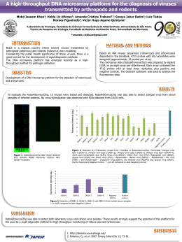
International Journal of Poultry Science 12 (11): 639-646, 2013
ISSN 1682-8356
© Asian Network for Scientific Information, 2013
Nephritis Associated with a S1 Variant Brazilian Isolate of Infectious Bronchitis
Virus and Vaccine Protection Test in Experimentally Infected Chickens
Filipe Santos Fernando1, Maria de Fátima da Silva Montassier1, Ketherson Rodrigues Silva1,
Cintia Hiromi Okino2, Elisabete Schirato de Oliveira1, Camila Cesário Fernandes1,
Márcio de Barros Bandarra3, Mariana Costa Mello Gonçalves4, Mariana Monezi Borzi1
Romeu Moreira dos Santos1, Rosemeri de Oliveira Vasconcelos3,
Antônio Carlos Alessi3, Hélio José Montassier1
1
School of Agricultural and Veterinary Sciences, São Paulo State University (FCAV-Unesp),
Department of Veterinary Pathology, Laboratory of Veterinary Immunology and Virology,
Jaboticabal 14884-900, SP, Brazil
2
Embrapa Swine and Poultry, Concórdia 89700-000, SC, Brazil
3
School of Agricultural and Veterinary Sciences, São Paulo State University (FCAV-Unesp),
Department of Veterinary Pathology, Laboratory of Veterinary Pathology, Jaboticabal 14884-900, SP, Brazil
4
Federal Institute of Goiás - Rio Verde 75901-970, GO, Brazil
Abstract: Infectious bronchitis virus (IBV) induces a significant negative impact on poultry production
worldwide, specially due to the continuous emergence of viral variants. This virus causes damage to the
respiratory tract, and depending on the virus strain, affects and damages the urogenital system. The objective
of this study is to characterize the pathotype and the cross-immunity with regard to Massachusetts vaccine
strain (H120) of a Brazilian IBV field isolate (IBVPR-05) previously S1 genotyped as a variant. The
pathogenicity test was conducted on two experimental groups of specific pathogen free chickens; one was
vaccinated with attenuated Massachusetts H120 strain and the other remained non-vaccinated. Three weeks
after vaccination, both groups were challenged with IBRPR-05. The tracheal ciliostasis, and the viral load,
histopathology and immunohistochemistry in trachea and kidney samples were evaluated. The viral loads,
measured by quantitative real time RT-PCR, were higher in kidney than in trachea, and the most prominent
histopathological changes were found in the kidneys. The renal lesions were characterized by the presence
of nephritis with intense inflammation, tubular epithelial cell degeneration and necrosis. The H120 vaccine
induced a partial protection against the infection of trachea and kidney tissues by this variant isolate. Thus,
the Brazilian variant isolate IBVPR-05 was characterized in this study as a nephropathogenic pathotype and
as a protectotype differing from Massachusetts vaccine strain of IBV. This indicates the importance to
determine these biologic characteristics of other Brazilian variant IBV isolates, in order to implement more
effective control measures of IBV infection in this country.
Key words: Avian infectious bronchitis virus, variant, nephropathogenic, nephritis, massachusetts vaccine
Cavanagh, 2005). In addition, the presence of IBV renal
epithelial cells generates an acute interstitial
inflammatory response with inflammatory cells
proliferation and the development of necrosis and
nephritis (Abdel-Moneim et al., 2006).
It has been shown that the spike glycoprotein (S) of
corona virus is a determinant of cell tropism (Kuo et al.,
2000). Because of the high rates of glycoprotein S1 gene
mutation, several IBV serotypes or antigenic variant
strains have been reported in many countries
(Cavanagh, 2007). In general, different IBV serotypes of
the virus do not confer cross protection against each
other (Ignjatovic and Sapats, 2000), and cross protection
tends to diminish as the degree of amino acid identity
INTRODUCTION
The avian infectious bronchitis virus (IBV) belongs to the
genus Gammacoronavirus of the Coronaviridae family
and is the etiologic agent of infectious bronchitis (IB),
and has been one of the major pathogen affecting the
global poultry industry. IBV infects chickens of all ages
and causes lesions in respiratory and urogenital organs
(Cavanagh, 2007; Cook, 2002).
IBV initially replicates the upper respiratory tract and may
then infect other epithelial cells of other organs of the
host (Dhinakar and Jones, 1997). Some IBV strains
replicate in the kidney, and due to their
nephropathogenic properties, have the potential to
cause severe losses (Cavanagh and Naqi, 2003;
Corresponding Author: Filipe Santos Fernando, School of Agricultural and Veterinary Sciences, São Paulo State University (FCAVUnesp), Department of Veterinary Pathology, Laboratory of Veterinary Immunology and Virology,
Jaboticabal 14884-900, SP, Brazil
639
Int. J. Poult. Sci., 12 (11): 639-646, 2013
between the S1 proteins of IBV strains decreases (Gelb
et al., 2005). However, some strains of the virus are
effective in inducing cross protection against other
serotypes or genotypes and these viruses are classified
in the same protectotypes (Dhinakar and Jones, 1996).
In Brazil, the presence of a significant number of genetic
variants of IBV with characteristics differing from
reference IBV strains from other countries, have been
reported with increasing frequency since the year 2000
(Montassier et al., 2008; Montassier et al., 2012;
Villarreal et al., 2007; Felippe et al., 2010). However, the
characteristics of these genetic variants were not
associated with phenotypic variations, such as those
related to cell tropism and pathogenesis. Another aspect
that remained unknown is the protection conferred
against these variant Brazilian IBV isolates by the
attenuated live vaccine strain from Massachusetts
serotype (H120 and Ma5), which is the only attenuated
vaccine authorized in Brazil.
This paper investigate and characterize the tissue
tropism and pathogenicity of a Brazilian IBV isolated
previously genotyped as a S1 variant (IBVPR-05 –
GenBank: GQ169242) (Montassier et al., 2008). In
addition, the protection induced by a commercially
available IBV vaccine in Brazil was also evaluated for this
variant isolate in order to characterize its protectotype
with regard to the Massachusetts vaccine strain.
control and was mocked vaccinated and challenged with
phosphate-buffered saline (PBS).
Inhibition of ciliary activity: For evaluation of tracheal
ciliostasis three fragments of approximately 1.5 mm of
each portion, nine rings per bird, were analyzed. The
rings were placed in a Petri dish containing Eagle
culture medium with 10% fetal bovine serum. They were
then analyzed on an inverted microscope, observing the
degree of integrity and preservation of the ciliary
movement of the tracheal epithelial cells, in accordance
with Andrade et al. (1982); Cook et al. (1999); Yashida et
al., 1985. The tracheal ciliary activity were classified in
scores from 0 to 3.
Histopathology: Fragments of kidney and portions from
the upper, middle and the lower part of the trachea of
approximately 0.5cm were collected on the fifth day after
challenge. They were fixed in 10% buffered
formaldehyde solution for 1 day and processed by the
standard histological procedure. Then were embedded
in paraffin wax and cut into 4 mm sections (Stevens,
2007). All of the sections were stained with haematoxylin
and eosin and were examined by light microscopy for
lesions due to IBV infection or untreated slides for use
in immunohistochemistry.
The samples of trachea were assigned scores. A score
of zero identifies the absence of injury, while cases with
mild, moderate and severe injury were classified as one,
two
and
three,
respectively.
Morphological
characteristics were observed for loss of cilia and
epithelial cells, degeneration of the epithelium and
glands depletion, inflammatory infiltration and epithelial
hyperplasia.
For kidney damage, injury was considered mild when
there was mild tubular dilation with minimal infiltration of
lymphocytic cells in the interstitial lumen. Moderate
histological lesions were defined as the presence of
small foci of necrosis and tubular dilatation with
moderate inflammatory infiltrate. Severe lesions are
defined as the presence of large foci of acute necrosis
with tubular dilatation and severe inflammatory infiltrate
around tubules as described by Nakamura et al. (1991)
and Chen et al. (1996). Tissue sections were randomly
photographed with a light microscope (Eclipse Moticam,
Nikon, Japan).
MATERIALS AND METHODS
Virus: The IBVPR-05 Brazilian field isolate used in this
study was originated from a broiler flock showing
respiratory clinical signs. This virus was genotyped by
RFLP
and
nucleotide
sequencing
(Genbank:
GQ169242), after amplification of S1 gene (Montassier
et al., 2008; Montassier et al., 2012). The virus was
propagated in 9-to 11-day-old embryonated chicken
eggs, and the embryo 50% infectious doses (EID50)
were determined according to Reed and Muench
(1938).
Experimental design: We used 18 one-day old “Specific
Pathogen Free” (SPF) birds irrespective of sex. The birds
were divided into three groups and maintained in
isolators with positive pressure in air-conditioned rooms
with negative pressure. One group (Non vac) was
composed of six birds and was challenged at the sixth
week of age with field strain IBVPR-05 with 50 µl
containing 1 x 104.0 EID50 by intranasal and intraocular
routes. They were euthanized five days after challenge.
The second group (Vac) was vaccinated with strain
Mass H120 at three weeks of age and subjected to
challenge at sixth weeks of age to the same field strain
in EID50 per bird dose 1 x 104.0 by intranasal and
intraocular route. This group too was euthanized five
days after challenge. Finally, a third group also
comprising six birds was maintained as a negative
RNA isolation and Real Time RT-qPCR: The total RNA of
tracheal and kidney tissue samples were extracted with
TRIZOL Reagent® (Invitrogen, USA) and then cDNAs
were obtained in the RT with a Superscript III Kit
(Invitrogen, USA), as described previously. The cDNA
samples were submitted to real time quantitive PCR for
the absolute quantification of viral load, and this
technique was conducted as recommended Okino et al.
(2013), except that the primers described by Wang and
640
Int. J. Poult. Sci., 12 (11): 639-646, 2013
Tsai (1996) were used in place of HV+ and HV- primers.
A linear regression was determined plotting the
logarithmic values of the number of copy of plasmid DNA
containing the insert of gene S1 against the cycle
threshold (Ct) values, in order to convert the Ct values
from tissue samples into S1 gene copy number (Okino
et al., 2013).
of both vaccinated and unvaccinated groups. Slight
hyperaemia with catarrhal exudate was seen in the
trachea in unvaccinated group. There were no changes
in other organs.
Inhibition of ciliary activity: The test of inhibition of ciliary
activity was measured in three fragments of skull
portions, medial and distal. Ciliostasis scores were very
similar between the two challenged groups, with no
significant difference between them (Fig. 1a).
Immunohistochemistry: IHC was performed on kidney
and trachea. The general procedure was initiated by
deparaffinization of the sections followed by two baths in
xylol. Then the samples were rehydrated in solutions of
decreasing alcohol. Antigen retrieval was performed by
heat, using a pressure chamber (Pascal, Dako, USA).
Following this, the endogenous phosphatase was
blocked through the addition of 200uL blocking solution
commercial Dual Endogenous Enzyme Block (Dako,
USA) for 20 min. The blocking of nonspecific reactions
was done with Protein Block (Dako, USA). Incubation
was performed with the primary antibody anti-IBV
hyperimmune polyclonal antiserum, produced by our
laboratory in goats, against the recombinant
nucleoprotein of infectious bronchitis virus for 2 hr at
37ºC in a humid chamber. Reaction was developed with
an Envision-HRP Kit and EnVisionTM G | 2 System / AP,
Rb / Mo (Dako, USA). Counter-staining was made with
Harris hematoxylin and the slides were mounted with
Entellan (Merck). Between each of the steps, the slide
was rinsed with a solution of distilled water and Tris HCl
buffer solution, pH 7.4. The negative control technique
was performed with Tris-HCl buffer, instead of the
primary antibody, and another set of slides of the birds
kept as the negative control of infection.
Histopathology: In the tracheas, the histological
changes were more evident in animals of the
unvaccinated group; five out of the six birds showed
moderate desquamation of ciliated cells (++), epithelial
hyperplasia and degeneration of the mucus-producing
cells and infiltration of heterophils and lymphocytes. The
vaccinated group had only mild (+) infiltration of
heterophils in the lamina propria, epithelial hyperplasia
and moderate desquamation of ciliated cells across the
group (Fig. 1b).
With respect to renal histopathology, all birds of the
vaccinated and unvaccinated groups showed intense
multifocal nephritis (++), with lymphoplasmacytic
infiltration, especially in the distal collecting tubules. We
also observed mild necrosis of renal epithelial cells (Fig.
2a), deposition of urate crystals and proteinaceous
material in the vaccinated group. In the unvaccinated
group, moderate necrosis and epithelial degeneration
was observed in four of the six birds (Table 1).
Real time RT-qPCR: The software generated a standard
curve by plotting the cycle threshold (Ct) against the
values of each dilution of the standard reference strain.
The values were expressed as Log10 S1 gene copy
number. All tracheas and kidneys analyzed were positive
for the presence of the S1 gene of IBV, except the control
group.
Figure 3a shows greater presence of viral RNA in the
trachea of the group non-vaccinated (p<0.01). This
result was similar in renal analyzing samples (Fig. 3b)
which contained greater number of viral copies in nonvaccinated group (p <0.05). Taking into account the
numbers of copies of viral RNA present in the trachea
and kidneys from non-vaccinated group, one notable
difference was observed between them, in which the
viral load in the kidneys was larger than the trachea (p
<0.05).
Statistical analysis: Results were expressed as
Mean±SEM. Data were analyzed statistically using
GraphPad Prism software version 5.0 for Windows
(GraphPad Prism Software Inc., San Diego, CA, USA).
The non-parametric test (Mann Whitney test) was used
to determine the statistical significance and p<0.05 was
considered statistically significant.
RESULTS
Clinical signs and gross pathology: All birds except
those from the infection control group, showed sneezing
and tracheal and bronchiolar rales with moderate
hoarseness. Two to three days after challenge, four
birds in the unvaccinated and challenged group were
weak and depressed and one bird died. Only one bird
in the vaccinated and challenged group had the same
symptoms. At 4 dpi both groups showed increase in
water consumption and slight watery diarrhea.
At necropsy, morphological changes were observed in
the kidneys of both the vaccinated and unvaccinated
groups, as well as edematous and pale spots. Mucus
and only mild congestion were present in the tracheas
Immunohistochemistry: The presence of IBV antigen
was detected in all tissue samples from trachea and
kidney. The cytoplasm of epithelial cells of the mucosa
and lamina propria of the trachea were labeled and also
the cilia left over. More intensive staining of viral antigen
tracheal was noted within the group non-vaccinated, as
641
Int. J. Poult. Sci., 12 (11): 639-646, 2013
Table 1:
Histopathology and IBV antigen detection by immunohistochemistry in tracheal and renal samples from vaccinated and non-vaccinated
chickens challenged with the Brazilian variant IBVPR-05 isolate
Tissue
Trachea
Kidney
-----------------------------------------------------------------------------------------------------------------------------------------------------------------------------------Lesion
Epitelial deciliation
Heterophil infiltration
IBV antigen
Epitelial degeneration
Heterophil infiltration
IBV antigen
Non Vacc
++a
++
++b
++
+++
++
Vac
+
+
+
+
+++
++a
a
Mean of the severity index: -, no change; +, mild; ++, moderate; +++, severe.
b
Mean number of antigen positive cells per microscopic field IBV: -, no antigen; +, 1 to 4; ++, 5 to 10; +++, over 10.
c
Experimental groups.
shown in Table 1. In kidney is shown in the presence of
IBVPR-05 antigen was detected in renal tubular
epithelium by IHC (Fig. 2b). Vaccinated group and nonvaccinated showed the same score of IBV-labeling by
IHC in the tubular epithelial cells of the kidney (Table 1).
Additionally necrosis and desquamation of tubular
epithelial cells were detected in the kidney samples. Our
finding with regard to the presence of acute interstitial
nephritis at 5 dpi, further characterize this variant isolate
as a nephropathogenic pathotype. Similar renal lesions
were reported for an IBV field isolate from Egypt, which
induces prominent lesions in the kidneys, at 5 and 7 dpi
in birds vaccinated with H120, despite this Egyptian
isolated has 97% homology on the nucleotide sequence
of S1 gene from Massachusetts strain M41 (AbdelMoneim et al., 2006).
Contrary to the Egyptian IBV isolate, genetically or
antigenically different IBV strains can not have crossimmunity, and, consequently, the existing vaccine strains
do not induce efficient protection for different organs of
the chicken host against most of the heterologous
strains or isolates (Ignjatovic and Sapats, 2000; Liu et
al., 2006; Liu et al., 2009). Thus, other hypothesis for the
low vaccine protection elicited by the H120 attenuated
strain to the kidney tissues in this study, could be related
to the fact that the IBVPR05 has relevant differences in
its amino acid sequence of S1 glycoprotein with regard
to H120 strain. Therefore, the degree of cross-protection
tends to decrease as decreasing the extent of the
identity of the amino acid sequences of S1 glycoprotein
from two different strains of IBV (Cavanagh et al., 1997).
IHC used in this study was able to detect the presence
of viral antigen in both trachea and kidney samples from
challenged birds. The IBV antigen was found in the
cytoplasm of epithelial cells and also in the lamina
propria of the trachea and in the cytoplasm of tubular
epithelial cell. This finding further confirmed the results
of histopathology with regard to the predominant IBV
replication site in the kidneys. This site coincided with
the distal tubules and collecting ducts described by
Chen et al. 1996 by characterizing the pathogenicity of a
nephropathogenic IBV field isolate. The IHC technique
applied here also showed high sensitivity and specificity,
as well as other protocols previously applied for antigen
detection in different tissues (Naqi, 1990; Nakamura et
al., 1991; Kapczynski et al., 2002; Benyeda et al., 2010).
The results of RT-qPCR showed that the viral loads
were higher in the kidney than in the trachea, for both
vaccinated and non-vaccinated groups. Such finding
reinforces the nephropathogenic character of this
Brazilian IBV isolate. The results of RT-qPCR also
showed that there was significant difference in the mean
DISCUSSION
To our knowledge, this is the first report characterizing a
Brazilian field isolate, previously classified in a S1
variant genotype, as a nephropathogenic pathotype.
Since the first description of IBV in Brazil reported by
Hipólito in 1957, antigenic and phylogenetic analyses
have revealed, in the last two decades, IBV genotypes
and serotypes in Brazil differing from those identified in
North America, Europe, Asia, and Australia (DiFabio,
1993; Villarreal et al., 2007; Montassier et al., 2008;
Montassier et al., 2012; Abreu et al., 2006). However,
these isolates have not been tested to determine their
pathotype, neither their protectotype with regard to the
conventional vaccine strains used in Brazil.
The results of this study were surprising because of the
marked nephropathogenicity exhibited by this Brazilian
IBV variant isolate. After challenge with this isolate, birds
in the vaccinated group developed symptoms common
to IBV infection in the same intensity as the nonvaccinated group. The same relationship was seen in
necropsy examinations of these birds. The primary
difference was due to the death of one bird in the nonvaccinated group.
By comparing the scores of inhibition of tracheal ciliary
activity of vaccinated and non-vaccinated groups, no
difference in tracheal protection status was observed.
Besides, the scores of histopathology in the trachea
showed that the damage caused by the Brazilian variant
strain IBVPR05 was higher in the non-vaccinated group
than in vaccinated birds, which showed moderate
inflammatory infiltration and epithelial degeneration.
In the kidneys, no difference was recorded in the
intensity of nephritis, when vaccinated and nonvaccinated groups were compared. The kidney lesions
caused by the IBVPR05 strain were similar to those
described in previous reports, which characterized IBV
nephropathogenic strains (Albassam et al., 1985;
Ziegler et al., 1997-2000). Both experimental groups
had, at 5 dpi, abundant renal lesions with diffuse
lympho-histiocytic infiltration between the tubules, and
adjacent to the distal tubules and the collecting ducts.
642
Int. J. Poult. Sci., 12 (11): 639-646, 2013
Fig. 1:
Mean scores of the ciliary activity test and histopathological representation. (A) Score of ciliary stasis caused
by IBVPR-05, there was no significant difference between the groups vaccinated and unvaccinated (p>0.05).
(B) Focuses of infiltration of inflammatory cells and hyperplasia in the trachea from the vac birds (+) and no
lesions for mock group.
number of S1 gene copy in the tracheal samples of
vaccinated group and that showed by non-vaccinated
group (P< 0.01). Likewise, the mean viral load detected
in the kidneys from birds of non-vaccinated group was
higher, and showed a significant difference compared to
that observed in the vaccinated group (P<0.05).
Although there is a significant difference in the number
of S1 viral gene copy in the kidney samples from
vaccinated and non-vaccinated group, the vaccinated
group still demonstrated a high viral load, and its kidney
lesions were indistinct from those recorded in nonvaccinated birds, except for the epithelia degeneration.
This indicates that the greater viral load in this organ
could be related to the development of renal lesions.
Like in infections by other coronaviruses, it is known that
the IBV S1 glycoprotein is largely responsible for the
tissue tropism of an IBV strain (Kuo et al., 2000). This
glycoprotein plays a critical role in the viral infectivity and
contains the antigenic determinants that induce the
formation of neutralizing antibodies. Thus, the variation
in the composition of amino acids located in some
regions of the S1 glycoprotein could result in change in
tissue tropism, virulence and antigenicity of a given IBV
strain and constitutes the main strategy for IBV escapes
from the host defense mechanisms (Cavanagh et al.,
1997; Keeler et al., 1998; Cook et al., 1999).
Thus, the genetic alterations in the composition of S1
glycoprotein of IBVPR-05 resulted in a marked
predilection for infection of epithelial cells which do not
belong to the respiratory tract, possibly through an
escape mechanism in the host, which in the case of this
variant increased the tropism for kidney epithelial cells,
and this led to the development of nephritis.
643
Int. J. Poult. Sci., 12 (11): 639-646, 2013
Fig. 2(a-b): (a) Photomicrograph of the kidney of a bird from vaccinated group showing the characteristic
lymphoplasmacellular interstitial nephritis (black/white arrow), and mild degeneration of renal tubular
epithelial cells (black arrow), Hematoxylin-eosin. (b) IBV antigen detection by immunohistochemistry in
renal tubular epithelium of a bird from non-vaccinated group (black arrow). Note the specificity of
immunostaining for IBV in the cytoplasm of the tubular epithelium associated to inflammatory cells. Scale
bar = 50 µm.
Fig. 3(a-b): Detection of viral load (Log10 S1 gene copy number) by RT-qPCR in trachea (a) and kidney (b) samples
collected from vaccinated (Vac), non-vaccinated (Non Vac) and control groups of chickens experimentally
infected or not (Control group) with Brazilian variant IBVPR-05 isolate, at 5 dpi. A. Statistical differences
between Vac and Non Vac groups are represented by ** (p<0.01) and * (p<0.05).
This study assessed the immune-protection status
induced by the conventional Massachusetts IBV strain
(H120) against this Brazilian variant isolate. The results
indicate that there was a better vaccine protection in the
trachea due to lower pathogenicity of this Brazilian
variant isolate for this organ compared to the kidney. A
drastic drop in the number of S1 viral gene copy was
detected in the tracheas of vaccinated birds, but not in
the kidney samples of these birds. In fact, although there
is a significant reduction in the number S1 gene viral
copy and lesions in the kidneys of vaccinated birds, they
still showed a severe nephritis.
The best protection against virulent challenge with
nephropathogenic IBV strain is usually achieved by
vaccination
with
homologous
serotype
strain
(Lambrechts et al., 1993; Pensaert and Lambrechts,
1994). Due to the increasing isolation and
identification of nephropathogenic IBV variants,
including the Brazilian isolate IBRPR-05, it would be
interesting to test this variant strain as a vaccine
against this and others nephropathogenic Brazilian
isolates, and compare to the status of crossimmunity induced by the reference Massachusetts
vaccine strain.
644
Int. J. Poult. Sci., 12 (11): 639-646, 2013
In conclusion, the IBVPR05 variant isolate is
characterized in this study as a nephropathogenic strain
and based in its partial cross-immune protection with
H120 Massachusetts strain, this virus must be
classified in a different protectype. This indicates also
the importance of performing biologic analysis besides
the genotyping for the Brazilian IBV variant isolates, in
order to determine the efficacy of current IBV vaccines
against these viruses ant to implement more effective
control measures.
Cavanagh, D. and S. Naqi, 2003. Infectious bronchitis.
In: Calnek B.W., Barnes H.J. and Beard C.W. (ed.),
Diseases of Poultry. 11th ed. Iowa State University
Press, Ames, pp: 101-119.
Cavanagh, D., 2007. Coronavirus avian infectious
bronchitis virus. Vet. Res., 38: 281-297.
Chen, B.Y., S. Hosi., T. Nunoya and C. Itakura, 1996.
Histopathology and immunohistochemistry of renal
lesions due to infectious bronchitis virus in chicks.
Avian Path., 25: 269-283.
Cook, J.K.A., S.J. Orbell., M.A. Woods and M.B.
Huggins, 1999. Breadth of protection of the
respiratory tract provided by different live-attenuated
infectious bronchitis vaccines against challenge
with infectious bronchitis virus of heterologous
serotypes. Avian Path., 28: 477-485.
Cook, J., 2002. Coronaviridae. In: Jordan, F., Pattison,
M., Alexander, D., Faragher, T. (Eds.), Poultry
diseases. WB Saunders, New York, pp: 298-306.
Dhinakar Raj, G. and R.C. Jones, 1996. Protectotypic
differentiation of avian infectious bronchitis viruses
using an in-vitro challenge model. Vet. Microbiol.,
53: 239-252.
Dhinaker Raj, G., and R.C. Jones, 1997. Infectious
bronchitis virus: immunopathogenesis of infection
in the chicken. Avian Path., 26: 677-706.
DiFábio, J., 1993. Bronquite infecciosa das galinhas:
aspectos clínicos e controle da BI no Brasil. In:
Conferência Apinco de ciência e tecnologia
avícolas, Santos, SP. Anais, Campinas : FACTA,
1993, p:1-8.
Felippe, P.A.N., L.H.A. da Silva, M.M.A.B. Santos, F.R.
Spilki and C.W. Arns, 2010. Genetic Diversity of
Avian Infectious Bronchitis Virus Isolated from
Domestic Chicken Flocks and Coronaviruses from
Feral Pigeons in Brazil Between 2003 and 2009.
Avian Dis., 54: 1191-1196.
Gelb, J. Jr., Y. Weisman, B.S. Ladman and R. Meir, 2005.
S1 gene characteristics and efficacy of vaccination
against infectious bronchitis virus field isolates from
the United States and Israel (1996 to 2000)., Avian
Path., 34: 194-203.
Hipólito, O., 1957. Isolamento e identificação do vírus
da bronquite infeciosa das galinhas no Brasil.
Arq. Esc. Sup. Vet. (MG)., 10: 131-63.
Ignjatovic, J. and S. Sapats, 2000. Avian infectious
bronchitis virus. Rev. Sci. Technol., 19: 493-508.
Kapczynski, D.R., H.S. Sellers., G.N. Rowland and M.W.
Jackwood, 2002. Detection of in ovo-inoculated
infectious bronchitis virus by immunohistochemistry
and in situ hybridization with a riboprobe in
epithelial cells of the lung and cloacal bursa. Avian
Dis., 46: 679-685.
Keeler, C.L.Jr., K.L. Reed, W.A. Nix and J.Jr. Gelb, 1998.
Serotype identification of avian infectious bronchitis
virus by RT-PCR of the peplomer (S-1) gene. Avian
Dis., 42: 275-284.
ACKNOWLEDGMENTS
We would like to thank Fundação de Amparo à Pesquisa
do Estado de São Paulo (FAPESP - grant no.
2011/047432) and CNPq for their financial support and
FortDodge (Pfizer) and Merial for supplying SPF eggs
and SPF chicks.
Ethics Committee and Biosafety: This study was
approved by the Institutional Ethics and Animal Welfare
Committee (Comissão de Ética no Uso de Animais CEUA, UNESP, process number 011467/11).
REFERENCES
Abdel-Moneim, A.S., M.F. El-Kady., B.S. Ladman and J.Jr.
Gelb, 2006. S1 gene sequence analysis of a
nephropathogenic strain of avian infectious
bronchitis virus in Egypt. Virol. J., 3: 78.
Abreu, J.T., J.S. Resende., R.B. Flatschart., A.V.
Folgueras-Flatschart., A.C. Mendes. and N.R.
Martins, 2006. Molecular analysis of Brazilian
infectious bronchitis field isolates by reverse
transcription- polymerase chain reaction, restriction
fragment length polymorphism, and partial
sequencing of the N gene. Avian Dis., 50: 494-501.
Albassam, N.R., R.W. Winterfield and H.L. Thacker,
1985. Comparison of the nephropathogenicity of
four strains of infectious bronchitis virus. Avian Dis.,
30: 468-476.
Andrade, L.F., A.P. Villegas., O.J. Fletcher. and R.
Laudencia, 1982. Evaluation of ciliary movement in
tracheal rings to assess immunity against
infectious bronchitis virus. Avian Dis., 26: 805-814.
Benyeda, Z.S., L. Szeredi., T. Mato´., T. Su¨ Veges., G.
Balka., Z. Abonyi-To´TH., M. Rusvai and V. Palya,
2010.
Comparative
Histopathology
and
Immunohistochemistry of QX-like, Massachusetts
and 793/B Serotypes of Infectious Bronchitis Virus
Infection in Chickens. J. Comp. Path., 143: 276283.
Cavanagh, D., M.M. Ellis and J.K.A. Cook, 1997.
Relationship between variation in the S1 spike
protein of infectious bronchitis virus and the extent
of cross-protection. Avian Path., 26: 63-74.
Cavanagh, D., 2005. Coronaviruses in poultry and other
birds. Avian Path., 34: 439-448.
645
Int. J. Poult. Sci., 12 (11): 639-646, 2013
Kuo, L., G.J. Godeke, M.J. Raamsman, P.S. Masters, P.J.
Rottier, 2000. Retargeting of coronavirus by
substitution of the spike glycoprotein ectodomain:
crossing the host cell species barrier. J. Virol., 74:
1393-1406.
Lambrechts, C., M. Pensaert and R. Ducatelle, 1993.
Challenge experiments to evaluate cross-protection
induced at the trachea and kidney level by vaccine
strains and Belgian nephropathogeni c isolates of
avian infectious bronchitis virus. Avian Path., 22:
577-590.
Liu, X.L., J.L, Su, J.X, Zhao and G.Z, Zhang, 2006.
Complete genome sequence analysis of a
predominant infectious bronchitis virus (IBV)
strain in China. Virus Gen, Secaucus. 38: 56-65.
Liu, S., X. Zhang, Y. Wang, C. Li, Q. Liu, Z. Han, Q.
Zhang, X. Kong and G. Tong, 2009. Evaluation of the
protection conferred by commercial vaccines and
attenuated heterologous isolates in China against
the CK/CH/LDL/97I strain of infectious bronchitis
coronavirus. Vet. J., 179: 130-136.
Montassier, M.F.S., L. Brentano., H.J. Montassier and
L.J. Richtzenhain, 2008. Genetic grouping of avain
infectious bronchitis virus isolated in Brazil based
on RT-PCR/RFLP analysis of the S1 gene. Pesq.
Vet. Bras., 28: 190-194.
Montassier, M.F.S., L. Brentano, L.J. Richtzenhain, H.
Montassier, 2012. Molecular analysis and evolution
study of infectious bronchitis viruses isolated in
Brazil over a twenty-one-year period. Proceedings of
the 7th International Symposium on Avian Corona
and Pneumoviruses and Complicating Pathogens;
2012; Wetter, Germany. Druckerei Schoreder., 1:
19-30.
Nakamura, K., J.K.A. Cook, K. Otsuki, M.B. Huggins, and
J. Frazier, 1991. Comparative study of respiratory
lesions in two chicken lines of different
susceptibility infected with infectious bronchitis
virus:
histology,
ultrastructure
and
immunohistochemistry. Avian Path., 20: 241-257.
Naqi, S.A., 1990. A monoclonal antibody-based
immunoperoxidase procedure for rapid detection of
infectious bronchitis virus in infected tissues. Avian
Dis., 34: 893-898.
Okino, C.H., A.C. Alessi, M.F.S. Montassier, A.J.M. Rosa,
X. Wang and H.J. Montassier, 2013. Humoral and
Cell-Mediated Immune Responses to Different
Doses of Attenuated Vaccine Against Avian
Infectious Bronchitis Virus. Viral. Immunol., 26: 259267.
Pensaert, M. and C. Lambrechts, 1994. Vaccination of
chickens againsta Belgiann ephropathogenics train
of infectious bronchitis virus B 1648 using
attenuated homologous and heterologous strains.
Avian Path., 23: 631-641.
Reed, L.J and H. Muench, 1938. A simple method of
estimating fifty per cent end points. Am. J. Hyg., 27:
493-497.
Stevens, A., 2007. The haematoxylins. In J.D. Bancroft &
A. Stevens (Eds.), Theory and Practice of
Histological Techniques, 6th edn Edinburgh,
Churchill Livingstone, pp: 107-118.
Villarreal, L.Y., P.E. Brandao, J.L. Chacon, A.B.
Saidenberg, M.S. Assayag, R.C. Jones and A.J.
Ferreira, 2007. Molecular characterization of
infectious bronchitis virus strains isolated from the
enteric contents of Brazilian laying hens and
broilers. Avian Dis., 51: 974-978.
Wang, C.H. and C.T. Tsai, 1996. Genetic grouping for
the isolates of avian infectious bronchitis virus in
Taiwan. Arch Virol., 141: 1677-1688.
Yashida, S., S. Aoyama, K. Sawaguchi, N. Takahashi,
Y. Iritani and Y. Hayashi, 1985. Relationship
between several criteria of challenche-immunity
and humoral immunity in chickens vaccinated
with avian infectious bronchitis vaccines. Avian
Pathol., 14: 199-211.
Ziegler, A.F., B.S. Ladman, P.A. Dunn, A. Schneider, S.
Davison, P.G. Miller, H. Lu, D. Weinstock, M. Salem,
R.J. Eckroade, and J. Gelb Jr., 1997-2000.
Nephropathogenic
infectious
bronchitis
in
Pennsylvania chickens 1997-2000. Avian Dis., 46:
847-858.
646
Baixar