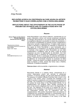
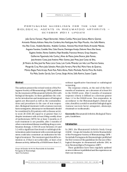
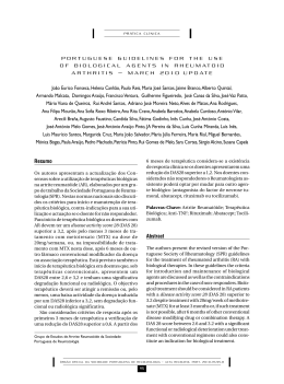
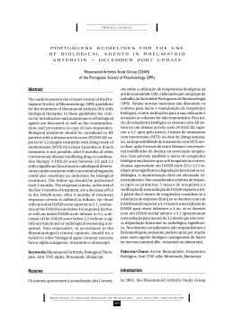
ISSN 0482-5004
Impact Factor 2012: 0.864
© Thomson Reuters Journal Citation Reports,
Science Edition (2012)
REVISTA BRASILEIRA DE REUMATOLOGIA
BRAZILIAN JOURNAL OF RHEUMATOLOGY
SEPTEMBER/OCTOBERt7PMVNFt/VNCFS
4&5&.#300656#30t7PMVNFt/ÞNFSP
www.reumatologia.com.br
REVISTA BRASILEIRA DE
REUMATOLOGIA
www.reumatologia.com.br
Volume 53. Number 5. September/October 2013
Volume 53. Número 5. Setembro/Outubro 2013
CONTENTS | SUMÁRIO
Editorial | Editorial
Group for Research and Assesment of Psoriasis and Psoriatic Arthritis-GRAPPA (2003-2013)
Grupo para Pesquisa e Avaliação da Psoríase e da Artrite Psoriásica – GRAPPA (2003-2013)
Morton Scheinberg .....................................................................................................................................
375
Original Articles | Artigos Originais
Subclinical atherosclerosis in ankylosing spondylitis: is there a role for inflammation?
Aterosclerose subclínica em pacientes com espondilite anquilosante: há um papel para a inflamação?
Renato Leandro Mattar Valente, Jamil Mattar Valente, Gláucio Ricardo Werner de Castro,
Adriana Fontes Zimmermann, Sonia Cristina de Magalhães Souza Fialho, Ivânio Alves Pereira ................
377
EpiFibro – a nationwide databank for fibromyalgia syndrome – the initial analysis of
500 women
EpiFibro – um banco de dados nacional sobre a síndrome da fibromialgia – análise inicial de 500 mulheres
Marcelo C. Rezende, Eduardo S. Paiva, Milton Helfenstein Jr., Aline Ranzolin, José Eduardo Martinez,
Jose Roberto Provenza, Carlos Eugênio Ribeiro Parolini, Luiz Severiano Ribeiro, Eduardo J. R. Souza,
Daniel P. Feldman, Marcos Renato de Assis, Roberto E. Heymann...............................................................
382
Radiographic changes of the cervical spine in rheumatoid arthritis
Alterações radiográficas da coluna cervical em artrite reumatoide
Juan Marcelo Fernandez Alcala, Diogo Douat, Diogo Lago Pinheiro, Douglas Jun Kamei,
Fábio Raimundo M dos Santos, Marilia B Silva, Thelma L. Skare ................................................................
388
Physical activity among patients from the Brasilia cohort of early rheumatoid arthritis
Prática de atividade física entre pacientes da Coorte Brasília de artrite reumatoide inicial
Carolina Rocha Silva, Thaís Ferreira Costa, Tatiane Teixeira Vaz de Oliveira, Luciana Feitosa Muniz,
Licia Maria Henrique da Mota ...................................................................................................................
394
Evaluation of platelet aggregation in the presence of antiphospholipid antibodies:
anti-β2GP1 and anticardiolip
Avaliação da agregação plaquetária em presença de anticorpos antifosfolípides: anti-β2GP1 e anticardiolipina
Harleson Lopes de Mesquita, Giuliano Reder de Carvalho, Fernando Monteiro Aarestrup,
José Otávio do Amaral Corrêa, Maria Regina Andrade Azevedo ...............................................................
400
Symptoms of disease and psychological adaptation in Brazilian scleroderma patients
Sintomas de doença e adaptação psicológica em pacientes brasileiros com esclerodermia
Catarina Correia Leite, Ângela Costa Maia ...............................................................................................
405
Usefulness of anti-dsDNA antibody screening with chemiluminescence followed by
confirmation by indirect immunofluorescence
Utilidade da triagem dos anticorpos anti-dsDNA por quimioluminescência, seguida de confirmação por
imunofluorescência indireta
Maria Roseli Monteiro Callado, José Rubens Costa Lima, Maria Nancy de Alencar Barroso,
Antonio Tiago Mota Pinheiro, Moisés Francisco da Cruz Neto, Maria Arenilda de Lima Abreu,
Walber Pinto Vieira ....................................................................................................................................
412
Adalimumab in rheumatoid arthritis treatment: a systematic review and meta-analysis of
randomized clinical trials
Adalimumabe no tratamento da artrite reumatoide: uma revisão sistemática e metanálise de ensaios clínicos
randomizados
Marina Amaral de Ávila Machado, Alessandra Almeida Maciel, Lívia Lovato Pires de Lemos,
Juliana Oliveira Costa, Adriana Maria Kakehasi, Eli Iola Gurgel Andrade , Mariangela Leal Cherchiglia,
Francisco de Assis Acurcio .........................................................................................................................
419
Psychiatric comorbidities in patients with systemic lupus erythematosus: a systematic review
of the last 10 years
Comorbidades psiquiátricas em pacientes com lúpus eritematoso sistêmico: uma revisão sistemática dos
últimos 10 anos
Nadja Maria Jorge Asano, Maria das Graças Wanderley de Sales Coriolano, Breno Jorge Asano,
Otávio Gomes Lins .....................................................................................................................................
431
Case Reports | Relatos de Caso
Bullous systemic lupus erythematosus in a pregnant woman: a case report
Lúpus eritematoso sistêmico bolhoso em gestante: relato de caso
Cristiane Engel dos Santos, Pedro Henrique Isaacsson Velho, Fabrício Machado Marques, Betina Werner,
Salun Coelho Aragão, Acir Rachid Filho...........................................................................................................
438
c-ANCA-associated vasculitis in patients with ulcerative colitis: a case report
Vasculite c-ANCA-relacionada em paciente com retocolite ulcerativa: relato de caso
Cristiane Engel dos Santos, Vanessa Irusta Dal Pizzol, Salun Coelho Aragão, Acir Rachid Filho,
Fabrício Machado Marques .......................................................................................................................
441
Brief communication | Comunicação breve
Lipid profile and anti-TNF-α use
Perfil lipídico e uso de anti-TNF-α
Antonio Carlos Ferraz Filho, Luize Pereira dos Santos, Marilia B. Silva, Thelma L. Skare..............................
444
Letter to the editors | Carta aos editores
The use of ustekinumab in refractory treatment of psoriatic arthritis
Uso de ustequinumabe no tratamento refratário da artrite psoriásica
Karen Vega-Villanueva, Nathaly Cortez-Bazán, Angela Alvarado-Molina.....................................................
448
R E V B R A S R E U M AT O L . 2 0 1 3 ; 5 3 ( 5 ) : 3 7 5 – 3 7 6
REVISTA BRASILEIRA DE
REUMATOLOGIA
www.reumatologia.com.br
Editorial
Group for Research and Assesment of Psoriasis and
Psoriatic Arthritis-GRAPPA (2003-2013)
The common concept of rheumatologists and dermatologists
to deal with the combined evaluation of patients with psoriasis (PsO) and psoriatic arthritis (PsA) have led to the foundations of Group for Research and Assessment of Psoriasis and
Psoriatic Arthritis (GRAPPA), now in its 10th anniversary. This
editorial looks over the past ten years until the last meeting
in Toronto last July.
A history of success
The association of PsO with articular manifestations, similarly to what was also known with inflammatory bowel disease and uveitis, have been known for long time, although the
mechanism behind these clinical observations were and still
are only partially understood. It was only in 1973 that John
Moll and Verna Wright have tried to create fundamentals that
would identify in a patient with psoriasis that he was also
suffering from PsA (Table 1).1
Using these diagnostic criteria, Moll and Wright described
five subgroups of PsA: distal interphalangeal (DIP), asymmetrical oligoarthritis, polyarthritis, spondylitis, and arthritis
mutilans.
In the 90’s several other classifications were proposed by
different authors and were reviewed by Gladman and Espinoza.2 In the past decade an international group assembled
what is now known as the Classification of Psoriatic Arthritis
criteria (CASPAT).The original diagnostic criteria of Moll and
Wright are now being replaced by the CASPAR criteria.3 CASPAR takes into account the presence of articular manifestations, the presence of skin disease or familial history, typical
nail changes, the presence of swelling of the digit and juxtarticular bone in formation. It was shown that CASPAR has a
sensibility of 98.7% and a specificity of 91.4%.
GRAPPA is a non-profit educational and scientific organization, created to facilitate sharing of information that relates
to PsO and PsA. It was initiated 10 years ago with their first
meeting in New York City (Fig. 1). Its objectives include: 1) Promote the development of national and international collaborative registries of PsA and PsO patients to standardize the
data being obtained and learn more about the natural history
of the disease as well as its genetic underpinnings; 2) Work
closely with representatives of patient service leagues to promote public education and awareness of PsA and PsO; 3) Work
closely with representatives of biopharmaceutical companies to
promote and conduct research on effective therapies for PsA
and PsO; 4) Work closely with representatives of regulatory
agencies to establish appropriate guidelines for regulatory
approval of new therapies; 5) Work with other professional
Table 1 – Moll and Wright criteria for identification of
psoriatic arthritis.
Inflammatory arthritis (peripheral arthritis and/or sacoiliitis and
spondylitis)
The presence of psoriasis
The usual absence of serological tests for rheumatoid factor
Fig 1 – First GRAPPA meeting, New York, 2003. From left
to right Mease, Zimmerman, Gladman, third row Khan,
Helliwell, Nash, Ritchlin, Landwee, Espinoza, Smolen,
Fitzgerald, Braun, Kalden, Antoni van der Bosch and
Kavanaugh..
0482-5004/$ - see front matter. © 2013 Elsevier Editora Ltda. All rights reserved.
376
R E V B R A S R E U M AT O L . 2 0 1 3 ; 5 3 ( 5 ) : 3 7 5 – 3 7 6
bodies, such as the American College of Rheumatology, American Academy of Dermatology, and OMERACT to promote
knowledge of research about PsA and PsO within the context
of those disciplines; and 6) Develop treatment guidelines for
governmental and other interested parties.
Important publications that are now standard references were
published during the past five years and are listed in Table 2:
Brazil have been an active participant in the mission of
GRAPPA in Latin America with national meetings throughout
in Brazil and Latin America; several Brazilian rheumatologists
and dermatologists are now working together for the benefit
of patients with both diseases.The first joined meeting was
held in Porto Alegre (RS, Brazil) together with the Brazilian
Congress; the second meeting was held in Fortaleza (CE, Brazil) in 2012 and the third will be in Salvador (BA, Brazil), on
the second semester of this year.4 Last July, together with the
35th anniversary of the creation of the Psoriatic Arthritis Clinic
at the University of Toronto, GRAPPA was commemorating its
10th anniversary since its inception in New York. The Brazilian rheumatologists Claudia Schainberg, Roberto Ranza, Sueli
Carneiro, Penelope Pelarminos, Rachel Gryzpan and myself
attended the event (Fig. 2).
As of today Brazil has 29 members affiliated with GRAPPA
15 of them are rheumatologists.New projects are being designed with participation of Brazilian rheumatologists in the
upcoming years. The projects are listed below.
• New projects are being developed in refinement and validation of arthritis outcome measures in PsA
• Development of instruments to assess QOL, function, and
participation
• Standardization of histologic and immunohistochemical
assessments in PsA and PsO
• Updated criteria for classification of PsA (CASPAR)
• Imaging in PsA.
Morton Scheinberg
Hospital Israelita Albert Einstein, São Paulo, SP, Brazil
Hospital Abreu Sodré (AACD), São Paulo, SP, Brazil
E-mail: [email protected]
REFERENCES
1. Moll JM, Wright V. Psoriatic Arthritis Seminars in Arthritis
and Rheumatism. 1973,3:55-78.
Table 2 – ARD Supplement - PsA and PsO: state of the art
review and research advances.
ARD Treatment Recommendations for Psoriatic Arthritis, 2008
GRAPPA Newsletter, Primavera de 2012
JRheum Supplement - 2007 Updates; Screening & Assessment
Tools, Quality etc
JRheum Supplement - 2008 Annual Meeting Imaging; Comp
Measures; Biomarkers
JRheum Supplement - 2009 Annual Meeting of GRAPPA,
Estocolomo, Suécia
JRheum Supplement - 2010 Annual Meeting of GRAPPA, Miami
Beach, FL
JRheum Supplement - Systematic Review of Treatments for PsA
Patient Global Assessment in PsA: A Multicenter GRAPPA and
OMERACT Study
Fig 2 – Tenth GRAPPA meeting, Toronto, 2013. From left to
right: Luis Espinoza, Claudia Schainberg, John Moll and
Morton Scheinberg.
2. Gladman DD, Espinoza LR. International symposium on
psoriatic arthritis. J Rheumatol. 1992;19(2):290-1.
3. Taylor W, Gladman D, Helliwell P, Marchesoni A, Mease P,
Mielants H; CASPAR Study Group. Classification criteria for
psoriatic arthritis: development of new criteria from a large
international study. CASPAR Study Group Arthritis Rheum.
2006;54(8):2665-73.
4. Goldenstein-Schainberg C, Ranza R, Bonfiglioli R, Carneiro
S, Azevedo VF, Goldenberg J, et al. The presence of the
Brazilian rheumatology in the GRAPPA. Rev Bras Reumatol.
2011;51(5):533-4
R E V B R A S R E U M AT O L . 2 0 1 3 ; 5 3 ( 5 ) : 3 7 7 – 3 8 1
REVISTA BRASILEIRA DE
REUMATOLOGIA
www.reumatologia.com.br
Original article
Subclinical atherosclerosis in ankylosing spondylitis: is there
a role for inflammation?
Renato Leandro Mattar Valente a, Jamil Mattar Valente b, Gláucio Ricardo Werner de Castro c,
Adriana Fontes Zimmermann c, Sonia Cristina de Magalhães Souza Fialho c,*,
Ivânio Alves Pereira c
a
Internal Medicine Division, Hospital Universitário, Universidade Federal de Santa Catarina, Florianópolis, SC, Brasil
Cardiology Division, Hospital Universitário, Universidade Federal de Santa Catarina, Florianópolis, SC, Brasil
c
Rheumatology Division, Hospital Universitário, Universidade Federal de Santa Catarina, Florianópolis, SC, Brasil
b
article info
abstract
Article history:
Objectives: To evaluate the prevalence of subclinical atherosclerosis in patients with anky-
Received 1 September 2012
losing spondylitis (AS) in comparison to controls with similar cardiovascular risk factors.
Accepted 13 December 2012
Methods: Forty-two consecutive patients with AS and 42 controls matched for age (43.3 ±
11.7 vs. 43.7 ± 11.3, P = 0.89), gender, smoking, diabetes mellitus and arterial hypertension
Keywords:
were enrolled. Participants were excluded if a personal cardiovascular disease (CV) history
Ankylosing spondylitis
was present. A questionnaire recording demographic data, medical and medication history
Subclinical atherosclerosis
was fulfilled. Blood pressure, abdominal circumference, height and weight were measured.
Cardiovascular risk
Lipid profile was determined in a 12-hour fastened blood sample. Ultrasound analysis of
the common carotid artery was performed by one blind observer. The distance between the
lumen-intima interface and the leading edge of the media-adventitia interface (IMT) was
measured and participants were also evaluated for the presence of plaques.
Results: The comparative analysis of demographic and cardiovascular risk factors between
AS patients and controls did not reveal statistically significant differences. Also, no significant differences between groups were observed for TC, HDL-C, T-C/HDL-C, LDL-C, triglycerides, or dyslipidemia frequency. IMT measures were not different in AS and controls (0.62 ±
0.09 vs. 0.61 ± 0.09, P = 0.39) as well as plaques frequencies (19% vs. 17%, P = 0.78).
Conclusions: Subclinical atherosclerosis assessed through carotid ultrasound imaging was
not more prevalent in the AS group when compared to controls with similar cardiovascular
risks. Our observations may imply that CV risk factors may have more influence on the CV
system than AS itself. These findings should be confirmed in a larger population with a
prospective study design.
© 2013 Elsevier Editora Ltda. All rights reserved.
* Corresponding author.
E-mail: [email protected] (S.C.M.S. Fialho).
0482-5004/$ - see front matter. © 2013 Elsevier Editora Ltda. All rights reserved.
378
R E V B R A S R E U M AT O L . 2 0 1 3 ; 5 3 ( 5 ) : 3 7 7 – 3 8 1
Aterosclerose subclínica em pacientes com espondilite anquilosante:
há um papel para a inflamação?
resumo
Palavras-chave:
Objetivos: Avaliar a prevalência de aterosclerose subclínica em pacientes com espondilite
Espondilite anquilosante
anquilosante (EA) em comparação com controles com fatores de risco cardiovasculares
Aterosclerose subclínica
similares.
Risco cardiovascular
Métodos: Foram recrutados 42 pacientes consecutivos com EA e 42 controles equiparados
para idade (43,3 ± 11,7 vs. 43,7 ± 11,3, P = 0,89), gênero, tabagismo, diabetes mellitus e hipertensão arterial. Qualquer participante seria excluído se estivesse presente uma história
pessoal de doença cardiovascular (CV). Foi preenchido um questionário registrando dados
demográficos e histórias médica e de medicação. Foram determinados: pressão arterial,
circunferência abdominal, altura e peso. O perfil lipídico foi determinado em uma amostra
de sangue com 12 horas em jejum. Foi realizada uma análise ultrassonográfica da artéria
carótida comum por um observador desconhecedor da pesquisa. Foi medida a distância
entre a interface lúmen-íntima e a borda de ataque da interface média-adventícia (EIM) e
os participantes também foram avaliados para presença de placas.
Resultados: A análise comparativa dos fatores de risco demográficos e cardiovasculares entre pacientes com EA e controles não revelou diferenças estatisticamente significativas.
Também não foram observadas diferenças significativas entre grupos para TC, HDL-C, T-C/
HDL-C, LDL-C, triglicerídeos ou frequência de dislipidemia. As medidas de EIM não foram
diferentes em EA e controles (0,62 ± 0,09 vs. 0,61 ± 0,09, P = 0,39) e nem as frequências de
placas (19% vs. 17%, P = 0,78).
Conclusões: A aterosclerose subclínica avaliada por meio de imagens ultrassonográficas da
carótida não foi mais prevalente no grupo EA, em comparação com os controles com riscos
cardiovasculares similares. Nossas observações podem implicar que os fatores de risco CV
podem ter mais influência no sistema CV versus a própria EA. Esses achados devem ser
confirmados em uma população maior, por meio de um estudo prospectivo.
© 2013 Elsevier Editora Ltda. Todos os direitos reservados.
Introduction
Ankylosing spondylitis (AS) is a chronic, inflammatory rheumatic disease. Its musculoskeletal manifestations include
both inflammation and structural damage. Characteristic
extra-articular manifestations include aortitis, cardiac conduction defects, pulmonary fibrosis and inflammatory bowel
disease indicating that AS is a systemic disease.
AS is known primarily for causing a lifetime of pain, impaired physical function, work disability, and decreased
quality of life, rather than for shortening life itself. However,
patients with AS also experience premature mortality.1 The
standardized mortality rates (SMR) associated with AS are approximately 50% higher than in the general population.2,3 The
four non-inception cohort studies published to date quote
SMRs of 1.33,4,5 1.56 and 1.8.7
Increased mortality is largely attributable to cardiovascular diseases (CV).3 A recent large population based study has
shown more ischemic heart disease (prevalence ratio 1.2), peripheral vascular disease (ratio 1.6), atherosclerosis (ratio 1.5),
congestive heart failure (1.8) and more cardiovascular risk
factors (prevalence ratios between 1.3 and 1.7) in AS patients
compared to healthy controls.8
It is unclear whether the increased cardiovascular risk of
AS patients could be explained by traditional cardiovascular
risk factors alone. In fact, there is increasing evidence that
the underlying inflammatory process in chronic inflammatory conditions resembles the chronic inflammatory processes
that contribute to various stages of atherothrombosis, from
early atheroma formation to plaque instability and thrombus
formation.9 However, it is unknown whether AS patients without CV disease risk factors show early signs of large artery
damage compared to controls, and if so, what the determinants of such large-vessel abnormalities are. This knowledge
could prove useful for development of risk stratification, intervention strategies and for a better disease understanding.
High-resolution ultrasonography can be used to measure
the intima-media thickness (IMT) as well as vascular elasticity of the carotid artery. An increased carotid IMT reflects
the atherosclerotic burden and predicts the development of
(clinically apparent) CV disease in the general population.10,11
Hence, this study was designed to determine whether signs
of subclinical atherosclerosis are more prominent in a sample
of AS patients compared to controls without the disease but
with similar cardiovascular risks. Other studies assessing IMT
in AS patients and controls have been published but results
were contradictory12-18 and will be discussed further.
Methods
Study population: forty-two consecutive patients with AS attending the outpatient clinic of the University Hospital of the
379
R E V B R A S R E U M AT O L . 2 0 1 3 ; 5 3 ( 5 ) : 3 7 7 – 3 8 1
Federal University of Santa Catarina, Brazil. All patients fulfilled the modified New York diagnostic criteria for AS.19 Fortytwo volunteers (hospital staff or patients who attended the
General Clinic for the University employees) matched for age,
sex, smoking (current or in the last five years), diabetes mellitus and systemic arterial hypertension served as controls. All
participants gave written informed consent and the institutional ethics committees of the University Hospital approved
the study protocol.
Patients and controls were excluded if a personal CV disease history was present (myocardial infarction, percutaneous transluminal coronary angioplasty, surgery for ischemic
heart disease, stroke, transient ischemic attack, carotid endarterectomy, peripheral arterial reconstructive surgery, or
limb amputation).
Patients and controls were examined by a research physician. A questionnaire recording demographic data, medical
and medication history was fulfilled. Blood pressure, abdominal circumference, height and weight were measured. Body
mass index (BMI) was calculated as the ratio of weight and
height squared. We considered the patient/control as diabetic
if they referred hypoglycemic drug use or in the presence of
least two glycemic tests higher than 126 mg/dL. We considered the patient/control as hypertensive if they referred anti-hypertensive drugs use or a systolic blood pressure > 140
mmHg or diastolic blood pressure > 90 mmHg measured in
two different occasions. We considered the patient/control as
dyslipidemic if they referred hypolipemiants drugs use or if
they presented at least one of the following: LDL-C (low density lipoprotein) > 130 mg/dL; triglycerides > 150 mg/dL; HDLC (high-density lipoprotein) < 40 mg/dL.
Laboratory variables were determined in a 12-hour fastened blood sample and included: TC (total cholesterol),
HDL-C, LDL-C, triglycerides (all analyzed by enzymatic techniques); C-reactive protein (CRP by nephelometry method),
and erythrocyte sedimentation rate (ESR by Westergreen
method).
Arterial measurements were conducted in a quiet room
after 15 minutes of rest, with the subjects in supine position.
Ultrasound analysis of the common carotid artery (bilateral) was performed by a cardiologist who were unaware of
the participants’ clinical or laboratory characteristics. Measurements were performed using a B mode high resolution
ultrasound ATL HDI 3000 (Phillips Bothel, WA, USA) with a
5-12MHz linear probe. The distance between the lumen-intima interface and the leading edge of the media-adventitia
interface of the far wall corresponds with IMT. After localization of the common carotid artery, cross-sectional measurements were performed 10 mm proximal from the carotid
bulb. Sites with mural atherosclerotic plaque were excluded
while measuring. Three measurements were performed at
each side. A mean of each side (right or left) was calculated
and finally the mean of both sides (mean) was achieved. We
defined plaques as focal widening of the vessel wall of 50%
relative to adjacent segments with protrusion into the lumen or a IMT > 1.5 mm.
The distribution of each continuous variable was examined graphically and statistically for normality. Numerical
data are summarized as the mean and standard deviation
(SD). Variables not normally distributed were compared us-
ing the Wilcoxon nonparametric test for differences. Variables normally distributed were compared using students’
tests. Categorical data among groups were compared by the
chi-square or the Fischer exact test statistics when appropriate. Some results were evaluated according to the established
normal values and were subsequently ranked as elevated or
depressed. A statistical significance was set at P < 0.05. All statistical analyses were performed using NCSS software.
Results
Comparative analysis of demographic and cardiovascular risk
factors between AS patients and controls did not reveal statistically significant differences as demonstrated in table 1.
AS mean age was 43.3 ± 11.7 years-old and the mean time of
disease duration was 15.9 years.
Also, no significant differences between groups were observed for TC, HDL-C, T-C/HDL-C, LDL-C, triglycerides, or dyslipidemia frequency as showed in table 2. AS patients had a
significant elevation in CRP (38.1% vs. 14.3%, P = 0.01), but not
in the ESR.
In table 3 we demonstrate medications use in both groups.
31% against 11.9% of AS patients were on hypolipemiants (P =
0.03). Anti-TNF was used in 66.6% of all AS patients.
There was no difference in IMT measures in AS and controls (0.62 ± 0.09 vs. 0.61 ± 0.09, P = 0.39). Also no difference
was observed in the frequency of plaques (table 4).
Table 1 – Clinical and demographic features in AS and
controls.
Age (years) ± SD
Male, n (%)
Caucasian, n (%)
Smokers, n (%)
CV FH, n (%)
AH, n (%)
DM, n (%)
BMI (kg/m2) ± SD
AC (cm) ± SD
AS (n = 42)
Controls (n = 42)
P
43.3 ± 11.7
26 (61.9)
35 (83.3)
8 (19.04)
14 (33.3)
15 (35.7)
3 (7.1)
26.5 ± 3.9
90.1 ± 12.3
43.7 ± 11.3
26 (61.9)
40 (95.2)
8 (19.04)
12 (28.57)
11 (26.2)
3 (7.1)
27.6 ± 4.5
91.6 ± 12.8
0.89
1.0
0.08
1.0
0.64
0.35
1.0
0.25
0.58
AS, ankylosing spondylitis; SD, standard deviation; CV FH,
cardiovascular family history; AH, arterial hypertension; DM, diabetes
mellitus; BMI, body mass index; AC, abdominal circumference.
Table 2 – Laboratory profile in AS and controls.
Total-cholesterol (mg/dL) ± SD
HDL-C (mg/dl) ± SD
LDL-C (mg/dl) ± SD
Tryglicerides (mg/dL) ± SD
CT/HDL ± SD
Dyslipidemia, n (%)
Elevated CRP, n (%)
ESR (mm/1st hour) ± SD
AS
(n = 42)
Controls
(n = 42)
P
194.8 ± 36.0
52.7 ± 10.9
120.1 ± 32.4
109.7 ± 49.9
3.8 ± 0.9
25 (59.5)
16 (38.1)
22.2 ± 17.7
196.8±36.4
52.1±14.9
119.3 ± 35.1
126.8 ± 103.7
4.0 ± 1.3
26 (62)
6 (14.3)
20.3 ± 16.8
0.81
0.82
0.92
0.88
0.36
0.82
0.01
0.29
AS, ankylosing spondylitis; SD, standard deviation, CRP, C-reactive
protein; ESR, erythrocyte sedimentation rate.
380
R E V B R A S R E U M AT O L . 2 0 1 3 ; 5 3 ( 5 ) : 3 7 7 – 3 8 1
Table 3 – Medications use in AS and controls.
Glucocorticoid, n (%)
NSAID, n (%)
Sulfasalazine, n (%)
Methotrexate, n (%)
Anti-TNF, n (%)
Hypolipemianta, n (%)
AS (n = 42)
Controls (n = 42)
P
8 (19)
22 (52.3)
6 (14.3)
5 (11.9)
28 (66.6)
13 (31.0)
5 (11.9)
0.03
AS, ankylosing spondylitis; NSAID, non-steroidal anti-inflammatory
drugs.
a
All hypolipemiants used were from the statins group
Table 4 – Carotid ultrasound in AS and controls.
IMT (mm) ± SD
Plaques, n (%)
AS (n = 42)
Controls (n = 42)
P
0.62 ± 0.09
8 (19.0)
0.61 ± 0.09
7 (17.0)
0.39
0.78
AS, ankylosing spondylitis; SD, standard deviation.
Discussion
Given that inflammation has increasingly been acknowledged
as the reason rheumatic patients bear elevated CV risk,20 we
selected a study population of AS patients matched for five
major CV risk factors in order to better assign for this issue.
Our study revealed that CV risk among AS patients as indicated by IMT is not different from controls without the disease. On the opposite, Peters at al found a greater IMT in AS
patients in comparison with controls.12 However, the authors
also found a high CV risk factor profile in patients with AS,
and some of these risk factors (lipids and BMI) were associated
with a greater carotid IMT and increased arterial stiffness. No
association between large-vessel properties and higher Bath
AS indices or CRP values were found. Also, Mathieu et al.14
found significantly increased IMT in the AS group compared
with healthy controls. However, after adjustment for confounding factors, only an underlying trend towards increased
IMT was present. IMT was positively correlated with tobacco
use and blood pressure but not correlated with CRP level or
mSASS. In the AS group, IMT was correlated with traditional
risk factors, such as smoking and systolic blood pressure.
Although the cross-sectional study design does not permit a
good estimate of the cumulative inflammatory burden and the
small series of patients are associated with a low statistical power, their results suggest that an adverse CV risk profile may cause,
at least partly, the greater IMT found by them and there is no
sufficient evidence to support a role of biological inflammation.
Gonzalez et al., recruited 64 AS patients and 64 matched
controls with no cardiovascular morbid. Patients with AS exhibited greater carotid IMT than did matched controls (mean
± SD, 0.74 ± 0.21 mm vs. 0.67 ± 0.14 mm; P = 0.01; differences
of means, 0.077; 95% confidence interval [CI], 0.016-0.139). In
this case, although the best predictors for carotid plaques in
patients with AS were erythrocyte sedimentation rate (ESR) at
time of disease diagnosis (odds ratio [OR], 1.18; 95% CI, 1.041.33; P = 0.01) and duration of disease (OR, 1.39; 95% CI, 1.011.92; p = 0.05); there was no significant correlation between
carotid IMT and either ESR or C-reactive protein.13
Indeed, many of the traditional risk factors for cardiovascular disease are present in the AS versus the general
population, including a higher incidence of hypertension,
elevated lipids, increased fibrinogen and CRP levels, and
poorer physical activity levels. BMI and total cholesterol and
triglycerides have been positively correlated with IMT and/
or arterial stiffness.21
In accordance with our findings, Choe et al. found that carotid IMT and parameters related with arterial elastic properties in young AS patients without clinically evident cardiovascular risk factors were not different from those of sex- and
age-matched healthy controls. Serum levels of TNF-a, IL-6,
and MCP-1 did not reflect the degree of carotid subclinical
atherosclerosis.15 In addition, recently, Capkin et al. evaluated
a total of 67 AS patients, and age, sex, body mass index (BMI)
smoking status, lipid profiles and blood pressure-matched
healthy control subjects (n = 34). They also found no difference in IMT-C between groups.
Our study has some limitations. No disease activity index
was analyzed. However, the isolated disease activity measure would not allow drawing any conclusions about the inflammatory burden possibly associated with atherosclerosis.
Also, 66.6% of patients were on anti-TNF treatment. A subanalysis comparing AS patients suggested that the group on
anti-TNF treatment (n = 28) was not different from the group
treated with non-biologic drugs (n = 14) when it comes to
the IMT, however, they had numerically less plaques (10.7%
vs. 35.7%, P = 0.09). Although this study was not designed
to assign for this exact issue, our finding raises the question about the anti-TNF playing a part to a better cardiovascular outcome. Studies on ischemic heart disease related
mortality and morbidity following anti-TNF therapy have
shown mixed results. Ferrante et al.22 observed a significant
decrease of carotid IMT in anti-TNF-treated RA patients after two years but not in the group treated with methotrexate alone, although significant improvements were seen in
measures of disease activity, CRP and fibrinogen levels with
both type of treatments. It is thought though that anti-TNF
treatment has the potential not only to reduce inflammation but also to modify traditional cardiovascular risk factors
and endothelial dysfunction in RA.23,24 Additionally, one third
of the AS patients were more frequently on hypolipemiants
(31% vs. 11,9%), although dyslipidemia (as defined by this
study) was not more prevalent in AS patients. However, it
well known that statins’s vascular improvement is independent of statins’ cholesterol-lowering actions, fact that has
been associated with their anti-inflammatory and immunomodulatory properties.25 Similarly to the anti-TNF treatment, because of the study design we cannot be conclusive
about statins contribution for a possible better cardiovascular outcome on these AS cases.
In conclusion, our observations are in agreement with the
findings of others and may imply that CV risk factors have
more influence on the CV system than AS itself. However, we
could not be conclusive because of the anti-TNF and hypolipemiants use. These findings should be confirmed in a larger
population with a prospective study design. Further research
concerning the pathogenesis of increased cardiovascular risk
in AS patients should be high priority, as many risk factors are
likely to be modifiable.
R E V B R A S R E U M AT O L . 2 0 1 3 ; 5 3 ( 5 ) : 3 7 7 – 3 8 1
Conflicts of interest
14.
The authors declare no conflicts of interest.
REFERENCES
15.
1. Braun J, Pincus T. Mortality, course of disease and prognosis
of patients with ankylosing spondylitis. Clin Exp Rheumatol.
2002;20(Suppl. 28):S16-S22.
2. Sokka T, Abelson B, Pincus T. Mortality in rheumatoid
arthritis: 2008 update. Clin Exp Rheumatol.
2008;26(Suppl.51):S35-S61.
3. Zochling J, Braun J. Mortality in ankylosing spondylitis. Clin
Exp Rheumatol. 2008;26(Suppl.51):S80-S84.
4. Kaprover E, Little AH, Graham DC, Rosen PS. Ankylosing
spondylitis: survival in men with and without radiotherapy.
Arthritis Rheum. 1980;23:57-61.
5. Khan MA, Khan MK, Kushner I. Survival among patients
with ankylosing spondylitis: a life-table analysis. J
Rheumatol. 1981;8:86-90.
6. LehtinenK. Mortality and causes of death in 398 patients
admitted to hospital with ankylosing spondylitis. Ann
Rheum Dis. 1993;52:174-6.
7. Radford EP, Doll R, Smith PG. Mortality among patients with
ankylosing spondylitis not given x-ray therapy. N Engl J Med.
1977;297:572-6.
8. Han C, Robinson DW Jr, Hackett MV, Paramore LC, Fraeman
KH, Bala MV. Cardiovascular disease and risk factors in
patients with rheumatoid arthritis, psoriatic arthritis, and
ankylosing spondylitis. J Rheumatol. 2006;33:2167-72.
9. Libby P. Role of inflammation in atherosclerosis associated
with rheumatoid arthritis. Am J Med. 2008;121Suppl:S21-S31.
10. O’Leary DH, Polak JF. Intima-media thickness: a tool for
atherosclerosis imaging and event prediction. Am J Cardiol.
2002;90:18L-21L.
11. Bots ML, Dijk JM, Oren A, Grobbee DE. Carotid intima-media
thickness, arterial stiffness and risk of cardiovascular
disease: current evidence. J Hypertens. 2002;20:2317-25.
12. Peters MJ, van Eijk IC, Smulders YM, Serne E, Dijkmans
BA, van der Horst-Bruinsma IE, et al. Signs of accelerated
preclinical atherosclerosis in patients with ankylosing
spondylitis. J Rheumatol. 2010;37(1):161-6.
13. Gonzalez-Juanatey C, Vazquez-Rodriguez TR, MirandaFilloy JA, Dierssen T, Vaqueiro I, Blanco R, et al. The
high prevalence of subclinical atherosclerosis in
patients with ankylosing spondylitis without clinically
16.
17.
18.
19.
20.
21.
22.
23.
24.
25.
381
evident cardiovascular disease. Medicine (Baltimore).
2009;88(6):358-65.
Mathieu S, Joly H, Baron G, Tournadre A, Dubost J, Ristori JM,
et al. Trend towards increased arterial stiffness or intimamedia thickness in ankylosing spondylitis patients without
clinically evident cardiovascular disease. Rheumatology.
2008;47(8):1203-7.
Choe JY, Lee MY, Rheem I, Rhee MY, Park SH, Kim SK. No
differences of carotid intima-media thickness between young
patients with ankylosing spondylitis and healthy controls. Joint
Bone Spine. 2008;75:548-53.
Malesci D, Niglio A, Mennillo GA, Buono R, Valentini G, La
MG. High prevalence of metabolic syndrome in patients with
ankylosing spondylitis. Clin Rheumatol. 2007;26:710-4.
Sari I, Okan T, Akar S, Cece H, Altay C, Secil M, et al. Impaired
endothelial function in patients with ankylosing spondylitis.
Rheumatology. 2006;45:283-6
Capkin E, Kiris A, Karkucak M, Durmus I, Gokmen F, Cansu A,
et al. Investigation of effects of different treatment modalities
on structural and functional vessel wall properties in patients
with ankylosing spondylitis. Joint Bone Spine. 2011;78(4):378-82.
van der Linden S, Valkenburg HA, Cats A. Evaluation of
diagnostic criteria for ankylosing spondylitis. A proposal
for modification of the New York criteria. Arthritis Rheum
1984;27(4):361-8.
Sattar N, McCarey DW, Capell H, McInnes IB. Explaining low
“high-grade” systemic inflammation accelerates vascular risk
in rheumatoid arthritis. Circulation. 2003;108:2957-63.
Malesci D, Niglio A, Mennillo GA, Buono R, Valentini G, La
MG. High prevalence of metabolic syndrome in patients with
ankylosing spondylitis. Clin Rheumatol. 2007;26:710-4.
Ferrante A, GiardinaAR, Ciccia F, Parrinello G, LicataG, Giardina
E, et al. Long-term anti-tumor necrosis factor therapy reverses
the progression of carotid intima-media thickness in female
patients with active rheumatoid arthritis. Rheumatol Int.
2009;30(2):193-8.
Spanakis E, Sidiropoulos P, Papadakis J, Ganotakis E, Katsikas
G, Karvounaris S, et al. Modest but sustained increase of serum
high density lipoprotein cholesterol levels in patients with
inflammatory arthritides treated with infliximab. J Rheumatol.
2006;33:2440-6.
Hürlimann D, Forster A, Noll G, Enseleit F, Chenevard R, Distler
O, et al. Anti−Tumor necrosis factor- treatment improves
endothelial function in patients with rheumatoid arthritis.
Circulation. 2002;106:2184-7.
Hermann F, Forster A, Chenevard R, Enseleit F, Hürlimann
D, Corti, et al. Sinvastatin improves endothelial function
in patients with rheumatoid arthritis. J Am Coll Cardiol.
2005;45:461-4.
R E V B R A S R E U M AT O L . 2 0 1 3 ; 5 3 ( 5 ) : 3 8 2 – 3 8 7
REVISTA BRASILEIRA DE
REUMATOLOGIA
www.reumatologia.com.br
Original article
EpiFibro – a nationwide databank for fibromyalgia syndrome –
the initial analysis of 500 women
Marcelo C. Rezendea,b, Eduardo S. Paivac, Milton Helfenstein Jr.d, Aline Ranzoline,f,
José Eduardo Martinezg,*, Jose Roberto Provenzah, Carlos Eugênio Ribeiro Parolinii,
Luiz Severiano Ribeiroj,k, Eduardo J. R. Souzal,m, Daniel P. Feldmand,
Marcos Renato de Assisn, Roberto E. Heymannd
a
Post-graduate Program in Rheumatology, Universidade do Rio de Janeiro, Rio de Janeiro, RJ, Brazil
Unit of Rheumatology, Santa Casa de Campo Grande, Campo Grande, MS, Brazil
c
Discipline of Rheumatology, Department of Clinical Medicine, Universidade Federal do Paraná, Curitiba, PR, Brazil
d
Discipline of Rheumatology, Escola Paulista de Medicina, Universidade Federal do Estado de São Paulo, São Paulo, SP, Brazil
e
Post-graduation Program in Medicine, Universidade Federal do Rio Grande do Sul, Porto Alegre, RS, Brazil
f
Fibromyalgia Outpatient Clinic, Hospital das Clínicas de Pernambuco, Recife, PE, Brazil
g
Department of Medicine, Pontifícia Universidade Católica de São Paulo, São Paulo, SP, Brazil
h
Discipline of Rheumatology, Pontifícia Universidade Católica de Campinas, Campinas, SP, Brazil
i
Unit of Rheumatology, Universidade Federal de Uberlândia, Uberlândia, MG, Brazil
j
Rheumatology Residency Program, Hospital do Servidor Público de Minas Gerais, Belo Horizonte, MG, Brazil
k
Doctoral Program in Public Health, Universidade Federal de Minas Gerais, Belo Horizonte, MG, Brazil
l
Fibromyalgia Outpatient Clinic, Santa Casa de Belo Horizonte, Belo Horizonte, MG, Brazil
m
Rheumatology Residency Program, Santa Casa de Belo Horizonte, Belo Horizonte, MG, Brazil
n
Marília School of Medicine, Marília, SP, Brazil
b
article info
abstract
Article history:
Introduction: Fibromyalgia syndrome (FS) is a common painful condition of the musculo-
Received 13 June 2012
skeletal system that is typically accompanied by several symptoms in other systems. In
Accepted 7 March 2013
Brazil, the prevalence of FS is estimated at 2.5%. Here, we present the initial data from EpiFibro, a nationwide databank of FS patients seen in public and private settings.
Keywords:
Objective: The aims of this study were to assess how the diagnosis of FS was made, identify
Fibromyalgia
a set of clinical domains considered relevant by both clinicians and patients in cases of FS,
Quality of life
analyse the impact of disease on patient quality of life, and compare the findings among
Epidemiology
patients of public and private services.
Database
Methods: Based on the results of questionnaires, we analysed data corresponding to the first
500 women in the database. Questionnaires pertaining to demographic and clinical data
and the Fibromyalgia Impact Questionnaire (FIQ), which was translated and validated for
Brazilian patients, were completed by the clinicians and/or patients.
Results: Preliminary analysis of the EpiFibro databank revealed that female FS patients in
Brazil reported a high impact of disease, as measured by the FIQ, a high prevalence of associated symptoms, and a low degree of education (consistent with the public health care
* Corresponding author.
E-mail: [email protected] (M.C. Rezende)
0482-5004/$ - see front matter. © 2013 Elsevier Editora Ltda. All rights reserved.
R E V B R A S R E U M AT O L . 2 0 1 3 ; 5 3 ( 5 ) : 3 8 2 – 3 8 7
383
in Brazil used mainly by the underserved). In addition, most patients perceived their pain
as diffuse from the onset of disease.
Conclusion: Depression and anxiety were seen as the main triggers of FM symptoms, but a
significant proportion of the subjects perceived work strain as the initial trigger. We also observed a delay of a few years in seeking medical help and examination by a rheumatologist.
© 2013 Elsevier Editora Ltda. All rights reserved.
EpiFibro – um banco de dados nacional sobre a síndrome da fibromialgia –
análise inicial de 500 mulheres
resumo
Palavras-chave:
Introdução: A fibromialgia (FM) é uma condição dolorosa do sistema musculoesquelético, ge-
Fibromialgia
ralmente acompanhada de vários sintomas em outros sistemas, com uma prevalência no
Qualidade de vida
Brasil estimada em 2,5%. Apresentamos os dados iniciais do EpiFibro, um banco de dados
Epidemiologia
nacional de pacientes com FM atendidos em serviços públicos e privados.
Banco de dados
Objetivo: Avaliar como é feito o diagnóstico da doença, identificar um conjunto de domínios
clínicos considerados relevantes por médicos e por pacientes com FM, analisar o impacto
da doença na qualidade de vida dos pacientes e comparar os achados entre pacientes de
serviços público e privado.
Métodos: Foram analisadas as respostas das primeiras 500 mulheres nesse banco de dados. Esse banco de dados foi baseado em um questionário contendo dados demográficos e
clínicos. O Fibromyalgia Impact Questionnaire (FIQ), traduzido e validado para o Brasil, foi
preenchido pelos médicos e/ou pacientes.
Resultados: Uma análise preliminar do banco de dados EpiFibro revelou que as pacientes
com FM no Brasil têm um alto impacto da doença avaliada pelo FIQ, uma alta prevalência
de sintomas associados, um baixo grau de educação (um achado que pode ser explicado
pelo fato de a saúde pública no Brasil ser usada principalmente por aqueles desfavorecidos
socialmente) e a maioria percebe a sua dor como sendo difusa a partir do início da doença.
Conclusão: Depressão e ansiedade são percebidas como as principais causas dos sintomas
da FM, mas uma quantidade significativa considera o esforço no trabalho como o primeiro
gatilho. Há um atraso de poucos anos em busca de ajuda médica e para chegar ao reumatologista.
© 2013 Elsevier Editora Ltda. Todos os direitos reservados.
Introduction
Fibromyalgia syndrome (FS) is a painful condition that is
highly prevalent in the global population. In Brazil, the
prevalence of FS is estimated at 2.5%.1 FS is characterised by
chronic musculoskeletal pain and is usually accompanied by
various other symptoms unrelated to the locomotor system.
The manifestations of this disease depend on social, psychological, and cultural factors, among others, which makes the
clinical expression of this disease highly varied and requires
the use of different therapeutic approaches. Moreover, there
is critical need for epidemiological studies on FS in Brazil.
The EpiFibro (Estudo Epidemiológico da Fibromialgia no Brasil
[Epidemiological Study of Fibromyalgia in Brazil]) was created
to analyse the epidemiology of FS and its comorbidities across
Brazil. Using suitable on-line questionnaire, this database
sought to provide better information for the assessment of
diagnosis, treatment and impact of this disorder in the Brazilian society.
The objective of the current project was to assess how the
diagnosis of FS is carried out, including the time required to
perform a diagnosis; identify a set of potential clinical do-
mains in FS cases, which were considered relevant by doctors
and patients; and analyse the impact of the disease on the
quality of life of patients. We also sought to identify the most
commonly used treatments and assess whether there were
differences among private and public health care systems.
With this information, we hope to shorten the time required
for diagnosis, improve FS diagnosis, treat patients earlier, provide more adequate treatment, and improve the quality of life
of patients.
Materials and methods
The questionnaire was divided into 3 parts: 1) the registration form for the physician, which was completed only once
using registration data obtained by the assistant physician,
pertaining to how the diagnosis of FS was performed as well
as the physician’s opinion regarding the occupational aspect
of FS; 2) the registration form for the patient, containing an
initial page presenting data on diagnosis and treatment that
was completed by the physician, with the remainder to be
self-completed pertaining to registration and epidemiological
384
R E V B R A S R E U M AT O L . 2 0 1 3 ; 5 3 ( 5 ) : 3 8 2 – 3 8 7
data and the details of the patient’s symptoms; and 3) the patient follow-up sheet, which was completed during the first 3
consultations following the initial assessment; this form was
available online and was completed jointly by the physician
and the patient.
All patients completed an informed consent form, and the
project was approved by the Research Ethics Committee (Comitê de Ética em Pesquisa – CEP) of the Hospital das Clínicas
of the Universidade Federal do Paraná.
A standardised questionnaire was also used in the investigation, which was completed via the internet by physicians
who provided care to FS patients, both in the public and private sectors. All information was entered online in the respective databases for tabulation. Subsequently, these data
were scanned on the website (www.renaprom.com.br) and
then encrypted for access only with permission of the Brazilian Society of Rheumatology (Sociedade Brasileira de Reumatologia – SBR), which was responsible for the reliability of the
project.
Based on the results of these questionnaires, data from the
first 500 women were analysed. All patients were assessed by
rheumatologists, and all met the FS criteria of the American
College for Rheumatology (ACR) 1990.2 A questionnaire containing demographic and clinical data and the Fibromyalgia
Impact Questionnaire (FIQ), which was translated and validated for Brazilian patients, were completed by the respective
physicians.
The results of quantitative variables were expressed as the
mean, median, minimum value, maximum value, and standard deviation. The results of qualitative variables were expressed as frequencies and percentages. For comparison of
groups defined by the health care setting (public or private)
with quantitative variables, Student’s t-test for independent
samples or the nonparametric Mann-Whitney test were used
as appropriate. Comparisons of the qualitative variables were
performed using the Chi-square or Fisher’s exact test. P values < 0.05 were considered statistically significant. Data were
analysed using the Statistica v.8.0 computer software.
6%
5%
Married
6.4%
Single
8.4%
Divorcied
Widow
59.4%
14.8%
Separated
Not informed
Fig. 1 – Marital status
Did not finish
Elementary School
9%
Finished High School
11%
Did not finish High School
37%
2%
3%
Finished College/University
Did not finish College/
University
3%
Illiterate
8%
Finished Post-Graduation
10%
Did not finish
Post-Graduation
17%
Not informed
Fig. 2 – Educational level.
35
31
30
25
21
20
%
17,6
16
15
11.2
10
3.2
5
in
fo
rm
In
ed
fo
rm
al
em
pl
oy
m
en
t
ot
N
of
ab
se
nc
e
Le
av
e
U
ne
m
pl
oy
ed
H
om
em
ak
er
Fig. 3 – Occupational activity.
50
41.6
40
31.8
30
20
9.2
10
7.4
3.8
1.8
2.6
0.8
1
Fig. 4 – Percentage of family income.
N
I
>1
0,
00
0
0
1,
00
12,
00
2,
0
00
13,
00
0
3,
00
14,
00
4,
0
00
15,
00
0
5,
00
17,
50
7,
0
50
010
,0
00
Patients treated in the public setting comprised 70% of the
sample. The mean age was 50.16 years (± 10.85), with a minimum age of 17 years and maximum of 89 years (median of 51
years). The majority (59.4%) of patients were married (Fig. 1),
and approximately one-third had not completed elementary
education (Fig. 2). Regarding the occupational activity of the
patients, 31% were employed, 21% were housewives, and 34%
were unemployed or retired (Fig. 3). The household income for
the vast majority of the families of the patients (73.4%) was
less than R$ 2,000.00 per month (Fig. 4).
Regarding the patients’ perception of what triggered their
FS symptoms, 39.4% considered certain working conditions
and 51% consider anxiety or depression to be the trigger.
Some of the patients believed their symptoms had resulted
from more than a single cause (Fig. 5).
Most of the patients stated that their pain had started as
diffuse (70.2%). Approximately 25% of the patients waited
more than 3 years to consult a doctor, and 42% waited more
<1
,0
00
Results
H
ou
se
m
ai
d
0
Reais/month
R E V B R A S R E U M AT O L . 2 0 1 3 ; 5 3 ( 5 ) : 3 8 2 – 3 8 7
9%
17%
Depression/anxiety
51%
Working conditions
Exertion at home
Trauma
39%
Fig. 5 – Patients’ opinion regarding triggering factor.
than 3 years to consult a rheumatologist. Most patients first
consulted with a general practitioner or an orthopaedist to
evaluate their complaints.
Approximately 55% of patients were diagnosed at the
time they entered the databank, and approximately 43%
were follow-up patients; we did not obtain this type of information for approximately 2% of patients. Among the 277 patients who were diagnosed when they entered the databank,
approximately three-quarters (74.37%) reported pain for
more than 3 years, 70% had visited more than 3 physicians
before the diagnosis was made, and 44% of patients had only
consulted a rheumatologist after a period of 3 years from the
first consultation with another medical professional.
The main symptoms associated with FS included sleep
disorders (86%), fatigue (84.6%), anxiety (77.2%), paresthesia (75%), and headache (72.6). The average number of
symptoms associated with chronic musculoskeletal pain in
these patients was 8.6 ± 3.2 (0-15). These patients had an
average of 13.74 tender points. The average FIQ score was
60.82 (with a maximum value possible of 100), which was
considered to indicate a significant impact of FS on the patients’ quality of life.
Table 1 shows that the patients from the public sector reported lower levels of education compared to private-sector
patients, who also reported more diffuse pain in the beginning of the disease (77.71% vs. 55.49% in private patients).
However, there was no difference between these groups regarding the FIQ scores (60.65 vs. 62.67, respectively).
Discussion
Public health care patients represented the majority of this
sample. In addition, a substantial proportion of the patients
analysed did not complete elementary education, indicating
Table 1 – Public and private patients.
Finished elementary school
Initially diffuse pain
Mean FIQ
Public
Private
P
32.6%
78%
60.65%
4.4%
55%
62.67%
0.001
0.001
ns
385
that they possessed a low educational level, which put them
at a disadvantage in the labour market and in other life situations. More than half of the patients were not employed and
were considered housewives, retired or unemployed. Moreover, approximately three-quarters of the patients reported
a low family income, and psychosocial factors such as these
have been shown to interfere with FS symptoms.3
Many patients cited working conditions and/or psychological disorders, particularly depression and anxiety, as factors
responsible for the onset of their disease symptoms. However,
there is no scientific evidence to suggest that FS has an occupational origin or that this chronic pain syndrome is caused
by depression, anxiety, or other psychogenic illness. Although
a few articles published in the 1990´s related trauma in the
workplace to the onset of FS,4-6 there is insufficient data in the
literature to characterise such causality. Moreover, previous
studies have been conducted with small patient samples or
were documented as isolated case reports.7,8
It remains unclear whether FS symptoms worsen when
patients are subjected to strict criteria of productivity and
pressure in the workplace. However, the biomechanical limits of these patients have yet to be established, and well as
the impact of any reduction, modification, or elimination of
specific tasks they perform. Systematic reviews suggest that
there is currently no work-related intervention strategy that
can be considered effective for the occupational aspects of
disease in these patients.9,10
Importantly, as with any other chronic pain syndrome, motivation at work can be influenced by social and psychological
factors. For example, it is known that patients with chronic
pain tend to experience worse symptoms when they are also
affected by associated psychological disorders. A study in Brazil found that 30% of FS patients exhibited severe depression
and 34% moderate depression, and this study further showed
that 70% of patients demonstrated significant anxiety and
88% showed a high state of anxiety.11 Data from the present
study are in full agreement with the findings of other studies, which have shown that FS patients have a significantly
higher prevalence of depressive disorders and anxiety when
compared to controls. In addition to their high prevalence,
psychiatric disorders and FS also exhibit sociodemographic
and clinical similarities.12 In particular, a heterogeneous series of disorders, mainly involving the autonomic, neuroendocrine, and neuropsychiatric systems, can be observed in these
patients, and the chronic state of psychophysical pain in FS
adversely affects the quality of life, performance and mood
state of patients. Therefore, many factors indicate a common
pathophysiology between FS and various types of psychiatric illnesses, including changes in neurotransmitter systems,
which may represent a common underlying mechanism.13,14
Furthermore, many FS cases may benefit from the help of an
expert in the field of mental disorders. However, it should be
noted that there is no scientific literature to definitively show
that FS is caused by a psychiatric disorder.15
Most patients in the present study reported that their condition began with diffuse pain, yet a substantial portion of
patients took more than 3 three years to seek medical help
and consult a rheumatologist. These findings indicate considerable delayed diagnosis and hence delayed onset of treatment. It was also observed that a significant number of pa-
386
R E V B R A S R E U M AT O L . 2 0 1 3 ; 5 3 ( 5 ) : 3 8 2 – 3 8 7
tients consulted more than three doctors before receiving a
diagnosis, which likely resulted in wasted time, unnecessary
direct and indirect costs, and increased levels of stress. These
data show that patients need better guidance and better access to specialists.
This study also found that the main symptoms associated
with FS included sleep disorders, fatigue, anxiety, paresthesia,
and headaches in over 70% of patients. The average number
of associated symptoms (approximately nine) was another
important finding, and this high prevalence of associated
symptoms is similar to that reported in another study conducted in Brazil, which observed, in decreasing order of prevalence, stiffness, sleep disorder, fatigue paresthesia, impaired
memory, headache, palpitation, dizziness, bloating, and chest
pain in at least 70% of FS patients examined.16 These findings
reflect the major symptoms of this syndrome and the care
that must be taken in diagnostic assessment, particularly the
use of good propaedeutics and proper knowledge of the wide
range of differential diagnosis.17,18
In addition to the large number of reported symptoms, the
elevated tender point and FIQ values that were obtained in
this study reflect the significant impact of this disease on the
quality of life of patients. These data further indicate that the
therapeutic approach should be quickly established and that
treatment should be comprehensive and involve multiple
strategies (both with and without medication).19,20
Although public health care patients demonstrated lower
levels of education and reported more diffuse pain since the
onset of symptoms compared to private care patients, they
did not differ in FIQ values. This finding suggested that the
impact of the disease was equally significant in both patient
groups, a result that differs from that observed previously in
the international literature.
Conclusions
This pioneering program entitled EpiFibro was created with
the goal of improving the quality of care for patients with FS.
FS is a chronic pain disorder characterised by multiple
symptoms, particularly diffuse pain in the musculoskeletal
system. This syndrome has a negative impact on many domains of the patients’ lives, including performance, motivation, and quality of life. FS also has financial consequences for
patients and the health care system due to repeated spending
on diagnostic assessment and treatment.
This preliminary analysis of the EpiFibro databank revealed that many women with FS in Brazil have a low educational level, are poorly informed about their disease, and are
slow to seek medical help and the advice of a rheumatologist.
In fact, we found that diagnosis could take several years to
be performed. Our findings also revealed that these patients
have a high prevalence of associated symptoms, experience a
significant negative impact on their quality of life, and suffer
from a significant delay in initiating treatment.
Conflicts of interest
The authors declare no conflicts of interest.
REFERENCES
1. Senna ER, De Barros AL, Silva EO, Costa IF, Pereira LV,
Ciconelli RM, et al. Prevalence of rheumatic diseases in
Brazil: a study using the COPCORD approach. J Rheumatol.
2004; 31:594-7.
2. Wolfe F, Smythe HA, Yunus MB, Bennett RM, Bombardier C,
Goldenberg DL, et al. The American College of Rheumatology
1990 criteria for the classification of fibromyalgia. Report
of the multicenter criteria committee. Arthr Rheum.
1990;33:160-72.
3. Reisine S, Fifield J, Walsh SJ, Feinn R. Do employment
and family work affect the health status of women with
fibromyalgia? J Rheumatol. 2003;30(9):2045-53.
4. Whorton D, Weisenberger BI, Milroy WC. Does fibromyalgia
qualify as a work-related illness or injury? J Occup Environ
Med. 1992;34:968-72.
5. Bruusgaard D, Evensen AR, Bjerkedal T. Fibromyalgia – a new
cause for disability pension. Scand J Soc Med. 1993;21:116-9.
6. Bennett RM. Disabling fibromyalgia: appearance versus
reality. J Rheumatol. 1993;11:1821-5.
7. Romano TJ. Clinical experiences with post-traumatic
fibromyalgia syndrome. West Virg Med J. 1990;86:198-202.
8. Moldofsky H, Wong MTH, Lue FA. Litigation, sleep,
symptoms and disabilities in postaccident pain
(fibromyalgia). J Rheumatol. 1993;20:1935-40.
9. Boocock, MG, McNair PJ, Larmer PJ, Armstrong B, Collier J,
Simmonds M, et al. Interventions for the prevention and
management of neck/upper extremity musculoskeletal
conditions: a systematic review. Occup Environ Med.
2007;64(5):291-303.
10. Henriksson CM, Liedberg GM, Gerdle B. Women with
fibromyalgia: work and rehabilitation. Disabil Rehabil.
2005;27(12):685-94.
11. Helfenstein M, Feldman D. Prevalência da síndrome da
fibromialgia em pacientes diagnosticados como portadores
de lesões por eFMorços repetitivos (LER) [Prevalence of
fibromyalgia in patients with a diagnosis of repetitive strain
injury]. Rev Bras Reumatol. 1998;38:71-7.
12. González E, Elorza J, Failde I. Fibromyalgia and psychiatric
comorbidity: their effects on the quality of life of patients.
Actas Esp Psiquiatr. 2010;38(5):295-300.
13. Fietta P, Fietta P, Manganelli P. Fibromyalgia and psychiatric
disorders. Acta Biomed. 2007;78(2):88-95.
14. Arnold LM, Hudson JI, Keck PE, Auchenbach MB, Javaras
KN, Hess EV. Comorbidity of fibromyalgia and psychiatric
disorders. J Clin Psychiatry. 2006;67(8):1219-25.
15. Arnold LM, Bradley LA, Clauw DJ, Glass JM, Goldenberg
DL. Evaluating and diagnosing fibromyalgia and comorbid
psychiatric disorders. J Clin Psychiatry. 2008;69(10):e28.
16. Helfenstein M, Feldman D. Síndrome da fibromialgia:
características clínicas e associações com outras síndromes
funcionais [Fibromyalgia syndrome: clinical characteristics
and associations with other dysfunctional syndromes]. Rev
Bras Reumatol. 2002;42(1):8-14.
17. Helfenstein M. Diagnóstico diferencial da síndrome
da fibromialgia [Differential diagnosis of fibromyalgia
syndrome]. In: Roberto E. Heymann (ed.). Dores
musculoesqueléticas localizadas e difusas [Localized
and diffuse musculoskeletal pain]. São Paulo: Planmark;
2010,p.83-6.
18. Helfenstein M. Fibromialgia, LER, entre outras confusões
diagnósticas [Fibromyalgia, repetitive strain injury
and other diagnostic confusions]. Rev Bras Reumatol.
2006;46:70-2.
19. Clark P, Gentile MJ, Helfenstein M, Jannaut MJ, Liendo V, Ríos
C, et al. Tratamiento farmacológico y no farmacológico de la
R E V B R A S R E U M AT O L . 2 0 1 3 ; 5 3 ( 5 ) : 3 8 2 – 3 8 7
fibromialgia – Síntesis de la mejor evidencia [Fibromyalgia:
diagnosis and treatment. Current knowledge]. Drugs of Today.
2011;47(supplA):1-28.
387
20. Heymann RE, Paiva ES, Helfenstein M, Pollak DF, Martinez
JE, Provenza JR, et al. Consenso brasileiro do tratamento
da fibromialgia [Brazilian consensus on the treatment of
fibromyalgia]. Rev Bras Reumatol. 2010;50:56-66
R E V B R A S R E U M AT O L . 2 0 1 3 ; 5 3 ( 5 ) : 3 8 8 – 3 9 3
REVISTA BRASILEIRA DE
REUMATOLOGIA
www.reumatologia.com.br
Original article
Radiographic changes of the cervical spine in rheumatoid
arthritis
Juan Marcelo Fernandez Alcalaa, Diogo Douatb, Diogo Lago Pinheirob, Douglas Jun Kameia,
Fábio Raimundo M dos Santosa, Marilia B Silvaa, Thelma L Skarea,*
a
b
Faculdade Evangélica de Medicina do Paraná, Curitiba, PR, Brazil
Unit of Diagnostic Imaging, Hospital Universitário Evangélico de Curitiba, Curitiba, PR, Brazil
article info
abstract
Article history:
Introduction: The involvement of the cervical spine is a common feature of rheumatoid
Received May 13 2012
arthritis (RA).
Accepted March 14 2013
Objective: To study the prevalence of radiographic changes of the cervical spine in patients
with RA and their association with clinical and serological profiles of the disease.
Keywords:
Methods: We analysed lateral (neutral position, hyperextension, hyperflexion) and tran-
Rheumatoid arthritis
soral views of cervical spine radiographs from 80 individuals with RA to investigate the
Cervical spine
presence of atlanto-axial subluxation (AAS), basilar invagination (BI), and subaxial insta-
Atlanto-axial luxation
bility (SAI). Demographic, clinical (nodules, interstitial pneumonitis, secondary Sjögren’s
Basilar invagination
syndrome, medications etc.), and serologic (rheumatoid factor - RF, cyclic citrullinated
Subaxial instability
peptide antibody – anti-CCP, and antinuclear factor - ANF) data were obtained from the
clinical records.
Results: Cervical spine misalignments were identified in 26/80 (32.5%) participants; AAS
occurred in 12/80 (15%) participants, BI in 6/80 (7.5%), and SAI in 13/80 (32.5%). Odontoid
erosions were identified in 16/80 (20.0%) participants. Cervical spine misalignment exhibited associations with age at onset and disease duration (P = 0.03 and 0.02, respectively).
No associations were identified between the cervical spine changes and the participants’
ethnicity or gender, presence of nodules, interstitial pneumonitis, secondary Sjögren’s
syndrome, RF, ANF, or anti-CCP. The participants with cervical spine misalignment exhibited higher frequencies of odontoid erosion (P = 0.03).
Conclusions: Cervical spine misalignment was a common radiographic finding and occurred more frequently in participants with earlier onset and longer length of RA.
© 2013 Elsevier Editora Ltda. All rights reserved.
* Corresponding author.
E-mail: [email protected] (T.L. Skare).
0482-5004/$ - see front matter. © 2013 Elsevier Editora Ltda. All rights reserved.
R E V B R A S R E U M AT O L . 2 0 1 3 ; 5 3 ( 5 ) : 3 8 8 – 3 9 3
389
Alterações radiográficas da coluna cervical em artrite reumatoide
resumo
Palavras-chave:
Introdução: O envolvimento da coluna cervical é comum na artrite reumatoide (AR).
Artrite reumatoide
Objetivo: Estudar a prevalência das alterações radiológicas de coluna cervical em pacientes
Coluna cervical
com AR e sua associação com perfil clinico e sorológico da doença.
Luxação atlanto-axial
Métodos: Analisaram-se as radiografias de coluna cervical em perfil neutro hiperextensão,
Invaginação basilar
hiperflexão e transoral de 80 pacientes com AR para presença de subluxação atlanto-axial
Instabilidade subaxial
(LAA), invaginação basilar (IB) e instabilidade subaxial (ISA). Dados de perfil demográfico,
clínico (nódulos, pneumonite intersticial, síndrome Sjögren secundária, uso de medicamentos etc.) e sorológico (FR, anti-CCP e FAN) foram obtidos por revisão de prontuários.
Resultados: Havia alguma alteração de eixo de coluna cervical em 26/80 (32,5%); em 12/80
(15%) havia LAA; em 6/80(7,5%) existia IB; em 13/80 (16,2%) existia ISA. Erosões em odontoide foram vistas 16/80 (20,0%). As alterações do eixo cervical estavam associadas com
idade de início da doença e duração da mesma (P = 0,03 e 0,02, respectivamente). Não se
encontrou associação das alterações em coluna cervical com raça, gênero, nódulos, pneumonite intersticial, Sjögren secundário, FR, FAN ou anti-CCP. Pacientes com alterações do
eixo cervical apresentavam mais erosões de odontoide (P = 0,03).
Conclusões: Alterações radiológicas em eixo de coluna cervical são comuns e aparecem mais
frequentemente em indivíduos com diagnóstico mais precoce de AR e maior tempo de doença.
© 2013 Elsevier Editora Ltda. Todos os direitos reservados.
Introduction
One of the characteristics of rheumatoid arthritis (RA) is the
involvement of the cervical spine.1 The main alterations occur
in the spine’s most mobile region: its upper portion.1 Typical
alterations include anterior atlanto-axial subluxation, atlanto-axial impaction or basilar invagination (also known as vertical atlanto-axial subluxation), and subaxial disease.1,2 All of
these alterations are due to chronic local inflammation.1,2
Anterior atlanto-axial luxation occurs when the ligaments
that stabilise this area are damaged. Under such circumstances, whenever the neck is moved, the head weight pulls the atlas away from the axis.2 When inflammation affects the atlanto-axial joints by destroying the cartilage and bone structure,
the skull presses the atlas down towards the axis, causing
basilar invagination.2 Subaxial disease is less frequently observed and usually occurs in association with the remainder
of the deformities.1 This pathology is due to inflammation of
the facet joints below the second cervical vertebra.3
One meta-analysis reported that cervical spine alterations
are frequent in patients with RA, occurring in 5.5 to 73% of the
cases (average of 32%). These alterations are associated with
neurologic signs and/or symptoms in 17% of cases.1
Atlanto-axial subluxation occurs early,1,2 within the first
two years of disease onset.2 Diagnosis is established when a
distance greater than 3 mm is measured between the anterior arch of the atlas and the odontoid process of the axis.
When the diameter between the posterior arch of the atlas
and the odontoid process of the axis is equal to or less than
14 mm, the risk of myelopathy is high. The first neurologic
sign associated with atlanto-axial subluxation is headache in
the occipital area due to compression of the greater occipital nerve (Arnold’s neuralgia), which is followed by sensory
and motor deficit affecting the arms and legs.4 Further com-
plaints include neck stiffness, earache due to compression of
the greater auricular nerve, vertigo, gait abnormalities, loss of
balance, and tinnitus due to alterations of the vertebral artery
flow.5 When the neck is bent forwards, Lhermitte’s sign could
be triggered, which is an electric-like shock sensation running
down the back into the limbs.5 Quadriparesis, chronic hydrocephalus, cerebral infarction, and sudden death are complications of established disease.2,4
The problems associated with basilar invagination tend
to appear later in the progression of disease and occur more
commonly in the most severe cases of RA.2 These symptoms
appear in 4 to 34% of the cases, and the upward migration
of the odontoid process might result in compression of the
brainstem.1,6 The prevalence of axial subluxation varies from
7 to 29%1,6 and might occur as an isolated deformity or may
affect multiple levels. The latter case results in the onset of a
deformity with a “staircase” appearance.1,6
Corbett et al.7 assessed 102 individuals who developed
atlanto-axial luxation within the first two years of RA and determined that it occurred in association with erosive disease
and resulted in poor functional prognosis. However, early
administration of disease-modifying antirheumatic drugs
(DMARDs) and the advent of more powerful agents for the
control of inflammation allows for alteration of the natural
disease progression of RA. Such modulation also affects the
severity of the cervical spine involvement. It is believed that
early and effective use of DMARDs might prevent or limit the
pannus growth, thus reducing the space it occupies and its
destructive potential.1,8 As a consequence, it is expected that
both the prevalence of and the risks associated with RA cervical complications will decrease.
In this study, we investigated the prevalence of radiographic changes in the cervical spine of Brazilian individuals
with RA and sought to establish the clinical, serological, and
demographic characteristics associated with these changes.
390
R E V B R A S R E U M AT O L . 2 0 1 3 ; 5 3 ( 5 ) : 3 8 8 – 3 9 3
Methods
This study was approved by the institutional research ethics
committee, and all participants signed an informed consent
form. Individuals from both genders who met at least four of the
classification criteria formulated in 1987 by the American College of Rheumatology9 were invited to participate in the study.
The participants were selected based on their order of consultations and their availability to participate in the study, which
was conducted from July to December 2011. In addition, their RA
diagnosis should have been established before age 16. Pregnant
women and individuals with histories of cervical spine trauma
or with intellectual incapacities hindering them from understanding the terms of the informed consent form were excluded.
Lateral-view radiographs of the cervical column were performed for all of the participants in the neutral position and
in hyperextension and hyperflexion. Two radiologists blinded
to the participants’ clinical data independently analysed all
of the images.
The parameters assessed were: atlanto-axial subluxation,
basilar invagination, erosion of the odontoid process of the
axis, and subaxial instability. Atlanto-axial subluxation was
considered to be present when the distance between the anterior arch of the atlas and the odontoid process of the axis
was larger than 3 mm (Fig. 1).10,11
Basilar invagination was assessed using the RedlundJohnell and Pettersson method12, which involves drawing a
line from the posterior margin of the hard palate to the inferior cortical surface of the occipital bone on a lateral radiograph
and measuring the distance between that line and the centre
of the lower end plate of C2 vertebral body. The normal values
of that line are 34 mm or more in males and 29 mm or more
in females. This method is appropriate for the assessment of
basilar invagination because it avoids the use of measurements involving the tip of the odontoid process as reference,
as it is frequently eroded in individuals with RA (Fig. 2).12
Subaxial instability was diagnosed when vertebral slippage was greater than 3 mm (Fig. 3).13,14
The participants’ demographic data, along with duration,
age at onset of disease, clinical profile (i.e., presence of nodules, associated interstitial lung disease, secondary Sjögren’s
syndrome, peripheral neuropathy, and eye involvement), and
medication use, were collected from the clinical records. To
establish the presence of secondary Sjögren’s syndrome, the
McGregor
line
Redlund Johnell
Fig. 2 – Redlund-Johnell’s method for identifying basilar
invagination. On a lateral radiograph, a line is drawn from
the posterior margin of the hard palate (A) to the inferior
cortical surface of the occipital bone (B). The distance
from the centre of the lower end plate of C2 vertebral
body (C) and line A-B is measured on a line parallel to
the longitudinal axis of the odontoid process. The normal
values of that distance are 34 mm or more in males and 29
mm or more in females.
Fig.1 – Lateral radiograph (hyperflexion) showing increased
space between the posterior margin of the anterior arch of
the atlas and the anterior surface of the odontoid process
of the axis, indicative of atlanto-axial subluxation.
Fig. 3 – Lateral cervical spine radiographs in the neutral
position (A), hyperextension (B), and hyperflexion (C). The C4
vertebral body exhibits anterior slippage of 3.5 mm relative
to the C5 vertebral body only on the radiograph performed
in hyperflexion (C), indicating subaxial instability.
391
R E V B R A S R E U M AT O L . 2 0 1 3 ; 5 3 ( 5 ) : 3 8 8 – 3 9 3
classification criteria formulated by the American-European
consensus group15 were used. The participants were asked
about any clinical complaints they had regarding the cervical
region (i.e., pain, stiffness, paresthesia, and weakness of the
upper limbs).
The data were grouped into frequency and contingency
tables; the measures of central tendency used were medians and interquartile ranges in cases of non-parametric data
and means and standard deviations in cases of parametric
data. Associations between nominal data were assessed using
Fisher’s exact test, and the Mann-Whitney test and unpaired
Student’s t-test were used in cases of numerical data. The
level of significance was established as 5%, and analyses were
performed using the Graph Pad Prism version 5.0 software.
No participant exhibiting radiographic alterations reported clinical complaints that could be attributed to them.
Results
The sample assessed in this study exhibited a high prevalence of cervical spine abnormalities (31%). In agreement with
the literature,1 basilar invagination was the least frequent alteration. Interestingly, all of the participants in this case series were clinically asymptomatic, and the literature indeed
stresses that silent development is characteristic of basilar
invagination.2 Therefore, clinicians must actively evaluate patients for this pathology.1,2
Therefore, routine follow-up of patients with RA must include radiographs of the cervical spine, which should be performed in other positions in addition to the neutral position;
otherwise, many alterations might not be identified. According to Kauppi et al.,10 50% of subluxations are undiagnosed
when radiographs are taken in the neutral position only. In
this case series, atlanto-axial luxation would have been diagnosed in only one participant if radiographs had been performed in only the neutral position. There are no definite
guidelines in the literature for the interval between radiographic assessments. Although the results of this study do
not allow for any conclusions to be drawn in that regard, they
indicate that individuals with longer disease duration or earlier disease onset should be monitored more carefully.
Cervical myelopathy usually appears many years after the
onset of atlanto-axial subluxation.1,4 This delay is believed to
be due to the accumulated effects of repeated microtrauma
on an unstable cervical spine over the course of many years,
Descriptive analysis of the investigated population
Among the 80 individuals included in the study, 10 (12.5%)
were male, and 70 (87.5%) were female; 18 (22.5%) were Afrodescendants, and 62 (77.5%) were Caucasian. The average age
of the sample was 55.4 ± 11.9 years (26-82 years), the average
duration of disease was nine years (1-29 years), and the age
at disease onset ranged from 17 to 75 years (average of 45.0
± 12.6 years).
Eleven (13.7%) participants exhibited interstitial lung disease on chest radiograph or tomography, nine (11.2%) exhibited subcutaneous nodules, two (2.5%) had scleritis, one
(1.25%) had vasculitis, and one (1.25%) had peripheral neuropathy. Nineteen (23.7%) participants exhibited secondary
Sjögren’s syndrome. Fifty-four participants tested positive
for rheumatic factor (RF), 19 (25.0%) for antinuclear factor
(ANF), and 22 (70.9%) for the cyclic citrullinated peptide antibody (anti-CCP). Regarding treatment, 66 (82.5%) participants
used methotrexate, 39 (48.7%) used antimalarial agents, 37
(46.2%) used glucocorticoids, 27 (33.7%) used leflunomide,
nine (11.2%) used anti-tumour necrosis factor (TNF)-α agents,
and four (5%) used azathioprine. Ten (12.5%) participants used
four DMARDs, 24 (30.0%) used three, 31 (38.7%) used two, and
15 (18.7%) used only one.
The prevalence rates of the various types of radiographic
changes observed in the cervical spine are depicted in Figure 4.
The distance between the anterior arch of the atlas and
the anterior surface of the odontoid process in the individuals
with atlanto-axial luxation varied from 3.5 to 5.5 mm (average
of 4.3 ± 0.7 mm), and the distance between the odontoid process and the posterior arch of the atlas varied from 14.0 to 23.0
mm (average of 20.4 ± 2.1 mm). This alteration was noted on
the lateral radiograph in the neutral position in only one of the
12 participants with atlanto-axial luxation. The distance from
the anterior arch of the atlas to the odontoid process exhibited
a median difference of 2.7 mm between the radiographs acquired in the neutral position and neck hyperflexion.
The measure of vertebral slippage in the six participants
with basilar invagination varied from 20.0 to 28.0 mm (average of 25.6 ± 2.8 mm). Vertebral slippage in the 13 participants
with subaxial instability varied from 3.5 to 5.0 mm (median
of 4.0 mm).
The association between radiographic changes in the cervical
spine and the clinical and laboratory profiles
The results of the comparison of the group of participants
with some type of cervical spine misalignment with the remainder of the participants are described in Table 1. The
prevalence of radiographic changes in the cervical spine was
higher in the participants with longer disease duration and
with earlier disease onset.
Discussion
35
30
25
20
15
10
5
0
any kind of
misalignment
atlanto-axial
subluxation
basilar
invagination
subaxial
instability
odontoid
erosion
Fig. 4 – Prevalence rates (%) of radiographic changes in
the cervical spine of 80 individuals with RA (atlanto-axial
subluxation in 12/80, basilar invagination in 6/80, subaxial
instability in 13/80, and odontoid erosion in 16/80).
392
R E V B R A S R E U M AT O L . 2 0 1 3 ; 5 3 ( 5 ) : 3 8 8 – 3 9 3
Table 1 – Comparison of the demographic, clinical, and serological profiles of patients with and without cervical spine
misalignment.
Age (years)
Disease duration (years)
Age at disease onset (years)
Gender
Ethnicity
Nodules
Lung fibrosis
Secondary Sjögren’s
Rheumatoid factor
Antinuclear factor
Cyclic citrullinated peptide antibody
Odontoid erosion
With some luxation
n = 26/80 = 32.5%
Without luxation
n = 54/80 = 67.5%
P
26-75
mean 53.9 ± 13.9
2-29
mean 11.0
IQR 7.5-16.5
17- 63
mean 40.5 ± 13.4
4 males
22 females
21 Caucasian
5 Afro-descendants
2/26 (7.6%)
4/26 (15.3%)
5/26 (19.2%)
15/26 (57.6%)
4/26 (15.3%)
8/13 (61.5%)
9/26 (34.6%)
34-82
mean 56.0 ± 10.9
1-27
mean 7,0
IQR 4.0-12.0
23-75
mean 47.0 ±11.8
6 males
48 females
41 Caucasian
13 Afro-descendants
7/54 (12.9%)
7/54 (12.9%)
14/54 (25.9%)
39/53 (73.5%)
15/50 (30.0%)
14/18 (88.8%)
7/54 (12.9%)
0.46
which results in both neuronal and glial cell death and spinal
cord atrophy.1,4,16 Microtrauma seems more relevant for the
genesis of myelopathy than ischaemic injury.5 Once myelopathy manifests itself, the clinical state of deteriorates rapidly,
and the prognosis becomes poorer.1
In one study including 37 individuals with RA and cervical myelopathy,17 19 participants died; 15 deaths occurred six
months after the onset of symptoms. In that same case series, all of the individuals who had not received cervical collar
treatment and half the individuals who had received treatment died within 12 months. In another study18 including
nine individuals with myelopathy and subjected to conservative treatment, all of the participants died within 12 months;
the cause of death was attributed to spinal cord compression
in four cases.
In the study conducted by Neva et al.6 of Finnish patients
with RA who died, review of the clinical records revealed that
cervical spine abnormalities had been diagnosed in only 38
out of 853 individuals and that cervical spine deformities
were severe enough to be a potential cause of death in 17 cases. Despite these findings, according to the official death certificates, cervical spine disorder was not the cause of death of
any of those individuals. The data indicate that cervical myelopathy is often not given proper consideration.
There is no consensus on the treatment of cervical spine
instability in patients with RA, while the available opinions
and recommendations on early and prophylactic surgical stabilisation are exclusively based on retrospective studies.4,19
As a rule, conservative treatment is performed in asymptomatic individuals, while indications for surgical intervention
include intractable pain, neurologic disorders, involvement
of the vertebral artery, and high signal intensity in the spinal cord on T1-weighted magnetic resonance imaging.20,21
Although conservative treatment is only used in the milder
cases, close monitoring to detect the onset of cervical spine
instability is mandatory, especially in the individuals subjected to manipulation of the cervical spine, such as patients
requiring orthopaedic surgery.22
0.02
0.03
0.72
0.77
0.71
0.74
0.28
0.19
0.10
0.43
0.03
Some authors19,23 have reported associations between
cervical spine subluxation and some features of RA, such as
positive RF and the presence of subcutaneous nodules. In the
present study, neither these nor other clinical features exhibited such associations, except for earlier age at onset and longer disease duration. Association with a longer-duration ECD
disease has already been reported in the literature,17 although
contradictory findings have been described.24 The disagreement relative to associations with positive RF, the presence of
subcutaneous nodules, and disease duration might possibly
be accounted for in more aggressive RA treatments resulting
from novel data on its physiopathology. As has already been
mentioned, that type of treatment tends to modify the natural history of RA, including its effects on the cervical spine.8,25
To conclude, in this study, a sample of individuals with RA
exhibited high prevalence rates of asymptomatic disorders of
cervical spine alignment. These disorders were more frequent
in individuals with longer disease durations.
Conflicts of interest
The authors declare that there is no conflicts of interest.
REFERENCES
1. Casey ATH, Crockard HA. The cervical spine. In: Firestein GS,
Panayi GS, Wollheim FA (eds). Rheumatoid Arthritis. 2.ed.
London: Oxford University Press, 2006; p.475-84.
2. Kaupasi MJ, Barcelos A, da Silva JAP. Cervical complications of
rheumatoid arthritis. Ann Rheum Dis. 2005;64:355-8.
3. Neva MH, Kotaniemi A, Lehtinen JT, Belt EA, Kauppi M.
Atlanto-axial disorders in rheumatoid arthritis associate
with the destruction of peripheral and shoulder joints,
and decreased bone mineral density. Clin Exp Rheumatol.
2003;21:179-84.
4. Wolfs JFC, Kloppemburg M, Fehlings MG, von Tulder MW,
Boers M, Peul WC. Neurologic outcome of surgical and
R E V B R A S R E U M AT O L . 2 0 1 3 ; 5 3 ( 5 ) : 3 8 8 – 3 9 3
5.
6.
7.
8.
9.
10.
11.
12.
13.
14.
conservative treatment of rheumatoid cervical spine
subluxation: a systematic review. Arthritis Rheum.
2009;61:1743-52.
Wasserman BR, Moskovicich R, Razi AE. Rheumatoid arthritis
of cervical spine. Bull NYU Hosp Joint Dis. 2011;69:136-48.
Neva H, Myllykangas-Luosujärvi R, Kautiainen H, Kauppi
M. Mortality associated with cervical spine disorders: a
population-based study of 1666 patients with rheumatoid
arthritis who died in Finland in 1989. Rheumatology.
2001;40:123-7.
Corbett M, Dalton S, Young A, Silman A, Shipkley M. Factors
predicting death, survival and functional outcome in a
prospective study of early rheumatoid disease over fifteen
years. J Rheumatol. 1993;32:717-23.
Neva MH, Kauppi MJ, Kautiainen H, Luukkainen R, Hannonen
P, Leirisalo-Rapo M, et al. Combination drug therapy retards
the development of rheumatoid atlanto-axial subluxation.
Arthritis Rheum. 2000;43:2397-401.
Arnett FC, Edworthy SM, Bloch DA, McShane DJ, Fries JF,
Cooper NS, et al. The American Rheumatism Association 1987
revised criteria for the classification of rheumatoid arthritis.
Arthritis Rheum. 1988;31:315-24.
Kauppi M, Neva MH. Sensitivity of lateral view cervical
spine radiographs taken in the neutral position in atlantoaxial subluxation in rheumatoid diseases. Clin Rheumatol.
1998;17:511-4.
Komusi T, Munro T, Harth M. Radiological review: the
rheumatoid cervical spine. Semin Arthritis Rheum.
1985;14:187-95.
Redlund-Johnell I, Pettersson H. Radiographic measurements
of the craniovertebral region. Designed for evaluation of
abnormalities in rheumatoid arthritis. Acta Radiol Diagn
(Stockh). 1984;25:23-8.
Eijk IC, Nielsen MM, van Soesbergen RM, Haumburger
HL, Kertens PJSM, Dijkmans BAC, et al. Cervical spine
involvement is rare in early rheumatoid arthritis. Ann Rheum
Dis. 2006;65:973-4.
Souza CP, Delfino HLA. Radiographic study of cervical spine
alterations and its clinical correlation in patients with
rheumatoid arthritis. Acta Ortop Bras. 2005;13:38-41.
393
15. Vitali C, Bomardieri S, Jonsson R, Moutsopoulos HM,
Alexander EL, Carson SE, et al. Classification criteria for
Sjögren’s syndrome: a revised version of the European
criteria proposed by the American European consensus
group. Ann Rheum Dis. 2002;61:554-8.
16. Henderson FC, Geddes JF, Crockard HA. Neuropathology
of brain stem and spinal cord in end stage rheumatoid
arthritis: implications for treatment. Ann Rheum Dis.
1993;52:629-37.
17. Marks JS, Sharp J. Rheumatoid cervical myelopathy. Q J Med.
1981;50:307-19
18. Meijers KA, van Beusekom GT, Luyendijk W, Duijfjes F.
Dislocation of the cervical spine with cord compression in
rheumatoid arthritis. J Bone Joint Sur (Br). 1974;56B:668-80.
19. Halla JT, Hardin JG. The spectrum of atlanto-axial facet
joint involvement of rheumatoid arthritis. Arthritis Rheum.
1990;33:325-9.
20. Schmitt-Sody M, Kirchhoff C, Buhmann S, Metz P,
Birkenmaier C, Troullier H, et al. Timing of cervical spine
stabilization and outcome in patients with rheumatoid
arthritis. Int Orthop. 2008; 32: 511–516
21. Christensson D, Saveland H, Rydholm U. Cervical spine
surgery in rheumatoid arthritis: A Swedish nation-wide
registration of 83 patients. Scand J Rheumatol. 2000;29:314-9.
22. Neva MH, Häkkinen A, Mäkinen H, Hannonen P, Kauppi M,
Sokka T. High prevalence of asymptomatic cervical spine
subluxation in patients with rheumatoid arthritis waiting
for orthopaedic surgery. Ann Rheum Dis. 2006;65:884-8.
23. Rasker JJ, Cosh JA. Radiological study of cervical spine and
hand in patients with rheumatoid arthritis of 15 years
duration: an assessment of the effects of corticosteroids
treatment. Ann Rheum Dis. 1978;37:529-35.
24. de Souza MC, de Ávila Fernandes E, Jones A, Lombardi
I Jr, Natour J. Assessment of cervical pain and function
in patients with rheumatoid arthritis. Clin Rheumatol.
2011;30:831-6.
25. Kaito T, Hosono N, Ohsima S, Ohwaki H, Takenaka S,
Fujiwara S, et al. Effect of Biological Agents on Cervical
Spine Lesions in Rheumatoid Arthritis. Spine, 2012 Apr 2.
[Epub ahead of print]
R E V B R A S R E U M AT O L . 2 0 1 3 ; 5 3 ( 5 ) : 3 9 4 – 3 9 9
REVISTA BRASILEIRA DE
REUMATOLOGIA
www.reumatologia.com.br
Original article
Physical activity among patients from the Brasília cohort of
early rheumatoid arthritis
Carolina Rocha Silvaa,*, Thaís Ferreira Costaa, Tatiane Teixeira Vaz de Oliveirab,
Luciana Feitosa Munizc, Licia Maria Henrique da Motad
a
Rheumatology Service Residency Program, Hospital Universitário de Brasília, Brasília, DF, Brazil
Department of Physical Therapy, Universidade Estadual da Paraíba, João Pessoa, PB, Brazil
c
Service of Rheumatology, Hospital Universitário de Brasília, Brasília, DF, Brazil
d
Medical Science Post-Graduation Program, School of Medicine, Universidade de Brasília, Brasília, DF, Brazil
b
article info
abstract
Article history:
Introduction: The 2012 Consensus of the Brazilian Society of Rheumatology (SBR) for the
Received 30 August 2012
treatment of Rheumatoid Arthritis (RA) recommends that patients should regularly per-
Accepted 18 March 2013
form physical exercises. There have been no studies in Brazil on physical activity among
patients with early RA.
Keywords:
Objective: To investigate the physical activity practice among patients with early RA and the
Rheumatoid arthritis
possible association between physical activity, disease activity and functional disability.
Physical activity
Methods: Cross-sectional study of patients from the Brasilia cohort of early RA. Demo-
Physical exercises
graphic data (sex, age and level of schooling), physical activity practice, Disease Activity
Sedentary life style
Score 28 (DAS 28), functional disability (Health Assessment Questionnaire - HAQ), as well
as data on smoking status, alcohol consumption, comorbidities and RA treatment were
analyzed.
Results: A total of 72 patients were evaluated, 90.27% females, mean age 50.2 ± 13.3 years,
mean DAS 28: 3.66 and HAQ: 0.69. Of them, 43.05% were regularly active, with walking being the most often practiced exercise (80.64%). The mean duration of exercise was 48.22 ±
27.18 min, with a frequency of 3.7 ± 1.64 times per week. There was no association between
physical activity and gender, age, educational level, disease activity, functional disability,
alcoholism or smoking, presence of comorbidities and treatment with drugs that alter the
course of disease.
Conclusion: Given the importance of regular physical activity practice, it is necessary to
recommend it to patients, especially resistance physical activities, which are not frequent
among the patients in our study.
© 2013 Elsevier Editora Ltda. All rights reserved.
* Corresponding author.
E-mail: [email protected] (C.R. Silva)
0482-5004/$ - see front matter. © 2013 Elsevier Editora Ltda. All rights reserved.
R E V B R A S R E U M AT O L . 2 0 1 3 ; 5 3 ( 5 ) : 3 9 4 – 3 9 9
395
Prática de atividade física entre pacientes da Coorte Brasília de artrite
reumatoide inicial
resumo
Palavras-chave:
Introdução: O Consenso 2012 da Sociedade Brasileira de Reumatologia para Tratamento da
Artrite reumatoide
Artrite Reumatoide (AR) recomenda que os pacientes realizem exercícios físicos de forma
Atividade física
regular. Não há estudos no Brasil sobre a prática de atividade física entre pacientes com
Exercícios físicos
AR inicial.
Sedentarismo
Objetivo: Investigar a prática de atividade física entre pacientes com AR inicial e a possível
relação entre atividade física, atividade da doença e incapacidade funcional.
Métodos: Estudo transversal incluindo pacientes da Coorte Brasília de AR inicial. Foram analisados dados demográficos (sexo, idade e escolaridade), prática de atividade física, índice
de atividade da doença (Disease Activity Score 28 – DAS 28), incapacidade funcional (Health
Assessment Questionnaire - HAQ), além de dados sobre tabagismo, etilismo, presença de
comorbidades e tratamento da AR.
Resultados: Foram avaliados 72 pacientes, sendo 90,27% do sexo feminino, com média de
idade de 50,2 ± 13,3 anos, média do DAS 28: 3,66 e a do HAQ: 0,69. Estavam regularmente
ativos 43,05%, sendo que a caminhada foi o exercício mais praticado (80,64%). A média de
tempo de exercício físico foi de 48,22 ± 27,18 min, periodicidade de 3,7 ± 1,64 vezes na semana. Não houve associação entre atividade física com sexo, idade, escolaridade, atividade
da doença, incapacidade funcional, tabagismo ou etilismo, presença de comorbidades e
tratamento com drogas modificadoras do curso da doença.
Conclusão: Dada a importância da prática regular de atividade física, há necessidade de
orientação dos pacientes, em especial quanto à prática de atividades resistidas, pouco frequente entre os pacientes do nosso estudo.
© 2013 Elsevier Editora Ltda. Todos os direitos reservados.
Introduction
Rheumatoid arthritis (RA) is a systemic, chronic and inflammatory disease, which mainly affects the synovium of peripheral joints, with great impact on quality of life, regarding
the social, economic and psychological aspects.1 The name
early RA comprehends the first few weeks or months of joint
symptoms or signs (usually less than 12 months), with the
first twelve weeks of disease manifestations being the critical
period.2 In the last decade, in addition to the development of
new drugs and therapeutic strategies,3 physical activity has
become an important treatment component.4
Most dynamic exercise programs for patients with RA follows the recommendations of the American College of Sports
Medicine (ACSM).5 It is recommended for healthy individuals, including patients with RA, to perform exercises lasting
20 minutes or more, at least twice a week, leading to a 60%
increase of heart frequency expected for age, to achieve positive clinical effects without detriment to the disease, that is,
without worsening RA activity and without causing pain.3
When comparing the dynamic exercise to the conventional
joint rehabilitation program, there is an improvement in the
quality of life of RA patients.6,7
Aerobic activities such as, walking, jogging, cycling, water
aerobics and swimming can improve cardiovascular fitness
and help in the prevention of limitations related to RA.8
Among patients diagnosed with RA, according to Stenström
et al., 20-30% have reduced aerobic capacity and consequent
decrease in the rate of muscle strength and mass due to pain,
fatigue and limited joint function caused by the disease.4 The
physical limitations shown by patients with RA can lead to a
decrease in the practice of physical exercises and increased
risk of cardiovascular events.9 Although the practice of physical
activity is an important factor in the treatment of patients with
RA, there have been no Brazilian studies on the prevalence of
physical activity among patients with early RA.
The aim of the present study was to investigate the percentage of patients with early RA who practice some type of
structured physical activity on a regular basis, as well as determine the type of physical activity most commonly practiced.
The secondary objective was to evaluate the possible association between physical activity and demographic parameters
(gender, age, level of education), disease activity, functional
disability, clinical data (smoking status, alcohol consumption, comorbidities) and drug treatment of RA, including synthetic and biological disease-modifying anti-rheumatic drugs
(DMARDs).
Patients and methods
Patients from the Brasilia cohort of RA were evaluated,10,11
which is an incident cohort of patients with early RA, followed in the Outpatient Rheumatology Clinic of Hospital Universitário de Brasília, Universidade de Brasília. For inclusion
in this cohort, early RA is defined as the occurrence of joint
symptoms compatible with pain and joint swelling with an
inflammatory pattern, with or without morning stiffness or
other manifestations suggestive of inflammatory joint disease, assessed by a single observer,12,13 lasting more than 6
396
R E V B R A S R E U M AT O L . 2 0 1 3 ; 5 3 ( 5 ) : 3 9 4 – 3 9 9
weeks and less than 12 months, regardless of meeting the
criteria of the American College of Rheumatology (ACR).14 All
selected patients retrospectively met the EULAR / ACR criteria
2010.15
The patients received the standard treatment regimen
used in the service, including synthetic or biological DMARDs,
according to their needs. The study was carried out from
March to May 2012, through direct interviews and medical file
review. A structured questionnaire was applied to each patient to obtain information on gender, age, years of education,
physical activity practice, index of disease activity (Disease
Activity Score 28 - DAS28),16 Health Assessment Questionnaire (HAQ),17 lifestyle habits such as smoking or alcohol consumption, presence of comorbidities and drug treatment.
To identify the type of exercise, regularity and duration
of physical activity, a questionnaire was adapted from that
from the study by Valkeinen et al., which consists of three
questions18:
1) Over the last six months, what kind of physical exercise do
you regularly do during one week?
2) How often during the week do you perform this previously
mentioned exercise?
3) What is the mean duration, in minutes, of a single session
of exercise?
Patients consecutively selected in the cohort participated
voluntarily in the study after explanations on the content of the
research and after having signed an informed consent form.
The study was approved by the Ethics Committee of the School
of Medicine of Universidade de Brasília (CEP-FM 074/2005).
Descriptive statistical analysis was used to evaluate the
general characteristics of the study population. Student’s t
test or Mann-Whitney test was used to analyze continuous
variables. To evaluate the association between physical activity and educational level, smoking status, alcohol consumption, presence of comorbidities and treatment, the chi-square
or Fisher’s exact test were used when appropriate. The significance level was set at 5% (P < 0.05) for all statistical tests.
Results
From March to May 2012, 72 patients with early RA from the
Brasilia cohort were interviewed at HUB, of which 90.27%
were females (n = 65). Mean age was 50.2 ± 13.3 years, mean
follow-up (after the diagnosis of RA) was 4.2 ± 2.7 years,
mean DAS28 was 3.66 and mean HAQ score was 0.69. The
general characteristics of the study population regarding demographic data (gender, age and education), lifestyle habits such as smoking status, alcohol consumption and drug
treatment are described in Table 1. Table 2 shows the comparative characteristics between the physically active and
sedentary groups regarding disease activity, functional disability, comorbidities and drug treatment with synthetic and
biological medications.
Of the total number of patients, 43.05% (31/72) were regularly active, according to the questionnaire response. Among
patients who regularly performed physical exercises, 25 subjects (80.64%) walked, which was the most frequent physical
activity in this study. The practice of other forms of physical
activity was less frequent, including Pilates (2/31-6.4%), water
Table 1 – General characteristics of patients with
diagnosis of early RA (n = 72).
General characteristics
n (%) or
mean ± SD (n = 72)
Female sex
Mean age (years)
Level of schooling
Illiterate
Elementary School
High School
Finished College/University
Did not finish College/University
Smoking
Current smoker
Ex-smoker
Non-smoker
Alcohol consumption
Currently consumes alcohol
Used to consume alcohol
Non-consumer of alcohol
Treatment with synthetic DMARDs
Treatment with biological DMARDs
65 (90.27%)
50.2 ± 13.3
3 (4.16%)
27 (37.5%)
23 (31.9%)
6 (8.33%)
5 (6.94%)
8 (11.1%)
19 (26.3%)
45 (62.5%)
5 (6.9%)
3 (4.1%)
64 (88.8%)
71 (98.6%)
15 (20.8%)
RA, rheumatoid arthritis; DMARDs, disease-modifying antirheumatic drugs.
Table 2 – Comparative characteristics between groups of
physically active and sedentary patients.
Physical Sedentary
activity
n = 41
n = 31
DAS-28
HAQ
Comorbidities
SAH
Diabetes
Fibromyalgia
Osteoarthritis
Osteoporosis
Dyslipidemia
Sjögren’s Syndrome
Depression
Anxiety
Hypothyroidism
Neoplasias
Treatment with synthetic DMARDs
Methotrexate
Antimalarial drugs
Leflunomide
Sulfasalazine
Treatment with biological DMARDs
Infliximab
Etanercept
Adalimumab
Rituximab
Tocilizumab
Abatacept
Prednisone
P
3.51
0.60
14
1
15
0
1
7
1
5
3
4
0
3.78
0.75
19
4
10
3
4
10
1
6
8
4
1
0.32
0.27
0.88
0.38
0.062
0.25
0.38
0.91
1.0
1.0
0.33
0.7
1.0
30
24
8
9
3
5
2
0
1
2
0
0
6
41
34
9
8
5
10
3
1
3
1
0
2
9
0.4
0.77
0.9
0.5
1.0
0.5
1.0
1.0
0.6
0.5
0.5
0.9
SAH, systemic arterial hypertension; DMARDs, disease-modifying
anti-rheumatic drugs.
R E V B R A S R E U M AT O L . 2 0 1 3 ; 5 3 ( 5 ) : 3 9 4 – 3 9 9
aerobics (3/31- 9.6%) aerobics and weight lifting (1/31 - 3.22%),
which were similar.
In the physically active group, the mean exercise duration
was 48.22 ± 27.18 minutes, with a frequency of 3.7 ± 1.64 days/
week. The mean age in the group that practiced physical exercises was 49.12 years, the mean DAS28 was 3.51 and the mean
HAQ score was 0.60.
Among the sedentary individuals, the mean age was 51
years, mean DAS was 3.78 and mean HAQ score was 0.75.
There was no association between physical activity and
demographic data (gender, age, social class and education),
time of follow-up (after the diagnosis of RA), disease activity,
functional disability and other lifestyle habits, such as smoking and drinking (P > 0.05). Similarly, there was no difference
between the groups regarding the frequency of comorbidities,
including those that could influence the practice of physical
exercises, such as hypertension, diabetes mellitus, osteoarthrosis and osteoporosis. However, there was a tendency to
greater frequency of physical activity among patients with
fibromyalgia (32% vs. 48%, P = 0.06). There was also no association between physical activity and drug therapy (synthetic
and biological DMARDs).
Discussion
Physical activity can be considered a rehabilitation component in the treatment of early RA.3,4
In our study, 43% of the patients from the Brasilia cohort
of initial RA can be defined as physically active, with this
percentage being higher than that observed in other studies
involving patients with RA19 and even in epidemiological surveys carried out in the healthy population.20,21
In an international study of patients diagnosed with RA
carried out from January 2005 to April 2007 in 28 cities from 21
countries, with a total of 5235 patients, only 13.8% practiced
physical activities more than three times a week.19 In this
evaluation, according to Sokka et al., more than 80% of the
individuals in seven countries and between 60% and 80% in
other 12 countries did not practice regular physical activities,
suggesting a high prevalence of sedentary individuals, differently from what we observed in our population.
The practice of physical activity among the patients from
our cohort also appears to be higher than that reported in the
healthy population. A sedentary life style was verified in an
epidemiological survey of physical activity in the city of São
Paulo, in which one thousand individuals were interviewed,
and only 31.3% of respondents were engaged in some sort of
physical activity.20
Data from the Brazilian Institute of Geography and Statistics (IBGE) show that the practice of physical activity among
Brazilians varies according to age range, demographic region
and socioeconomic level of the population.21 In 2008, the contract between IBGE and the Ministry of Health, addressed
by PNAD (National Household Sample Survey), conducted a
supplemental health survey. This assessment consisted of
163 questions of several aspects, among which the practice
of physical activity was assessed in individuals older than
14 years and in the following domains: commuting to work,
work activity, cleaning the household and physical activity
397
during leisure. The number of individuals corresponded to
142,533,480. The prevalence of individuals classified as active
during leisure time was exactly the same as those considered
active during commuting, accounting for 10.5% of the population. Approximately one fifth of the population (20.2%) reported not practicing any type of physical activity at work, during
leisure, commuting or at home.21
Although the methodology used in the IBGE survey was
different than the one used in our study, the differences between the findings suggest that the population with early
RA followed in our cohort is more physically active than the
Brazilian population as a whole, a fact that is possibly due to
repeated medical advice about the importance of physical exercise to reduce the disease morbidity and mortality.
The practice of physical activity in the Brasília cohort of
early RA was equally prevalent in both genders, similar to
what was observed by Brodim et al., who also assessed patients with early RA.22 However, the international study in RA
patients performed by Sokka et al.19 showed that the female
gender was more associated with a sedentary life style. Considering the Brazilian population in general, the prevalence
studies on physical activity in healthy subjects have shown
that physical activity during leisure and commuting is more
prevalent in males, according to IBGE data.22
The mean age of patients from the Brasilia cohort that
practiced physical activity was 49.2 years, slightly younger
than the mean age in the study on established RA carried out
by Sokka et al (mean age of 57 years)19 and the study on early
RA performed by Brodin et al. (mean age of 55 years).22
According to IBGE data, individuals with higher educational level tend to practice more physical activity, an observation that brings up a social and economic aspect, in which
physical activity during leisure time is very often associated
with private entities and higher social classes.21 In our study,
we observed no association between physical activity and
social class or educational level, which may have been due
to the limited number of subjects evaluated, especially when
we consider those belonging to the higher social strata in our
sample.
According to the American Heart Association (AHA), it
is recommended for healthy individuals between 18 and 65
years old, practicing at least 30 minutes of moderate physical activity at least five times a week or 20 minutes of more
vigorous activity at least three times a week, or combination
of both modalities.23 The recommendations of the ACSM have
been mentioned before and are indicated for the healthy population and for patients diagnosed with RA. In our study, in
the physically active group, the mean time of physical exercise practice was 48.22 ± 27.18 minutes, at a frequency of 3.7
± 1.64 days/week.
Among the patients from Brasilia cohort of early RA, the
most often reported physical activity was walking, similar
to data observed in the epidemiological survey performed
by VIGITEL (Surveillance and Risk and Protective Factors for
Chronic Diseases through Telephone Survey),24 perhaps because this type of physical activity is practical and easy to
perform. Walking is an aerobic activity that can provide physical and cardiovascular fitness;25 however, in patients with RA
and active inflammation in the joints of the lower limbs, or
with comorbidities such as knee osteoarthritis, there may be
398
R E V B R A S R E U M AT O L . 2 0 1 3 ; 5 3 ( 5 ) : 3 9 4 – 3 9 9
relative contraindications to its practice.26 Thus, the practice
of physical activity, including walking, should be targeted individually and according to each patient’s level of physical fitness, disease activity and presence of comorbidities.26
In our cohort we observed no significant association between physical exercises and disease activity (DAS-28) or
functional disability (HAQ), a result similar to that of Brodim
et al. in patients with early RA. However, Hakkinen et al.27
observed a different outcome in a prospective randomized
study in 62 patients with early RA.27 In this study, patients
were divided into two groups. One group performed strength
training, focusing on all muscle groups, with 50% to 70% of
maximum repetition or the other trained only to improve
the range of motion, without increasing muscle strength, at
a frequency of 2-3 times per week. There was a significant improvement in the DAS 28 and HAQ in the group submitted to
an increase in muscle strength in comparison to the rangeof-motion group.27
According to Ende et al.,28 the controlled, intense dynamic
training is more effective in increasing aerobic capacity, joint
mobility and muscle strength in patients with well-controlled
RA; however, in the Brasília cohort of early RA, we found a
low prevalence of resisted exercises, including weight lifting
or any other activity of strength training, such as Pilates (only
three patients). One hypothesis to explain this fact is the cost
of practicing resisted exercises (usually performed in gyms
and private studios or centers) and the absence of a formal
rehabilitation program at the HUB, where patients could be
advised on resisted exercises.
Generally, in RA, it is possible to attain aerobic fitness and
improve muscle strength without any negative effects on
pain intensity and disease activity.29 Some studies have also
observed an improvement in joint strength and mobility.30
Our study showed no association between smoking habits
and alcohol consumption with physical activity, but it is important to note that the prevalence of smoking and drinking
in our cohort was considerably low.
Physical exercise is well indicated for patients with increased cardiovascular risk, such as hypertension and diabetes.31 In RA patients, and considering some of these comorbidities, physical activity becomes more important because
cardiovascular risk may be even higher in this subgroup of
patients. However, in our study, it was not observed that patients with comorbidities (such as hypertension and diabetes
mellitus) practiced more physical activity.
Physical activity is also indicated in patients with other
rheumatic diseases, such as osteoarthritis and osteoporosis,
aiming, among other factors, muscle strengthening and joint
protection.26 On the other hand, these conditions can hinder
the practice of physical exercises, as certain activities and
modalities can cause joint pain in patients with osteoarthritis, and increase the risk of fractures in those with osteoporosis. In our study, we did not observe any influence of these
conditions on physical activity performance in patients with
early RA. The trend of higher frequency of physical activity
among patients with fibromyalgia may be the result of encouragement given to these patients, as physical activities
play an important role in the treatment of the disease.32
Drug treatment in early RA is of great importance to control the inflammatory process in the joints, to reduce pain, in-
flammation and consequent deformities.33 According to Reid
et al., drug treatment in RA with biological agents, when associated with physical activity provides better quality of life for
the patient.33 In our study, no association was found between
physical activity and drug treatment (synthetic and biological
DMARDs), which might have been due to the small number of
patients assessed.
Our study has some limitations: 1) this was a cross-sectional descriptive study, i.e., it does not allow us to hypothesize the cause and effect association; 2) the study was carried
out in only one hospital, so the generalizability of our results
to the general population of patients with early RA should
be made with due caution. Despite the methodological limitations, however, this is the first study on physical activity
among patients with early RA and the results observed can be
used as basis for future studies and interventions in the area.
Conclusion
In the Brasília cohort of early RA, there was a higher prevalence of sedentary individuals than those who engaged in
some kind of regular physical activity. However, when compared to international studies and data from the IBGE, the
prevalence of physical activity in this study was considered
high (43%) and walking was the most common physical activity. In this study, there was no association between physical
exercises and disease activity and/or functional disability.
Considering the results obtained, we suggest that encouraging patients with early RA to regularly practice physical
activities (including resisted activities) should be part of the
routine care of the rheumatologist, within the limitations and
peculiarities of each case, with the purpose of reducing cardiovascular morbidity and mortality and improving the quality of life of these patients.
Acknowledgements
We would like to thank the residents who helped us in data
collection: Meliane Cardoso and Gabriela Jardim.
Conflicts of interest
The authors declare no conflicts of interest.
REFERENCES
1. da Mota LMH, Cruz BA, Brenol CV, Pereira IA, Rezende-Fronza
LS, Bertolo MB, et al. Consenso 2011 da Sociedade Brasileira
de Reumatologia para o diagnóstico e avaliação inicial da
artrite reumatoide. Rev Reumatol. 2011;51(3):207-19.
2. da Mota LM, Laurindo IMM, Santos-Neto LL. Artrite
reumatoide inicial-conceitos. Rev Assoc Med Bras.
2010;56(2):227-9.
3. da Mota LMH, Cruz BA,Brenol CV, Pereira IA, Rezende-Fronza
LS, Bertolo MB, et al. Consenso 2012 da Sociedade Brasileira
de Reumatologia para o tratamento da artrite reumatoide.
Rev Bras Reumatol. 2012;52(2):135-74.
R E V B R A S R E U M AT O L . 2 0 1 3 ; 5 3 ( 5 ) : 3 9 4 – 3 9 9
4. Stenström CH, Minor MA. Evidence for the benefit of aerobic
and strengthening exercise in rheumatoid arthritis. Arthritis
Rheum. 2003;49(3):428-34.
5. Garber CE, Blissmer B, Deschenes MR, Franklin BA, Lamonte
MJ, Lee IM, et al. American College of Sports Medicine.
American College of Sports Medicine position stand. Quantity
and quality of exercise for developing and maintaining
cardiorespiratory, musculoskeletal, and neuromotor fitness in
apparently healthy adults: guidance for prescribing exercise.
Med Sci Sport Exerc. 2011;43(7):1334-59.
6. Baillet A, Payraud E, Niderprim V-A, Nissen MJ, Allenet B,
François P, et al. A dynamic exercise program to improve
patients disability in rheumatoid arthritis: a prospective
randomized controlled trial. Rheumatology. 2009;48:410-5.
7. Munneke M, de Jong Z, Zwinderman AH, Ronday HK, van
Schaardenburg D, Dijkmans BA, et al. Effect of a high-intensity
weight-bearing exercise program on radiologic damage
progression of the large joints in subgroups of patients with
rheumatoid arthritis. Arthritis Rheum. 2005;53(3):410-7.
8. Viet Vieland TPM. Rehabilitation of people with rheumatoid
arthritis. Best Pract Res Clin Rheumatol. 2003;17:847-61.
9. Solomon DH, Curhan GC, Rimm EB, Cannucio CC, Karlson
EW. Cardiovascular risk factors in women with and without
rheumatoid arthritis. Arthritis Rheum. 2004;59(12):3444-9.
10. da Mota LMH, Santos-Neto LL, Pereira IA, Burlingame R,
Ménard HA, Laurindo IM. Autoantibodies in early rheumatoid
arthritis: Brasília cohort: results of at three-year serial
analysis. Rev Bras Reumatol. 2011;51(6):564-71.
11. da Mota LMH, dos Santos Neto LL, Burlingame R, Ménard
HA, Laurindo IM. Laboratory characteristics of a cohort of
patients with early rheumatoid arthritis. Rev Bras Reumatol.
2010;50(4):375-88.
12. da Mota LMH, Laurindo IM, dos Santos Neto LL. Prospective
evaluation of the quality of life in a cohort of patients
with early rheumatoid arthritis. Rev Bras Reumatol.
2010;50(3):249-61.
13. da Mota LMH, Laurindo IM, dos Santos Neto LL. Demographic
and clinical characteristics of a cohort of patients with early
rheumatoid arthritis. Rev Bras Reumato.l 2010;50(3):235-48.
14. Funovits J, Aletaha D, Bykerk V, Combe B, Dougados M, Emery
P, et al. The 2010 American College of Rheumatoloy European
League against arthritis: methodological report phase I. Ann
Rheum Dis. 2010;69:1589-95.
15. Aletaha D, Neogi T, Silman AJ, Funovits J, Felson DT, Bingham
III CO, et al. 2010 Rheumatoid arthritis classification criteria:
an Americam College of Rheumatology/European League
Against Rheumatism collaborative initiative. Ann Rheum Dis.
2010;69:1580-8.
16. Pinheiro GDRC. Instrumentos de medidas da atividade da
Artrite Reumatoide – Por que e como empregá-los? Rev Bras
Reumatol. 2007;47(5):362-5.
17. Corbacho MI, Dapueto JJ. Avaliação da capacidade funcional
e da qualidade de vida de pacientes com artrite reumatoide.
Rev Bras Reumatol. 2010;50(1):31-43.
18. Valkeinen H, Hakkinen A, Alen M, Hannonen P, KukkonenHarjula K, Hakkinen K. Physical fitness in postmenopausal
women with fibromyalgia. Int Journal Sports Med.
2008;29(5):408-13.
399
19. Sokka T, Hakkinen A, Kautainen H, Maillefert JF, Toloza
S, Hansen TM, et al. Physical inactivity in patients with
rheumatoid arthritis: Data from twenty-one countries in
a cross sectional international study. Arthritis Rheum.
2008;59(1):42-59.
20. Mello MT, Fernandez AC, Tukif S. Levantamento
Epidemiológico da prática de atividade física na cidade de
São Paulo. Rev Bras Med Esporte. 2000;6(4):119-24.
21. Knuth AG, Malta DC, Dumith SC, Pereira CA, Neto OLM,
Temporão JG, et al. Prática de atividade física e sedentarismo
em brasileiros: resultados da pesquisa nacional por amostra
de domicílios (PNAD)-2008. Ciência e Saúde Coletiva
2011;16(9):3697-705.
22. Brodin N, Eurenius E, Jensen I, Nisell R, Opava CH. Coaching
patients with early rheumatoid arthritis to healthy physical
activity: a multicenter, randomized, controlled study.
Arthritis Rheum. 2008;59(3):325-31.
23. Dumith SC. Atividade física no Brasil: uma revisão
sistemática. Cad Saúde Pública. 2009;25(3):415-26.
24. Neto OLMD, M CA, Castro AM, Cruz DKA, Neto OLDM,
Monteiro CA. Padrão de atividade física em adultos brasileiros:
resultados de um inquérito por entrevista telefônica, 2006.
Epidemiol Serv Saúde. 2009;18(1):7-16.
25. Matsudo SM, Matsudo VKR, Neto TLB. Atividade física e
envelhecimento: aspectos epidemiológicos. Rev Bras Med.
2003;60(3):133-6.
26. Biasoli MC, Izola LNT. Aspectos gerais da reabilitação física
entre pacientes com osteoartrose. Rev Bras Med 2003; 60 (3):
133-36.
27. Hakkinen A, Sokka T, Kotaniemi A, Hannonen P. Randomized
two-year study of the effects of dynamic strength training
on muscle strength, disease activity, functional capacity, and
bone mineral density in early rheumatoid arthritis. Arthritis
Rheum. 2001;44(3):515-22.
28. Ende CHVD, Hazes JM, Cessie SL, Mulder WJ, Belfor DG,
Breedveld FC, et al. Comparison of high and low intensity
training in well controlled rheumatoid arthritis. Results of a
randomized clinical trial. Ann Rheum. 2005;53(1):48-55.
29. Eurenius E, Stenstrom CH. Physical activitity, physical fitness,
and general health perception among individuals with
rheumatoid arthritis. Arthritis Rheum 2005; 53 (1):48-55.
30. Hakkinen A, Sokka T, Kautiainen H, Kotaniemi A, Hannonen P.
Sustained maintenance of exercise induced muscle strength
and normal bone mineral density in patients with early
rheumatoid arthritis: a 5 year follow up. Ann Rheum Dis.
2004;63:910-6.
31. Knuth AG, Bielemann RM, da Silva SG, Borges TT, Del Duca
GF, Kremer MM, et al. Conhecimento de adultos sobre o papel
da atividade física na prevenção e tratamento de diabetes e
hipertensão: estudo de base populacional. Cad Saúde Pública.
2009;25(3):513-20.
32. Valim V. Benefícios dos exercícios físicos na fibromialgia. Rev
Bras Reumatol. 2006;46(1):49-55.
33. Reid A, Brady A, Blake C, Mongey AB, Veale DJ, FitzGerald
O, et al. Randomized controlled trial examining the effect
of exercise in people with rheumatoid arthritis taking
anti-TNF therapy medication. BMC Musculoskelet Disord.
2011;12(11):1-10.
R E V B R A S R E U M AT O L . 2 0 1 3 ; 5 3 ( 5 ) : 4 0 0 – 4 0 4
REVISTA BRASILEIRA DE
REUMATOLOGIA
www.reumatologia.com.br
Original article
Evaluation of platelet aggregation in the presence of
antiphospholipid antibodies: anti-β2GP1 and anticardiolipin
Harleson Lopes de Mesquitaa,*, Giuliano Reder de Carvalhoa, Fernando Monteiro Aarestrupb,
José Otávio do Amaral Corrêac, Maria Regina Andrade Azevedod
a
Department of Clinical Analyses, Universidade de Santo Amaro, São Paulo, SP, Brazil
Center in Biology of Reproduction, Universidade Federal de Juiz de Fora, Juiz de Fora, MG, Brazil
c
Faculty of Pharmacology, Universidade Federal de Juiz de Fora, Juiz de Fora, MG, Brazil
d
Department of Clinical Analyses, Universidade de São Paulo, São Paulo, SP, Brazil
b
article info
abstract
Article history:
Introduction: The antiphospholipid syndrome (APS) is an autoimmune condition charac-
Received 23 July 2012
terized by recurrent arterial and venous thrombosis, besides obstetric complications. The
Accepted 14 May 2013
pathogenesis is associated with the presence of antiphospholipid and/or anti-b2-glicoprotein I (anti-b2GPI) antibodies that appear to change the anticoagulant activity of b2GPI.
Keywords:
Antibody-induced dimerization of b2GPI seems to be related to the induction of platelet
Antiphospholipid syndrome
aggregation, contributing to the development of thrombosis in APS.
Antiphospholipid antibodies
Objectives: The objective of the present study is to demonstrate the influence of antiphos-
Platelet agreggation
pholipid antibodies in platelet aggregation tests with different agonists (ADP, collagen, and
adrenaline).
Methods: We analyzed platelet aggregation tests with different agonists (ADP, collagen,
adrenalin) when normal platelets were exposed to serum with different concentrations of
antiphospholipid antibodies.
Results: Results demonstrated a significant inhibition in adrenalin- and ADP-induced platelet aggregation curves (P < 0.05) in all antibody concentrations tested when compared to the
control. The paradox between the prothrombotic state and the presence of autoantibodies
that show anticoagulant activity in vitro was demonstrated in the literature, making it difficult to understand the pathophysiologic mechanism of the antiphospholipid syndrome.
Conclusion: Results showed that anticardiolipin and anti-b2GPI antibodies-rich serum, both
of which belonging to the IgG class, can interfere with platelet aggregation curves.
© 2013 Elsevier Editora Ltda. All rights reserved.
* Corresponding author.
E-mail: [email protected] (H.L. Mesquita).
0482-5004/$ - see front matter. © 2013 Elsevier Editora Ltda. All rights reserved.
R E V B R A S R E U M AT O L . 2 0 1 3 ; 5 3 ( 5 ) : 4 0 0 – 4 0 4
401
Avaliação da agregação plaquetária em presença de anticorpos
antifosfolípides: anti-β2GP1 e anticardiolipina
resumo
Palavras-chave:
Introdução: A síndrome antifosfolípide (SAF) é uma condição autoimune que apresenta fe-
Síndrome antifosfolípide
nômenos trombóticos arteriais e venosos de repetição além de complicações obstétricas.
Anticorpos antifosfolípides
Sua patogênese está associada à presença de anticorpos antifosfolípides e/ou anti-β2 glico-
Agregação plaquetária
proteína I (β2GPI) que aparentemente modificam o efeito anticoagulante da β2GPI. A dimerização da β2GPI induzida por anticorpos parece estar relacionada à indução da agregação
plaquetária contribuindo para o estado trombofílico na SAF.
Objetivos: O presente trabalho objetiva demonstrar a influencia dos anticorpos antifosfolípides em testes de agregação plaquetária com diferentes agonistas (ADP, colágeno e adrenalina).
Métodos: Foram analisados testes de agregação de plaquetas normais com diferentes agonistas (ADP, colágeno, adrenalina) na presença de soro contendo anticorpos antifosfolípides em diferentes concentrações.
Resultados: As análises obtidas mostraram uma inibição significativa (P < 0,05) nas curvas de
agregação plaquetária induzidas por ADP e adrenalina quando comparadas ao controle. O
paradoxo entre o estado protrombótico e a presença de autoanticorpos que in vitro apresentam atividade anticoagulante foi demonstrado na literatura, dificultando o entendimento
patofisiológico da síndrome antifosfolípide.
Conclusão: Os resultados obtidos demonstraram que o soro rico em anticorpos anticardiolipina e anti-β2GPI, ambas da classe IgG, interferem em testes de curvas de agregação plaquetária.
© 2013 Elsevier Editora Ltda. Todos os direitos reservados.
Introduction
Primary antiphospholipid syndrome (APS) is a clinical condition characterized by recurrent venous and arterial thrombosis and fetal mortality with recurrent miscarriage associated
with the presence of antiphospholipid antibodies (aPL).1,2
Laboratorial tests to characterize APS include immunoassay for anticardiolipin (aCL) IgM and IgG antibodies, anti-β2glycoprotein-I antibodies, and coagulation tests to determine
the presence of lupus anticoagulant (LA). Confirmation of
the presence of these antibodies 12 weeks after they are first
identified in the patient’s serum allows the exclusion of transitory cases and the classification of patients according to the
criteria of the XI International Congress on antiphospholipid
antibodies in Sydney, Australia, 2006.3,4,5
β2-glycoprotein-I, also known as apolipoprotein H, is a
phospholipid cofactor with anticoagulant characteristics. It
has an important role in the preservation of vascular endothelial surface, forming a complex with phospholipids and
prothrombin. It inhibits the activation of coagulation factor
XII and it seems to contribute with the activation of protein
C. On platelet surfaces, β2GPI inhibits the generation of factor
Xa, besides blocking platelet aggregation and conversion of
prothrombin to thrombin.6
The pathogenesis of APS is uncertain, but it is believed
that the interaction of autoantibodies against anionic phospholipids or against β2GPI present on platelets would be
associated with the release of thrombogenic components.
Antiphospholipid antibodies would occupy spaces on the
β2GPI-prothrombin complex, with the consequent endothelial and platelet activation.7
To understand the pathophysiology of this syndrome, one
should analyze the different clinical types of APS and the
elements considered to participate of the pathophysiological process of the disease, such as platelets, thrombin, and
β2GPI. The search for the main reasons of this phenomenon
will provide better therapeutic choices for these patients in
the future. Therefore, the study of laboratory tests used in the
diagnosis of this syndrome is paramount.
Material and methods
Female patient with APS
A female patient with APS, according to the criteria of the
XI International Congress on antiphospholipid antibodies in
Sydney, Australia, 2006,3 was selected to obtain the serum
with aPL. The study was approved by the ethics on research
committee of the Universidade Santo Amaro with registry
number 069/2011, and the patient signed an informed consent.
A 24-year old female patient who lived in Volta Redonda,
RJ, experienced a miscarriage in November 2000, in the 12th
week of pregnancy, and again in March 2002, in her second
pregnancy, approximately in the 18th week. The histopathology of the second miscarriage showed partial fetal and placental autolysis due to extensive ischemic infarctions, suggestive
of deep venous thrombosis of maternal origin. Soon after the
second miscarriage (May 2002), the patient developed hypertension and episodes of absence that were related to probable
cerebral ischemia, which made her clinician suspect of prothrombotic and circulatory disorders.
402
R E V B R A S R E U M AT O L . 2 0 1 3 ; 5 3 ( 5 ) : 4 0 0 – 4 0 4
Laboratory tests requested by her clinician in May 2002
showed: aPTT 28 sec. with normal values ranging from 2535 sec., and INR 1.0; antinuclear factor (ANF) and rheumatoid
factor non reactive; positive VDRL titers 1:4; FTA-ABS IgM and
IgG negative; positive direct Coombs; IgG aCL antibodies 65
GPL (negative < 10 GPL); IgM aCL antibodies 15 MPL (normal
< 10 MPL). Complete blood count showed 6,500 leukocytes/
mm3 (normal = 4,500-11,000) without left shift; mild anemia
with hematocrit of 35% (normal = 37-47%); hemoglobin 11.8
g/dL (normal = 12-16 g/dL); RBC = 3.5 million/mm3 (normal =
4-5.5 million/mm3), and mild thrombocytopenia, with 135,000
platelets/mm3 (normal = 150,00-400,000/mm3). Biochemistry
showed normal glucose and liver function tests (AST, ALT, and
bilirubin), but the patient had mild change in creatinine clearance, at 85 mL/min (normal = 97-130 mL/min).
Anticardiolipin antibodies were positive two other times
in routine tests, in September 2002 (IgG aCL 73 GPL and IgM
aCL 13.5 MPL) and October 2002 (IgG aCL 63 GPL and IgM aCL
11.5 MPL). Positive aCL at intervals greater than 12 weeks
(May, September, and October), associated with clinical criteria of recurrent miscarriages allowed her characterization as
having primary antiphospholipid syndrome, according to the
criteria established in Sydney, in 2006.3 Treatment with anticoagulants was instituted with periodic PT and INR.
aPL antibodies
During three months, one blood sample a month was collected
from the patient in a tube without anticoagulant. Blood samples were centrifuged at 3,000 rotations per minute (rpms) for
10 minutes, and the serum was labeled sample 1 (A1), sample
2 (A2), and sample 3 (A3), as it was collected. Since the patient
was on oral anticoagulant, under close supervision of her clinician and strict evaluation of possible risks, those medications were discontinued 7 days prior to the collection date to
avoid interference with the procedure.8
Lupus anticoagulant testing, performed through the Russell
viper venom test (Dade Behring®) was negative (integrated automated test which includes the screening and confirmation
testing) and confirmed by aPTT testing with silica activator.9
Quantification of aPL for aCL and anti-β2GPI antibodies was
done using the IgG/IgM EliA immunoassay (ImmunoCAP 250,
Phadia, Pharmacia Diagnostics®), with sensitivity and specificity similar to other immunoassay testing, such as ELISA, for
antiphospholipid antibodies.10 Results were reported in IgG
phospholipid units (GPL) and IgM phospholipid units (MPL)
where 1 unit is equal to 1 mg/mL of IgG or IgM (Tables 1 and 2).
control for aggregation curves. Platelet-poor plasma (PPP) was
obtained from these patients for calibration of the aggregometer after centrifugation at 2,500 rpm for 10 minutes. Agonists
used included: ADP (1 μL-1nM), collagen (0,5 μL-1 mg/mL), and
adrenaline (0,5 μL-1 mg/mL) (Chrono-par®, Chrono-log Corporation, USA) and platelet aggregation tests were performed
in a Chrono-log aggregometer model 530 (Biomédica S/C Ltda).
aPL-containing serum samples, A1, A2, and A3, were added
to the PRP with a final volume of 500 μL (Table 3) and underwent aggregation. The number of platelets in the samples
was verified and standardized in 300,000/mm3 in all samples,
an adequate number for the aggregation tests performed in
the equipment used.11 The Mann-Whitney test was used to
analyze the results, adopting a significance level of P < 0.05.
Results
Tables 4, 5, and 6 show the mean and standard deviation (M ±
SD) of the results of platelet aggregation expressed in % after
5 minutes using the pool of control PRP and after the addition
of the aPL in different concentrations, A1, A2, and A3.
Table 1 – Results of aPL antibodies in samples A1, A2,
and A3.
ACL,
IgM antibodies
A1
A2
A3
Reference values*
12.3 MPL
10.5 MPL
10.8 MPL
IgM:
Negative → Lower
than 10 MPL
Undetermined
→ 10-19 MPL
Moderate reactivity
→20-80 MPL
Strong reactivity →
Above 80 MPL
ACL, IgG
antibodies
71.1 GPL
71.9 GPL
72.4 GPL
IgG:
Negative → Lower
than 10 GPL
Undetermined →
10-19 GPL
Moderate reactivity
→ 20-80 GPL
Strong reactivity →
Above 80 GPL
*Reference value for EliA IgG/IgM (ImmunoCAP 250, Phadia,
Pharmacia Diagnostics®).
Table 2 – Results of anti-β2GPI antibodies in samples A1,
A2, and A3.
β2 GPI,
IgM antibodies
Platelet aggregation test
A1
The pool of platelet-rich plasma (PRP) was obtained from 20
healthy volunteers, ages 28-43 years, without a history of coagulation disorders, and with normal PT and aPTT. After the
pool was obtained, it underwent aPL testing (IgM and IgG anticardiolipin antibodies and IgM and IgG anti-β2GPI antibodies), using the same method used in the aPL-rich serum, with
negative results (less than 10 MPL, for IgM aCL, and less than
10 GPL, for IgG aCL). Blood samples were collected in tubes
with sodium citrate at a rate of 1:9 and centrifuged at 1,000
rpm for 15 minutes. The pool of plasma obtained was used as
A2
A3
Reference values
Lower than 5 U/mL
β2 GPI,
IgG antibodies
Greater than 100
U/mL
Lower than 5 U/mL
Greater than 100
U/mL
Lower than 5 U/mL
Greater than 100
U/mL
IgM:
IgG:
Negative → Lower
Negative → Lower
than 5 U/mL
than 5 U/mL
Undetermined → 5-8 Undetermined →
U/mL
5-8 U/mL
Positive → Above 8
Positive → Above 8
U mL
U/mL
403
R E V B R A S R E U M AT O L . 2 0 1 3 ; 5 3 ( 5 ) : 4 0 0 – 4 0 4
According to the Mann-Whitney test, a significant inhibition (P < 0.05)
was observed in samples A1, A2, and A3 for the agonist adrenaline.
lampsia, and spontaneous miscarriages) associated with the
presence of aPL.12
The name APS seems to be incorrect due to the discovery
that some forms of aPL are not directed against phospholipids, but against proteins with phospholipid complexes, such
as β2GPI, prothrombin, and annexin V.5,13-15 Prior studies suggest that anti-β2GPI in complexes with β2GPI activates platelets, potentially contributing for the thrombotic tendency of
patients with APS. However, the presence of aPL does not implicates in the development of APS, as they can be present
in up to 1% of the normal population and in 3% of elderly
individuals.16-18
To verify whether evidence of in vivo platelet hyperactivity would be reproduced in vitro, analysis of platelet aggregation curves with different agonists in the presence of
aPL-rich human serum of a patient with primary APS diagnosed according to the criteria of the XI International Congress on antiphospholipid antibodies held in Sydney, in 2006,
was performed. Samples collected from the patient at varying intervals showed elevated IgG aCL, which the literature
has indicated to be more relevant than the IgM aCL regarding
thrombotic phenomena.19 The titers of anti-β2GPI IgG antibodies were also elevated in all three samples (> 100 U/mL). These
antibodies are fundamental for the clinical aspects of APS,
since the β2GPI plasma protein to which they bind has a great
affinity for anionic phospholipids, working as a cofactor for
the development of APS.20
The results showed a statistically significant (P < 0.05) inhibition of platelet aggregation for adrenaline, in all antibody
concentrations, and ADP, in the two highest antibody concentrations. As for collagen, the inhibition was non-significant.
This apparent paradox between aPL and the inhibition of
platelet aggregation was also demonstrated in a recent study
in which elevated levels of anti-β2GPI antibodies reduced significantly platelet aggregation (this inhibition was proportional to the concentration of said antibodies) in the presence
of the aggregants, ADP and collagen.21
The inhibition of platelet aggregation after addition of aCLand anti-β2GPI antibodies-containing serum in vitro suggests
that other factors are implied in the thrombotic phenomena
that occur in vivo. Activation of endothelial cells, endothelial lesion mediated by oxidants, interference with proteins
that bind anionic phospholipids responsible for hemostasis,
interactions of these antibodies with monocytes or natural
anticoagulants, such as proteins S and C, can also justify the
vaso-occlusive phenomena.22 Another possibility would be
Table 5 – Mean aggregation % of samples A1, A2, and A3
after 5 minutes of aggregation with ADP.
Table 6 – Mean aggregation % of samples A1, A2, and A3
after 5 minutes of aggregation with collagen.
Table 3 – Volume of addition of samples A1, A2, and A3
in platelet-rich plasma for platelet aggregation.
Sample
Volume
added
Volume
of PRP
Volume and
concentration of the
aggregant added
A1/A2/A3
50 μL
450 μL
ADP (1 μL – 1 nM)
A1/A2/A3
100 μL
400 μL
ADP (1 μL – 1 nM)
A1/A2/A3
150 μL
350 μL
A1/A2/A3
50 μL
450 μL
A1/A2/A3
100 μL
400 μL
A1/A2/A3
150 μL
350 μL
ADP (1 μL – 1 nM)
Colagen
(0.5 μL – 1 mg/mL)
Colagen
(0.5 μL – 1 mg/mL)
Colagen
(0.5 μL – 1 mg/mL)
A1/A2/A3
50 μL
450
Adrenaline (1 μL – 5 nM)
A1/A2/A3
100 μL
400
Adrenaline (1 μL – 5 nM)
A1/A2/A3
150 μL
350
Adrenaline (1 μL – 5 nM)
Table 4, for the aggregant adrenaline, shows a statistically
significant fall (P < 0.05) in the values of platelet aggregation
in the presence of anti-β2GPI IgG- and aCL IgG-rich serum.
Regarding the agonist ADP, shown in Table 5, a statistically
significant fall was also observed with the two higher concentrations of anti-β2GPI IgG and aCL IgG (P < 0.05).
As for the agonist collagen, whose values are shown in
table 6, a statistically significant influence was not observed
with anti-β2GPI IgG and aCL IgG (P > 0.05).
Discussion
The characteristic of APS is the association of several clinical
aspects, including arterial or venous thrombosis and morbidity during pregnancy (recurring fetal loss, preeclampsia, ec-
Table 4 – Mean aggregation % of samples A1, A2, and A3
after 5 minutes of aggregation with adrenaline.
Control
A1 = 70%
A2 = 70%
A3 = 70%
Mean = 70%
Control
A1 = 74%
A2 = 74%
A3 = 74%
Mean = 74%
50 μL
100 μL
150 μL
A1 = 59%
A2 = 61%
A3 = 56 %
Mean = 59%
P = 0.016
A1 = 56%
A2 = 60%
A3 = 52%
Mean = 56%
P = 0.026
A1 = 53%
A2 = 57%
A3 = 50%
Mean = 53%
P = 0.014
50 μL
100 μL
150 μL
A1 = 74%
A2 = 58%
A3 = 58%
Mean = 63%
P = 0.184
A1 = 59%
A2 = 63%
A3 = 58%
Mean = 60%
P = 0.012
A1 = 58%
A2 = 56%
A3 = 51%
Mean = 55%
P = 0.012
According to the Mann-Whitney test, a significant inhibition
(P < 0.05) was observed for samples A1, A2, and A3 for the agonist
ADP in the volumes of 100 μL and 150 μL.
Control
A1 = 70%
A2 = 67%
A3 = 67%
Mean = 68%
50 μL
100 μL
150 μL
A1 = 69%
A2 = 65%
A3 = 65%
Mean = 66%
P = 0.378
A1 = 69%
A2 = 63%
A3 = 60%
Mean = 64%
P = 0.267
A1 = 68%
A2 = 62%
A3 = 53%
Mean = 61%
P = 0.246
According to the Mann-Whitney, test, significant inhibition was
not observed (P > 0.05) for samples A1, A2, and A3 for the agonist
collagen.
404
R E V B R A S R E U M AT O L . 2 0 1 3 ; 5 3 ( 5 ) : 4 0 0 – 4 0 4
that anti-β2GPI antibodies may inhibit the release of platelet
dense granules and inhibit the arachidonic acid metabolic
pathway.21
In a selection of clinical studies performed with aPL, only
LA was consistently related as a risk factor for clinical thrombotic phenomena. However, studies with aCL and anti-β2GPI
antibodies were inconclusive and partly controversial. aCL
IgG and anti-β2GPI IgG demonstrated greater relationship
with thrombotic processes (aCL related to more specific situations of stroke and myocardial lesions) than the respective
IgM antibodies, but they did not show the close relationship
that LA has with those phenomena.23
The aforementioned data seem important for the interpretation of our results of inhibition of platelet aggregation, since
the level of LA of the patient with primary APS was negative
in the samples used for the testing, and these antibodies are
more closely related to clinical thrombosis.
Besides, other authors have demonstrated, in patients
with thromboembolic complications, the presence of autoantibodies that, in vitro, showed to have anticoagulant activity
and caused prolongation of coagulation tests.20
The results of the present study suggest that the in vivo
thrombotic effect of aPL can be associated to the type of antibodies present in the patient with APS and other factors that
were not detected in the in vitro aggregation test. The use of
these aggregation tests for diagnosis can cause misinterpretation and failure to refer the patient to test for the presence
of aPL even in the presence of symptomatology compatible
with APS.
Acknowledgements
We are grateful to the professionals of the Clinical Analysis
Laboratory of the Universidade Federal the Juiz de Fora (UFJF),
of Hospital Samaritano (SP), and Universidade de Santo
Amaro (SP).
Conflicts of interest
The authors declare no conflicts of interest.
REFERENCES
4.
5.
6.
7.
8.
9.
10.
11.
12.
13.
14.
15.
16.
17.
18.
19.
20.
1. Visseaux B, Masliah-Planchon J, Fischer AM, Darnige L.
Antiphospholipid syndrome diagnosis: an update. Annales de
Biologie Clinique. 2011;69(4):411-8.
2. Sá EB, Passos AS, Cecconi M, Barbo MLP, Martinez JE,
Novaes GS. Gangrene of the auricle as the first signo f
antiphospholipid antibody syndrome. Rev Bras Reumatol.
2011;51(6):655-61.
3. Miyakis S, Lockshin MD, Atsumi T, Branch DW, Brey RL,
Cervera R, et al. Internacional consensus statement
on an update of the classification criteria for definite
21.
22.
23.
antiphospholipid sindrome (APS). J Thromb Haemost.
2006;4:295-306.
Sangle NA, Smock KJ. Antiphospholipid antibody syndrome.
Arch Pathol Lab Med. 2011;135(9):1092-6.
Perches PG, Domingues DP, Gomes AL, Ribeiro AM, Pereira
FMT, Rassi IE, et al. Avaliação da pesquisa de anticorpos
antifosfolipídios para o diagnóstico da síndrome
antifosfolípide. Rev Bras Reumatol. 2011;51(6):236-45.
Ranzolin A, Lotterman A, Bohn J, Von Mühlen CA, Staub, HL.
Anticorpos contra beta2-glicoproteína I, autoimunidade e
aterosclerose. Rev Bras Reumatol. 2004;44(2):139-49.
Santos MRF, Costa CRT, Tavares RM, Souza MVM. Síndrome
antifosfolípide:uma causa de neuropaita periférica? Rev Bras
Reumatol. 2007;47(4):281-5.
Rizzatti EG, Franco, RF. Tratamento do tromboembolismo
venoso. Medicina, Ribeirão Preto. 2001;34:269-75.
Pengo V, Tripodi A, Reber G, Rand JH, Ortel TL, Galli M, et al.
Update of the guidelines for lupus anticoagulant detection. J
Thromb Haemost. 2009;7:1737-40.
Villalta D, Alessio MG, Tampoia M, Da Re A, Stella S, Da Re M,
et al. Accuracy of the First Fully Automated Method for Anticardiolipin and Anti-β2 Glycoprotein I Antibody Detection for
the Diagnosis of Antiphospholipid Syndrome. Ann NY Acad
Sci. 2009;1173:21-7.
Cavalcante JWS, Santos PRM, Menezes MGF, Marques
HO, Cavalcante LP, Pacheco WS. Influência da cafeína
no comportamento da pressão arterial e da agregação
plaquetária. Arq Bras Cardiol. 2000;75(2):97-101.
Erkan D, Lockshin MD. Antiphospholipid syndrome. Curr
Opin Rheumatol. 2006;18(3):242-8.
Arnout J, Vermylen J. Current status and implications of
autoimmune antiphospholipid antibodies in relation to
thrombotic disease. J Thromb Haemost. 2003;1:931-42.
Nimmo MC, Carter CJ. The antiphospholipid antibody
syndrome a riddle wrapped in a mystery inside an enigma.
Clinical and Applied Immunology Rev. 2003;4(2):125-40.
Santamaria JR, Badziak D, Barros MF, Mandelli FL, Cavalin
LC, Sato MS. Síndrome antifosfolípide. An Bras Dermatol.
2005;3:225-39.
Shi T, Giannakopoulos B, Yan X, Yu P, Berndt MC, Andrews
RK, et al. Anti-β2-glycoproteína I antibodies in complex with
β2-glycoproteína I can activate platelets in a dysregulated
manner via glycoprotein Ib-IX-V. Arthritis Rheum.
2006;54:2558-67.
Cabral AR, Cabiedes J, Segovia DA. Heterogeneity of
antibodies to β2-glycoprotein 1 from patients with systemic
lupus erithematosus. Lupus. 2004;13(3):182-7.
Cuadrado MJ, Pedrera CL. Antiphospholipid syndrome. Clin
Exp Med. 2003;3(3):129-39.
Levine JS, Branch DW, Rauch J. The Antiphospholipid
syndrome. N Engl J Med. 2002;346:752-63.
Bas De Laat H, Derksen RHWM, De Groot PG. β2-glicoproteína
I, the playmaker of the antiphospholipid syndrome. Clinical
Immunology. 2004;112:161-8.
Palatinus AA, Ahuja KD, Adams MJ. Effects of
antiphospholipid antibodieson in vitro platelet aggregation.
Clin Appl Thromb Hemost. 2012;18:59-65.
Marai I, Goddard GZ, Shoenfeld Y. The systemic nature
of the antiphospholipid syndrome. Scand j Rheumatol.
2004;33(6):365-72.
Galli M. Antiphospholipid syndrome: association between
laboratory tests and clinical practice. Pathophysiol Haemost
Thromb. 2003;33:249-55.
R E V B R A S R E U M AT O L . 2 0 1 3 ; 5 3 ( 5 ) : 4 0 5 – 4 1 1
REVISTA BRASILEIRA DE
REUMATOLOGIA
www.reumatologia.com.br
Original article
Symptoms of disease and psychological adaptation in
Brazilian scleroderma patients
Catarina Correia Leite, Ângela Costa Maia*
Centro de Investigação em Psicologia, Escola de Psicologia, Universidade do Minho, Portugal
article info
abstract
Article history:
Objective: To characterize the prevalence and impact of symptoms of scleroderma in Brazil-
Received 5 August 2012
ian patients and to describe their satisfaction with medical care and psychological symp-
Accepted 20 May 2013
toms.
Methods: One-hundred and twenty eight Brazilian scleroderma patients participated in an
Keywords:
online survey by filling out a Portuguese version of the Canadian Scleroderma Patient Sur-
Medical care
vey of Health Concerns and Research Priorities. The mean age of participants was 38 years
Scleroderma
old (SD = 12.33), and most of the participants were females (n = 108, 88%).
Psychological symptoms
Results: Hardening/tightening of skin, itchy skin and joint pain were symptoms reported
as being most frequent, whereas muscle pain and difficulty climbing stairs were symptoms reported as having a higher impact. Participants reported dissatisfaction regarding
the medical care. Psychological evaluations suggested that participants who scored above
clinical values for depression was significantly high (90%; n = 77). In addition, 48% (n = 42)
of participants fit the clinical criteria for anxiety disorder, and 40% (n = 35) of participants
fit the clinical criteria of social phobia. Finally, body image disturbance was reported by 69%
(n = 88) of participants.
Conclusions: The physical and psychological symptoms associated with scleroderma have
a significant impact on patient quality of life. The Brazilian patients in the current sample
report higher levels of dissatisfaction with medical care than patients from Canada and European countries. These Brazilian patients also report more psychopathology, particularly
symptoms of depression. The current results suggest that there is a need for professionals
to consider and attend to the individual problems of scleroderma patients.
© 2013 Elsevier Editora Ltda. All rights reserved.
Sintomas de doença e adaptação psicológica em pacientes brasileiros
com esclerodermia
resumo
Palavras-chave:
Objetivo: Caracterizar a prevalência e o impacto dos sintomas de esclerodermia em pa-
Atendimento médico
cientes brasileiros e descrever sua satisfação com o atendimento médico e sintomas psi-
Esclerodermia
cológicos.
Sintomas psicológicos
Métodos: Cento e vinte e oito pacientes brasileiros com esclerodermia participaram em uma
pesquisa online preenchendo a versão portuguesa do Canadian Scleroderma Patient Sur-
* Corresponding author.
E-mail: [email protected] (A.C. Maia).
0482-5004/$ - see front matter. © 2013 Elsevier Editora Ltda. All rights reserved.
406
R E V B R A S R E U M AT O L . 2 0 1 3 ; 5 3 ( 5 ) : 4 0 5 – 4 1 1
vey of Health Concerns and Research Priorities. A média de idade dos participantes foi 38
anos (DP = 12,33) e a maioria dos participantes era constituída por mulheres (n = 108, 88%).
Resultados: Endurecimento/retesamento da pele, coceira na pele e dor articular foram sintomas informados como mais freqüentes, enquanto que dor muscular e dificuldade para
subir escadas foram sintomas informados como tendo maior impacto. Os participantes
informaram insatisfação em relação ao atendimento médico. As avaliações psicológicas
sugeriram que o percentual de participantes com pontuação acima dos valores clínicos
para depressão foi significativamente elevado (90%; n = 77). Além disso, 48% (n = 42) dos
participantes se enquadravam nos critérios clínicos para transtorno da ansiedade e 40% (n
= 35) dos participantes se enquadravam nos critérios clínicos de fobia social. Finalmente,
69% (n = 88) dos participantes informaram transtornos da imagem corporal.
Conclusões: Os sintomas físicos e psicológicos associados com esclerodermia têm impacto
significativo na qualidade de vida dos pacientes. Na presente amostra, os pacientes brasileiros informam níveis mais altos de insatisfação com o atendimento médico, em comparação com pacientes do Canadá e de países europeus. Esses pacientes brasileiros também
informam mais casos de psicopatologia, particularmente sintomas de depressão. Nossos
resultados sugerem que há necessidade que os profissionais levem em consideração os
problemas individuais dos pacientes com esclerodermia e cuidem desses problemas.
© 2013 Elsevier Editora Ltda. Todos os direitos reservados.
Introduction
Scleroderma, or systemic sclerosis, is a connective tissue disease that causes vascular, inflammatory and fibrotic dysfunction of organ systems.1 The etiology of this chronic disease
remains unknown. The characteristics of scleroderma include
microvascular damage, excessive deposition of collagen in
the skin and organs, Raynaud phenomenon and hardening of
the skin.2 Scleroderma can damage the functioning of respiratory, gastrointestinal, and cardiac system, in addition to damaging renal function. Consequently, scleroderma is highly
morbid and is associated with high rates of mortality.1 Other
scleroderma symptoms include musculoskeletal disorders
such as joint pain, arthritis, flexion contractures of the joints
and muscle weakness.3 Because scleroderma is a rare disease
with multiple symptoms, it may be difficult to diagnose and
to make an accurate prognosis from the symptoms.4
Rheumatic diseases involve visible physical changes that
correlate with the progression of the disease; however, scleroderma involves physical changes that are even more visible5
and that tend to worsen over time.6 In most cases, these physical disfigurements tend to localize in the face and hands. At
the initial stages of the disease, physical changes include:
swelling of fingers, loss of natural skin wrinkles, brightening
of skin tone, hypo- or hyper-pigmentation, and facial changes,
which include change in appearance of the eyes and nose, loss
of flexibility in the lips, loss of ability to fully open the mouth,
and difficulty in completely closing the lips.5 Dissatisfaction
with body image is more prevalent in younger patients with
severe symptoms and is associated with age and disability.7
The results from a Canadian study suggest that over three
quarters of patients reported concerns about body image due
to scleroderma.6
Regarding depressive symptoms, the same Canadian study
found that approximately half of the participants reported
feeling “down, depressed or hopeless” for at least several days
during the previous two weeks. It is noteworthy that depressive symptoms are very common in patients with scleroderma8
and clinical results are negatively influenced by the presence
of depressive symptoms. Studies show that female patients report being dissatisfied with their body image and depression
appears to be moderating this relation.7 Anxiety symptoms are
also very frequent in patients with scleroderma.9
Moreover, scleroderma patients have high physical and
psychological morbidity and there are costs associated with
regular use of healthcare with long periods of morbidity.10
The “Canadian Scleroderma Patient Survey of Health Concerns and Research Priorities” suggests that patients with
scleroderma are more dissatisfied with healthcare than other
chronically ill patients.6
However, until now, the frequency and impact of scleroderma symptoms and the psychological symptoms exhibited
by scleroderma patients were studied only in Canada and
Europe. This study intends to extend on this research by investigating the psychosocial impact on scleroderma patients
living under different geographical and cultural conditions.
Specifically, this study focuses on scleroderma patients living
in Brazil, a country in which climatic and cultural characteristics differ significantly from European countries and Canada.
For instance, evidence suggests that cold weather exacerbates
scleroderma symptoms by causing Raynaud’s phenomenon
and pain in joints and muscles. However, it is unclear how
patients in Brazil, where the climate is warm, fare relative to
patients living in Canada and European countries.
Another relevant cultural difference between these countries is related to body image. Specifically, Brazilian patients
may be more preoccupied with their body image because, due
to the hot climate, their bodies are more exposed throughout
the year. Moreover, body image is generally important in Brazil, and because the illness affects mainly women, we expect
higher rates of dissatisfaction with body image in the Brazilian sample. Finally, because scleroderma is a rare disease, it
can be related to specific difficulties in finding appropriate
R E V B R A S R E U M AT O L . 2 0 1 3 ; 5 3 ( 5 ) : 4 0 5 – 4 1 1
care, which may decrease patients’ satisfaction with medical
services and affect their global well-being. Thus, the current
study aimed to explore rates of satisfaction with medical care
amongst patients with scleroderma.
In light of the issues mentioned above, the objectives of
the current study were as follows: (1) to characterize the main
symptoms of scleroderma and their impact on scleroderma
patients from Brazil; (2) to assess who made the diagnoses
and how patients evaluated their satisfaction with medical
care in terms of general satisfaction, technical quality, interpersonal manner, communication, financial costs, time spent
with doctor, accessibility and convenience; (3) to describe the
psychological symptoms, including depressive, anxiety and social phobia symptoms; (4) to assess the impact of physical appearance on social functioning; and (5) to evaluate predictors
of depression, anxiety, social phobia and body image in Brazilian patients with scleroderma, controlling for education level,
a variable that is known to influence psychological adjustment.
Patients and methods
Participants
The sample consisted of 128 Brazilian scleroderma patients.
The mean age of the participants was 38 years old (SD = 12.33),
and the majority of the sample was female (n = 108, 88%) and
white (n = 99, 81%). The majority of participants were married or living as married (n = 71, 60%). Only two participants
lived alone. Twenty-seven percent (n = 31) of the sample were
working full-time. Thirty-nine percent (n = 24) of participants
had a college degree, 16% (n = 10) completed high school and
26% (n = 16) had the first level of education
The majority of participants reported being diagnosed
with diffuse scleroderma (n = 51, 46%) or limited scleroderma
(n = 18, 16%).
Instruments
Canadian Scleroderma Patient Survey of Health Concerns and Research Priorities.11 The questionnaire was distributed to us by
the original authors, and we received proper authorization to
translate the questionnaire. The questionnaire was translated, back-translated and validated into European Portuguese.
This questionnaire evaluates the most important aspects of
living with scleroderma and includes the following 11 sections: demographics, diagnosis and disease, healthcare services utilization, healthcare services reimbursement, healthcare services reimbursement needs, type of medical care,
symptoms, employment, sensations, physical appearance,
and commentaries. For the purpose of the current study, only
a subset of these sections was analyzed. The sections are described in Table 1.
Procedure
We contacted the Brazilian Association of Patients with Systemic Sclerosis (ABRAPES), who divulgated the study to their
associates. Patients were invited to complete the survey online, using Survey Monkey.
407
Confidentiality and anonymity were guaranteed. Participants did not provide any personal information, including
name, date of birth, or telephone number. Data were collected
between December 2010 and July 2011.
Statistical analysis
The data analysis was performed using the statistical software package SPSS 18 (for Windows). We ran an exploratory
data analysis to verify the normality and homogeneity of the
variables’ distribution, however, this was not verified in our
sample. Thus, we conducted parametric and non-parametric
tests, and found that the results were equivalent. Finally, we
choose to report the results of the parametric tests because
these were more robust.12
We used descriptive statistics to describe the data and
inferential statistics to test relations among the variables.
We ran a multiple regression analysis to assess predictors
of depression, anxiety, social phobia and body image. In this
model, variables that showed significant correlations with the
Table 1 – Sections and aims/content of each section
of the Canadian Scleroderma Patient Survey of Health
Concerns and Research Priorities (Taillefer SS, Bernstein
J, Schieir O, Buzza R, Hudson M, Scleroderma Society of
Canada, et al. Canadian Scleroderma Survey of Health
Concerns and Research Priorities. 2010. Portuguese
version, Leite C, Maia A, 2011. Escola de Psicologia,
Universidade do Minho) as was used in the study.
Sections
Diagnosis and
disease
Medical care
Aims /content
To describe the medical status of participants
To assess how scleroderma patients feel about
the medical care provided to them
Satisfaction with This part is organized according to
medical care
the following seven domains: general
satisfaction, technical quality of care,
interpersonal manner, communication,
financial aspects, time spent with physician,
and accessibility and convenience of
care. Response options ranged from 1
(strongly agree) to 5 (strongly disagree) and
items were scored so that higher scores
reflected satisfaction with medical care.
This section uses an existing survey: The
Patient Satisfaction Questionnaire (Marshall
GN, Hays RD. The patient satisfaction
questionnaire short-form (PSQ-18). Santa
Monica, CA: Rand Corporation; 1994).
Symptoms
The questions are based on The Scleroderma
Assessment Questionnaire (SAQ) (Ostojic
PS, Damjanov NS. The scleroderma
assessment questionnaire (SAQ). Z Reumatol.
2006;65:168-75) and several articles, with
the aim to list the frequency and impact
of 69 symptoms. The response options for
“symptom frequency” are as follows: never,
rarely, sometimes, most of the time, and
always. The response options for “symptom
impact” are as follows: no impact, minimal,
moderate, severe, and extremely severe
Employment
Patients’ employment
(continued on next page)
408
R E V B R A S R E U M AT O L . 2 0 1 3 ; 5 3 ( 5 ) : 4 0 5 – 4 1 1
Table 1 – Sections and aims/content of each section
of the Canadian Scleroderma Patient Survey of Health
Concerns and Research Priorities (Taillefer SS, Bernstein
J, Schieir O, Buzza R, Hudson M, Scleroderma Society of
Canada, et al. Canadian Scleroderma Survey of Health
Concerns and Research Priorities. 2010. Portuguese
version, Leite C, Maia A, 2011. Escola de Psicologia,
Universidade do Minho) as was used in the study.
(continued).
Sections
Sensations
Physical
appearance
Aims /content
Aims to evaluate levels of depression,
anxiety and social phobia. Depression was
assessed with the instrument Patient Health
Questionnaire (PHQ-2) (Kroenke K, Spitzer
RL. The PHQ-9, a new depression diagnostic
and severity measure. Psychiatric Annals.
2002;32:1-7). The cut-off of this two-item
instrument is three, higher than or equal to
three means sensitivity to severe depression.
The first item assesses anhedonia and
the second item assesses dysphoria. The
sum of the two items is a maximum of six.
Anxiety was assessed with the instrument
Generalized Anxiety Disorder (GAD) (Kroenke
K, Spitzer RL, Williems JBW, Monahan PO,
Lowe B. Anxiety disorders in primary care:
prevalence, impairment, comorbidity, and
detection. Annals of Internal Medicine.
2007;146:317-25). Includes two items and
the cut-off of this two-item instrument is
three, higher than or equal to three means
generalized anxiety disorder. The sum of the
two items is a maximum of six. Finally, social
phobia was evaluated with the instrument
Mini SPIN (Social Phobia Inventory) (Connor
KM, Kobak KA, Churchill LE, Katzelnick D,
Davidson JRT. Mini-SPIN: a brief screening
assessment for generalized social
anxiety disorder. Depression and Anxiety.
2001;14:137-40). The cut-off of the items of
this instrument is six. Higher than or equal
to six means general social anxiety disorder.
The sum of items varies between 0 and a
maximum of 12.
To assess concerns about body image. The
three questions used in this section were
taken from the instrument The Body Image
Disturbance Questionnaire (Cash TF, Phillips
KA, Santos MT, Hrabosky JI. Measuring
“negative body image”: validation of the
body image disturbance questionnaire
in a nonclinical population. Body Image.
2004;1:363-72).
predicted variables were entered as predictors. In all of our
analyses, we set the level of significance at P < 0.05 and the
level of marginal significance at P < 0.1.
Results
The frequency and impact of symptoms are summarized in
Table 2. Regarding the frequency of symptoms, the five most
common symptoms reported by patients were the following:
joint pain (96%), hardening/tightening of skin (90%), heartburn
(89%), difficulty concentrating (88%) and difficulty remembering things (88%). The five symptoms with the greatest impact
were the following: muscle pain (91%), joint pain (84%), Raynaud’s (84%), fatigue (83%) and difficulty sleeping (82%).
Some participants did not answer items related to how
frequently they experienced symptoms, however, all participants responded to items probing the impact of these symptoms.
Most participants reported being diagnosed with scleroderma by a rheumatologist (n = 70, 65%). Only 17% (n = 18)
of participants received their diagnosis from the first doctor
they consulted. Finally, 27% (n = 29) of participants consulted
more than 5 physicians before receiving their diagnosis.
Table 2 – Frequency and Impact (reported moderate or
extremely severe) of the most reported symptoms in
Brazilian scleroderma patients.
Hardening/tightening of
skin
Itchy skin
Joint pain
Muscle pain
Heartburn
Diarrhea
Fatigue
Difficulty sleeping
Difficulty concentrating
Difficulty remembering
things
Raynaud’s
Changes in skin color
Swollen joints
Numbness in feet or lower
legs
Carpal tunnel syndrome
Migraine headaches
Difficulty climbing stairs
Stiffness in the hands
Medication side effects
Skin pain
Difficulty swallowing
Difficulty walking
Shortness of breath
Chest pain
Vaginal dryness
Dry mouth
Hypersensitivity to the
sun
Bad taste in the mouth at
night
Difficulty turning on a
faucet
Nausea
Difficulty dressing
Difficulty getting in/out
of a car
Difficulty holding objects
Dry eyes
Difficulty self-washing
Difficulty opening the
mouth
Difficulty fully opening
the hand
Frequency n ( %)
Impact n ( %)
88 (90%)
97 (76%)
82 (87%)
92 (96%)
83 (87%)
89 (89%)
81 (84%)
83 (86%)
83 (80%)
85 (88%)
82 (88%)
92 (72%)
108 (84%)
116 (91%)
102 (80%)
88 (67%)
106 (83%)
105 (82%)
93 (73%)
100 (78%)
84 (86%)
81 (81%)
79 (81%)
78 (81%)
108 (84%)
88 (69%)
104 (81%)
104 (81%)
79 (81%)
76 (79%)
74 (76%)
72 (76%)
73 (75%)
72 (75%)
73 (75%)
70 (74%)
70 (74%)
73 (74%)
54 (74%)
71 (73%)
70 (73%)
106 (83%)
96 (75%)
110 (86%)
109 (85%)
105 (82%)
109 (85%)
94 (73%)
107 (84%)
104 (81%)
96 (75%)
101 (79%)
94 (73%)
102 (80%)
72 (73%)
95 (74%)
69 (73%)
104 (81%)
69 (72%)
68 (71%)
69 (70%)
95 (74%)
101 (79%)
103 (81%)
68 (70%)
67 (70%)
66 (70%)
66 (68%)
102 (80%)
109 (85%)
97 (76%)
105 (82%)
64 (66%)
97 (76%)
R E V B R A S R E U M AT O L . 2 0 1 3 ; 5 3 ( 5 ) : 4 0 5 – 4 1 1
The results taken from the section “health care satisfaction” are summarized in Table 3. In general, participants reported negative evaluations regarding the medical care provided to them. The area where they were the least satisfied
related to the financial costs of the disease.
The majority of participants (69%, n = 75) reported that
scleroderma affected their ability to work. In addition, 90% (n
= 77) of participants scored above the clinical criteria for depression, whereas 48% (n = 42) and 40% (n = 35) of participants
scored above the clinical criteria for anxiety and social phobia, respectively. Body image disturbance was reported by 69%
(n = 88) of participants, with 50% of these participants reporting that they were “very” or “extremely” concerned about the
appearance of some parts of the body, which they considered
to be especially unattractive due to scleroderma.
Correlation between age, education level, years since diagnosis, frequency and impact of symptoms, psychosocial variables and education level are presented in Table 4. As shown,
there is a significant correlation between frequency and impact of symptoms and among the psychological variables.
Education level of the participants is not correlated with others variables.
The multiple regression model that was used to test predictors of depression explained 18% of the variance related
to depression symptoms (R2aj = 0.11, P < 0.05; F(6,69) = 2.57, P
< 0.05). The “impact of symptoms” (t = 0.31, P < 0.05) variable
was the only significant predictor in this model (Table 5). Regarding anxiety, the current model explained 22% of variance
in anxiety symptoms (R2aj = 0.16, P < 0.01; F(5,70) = 3.85, P <
0.01). The “frequency of symptoms” (t = -2.53, P < 0.05) and
“body image” (t = 1.82, P < 0.1) variables were the two significant predictors in this model. Regarding social phobia symptoms, the current model explained 34% of variance related to
social phobia (R2aj = 0.28, P < 0.001) (F(6,69) = 5.96, P < 0.001),
where the “body image” (t = 4.45, P < 0.001) variable was the
only significant predictor of social phobia. Finally, regarding
body image, the current model explained 28% of the variance
related to body image (R2aj = 0.23, P < 0.001) (F(4,65) = 6.19, P <
0.001). In this model, “years of diagnosis” (t = 1.82, P < 0.1) and
“social phobia” (t = 3.76, P < 0.001) were the two significant
predictors of “body image” variability.
Discussion
In the present study we intended to assess the most frequent
symptoms experienced by Brazilian scleroderma patients and
the impact of these symptoms on patients’ everyday life. In
Table 3 – Satisfaction with medical care in Brazilian
scleroderma patients.
Mean (SD)
General satisfaction
Technical quality of care
Interpersonal manner
Communication
Financial aspects
Time spent with physician
Accessibility and convenience
2.24 (1.49)
2.30 (1.40)
2.59 (1.70)
2.40 (1.58)
1.93 (1.41)
2.29 (1.56)
2.17 (1.45)
409
addition, we aimed to assess participants’ level of satisfaction
with their medical care and to assess participants’ levels of
depression, anxiety, social phobia and body image. Finally, we
intended to evaluate predictors of depression, anxiety, social
phobia and body image.
Regarding the frequency of symptoms, our results were
similar to findings found in a Canadian sample and European
samples.6,13 Interestingly, while evidence suggest that Raynaud’s phenomenon is exacerbated by cold weather, we found
similar levels of prevalence of this symptom in Brazil (i.e., 86%
of patients compared to 94% in a Canadian sample and 90%
in a European sample6,13). It is worth noting that stress may
worsen Raynaud’s phenomenon, and there are other factors
that may contribute to the current pattern of results. Relatedly, symptoms such as joint pain also tend to worsen in cold
weather, however, this symptom was highly prevalent in our
sample from Brazil 96% (compared to 94% in Canada and Europe6,13). As mentioned, this pattern of results may be due to
other factors, including depression, whose contribution to the
disease or the report of symptoms need to be better explored
in future studies.
Scleroderma remains unknown to many health professionals and the symptoms tend to be confounded with symptoms of other diseases for a long time. This could explain why
only 17% of patients were diagnosed with scleroderma by the
first doctor they consulted. Relatedly, satisfaction with medical care of patients in Brazil is low and is considerably lower
than levels of satisfaction reported by Canadian and European
patients. For instance, on a 1 to 5 scale, which indicates “low”
to “high satisfaction”, respectively, Brazilian patients rated “financial aspects” at 1.93, while the European and Canadian patients rated this item at 3.2. These results were corroborated
when we compared reported satisfaction with medical care
in Canada, Europe and Brazil, founding that levels of satisfaction were higher in both Canada and Europe relative to Brazil.
Such differences in satisfaction with medical care may arise
from the distinct economic conditions of the populations, or
from the organization and /or availability of access to the National Health Service in Brazil.
Finally, our results show that 90% of participants reported
symptoms that are above the cut-off for depression, a finding
that supports the previous evidence that depression is common amongst patients with scleroderma.8 These results are
far more robust when compared to data found in the Canadian study, in which 48%6 of patients fit a diagnosis for depression, a difference that raises serious concerns. In the current
sample, depression was associated with the variables of age,
symptom “frequency” and “impact”, anxiety and social phobia, suggesting that the disease is affecting more the older
and the ones that have more symptoms. In this sample, the
symptom “impact” was the main predictor, showing the costs
in terms of mental health of the limitations in everyday life
associated with symptoms.
Regarding anxiety symptoms, the results indicate that
approximately half of participants (48%) reported symptoms
of anxiety. This result is consistent with the idea that anxiety is very prevalent in people with some type of physical
disfigurement14 and that anxiety is common in scleroderma
patients.9 In the current sample, anxiety was associated with
age, frequency of symptoms, body image, depression and so-
410
R E V B R A S R E U M AT O L . 2 0 1 3 ; 5 3 ( 5 ) : 4 0 5 – 4 1 1
Table 4 – Pearson correlations among disease variables and psychosocial variables in Brazilian scleroderma patients.
Variable
1. Age
2. Years since diagnosis
3. Frequency of symptoms
4. Impact of symptoms
5. Body image
6. Depression
7. Anxiety
8. Social phobia
9. Level of education
1
2
3
4
5
6
7
8
0.17a
-0.15a
-0.16a
0.10
0.22a
0.24b
0.25b
-0.18
0.02
0.02
0.23b
0.17
0.12
0.10
-0.10
0.95d
-0.04
-0.19a
-0.27b
-0.28c
-0.02
-0.09
-0.20a
-0.27*
-0.32c
-0.04
0.42d
0.31c
0.47d
-0.07
0.63d
0.35c
-0.18
0.22b
0.08
-0.15
a
P < 0.1.
P < 0.05.
c
P < 0.01.
d
P < 0.001
b
Table 5 – Results of multiple regression analysis to test
predictors of depression, anxiety, social phobia and
body image in Brazilian scleroderma patients.
Depression R2(R2aj) = 0,18 (0,11); F(6,69) = 2.57b
Age
Frequency of symptoms
Impact of symptoms
Body image
Anxiety
Social phobia
β
t
0.11
0.1
0.31
0.11
0.14
0.02
0.96
0.78
2.55b
0.84
1.15
0.12
Anxiety: R2(R2aj) = 0,22(0,16); F(5,70) = 3.85c
Age
Frequency of symptoms
Body image
Depression
Social phobia
0.16
-0.28
0.23
0.13
-0.05
1.45
-2,.53b
1.82a
1.15
-0.39
Social phobia: R2(R2aj) = 0,34(0,28); F(6,69) = 5.96d
Age
Frequency of symptoms
Impact of symptoms
Body image
Depression
Anxiety
0.14
-0.13
0.15
0.46
0.01
-0.04
1.32
-1.19
1.34
4.45d
0.12
-0.35
Body image: R2(R2aj) = 0,28(0,23);F(6,69) = 6.19d
Years of diagnosis
Social phobia
0.2
0.41
1.82a
3.76d
a
P < 0.1.
P < 0.05.
c
P < 0.01.
d
P < 0.001
b
cial phobia. The regression analysis showed that “frequency
of symptoms” and “body image” were the most significant
predictors of anxiety. Because scleroderma is characterized
by a complex set of symptoms (including the ones that contribute to the change in body image), the management of
them is a target that should be considered in order to reduce
anxiety.
Regarding social phobia, 40% of participants reported this
problem. Only part of scleroderma patients have face disfiguration, but changes in hands and other visible parts can be
a challenge to the ones that are affected. Previous literature
suggests that people with disfigurement tend to avoid social
situations,14 and the “body image” variable was the main predictor of social phobia in our sample, a result that can be understood in the dynamics between negative body image and
avoidance of social situations.
Finally, regarding body image disturbance, 69% of participants reported such a disturbance in our sample. This finding
complements the fact that scleroderma involves many physical changes on visible parts of the body.14 However, the results
in our sample are significantly higher than those found in a
Canadian sample (23%).6 This difference may be due to particular weather conditions in Brazil that require more bodily
exposure throughout most of the year, and the consequent
difficulty to disguise physical disfigurements.
Conclusions
The current study offers a number of challenging results,
including support for a serious impact of scleroderma on the
daily functioning of Brazilian patients, and a very high percentage of psychological suffering amongst these patients.
The total population in Brazil is over 194 million people and
scleroderma affects approximately 44 in every 100,000 people.15 These statistics suggest that 83,600 people in Brazil may
suffer from scleroderma. In light of this estimate, one main
limitation of the study is the relatively small number of participants used in the sample.
A second major limitation of the study is the method by
which participants were recruited. Specifically, data collection was performed through the collaboration of a patient`s
association (ABRAPES) and thus this study only included
patients who were somehow connected to this association. These participants may have characteristics that are
not representative of Brazilian scleroderma patients in the
general population. Furthermore, the fact that the questionnaire had to be filled out online may have significantly limited the range of our sample. Still, it is noteworthy that even
if our sample reflects a part of the population with higher
socio-economic status and higher social functioning, their
reported satisfaction with medical care is still lower than
that in patients from other countries and that psychological symptoms still had a negative impact on the majority
R E V B R A S R E U M AT O L . 2 0 1 3 ; 5 3 ( 5 ) : 4 0 5 – 4 1 1
of the Brazilian patients. Thus, as a whole, the current data
suggest that it is necessary that health professionals are better equipped to address patients with scleroderma and that
specialized support is provided to these patients. We hope
that the current study may motivate further research on
scleroderma patients in Brazil, calling attention to the areas
of concern highlighted by our data.
Acknowledgments
Associação Brasileira de Pacientes de Esclerose Sistêmica
(ABRAPES).
Conflicts of interest
The authors declare no conflicts of interest.
REFERENCES
1. Li Q, Sahhar J, Littlejohn G. Red flags in scleroderma. Aust
Fam Physician. 2008;37:831-4.
2. Hinchcliff M, Varga J. Systemic sclerosis/Scleroderma:
a treatable multisystem disease. Am Fam Physician.
2008;78:961-9.
3. Ostojic P, Zivojinovic S, Reza T, Damjanov N. Symptoms of
depression and anxiety in Serbian patients with systemic
sclerosis: impact of disease severity and socioeconomic
factors. Mod Rheumatol. 2010;20:353-7.
4. Ruzek MC, Jha S, Ledbetter S, Richards SM, Garman RD.
A modified model of graft-versus-host-induced systemic
sclerosis (scleroderma) exhibits all major aspects of the
human disease.Arthritis & Rheumatism. 2004;50:1319-31.
411
5. Malcarne VL, Hansdottir I, Greenbergs HL, Clements PJ,
Weisman. Appearance Self-Esteem in Systemic Sclerosis.
Cogn Ther Res. 1999;23:197-208.
6. Taillefer SS, Bernstein J, Schieir O, Buzza R, Hudson M;
Scleroderma Society of Canada, et al. Canadian scleroderma
patient survey of health concerns and research priorities.
Report. Montreal; 2010.
7. Benrud-Larson LM, Heinberg LJ, Boling C, Reed J, White B,
Wigley FM, et al. Body image dissatisfaction among women
with scleroderma: extent and relationship to psychological
function. Health Psychol. 2003;22:130-9.
8. Benud-Larson LM, Haythornthwaite JA, Heinberg LJ, Boling C,
Reed J, White B, et al. The impact of pain and symptoms of
depression in scleroderma. Pain. 2002;95:267-75.
9. Legendre C, Allanore Y, Ferrand I, Kahan A. Evaluation of
depression and anxiety in patients with systemic sclerosis.
Joint Bone Spine. 2005;72:825-9.
10. Hansdottir I, Malcarne VL, Furst DE, Weisman MH, Clements
PJ. Relationships of Positive and Negative Affect to Coping
and Functional Outcomes in Systemic Sclerosis. Cogn Ther
Res. 2004;28:593-610.
11. Taillefer SS, Bernstein J, Schieir O, Buzza R, Hudson M,
Scleroderma Society of Canada, et al. Canadian Scleroderma
Survey of Health Concerns and Research Priorities. 2010,
Portuguese version, Leite C, Maia A, 2011. Escola de Psicologia,
Universidade do Minho, Portugal.
12. Martins C. Manual de análise de dados quantitativos com
resurso ao IBM SPSS: saber decidir, fazer, interpretar e redigir.
Braga: Psiquilibrios Edições; 2011.
13. Leite C. Psychosocial characterization, symptoms and illness
perception in scleroderma patients: an international study
[dissertation]. [Braga]: Universidade do Minho; 2011. 45p.
14. Thompson A. Adjusting to disfigurement: processes involved
in dealing with being visibly different. Clin Psychol Rev.
2001;21:663-82.
15. Bernatsky S, Joseph L, Pineau CA, Belisle P, Hudson M,
Clarke AE. Scleroderma prevalence: demographic variations
in a population-based sample. Arthritis Care & Research.
2009;61:400-4.
R E V B R A S R E U M AT O L . 2 0 1 3 ; 5 3 ( 5 ) : 4 1 2 – 4 1 8
REVISTA BRASILEIRA DE
REUMATOLOGIA
www.reumatologia.com.br
Original article
Usefulness of anti-dsDNA antibody screening with
chemiluminescence followed by confirmation by indirect
immunofluorescence
Maria Roseli Monteiro Calladoa,*, José Rubens Costa Limab,
Maria Nancy de Alencar Barrosoc, Antonio Tiago Mota Pinheirod,
Moisés Francisco da Cruz Netod, Maria Arenilda de Lima Abreuc, Walber Pinto Vieirae,f
a
School of Medicine, Universidade de São Paulo, São Paulo, SP, Brazil
Epidemiological Surveillance Cell, Municipal Department of Health of Fortaleza, Fortaleza, CE, Brazil
c
Laboratory of Clinical Pathology, Hospital Geral de Fortaleza, Fortaleza, CE, Brazil
d
Universidade Federal do Ceará, Fortaleza, CE, Brazil
e
Rheumatology Unit, Hospital Geral de Fortaleza, Fortaleza, CE, Brazil
f
School of Medicine, Universidade Estadual do Ceará, Fortaleza, CE, Brazil
b
article info
abstract
Article history:
Objective: The purpose of this study was to evaluate the performance of a chemilumines-
Received 28 May 2012
cent immunoassay (CLIA) to detect anti-dsDNA antibodies, using the indirect immunofluo-
Accepted 23 April 2013
rescence test (IIF) on Crithidia luciliae as a reference.
Keywords:
specificity according to the intrinsic validation process performed using 179 consecutive
Anti-dsDNA
samples from 169 patients in the beginning of 2011. These patients were subsequently di-
Autoimmune diseases
vided into 3 groups according to the co-reactivity of anti-dsDNA results using the 2 meth-
Systemic lupus erythematosus
ods (reactive, non-reactive and discrepant results).
Chemiluminescence
Results: Upon data analysis, 77% (129/169) of the tests were requested by rheumatologists,
Indirect immunofluorescence
and 57% (97/169) of the samples were from lupus patients. Both the reactive and non-reac-
Methods: The automation system demonstrated 81% efficiency, 100% sensitivity and 82%
tive results of the CLIA were well defined and standardised, and automation reduced the
manual labor required by 70% in a safe and high-quality manner. Furthermore, the high
prevalence of patients with lupus and nephritis among the CLIA false-positive results corroborates the hypothesis that the actual index of CLIA false positivity is lower than that
initially found in this study.
© 2013 Elsevier Editora Ltda. All rights reserved.
Study conducted at the Unit of Rheumatology and Clinical Pathology of Hospital Geral de Fortaleza, Fortaleza, CE, Brazil.
* Corresponding author.
E-mail: [email protected] (M.R.M. Callado)
0482-5004/$ - see front matter. © 2013 Elsevier Editora Ltda. All rights reserved.
R E V B R A S R E U M AT O L . 2 0 1 3 ; 5 3 ( 5 ) : 4 1 2 – 4 1 8
413
Utilidade da triagem dos anticorpos anti-dsDNA por
quimioluminescência, seguida de confirmação por imunofluorescência
indireta
resumo
Palavras-chave:
Objetivo: Avaliar o desempenho de um imunoensaio quimioluminescente (CLIA) para os
Anti-dsDNA
anticorpos anti-dsDNA, utilizando como referência o teste de imunofluorescência indireta
Doenças autoimunes
(IFI) sobre Crithidia luciliae.
Lúpus eritematoso sistêmico
Métodos: O sistema de automação foi previamente aprovado com 81% de eficiência, 100%
Quimioluminescência
de sensibilidade e 82% de especificidade, por processo de validação intrínseca em 179
Imunofluorescência indireta
amostras consecutivas de 169 pacientes no início de 2011. A seguir, esses pacientes foram
subdivididos em três grupos de acordo com os resultados da pesquisa dos anticorpos anti-dsDNA nas duas metodologias (reagentes, não reagentes e resultados discrepantes).
Resultados: Na análise dos dados: 1) 77% (129/169) dos exames haviam sido solicitados por
médicos reumatologistas; 2) 57% (97/169) das amostras eram de pacientes lúpicos; 3) Os
resultados de CLIA, reagentes e não reagentes, estavam bem definidos e padronizados; 4)
A automação reduziu em 70% as passagens pela técnica manual com segurança e qualidade; 5) A alta prevalência de pacientes lúpicos e com nefrite entre os resultados de CLIA
falso-positivos corrobora a hipótese de que o índice real de falsa positividade do CLIA seja
menor que o encontrado inicialmente neste estudo.
© 2013 Elsevier Editora Ltda. Todos os direitos reservados.
Introduction
The study of anti-double-stranded DNA (anti-dsDNA) autoantibodies is useful for the diagnosis and management of
systemic lupus erythematosus (SLE),1 especially in patients
with lupus nephritis.2 Automated assays have been introduced as a more rapid alternative for anti-dsDNA antibody
screening in the major laboratories.3 Although radioimmunoassay tests are recognised as a more specific method, such
tests are less commonly used because they require the use
of radioactive material.4 Automated assays process large volumes of clinical samples quickly and at lower cost than traditional methods.5,6
The implementation of serological testing in a clinical pathology laboratory requires an intrinsic validation process
to evaluate test performance by comparison to a reference
method, according to sensitivity, specificity and efficiency
parameters. This validation process evaluates features of the
new test rather than those of the population to which it is
being applied, which enables the collection of consistent results that are independent of disease prevalence.7 These validation methods may be approved for replacing techniques
(change of reactive supplier), improving quality (an addition
to the technique in use) and/or reducing laboratory operating costs. This type of analysis does not require approval
of an ethics committee because the origin of the biological
sample should not be disclosed.
From 2002 to 2006, the prevalence of positive antinuclear antibody (ANA)-Hep-2 test results at the Hospital Geral
de Fortaleza (HGF) was studied. Among the 6,000 samples
analysed, negative results were obtained in 84% of cases,8
which justified the performance of autoimmune screening
tests using an automated method to reduce the test time
and chance of human error resulting from the interface with
the equipment.
The objective of this study was to analyse the performance of a chemiluminescent immunoassay (CLIA) for the
detection of anti-dsDNA antibodies, using the indirect immunofluorescence assay (IFA) on Crithidia luciliae as a reference. Upon approval of an internal protocol of the Clinical
Pathology Laboratory of HGF for the intrinsic validation of
an automation system for screening ANA and anti-dsDNA
antibodies, this project was developed to analyse medical
records of the clinical samples studied. This study was approved by the Research Ethics Committee of HGF under protocol number 060705/11; all authors signed the trustee statement and declared no conflicts of interest.
Materials and methods
Sample
From February to March 2011, serum samples sent to the
HGF laboratory for the detection of anti-dsDNA antibodies
were examined in the immunofluorescence unit and subsequently forwarded to the automation unit. The tests were
carried out independently by the two technical teams. The
IFA results were released in a timely manner without detriment to the patients. The intrinsic validation of the automation system was approved with 81% efficiency (results in
agreement with IFA), 100% sensitivity and 82% specificity.
Demographic (age and gender), epidemiological and clinical (reason for the request, diagnoses, duration of symptoms
and laboratory results) data for patients (n = 169) included in
this study were obtained from the laboratory database and
medical records after approval from the ethics committee.
414
R E V B R A S R E U M AT O L . 2 0 1 3 ; 5 3 ( 5 ) : 4 1 2 – 4 1 8
Anti-dsDNA antibody tests were requested in subsequent
consultations for patients with discrepant results between
the two methods during the CLIA validation period and were
monitored for one year (March/11 to March/12), without involvement of the study authors.
Laboratory analysis
Chemiluminescence assay (CLIA): LIAISON_dsDNA (DiaSorin, Saluggia, Italy) is a CLIA that uses magnetic particles
coated with a synthetic dsDNA oligonucleotide, which ensures the absence of contamination with histones and other
nuclear proteins. A monoclonal antibody labelled with an
isoluminol derivative is used as a conjugated antibody to
detect IgG anti-dsDNA antibodies.5 All test procedures were
performed automatically in a primary sample using the LIAISON® system. The reactivity pattern was defined by the
manufacturer as non-reactive (< 20 IU/mL), in the grey zone
(20-25 IU/mL) or reactive (> 25 IU/mL).
Indirect immunofluorescence assay (IFA): These tests
were performed using the commercially available method
(Euroimmun, Lubeck, Germany) according to the manufacturer’s technical recommendations. The sera were 1/10 diluted in phosphate-buffered saline solution and incubated
on glass slides with the antigen substrate (Crithidia luciliae),
where the anti-dsDNA antibodies present bind to the kinetoplast and are revealed by a specific fluorescein isothiocyanate-labelled anti-gamma-globulin. Internal positive
and negative controls were conducted in each test routine.
Cell staining was examined using a fluorescence microscope (model Nikon YS2H) under 400x magnification. Sera
with positive results in the 1/10 screening were expressed in
semiquantitative titres.
Statistical analysis
The data were collected in a Microsoft Excel® spreadsheet.
Sensitivity, specificity and efficiency tests were carried out
for validation of the serological test using the analysis of
anti-dsDNA antibodies by IFA as a reference test.
Results
The intrinsic evaluation was performed with 179 serum
samples, which were analysed using both techniques. The
CLIA was positive in 41 (23%) serum samples, negative in
132 (74%) and indeterminate (grey area) in six samples (3%).
The six indeterminate sera samples were grouped with the
positive samples for comparing sensitivity, specificity and
efficiency of the method compared to IFA. The comparison
between the two methods revealed that 15 samples (8.4%)
were positive in both techniques, 132 (73.7%) were double
negative, 32 (17.9%) were false positive in CLIA, and none
were false negative in CLIA, revealing a sensitivity of 100%,
specificity of 82% and an efficiency of 81% for CLIA. After this
analysis, the laboratory implemented screening of anti-dsDNA antibodies by automation, in which positive results were
re-evaluated by IFA for confirmation. In this new screening
process, the manual phase was reduced by 74% (132/179) of
the previous total test-bench effort, limiting the need for
manual testing in each of the 4 tests previously performed
using the IFA method.
The intrinsic evaluation (179 serum samples) comparing
the CLIA and IFA methods involved 169 patients with eight
duplicate sera and one triplicate sample. The CLIA results of
multiple samples were negative in seven patients and discrepant in one patient (32 and 13.8 IU/mL), with double serum
samples positive in the patient with three anti-dsDNA antibody test requests over a 2-month period (154.6, 46 and 37.5
IU/mL). All these sera samples were negative using the IFA
method. An analysis of these results will be presented later.
The epidemiological and demographic characteristics of
this patient sample are shown in Table 1. Patients were classified according to the diagnoses in their medical records.
One third of the sample (55 patients) comprised patients
under diagnostic investigation due to clinical suspicion of
SLE, where the test requests were made due to the presence
Table 1 – Epidemiological characteristics of the sample
(n = 169).
Clinical sample characteristics
Gender
Male
Female
Age range (years)
Children (< 11 )
Adolescents (12 to 19)
Adults
20-29
30-39
40-49
50-59
60-69
Clinic requesting test
Rheumatology
Medical clinic
Nephrology
Gynaecology/obstetrics
Paediatric rheumatology
Other clinicb
Patient diagnosis
SLE
SLE overlap syndrome
Investigation of autoimmune diseasec
Other autoimmune diseases
Primary APLS
Rheumatoid arthritis
Vasculitis
Devic’s disease
Mixed connective tissue disease
Sjögren’s syndrome
Ankylosing spondylitis
Linear systemic sclerosis
Autoimmune thyroiditis
Multiple sclerosis
n (%)
16 (9)
153 (91)
(n = 166)a
3 (2%)
20 (12%)
54 (33%)
43 (26%)
28 (17%)
12 (7%)
6 (4%)
123 (73%)
19 (11%)
7 (4%)
7 (4%)
6 (4%)
7 (4%)
92(54%)
5 (3%)
55 (33%)
17 (10%)
4
3
2
2
1
1
1
1
1
1
SLE, systemic lupus erythematosus; APLS, antiphospholipid
antibody syndrome.
a
Three patients did not have records and their ages were not
mentioned in the laboratory records.
b
Emergency, endocrinology, neurology and ICU.
c
The patients without medical records (n = 3) were included in this
group.
R E V B R A S R E U M AT O L . 2 0 1 3 ; 5 3 ( 5 ) : 4 1 2 – 4 1 8
of several signs or symptoms (e.g., arthralgia, arthritis, kidney failure, haemolytic anaemia, purpura, Raynaud’s disease
and paresthesias) related to or present in SLE.
Patients were divided into three groups as defined in
Table 2, according to the results obtained in the anti-dsDNA
antibody testing using the two methodologies. The sera from
Group I belonged to 15 lupus patients. Of these, 12 patients
had a previous diagnosis of lupus nephritis, one demonstrated serositis, and another had suffered from SLE for seven
months, presenting with evidence of positive inflammatory
activity, lymphopenia and consumption of the complement
components C3 and C4. The last patient, who had juvenile
rheumatoid arthritis (JRA) and had been in treatment for two
years, tested positive for ANA and anti-dsDNA antibodies in
this laboratory revaluation nine months before meeting the
criteria for a diagnosis of SLE. The results of the screening for
anti-dsDNA antibodies using the CLIA technique in the 15
sera samples from Group I remained in the 240 IU/mL to 32.6
IU/mL range with a mean of 167 IU/mL, median of 198 IU/mL
and mode of 240 IU/mL, and the IFA titres ranged from 1/640
to 1/20. Eleven sera samples demonstrated readings greater
than 125 IU/mL in the CLIA and IFA titres of 1/320 or 1/640.
The values for the four remaining sera samples (63, 39, 36
and 32.6 IU/mL) showed titres of 1/320, 1/20 1/160 and 1/320,
respectively.
The relevant information recorded in the medical records
of each patient in Group II (clinical condition and laboratory
changes, with reasons for requesting the anti-dsDNA tests)
is listed in Table 3. These data showed that 87% (26/30) of
the cases labelled as false-positive in CLIA had SLE, and 65%
(17/26) of these patients had a previous diagnosis of lupus nephritis, with a description of signs and/or symptoms of clinical progression of disease in 50% (13) of cases. In addition, isolated laboratory changes compatible with active disease were
present in 23% (6/26) of patients (cases 5, 8, 11, 19, 25 and 29).
The results of the anti-dsDNA antibody testing using CLIA
in Group II (Table 4) was in the range of 184-20 IU/mL, with
a mean of 59 IU/mL and median 45 IU/mL. Four sera samples (cases 1 to 4) were positive with values 5 times greater
Table 2 – Definition of strata for analysis (n = 169).
Group
I
II
III
Definition
Stratum
Samples positive
SLE
in both techniques
(CLIA and IFA)
Samples positive in
SLE
CLIA and negative in SLE overlap syndrome
IFA (false positives)
Autoimmune
thyroiditis
Investigation of
autoimmune diseases
Samples negative
SLE
in both techniques SLE overlap syndrome
(CLIA and IFA)
Other autoimmune
pathologies
Investigation of
autoimmune diseases
n
Total
15
15
24
2
1
30
3
53
3
16
124
52
CLIA,
chemiluminescent
immunoassay;
IFA,
indirect
immunofluorescence assay; SLE, systemic lupus erythematosus.
415
than the cut-off point indicated by the manufacturer. Sera
classified in the ‘grey zone’ accounted for 3.5% (6/169) of the
sample studied.
The clinical condition of each patient in Group II (Table
4) was paired with the historic presence of autoantibodies
and the progression of detection of anti-dsDNA antibodies
in sera since disease onset. The ANA results were available
and positive in 97% (29/30) of patients. Anti-Sm antibodies
were detected in 38% (10/26) of SLE patients in this group.
A previous history of reactivity to dsDNA (anti-dsDNA by
IFA) occurred in 50% (13/26) of lupus patients; however, this
information was not available for two patients from other
units (cases 25 and 26), and four patients under diagnostic
investigation were undergoing tests for the first time (cases
1, 4, 12 and 21).
Further evaluations of anti-dsDNA antibodies were requested in 77% (23/30) of patients within one year. Among the
seven remaining patients, three did not have SLE (cases 1, 12
and 21), three had lupus in clinical and laboratory remission
(3, 10 and 18) and case number 5, with clinical remission, had
haematuria at the time of intrinsic validation.
Two patients (cases 2 and 17) were studied using multiple sera samples. Case 2, which demonstrated a triple positive evaluation in CLIA (154.6, 46 and 37.5 IU/mL) had been
diagnosed with SLE and lupus nephritis for four years. This
patient showed a positive result for anti-dsDNA antibodies
by enzyme-linked immunosorbent assay (ELISA) (80 U) at the
onset of the disease (2007), which was not confirmed by IFA
in three tests conducted in the 2009 to 2010 period, although
the IFA test became positive after this patient experienced
convulsive symptoms for one month. Case 17, which demonstrated discrepant results in the CLIA (32 and 13.8 IU/mL), had
been diagnosed with SLE and lupus nephritis 2 months prior,
although anti-dsDNA results using both CLIA and IFA for this
patient remained negative after six months.
Ten patients became reactive by IFA within one year of
their first evaluation (cases 2, 9, 23 and 26 up to 3 months;
case 29 after five months; cases 14 and 15 after 9 months and
cases 7, 20 and 22 after an interval of 12 months).
The remainder of the sample (Group III) consisted of 58%
(56/97) of the total number of patients with SLE, 95% (52/55)
of the patients being tested for autoimmune diseases and the
majority (94%, 16/17) of patients affected by other autoimmune diseases. There were seven duplicate samples in the intrinsic evaluation of this group that belonged to five patients
with SLE, 1 under diagnostic investigation and one with rheumatoid disease. The CLIA results in Group III showed values in
the range of 19 to 0.5 IU/mL (Fig. 1), with a mean of 5.5 IU/mL,
median of 4 IU/mL and mode of 0.5 IU/mL.
A prior history of reactivity to dsDNA using IFA in all SLE
patients who participated in the study (n = 97) was investigated using the medical and/or laboratory records (Table 5), and
we found that 48% (47/97) of the samples were reactive, with
five patients in Group I (positive in both methodologies) undergoing anti-dsDNA testing for the first time. Clinical activity
measured using the Systemic Lupus Erythematosus Disease
Activity Index (SLEDAI) was not available in all records during
the test request period, which prevented the study of clinical
progression (periods of disease activity or remission) related
to the presence of anti-dsDNA antibodies.
416
R E V B R A S R E U M AT O L . 2 0 1 3 ; 5 3 ( 5 ) : 4 1 2 – 4 1 8
Table 3 – Relevant information for Group II patients (n = 30)
Pat.
G
Age
Reasons for requesting
anti-dsDNA test
Time
Current condition
Clinical
1
2
3
4
5
6
7
8
9
10
11
12
13
14
15
16
17
F
F
F
F
F
F
F
F
F
F
F
F
F
F
M
F
F
35
25
60
34
33
21
24
57
28
18
49
49
33
22
13
43
34
Undefined arthralgia
SLE + nephritis
SLE
Additive polyarthritis
SLE + nephritis
SLE + nephritis
Mucocutaneous SLE
SLE
SLE + nephritis
SLE + nephritis
SLE
Autoimmune thyroiditis
SLE + nephritis
SLE
SLE
SLE + nephritis
SLE + nephritis
1y
4y
15 y
2y
11 y
7m
2m
22 y
5y
2y
5y
3y
8y
10 m
2m
5y
2m
18
19
20
21
22
23
24
25
26
F
F
F
F
F
F
F
F
M
27
14
27
49
22
23
28
19
34
SLE + DM/DP
Mucocutaneous SLE
SLE + nephritis
Undefined kidney failure
SLE + nephritis
SLE + nephritis
SLE + nephritis
SLE + nephritis
SLE + nephritis
4y
5m
6y
1m
4y
7y
3y
5y
3y
27
28
29
30
F
F
F
F
46
29
61
22
SLE + nephritis
SLE + nephritis
SLE + SS
SLE + nephritis
5y
18 m
10 y
3y
Laboratory changes
under investigation
activity (convulsion)
remission
under investigation
remission
activity
remission
remission
activity
remissiona
remission
arthralgia
activity
remission
activity
activity
activity
ANA-reactive
lymphopenia, ↓ C ', proteinuria
no change
ANA-reactive
haematuria ++
↓C', haematuria, proteinuria
no change
lymphopenia
anaemia, ↓C', proteinuria
no change
CRP+, ↓C3
haematuria +
lymphopenia, ↓ C ', haematuria
no change
↓C'
lymphopenia, haematuria, proteinuria
lymphopenia, ↓C', ESR and CRP,
haematuria, proteinuria
remission
CRP
remission
↓C'
activity
↓C', proteinuria
under investigation
proteinuria
haematuria, proteinuria
activitya
proteinuria
activitya
haematuria, proteinuria
activitya
remission
lymphopenia
activitya, haemodialysis
↓C', haematuria, proteinuria,
creatinine
leukocyturia
activitya
activity, haemodialysisb
↓C3, proteinuria, leukocyturia
remission
lymphopenia, ↓C',
evaluation after
no change
pregnancy
Pat, patient; G, gender; F, female; y, year; ANA, antinuclear autoantibody; SLE, Systemic lupus erythematosus; C’, complement; m, month; CRP,
C-reactive protein; M, male; ESR, erythrocyte sedimentation rate; DM/DP, dermatomyositis/dermatopolymyositis; SS, systemic sclerosis
a
In the presence of pulse therapy with methylprednisolone and cyclophosphamide.
b
Use of rituximab in previous year.
Discussion
Currently, the most commonly used techniques for detecting
anti-dsDNA antibodies are immunoenzymatic assay and IFA,
the latter being more specific and capable of detecting antibodies with moderate and high affinity related to SLE activity.9
ELISA-based methods, although quantitative, reproducible
and automated, exhibit lower precision in terms of clinical
performance because they detect low-avidity anti-dsDNA
autoantibodies, which generally have little clinical relevance
and may be present in other connective tissue diseases, inflammatory or infectious diseases and in normal subjects.10
However, in recent years, a new generation of ELISAs for the
detection of anti-dsDNA antibodies has been introduced into
the market, and these new reagents provide greater antigen
purification, making them more selective for the detection of
intermediate- and high-avidity antibodies.5 The CLIA method
evaluated in this study is included in this group.
The performance of the CLIA-LIAISON assay in this intrinsic evaluation was satisfactory and produced 100% sen-
sitivity, 82% specificity and 81% efficiency when compared
to IFA. This same CLIA reagent has been tested by the Italian
Society of Laboratory Medicine Study Group on Autoimmune
Diseases5 in an extrinsic evauation7 with a clinical samples
from 52 patients with SLE, 28 patients with other connective
tissue diseases, 36 patients with hepatitis C virus (HCV) and
24 patients with other acute viral diseases. These authors reported 84.6% sensitivity, 82.9% specificity and 83.6% efficiency
of the method, which is similar to the results obtained in the
present study, although the difference in sensitivity may be
attributed to the clinical samples examined. This study also
analysed the performance of the automated test for the detection of anti-dsDNA antibodies, according to the reality experienced by the local population, where the majority (57%) of
patients who undergo this exam have SLE. The Italian study
also included patients with HCV who eventually had positive
CLIA testing for anti-dsDNA antibodies.5
The performance of the CLIA test in identifying a negative
reaction was adequate in this study, with measures of central
tendency in Group III convergent with values less than three
times the maximum negativity suggested by the manufac-
417
R E V B R A S R E U M AT O L . 2 0 1 3 ; 5 3 ( 5 ) : 4 1 2 – 4 1 8
Table 4 – Presence of autoantibodies in the serum of Group II patients (n = 30).
Pat.
1
2
3
4
5
6
7
8
9
10
11
12
13
14
15
16
17
18
19
20
21
22
23
24
25
26
27
28
29
30
Current clinical
condition
Previous history of other
autoantibodies
Investigation
Activity
Remission
Investigation
Remission
Activity
Remission
Remission
Activity
Remissiona
Remission
Investigation
Activity
Remission
Activity
Activity
Activity
Remission
Remission
Activity
Investigation
Activitya
Activitya
Activitya
Remission
Activitya
Activitya
Activity
Remission
Remission
ANA
ANA
ANA
ANA, Cardio G and M
ANA, Sm, Cardio G and M
ANA, Sm, RNP
ANA
ANA
ANA, Ro, La
ANA
ANA, Sm
ANA, Ro
ANA, Sm
ANA
ANA, Sm, Ro
ANA, Ro, La
ANA
ANA
ANA, Sm, Cardio G
ANA, Sm, RNP
tnc
ANA
ANA
ANA, Sm, RNP, anti-p
un
ANA, Ro, Cardio G
ANA, RNP
ANA, Sm, Cardio G
ANA, Sm, RNP
ANA
Anti-dsDNA history
Previous
Current
Later evaluations
(IFA)
CLIA (IU/mL)
CLIA (IU/mL)
IFA (titre)
(interval)
tnc
NR
R
tnc
R
NR
NR
R
NR
R
R
tnc
NR
NR
NR
R
NR
R
tnc
R
tnc
R
R
NR
un
un
R
NR
R
R
183.5
154.6
134.1
125.5
111.1
92.5
92.1
85.6
84.6
68.6
51.5
50.9
49.5
45.2
44.9
36.1
32.0
31.4
31.1
30.2
28.7
26.6
26.5
25.4
22.7
22.1
22.0
20.9
20.6
20.3
tnc
49.0
tnc
tnc
tnc
199.9
54.1
19.0
189.3
tnc
96 e 15.2
tnc
38. 2 e 52
45.1
26.7
4.75
12.5
tnc
28.4
45.7
tnc
33.6
34.4
tnc
22.2
56.3
45.6
8.33
32.5
18.3
tnc
1:80
tnc
NR
tnc
NR
1:80
tnc
1:320
tnc
NR
tnc
NR
1:160
1:80
tnc
NR
tnc
NR
1:80
tnc
1:40
1:160
NR
NR
1:40
NR
tnc
1:40
tnc
1m
5m
10 m
12 m
5m
3m
2e6m
3 e 12 m
9m
9m
12 m
6m
3m
12 m
12 m
1m
2m
10 m
2m
2m
10 m
5m
11 m
Pat, patient; IFA, indirect immunofluorescence assay; CLIA, chemiluminescent immunoassay; ANA, antinuclear autoantibody; tnc, test not
conducted; NR, non-reactive; m, month; R, reactive; Cardio G, anticardiolipin G; Cardio M, anticardiolipin M; Sm, anti-Sm; RNP, anti-RNP; Ro,
anti-SSA(Ro); La, anti-SSB(La); un, evaluation unknown; anti-p, anti-ribossomal p.
a
In the presence of pulse therapy with methylprednisolone and cyclophosphamide, anti-ribosomal p.
turer (up to 19 IU/mL). In addition, the low frequency (3.5%)
of grey zone results enabled a clear definition of the positivity of the method. The identification of patients who constituted Group III demonstrated the specificity for anti-dsDNA
antibodies used in the test; of the 45 CLIA-reactive sera, 91%
(41/45) were from SLE patients.
IU/mL
18
16
The availability of the clinical samples also facilitated
qualitative analysis of the type of patient who receives
anti-dsDNA testing in the hospital, and these results revealed a clinical and epidemiological profile similar to that
found in the pathology of lupus where this autoantibody
is prevalent.11 The vast majority of patients were female
and in the young adult age range (20-39 years), with the
Rheumatology Unit accounting for over 70% of the test
requests. The low prevalence of children and adolescents
may be explained by the hospital’s focus on the tertiary
care of adults.
14
12
10
Table 5 – Previous history of anti-dsDNA (IFA) in lupus
patients (n = 97).
8
6
Group
4
2
0
0
5
10
15
20
Fig. 1 – Frequency of CLIA results (IU/mL) in Group III
(n = 124).
25
n
I
II
III
Total
Reactive
10
13
19
42
un, evaluation unknown.
Non1st time UN
reactive
0
10
32
42
5
1
5
11
0
2
0
2
Total
15
26
56
97
418
R E V B R A S R E U M AT O L . 2 0 1 3 ; 5 3 ( 5 ) : 4 1 2 – 4 1 8
After the establishment of automated screening for antidsDNA antibodies, positive samples and those with results in
the grey zone in CLIA were tested by IFA, using Crithidia luciliae
as a substrate. The implementation of this routine led to the
optimisation of time and laboratory personnel,6 reducing the
requirement for manual procedures by more than 70% and
also reducing the likelihood of procedural and random errors
that could compromise the quality and accuracy of the released tests. The potential for cost reduction with this new
detection approach will be analysed in a subsequent study.
International trials recommend the use of automated
reagents for the detection of anti-dsDNA antibodies,3,12-15 although the gold standard method in clinical and laboratory
research remains IFA.12,16 Because the array of laboratory
methods for the detection of anti-dsDNA antibodies is continuously increasing, tests traditionally used in routine work
are still far from becoming standardised and widely accepted.
Moreover, physicians should be aware that the agreement
rates between laboratories, the interpretation of results and
the diagnostic accuracy are dependent on the analytical variability and the population of patients being studied.17 In the
present study, the technical laboratory conditions and the referral of patients’ serum samples were maintained within the
normal working routine of the institution.
This study demonstrated that screening of anti-dsDNA autoantibodies using CLIA is a safe (100% sensitivity) and rapid
method that could improve the quality of tests available to patients. Among the study findings, it should be noted that most
of the CLIA results labelled as false positives belonged to lupus
patients with clinical and/or laboratory disease activity, some
of whom were confirmed months later as positive by IFA.
Conflicts of interest
The authors declare no conflicts of interest.
REFERÊNCIAS
1. Ghirardello A, Villalta D, Morozzi G, Afeltra A, Galeazzi M,
Gerli R, et al. Diagnostic accuracy of currently available
anti-double-stranded DNA antibody assays. An Italian
multicentre study. Clin Exp Rheumatol. 2011;29(1):50-6.
2. Heidenreich U, Mayer G, Herold M, Klotz W, Al-Jazrawi SK,
Lhotta K. Sensitivity and specificity of autoantibody tests
in the differential diagnosis of lupus nephritis. Lupus.
2009;18(14):1276-80.
3. Lemarié R, Jacomet F, Goutte B, Bonnafoux C, Tridon A,
Evrard B. The anti-dsDNA antibodies: validation of an
original two step strategy of detection. Ann Biol Clin (Paris).
2011;69(1):47-53.
4. Launay D, Schmidt J, Lepers S, Mirault T, Lambert M, Kyndt
X, et al. Comparison of the Farr radioimmunoassay, 3
commercial enzyme immunoassays and Crithidia luciliae
immunofluorescence test for diagnosis and activity
assessment of systemic lupus erythematosus. Clin Chim
Acta. 2010;411(13-14):959-64.
5. Antico A, Platzgummer S, Bassetti D, Bizzaro N, Tozzoli
R, Villalta D. Diagnosing systemic lupus erythematosus:
new-generation immunoassays for measurement of antidsDNA antibodies are an effective alternative to the Farr
technique and the Crithidia luciliae immunofluorescence
test. Lupus. 2010;19(8):906-12.
6. Meroni PL, Schur, PH. ANA screening: an old test with new
recommendations. Ann Rheum Dis. 2010;69(8):1420-2.
7. Ferreira AW, Ávila SLM. Diagnóstico Laboratorial das
principais doenças infecciosas e autoimunes. 2.ed. Rio de
Janeiro: Guanabara Koogan; 2001.
8. Callado MRM, Vieira RMRA, Araújo VMA, Callado CM, Costa
Lima JR, Rodrigues JNA, et al. Prevalência dos anticorpos
antinucleares (ANA) no Hospital Geral de Fortaleza
no período de jan/2002 a dez/2006. Jornal da Liga dos
Reumatologistas do Norte-Nordeste (LIRNNE). 2007;3:118-22.
9. Kim KH, Han JY, Kim JM, Lee SW, Chung WT. Clinical
significance of ELISA positive and immunofluorescence
negative anti-dsDNA antibody. Clin Chim Acta.
2007;380:182–5.
10. Smeenk RJT. Detection of autoantibodies to dsDNA:
Current insights into its relevance. Clin Exp Rheumatol.
2002;20:294-300.
11. Pisetsky DS. In: JH, Stone JH, Crofford LJ, White PH (eds.).
Primer on the Rheumatic Diseases. 13.ed. Springer/
Arthritis Foundation; 2008.
12. Yang JY, Oh EJ, Kim Y, Park YJ. Evaluation of Anti-dsDNA
antibody tests: Crithidia luciliae immunofluorescence
test, immunoblot, enzyme-linked immunosorbent assay,
chemiluminescence immunoassay. Korean J Lab Med.
2010;30(6):675-84.
13. Fiegel F, Buhl A, Jaekel HP, Werle E, Schmolke M, Ollert M, et
al. Autoantibodies to double-stranded DNA--intermethod
comparison between four commercial immunoassays and
a research biosensor-based device. Lupus. 2010;19(8):
957-64.
14. El-Chennawi FA, Mosaad YM, Habib HM, El-Degheidi T.
Comparative study of antinuclear antibody detection by
indirect immunofluorescence and enzyme immunoassay
in lupus patients. Immunol Invest. 2009;38(8):839-50.
15. Suh-Lailam BB, Chiaro TR, Davis KW, Wilson AR, Tebo AE.
Evaluation of a high avidity anti-dsDNA IgG enzyme-linked
immunosorbent assay for the diagnosis of systemic lupus
erythematosus. Int J Clin Exp Pathol. 2011;4(8):748-54.
16. Chiaro TR, Davis KW, Wilson A, Suh-Lailam B, Tebo AE.
Significant differences in the analytic concordance
between anti-dsDNA IgG antibody assays for the diagnosis
of systemic lupus erythematosus-Implications for interlaboratory testing. Clin Chim Acta. 2011;412(11-12):1076-80.
17. Ghirardello A, Villalta D, Morozzi G, Afeltra A, Galeazzi
M, Gerli R, et al. Evaluation of current methods for the
measurement of serum anti double-stranded DNA
antibodies. Ann N Y Acad Sci. 2007;1109:401-6.
R E V B R A S R E U M AT O L . 2 0 1 3 ; 5 3 ( 5 ) : 4 1 9 – 4 3 0
REVISTA BRASILEIRA DE
REUMATOLOGIA
www.reumatologia.com.br
Original article
Adalimumab in rheumatoid arthritis treatment: a systematic
review and meta-analysis of randomized clinical trials
Marina Amaral de Ávila Machadoa,*, Alessandra Almeida Maciela,
Lívia Lovato Pires de Lemosb, Juliana Oliveira Costaa, Adriana Maria Kakehasic,
Eli Iola Gurgel Andrade d, Mariangela Leal Cherchigliae, Francisco de Assis Acurcio f
a
Post-Graduation Program in Public Health, School of Medicine, Universidade Federal de Minas Gerais, Belo Horizonte, MG, Brazil
Post-Graduation Program in Medications and Pharmaceutical Care, School of Pharmaceutical Sciences, Universidade Federal de Minas
Gerais, Belo Horizonte, MG, Brazil
c
Department of Musculoskeletal System, School of Medicine, Universidade Federal de Minas Gerais, Belo Horizonte, MG, Brazil
d
Post-Graduation Program in Demographics, Universidade Federal de Minas Gerais, Belo Horizonte, MG, Brazil
e
Post-Graduation Program in Public Health, Universidade de São Paulo, São Paulo, SP, Brazil
f
Post-Graduation Program in Animal Sciences, School of Veterinary Medicine, Universidade Federal de Minas Gerais, Belo Horizonte, MG, Brazil
b
article info
abstract
Article history:
Since the discovery of the role of tumor necrosis factor in the physiopathological process
Received 6 September 2012
of rheumatoid arthritis, five drugs that block this cytokine have been used as therapeutic
Accepted 23 April 2013
options. To evaluate the efficacy and safety of adalimumab in the treatment of rheumatoid arthritis we performed a systematic review and meta-analysis of randomized con-
Keywords:
trolled trials. A search of relevant studies in Medline (through PubMed) and LILACS in June
Rheumatoid arthritis
2011 was carried out. Study selection, data collection and analysis were performed in pairs
Adalimumab
and independently by two reviewers and by a third reviewer in cases of disagreement. The
Tumor necrosis factor
meta-analysis was performed using the software Review Manager® 5.1 using the random
Systematic review
effects model. Eleven articles related to adalimumab were included and considered nine
Meta-analysis
studies with 3461 patients. Ten studies showed low risk of bias regarding the blinding of
participants and personnel and blinding of outcome assessment. Patients who received
the combination treatment of adalimumab and methotrexate showed better efficacy results and lower radiographic progression when compared to placebo + methotrexate in
24-104 weeks. Patients who received adalimumab as monotherapy showed better efficacy
outcomes when compared to placebo in 24 and 26 weeks. The results of the meta-analyses
of adverse events were not statistically significant, except for reactions at the injection site,
which favored the control group. Adalimumab efficacy was demonstrated in monotherapy
and when associated to a DMARD, but the evidence for combined use is more robust.
© 2013 Elsevier Editora Ltda. All rights reserved.
Study conducted at the Department of Social Pharmacy, School of Pharmaceutical Sciences, Universidade Federal de Minas Gerais and at the Department of Preventive and Social Medicine, School of Medicine, Universidade Federal de Minas Gerais, Belo Horizonte, MG, Brazil.
* Corresponding author.
E-mail: [email protected] (M.A.A. Machado).
0482-5004/$ - see front matter. © 2013 Elsevier Editora Ltda. All rights reserved.
420
R E V B R A S R E U M AT O L . 2 0 1 3 ; 5 3 ( 5 ) : 4 1 9 – 4 3 0
Adalimumabe no tratamento da artrite reumatoide: uma revisão
sistemática e metanálise de ensaios clínicos randomizados
resumo
Palavras-chave:
Desde a descoberta do papel do fator de necrose tumoral no processo fisiopatológico da artri-
Artrite reumatoide
te reumatoide, cinco medicamentos bloqueadores dessa citocina têm sido empregados como
Adalimumabe
opção terapêutica. Para avaliar a eficácia e a segurança do adalimumabe no tratamento da ar-
Fator de necrose tumoral
trite reumatoide foi conduzida uma revisão sistemática com metanálise de ensaios clínicos
Revisão sistemática
controlados e randomizados. Foi realizada busca de estudos relevantes nas bases de dados
Metanálise
Medline (via PubMed) e LILACS em junho de 2011. A seleção dos estudos, coleta e análise de
dados foram realizadas de forma pareada e independente por dois revisores e por um terceiro revisor em casos de discordância. A metanálise foi conduzida no software Review Manager® 5.1 usando o modelo de efeitos aleatórios. Onze artigos referentes ao adalimumabe
foram incluídos e contemplaram nove estudos com 3461 pacientes. Dez estudos mostraram
baixo risco de viés quanto ao cegamento dos participantes e pessoal e cegamento de avaliação de resultados. Os pacientes que receberam tratamento da associação de adalimumabe
e metotrexato apresentam melhores resultados de eficácia e menor progressão radiográfica
quando comparados ao grupo placebo + metotrexato em 24 a 104 semanas. Os pacientes que
utilizaram adalimumabe em monoterapia apresentaram melhores resultados de eficácia em
relação ao placebo em 24 e 26 semanas. Os resultados das metanálises de eventos adversos
não foram estatisticamente significantes, exceto para reações no local de aplicação, na qual
favoreceu o grupo controle. A eficácia do adalimumabe foi demonstrada em monoterapia e
associado a algum MMCD, porém as evidências para o uso combinado são mais robustas.
© 2013 Elsevier Editora Ltda. Todos os direitos reservados.
Introduction
Evidence-based Medicine is the conscientious, explicit and
sensible use of best evidence for decision-making in patient
care. The practice of evidence-based Medicine integrates the
individual experience of the physician with the best evidence
available through systematic research.1
Systematic reviews are considered Level I evidence and
have stringent methods that decrease the occurrence of biases when compared to narrative reviews.2 The benefits of the
monoclonal antibody adalimumab in the control of rheumatoid arthritis (RA) have been widely reported in the literature
and, in Brazil, this is the second most used drug of this class of
biological agents for the treatment of this disease.3-5 The annual cost of this treatment is high, being estimated in Brazil
at R$ 71,117.00 and with a ratio of incremental cost-effectiveness per quality-adjusted life year (QALY), when compared to
therapy with methotrexate (MTX), of R$ 628.124,00.6 This high
cost emphasizes the importance of systematization of all the
evidence available to aid decision-making in health care.
RA is a systemic inflammatory, chronic and progressive
disease of unknown etiology that affects the synovial membrane of joints, leading to cartilage and bone destruction. This
autoimmune disorder affects the joints, often in the hands
and feet, on both sides equally and symmetrically.3,7 The prevalence is estimated at 0.5-1.0% of the population and is more
frequent in women, according to studies performed in the
United States, Europe and Brazil.8,9
The care of patients with RA includes the use of diseasemodifying antirheumatic drugs (DMARDs), nonsteroidal anti-
inflammatory drugs (NSAIDs) and corticosteroids, in addition to non-pharmacological treatment such as occupational
therapy and physical therapy.10 Biological DMARDs represent
a breakthrough in therapy and RA and have been indicated in
cases where patients do not respond to conventional treatment.
The tumor necrosis factor (TNF) blockers adalimumab,
etanercept, infliximab, certolizumab and golimumab are included in this class.3,10,11
Aiming to contribute to the practice of evidence-based
Medicine, we performed a systematic review and meta-analysis of randomized controlled trials to evaluate the efficacy
and safety of Adalimumab in the treatment of RA.
Methods
This study is part of a systematic review of randomized controlled trials on the efficacy and safety of the drugs adalimumab, etanercept, infliximab and rituximab in the treatment
of rheumatoid arthritis.
Eligibility criteria
Randomized controlled trials written in Portuguese, English
and Spanish were selected for the review. We considered
comparisons of Adalimumab 40 mg once every 15 days as
monotherapy or combined with DMARDs vs. control group in
patients with rheumatoid arthritis diagnosis according to the
revised criteria of the American College of Rheumatology and
active disease.12
421
R E V B R A S R E U M AT O L . 2 0 1 3 ; 5 3 ( 5 ) : 4 1 9 – 4 3 0
Article search
The search for studies was carried out in the Medline database (through Pubmed) and LILACS in June 2011 and supplemented by manual searching in references of systematic
reviews and the studies that were found. The search strategy consisted of the following words: rheumatoid arthritis,
monoclonal antibodies, D2E7 antibody, Humira®. The search
in Pubmed was structured from Mesh (Medical Subject
Headings) terms and a sensitive search was performed for
randomized controlled trials.
the potential factors that influenced this phenomenon were
investigated.16
Results
The search for studies of the four drugs (Adalimumab, Etanercept, Infliximab and Rituximab) resulted in 3620 articles in
Pubmed and 84 in LILACS, as well as nine articles found by
manual search. Eleven articles related to Adalimumab were
included and considered nine studies with 3461 patients
(Fig. 1).
Study selection and data collection
Study characteristics
Study selection was carried out by analysis of the titles and
abstracts of studies selected by the search. Data were collected using a standardized form.
Two reviewers independently assessed and extracted data
from each study and disagreements were resolved by consensus or by a third reviewer. Data on characteristics of the study
design and the population, duration of disease, previous or
concomitant use of DMARDs, intervention and outcomes
were collected for each trial.
The primary outcome was the ACR20 response defined
by the American College of Rheumatology (ACR). ACR20 response occurs when there is a decreased of 20% in the count
of joints with pain and edema and improvement in 3 of the
5 variables: overall assessment by the patient and physician,
pain, Health Assessment Questionnaire (HAQ) scale and acute
phase inflammatory markers (C-reactive protein or erythrocyte sedimentation rate - ESR).13 The secondary outcomes
were ACR50 and ACR70 responses, in which there are 50%
and 70% improvement in the same parameters, in addition to
functionality, measured by the HAQ scale, radiographic outcomes, loss to follow-up and safety. The authors, if necessary,
were contacted to provide additional information.
Seven studies evaluated groups of patients treated with
Adalimumab (ADA) 40 mg every 2 weeks combined with some
DMARDs vs. DMARDs as monotherapy (plus placebo): in six
studies patients used MTX and in the STAR study subjects
received some DMARDs, among them MTX, chloroquine,
hydroxychloroquine, leflunomide, parenteral gold, oral gold
Search result: 3704
Manual search: 9
PubMed: 3620
Lilacs: 84
Total number of articles included in the
searches: 3713
Duplicates: 11
Excluded by title: 1203
Excluded by type of study: 417
Excluded by type of participant: 115
Total number of articles included after
removing the duplicates: 3702
Excluded by type of intervention: 71
Excluded by type of outcome: 600
Total number of articles excluded by
abstract: 1594
Excluded by type of study: 1186
Methodological quality and risk of bias
Excluded by type of participant: 78
The assessment of methodological quality and risk of bias
was performed independently by two reviewers with access
to the author’s name, institution and the journal that published the study and disagreements were resolved by consensus. Quality assessment by the modified Jadad scale and risk
of bias assessment proposed by the Cochrane Collaboration
were employed. These tools assess methodological aspects,
such as randomization, blinding and loss of participants. The
modified Jadad scale scores clinical trials from 0-6 and the
higher the score, the better the methodological quality.14, 15
Excluded by type of intervention: 77
Total number of articles included by title:
2499
Total of articles included by abstract: 905
Excluded by type of outcome: 253
Total of observational studies that will
be analyzed at the next phase: 820
Total number of articles excluded
by the full text: 30
Excluded by type of study: 22
Total number of controlled and randomized
clinical trials included by abstract: 85
Excluded by type of outcome: 01
Excluded by type of intervention: 07
Meta-analysis
The meta-analysis was performed using the Review Manager® 5.1 software. We used the weighted difference in means
for continuous outcomes and relative risk for dichotomous
data, both considering a confidence interval of 95%.
The presence of heterogeneity between studies was considered a premise and therefore the random effects model
was applied. Statistical heterogeneity was considered if P <
0.10 for the chi-square test and I2 > 40% and in those cases,
Total number of articles included
by the full text: 30
Adalimumab: 11
Infliximab: 10
Etanercept: 20
Rituximab: 14
Fig. 1 – Diagram of the selection process for the inclusion of
randomized controlled trials in the systematic review.
422
R E V B R A S R E U M AT O L . 2 0 1 3 ; 5 3 ( 5 ) : 4 1 9 – 4 3 0
compounds, sulfasalazine, or any combination of these.17
Most patients (82.1% group ADA + DMARDs and 84.9% group
placebo + DMARDs) used one or more DMARDs during the
study and MTX was the most common (56.0% group ADA
+ DMARDs and 62.6% group placebo + DMARDs). Two trials
were performed in groups using ADA 40 mg every 2 weeks as
monotherapy compared to placebo. Only the PREMIER study
included arms of comparison between ADA monotherapy vs.
MTX monotherapy.18
Patients had active RA in all studies. The study GUEPARD
defined active disease by DAS28 (disease activity score) greater than or equal to 5.1.19 The other studies defined it by counting the joints involved, ranging from 9 to 12 tender joints and
6 and 10 swollen joints. Furthermore, the PREMIER and DE019
studies included patients with positive rheumatoid factor or
at least one joint with erosion.18,20-22
The GUEPARD and PREMIER studies evaluated treatmentnaïve patients with MTX. 18,19,20 The STAR study included
treatment-naïve patients or those who had failed MTX therapy, whereas others showed data from individuals with previous use or treatment failure with DMARDs.17 The GUEPARD
and PREMIER studies considered patients with short disease
duration, with a mean ranging from 4-8 months in the randomized groups, while the mean in the remainder ranged
from 7-11 years.18,19,20 The number of swollen and tender
joints at baseline was similar among trials, except in the
GUEPARD study, which showed lower values for these measures (Table 1).19
No authors declared freedom from conflicts of interest. The
GUEPARD study was funded by the French Society of Rheumatology and the treatment with Adalimumab was provided
by the Abbott laboratory.19 Chen et al. reported no source of
funding and all other studies declared Abbott pharmaceutical
industry support.23
Methodological quality and risk of bias
The GUEPARD study showed a value in the modified Jadad
scale equal to three (low quality) by not being double-blind,
two (11.1%) had a score equal to four (appropriate quality),
while five studies (55.6%) showed score of five and one study,
by van de Putte et al., had a score of six, indicating high quality.19.24 The mean score was 4.66 (Table 2).
Only in the study by van de Putte et al., methods of randomization and allocation concealment for interventions
after randomization were reported, even though all studies
were described as randomized.24 Therefore, as these methods
were considered appropriate, only that study showed low risk
of bias in allocation concealment (selection bias) and in the
generation of random allocation sequences (selection bias).
The allocation concealment is an important aspect in the design of a study, because when this procedure is appropriately
carried out, one can prevent selection bias in the allocation
of intervention, by protecting the allocation sequence until
interventions are allocated (Table 2).15
The study of van de Putte et al. showed a high risk of bias
in relation to incomplete reporting of outcomes, as the loss
to follow-up was significantly different between the groups
(56.4% in the placebo group and 28.3% in the ADA group).24
The GUEPARD study, for not being double-blind, showed a
high risk of bias in the criteria of blinding of participants and
personnel (performance bias), and blinding of outcome assessment (detection bias).19 The other studies showed low risk
of bias in these two items (Table 2).
The inter-examiner agreement level showed an almost
perfect agreement (kappa = 0.831, SD = 0.675) in the assessment of the methodological quality by the modified Jadad
scale and substantial agreement (kappa = 0.654, SD = 0.571) in
the analysis of risk of bias.25
Efficacy
Adalimumab 40 mg + DMARDs vs. placebo + DMARDs
Patients who used ADA + DMARDs were more likely to achieve
ACR20, ACR50 and ACR70 responses at 24 weeks when compared to patients from the group placebo + DMARDs (Fig. 2).
The relative risk (RR) with confidence interval (CI) of 95% to
achieve ACR20 response was 1.92 (1.50; 2.47) with high heterogeneity (I2 = 66%, P = 0.01). We excluded studies to assess
which of them would be influencing heterogeneity and it was
observed that after removing the ARMADA study 26 the RR
(95%CI) was 1.73 (1.48; 2.02) with no statistically significant
heterogeneity (I2 = 24%, P = 0.26) (Fig. 2).17,19,21,23,27
In up to 24 weeks, the ACR50 response showed RR (95% CI)
of 2.91 (2.00; 4.24), with high heterogeneity (I2 = 59%, P = 0.03).
The exclusion of the study by Chen et al.23 increased the RR
(95% CI) to 3.23 (2.35; 4.44) and statistical heterogeneity may
not be significant (I2 = 41%, P = 0.15).17,19,21,26,27 In up to 24 weeks,
the ACR70 response was 4.02 (2.77; 5.96), with no statistically
significant heterogeneity (I2 = 3%, P = 0.40) (Fig. 2).17,19,21,23,26,27
The sources of heterogeneity for the studies by Chen et al.
and ARMADA are not clear.23,26 The exclusion of the GUEPARD
study, the only non-double-blind trial, from the three metaanalyses did not alter the level of heterogeneity and statistical significance of the results.19
In 52 weeks of treatment, the results of meta-analyses
showed no statistical significance and showed high heterogeneity (I2= 90-96% and P ≤ 0.001).18,21 The isolated studies
indicated statistically significant differences between the
comparison groups, favoring ADA + MTX therapy. The magnitude of the response was greater in the DE019 study, when
compared to PREMIER. This difference, as well as the high heterogeneity found in the meta-analyses, can be explained by
the fact that the PREMIER study included patients with early
disease (up to one year of diagnosis) and treatment-naïve patients with MTX, while the DE019 study evaluated patients
with a mean of 11 years of disease and treatment failure with
DMARDs.18,21
Jamal et al., in a continuation of the DE019 study, found
that patients with early disease (≤ 3 years) and with established disease (> 3 years) showed the same response profile,
always in favor of the intervention group.22
In the analysis of 104 weeks, only the PREMIER study was
included.18 The RR (95% CI) for ACR20 was 1.23 (1.08; 1.41), for
ACR50 was 1.36 (1.15; 1.62) and for ACR70 was 1.68 (1.33; 2.12),
favoring the ADA + MTX group.
The comparison of ADA monotherapy vs. MTX was performed only by the PREMIER study, which showed no statistical difference favoring the MTX group, only for ACR20 response at 52 weeks, although with a borderline confidence
STAR (Furst et al.,
2003) - 24 weeks17
ADA 40 mg every 2
weeks + DMARDs
MMCD
ARMADA (Weinblatt
et al., 2003) - 24
weeks26
ADA 40 mg every 2
weeks + MTX
MTX
DE019 (Keystone et al.,
2004)-52 weeks21
ADA 40 mg every 2
weeks + MTX
MTX
DE019 (Jamal et al.,
2009) - 52 weeks22
ADA 40 mg every 2
weeks + MTX≤3
years
MTX ≤ 3 years
ADA 40 mg every 2
weeks + MTX > 3
years
MTX > 3 years
Kim et al. (2007) - 24
weeks27
ADA 40 mg every 2
weeks + MTX
MTX
PREMIER (Breedveld
et al., 2006) - 104
weeks18
ADA 40 mg every 2
weeks + MTX
ADA 40 mg every 2
weeks
MTX
Study (time of
follow-up)
55.0(12.8)
55.8(12.4)
57.2(11.4)
569(10.8)
56.1(13.5)
56.1(12.0
49.7
52.6
57
56.3
48.5(10.2)
49.8(10.5)
51.9(14.0)
52.1(13.5)
52.0(13.1)
318
271
67
62
619
207
200
407
41
37
166
163
128
65
63
799
268
274
257
Age (years)
mean (SD)
318
636
Patients
(n)
0.8(0.9)
0.7(0.8)
0.7(0.8)
6.9(4.5)
6.8(4.2)
12.9
1.9
13.3
1.8
10.9(8.8)
11(9.2)
11.1(8.0)
12.2(11.1)
11.5(9.7)
9.3(8.8)
Time of disease
duration (years)
mean (SD)
81(31.5)
91(33.2)
87(32.5)
63(100)
65(100)
NI
NI
NI
NI
NI
NI
NI
NI
295(92.8)
292(91.8)
22.1 (11.7)
21.8 (10.5)
21.1 (11.2)
12.8(5.8)
12.2(5.6)
19.1
19.2
18.7
22.1
19.0(9.5)
19.3(9.8)
16.9(9.5)
17.3(8.6)
21.3(11.2)
20.9(11.0)
Patients that Edematous joints
used DMARDs
mean (SD)
previously n (%)
Table 1 – Basal characteristics of the studies included in the systematic review.
32.3 (14.3)
31.8 (13.6)
30.7 (14.2)
20.3(8.6)
19.2(9.2)
28
30.1
26.9
29.5
28.1(13.8)
27.3(12.7)
28.7(15.2)
28.0(12.7)
27.6(13.8)
27.3(13.0)
Painful joints
mean (SD)
91(35.4)
100(36.5)
96(35.8)
NI
NI
58(35.6)
13(35.1)
74(44.6)
23(56.1)
99(49.5)
SI
36(58.1)
NI
173(54.4)
162(50.9)
Patients using
steroids n (%)
NI
NI
NI
NI
NI
NI
NI
NI
NI
NI
NI
NI
NI
1.5(0.6)
1.6(0.6)
1.5(0.6)
1.3(0.6)
1.4(0.6)
1.5
1.5
1.4
1.5
1.48(0.59)
1.45(0.63)
1.64(0.63)
1.55(0.61)
1.43(0.60)
1.37(0.62)
HAQ mean
(SD)
(continued on next page)
203(63.8)
198(62.3)
Patients using
NSAIDs n (%)
R E V B R A S R E U M AT O L . 2 0 1 3 ; 5 3 ( 5 ) : 4 1 9 – 4 3 0
423
51.9(14.0)
52.0(13.1)
53.0(29.0-75.0)≠
53(35.0-73.0)≠
46.3(16.3)
49.3(15.2)
52.7(13.3)
53.5(13.2)
56.9(10.3)
53.4(12.8)
257
47
35
12
65
33
32
544
113
110
352
91
87
Idade (anos)
média (DP)
268
Pacientes
(n)
8.4(8.2)
9.9(7.9)
11.6(9.3)
10.6(6.9)
0.4(0.3-0.4)±
0.4(0.2-0.5)±
8.3(1.3-15.6)≠
6.2(0.3-19.1)≠
0.8(0.9)
0.7(0.8)
87(100)
91(100)
NI
NI
NI
SI
12(100)
35(100)
81(31.5)
87(32.5)
Tempo de duração Pacientes que
doença (anos)
usaram MMCD
média (DP)
prévio n (%)
19.3(7.0)
19.1(7.3)
19.8(9.3)
20.5(10.6)
10.8
9.5
24.1
21.9
22.1 (11.7)
21.1 (11.2)
Articulações
edemaciadas
média (DP)
23.7(8.8)
24.4(10.7)
35.5(14.2)
33.7(15.9)
14.1
13.8
37.2
32.5
32.3 (14.3)
30.7 (14.2)
Articulações
dolorosas
média (DP)
NI
NI
74(67.3)
77(68.1)
NI
NI
NI
NI
91(35.4)
96(35.8)
Pacientes em
uso de esteroides
n (%)
NI
NI
92(83.6)
93(82.3)
10(31.3)
10(30.3)
NI
NI
NI
NI
Pacientes em
uso de AINES
n (%)
1.39(0.75)
1.64(0.70)
1.88(0.64)
1.83(0.59)
1.41(0.74)
1.69(0.59)
1.8(1.5-2.1)≠
1.7(1.5-1.9)≠
1.5(0.6)
1.6(0.6)
HAQ média
(DP)
ADA, adalimumab; NSAIDs, nonsteroidal anti-inflammatory drugs; SD, standard deviation; HAQ, Health Assessment Questionnaire; DMARDs, disease-modifying antirheumatic drugs; MTX,
methotrexate; NI, no information.
± median (interquartile interval).
≠ median (amplitude).
PREMIER (Kimel et al.,
2008) - 104 weeks 20
ADA 40 mg every 2
weeks + MTX
MTX
Chen et al. (2009) - 12
weeks26
ADA 40 mg every 2
weeks + MTX
MTX
GUEPARD (Soubrier et
al., 2009) - 52 weeks19
ADA 40 mg every 2
weeks + MTX
MTX
Van de Putte et al.
(2004) - 26 weeks24
ADA 40 mg every 2
weeks
Placebo
CHANGE (Miyasaka et
al., 2008) - 24 weeks28
ADA 40 mg every 2
weeks
Placebo
Estudo (tempo de
acompanhamento)
Table 1 – Basal characteristics of the studies included in the systematic review (continued).
424
R E V B R A S R E U M AT O L . 2 0 1 3 ; 5 3 ( 5 ) : 4 1 9 – 4 3 0
425
R E V B R A S R E U M AT O L . 2 0 1 3 ; 5 3 ( 5 ) : 4 1 9 – 4 3 0
Table 2 – Risk of bias proposed by the Cochrane Collaboration11 and modified Jadad scale score10 of the methodological
quality of the studies included in the systematic review.
Study
Van de Putte et al.,
200424
PREMIER
(Breedveld 2006;
Kimel 2008)18,20
DE019 (Keystone
et al., 2004; Jamal
et al.,2009)21,22
Kim et al., 200727
ARMADA
(Weinblatt et al.,
2003)16
CHANGE
(Miyasaka et al.,
2008)28
Chen et al., 200923
STAR (Furst et al.,
2003)17
GUEPARD
(Soubrier et al.,
2009)19
Random
Allocation
Blinding of
Blinding of
Incomplete
generation concealment participants
outcome
outcome data
of allocation
(selection
and personnel
assessment
sequence
bias)
(performance (detection bias)
(selection bias)
bias)
Selective
reporting of
outcomes
Modified
Jadad scale
Low risk
Low risk
Low risk
Low risk
High risk
Uncertain
6
Uncertain
Uncertain
Low risk
Low risk
Low risk
Low risk
5
Uncertain
Uncertain
Low risk
Low risk
Low risk
Low risk
5
Uncertain
Uncertain
Uncertain
Uncertain
Low risk
Low risk
Low risk
Low risk
Low risk
Low risk
Low risk
Low risk
5
5
Uncertain
Uncertain
Low risk
Low risk
Low risk
Low risk
5
Uncertain
Uncertain
Uncertain
Uncertain
Low risk
Low risk
Low risk
Low risk
Low risk
Low risk
Low risk
Uncertain
4
4
Uncertain
Uncertain
High risk
High risk
Low risk
Low risk
3
interval (RR = 0.86, 95% CI: 0.74; 0.99). ACR50 and ACR70 responses at 52 weeks, and ACR20, ACR50 and ACR70 responses
at 104 weeks were not statistically significant. This was also
the only study that compared the combination ADA + MTX
with ADA as monotherapy and the first group showed better
ACR20, ACR50 and ACR70 responses at 52 and 104 weeks.18
Patients undergoing combination therapy (ADA+
DMARDs) showed greater reduction in HAQ scale. The difference in means between groups at 24 weeks was -0.32 (-0.40;
-0.24) and at 52 weeks of -0.32 (-0.39; -0.24).18,19,21,26,27 There
was no significant heterogeneity (I2 = 0% P = 0.99 and 0.60 for
24 and 52 weeks, respectively) (Table 3). Only the PREMIER
study reported HAQ outcome at 104 weeks and the difference in means (95% CI) was -0.10 with non-significant 95%
CI (-0.21; 0.01).18
The comparison of ADA vs. MTX was performed only by
PREMIER study, which showed a difference in HAQ scale of
zero between the groups at 52 and 104 weeks. This was also
the only study that compared the combination with ADA +
MTX with ADA as monotherapy. At 52 weeks, the first group
showed greater reduction in HAQ scale (difference in means
of -0.30, 95% CI: -0.41; -0.19); however, this result was not
maintained at 104 weeks (difference in means of -0.10, 95%
CI: -0.22; 0.02).18
Jamal et al. showed that the difference between ADA +
MTX and placebo + MTX in relation to HAQ scale was higher
in patients with up to three years of disease, when compared to patients with established RA, albeit not significant
(P > 0.05).22
Kimel et al. used data from the PREMIER study and reported that patients with RA treated with ADA + MTX and MTX
monotherapy had lower scores on the physical component
summary of the SF-36 when compared with the reference
population of the United States at 12 weeks. At 104 weeks of
treatment, there was a difference only for the MTX group.20
It was not possible to perform a meta-analysis for radiological outcomes, as the articles did not indicate standard deviation or other measure of variability that would
allow combination of data. Keystone et al. showed that at
52 weeks, the patients who received ADA + MTX showed
better radiographic progression measured by the modified
Sharp score, when compared with the placebo + MTX group
(increase of 0.8 vs. 2.7, P ≤ 0.001). Improvements were also
found in the scores for erosion (increase of 0.4 versus 1.6, p
≤ 0.001) and joint space narrowing (increase of 0.1 vs. 1.0, P
≤ 0.01).21
In the PREMIER study, at 26 weeks, the increase in the
modified Sharp index was 0.8, 2.1 and 3.5 for patients on
the combination therapy, MTX and ADA, respectively (P <
0.05 for all comparisons). At 52 and 104 weeks of treatment,
these values were 1.3 and 1.9 (ADA + MTX), 3.0 and 5.5 (ADA)
and 5.7 to 10.4 (MTX, with P < 0.05 for all comparisons).18
The meta-analysis showed a greater risk of loss to follow-up for lack of efficacy for placebo + DMARDs group in up
to 104 weeks (RR 0.31 95% CI: 0.21; 0.45) with no statistical
heterogeneity, I2 = 0% and P = 0,80.17,18,21,27 Loss to follow-up
due to adverse reactions showed RR of 1.55 (95% CI: 1.08;
2.21) in up to 104 weeks, favoring the placebo + DMARDs
group, with no statistical heterogeneity, I2 = 0%, P = 0.61 (Table 3).17,18,21,23,26,27
The PREMIER study showed no difference in the risk of
loss to follow-up due to lack of efficacy and adverse reactions between groups ADA and MTX. On the other hand,
when comparing the group ADA + MTX with ADA monotherapy, RR (95% CI) of 3.91 (2.18; 7.02) was observed for loss to
follow-up due to lack of efficacy, favoring the combination.18
426
R E V B R A S R E U M AT O L . 2 0 1 3 ; 5 3 ( 5 ) : 4 1 9 – 4 3 0
Fig. 2 – Meta-analysis of ACR20, ACR50 and ACR70 responses in up to 24 weeks. Adalimumab 40 mg every two weeks +
DMARDs vs. placebo + DMARDs.
DMARDs, disease-modifying antirheumatic drugs; MTX, methotrexate. ADA, adalimumab.
Statistics I2 >40% indicates statistical heterogeneity between the studies. A P value < 0.10 in the Chi-square test indicates
heterogeneity.
Adalimumab 40 mg versus placebo
The results of this comparison are shown for the period of 24/26
weeks. The combination of these studies resulted in RR (95% CI)
of 2.67 (1.89; 3.77), 3.19 (1.81; 5.62) and 7.90 (2.42; 25.80) for ACR20,
ACR50 and ACR70, respectively, favoring the ADA group. There
was no statistical heterogeneity (I2 = 0%, P = 0.45, 0.46 and 0.73
for ACR20, ACR50 and ACR70, respectively) (Table 3).24,28
The difference in means (95% CI) in HAQ scale between
the ADA and placebo groups was -0.31 (-0.42; -0.19), I2 = 0%,
P = 0.93, favoring the ADA group. The RR (95% CI) for loss to
follow-up due to adverse events was 3.34 (1.27; 8.80), favoring
the placebo group and with no significant heterogeneity (I2 =
0%, P = 0.55, Table 3).24,28
Safety
Adalimumab 40 mg + DMARDs vs. placebo + DMARDs
The results of meta-analyses of adverse events showed no
statistical significance, except for the reaction at the injection
site at up to 52 weeks, which favored the placebo + DMARDs
group, but with borderline confidence interval (RR: 1.32; 95%
CI: 1.02; 1.71).17,21,23,26 All meta-analyses showed low statistical
heterogeneity (Table 3).
Adalimumab 40 mg vs. placebo
Only reaction at the injection site showed a statistically significant result with RR (95% CI) of 12.45 (3.92; 39.52) at 24/26 weeks,
with no statistical heterogeneity (I2 = 0%, P = 0.86). The RR (95%
CI) for serious adverse reactions was 1.24 (0.49; 3.13), with substantial heterogeneity (I2 = 68% and P = 0.08, Table 3).24,28
The study by van de Putte et al. at 26 weeks, reported that
more patients that used ADA, when compared to the placebo
group, reported headache (18.6% vs. 10.9%), skin rash (20.4%
vs. 5.5%) and pruritus (11.5% vs. 0.9%, P < 0.05 for all comparisons). Severe infections occurred at similar frequencies in
both groups (2.3% ADA vs. 0.0% placebo).24
In the CHANGE study, at 24 weeks, the frequency of infections (45.1% vs. 36.8%) and severe infections (6.6% vs. 1.1%)
427
R E V B R A S R E U M AT O L . 2 0 1 3 ; 5 3 ( 5 ) : 4 1 9 – 4 3 0
Table 3 – Results of the meta-analyses for HAQ, adverse events and loss to follow-up for comparisons ADA 40 mg every
two weeks+ DMARDs vs. placebo + DMARDs and ADA 40 mg every two weeks vs. placebo.
Outcome
Period (weeks)
ADA 40 mg + MMCD vs. placebo + MMCD
HAQ
Up to 24
HAQ
52
Loss due to lack
Up to 104
of efficacy
Loss due to
Up to 104
adverse
reaction
Adverse reactions
Up to 104
Severe adverse
Up to 24
reactions
Infections
Up to 24
Severe infections
Up to 104
Reaction at the
Up to 52
injection site
Tuberculosis
Up to 104
Cancer
Up to 104
Death
Up to 104
ADA 40 mg vs. placebo
ACR20
24/26
HAQ
24/26
Loss due to
24/26
adverse
reaction
Severe adverse
24/26
reactions
Reaction at the
24/26
injection site
Studies
Participants
Measure of effect
(95%CI) *
I2 (%)a
P valueb
419,21,26,27
218,21
417,18,21,27
729
932
1696
-0.32 (-0.40; -0.24)
-0.32 (-0.39; -0.24)
0.31 (0.21; 0.45)
0
0
0
0.99
0.60
0.80
617,18,21,23,26,27
1872
1.55 (1.08; 2.21)
0
0.61
517,18,21,23,27
317,23,27
1955
811
1.03 (1.00; 1.05)
0.84 (0.58; 1.20)
0
0
0.67
0.54
317,23,27
617,18,21,23,26,27
417,21,23,26
1171
2014
1219
1.07 (0.93; 1.24)
1.73 (0.72; 4.14)
1.32 (1.02; 1.71)
0
27
2
0.59
0.23
0.38
517,18,21,23,27
617,18,21,23,26,27
517,18,21,23,27
1743
2226
1743
2.25 (0.46; 11.02)
1.02 (0.30; 3.47)
2.38 (0.52; 10.84)
0
0
0
0.96
0.53
0.88
224,28
224,28
224,28
401
401
401
2.67 (1.89; 3.77)
-0.31 (-0.42; -0.19)
3.34 (1.27; 8.80)
0
0
0
0.45
0.93
0.55
224,28
401
1.24 (0.49; 3.13)
68
0.08
224,28
401
12.45 (3.92; 39.52)
0
0.68
ACR, American College of Rheumatology; ADA, adalimumab; DMARDs, disease-modifying antirheumatic drugs; HAQ, Health Assessment
Questionnaire.
* Data on relative risk for dichotomous outcomes and difference of means for continuous outcomes with a confidence interval of 95%.
Statistics I2 > 40% indicates statistical heterogeneity between studies.
b
P-value < 0.10 at the chi-square test indicates statistical heterogeneity between studies. Superscript numbers indicate the studies used in the
meta-analyses.
showed no statistically significant differences between the
ADA and placebo groups.28
Discussion
The results of the systematic review and meta-analysis
showed that patients who were treated with ADA 40 mg every two weeks associated with MTX showed better efficacy
results and lower radiographic progression when compared
to patients receiving placebo + MTX. The risk of occurrence
of loss to follow-up due to lack of efficacy was higher in the
placebo + MTX group, while the loss due to adverse reactions
was higher in the ADA + MTX group. However, these results
are more robust for a follow-up of 24 weeks, as only two studies evaluated the patients for 52 and only one for 104 weeks.
There was no statistically significant difference regarding the efficacy and loss to follow-up due to lack of efficacy
between the ADA monotherapy group with ADA 40 mg every
two weeks and MTX monotherapy, whereas radiographic progression for the group that used ADA showed better results.
The combination of ADA 40 mg every other week + MTX when
compared to ADA 40 mg every two weeks as monotherapy
showed better outcomes in ACR response and radiographic
progression, whereas in the HAQ scale the result was statistically significant only at 52 weeks and also favorable to the
combination. The risk of loss to follow-up due to lack of efficacy was higher for the monotherapy. These comparisons
were evaluated by only one trial.18
Patients that received ADA 40 mg every two weeks as
monotherapy showed better efficacy outcomes when compared to placebo at 24/26 weeks, and loss due to adverse reactions favored the placebo group.
Meta-analyses of adverse events were not statistically significant when comparing “ADA 40 mg every two weeks + MTX
versus placebo + MTX” and “ADA 40 mg every two weeks vs.
placebo,” with the exception of reactions at the injection site,
which is expected to be higher in the group that used the antiTNF agent. It is noteworthy that the RR in the comparison of
“ADA 40 mg every two weeks + MTX versus placebo + MTX”
showed borderline confidence interval for this event.
An inherent characteristic of clinical trials is that they
are carried out in a carefully selected population and, therefore, do not represent the actual population. Furthermore,
most of the studies included were performed for a short
period of time (one study lasted two years). Thus, the results of clinical trials have low external validity and should
be extrapolated to clinical practice with caution, especially
428
R E V B R A S R E U M AT O L . 2 0 1 3 ; 5 3 ( 5 ) : 4 1 9 – 4 3 0
safety outcomes, as rare adverse events are often reported
in post-marketing studies.
Therefore, it is important to consider the results of adverse
events from studies with longer time of follow-up. An open
label extension of five years of the PREMIER study showed
that the rate of severe infections was 3.3 events per 100 patient-years, and that there were two cases of tuberculosis (0.1
/ 100 patient-years), a case of lymphoma (< 0.1/100 patientyears) and one non-melanoma skin cancer (< 0.1/100 patientyears), in addition to 11 other reports of malignant tumors.29
The open label phase of the DE019 study, which also lasted
five years, reported a rate of severe infections of 4.4 per 100
patient-years and two cases of tuberculosis. The rate of nonmelanoma skin cancer was 1.1/100 patient-years and other
types of cancer, 1.5/100 patient-years.30 In the early open-label
stage of the two studies, all patients started using ADA 40 mg
once every 15 days.
A meta-analysis of cohort studies indicated that patients
with RA that used TNF antagonists showed a 40% increase in
the risk of severe infections when compared with patients
who used DMARDs (RR: 1.37, 95% CI: 1.18; 1.60).31
In clinical trials with TNF blockers, it is common to perform
screening for latent TB infection and prophylactic treatment
in positive cases. This is also recommended by the treatment
guidelines and occurs in clinical practice.11 Nevertheless,
there have been cases of tuberculosis related to the use of
these drugs. The Spanish registry of adverse events of biological therapy in rheumatic diseases reported that the incidence
of tuberculosis before 2002 was 472 per 100,000 patient-years
and, from 2002 to January 2006, when recommendations for
screening and prophylactic treatment of patients with latent
tuberculosis began to be disclosed, the incidence decreased to
172 cases per 100,000 patient-years.32
The British Society for Rheumatology reported in 2008 that
the risk of tuberculosis in patients treated with adalimumab
was 217/100.000 person-years, while the mean annual incidence of the UK population was 13.2 events/ 100.000 personyears. Almost half of the cases were diagnosed after the end
of treatment, indicating that the surveillance for tuberculosis
should continue even after therapy cessation.33
A systematic review of clinical trials and cohorts shows
that the combination of adalimumab (or other biological
agent, such as etanercept, infliximab, or rituximab) with MTX
achieves better clinical responses than monotherapy with the
biological agent alone.34 Clinical trials that have shown the
benefit of ADA monotherapy compared the biological agent
with placebo and in the PREMIER study, in general, there was
no difference between the ADA and MTX groups.18,24,28
The GUEPARD study showed that, although the ADA + MTX
combination provides faster responses, it does not offer the
best results of efficacy and radiological indices after one year,
when compared with patients who started treatment with
MTX as monotherapy, which would not justify initiating treatment of RA with the biological agent.19
Comparisons with other systematic reviews
Other systematic reviews corroborate the results shown here,
demonstrating greater efficacy of ADA compared to control
in the short and long term. However, caution is needed when
interpreting long-term results, as studies lasting more than
52 weeks are scarce and the meta-analyses usually show high
heterogeneity.4,35-37
Wiens et al. showed that the result of the meta-analysis
for ACR responses at 52 weeks is statistically significant and
favorable to the group using ADA, which differs from the results found in this review.38 This difference regarding the direction of results may be related to the method used by the
authors, using both arms with ADA of the DE019 study, counting the results of placebo group twice, in addition to combining groups with different doses of ADA, which may have
skewed the results.21
Jamal et al. showed that there is no difference in ACR response when comparing patients with early and established
disease.22 On the other hand, systematic reviews of TNF blockers have found that the ACR response was better in patients
with more than two years of disease and better results were
seen in patients with previous MTX use.33,37
Limitations
The publication bias is a concern in any systematic review. It
is possible that studies suggesting benefits of the intervention
of interest are published, while those of which results point
in another direction remain unpublished. In this situation, a
systematic review of published studies can identify a spurious benefit of an important effect or fail to indicate important
adverse events.39
Overall, the evidence can be considered strong, as the studies included in this review had good methodological quality
and low risk of bias, except for the GUEPARD study, of which
design was not double-blind.19 It was observed that the results
remain similar after the exclusion of this study.
Implications for research and clinical practice
No studies were found comparing Adalimumab with other
biological agents, demonstrating a lack of knowledge in this
area, as it would be important for clinical practice to know
the comparative efficacy and safety profiles between biological agents, considering that in relation to placebo and MTX,
they are already well established.
This study showed that Adalimumab at a dose of 40 mg
every two weeks is effective in the treatment of RA and well
tolerated in the short term. The efficacy of Adalimumab was
demonstrated in monotherapy and associated to DMARDs,
particularly MTX, although the evidence for combined use is
more robust. The available scientific evidence corroborates the
recommendations of the Brazilian Society of Rheumatology for
RA treatment: the use of biological agents is indicated for patients who persist with disease activity despite treatment including at least two regimens with DMARDs, of which at least
one is a combination of DMARDs. The use of biological agents
must be performed associated to a DMARD, preferably MTX.3
Conflicts of interest
A.M.K declares to have received an educational grant from
Abbott. The other authors declare no conflicts of interest.
R E V B R A S R E U M AT O L . 2 0 1 3 ; 5 3 ( 5 ) : 4 1 9 – 4 3 0
Financial support
16.
CNPq.
REFERENCES
17.
1. Sackett DL, Rosemberg WMC, Gray JAM. Evidence based
medicine: what it is and what it isn’t. BMJ. 1966;326:756-8.
2. Dib RPE. Como praticar a medicina baseada em evidências. J
Vasc Bras. 2007;6(1):1-4.
3. Mota LMH, Cruz BA, Brenol CV, Pereira IA, Rezende-Fronza LS,
Bertolo MB, et al. Consenso 2012 da Sociedade Brasileira de
Reumatologia para o tratamento da artrite reumatoide. Rev
Bras Reumatol. 2012;52(2):135-74.
4. Chen Y-F, Jobanputra P, Barton P, Jowett S, Bryan S, Clark W, et al. A
systematic review of the effectiveness of adalimumab, etanercept
and infliximab for the treatment of rheumatoid arthritis in adults
and an economic evaluation of their cost-effectiveness. Health
Technol Assess. 2006;10(42):iii-iv, xi-xiii, 1-229.
5. Titton DC, SilveiraI IG, Louzada-Junior P; Hayata AL, Carvalho
HMS, Ranza R, et al. Registro brasileiro de biológicos:
processo de implementação e resultados preliminares do
BiobadaBrasil. Rev Bras Reumatol. 2011;51(2):145-60.
6. Venson R, Wiens A, Correr CJ, Otuki MF, Grochocki MC,
Pontarolli DRS, et al. Avaliação econômica das anticitocinas
adalimumabe, etanercepte e infliximabe no tratamento
da artrite reumatoide no Estado do Paraná. Physis. (RJ)
2011;21(2):359-76.
7. Wolfe F. The natural history of rheumatoid arthritis. J
Rheumatol Suppl. 1996;44:13-22.
8. Alamanos Y, Voulgari PV, Drosos AA. Incidence and
prevalence of rheumatoid arthritis, based on the 1987
American College of Rheumatology criteria: a systematic
review. Semin Arthritis Rheum. 2006;36(3):182-8.
9. Marques Neto JF, Gonçalves ET, Barros EFO, Cunha MF,
Radominski S, Oliveira SM, et al. Estudo multicêntrico da
prevalência da artrite reumatóide do adulto em amostras da
população brasileira. Rev Bras Reumatol. 1993;33(5):169-73.
10. Smolen JS, Landewé R, Breedveld FC, Dougados M, Emery
P, Gaujoux-Viala C, et al. EULAR recommendations for the
management of rheumatoid arthritis with synthetic and
biological disease-modifying antirheumatic drugs. Ann
Rheum Dis. 2010;69(6):964-75.
11. Singh JA, Furst DE, Bharat A, Curtis JR, Kavanaugh AF,
Kremer JM, et al. 2012 update of the 2008 American College
of Rheumatology recommendations for the use of diseasemodifying antirheumatic drugs and biologic agents in
the treatment of rheumatoid arthritis. Arthritis Care Res
(Hoboken). 2012;64(5):625-39.
12. Arnett FC, Edworthy SM, Bloch DA, McShane DJ, Fries JF,
Cooper NS, et al. The American Rheumatism Association 1987
revised criteria for the classification of rheumatoid arthritis.
Arthritis Rheum. 1988;31(3):315-24.
13. Felson DT, Anderson JJ, Boers M, Bombardier C, Furst D,
Goldsmith C, et al. American College of Rheumatology
preliminary definition of improvement in rheumatoid
arthritis. Arthritis Rheum., 1995;38(6):727–35.
14. Woodroffe R, Yao GL, Meads C, Bayliss S, Ready A,
Raftery J, et al. Clinical and cost-effectiveness of newer
immunosuppressive regimens in renal transplantation:
a systematic review and modelling study. Health Technol
Assess. 2005;9(21):1-179.
15. Higgins JPT, Altman DG, Sterne JAC. Assessing risk of bias in
included studies. In: Higgins JPT, Green S (editors). Cochrane
Handbook for Systematic Reviews of Interventions Version
18.
19.
20.
21.
22.
23.
24.
25.
26.
27.
429
5.1.0 (updated March 2011). The Cochrane Collaboration, 2011.
Available from www.cochrane-handbook.org.
Deeks JJ, Higgins JPT, Altman DG. Analysing data and
undertaking meta-analyses. In: Higgins JPT, Green S
(eds.). Cochrane Handbook for Systematic Reviews of
Interventions Version 5.1.0 (updated March 2011). The
Cochrane Collaboration, 2011. Available from www.
cochrane-handbook.org.
Furst DE, Schiff MH, Fleischmann RM, Strand V, Birbara
CA, Compagnone D, et al. Adalimumab, a fully human
anti tumor necrosis factor-alpha monoclonal antibody,
and concomitant standard antirheumatic therapy for the
treatment of rheumatoid arthritis: results of STAR (Safety
Trial of Adalimumab in Rheumatoid Arthritis). J Rheumatol.
2003;30(12):2563-71.
Breedveld FC, Weisman MH, Kavanaugh AF, Cohen SB,
Pavelka K, van Vollenhoven R, et al. The PREMIER study:
A multicenter, randomized, double-blind clinical trial of
combination therapy with adalimumab plus methotrexate
versus methotrexate alone or adalimumab alone in patients
with early, aggressive rheumatoid arthritis who had not
had previous methotrexate treatment. Arthritis Rheum.
2006;54(1):26-37.
Soubrier M, Puéchal X, Sibilia J, Mariette X, Meyer O, Combe
B, et al. Evaluation of two strategies (initial methotrexate
monotherapy versus its combination with adalimumab)
in management of early active rheumatoid arthritis:
data from the GUEPARD trial. Rheumatology (Oxford).
2009;48(11):1429-34.
Kimel M, Cifaldi M, Chen N, Revicki D. Adalimumab plus
methotrexate improved SF-36 scores and reduced the effect
of rheumatoid arthritis (RA) on work activity for patients
with early RA. J Rheumatol. 2008;35(2):206-15.
Keystone EC, Kavanaugh AF, Sharp JT, Tannenbaum H,
Hua Y, Teoh LS, et al. Radiographic, clinical, and functional
outcomes of treatment with adalimumab (a human antitumor necrosis factor monoclonal antibody) in patients
with active rheumatoid arthritis receiving concomitant
methotrexate therapy: a randomized, placebo-controlled, 52week trial. Arthritis Rheum. 2004;50(5):1400-11.
Jamal S, Patra K, Keystone EC. Adalimumab response in
patients with early versus established rheumatoid arthritis:
DE019 randomized controlled trial subanalysis. Clin
Rheumatol. 2009;28(4):413-9.
Chen DY, Chou SJ, Hsieh TY, Chen YH, Chen HH, Hsieh
CW, et al. Randomized, double-blind, placebo-controlled,
comparative study of human anti-TNF antibody adalimumab
in combination with methotrexate and methotrexate alone
in Taiwanese patients with active rheumatoid arthritis. J
Formos Med Assoc. 2009;108(4):310-9.
van de Putte LB, Atkins C, Malaise M, Sany J, Russell AS,
van Riel PL, et al. Efficacy and safety of adalimumab as
monotherapy in patients with rheumatoid arthritis for whom
previous disease modifying antirheumatic drug treatment
has failed. Ann Rheum Dis. 2004;63(5):508-16.
Landis JR, Koch GG. The measurement of observer agreement
for categorical data. Biometrics. 1977;33(1):159-74.
Weinblatt ME, Keystone EC, Furst DE, Moreland LW, Weisman
MH, Birbara CA, et al. Adalimumab, a fully human anti-tumor
necrosis factor alpha monoclonal antibody, for the treatment
of rheumatoid arthritis in patients taking concomitant
methotrexate: the ARMADA trial. Arthritis Rheum.
2003;48(1):35-45.
Kim H-Y, Lee S-K, Song YW, Yoo D-H, Koh E-M, Yoo B, et al.
A randomized, double-blind, placebo-controlled, phase III
study of the human anti-tumor necrosis factor antibody
adalimumab administered as subcutaneous injections
in Korean rheumatoid arthritis patients treated with
methotrexate. APLAR Journal of Rheumatology. 2007;10(1):9-16.
430
R E V B R A S R E U M AT O L . 2 0 1 3 ; 5 3 ( 5 ) : 4 1 9 – 4 3 0
28. Miyasaka N; CHANGE Study Investigators. Clinical
investigation in highly disease-affected rheumatoid arthritis
patients in Japan with adalimumab applying standard and
general evaluation: the CHANGE study. Mod Rheumatol.
2008;18(3):252-62.
29. van der Heijde D, Breedveld FC, Kavanaugh A, Keystone EC,
Landewé R, Patra K, et al. Disease activity, physical function,
and radiographic progression after longterm therapy with
adalimumab plus methotrexate: 5-year results of PREMIER. J
Rheumatol. 2010;37(11):2237-46.
30. Keystone EC, Kavanaugh A, Weinblatt ME, Patra K,
Pangan AL. Clinical consequences of delayed addition
of adalimumab to methotrexate therapy over 5 years
in patients with rheumatoid arthritis. J Rheumatol.
2011;38(5):855-62.
31. Bernatsky S, Habel Y, Rahme E. Observational studies of
infections in rheumatoid arthritis: a metaanalysis of tumor
necrosis factor antagonists. J Rheumatol. 2010;37(5):928-31.
32. Gómez-Reino JJ, Carmona L, Descalzo AM; Biobadaser
Group. Risk of tuberculosis in patients treated with tumor
necrosis factor antagonists due to incomplete prevention
of reactivation of latent infection. Arthritis Rheum.
2007;57(5):756-61.
33. Dixon WG, Hyrich KL, Watson KD, Lunt M, Galloway J,
Ustianowski A, et al. Drug-specific risk of tuberculosis in
patients with rheumatoid arthritis treated with anti-TNF
34.
35.
36.
37.
38.
39.
therapy: results from the British Society for Rheumatology
Biologics Register (BSRBR). Ann Rheum Dis. 2010;69(3):522-8.
Donahue KE, Gartlehner G, Jonas DE, Lux LJ, Thieda P, Jonas BL,
et al. Systematic review: comparative effectiveness and harms
of disease-modifying medications for rheumatoid arthritis. Ann
Intern Med. 2008;148(2):124-34.
Navarro-Sarabia F, Ariza-Ariza R, Hernández-Cruz B, Villanueva
I. Adalimumab for treating rheumatoid arthritis. J Rheumatol.
2006;33(6):1075-81.
Aaltonen KJ, Virkki LM, Malmivaara A, Konttinen YT, Nordstro
DC, Blom M. Systematic Review and Meta-Analysis of the
Efficacy and Safety of Existing TNF Blocking Agents in
Treatment of Rheumatoid Arthritis. PLoS ONE. 2012;7(1):e30275.
Alonso-Ruiz A, Pijoan JI, Ansuategui E, Urkaregi A, Calabozo M,
Quintana A. Tumor necrosis factor alpha drugs in rheumatoid
arthritis: systematic review and metaanalysis of efficacy and
safety. BMC Musculoskelet Disord. 2008;9:52.
Wiens A, Correr CJ, Venson R, Otuki MF, Pontarolo R. A
systematic review and meta-analysis of the efficacy and safety
of adalimumab for treating rheumatoid arthritis. Rheumatol Int.
2010;30(8):1063-70.
Sterne JAC, Egger M, Moher D. Addressing reporting biases. In:
Higgins JPT, Green S (eds.). Cochrane Handbook for Systematic
Reviews of Intervention. Version 5.1.0 (updated March 2011). The
Cochrane Collaboration, 2011. Available from www.cochranehandbook.org.
R E V B R A S R E U M AT O L . 2 0 1 3 ; 5 3 ( 5 ) : 4 3 1 – 4 3 7
REVISTA BRASILEIRA DE
REUMATOLOGIA
www.reumatologia.com.br
Original article
Psychiatric comorbidities in patients with systemic lupus
erythematosus: a systematic review of the last 10 years
Nadja Maria Jorge Asano a,*, Maria das Graças Wanderley de Sales Coriolano b,
Breno Jorge Asano c, Otávio Gomes Lins d
a
Department of Internal Medicine, Universidade Federal de Pernambuco, Recife, PE, Brazil
Department of Anatomy, Universidade Federal de Pernambuco, Recife, PE, Brazil
c
School of Medicine, Universidade Federal de Pernambuco, Recife, PE, Brazil
c
Department of Neuropsychiatry, Universidade Federal de Pernambuco, Recife, PE, Brazil
b
article info
abstract
Article history:
Objective: To analyze the frequency of psychiatric comorbidities in patients with systemic
Received 23 August 2012
lupus erythematosus (SLE) using the systematic review method.
Accepted 14 May 2013
Methods: A systematic literature search was performed between April and July 2011 in the
following databases: BIREME, PubMed and CAPES thesis database. This search prioritized
Keywords:
studies published over the last ten years (2001-2011), involving the presence of psychiatric
Systemic lupus erythematosus
comorbidities in patients with SLE.
Psychiatry
Results: Out of 314 articles published in scientific journals (PubMed) and 29 (BIREME), previ-
Mental disorders
ously identified ones, 13 articles on psychiatric disorders and SLE were selected so they could
be submitted to the systematic review methodological approach. The articles indicated high
frequency of psychiatric comorbidities, especially mood and anxiety disorders. There is no
consensus between the disease activity and psychiatric disorders. Patients with active SLE
showed a higher risk of developing mood disorders than patients with inactive SLE.
Conclusion: Patients with SLE had a higher suicide risk than the general population. More
thorough studies to evaluate the psychological and genetic role, specific and non-specific
autoimmune inflammatory mechanisms in mood and anxiety disorders are needed.
© 2013 Elsevier Editora Ltda. All rights reserved.
Comorbidades psiquiátricas em pacientes com lúpus eritematoso
sistêmico: uma revisão sistemática dos últimos 10 anos
resumo
Palavras-chave:
Objetivo: Verificar a frequência de comorbidades psiquiátricas em pacientes com lúpus eri-
Lúpus eritematoso sistêmico
tematoso sistêmico (LES), a partir do método da revisão sistemática.
Psiquiatria
Métodos: Uma busca sistemática na literatura foi realizada no período entre abril e julho de
Transtornos mentais
2011, nos portais: BIREME, PubMed e banco de teses da CAPES. Essa busca priorizou estudos
publicados nos últimos 10 anos (2001-2011), que envolvessem a presença de comorbidades
psiquiátricas em pacientes com LES.
Resultados: De 314 artigos publicados em periódicos científicos (PubMed) e 29 artigos (BI-
* Corresponding author.
E-mail: [email protected], [email protected] (N.M.J. Asano).
0482-5004/$ - see front matter. © 2013 Elsevier Editora Ltda. All rights reserved.
432
R E V B R A S R E U M AT O L . 2 0 1 3 ; 5 3 ( 5 ) : 4 3 1 – 4 3 7
REME) previamente identificados e selecionados, foram selecionados 13 artigos sobre
transtornos psiquiátricos e LES para submissão à abordagem metodológica de uma revisão
sistemática. Os artigos indicaram alta frequência de comorbidades psiquiátricas, principalmente transtornos do humor e de ansiedade. Não há um consenso entre a atividade da
doença e os transtornos psiquiátricos. Pacientes com atividade da doença apresentaram
um risco maior de desenvolver transtorno do humor do que pacientes com doença inativa.
Pacientes com LES apresentaram mais risco de suicídio do que a população em geral.
Conclusão: Estudos mais detalhados para avaliar o papel psicológico, genético, mecanismos
autoimunes específicos e não específicos inflamatórios nos transtornos do humor e de ansiedade são necessários.
© 2013 Elsevier Editora Ltda. Todos os direitos reservados.
Introduction
Systemic lupus erythematosus (SLE) is a chronic inflammatory and autoimmune disease of multifactorial etiology that
can affect many organs and systems.1 The studies show a
higher prevalence in women (approximately 90% of cases),
especially during childbearing years, i.e. between 15 and 45
years of age; more common in women of black ethnicity than
in Caucasian ones at a ratio of 3:1 , but it can occur in all ethnic groups and geographic regions.2
The central nervous system is often affected by this disease, causing neurological and/or psychiatric symptoms.1-3
The prevalence of neuropsychiatric disorders in SLE reported
in the literature is variable (14-75%), reflecting the variation
in diagnostic criteria and selection of the study population,
which can manifest at any time during the course of the disease with different clinical forms, from mild to severe ones.4
Psychological stress is considered by many academics as being of particular importance in triggering the disease and its
exacerbations.5-7 Psychiatric syndromes in patients with SLE
include a variety of psychiatric findings causing chronic disabilities.8
The American College of Rheumatology (ACR) classified 19
neuropsychiatric syndromes related to SLE, describing them
as psychiatric manifestations, psychosis, mood disorders,
anxiety disorders and acute confusional state.1
Thus, during the clinical treatment of patients, in addition
to the need to consider the disease itself, it would be appropriate to observe the specific psychiatric manifestations and
determine how these comorbidities result in daily life activity
limitations, affecting the quality of life of the individual.9
The aim of this review is to assess the frequency of psychiatric comorbidities in patients with SLE.
Material and methods
A systematic literature search was performed between
April and July of 2011 at the following databases: BIREME,
PubMed and CAPES thesis database. This search prioritized
studies published in the last 10 years (2001-2011) that included the presence of psychiatric comorbidities in patients with SLE.
The aim of this study was to collect and synthesize research results systematically. For this purpose, our guiding
question was: What is the frequency of psychiatric comorbidities in patients with SLE? Thus, the expected primary
outcome is that psychiatric comorbidities are frequent and
identified at some level of SLE activity. As secondary outcome, it is likely that among the observed comorbidities,
depression is the most frequent among SLE patients.
Aiming to clearly define the pertinence of the literature
found for this review study, the following inclusion criteria were established: a) articles with human subjects; b)
articles published in the last 10 years; c) patients of both
genders; d ) aged 19 years and older; e) articles published in
English, Portuguese and Spanish; and f) prospective studies.
The exclusion criteria were developed to eliminate articles that did not follow these parameters in their methodology: a) review studies; b) short communications; c)
qualitative studies; d) publications in other languages, even
when they had an abstract in English; e) intervention studies; f) and case reports.
The search was performed by three researchers. Two researchers (NMJA and MGWS) were responsible initially for
independent and blind searches. A third investigator (OGL),
the reviewer, was consulted in cases of disagreement to establish a consensus. The data collection forms were standardized and created before the search was started.
The key words were chosen according to the DeCS/
MeSH list. The DeCS list had the following key words: Systemic Lupus Erythematosus and Psychiatry. The MeSH list
had the following ones: Lupus Erythematosus, Systemic
and Psychiatry (Table 1). The references from selected articles were also reviewed to identify other relevant studies
that might have been omitted during the electronic search.
The search was carried out in more than one site and
several databases including thesis and dissertation databases. The search strategy followed the recommendations
by Castro et al.,10 Dickersin et al.11 and the Cochrane Collaboration.
All articles obtained in the search were organized in
tables and evaluated regarding the condition of being included or excluded based on the eligibility criteria. The
Jadad Scale12 was applied to evaluate the included articles,
in which each positive response generates 1 point on the
scale, resulting in a score of 0-5 points:
1 a. Was the study described as randomized?
1 b. Was the method adequate?
R E V B R A S R E U M AT O L . 2 0 1 3 ; 5 3 ( 5 ) : 4 3 1 – 4 3 7
2 a. Was the study described as double-blind?
2 b. Was the method adequate?
3. Was there a description of losses and exclusions?
Results
After the first search, using the expression from item 3 (Table 1) and without using the inclusion criteria, a total of
1,504 articles were identified in the PubMed site and 5,179
articles in the BIREME site.
Of the 1,504 items in the PubMed site, 266 articles were
immediately eliminated as they were review articles, of
which 1,238 remained to be assessed. After a more careful analysis following the inclusion criteria, of the 1,238 articles, 314 were selected.
Of the 314 articles analyzed at PubMed, 290 were excluded for the following reasons: a) case reports (18); b) neuroimaging study (33); c) autoantibody research (26); d) qualitative study (32); e) experimental study (13); f) evaluation of
drug treatment (32); g) study with children and adolescents
(7); h) analysis of cognitive deficits (32); i) studies with other
autoimmune diseases (97). A total of 11 articles remained in
the systematic review.
In the BIREME database, of the 5.179 articles that were
identified, 29 were selected, of which four were identical to
those found in PubMed database; thus, of the 25 remaining
articles, only one was included in this review, for meeting
the inclusion criteria.
Two theses were found in the CAPES thesis database, but
only one was selected; however, it was not possible to obtain its full text. The other thesis was excluded after reading its title.
The references of the 11 articles (PubMed) and one article (BIREME) included were analyzed and only one article met the inclusion criteria, and thus it was selected.
Therefore, the study of this systematic analysis included 13
articles (Fig. 1).
In this systematic review, 11 articles achieved the highest score 5 on the Jadad scale.12 The number of subjects varied from 46-1,206. The age studied had a wide range (16-83
years). Mean age ranged from 32-48 years. The vast majority
of subjects in the different studies (87-100%) were females
(Table 2). Only three studies evaluated the age of diagnosis,
which was around 30 years. Disease duration ranged from a
few months in the study by Hanly et al.24 to 47 years in the
study by Bachen et al.22 The mean duration of the disease
ranged around nine years.
Most articles used the SLEDAI25 tool to evaluate disease
activity, whereas many tools were indicated for psychiatric
evaluation, considering the terms of the DSM-IV.26
The most frequent psychiatric comorbidities were mood
disorders and anxiety disorders. Major depressive episode
(MDE) was the most frequent mood disorder, ranging from
18.3-75%, whereas anxiety disorder not otherwise specified
(AD-NOS) was the most frequent among the anxiety disorders, ranging from 3.6-74.6%.
Subsequently, the most commonly found anxiety Disorders were: generalized anxiety disorder (GAD) 9.9%,21
4.3%22 and 2.4%, 8 obsessive-compulsive disorder (OCD)
8.9%, 22 42%19 and 3.6%,8 social phobias and specific phobia
(SCP and SP) 12.7% and 25.4%, 21 15.6% and 23.9% 22 and
1.2% and 1.2%.8
Among other psychiatric comorbidities found, suicide
risk (SR) was observed by Ishikura et al. (8.3%)14 and Jarpa et
al. (9.6%),8 psychotic syndrome (SP) by Hanly et al. (5.0%)24
and Jarpa et al. (1.2%),8 adaptation disorder (AD) by Nery
et al. (8.4%)21 and Jarpa et al. (2.4%)8. Table 3 summarizes
the frequency of psychiatric comorbidades and evaluates
articles according to Jadad criteria.12
Table 1 – Expressions used in the search (Mesh).
Expressions
1: lupus erythematosus, systemic
2: psychiatry OR psychiatric OR mental
disorders OR depression OR emotional
disorders OR anxiety disorders OR
mood disorders
3: #1 AND #2
Limits: human, English, Portuguese or
Spanish, male, female, age older than
19 years, published over the past 10
years
Maintained after exclusion criteria
Added after reference consultation
Added after consultation
Included in this review
N. of articles
(Database)
49,637 (PubMed)
1,148,148 (PubMed)
1,504 (PubMed)
314 (PubMed)
11 (PubMed)
1 (PubMed)
1 (BIREME)
13
433
Fig. 1 – Study search and selection for the systematic
review according to Cochrane Collaboration
434
R E V B R A S R E U M AT O L . 2 0 1 3 ; 5 3 ( 5 ) : 4 3 1 – 4 3 7
Table 2 – Clinical and demographic characteristics of the 13 articles of the systematic review in patients with SLE in the
past 10 years.
Author
Ainiala
Ishikura
Brey
Iverson
Appenzeller
Doria
Slattery
Nery
Nery
Bachen
Philip
Hanly
Jarpa
n
Age (years)
Gender
Age at diagnosis (years)
Duration (years)
DAI
46
84
128
103
40
126
50
71
71
326
154
1206
83
45 ± 13 (20-64)
41 ± 12 (20-68)
43 (21-71)
48 ± 13
32
39 ± 12 (18-65)
42.1 ± 11.1 (20-71)
35 ± 10 (19-65)
35 ± 10 (19-65)
48 ± 11 (18-83)
52 ± 15
35 ± 13
39 (16-72)
39♀, 7♂
84♀
120♀, 8♂
102♀, 1♂
37♀, 3♂
110♀, 16♂
45♀, 5♂
71♀
71♀
326♀
140♀, 14♂
1080♀, 126♂
76♀, 7♂
31 ± 10 (14-60)
26.8 ± 10.3(7-60)
33 ± 12 (1-73)
-
14 ± 8 (2-37)
11 ± 7 (1-29)
8 ( 0.2-37)
10 ± 6 (1-32)
15.3 ± 9.1(1-34)
10 ± 7 (0-29)
10 ± 7 (0-29)
15 ± 10 (1-47)
15 ± 10
5(4) months
5 (0.1-40)
ECLAN
LACC
SLEDAI SLICC
SLEDAI
ECLAN SLICC
SLEDAI SLICC
SLEDAI SLICC
SLAC
SLEDAI SLICC
SLEDAI-2K
DAI, disease activity index; ECLAN, European Consensus Lupus Activity Measure; LACC, Lupus Activity Criteria Count; SLEDAI, Systemic Lupus
Erythematosus Disease Activity Index; SLICC, Systemic Lupus International Collaborating Clinics/ACR Damage Index; SLAC, Systemic Lupus
Activity Questionnaire.
Discussion
Psychiatric symptoms are commonly reported in patients
with SLE, contributing to the physical and functional morbidity. That was verified in the articles analyzed in this systematic review.8,13-24
Although the study design did not represent an exclusion
criterion, only Jarpa et al.8 and Hanly et al.24 described their
studies as being prospective. However, considering the description of the procedures in the other studies, one could say
that the design would also be prospective, cross-sectional or
longitudinal.
The eligibility criteria were described in detail in all articles, providing subsidies for future studies. By applying more
stringent inclusion criteria, thus substantially reducing the
number of articles, allowed greater consistency, homogeneity
and reliability of the analyzed findings.
The size of the samples studied in the thirteen articles
was widely variable, depending mainly on the study aim and
methodology used. While Ainiala et al.13 described the prevalence of neuropsychiatric syndromes in a Finnish population
represented by 46 subjects, Hanly et al.24 carried out a multicenter study to determine the frequency, the monitoring of
neuropsychiatric events and their impact on quality of life in
the first three years of the disease, obtaining a much larger
sample of 1,206 subjects.
The mean disease duration was similar between articles,
lasting approximately 10 years. The samples of subjects with
SLE were predominantly female in all articles (87-100%).
Male subjects were analyzed in 9 articles, but no characteristics of this group were recorded separately among the results.8,13,15-19,23,24 Some articles had in common an evaluation
regarding the contribution of social factors, mainly those related to ethnicity in subjects with SLE.8,15,24,14,18
Jarpa et al.8 described for the first time the prevalence
of psychiatric disorders in Chilean patients of mixed blood
(Amerindian/Spanish) with a diagnosis of SLE, observing considerably higher frequencies than those seen in the general
population and no association with disease activity. Brey et
al.15 studied a predominantly Mexican-American population,
comprising much of the San Antonio (Texas) region, where
psychiatric disorders in patients with SLE were also very frequent.
Ainiala et al.,13 when analyzing 46 Finnish subjects with a
diagnosis of SLE, found that 42 patients met at least one neuropsychiatric criterion established by the ACR.
The studies by Iverson16 and Hanly et al.24 observed a predominance of Caucasians among the several ethnic groups
studied. Bachen et al.22 described the prevalence of mood and
anxiety disorders in Caucasian women and Slattery et al.,19 analyzing the prevalence of OCD in SLE patients, found that 71%
of patients were Caucasians.
The most widely used tool in the assessment of disease activity was SLEDAI,25 which has been used for the assessment of disease activity in multiple centers with good
results regarding its validity and reproducibility. In some
studies this tool did not contribute to the association between the presence of psychiatric disorders and disease activity, perhaps by the diversity of disorders present in this
sample, including different forms of anxiety and alcoholism or probably because mechanisms that are intrinsic to
SLE may participate in the pathogenesis of each psychiatric
disorder.8,21 On the other hand, the study by Nery et al. 20
reported a trend of association between MDE with disease
activity.
In this review, the studies by Iverson,16 Slattery et al.19 and
Philip et al.23 have not applied any tool to assess disease activity, as their objectives did not include the analysis of this
activity.
Articles trying to define the prevalence of psychiatric disorders showed variations regarding patient selection, type of
study and clinical definitions of psychiatric comorbidities,
contributing to different results.8, 13-24
During the course of the disease, depression and anxiety symptoms have been often observed by several authors.8,17,18,21,22 In this systematic review, the most common
psychiatric comorbidity was MDE, ranging from 18.3-75% in
different studies.8,13,15-18,20,22,23 The negative perception of the
disease would be associated with different levels of depres-
435
R E V B R A S R E U M AT O L . 2 0 1 3 ; 5 3 ( 5 ) : 4 3 1 – 4 3 7
Table 3 – Frequency of psychiatric comorbidity in patients with SLE from 13 articles of the systematic review of the past
10 years.
Author
Year
PAT
Ainiala
2001
BDI
Ishikura
2001
Brey
2002
SDS
STAI
CMI
SCID
Iverson
2002
Appenzeller
2003
Doria
2004
Slattery
Nery
2004
2007
BCMDI
BDI
BPRS
HAD
BECK
HAS
HAM-D
Y-BOCS
SCID
Nery
2008
SCID
Bachen
2009
Philip
Hanly
Jarpa
2009
2010
2011
Mood disorders
39.6%
4.4%
40.5%
MDE
BD-NOS
DD-NOS
28%
19%
4%
39%
Anxiety disorders
Others
Jadad
13%
AD-NOS
5
51.2%
AD-NOS
8.3%
SR
5
MDE
DD-NOS
BD-NOS
MDE
24%
AD-NOS
5%
PD-NOS
5
75%
MDE
70%
AD-NOS
3
40.5%
MDE
74.6%
AD-NOS
5
42%
OCD
22.5%
4.2%
22.5%
18.3%
4.2%
4.2%
MDE
DD-NOS
MD
MDE
THCMG
DD-NOS
5
5
1.4%
12.7%
25.4%
9.9%
12.7%
AG
SCP
SP
GAD
AD-NOS
CIDI
47%
6%
3.3%
MDE
BD I
DD
1.2%
4.3%
8.9%
15.6%
23.9%
15.6%
AG
GAD
OCD
SCP
SP
PD
CDS
ACR
MINI- plus
27%
18.2%
21.7%
4.8%
2.4%
MDE
MD-NOS
MDE
DD
ME
4
3.6%
3.6%
3.6%
2.4%
2.4%
1.2%
1.2%
AD-NOS
AG
OCD
GAD
PTSD
SCP
SP
1.4%
1.4%
8.4%
SD
AA
AD
5
5
5.0%
11.7%
3.6%
1.2%
9.6%
6.0%
2.4%
SP
PDD
MADD
SP
SR
BDD
AD
5
5
5
PAI, Psychiatric Assessment Tool; AA, Alcohol Abuse; AG, Agorafobia; BCMDI, British Columbia Major Depression Inventory; BDI, Beck
Depression Inventory; CDS, Cardiac Depression Scale; CIDI, Composite International Diagnostic Interview; CMI, Cornell Medical Index; MD,
Major Depression; DSM-IV, Diagnostic and Statistical Manual of Mental Disorders IV; MDE, Major Depressive Episode; ME, Maniac Episode; SP,
Specific Phobia; SCP, Social Phobia; Y-BOCS, Yale-Brown Obsessive Compulsive Scale; MADD, Mixed anxiety-depressive disorder; MINI-plus,
Mini International Neuropsychiatric Interview; SR, Suicide Risk; SCID, Structured Clinical Interview for Psychiatric Diagnosis; SDS, Self-rating
Depression Scale; PS, Psychotic Syndrome; STAI, State-Trait Anxiety Inventory; AD, Adaptation Disorder; GAD, Generalized Anxiety Disorder;
AD-NOS, Anxiety Disorder-Not Otherwise Specified; BD I, Bipolar Disorder I; BD-NOS, Bipolar Disorder-Not Otherwise Specified; DD, Dysthymic
Disorder; BDD, Bodily Dysmorphic Disorder; PDD, Premenstrual Dysphoric Disorder; DD-NOS, Depressive Disorder Not Otherwise Specified;
PTSD, Posttraumatic stress disorder; MD-NOS, Mood disorder not otherwise specified; OCD, Obsessive-Compulsive Disorder; PD,Panic Disorder;
PD-NOS, Psychotic Disorder Not Otherwise Specified; SD, Somatoform Disorder.
sion in these patients. 23 Several articles have also found a
significant prevalence of depressive disorder not otherwise
specified in these patients.14,15,20,21
Anxiety disorders have also been described as having a
high prevalence in SLE among the several articles,8,13-15,17-19,21,22
especially AD-NOS, Phobias, OCD and GAD. According to
these authors, the reason why the high prevalence of anxiety disorders is considered unknown, is justified by the fact
that anxiety has not been well studied in patients with SLE.
Patients with anxiety disorder often find it difficult to reveal
their symptoms, thus necessitating other evaluation methods
such as self-reporting questionnaires for the identification of
this comorbidity.22
The OCD is a common comorbidity in patients with SLE,
according to the articles by Jarpa et al.,8 Bachen et al.22 and
mainly Slattery et al.19 The purpose of this latter study would
be to identify, among psychiatric symptoms in subjects with
SLE, which specific prevalence of this disorder could be higher
than in community-based studies. Neuroimaging studies indicate changes in the basal ganglia in patients with OCD.27,28
Evidence suggests an association between these abnormalities and psychiatric symptoms in this population.29
436
R E V B R A S R E U M AT O L . 2 0 1 3 ; 5 3 ( 5 ) : 4 3 1 – 4 3 7
Other psychiatric comorbidities were reported as: SR,
psychotic disorder, AD, body dysmorphic disorder.8,14,15,21,24 SR
is not evaluated by the criteria of the ACR30 and appears to
be neglected in this study population. Jarpa et al.8 found a
high prevalence (9.6%) compared with the general population. Suicidal ideation was observed in 8.3% in the study by
Ishikura et al.14 and was correlated with troubled relationships with family members, emphasizing the importance of
family support for these patients.
Articles in this systematic review indicated a high frequency of psychiatric comorbidities in subjects with SLE,
especially mood and anxiety disorders.
There is no consensus yet for the correlation between
disease activity and several mental disorders, although the
articles showed some methodological differences, mainly
related to the description of the study design, the characterization of the sample and the different evaluation tools and
questionnaires. Patients with disease activity had a higher
risk of developing mood disorders than patients with inactive disease, regardless of the occurrence of stressful events
or susceptibility to recurrent major depressive disorder.
The recognition of these associations can provide a more
appropriate treatment of these patients and may also bring
new knowledge to the understanding of the mechanisms involved in this important clinical presentation of SLE.
In this review study previous observations of the high frequency of depressive and anxiety disorders in SLE without
concomitant neurological manifestations were evident.
The association between SLE and depression deserves
special attention, especially concerning suicide risk, as it can
be observed that SLE patients have a higher risk of suicide
than that of the general population. In this review, the most
often observed anxiety disorder was the anxiety disorder not
otherwise specified.
More detailed studies to assess the psychological and
genetic role, specific and non-specific inflammatory autoimmune mechanisms in mood and anxiety disorders are
needed.
Despite the technological and scientific advances, SLE remains a threatening disease with a chronic evolution, resulting in intense physical, psychological and social suffering.
Further studies are necessary, with a larger number of subjects with SLE, using standardized scales and tools.
Even non-psychiatrists should be able to recognize symptoms suggestive of mental disorders, especially in SLE outpatient clinics and refer these patients to specialized treatment, in order to reduce suffering caused by this disease.
REFERENCES
1. Sato EL, Bonfá ED, Costallat LTL, Silva NA, Brenol JCT.
Consenso Brasileiro para o tratamento do Lúpus Eritematoso
Sistêmico. Rev Bras Reumatol. 2002;42(6):362-36.
2. Bonfá ESDO Borba Neto, EFB. Lúpus Eritematoso Sistêmico.
In: Bonfá ESDO, Yoshinari NH, editors. Reumatologia para o
clínico. São Paulo: Roca; 2011. p.25-33.
3. Miguel Filho EC. Alterações Psicopatológicas no Lúpus
Eritematoso Sistêmico. [Dissertação]. São Paulo (SP):
Universidade de São Paulo; 1992.
4. Afeltra A, Garzia P, Mitterhofer AP. Neuropsychiatric lupus
syndromes: relationship with antiphospholipid antibodies.
Neurology. 2003;61(1):108-10.
5. Iverson GL, Anderson KW, McCracken LM. Research
methods for investigating causal relations between SLE
disease variables and psychiatric symptomatology. Lúpus
1995;4(4):249-54.
6. Moreira MD, Mello FJ. Psicossomática hoje. 2. ed. Porto Alegre:
Artmed; 1992.
7. Araújo GRB. De “lupus” et homine; contribuições a um espaço
de atuação do psiquiatra em Hospital Geral. [Dissertação].
Rio de Janeiro (RJ): Universidade Federal do Rio de
Janeiro; 1989.
8. Jarpa E, Babul M, Calderón J, González M, Martinez ME,
Zehnder-Bravo M, et al. Common mental disorders and
psychological distress in systemic lupus erythematosus are
not associated with disease activity. Lupus. 2011;20(1):58-66.
9. Hanly JG, McCurdy G, Fougere L, Douglas JA, Thompson K.
Neuropsychiatric events in systemic lupus erythematosus:
attribution and clinical significance. J Rheumatol.
2004;31(11):2156-62.
10. Castro AA, Clark OAC, Atallah AN. Optimal search strategy for
clinical trials in the Latin American and Caribbean Health
Science Literature Database (LILACS database): Update. Med
J/Rev Paul Med. 1992;117(3):138-9.
11. Dickersin K, Scherer R, Lefebvre C. Identifying relevant
studies for systematic reviews - Systematic Reviews BMJ.
1994;309(6964):1286-91.
12. Jadad AR. Meta-analysis of walking for preservation of bone
mineral density in postmenopausal women. Control Clin
Trials. 1996;17(1):1-12.
13. Ainiala H, Loukkola J, Peltola J, Korpela M, Hietaharju A. The
prevalence of neuropsychiatric syndromes in systemic lupus
erythematosus. Neurology. 2001;57(14):496-500.
14. Ishikura R, Morimoto N, Tanaka K. Factors associated
with anxiety, depression and suicide ideation in female
outpatients with SLE in Japan. Clin Rheumatol 2001;20(6):394400.
15. Brey RL, Holliday SL, Saklad AR, Navarrete MG, HermosilloRomo D, Stallworth CL. Neuropsychiatric syndromes in
lupus: prevalence using standardized definitions. Neurology.
2002;58(8):1214 -20.
16. Iverson G. Screening for depression in systemic lupus
erythematosus with the British Columbia Major Depression.
Psychol Rec. 2002;90(3):1091-6.
17. Appenzeller S, Costallat LTL. Comprometimento primário do
sistema nervoso central no lúpus eritematoso sistêmico. Rev
Bras Reumatol. 2003;43(1):20-5.
18. Doria A, Rinaldi S, Ermani M, Salaffi F, Iaccarino L,
Ghirardello A, et al. Health-related quality of life in Italian
patients with systemic lupus erythematosus. II. Role of
clinical, immunological and psychological determinants.
Rheumatology. 2004;43(12):1580-6.
19. Slattery MJ, Dubbert BK, Allen AJ, Leonard HL, Swedo SE,
Gourley MF. Prevalence of obssessive-compulsive disorder
in patients with systemic lupus erythematosus. J Clin
Psychiatry. 2004;65(3):301-6.
20. Nery FG, Borba EF, Hatch JP, Soares JC, Bonfá E, Neto FL. Major
depressive disorder and disease activity in systemic lupus
erythematosus. Compr Psychiatry. 2007;48(1):14-9.
21. Nery FG, Borba EF, Viana VST, Hatch JP, Soares JC, Bonfá
E, et al. Prevalence of depressive and anxiety disorders
in systemic lupus erythematosus and their association
with anti-ribosomal P antibodies. Progress in NeuroPsychopharmacology & Biological Psychiatry. 2008;32(3):
695-700.
22. Bachen EA, Chesney MA, Criswell LA. Prevalence of mood
and anxiety disorders in women with systemic lupus
erythematosus. Arthritis Rheum. 2009;61(16):822-9.
R E V B R A S R E U M AT O L . 2 0 1 3 ; 5 3 ( 5 ) : 4 3 1 – 4 3 7
23. Philip EJ, Lindner H, Lederman L. Relationship of illness
perceptions with depression among individuals diagnosed
with lupus. Depression and Anxiety. 2009;26(6):575-82.
24. Hanly JG, Urowitz MB, Su L. Prospective analysis of
neuropsychiatric events in an international disease inception
cohort of patients with systemic lupus erythematosus. Ann
Rheum Dis. 2010;69(3):529-35.
25. Bombardier C, Gladman DD, Urowitz MB. Derivation of the
SLEDAI. A disease activity index for lupus patients. Arthritis
Rheum. 1992;35(6):630-40.
26. Diagnostic and Statistical Manual of Mental Disorders. 4. ed.
American Psychiatric Association; Washington D.C.: 1994.
437
27. Saxena S, Brody L, Schwartz JM. Neuroimaging and frontalsubcortical circuitry in obsessive-compulsive disorder. Br J
Psychiatry Suppl. 1998;(35):26-37.
28. Rauch SL. Neuroimaging in OCD: clinical implications CNS.
Spectrums. 1998;3(5):26-9.
29. Miguel EC, Pereira RM, Pereira CA. Psychiatric manifestations
of systemic lupus erythematosus: clinical features,
symptoms, and signs of central nervous system activity in 43
patients. Medicine (Baltimore). 1994;73(4):224-32.
30. The American College of Rheumatology nomenclature and
case definitions for neuropsychiatric lupus syndromes.
Arthritis Rheum. 1992;42(4):599-608.
R E V B R A S R E U M AT O L . 2 0 1 3 ; 5 3 ( 5 ) : 4 3 8 – 4 4 0
REVISTA BRASILEIRA DE
REUMATOLOGIA
www.reumatologia.com.br
Case report
Bullous systemic lupus erythematosus in a pregnant woman:
a case report ☆
Cristiane Engel dos Santosa,*, Pedro Henrique Isaacsson Velhob,
Fabrício Machado Marquesc, Betina Wernerc, Salun Coelho Aragãoa, Acir Rachid Filhoa
a
Unit of Rheumatology, Hospital de Clínicas, Universidade Federal do Paraná, Curitiba, PR, Brazil
Unit of Internal Medicine, Hospital de Clínicas, Universidade Federal do Paraná, Curitiba, PR, Brazil
c
Unit of Pathology, Hospital de Clínicas, Universidade Federal do Paraná, Curitiba, PR, Brazil
b
article info
abstract
Article history:
Systemic lupus erythematosus (SLE) can cause numerous skin lesions. Despite being rare,
Received 21 September 2011
lupus-specific bullous lesions demonstrate characteristic clinical and immunopathological
Accepted 14 May 2013
features and require differential diagnosis among numerous bullous conditions that may
overlap with SLE. The present study presents a case of bullous systemic lupus erythemato-
Keywords:
sus (BSLE) in a pregnant woman.
© 2013 Elsevier Editora Ltda. All rights reserved.
Systemic lupus erythematosus
vesiculobullous skin diseases
Pregnant women
Lúpus eritematoso sistêmico bolhoso em gestante: relato de caso
resumo
Palavras-chave:
O lúpus eritematoso sistêmico pode apresentar inúmeras lesões cutâneas. As lesões bolho-
Lúpus eritematoso sistêmico
sas específicas do lúpus, apesar de raras, apresentam características clínicas e imunopato-
Dermatopatias vesiculobolhosas
lógicas próprias e implicam em diagnóstico diferencial entre inúmeras patologias bolhosas
Gestantes
que podem sobrepor-se ao lúpus eritematoso sistêmico. Apresenta-se um caso de lúpus
eritematoso sistêmico bolhoso em gestante.
© 2013 Elsevier Editora Ltda. Todos os direitos reservados.
Introduction
The skin lesions caused by systemic lupus erythematosus
(SLE), such as malar rash, oral ulcers, discoid lesions and photosensitivity, are among the most common manifestations
☆
of the disease. However, bullous eruptions are rare and occur
in less than 5% of SLE patients.1-4 For this type of collagenosis, the vesiculobullous lesions that display distinct clinical
and immunopathological features are described as bullous
systemic lupus erythematosus (BSLE) and are part of the differential diagnosis for numerous bullous diseases, such as
Study conducted at Hospital de Clínicas, Universidade Federal do Paraná, Curitiba, PR, Brazil.
* Corresponding author.
E-mail: [email protected] (C.E. Santos).
0482-5004/$ - see front matter. © 2013 Elsevier Editora Ltda. All rights reserved.
R E V B R A S R E U M AT O L . 2 0 1 3 ; 5 3 ( 5 ) : 4 3 8 – 4 4 0
dermatitis herpetiformis, bullous pemphigoid, epidermolysis
bullosa acquisita and gestational pemphigoid. Here, we present the case of a patient with SLE who presented with vesiculobullous lesions during the third trimester of pregnancy.
Case report
The patient was a 25-year-old-woman who had been diagnosed with SLE, according to the criteria of the American
College of Rheumatology (ACR), approximately one year previously. The patient regularly used chloroquine diphosphate
250 mg/day, prednisone 5 mg/day and enalapril 40 mg/day.
Ten months prior, the patient received a routine consultation
for a confirmed 12-week pregnancy and presented a malar
rash. Enalapril was then replaced by methyldopa, chloroquine
was replaced by hydroxychloroquine, and the prednisone
dose was increased to 20 mg/day. The patient’s rash subsequently improved.
Four months later, she presented with extensive erythemato-squamous lesions on sun-exposed areas, with mild
pain and itching. The prednisone dose was increased to 60
mg (approximately 1 mg/kg), and an early outpatient reassessment with laboratory tests was scheduled. On the follow-up visit, the patient demonstrated bullous, hyperaemic
and squamous lesions on the face, trunk and upper limbs
(Fig. 1). She did not report any association between the disease onset and concomitant infections or use of other drugs.
439
There was also no hypertension, abdominal pain or lower
extremity oedema.
Blood count analysis showed mild anaemia and lymphopenia, with normal platelet numbers. No alterations were observed in the peripheral blood analysis. Liver enzymes and
renal function were normal. Lactate dehydrogenase, uric acid
and C3 and C4 levels were also normal. The anti-DNA antibody titre was 1:80. The extractable nuclear antigen (ENA)
profile was negative, and partial urine samples were normal.
A skin biopsy was performed, and it showed significant necrosis of keratinocytes in the epidermis and a subepidermal
cleft with a perivascular lymphocytic inflammatory infiltrate
at the dermis/epidermis interface (Fig. 2). The direct immunofluorescence (DI) results showed granular, dense and continuous deposits of moderate IgG positivity in the basement
membrane zone.
The patient developed fever due to secondary skin infection. Prednisone 60 mg/day was maintained, and therapy with
broad-spectrum antibiotics was initiated. Because the foetus
had a severe intrauterine growth restriction, an emergency
caesarean was performed. The surgery was uneventful, and
the child did not demonstrate any complications.
During hospitalisation, there was slight progression of the
injuries to the lower limbs and involvement of the oral mucosa. The high dose of corticosteroids was maintained, with
gradual improvement of the skin condition. After two months,
the prednisone dose was tapered. The patient has since been
monitored as an outpatient, and her skin symptoms did not
return, leaving only hyper- and hypochromic scars on the upper limbs and trunk.
Discussion
Skin involvement occurs in 70-85% of all patients with SLE.
Cutaneous manifestations can be classified as specific and
non-specific, according to morphological and histological assessment,5 and BSLE is classified as a specific acute cutaneous
manifestation.5,6
In 1973, Pedro and Dahl described the first case of BSLE.4
This rare form of skin involvement in SLE occurs at an inci-
Fig. 1 – View of the back of the pregnant patient with SLE
presenting bullous lesions.
Fig. 2 – Back skin biopsy showing a subepidermal cleft.
440
R E V B R A S R E U M AT O L . 2 0 1 3 ; 5 3 ( 5 ) : 4 3 8 – 4 4 0
dence of less than 0.2 cases per million/year.3,7 The clinical
condition is characterised by vesicles or bullae with serous or
haemorrhagic content,4,7 in areas both exposed and non-exposed to the sun. Mild to severe itching and mucosal involvement may also be found.4 In addition, the bullae can progress
without scarring4 or with hypo- or hyperpigmented scarring.7
The histopathology of BSLE is characterised by subepidermal bullae with microabscesses of neutrophils in the dermal
papillae, similar to those found in dermatitis herpetiformis.
There is also dermal oedema with a perivascular inflammatory infiltrate and a predominance of lymphocytes. Some cases
present with leukocytoclastic vasculitis and extravasation of
erythrocytes.2,4,6,8,9 In our patient, biopsy revealed necrotic keratinocytes in the epidermis, lymphocytic infiltration at the
dermis/epidermis interface and in the subepidermal cleft and
a perivascular inflammatory infiltrate in the dermis. These
findings can also be found in erythema multiforme and in
Rowell’s Syndrome, the latter being described as an association of lupus and erythema multiforme in patients with anti-Ro/SS-A antibodies and rheumatoid factor.1,10 Because the
DI results for our patient showed a moderate degree of IgG
deposition in the basement membrane zone, which is characteristic of BSLE, and because the antibodies often present
in Rowell Syndrome were not detected, the diagnosis of BSLE
was the most suitable.
The diagnostic criteria for BSLE proposed by Camisa and
Sharma include a diagnosis of SLE based on the following criteria of the ACR: vesicles and bullae mainly located on sunexposed areas; histopathological findings similar to those
found in dermatitis herpetiformis; and deposition of IgG, IgM
or both and often IgA in the basement membrane zone.4,8,9
Gammon and Buggaman classified BSLE into two different
subtypes: type 1, in which patients have circulating anti-collagen VII antibodies, and type 2, in which no specific antibodies are present.8,9
The pathology of BSLE is thought to be associated with antibodies against the non-collagenous domain of collagen VII,
as well as other antibodies against different components of
the basement membrane. In particular, these immunoglobulins are thought to block the connection between the basement membrane and the dermal papillae by complement
activation and recruitment and activation of neutrophils, resulting in the formation of a subepidermal bulla.3,8,9,11
It remains unclear whether there is a relationship between
bullous eruptions and symptom flares in SLE. Vesiculobullous
lesions can develop without clinical or laboratory evidence of
disease worsening; however, in some cases, there is a clear association between skin manifestations and renal activity.2-4,7,9
The patient described herein demonstrated no other signs of
SLE activity in other organs.
The behaviour of SLE during pregnancy has been discussed,12,13 and most patients are believed to exhibit exacerbation of skin lesions during pregnancy, with a 60% chance of
prematurity and 2- to 4-fold increased likelihood of miscarriage when the disease is active. One important differential
diagnosis in the context of bullous diseases in pregnancy is
gestational pemphigoid, also known as herpes gestationis.
This clinical presentation is very similar to BSLE, including
histopathology results showing subepidermal bulla, which,
in this case, contained numerous eosinophils. The DI results
generally also show linear deposition of C3 in the basement
membrane. In general, however, pregnancy does not seem to
be a risk factor for the appearance of bullous lesions, given
that we did not find other case reports of BSLE in pregnant
women.
Dapsone is the drug of choice for BSLE treatment.2,3,9,11 Patients generally respond dramatically to this drug, as the formation of new bullae is interrupted within 1-2 days of treatment and the existing lesions are healed in a few days, even
with low doses of 25-50 mg/day.3,9 Dapsone has been assigned
to pregnancy category C. Other drugs such as prednisone, azathioprine, cyclophosphamide, mycophenolate mofetil and antimalarial drugs may also be effective for BSLE treatment.3 For
example, Malcangi et al. described the case of a BSLE patient
who demonstrated a good response to the use of methotrexate.14 However, because our patient was pregnant and using
hydroxychloroquine, we opted for treatment with high-dose
corticosteroids and obtained a satisfactory response.
REFERENCES
1. Mukai M, Tokarski T, Silva MB, Skare TL. Síndrome de Rowell
e lúpus eritematoso sistêmico: um diferencial. Rev Bras
Reumatol. 2003;43(3):190-3.
2. Ng YY, Chang T, Chen TW, Liou HN, Yang AH, Yang WC.
Concomitant lupus nephritis and bullous eruption in
systemic lupus erythematosus. Nephrol Dial Transpalnt.
1999;14:1739-43.
3. Tincopa M, Puttgen KB, Sule S, Cohen BA, Gerstenblith MR.
Bullous Lupus: An Unusual Initial Presentation of Systemic
Lupus Erythematosus in an Adolescent Girl. Pediatric
Dermatology. 2010;4:373-6.
4. Cato EE, Lima AS, Pontes ALL, Vanucci AB, Levites J. Lúpus
eritematoso sistêmico bolhoso associado à nefrite lúpica:
relato de dois casos. An Bras Dermatol. 2007;82(1):57-61.
5. Cardinali C, Caproni M, Bernacchi E, Amato L, Fabbri P.
The spectrum of cutaneous manifestations in lupus
erythematosus - the Italian experience. Lupus. 2000;9:417-23.
6. Obermoser G, Sontheimer RD, Zelger B. Overview of
common, rare and atypical manifestations of cutaneous
lupus erythematosus and histopathological correlates.
2010;19:1050-70.
7. Mendes RVM, Silva LM, Amoras JAP, Ribeiro MCM. Diagnóstico
e evolução de adolescente com lúpus eritematoso sistêmico
bolhoso e nefrite lúpica. Brasilia Med. 2009;46(4):399-402.
8. Stith RH, Erickson QL, Elston DM, Bajar KD. Bollous Eruption:
A Manifestation of Lupus Erythematosus. Cutis. 2003;72:31-7.
9. Fujimoto W, Hamada T, Yamada J, Matsuura H, Iwatsuki
K. Bullous Systemic Lupus Erythematosus as na Initial
Manifestation of SLE. The Journal of Dermatology.
2005;32:1021-27.
10. Perera GK, Black MM, McGibbon DH. Bullous subacute
cutaneous lupus erythematosus. Clinical and Experimental
Dermatology. 2004;29:265-7.
11. Ludgate MW, Greig DE. Bullous systemic lupus erythematosus
responding to dapsone. Australasian Journal of Dermatology.
2008;49:91-3.
12. Carneiro SCS, Abulafia LA. Pele na gestação. Rev Bras
Reumatol. 2005;45:146-52.
13. Alves GF, Nogueira LSC, Varella TCN. Dermatologia e gestação.
An Bras Dermatol. 2005;80:179-86.
14. Malcangi G, Brandozzi G, Giangiacomi M, Zampetti M, Danieli
MG. Bollous SLE: response to methotrexate and relationship
with disease activity. Lupus. 2003;12:63-6.
R E V B R A S R E U M AT O L . 2 0 1 3 ; 5 3 ( 5 ) : 4 4 1 – 4 4 3
REVISTA BRASILEIRA DE
REUMATOLOGIA
www.reumatologia.com.br
Case report
c-ANCA-associated vasculitis in patients with ulcerative
colitis: a case report ☆
Cristiane Engel dos Santosa,*, Vanessa Irusta Dal Pizzola, Salun Coelho Aragãoa,
Acir Rachid Filhoa, Fabrício Machado Marquesb
a
b
Department of Rheumatology, Hospital das Clínicas, Universidade Federal do Paraná, Curitiba, PR, Brazil
Department of Anatomic Pathology, Hospital das Clínicas, Universidade Federal do Paraná, Curitiba, PR, Brazil
article info
abstract
Article history:
The pulmonary manifestations of ulcerative colitis (UC) are rare and include inflammation
Received 19 June 2011
of small and large airways, parenchymal disease and serositis among others. A substantial
Approved 14 May 2013
proportion of patients with inflammatory bowel disease, particularly those with ulcerative
colitis presents positive ANCA, most p-ANCA pattern. We present a case of patient with
Keywords:
ulcerative colitis, with positive c-ANCA, which progressed to hemoptysis associated with
Ulcerative colitis
radiological findings consistent with pulmonary vasculitis.
© 2013 Elsevier Editora Ltda. All rights reserved.
Vasculitis
Anti-neutrophil cytoplasmic
antibody-associated vasculitis
Vasculite c-ANCA-relacionada em paciente com retocolite ulcerativa:
relato de caso
resumo
Palavras-chave:
As manifestações pulmonares da retocolite ulcerativa (RCU) são raras e incluem inflamação
Colite ulcerativa
de pequenas e grandes vias aéreas, doença parenquimatosa e serosite, entre outras. Uma
Vasculite
proporção substancial de pacientes com doença inflamatória intestinal, particularmente
Anticorpos anticitoplasma de
aqueles com RCU, apresenta ANCA positivo, a maioria padrão p-ANCA. Apresentamos um
neutrófilos
caso de paciente com RCU, com c-ANCA positivo, que evoluiu com hemoptise, associada a
alterações radiológicas compatíveis com vasculite pulmonar.
© 2013 Elsevier Editora Ltda. Todos os direitos reservados.
Introduction
Pulmonary manifestations of ulcerative colitis (UC), which are
rare, include pulmonary infections, bronchiectasis, chronic
☆
bronchitis and cryptogenic organising pneumonia (which was
previously described as bronchiolitis obliterans organising
pneumonia (BOOP)).1 Although pulmonary lesions are rarely
recognised in UC patients, these lesions are not only capable of
producing persistent and significant symptoms but also caus-
This study was performed at Hospital das Clínicas, Universidade Federal do Paraná, Curitiba, PR, Brazil.
* Corresponding author.
E-mail: [email protected] (C.E. Santos).
0482-5004/$ - see front matter. © 2013 Elsevier Editora Ltda. All rights reserved.
442
R E V B R A S R E U M AT O L . 2 0 1 3 ; 5 3 ( 5 ) : 4 4 1 – 4 4 3
ing destructive and irreversible lesions in airways. It is difficult
to diagnose these abnormalities in UC patients, largely because
pulmonary manifestations can arise when UC is in remission
and can even develop after colectomies have been performed.2
The presence of anti-neutrophil cytoplasmic antibodies
(ANCA) suggests an immune system disorder. A cytoplasmic
ANCA (c-ANCA) pattern is observed in 70-80% of patients with
Wegener’s granulomatosis (WG), whereas a perinuclear ANCA
(p-ANCA) pattern is primarily observed in patients with microscopic polyangiitis (MPA) or Churg-Strauss syndrome and
in 60-70% of patients with UC.3 This phenomenon indicates
that these autoimmune diseases may exhibit similar pathophysiological characteristics.
The authors describe the case of a c-ANCA-positive UC patient with pulmonary manifestations.
Case report
The patient was a 22-year-old female who had been diagnosed
with UC for six years (based on bloody diarrhoea and abdominal pain). Two years prior to the current study, the patient presented with ulcerated skin lesions on the lower limbs; biopsy
results were compatible with leukocytoclastic vasculitis (fig. 1).
The patient was admitted with complaints of one day of
coughing, large-volume haemoptysis and dyspnoea following
moderate- to low-effort activities. She did not present with
fever or other symptoms, and neither recurrent sinusitis nor
ocular/otological complications were reported.
Daily doses of 10 mg prednisone, 200 mg azathioprine, 50
mg amitriptyline and 20 mg omeprazole were prescribed.
A physical examination revealed pallor and tachypnoea
(with a respiratory rate of 44). Pulmonary auscultation identified diminished breath sounds in the right hemithorax and
diffuse crackles.
Laboratory tests upon admission produced the following results: Hb = 7.6 g/dL; haematocrit = 24.7%; 7,260 leukocytes/mm3
with 38% band neutrophils; 89,000 platelets/mm3; creatinine =
0.9 mg/dL; and ESR (erythrocyte sedimentation rate) = 100 mm.
Electrolyte levels were normal. Two sputum samples were neg-
ative for acid-fast bacilli. Tests were positive for c-ANCA (titre
not available). Partial urinalysis revealed leukocyturia, hematuria (without dysmorphic erythrocytes), nitrituria and bacteriuria but the absence of proteinuria. Urine cultures contained
100,000 CFU of Escherichia coli. Tests of 24-hour proteinuria produced negative results.
Chest X-rays revealed multiple alveolar condensations.
Computed tomography (CT) scans of the chest indicated areas
of confluent consolidation in the right lung, in the basal segments of the left lower lobe and in the lingula; this consolidation was associated with regions with a “ground-glass-like” appearance. In accordance with radiological descriptions, the CT
findings suggested diffuse pulmonary haemorrhaging.
The multisystem clinical presentation suggested a diagnosis of vasculitis that predominantly involved small vessels.
Other possible diagnoses, including WG, primary amoebic meningoencephalitis (PAM), Churg-Strauss syndrome, cryoglobulinaemia, systemic lupus erythematosus and systemic infections (such as leptospirosis), were also considered. The positive
c-ANCA findings strongly suggested a diagnosis of primary
vasculitis. Antibiotic therapy with ceftriaxone was initiated to
treat the observed urinary tract infection. Due to the severity of
the lung injury, we opted to perform pulse therapy with methylprednisolone and cyclophosphamide. The patient’s symptoms went into complete remission, and her tomographic
results improved. Because the patient responded fully to treatment, no lung biopsy was performed. Maintenance treatment
involved the administration of monthly cyclophosphamide
pulses, which produced a sustained response.
The evolution of laboratory test results
Table 1 indicates how the patient’s laboratory test results
evolved over time.
Discussion
UC and Crohn’s disease are idiopathic inflammatory bowel diseases that are associated with a variety of extraintestinal pul-
Table 1 – Patient’s laboratory test results.
Tests
CBC
Hb
Haematocrit
Leukocytes
Tests for AFB
in sputum
Partial
urinalysis
Leukocytes
RBCs
Protein
Bacterioscopy
1st DA
5th DA
9th DA
7.6 g/dL
24.7%
7,260 × 103
9.4 g/dL
9.0 g/dL
10 g/dL
29.5%
28.3%
31.7%
11,290 × 103 10,300 × 103 7,370 × 103
1st sample
negative
2nd sample
negative
32,000/mL
40,000/mL
Negative
Intense
bacteriuria
c-ANCA
Fig. 1 – c-ANCA-associated vasculitis in a patient with UC.
A skin biopsy indicating leukocytoclastic vasculitis.
3rd DA
DA, day of admission.
12,800/mL
6,000/mL
Negative
Absence of
bacteria
Positive
R E V B R A S R E U M AT O L . 2 0 1 3 ; 5 3 ( 5 ) : 4 4 1 – 4 4 3
monary manifestations. In particular, upper airway stenosis,
tracheobronchitis, bronchiectasis, constrictive bronchiolitis,
nodules, interstitial lung disease, BOOP, pulmonary vasculitis, eosinophilic infiltration, WG and apical pulmonary fibrosis
have been reported in UC patients.1,4,5 The most frequently reported pulmonary symptoms associated with UC are nonspecific and include cough, purulent sputum and dyspnoea.6 Mahadeva et al. studied UC patients and reported that the primary
abnormality observed in high-resolution chest CT scans of
these patients was bronchiectasis, followed by air trapping and
“tree-in-bud” patterns. Among the series of cases examined
by Mahadeva et al., only two patients exhibited abnormalities
that were suggestive of fibrosis; in addition, no associations between bowel disease activity and lung injury were reported.4
Extant literature has indicated that 70-80% of individuals
with WG present with c-ANCA and that 10% of individuals
with WG present with p-ANCA.2 ANCA positivity increases as
disease involvement becomes more diffuse. Positive p-ANCA
patterns have been observed in 60-70% of UC cases.3 This type
of ANCA pattern is associated with MPA and Churg-Strauss
syndrome. A c-ANCA pattern suggests the presence of antiproteinase 3 ANCA in serum, whereas a p-ANCA pattern is defined as any perinuclear fluorescence associated with ANCA;
this perinuclear staining mostly corresponds to anti-myeloperoxidase ANCA.2 The p-ANCA subtype found in individuals
with UC does not react to the same antigens that are found
in patients with WG.3 Instead, in the context of UC, p-ANCA
appear to be directed against a myeloid cell-specific 50 kD
nuclear envelope protein.7 The importance of this finding has
not yet been completely characterised.
Rosa et al. determined that no renal injuries were reported
during a one-year follow-up of ANCA-positive patients with
UC. This finding supports the hypothesis that if ANCA have
the potential to induce renal injury, this potential is dependent on the antigenic specificity of these antibodies, particularly given that ANCA have different antigenic targets in WG
and UC. In the patient examined in the current case study, no
renal involvement was detected.8
Prior reports have described cases in which the coexistence of WG and UC has been confirmed by lung biopsies
without accompanying ANCA tests. The extant literature has
443
also described pulmonary complications in UC patients who
are similar to the pulmonary complications observed in WG
patients. In these UC patients, certain histopathological tests
revealed the presence of BOOP. Focal areas of BOOP have been
observed in 44% of WG cases, and the aforementioned BOOPlike manifestations observed in UC patients may represent a
variant of WG.1
The authors report the case of a c-ANCA-positive UC patient who presented with pulmonary manifestations and
whose imaging results were consistent with pulmonary vasculitis. The patient exhibited excellent clinical response to
conventional treatments for ANCA-associated vasculitis. The
clinical presentation suggested an overlap between UC and cANCA-associated vasculitis isolated to the lungs, which may
represent a limited form of WG.
REFERENCES
1. Kasuga A, Mandai Y, Katsuno T, Sato T, Yamaguchi T,
Yokosuka O. Pulmonary Complications Resembling
Wegener’s Granulomatosis in Ulcerative Colitis with Elevated
Proteinase-3 Anti-Neutrophil Cytoplasmic Antibody. Inter
Med. 2008;47:1211-4.
2. Bosch X, Guilabert A, Font J. Antineutrophil Cytoplasmic
Antibodies. Lancet. 2006;368:404-18.
3. Targan SR. The Utility of ANCA and ASCA in Inflammatory
Bowel Disease. Inflammatory Bowel Diseases. 1999;5:61-3.
4. Mahadeva R, Walsh G, Flower CDR, Shneerson JM. Clinical
and Radiological characteristics of Lung Disease in
Inflammatory Bowel Disease. Eur Respir J. 2000;15:41-8.
5. Stebbing J, Askin F, Fishman E and Stone J. Pulmonary
Manifestations of Ulcerative Colitis Mimicking Wegener’s
Granulomatosis. J Rheumatol. 1999;26:1617-21.
6. Camus Ph, Colby TV. The Lung in Inflammatory Bowel
Disease. Eur Respir J. 2000;15:5-10.
7. Locht H, Skogh T, Wiik A. Characterisation of autoantibodies
to neutrophil granule constituents among patients with
reative arthritis, rheumatoid arthritis, and ulcerative colitis.
Ann Rheum Dis. 2000;59:859.
8. Rosa M, Esposito C, Caglioti A, Mazza G, Capria M, Comi N,
et al. Does the presence of ANCA in patients with ulcerative
colitis necessarily imply renal involvement? Nephrol Dial
Transplant. 1996;11(12):2426-9.
R E V B R A S R E U M AT O L . 2 0 1 3 ; 5 3 ( 5 ) : 4 4 4 – 4 4 7
REVISTA BRASILEIRA DE
REUMATOLOGIA
www.reumatologia.com.br
Brief communication
Lipid profile and anti-TNF-α use
Antonio Carlos Ferraz Filho, Luize Pereira dos Santos, Marilia B. Silva, Thelma L. Skare*
Rheumatology Service of Hospital Universitário Evangélico de Curitiba, Curitiba, PR, Brazil
article info
abstract
Article history:
The use of anti-TNF-α has been associated with several changes in lipid profile, although
Received 24 June 2012
some study results are conflicting. The knowledge of this fact is of great importance when
Accepted 30 November 2012
one observes at the association between rheumatic diseases and accelerated atherogenesis.
The aim of this analysis was search for changes in lipid profile in anti TNF-α users in the
Keywords:
population of Southern Brazil and its association with duration of use, indications, patient
Anti-TNF-alfa
gender and type of anti-TNF. For this purpose, we studied the profiles of total cholesterol
Rheumatoid arthritis
(TC), HDL cholesterol (HDLc), LDL cholesterol (LDLc), atherogenic index (ATI) and triglycerides
Spondiloarthritis
(TGs) of 58 patients (42 with rheumatoid arthritis and 16 with spondyloarthritis) before and
Atherogenesis
after using this drug for a median of 16.0 months. There were no changes in the levels of TC,
HDLc, LDLc and ATI (P = NS). However, there was a significant increase in TG levels (P = 0.03).
The median difference between first and second TG measurements was 16 mg/dL and this
increase was not associated with gender, time of use, use indication or type of anti TNF-α (P
= NS). It was concluded that the use of anti TNF-α is associated with increased values of TG.
© 2013 Elsevier Editora Ltda. All rights reserved.
Perfil lipídico e uso de anti-TNF-α
resumo
Palavras-chave:
O uso do anti-TNF alfa tem sido associado a várias alterações no perfil lipídico, embora o es-
Anti-TNF-alfa
tudo dessas alterações tenha gerado resultados que ainda são conflitantes. O conhecimento
Artrite reumatoide
desse fato é de grande importância quando se observa a associação entre doenças reumáti-
Espondiloartrites
cas e aterogenêse acelerada. Esta pesquisa foi feita com o intuito de verificar alterações no
Aterogênese
perfil lipídico de usuários de anti-TNF-α na população do sul do Brasil e sua associação com
tempo de uso, indicações, gênero do paciente e tipo de anti-TNF. Para tanto, analisaram-se os
perfis de colesterol total (TC), HDL colesterol (HDLc), LDL colesterol (LDLc), índice aterogênico
(IAT) e triglicerídeos (TGs) de 58 pacientes (42 com artrite reumatoide e 16 com espondiloartrites) antes e depois do uso desse medicamento por um tempo mediano de 16,0 meses. Não
se observaram alterações nos níveis de CT, HDLc, LDLc e IAT (P = NS). Todavia, houve um aumento significativo nos níveis de TGs (P = 0,03). A diferença mediana dos valores de TGs entre
primeira e segunda medidas foi de 16 mg/dL, e esse aumento não estava associado ao gênero
do paciente, tempo de uso, indicação de uso ou tipo de anti-TNF-α (P = NS). Concluiu-se que
o uso de anti TNF-α está associado com aumento nos valores de TGs.
© 2013 Elsevier Editora Ltda. Todos os direitos reservados.
* Corresponding author.
E-mail: [email protected] (T.L. Skare)
0482-5004/$ - see front matter. © 2013 Elsevier Editora Ltda. All rights reserved.
R E V B R A S R E U M AT O L . 2 0 1 3 ; 5 3 ( 5 ) : 4 4 4 – 4 4 7
445
Introduction
Methods
There is an increased cardiovascular risk in chronic inflammatory diseases such as rheumatic ones.1 This is evident
in patients with rheumatoid arthritis (RA), when one observes that patients have a 1.7-fold higher chance of having myocardial ischemia than the general population, a risk
comparable to that shown by patients with type 2 diabetes
mellitus.2
The knowledge that inflammation and atherogenesis
are closely associated has generated a new way to approach
these patients from the therapeutic point of view, aiming
to control the inflammatory activity and reduce these complications. Thus, the aggressive use of traditional DMARDs
or new drugs such as biological ones has been offered as
an attractive option.3,4 In a study of 49 patients with RA,
in which 30 were treated satisfactorily with anti-TNF-α for
12 months, it was observed that the thickness of the carotid intima-media in subjects who received this medication and monitored the disease was significantly smaller
than in those not receiving it.4 This effect was attributed
to the improvement in the inflammatory process. However,
the anti TNF-α drugs also seem to have a direct effect on
lipid metabolism.5
The administration of TNF-α to rodents is followed by an
increase in hepatic synthesis of cholesterol and its blood
concentrations, due to an increase in the activity of HMGCoA reductase. (5) This increase is not higher only because
HMG-CoA activity is partially offset by the production and
synthesis of squalene synthase (also known as farnesyl-diphosphate farnesyltransferase), which is the first enzyme
to act on the mevalonate pathway.5
However, in humans and other primates, the administration of this cytokine has not caused changes in the levels
of total serum cholesterol (TC) and LDL-cholesterol (LDLc).5 Patients with cancer undergoing TNF-α infusion for
five days showed a 7% reduction in TC and 43% reduction
in HDL-cholesterol (HDL-c).6 In addition to these changes,
TNF-α increases levels of triglycerides (TGs) by a lipolytic
action in adipose tissue, as well as by the increased hepatic
synthesis of TGs, caused by an increase in the concentration of their precursors.5 This cytokine also promotes decreased clearance of TG-rich lipoproteins.5
The study of the lipid profile after administration of
anti TNF-α in patients with spondyloarthritis (SA) and RA
has shown conflicting findings. Van Eijk et al.7 studied 92
patients with SA and observed increase in TC, HDL-c and
apolipoprotein A1, resulting in a better TC/HDL-c ratio. Castro et al.8 studied 15 patients with psoriatic arthritis and
observed increased levels of TGs after 3 months of infliximab. Results of a meta-analysis9 of other 32 studies (13 of
which were prospective) showed that inhibition of TNF-α in
RA patients was associated with increased levels of TC and
HDL-c, whereas LDL-c and the atherogenic index remained
unchanged; the prolonged use resulted in increased levels
of TGs and decreased Apo B/Apo A ratio.
In this context, the present study aims to analyze the
lipid profile in the local population of patients with RA and
spondyloarthritis (SA) treated with anti TNF-α.
This is a retrospective study, which was approved by the
local Research Ethics Committee. Patients were included
when they had used anti-TNF-α drugs (infliximab, etanercept and adalimumab) for more than three months, for
the treatment of RA and SA, older than 18 years, of both
genders, who had a lipid profile assessment performed immediately before the use of anti-TNF-α drugs and another
after their introduction and had not undergone changes in
basal medication doses (including corticosteroids), or introduction or withdrawal of agents with potential to alter the
lipid profile, except for anti TNF-α, during the observation
period between the assessment of the two lipid profiles.
Demographic data, as well as data on time of use and
indication of the drug, erythrocyte sedimentation rate
(ESR), C-reactive protein (CRP), TC, HDL-c, LDL-c and TG
levels were collected and the atherogenic index (ATI = TC/
LDL-c) was calculated. In our institution the lipid profile is
assessed in fasting and TC, TGs, LDL-c and HDL-c are measured by enzymatic/colorimetric methods. Normal values
were considered as TC up to 200 mg/dL, HDL-cholesterol
levels above 40 mg/dL, LDL-cholesterol up to 110 mg/dL and
TG up to 150 mg/dL.
The data were collected in spreadsheets and analyzed
using the Graph Pad Prism program, release 5.0. Student’s t
test, Mann Whitney and Krukall Wallis tests were used for
association studies, whereas Spearman’s test was used for
the correlation study. The significance level was set at 5%.
Results
Analyzing the medical records of 609 patients with RA and
134 patients with SA, 125 users of anti TNF-α were identified. Of these, 58 patients met the requirements described
above for data analysis and constituted the sample that
was studied. There were 31.0% of men and 68.9% women,
mean age 47.1 ± 12.9 years and mean disease duration of
12.7 ± 7.4 years. The indication was RA in 72.4% (42/58) and
SA in 27.5% (16/58); 50% (29/58) used etanercept, 6.8% (4/58)
adalimumab and 43.1% (25/58) used infliximab. The duration of drug use ranged from 8.1-24.6 months (median of
16.04 months).
Prednisone was used by 70.68% (41/58) of patients, with
a median dose of 10 mg/day; methotrexate was used by
37.93% (22/58) of patients, leflunomide by 29.31% (17/58);
antimalarial drugs by 25.86% (15/58); sulfasalazine by 8.62%
(5/58) and azathioprine by 3.44% (2/58) of patients.
The lipid profile values obtained before and after TNF-α
use can be seen in Table 1, which showed there was a significant increase in TG values after the use of these drugs.
The median variability in TG values (ΔTGs) was 16.0 mg/
dL. When analyzing this variation in relation to patient
gender, type of anti TNF-α and disease indication, no differences were found, as seen in Table 2.
When analyzing the variability in TG values (ΔTGs) in
relation to time of use, there was no correlation between
these two variables (R = 0.008, 95%CI: -0.25 to +0.27, P = 0.94).
446
R E V B R A S R E U M AT O L . 2 0 1 3 ; 5 3 ( 5 ) : 4 4 4 – 4 4 7
Table 1 – Variation in lipid profile values and
inflammatory activity before and after the use of antiTNF-α.
Before
Total cholesterol
(mg/dL)
HDL cholesterol
(mg/dL)
LDL cholesterol
(mg/dL)
Atherogenic index
Triglycerides
(mg/dL)
ESR (mm /1st hour)
C-reactive protein
(mg/dL)
Mean of
179.1 ± 38.12
Mean of
55.7 ± 16.5
Median
of 102.4
Median
of 3.1
Median
of 90.5
Median
of 28.5
Median
of 1.35
After
P
Mean of
177.4 ± 32.3
Mean of
55.4 ± 14.86
Median
of 95.8
Median
of 3.2
Median
of 105.0
Median
of 19.0
Median
of 0.12
1.00
0.90
0.31
0.96
0.03
0.02
0.03
ESR, erythrocyte sedimentation rate.
Table 2 – Variability of levels of triglycerides (ΔTG)
according to gender, indication for use and type of anti
TNF-α.
Gender
Type of anti-TNF-α
Indication for use
Median ΔTG in males
of 10.0 mg/dL
Median ΔTG in females
of 21.5 mg/dL
Median ΔTG of etanercept
of 21 mg/dL
Median ΔTG of infliximab
of 19.5 mg/dL
Median ΔTG of adalimumab
of 4.5 mg/dL
Median ΔTG in rheumatoid
arthritis of 20.25 mg/dL
Median ΔTG in spondyloarthritis
of 10.5 mg/dL
in TG levels between two measurements increased this risk.18
Furthermore, it is known that hypertriglyceridemia is associated with glucose intolerance and insulin resistance.17
No differences could be demonstrated between the several forms of TNF-α inhibition in the present study regarding
TG levels. Garcês et al.19 studied the effects of etanercept and
infliximab on the lipid profile in a sample of patients with RA,
psoriatic arthritis and SA, observed differences between the
two agents, the first being associated with increases in TC
and LDL-c and the second with increased HDL-c levels. These
authors attributed a class-specific effect to these drugs, secondary to the different capabilities of blocking lymphotoxin-α,
which would have a pro-atherogenic effect. Other studied variables, such as gender and time of use, did not influence the
changes observed in the present study. Interestingly, Jacobsson
et al.20 found that the use of anti-TNF-α improves survival in
women, but not in men with RA.
It is quite likely that the cardiovascular protective factor of
anti-TNF was not due to changes in the lipid profiles of patients, given the pleomorphic actions of TNF-α in the cardiovascular system. However, further studies are necessary to
clarify the importance of these metabolic changes, especially
in cases of long-term use of this drug group.
P = 0.46
Conflicts of interest
P = 0.71
The authors declare no conflicts of interest.
REFERENCES
P = 0.34
Discussion
The effect of anti-TNF-α on the lipid profile of its users is controversial. In the present study, there were no changes in the
profile of TC, HDL-c, LDL-c or atherogenic index. In contradiction to what would be expected considering the known mechanisms of TNF-α effect on the lipid profile, there was an increase
in TGs. These findings are in agreement with those by Castro et
al.,8 Stern et al.,10 Kiortsis et al.11 and Tam et al.12 However, in at
least two other studies13-14 there were no significant changes in
TG levels, while in two others15,16 found a decrease in TG levels.
Although the contribution of hypertriglyceridemia to atherosclerotic disease has not been much appreciated in the
past, it is known today that the increase in TG levels is independently associated with cardiovascular risk, particularly the
coronary type.17 However, to date, a causal link between these
two variables has not been established, as the accumulation of
TGs in atherosclerotic plaques is very small when compared to
the accumulation of cholesterol.17
Notwithstanding these concerns, a study carried out in 2007
with 14,000 young men showed that hypertriglyceridemia was
associated with increased coronary risk and that the increase
1. Van Doornum S, McColl G, Wicks IP. Accelerated
atherosclerosis: an extra articular feature of rheumatoid
arthritis? Arthritis Rheum. 2002;46:862-73.
2. Lindhardsen J, Ahlehoff O, Gislason GH, Madsen OR, Olesen
JB, Torp-Pedersen C, et al. The risk of myocardial infarction
in rheumatoid arthritis and diabetes mellitus: a Danish
nationwide cohort study. Ann Rheum Dis. 2011;70:929-34.
3. van Halm VP, Nurmohamed MT, Twisk JW, Dijkmans BA,
Voskuyl AE. Disease-modifying antirheumatic drugs are
associated with a reduced risk for cardiovascular disease
in patients with rheumatoid arthritis: a case control study.
Arthritis Res Ther. 2006;8:R151.
4. Del Porto F, Laganà B , Lai S, Nofroni I, Tinti F, Vitale M, et al.
Response to anti-tumour necrosis factor alpha blockade is
associated with reduction of carotid intima-media thickness
in patients with active rheumatoid arthritis. Rheumatology.
2007;46:1111-5.
5. Popa C, Netea MG, van Riel PLCM, van der Meer JWM,
Stalenhoef AFH. The role of TNF-α in chronic inflammatory
conditions, intermediary metabolism and cardiovascular risk.
J Lipid Res. 2007;48:751-60.
6. Sherman ML, Spriggs DR, Arthur KA , Imamura K, Frei III
E, Kufe DW. Recombinant human tumor necrosisfactor
administered as a five-day continuous infusion in cancer
patients: phase I toxicity and effects on lipid metabolism. J
Clin Oncol. 1988;6:344-50.
7. van Eijk IC, de Vries MK, Levels JHM, Peters MJL, Huizer
EE, Dijkmans BAC, et al. Improvement of lipid profile is
accompanied by atheroprotective alterations in highdensity lipoprotein composition upon tumor necrosis
factor blockade. A prospective cohort study in ankylosing
spondylitis. Arthritis & Rheum. 2009;60:1324-30.
R E V B R A S R E U M AT O L . 2 0 1 3 ; 5 3 ( 5 ) : 4 4 4 – 4 4 7
8. Castro KR, Aikawa NE, Saad CG, Moraes JCB, Medeiros AC,
Mota LMH, et al. Infliximab induces increase in triglyceride
levels in psoriatic arthritis patients. Clin Dev Immunol. 2011.
[In Press].
9. Daïen CI, Duny Y, BarnetcheT, Daurès JP, Combe B, Morel
J. Effect of TNF inhibitors on lipid profile in rheumatoid
arthritis: a systematic review with meta-analysis. Ann
Rheum Dis. 2012;71:862-8.
10. Popa C, van den Hoogen FHJ, Radstake TRDJ, Netea MG,
Eijsbouts AE, Den Heijer M, et al. Modulation of lipoprotein
plasma concentrations during long-term anti-TNF therapy
in patients with active rheumatoid arthritis. Ann Rheum Dis.
2007;66:1503-7.
11. Kiortsis DN, Mavridis AK, Filippatos TD, Vasakos S, Nikas SN,
Drosos AA. Effects of infliximab treatment on lipoprotein
profile in patients with rheumatoid arthritis and ankylosing
spondylitis. J Rheumatol. 2006;33:921-3.
12. Tam LS, Tomlinson B, Chu TT, Li TK, Li EK. Impact of TNF
inhibition on insulin resistance and lipids levels in patients
with rheumatoid arthritis. Clin Rheumatol. 2007;26:1495-8.
13. Soubrier M, Jouanel P, Mathieu S, Poujol D, Claus D, Dubost
JJ, et al. Effects of anti-tumor necrosis factor therapy on lipid
profile in patients with rheumatoid arthritis. Joint Bone
Spine. 2008;75(1):22-4.
14. Peters MJL, Vis M, Van Halm VP, Wolbink GJ, Voskuyl AE, Lems
WF, et al. Changes in lipid profile during infliximab and
15.
16.
17.
18.
19.
20.
447
corticosteroid treatment in rheumatoid arthritis. Ann Rheum
Dis. 2007;66:958-61.
Perez-Galan MJ, Salvatierra-Ossorio J, Caliz-Caliz R, GuzmanUbeda MA. Influence of tumor necrosis alpha blockade with
infliximab on lipid profile in patients with active rheumatoid
arthritis. Med Clin (Barc). 2006;126:757-8.
Wijbrandts CA, van Leuven SI, Boom HD, Gerlag DM, Stroes
ES, Kastelein JJ, et al. Sustained changes in lipid profile and
macrophage migration inhibitory factor (MIF) levels after
anti- TNF therapy in rheumatoid arthritis. Ann Rheum Dis.
2009;68:1316-21.
Rosenson RS. Approach to the patient with
hypertriglyceridemia. In: Freeman MW, Rind DM (eds.). Up
to date. Disponível em: www.uptodate.com. [Acessado em
junho de 2012].
Tirosh A, Rudich A, Shochat T, Tekes-Manova D, Israeli E,
Henkin Y, et al. Changes in triglyceride levels and risk of
coronary heart disease in young men. Ann Intern Med.
2007;147:377-85.
Garcês SP, Santos MJP, Vinagre FMR, Roque RM, da Silva JAC.
Anti-tumour necrosis factor agents and lipid profile: a class
effect? Ann Rheum Dis. 2008;67:895-6.
Jacobson LTH, Turesson C, Milsson JA, Petersson IF, Lindqvist
E, Saxne T, et al. Treatment with anti TNF blockers and
mortality risk in patients with rheumatoid arthritis. Ann
Rheum Dis. 2007;66:670-5.
R E V B R A S R E U M AT O L . 2 0 1 3 ; 5 3 ( 5 ) : 4 4 8 – 4 4 9
REVISTA BRASILEIRA DE
REUMATOLOGIA
www.reumatologia.com.br
Letter to the editors
The use of ustekinumab in refractory treatment of psoriatic
arthritis
Uso de ustequinumabe no tratamento refratário da artrite psoriásica
To the editors:
We had the opportunity of read the review article of
Goldenstein-Schainberg et al.1 about the most important
aspects of psoriatic arthritis (PA) over the years. Related
to the therapeutic aspects, it is concluded that biologic
agents, especially TNF inhibitors (infliximab, etarnecept,
adalimumab and golimumab), are used as drugs of last
line in refractory cases of the disease. Then, based on existing literature, we show our agreement in this last statement.2,3
However, we consider important to mention alternative
therapies. In the clinical trial of Griffiths et al., the autors studied 900 patients with PA who didn’t respond to
treatment with one biologic agent. In order to get a better clinical response, they compared two biologic agents:
ustekinumab – last biologic agent (monoclonal antibody)
approved in 2009 – and etanercept. As a result, they found
that patients with ustekinumab had a better and faster
clinical response, with both dermatological and joint
improvement.4 Cuchacovich reported the same comparison in 2011 and reassured the findings of Griffiths with
ustekinumab.2 Furthermore, in the case report of Cuchacovich, the clinical improvement was demonstrated with the
use of the combination of the two biologic agents mentioned earlier: ustekinumab and etanercept.3 Above all,
to demonstrate the safety of ustekinumab, Cuchacovich
reported its use in patients who were refractory to phototherapy, systemic corticosteroids and biologic therapy (including TNF inhibitors). The result not only was favorable
but also improved the clinical response in the psoriasis
area and in the severity index.2
Furthermore, another biologic agent recommended
in this review, efalizumab, was withdrawn from the U.S
market in June 2009 by the FDA because of potential risk
to patients of developing progressive multifocal leukoencephalopathy (PML).5 This information is necessary to emphasize that this drug should not be recommended as a
treatment for skin manifestations in PA patients as it was
suggested in the study of Goldenstein-Schainberg et al. 1
In fact, PML is known as a serious life-threatening condi-
tion and a devastating neurological disease. It is usually
caused by the JC virus but years ago it was emerged as a
result of treatment with biologic agents in several rheumatic diseases.6
In conclusion, ustekinumab should be included in therapeutic protocols for PA treatment refractory to biological
drugs. It is also important not to encourage the use of efalizumab for any kind of rheumatic disease. We need more
studies to demonstrate the safety of new biological drugs
different from ustekinumab.
Karen Vega-Villanueva, Nathaly Cortez-Bazán*,
Angela Alvarado-Molina
Faculty of Medicine, Universidad Peruana de Ciencias Aplicadas,
Lima, Peru
* Corresponding author.
E-mail: [email protected] (N. Cortez-Bazán).
© 2013 Elsevier Editora Ltda. All rights reserved.
REFERENCES
1. Goldenstein-Schainberg C, SampaioFavarato M, Ranza R.
Current and relevant concepts in psoriatic arthritis. Rev Bras
Reumatol. 2012;52(1):92-106.
2. Cuchacovich R, Garcia Valladares I, Espinoza L. Comparative
assessment of biologics in treatment of psoriasis: drug
design and clinical effectiveness of ustekinumab. Drug Des
DevelTher. 2011;5:41-9.
3. Cuchacovich R, Garcia Valladares I, Espinoza L. Combination
Biologic Treatment of Refractory Psoriasis and Psoriatic
Arthritis. J Rheumatol. 2012;39:187-93.
4. Griffiths CE, Strober BE, van de Kerkhof P, Ho V, FidelusGort R, Yeilding N, et al. Comparison of Ustekinumab and
Etanercept for moderate to severe Psoriasis. N Engl Med.
2010;362(2):118-28.
5. Chappelle R. FDA Statement on the Voluntary Withdrawal
of Raptiva From the U.S. Market [internet]; 2009 Apr 4.
Available from: http://www.fda.gov/Drugs/DrugSafety/
PostmarketDrugSafetyInformationforPatientsandProviders/
ucm143347.htm.
R E V B R A S R E U M AT O L . 2 0 1 3 ; 5 3 ( 5 ) : 4 4 8 – 4 4 9
6. Boren EJ, Cheema GS, Naguwa SM, Ansari AA, Gershwin
ME. The emergence of progressive multifocal
leukoencephalopathy (PML) in rheumatic diseases.
J Autoimmun. 2008;30(1-2):90-8.
449
Baixar