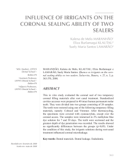
EVALUATION OF THE MARGINAL
LEAKAGE USING MODIFIED GLASS
IONOMER RESIN AS TEMPORARY
SEALER OF ENDODONTICS CAVITIES
Kalena de Melo Maranhão1
Eliza Burlamaqui Klatau2
Suely Maria Santos Lamarão3
UFPA’S Dental School –
Belém-PA
2
Assistant Professor,
UFPA’S Dental School –
Belém-PA
3
Adjunct Professors,
UFPA’S Dental School –
Belém-PA
1
Recebido em: april de 2007
Aceito em: january de 2008
MARANHÃO, Kalena de Melo, KLAUTAU, Eliza Burlamaqui e
LAMARÃO, Suely Maria Santos. Evaluation of the marginal leakage using modified glass ionomer resin as temporary sealer of endodontics cavities. Salusvita, Bauru, v. 28, n. 1, p. 31-39, 2009.
ABSTRACT
This study evaluates the marginal microleakage of resin modified
glass ionomer – Vitremer/3M, used as temporary sealer after
endodontic treatment. 36 molars were selected, in which, after
endodontic treatment, cavities with 4mm of depth were standardized
for the sealer material insertion, and divided into two groups of 18
teeth each: Group1 (conventional technique): primer + glass ionomer
+ protective of surface and Group2 (hybridization technique): 37%
phosphoric acid + adhesive system Single Bond + glass ionomer
+ protective of surface. After restoration, all the specimens were
thermocycled, impermeabilized and immersed in 2% of methylene
blue solution for 7 days. After this period, the samples were washed
in current water for 4 hours, sectioned mesiodistally and evaluated
in stereomicroscope. The data were analyzed using Mann-Whitney
Test at a significant level of 5%. The results revealed no statistically
significant difference between the groups (p> 0,05) and that none
of the tested groups were capable to completely eliminate marginal
leakage. The authors concluded that the association of the adhesive
31
system with a resin modified glass ionomer did not influence the
sealing ability of those restorations.
Key words: Glass ionomer cement. Marginal leakage. Endodontic.
RESUMO
Este estudo avaliou a infiltração marginal do ionômero de vidro
modificado por resina – Vitremer, utilizado como selador provisório após tratamento endodôntico. Foram selecionados 36 molares
íntegros, nos quais, após o tratamento endodôntico, padronizou-se
cavidades com 4mm de profundidade para inserção do material selador; originando dois grupos com 18 corpos-de-prova cada: Grupo
1 (técnica convencional) primer + ionômero + protetor de superfície
e Grupo 2 (técnica com hibridização) ácido fosfórico 37% + adesivo
dentinário Single Bond + ionômero + protetor de superfície. Procedeu-se então a termociclagem e em seguida a imersão em azul de
metileno a 2% por 7 dias. Decorrido o prazo experimental os corposde-prova foram lavados em água corrente por 4 horas, seccionados
longitudinalmente no sentido Mesio-Distal e levados à leitura em
um esteromicroscópio. Os dados obtidos foram submetidos à análise
estatística por meio do teste não paramétrico de Mann-Whitney com
nível de significância de 5%, observando-se não haver diferença significante entre os grupos (p>0,05) e que nenhum dos grupos testados
foi capaz de impedir a microinfiltração marginal. Os autores concluem que a associação do sistema adesivo ao ionômero de vidro modificado por resina não influenciou no selamento das restaurações.
Palavras-chave: Cimento ionômero de vidro. Infiltração marginal.
Endodontia.
INTRODUCTION
Studies have demonstrated that the coronal filling is as important
as the apical, preventing the entry of fluids and microorganisms
into the root canal space (HOMMEZ et al., 2002; SEGURA-EGEA
et al., 2004).
The leakage is still considered as a factor in the failure of endodontic
treatment. Thus, the search for an effective material has been a
constant concern in the endodontics. In reality, a clean surface is
essential for an effective sealing, therefore the presence of smear layer,
32
MARANHÃO,
Kalena de Melo,
KLAUTAU, Eliza
Burlamaqui e
LAMARÃO, Suely
Maria Santos.
Evaluation of the
marginal leakage
using modified glass
ionomer resin as
temporary sealer of
endodontics cavities.
Salusvita, Bauru,
v. 28, n. 1, p. 31-39,
2009.
MARANHÃO,
Kalena de Melo,
KLAUTAU, Eliza
Burlamaqui e
LAMARÃO, Suely
Maria Santos.
Evaluation of the
marginal leakage
using modified glass
ionomer resin as
temporary sealer of
endodontics cavities.
Salusvita, Bauru,
v. 28, n. 1, p. 31-39,
2009.
after the chemical-surgical preparation, can influence the coronal
restoration sealing process. Thus, the united action of endodontic
instruments and chemical substances during the biomechanical
preparation provides the increase of the dentin permeability, due to
the removal of smear layer, opening the dentinal tubules, providing
the union between the material and tooth (STEWART et al., 1969;
PAIVA; ANTONIAZZI, 1973; ÇOBANKARA et al., 2004).
Several temporary fillings are proposed to block the passage of
fluids, microorganisms and substances in the interface between the
restorative material and the dentin. Among the existent restorative
materials, the glass ionomer cement, described in the 70’s by Wilson
and Kent offered various advantages such as chemically bonding to
dental structure, thermal expansion coefficient similar to the tooth,
antimicrobial activity and reduced microleakage (BUSSADORI;
MUENCH, 1999; FORMOLO et al., 2001).
In the 80’s, resin modified glass ionomer cement was formulated
to overcome the moisture sensitivity problems of composites and the
low mechanical strength of glass ionomers, while maintaining the
clinical advantages of conventional glass ionomer cement (LIMA et
al., 2002).
Different ways of restoring procedures have been proposed in the
attempt of obtaining a good retention and consequently to reduce the
marginal infiltration, among them the adhesive systems have been
combined with the resin modified glass ionomers cements (NOVAES
JR et al., 1998; SALLES et al., 1999; KRAMER et al., 2003).
Nowadays, despite all technological efforts, it was not possible
to produce an adhesion system effective to bond restorative material
with dentin.
The aim of this study was to evaluate in vitro the marginal leakage
of the resin modified glass ionomer cement after root canal treatment
comparing two different techniques: the conventional and with the
hybridization technique.
MATERIAL AND METHOD
Thirty six caries-free extracted human maxillary and mandibular
molar teeth were autoclaved and stored in distilled water. The Ethics
Committee approved the study design. Access was made using a high
speed air turbine under water coolant with a nº1014 round bur (KG
Sorensen) for initial entry and an Endo-Z bur (Dentsply) to extend
the preparation to the desired occlusal outline. The cervical third of
the root canal was prepared with Gates Glidden drills (Dentsply),
33
Endo-PTC cream and 0,5% of sodium hypochlorite (Formula &
Ação Farmácia), according to Paiva and Antoniazzi (1993).
A final irrigation with 30ml of 15% EDTA-T (Formula & Ação
Farmácia) to remove the smear layer was made. The prepared opening
was air-dried; cotton pellets and gutta-percha (G-C Chemical) were
placed on the floor of the pulp chamber. The depth of the cavity was
measured with a periodontal probe and allowed for at least 4 mm of
temporary filling material (WEBBER et al., 1978).
The teeth were divided randomly into two groups of 18 teeth each.
Group I-(conventional Technique): the primer was applied for 30
seconds using a microbrush and light cured for 20 seconds, with the
apparel Curin Light XL 1500-3M, with potency of 550mw/cm². The
resin modified glass ionomer cement was manipulated according to
the manufacturer’s instructions and inserted into the cavity using a
syringe (Centrix Incorporated) and light cured for 40 seconds.
Group II-(hybridization Technique): the specimens were etched
with 37% phosphoric acid for 15 seconds, washed for 10 seconds and
dried with absorbent paper. A thin layer of adhesive system Single
Bond (3M) was applied. The dentine bonding agent was light cured for
20 seconds. The material was prepared following the manufacturer’s
instructions. The resin modified glass ionomer cement was placed
into the pulp chamber by a syringe (Centrix Incorporated) and light
cured for 40 seconds.
In both groups the application and polymerization of the finishinggloss were accomplished.
The sealers were stored in an incubator for 24 hours at 37ºC and
100% humidity. Then the specimens were thermocycled at 500 cycles
to temperatures of 5ºC and 55ºC, with 30 seconds of immersion in
each bath.
The teeth were sealed with application of three super bonder
layers, except over the coronal access. The teeth were then immersed
in 2% methylene blue solution (pH 7,2) and stored in an incubator,
maintained at 37ºC for 7 days. The specimens were sectioned in a
mesiodistal direction along their longitudinal axis with a low speed
diamond cutter. After being sectioned, the samples were rinsed in
tap water for 10 min to ensure removal of the debris and the smear
layer created by the cutting.
The maximum linear coronal dye penetration was measured in
millimeters, using a stereomicroscope (Technival Carl Zeiss) at a
X25 magnification. One examiner, who had no knowledge of the
treatment, analyzed the sections. The section that had the greatest
depth of dye penetration was used as the final score for that specimen.
The results were tabulated, and the mean value for each group was
recorded.
34
MARANHÃO,
Kalena de Melo,
KLAUTAU, Eliza
Burlamaqui e
LAMARÃO, Suely
Maria Santos.
Evaluation of the
marginal leakage
using modified glass
ionomer resin as
temporary sealer of
endodontics cavities.
Salusvita, Bauru,
v. 28, n. 1, p. 31-39,
2009.
MARANHÃO,
Kalena de Melo,
KLAUTAU, Eliza
Burlamaqui e
LAMARÃO, Suely
Maria Santos.
Evaluation of the
marginal leakage
using modified glass
ionomer resin as
temporary sealer of
endodontics cavities.
Salusvita, Bauru,
v. 28, n. 1, p. 31-39,
2009.
The obtained data were submitted to a statistical analysis using
the Mann-Whitney test, with a significance level of 5%.
RESULTS
The results of the quantitative marginal analysis are presented
in Figure 1, observing the comparison between the medium values
obtained and the applied methods.
The Mann-Whitney nonparametric test showed no significant
differences among groups (p> 0,05). However, the leakage values
were significantly higher in the conventional technique than in the
hybridization technique.
0.5
0.4
0.43
0.3
0.2
0.23
0.1
0
Hybridization Technique
Conventional Technique
Figure 1 - Coronal leakage (mm) of the techniques tested
DISCUSSION
The resin modified glass ionomer cement has further created great
interest for temporary restoration material, due to its advantages of
fluoride release as well as the largest durability of the material, due
to the resin incorporation, which provide a larger resistance to wear
and fractures.
Thus, to keep the positive characteristics of resin modified glass
ionomer cement, and also to improve bond strength, a combination
with the adhesive system was developed.
The results of the present study showed leakage in different
degrees among techniques, but it has no significant difference
35
between the groups (p> 0,05). Same results have been reported
in previous studies (BUSSADORI; MUENCH, 1999; SALLES et
al., 1999; LIMA et al., 2002; KRAMER et al., 2003) using similar
methodology. In addition, Wang et al. (2006) also confirmed similar
results while investigating the shear bond strength of resin-modified
glass ionomer cements in human dentin using two adhesive systems.
However, Novaes Jr et al. (1998), Cacciafesta et al. (2003) and
Wang et al. (2004) demonstrated that the modified technique showed
better results than the conventional technique. Bernardo et al. (2000)
have reported that the adhesion force for the acid conditioning
technique is larger than the conventional technique.
Several studies have noticed the importance of the smear layer
removal, to obtain better sealing ability of the restorative material.
However, the results in this study didn’t confirm these data, because
the hybridization technique, that provide a larger removal of smear
layer than the conventional technique, didn’t improve the marginal
sealing in endodontically treated teeth.
This result can be justified for the use of the irrigant solution during
the endodontic therapy, which increases the dentin permeability.
This is in accordance with other studies of the dentinal permeability
that evidenced better results with the use of irrigant solution in
biomechanical preparation. (NIKIFORUK; SREEBNY, 1953;
PAIVA; ANTONIAZZI, 1973; ROBAZZA et al., 1981; VIVACQUAGOMES et al., 2002; ÇOBANKARA et al., 2004)
Thus, was observed that the leakage in group I was not influenced
by adhesive system, but by auxiliary substances that removed smear
layer and prepared the dentin surface for better adhesion of the
temporary restoring material. In relation to group II, the leakage is
similar to group I, possibly originated from an inherent failure to the
adhesive system (CARVALHO et al., 2004; WANG et al., 2006). In
addition, Souza et al. (2005) confirmed similar results when liquid
adhesives were applied at the pulp chamber walls, using similar
methodology in this paper. Ozturk et al. (2004) also observed similar
results, using the fluid filtration method.
Perdigão et al. (2000) and Uceda-Gomez et al. (2003) related that
the residual NaOCl in the subsurface porous dentin could result
in incomplete polymerization of resin monomers at the interface
between the adhesive and dentin. Thus, these endodontic irrigants
could interfere with the retention of the adhesive system.
Moreover, the results showed that none of the tested groups
were capable to completely eliminate the marginal leakage in
endodontically treated teeth.
36
MARANHÃO,
Kalena de Melo,
KLAUTAU, Eliza
Burlamaqui e
LAMARÃO, Suely
Maria Santos.
Evaluation of the
marginal leakage
using modified glass
ionomer resin as
temporary sealer of
endodontics cavities.
Salusvita, Bauru,
v. 28, n. 1, p. 31-39,
2009.
MARANHÃO,
Kalena de Melo,
KLAUTAU, Eliza
Burlamaqui e
LAMARÃO, Suely
Maria Santos.
Evaluation of the
marginal leakage
using modified glass
ionomer resin as
temporary sealer of
endodontics cavities.
Salusvita, Bauru,
v. 28, n. 1, p. 31-39,
2009.
CONCLUSION
According to the methodology used, it was possible to conclude
that none of the tested groups were capable to completely eliminate
marginal leakage in endodontically treated teeth and the association
of the adhesive system to resin modified glass ionomer did not
influence the sealing ability of those restorations. Additional studies
may be needed to verify the sealing quality provided by these
techniques in clinical applications.
REFERENCES
BERNARDO, P. C. et al. Avaliação clínica de um cimento de ionômero de vidro utilizado como selante oclusal. Pesq Odont Brás, v.14,
p. 53-57, 2000.
BUSSADORI, S. K.; MUENCH, A. Microinfiltração em dentes decíduos em função de materiais restauraores e condicionamento ácido. Rev Odontol Univ São Paulo, v.13, p. 369-373, 1999.
CACCIAFESTA, V. et al. Use of a self-etching primer in combination with a resin-modified glass ionomer: Effect of water and saliva
contamination on shear bond strength. Am J Orthod Dentofacial Orthop, v. 124, p. 420-6, 2003.
CARVALHO, R. M. et al. Sistemas adesivos: Fundamentos para
aplicação clínica. Biodonto, v. 2, p. 125-30, 2004.
ÇOBANKARA, F. K. et al. Evaluation of the influence of smear
layer on the apical and coronal sealing ability of two sealers. J Endod, v. 30, p. 406-409, 2004.
FORMOLO, E. et al. Infiltração marginal em cavidades de classe V
com o uso de diferentes materiais adesivos. Rpg Rev Pos-Grad, v. 8,
p. 306-312, 2001.
HOMMEZ, G. M. et al. Periapical health related to the quality of
coronal restorations and root fillings. Int Endod J, v. 35, p. 680-89,
2002.
KRAMER, P. F. et al. Microleakage between two filling restorative
techniques using glass ionomer cement in primary molars: comparative “in vitro” study. J Appl Oral Sci, v. 11, p. 114-119, 2003.
LIMA, D. R. et al. Avaliação do selamento de restaurações com cimento de ionômero de vidro resina-modificado em pregando como
37
pré-tratamento o ácido poliacrílico, ácido tânico e laser de ND:YAG.
PGRO-Pos-Grad Rev Odontol, v. 5, p. 29-34, 2002.
NIKIFORUK, G.; SREEBNY, L. Demineralization of hand tissues
by organic chelating agents at neutral pH. J Dent Res, v. 32, p. 85967, 1953.
NOVAES JR, J. B. et al. Cimentos de ionômeros de vidro convencionais e modificados. Influência de adesivos e da umidade, sobre a
adesão à dentina. Rev FOB, v. 66, p. 35-40, 1998.
OZTURK, B. et al. An in vitro comparison of adhesive systems to
seal pulp pulp chamber walls. Inter Endod J, v. 37, p. 297-306, 2004.
PAIVA, J. G.; ANTONIAZZI, J. H. O uso de uma associação de
peróxido de uréia e detergente (Tween 80) no preparo químico-mecânico dos canais radiculares. Rev Assoc. Paul. Cir. Dent, v. 27, n. 7,
p. 416-422, 1973.
PAIVA, J. G,; ANTONIAZZI, J. H. Endodontia: Bases para a prática
clínica. 2ª. ed. São Paulo: Artes Médicas; 1993.
PERDIGÃO, J. et al. Effect of a sodium hypochorite gel on dentin
bonding. Dent Mater, v. 16, p. 311-323, 2000.
ROBAZZA, C. R. C. et al. Variações na permeabilidade da dentina radicular quando do emprego de alguns fármacos auxiliares no
preparo Endodontico. Contribuição ao estudo. Rev Ass Paul Cirurg
Dent, v. 35, p. 528-533, 1981.
SEGURA-EGEA, J. I. et al. Periapical status and quality of root
fillings and coronal restorations in an adult spanish population. Int
Endod J, v. 37, n. 8, p. 525-30, 2004.
SALLES, V. et al. Avaliação in vitro da microinfiltração marginal de
restaurações realizadas com um cimento de ionômero de vidro modificado por resina e uma resina composta modificada por poliácidos
associadas a dois sistemas adesivos. Rev FOB, v. 7, p. 1-6, 1999.
SOUZA, F. D. et al. The effect on coronal leakage of liquid adhesive
application over root fillings after smear layer removal with EDTA or
Er:YAG laser. Oral Surg Oral Med Oral Pathol Oral Radiol Endod,
v. 99, p. 125-128, 2005.
STEWART, G. G. et al. EDTA and urea peroxide for root canal preparation. JADA, v. 78, p. 335-338, 1969.
UCEDA-GOMEZ, N. et al. Effect of sodium hypochlorite on the
bond atrength of an adhesive system to superficial and deep dentin.
J Appl Oral Sci, v. 11, n. 3, p. 223-228, 2003.
38
MARANHÃO,
Kalena de Melo,
KLAUTAU, Eliza
Burlamaqui e
LAMARÃO, Suely
Maria Santos.
Evaluation of the
marginal leakage
using modified glass
ionomer resin as
temporary sealer of
endodontics cavities.
Salusvita, Bauru,
v. 28, n. 1, p. 31-39,
2009.
MARANHÃO,
Kalena de Melo,
KLAUTAU, Eliza
Burlamaqui e
LAMARÃO, Suely
Maria Santos.
Evaluation of the
marginal leakage
using modified glass
ionomer resin as
temporary sealer of
endodontics cavities.
Salusvita, Bauru,
v. 28, n. 1, p. 31-39,
2009.
VIVACQUA-GOMES, N. et al. Influence of irrigants on the coronal
microleakage of laterally condensed gutta-percha root fillings. Int
Endod J, v. 35, p. 791-795, 2002.
WANG, L. et al. Effect of one-bottle adhesive systems on the fluoride release of a resin-modified glass ionomer. J Appl Oral Sci, v. 12,
p. 12-17, 2004.
WANG, L. et al. Effect of adhesive systems associated with resinmodified glass ionomer cements. J Oral Rehabil, v. 33, n. 2, p. 110-6,
2006.
WEBBER, R. T. et al. Sealing quality of a temporary filling material.
Oral Surgery, v. 46, p. 123-30, 1978.
WILSON, A. D.; KENT, B.E. A new translucent cement for dentistry. The glass ionômero cement. Brit Dent J, v. 132, n. 4, p.
133-5, 1972.
39
Baixar