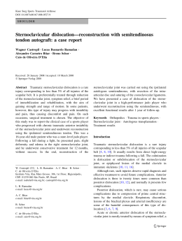
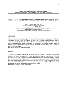
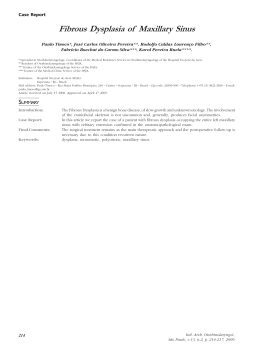
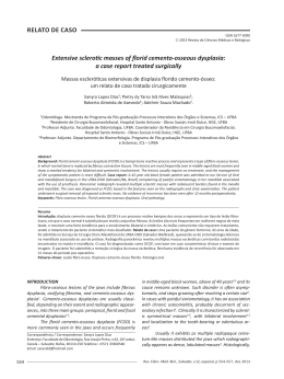
International Orthopaedics (SICOT)
DOI 10.1007/s00264-014-2357-3
ORIGINAL PAPER
Medial patellofemoral ligament anatomy: is it a predisposing
factor for lateral patellar dislocation?
Victor de Oliveira & Vanessa de Souza & Ricardo Cury &
Osmar Pedro Camargo & Osmar Avanzi &
Nilson Severino & Patricia Fucs
Received: 11 February 2014 / Accepted: 9 April 2014
# Springer-Verlag Berlin Heidelberg 2014
Abstract
Purpose Because the medial patellofemoral ligament (MPFL)
is the primary restraint to lateral dislocation of the patella, we
aimed, in this controlled study, to verify whether the MPFL
with different measurements could be considered another
predisposing factor for patellar dislocation.
Methods A group of 100 consecutive individuals without the
criteria for patellar dislocation (trochlear dysplasia, patella alta
and lateral patellar tilt) was recruited as a control group and
underwent magnetic resonance imaging (MRI) study and
another group of 50 patients with patellar instability. Femoral
condyles, interepicondylar distance, length and thickness of
the MPFL were measured.
Results In the control group, the MPFL was 38–60 mm long.
Individuals with patellar instability who had no episode of
patellar dislocation had a 4.11-mm longer ligament than controls (p=0.032), while patients with instability with a previous
history of dislocation had a 13.54-mm longer MPFL than
controls (p<0.001). Thickness of the MPFL at the patellar
insertion was lower in individuals with patellar instability with
a history of dislocation (p<0.001). An instability coefficient
(IC) less than 1.3 indicates that the MPFL is insufficient.
Conclusions Individuals with patellar instability and previous
patellar dislocation present with longer MPFL when compared to controls, and an associated IC less than 1.3 can be
considered a predisposing factor for patellar dislocation.
Treatment of patellar instability is a challenge, and it is
V. de Oliveira : R. Cury : O. P. Camargo : O. Avanzi : N. Severino :
P. Fucs (*)
Orthopaedic and Traumatology, Santa Casa Medical School and
Hospitals, São Paulo, Brazil
e-mail: [email protected]
V. de Souza
Radiology Department, Santa Casa Medical School and Hospitals,
São Paulo, Brazil
difficult to identify what is the predisposing factor. This study
has verified the measurements of the MPFL for the first time
and presents values of thickness and length that can be considered as indications for surgical reconstruction.
Level of evidence: III.
Keywords Patella . Patellar ligament . Joint instability . Knee
Introduction
Patellar instability results in subluxation and dislocation of the
patella, causing pain and functional impairment, both in athletes and non-athletes. A number of factors are involved in this
instability: trochlear dysplasia in 85–96 % of cases, increased
lateral patellar tilt in 83 % of cases, patella alta in 24 % and an
increase in the distance between the tibial tuberosity and the
centre of the trochlear groove (TT-TG) in 56 % of cases as
well as other, less frequent problems [1–6]. These anatomical
changes are described in the literature as “factors for patellar
instability”.
The congruence of the patellofemoral joint is maintained
by static and dynamic stabilising structures. The medial
patellofemoral ligament (MPFL) is the primary passive soft
tissue restraint to lateral dislocation of the patella [7–11].
Based on biomechanical and clinical studies, rupture of the
MPFL is an essential lesion in lateral patellar dislocation
[8–12].
The MPFL is a retinacular band of tissue located on a lower
plane than the vastus medialis oblique muscle. The MPFL has
its insertions over the medial upper margin of the patella and
in the medial femoral epicondylar region. In the femur, the
insertion could also be located between the adductor tubercle
and the medial epicondyle [8, 11, 13]. The MPFL is approximately 55 mm in length and between 3 and 30 mm in width. It
can bear a force of 208 N before rupturing [8]. The role of the
International Orthopaedics (SICOT)
MPFL in preventing lateral dislocation of the patella is more
marked at between 0 and 30° of knee flexion, when the
ligament is more tense [7, 8].
The MPFL is an anatomical structure that is difficult to
isolate in cadaveric studies, some of which report that this
ligament is not present [11, 14, 15]. However, the MPFL is
easily identifiable in magnetic resonance imaging (MRI) studies [3, 5, 13, 16–23]. In the literature, only two studies have
correlated the MPFL measurements with other anatomical
structures in the knee [14, 23]. In a literature search, no studies
were found analysing variations in the MPFL in cases in
which factors for patellar instability were identified.
Due to the importance of the MPFL in the stabilisation of
the patella, the hypothesis of this study was that an abnormality
in the ligament’s structure may be a predisposing factor for the
occurrence of primary dislocation of the patella and, subsequently, progression to chronicity. This study aimed, therefore,
to investigate the morphological characteristics of the MPFL,
comparing normal knees with knees with three risk factors for
patellar instability: trochlear dysplasia, patella alta and increased lateral patellar tilt. MRI was chosen due to the difficulty in isolating this ligament in cadavers and also because the
incidence of abnormal parameters is very low in the general
population, e.g. 2 % cited for trochlear dysplasia [2, 16].
The objective of the study was to verify whether individuals with the risk factors for patellar instability (trochlear
dysplasia, patella alta and lateral patellar tilt greater than
20°) have altered MPFL measurements, compared to a control
group (CG) of normal individuals, and also to verify whether
morphological characteristics of the MPFL might be considered a possible predisposing factor for patellar dislocation.
Materials and methods
This was an observational, case-control study carried out in a
public hospital. All patients attending the knee clinic between
August 2011 and September 2012 and submitted to MRI of
the knee were included. All of the patients and control subjects
signed informed consent forms for participation, and the study
was approved by the Ethics Committee of the Institution.
Patients with fracture sequelae or tumours were excluded,
as were those who had undergone knee or leg surgery before
admission. Patients with a history of patellar dislocation in the
previous 12 months were also excluded, because the oedema
of a recent episode of dislocation could interfere with MRI
signals and make it difficult to measure the ligament.
Participants were divided into two groups: the CG
consisting of patients with diagnoses such as anterior cruciate
ligament (ACL) tears, meniscal or femorotibial chondral lesions and no symptoms of patellar instability and the instability group (IG) comprising all cases with anterior knee pain and
signs of malalignment of the extensor mechanism. The latter
group was subdivided into two subgroups: the first with
patients presenting risk factors for instability, but no patellar
dislocation had been reported. This was called the “potential
instability” subgroup. The second subgroup, called “objective
instability”, contained patients with at least one previous
episode of patellar dislocation.
The study group was comprised of 146 patients and 150
knee MRI examinations. Of this group, 25 patients were
excluded, leaving 121 patients who were studied, including
four cases of bilateral involvement, with a total of 125 knees.
In the CG, 88 MRIs were performed and in the IG, 38 were
performed, with 19 cases in each subgroup: potential and
objective instability. The mean age at the time of the examination was 38.6 years (± 15.8), and 66 (54.4 %) were women.
All of the MRI measurements were conducted by two
radiologists specialised in musculoskeletal system radiology.
As the intra-class correlation coefficient (ICC) between them
was higher than 95 %, the mean measurement was registered
for statistical analysis.
On MRI, the MPFL was characterised as a band with a
focal, continuous thickness, greater than the other portions of
the medial patellar retinaculum, and with low signal sequences in proton density, extending from the medial border
of the upper part of the patella to the posterior portion of the
medial epicondyle of the femur. The route and the patellar and
femoral insertions of the MPFL were best evaluated in axial
sequences. The route of the MPFL was seen more clearly in
the coronal sequences than in the sagittal sequences.
All of the MRI exams were performed in the same manner,
with a 1.5 T device (Intera, Phillips), eight-channel coil and
proton density-weighted sequences in three planes (sagittal,
coronal and axial), with and without fat saturation, using the
following parameters: echo time (TE) 1,642, TE 30, matrix
512×256, field of view (FOV) 16×16, slice thickness of
3.5 mm, with an interval of 0.3 mm. During the exam, the
knee was placed in a standardised position, in extension, with
the patient supine but without any other special positioning.
The image analysis was performed on workstations using the
PACS/RIS Agfa software.
The following measurements were obtained:
–
–
–
–
Size of the lateral and medial femoral condyles, on the
sagittal plane, in the section of the longer anteroposterior
axis, measuring bone to bone surface (Fig. 1)
Interepicondylar distance, in the axial and coronal planes
(Figs. 2 and 3), at the level of the confluence of the medial
collateral ligament, adductor magnus and MPFL
insertions
Patellar height in the sagittal section, according to the
Caton-Deschamps index (with values >1.2 indicating
patella alta [1, 5])
Trochlear dysplasia: trochlear depth, measured in the
axial section 3 cm proximally to the joint surface
International Orthopaedics (SICOT)
Fig. 1 Measurement of the anteroposterior distance in the medial condyle in the sagittal cut of MRI exam
–
(<3 mm indicating trochlear dysplasia [3, 5, 16]); ventral
trochlear prominence, measured in the sagittal section
(>6.9 mm indicating trochlear dysplasia [16]); trochlear
groove angle, measured in the axial section, 3 cm above
the joint surface (>150° indicating trochlear dysplasia [2,
3, 5]); inclination of the lateral facet, corresponding to the
angle of inclination of the lateral facet relative to the
coronal plane, tangent to the posterior region of the femoral condyles (<11° indicating trochlear dysplasia [3, 5])
Ratio of the trochlear facets, medial to lateral, measured
in the axial section 3 cm above the joint surface (<0.4
indicating trochlear dysplasia [3, 5, 16])
Fig. 2 Measurement of the interepicondylar distance in the axial cut of
MRI exam
Fig. 3 Measurement of the interepicondylar distance in coronal cut of
MRI exam
–
–
–
Inclination angle of the patella, measured in the axial
plane (>20° indicating instability) [1]
Wiberg patellar classification [24]
Measurements of the MPFL in millimetres: thickness at
5 mm from the patellar insertion, in the midpoint and at
5 mm to the femoral insertion, full length from insertion
to insertion, using the Higuchi et al. technique [22]
(Fig. 4)
Qualitative variables were described as relative and absolute frequencies, while quantitative variables were described
as means, standard deviations, medians, minimum and
Fig. 4 Measurement of the MPFL full length in the axial cut of MRI
exam
International Orthopaedics (SICOT)
maximum values. Pearson’s correlation was calculated between the ligament measurements and patient’s age, femoral
condyle size and epicondylar distance. Student’s t test was
used to determine the association between the measurements
and the patient’s gender. Analysis of covariance (ANCOVA)
was used to determine the personal and femoral condyle
characteristics that could influence the ligament measurements, followed by Bonferroni multiple comparisons, to detect any differences in the ligament measurements. SPSS
software was used in the statistical analysis, adopting a level
of significance of 5 %.
Results
In the CG, MPFL length ranged from 36.1 to 61.8 mm, with a
mean of 49.0 mm and thickness of between 0.5 and 3.7 mm at
the different measurement points. In the IG, MPFL length
ranged from 43.7 to 76.6 mm, with a thickness of between
0.4 and 3.1 mm.
MPFL length correlated inversely with age (p<0.001),
with older individuals having shorter ligaments. There was a
direct correlation between thickness of the middle third and at
the patellar insertion and the size of the medial femoral condyle (p<0.05) and between the femoral insertion and the size
of the lateral femoral condyle (p=0.021) (Table 1).
The MPFL was significantly thicker in men than in women
(p=0.045) at the femoral insertion. The other measurements
were not significantly influenced by sex. Comparing the CG
with the subgroups of the IG, with and without any previous
episode of patellar dislocation, or objective and potential
instability, it was observed that the mean MPFL length and
the mean thickness at the patellar insertion were statistically
different (p<0.001), regardless of other characteristics that
could influence the ligament measurements (Table 2).
MPFL length and thickness at the patellar insertion was
significantly different between CG and the subgroups of IG,
Table 1 Pearson’s correlations
(r) between age and measurements of the femoral condyles
(n=125) and statistical
significance (p)
potential and objective instability. Cases with potential instability had longer MPFL than the controls (average increase of
4.11 mm) (p<0.032). Even longer lengths were seen in the
subgroup with objective instability, i.e. cases with previous
episodes of dislocation (average increase of 13.54 mm)
(p<0.001). The mean thickness at the patellar insertion was
significantly less than in the IG cases with objective instability
compared with the CG (p<0.001) (Table 3).
Analysing only the 35 cases with trochlear dysplasia and
the 15 cases with patellar lateral inclination over 20°, the mean
length of the MPFL was significantly greater than in the CG,
and the mean thickness at the patellar insertion was statistically less than that of the controls, independently of previous
episodes of patellar dislocation (p<0.001). Based on our
findings for MPFL length, any length greater than 60 mm
was considered abnormal and insufficient.
To minimise the differences in bone sizes among individuals, a coefficient that would more accurately reflect whether
the ligament is longer compared with the condyle’s size was
calculated. The interepicondylar distance in the axial and
coronal planes was chosen, to correlate with MPFL length.
The coefficients found ranged from 1.30 to 1.95; the smaller
the coefficient, the longer the ligament, in both planes. Values
lower than 1.3 indicate insufficient MPFL length to prevent
patellar dislocation (Table 4).
Discussion
Although the existence and importance of the MPFL for
stabilisation of the patella was first described in 1979 by
Warren and Marshall [25], it has only been in recent decades
that studies have emerged proving this theory and describing
the first techniques for repair and reconstruction of this ligament in the treatment of patellofemoral joint instability [7–9,
14, 15, 26–29]. As in other publications [3, 5, 13, 16–23], it
Correlation
Total
length
Thickness at
medial third
Thickness at
patellar insertion
Thickness
at femoral
insertion
Age (years)
r
p
−0.375
< 0.001
−0.057
0.525
0.098
0.278
0.038
0.672
Lateral femoral condyle size, sagittal plane
r
p
r
p
r
p
r
p
0.079
0.383
0.095
0.294
0.146
0.103
0.153
0.089
0.173
0.054
0.196
0.028
0.082
0.361
0.115
0.203
0.151
0.094
0.230
0.010
0.093
0.303
0.130
0.147
0.207
0.021
0.158
0.079
0.119
0.188
0.102
0.255
Medial femoral condyle size, sagittal plane
Interepicondylar distance, axial
Interepicondylar distance, coronal
International Orthopaedics (SICOT)
Table 2 Measurements of the MPFL (mm) and correlations with controls, with and without episodes of patellar dislocation
Variable
Groups
Mean
SD
Median
Minimum
Maximum
n
p
Total length
Control
Potential instability
Objective instability
Control
Potential instability
Objective instability
Control
Potential instability
Objective instability
Control
Potential instability
Objective instability
49.04
53.63
63.81
1.27
1.12
0.98
1.71
1.37
0.90
1.14
1.00
0.91
5.64
6.97
7.98
0.44
0.37
0.39
0.59
0.72
0.40
0.46
0.39
0.31
48.8
55.3
65.5
1.2
1.1
0.9
1.6
1.2
0.8
1.1
0.9
0.9
36.1
43.7
51.1
0.6
0.6
0.4
0.7
0.4
0.4
0.5
0.6
0.6
61.8
64.6
76.6
3.3
1.8
2.3
3.2
3.1
1.9
3.7
2.0
2.0
87
19
19
87
19
19
87
19
19
87
19
19
< 0.001
Thickness at medial third
Thickness at patellar insertion
Thickness at femoral insertion
0.101
< 0.001
0.339
SD standard deviation
was possible to identify the MPFL in all of the MRI exams in
our sample.
The results showed that in patients who had never had an
episode of patellar dislocation only the length of the MPFL
was altered. MPFL length was also changed in individuals
with at least one of the criteria for patellar instability, regardless of other variables that might influence the measurements
of this ligament. No other study was found measuring the
length and thickness of the MPFL in normal individuals and in
patients with the criteria of instability and performing the
comparisons made here. Therefore, this is the first study to
objectively measure, by MRI, MPFL length and thickness in
both groups of patients, with and without patellar instability.
The values registered here can be used for comparisons with
other populations in other settings as the coefficient was
normalised to minimise differences in body sizes.
In our results, in individuals with any of the criteria of
instability and at least one previous episode of patellar dislocation, the MPFL was around 30 % longer, but with only half
the thickness near the point of patellar insertion. These are
evident signs of increased risk for patellar dislocation and
should be watched carefully in the MRI scans of patients with
knee problems as part of routine care, independently of history
of patellar dislocation. Other parameters can be observed on
MRI though.
The changes in the MPFL were even more marked in
patients with trochlear dysplasia and with patellar lateral inclination over 20°, with longer length and less thickness at the
patellar insertion, independently of history of previous patellar
dislocation. The conclusion is that in the presence of these two
factors, attention should be paid to the MPFL morphology,
which will most likely be altered, with patellar instability
increasing the risk for dislocation.
In patients of the potential instability group, the MPFL was
much longer at 4.11 mm greater than in the CG. This significant difference suggests that the altered ligament is a predisposing factor for dislocation. In the other cases of IG with a
history of patellar dislocation (objective instability), the
changes in the MPFL were even more marked, with a mean
length greater than 13.54 mm and thickness 0.78 mm less than
the controls. A temporal relationship is difficult to establish
here as the measurements prior to the episode of dislocation
were not known (if they were yet abnormal, increasing the risk
of dislocation) or the ligament healing after dislocation was
insufficient, leading to longer and thinner ligaments. Thus, in
patients with abnormal MPFL documented after an episode of
Table 3 Results of the Bonferroni multiple comparison test and association between measurements of the length and thickness of the MPFL (mm)
between groups
Variable
Comparison groups
Mean difference
Standard error
p
Total length
Control x potential instability
Control x objective instability
Potential instability x objective instability
Control x potential instability
Control x objective instability
Potential instability x objective instability
−4.11
−13.54
−9.43
0.31
0.78
0.47
1.59
1.71
2.05
0.15
0.16
0.19
0.032
< 0.001
< 0.001
0.126
< 0.001
0.051
Thickness at patellar insertion
International Orthopaedics (SICOT)
Table 4 Correlation between
interepicondylar distance, axial
and coronal planes, and the full
length of the MPFL, components
of the instability coefficient (IC)
Variable
Axial IC
Statistical method: ANCOVA
n number of subjects, NI normal
interval
Coronal IC
Group
Control
Potential instability
Objective instability
Control
Potential instability
Objective instability
patellar dislocation, surgical treatment should be considered,
even after the first episode.
This indication for patients with previous dislocation is
corroborated by the results of other studies [19, 22, 28, 29],
which conclude that after the first episode of patellar dislocation the MPFL becomes insufficient, predisposing to recurrence. A study by Lewallen et al. [4], in 2013, showed that
among individuals under 18 years of age with trochlear dysplasia, after an acute dislocation event and conservative treatment, 69 % relapsed and would therefore benefit from early
surgical treatment.
Although the MPFL has shown to be thinner throughout its
length in individuals with any of the criteria for instability, it
was right next to the patella that a significant difference in
thickness from the CG was seen. This suggests that this is the
most frequent site of rupture. Some studies have confirmed
this hypothesis [3, 13, 20, 30, 31]; however, others have
reported the femoral insertion as the most frequent site of
MPFL rupture [19, 21, 28, 29, 32]. Balcarek et al. [19], in
2010, in a study of acute patellar dislocations, observed that in
patients with trochlear dysplasia and patella alta injuries were
predominantly near the patella or the femur, with concomitant
lesions in the patella and femur. Few patients had lesions in the
central region of the ligament. Patients with an increase of
more than 20 mm in the TT-TG had lesions near the patella.
However, Weber-Spickschen et al. [21], in 2011, analysing 59
patients with acute patellar dislocation, found no relationship
between injury site and morphology of the trochlea.
Dejour et al. [1], an experienced group of researchers,
stated that there are four factors of symptomatic patellar
instability: trochlear dysplasia, patellar lateral tilt greater than
20°, patella alta and TT-TG of more than 20 mm [33]. However, an important fifth determinant of patellar instability was
found: a longer MPFL—as observed in patients with the same
predisposing factors for dislocation but no episodes of dislocation—could represent, in our view, not a result of the event
of dislocation, but a risk factor.
To establish whether increased MPFL length is the cause or
effect of the instability can only be done, in the authors’ view,
by a longitudinal study, with clinical and sequential MRI
measurements, measuring ligaments from the first symptoms
Mean
1.63
1.41
1.17
1.60
1.39
1.15
SD
0.16
0.19
0.17
0.16
0.18
0.16
n
87
19
19
87
19
19
p
NI (95 %)
Inferior
Superior
< 0.001
1.30
1.95
< 0.001
1.28
1.92
and signs of patellofemoral instability, such as anterior knee
pain, and after the patellar episode of dislocation.
In face of a patient with patellar dislocation risk or history,
it is useful to calculate the coefficient of instability proposed
here. This coefficient, when less than 1.30 in the axial section
and less than 1.28 in the coronal view, indicates reconstruction
surgery, and many different techniques [34–36] can be
chosen.
Conclusion
Individuals with risk factors for patellar instability (presence
of trochlear dysplasia, patella alta and patellar tilt greater than
20°) and previous patellar dislocation present abnormally
longer MPFLs when compared to controls and to individuals
with risk factors for patellar instability and no history of
patellar dislocation. Furthermore, when these abnormalities
are associated with a coefficient of instability less of than 1.3,
longer MPFL can and should be considered a predisposing
factor for patellar dislocation.
References
1. Dejour H, Walch G, Nove-Josserand L, Guier C (1994) Factors of
patellar instability: an anatomic radiographic study. Knee Surg Sports
Traumatol Arthrosc 2(1):19–26
2. Bollier M, Fulkerson JP (2011) The role of trochlear dysplasia in
patellofemoral instability. J Am Acad Orthop Surg 19(1):8–16
3. Diederichs G, Issever AS, Scheffler S (2010) MR imaging of patellar
instability: injury patterns and assessment of risk factors.
Radiographics 30(4):961–981
4. Lewallen LW, McIntosh AL, Dahm DL (2013) Predictors of recurrent instability after acute patellofemoral dislocation in pediatric and
adolescent patients. Am J Sports Med 41(3):575–580
5. Charles MD, Haloman S, Chen L, Ward SR, Fithian D, Afra R (2013)
Magnetic resonance imaging-based topographical differences between control and recurrent patellofemoral instability patients. Am
J Sports Med 41(2):374–384
6. Arendt EA (2005) Anatomy and malalignment of the patellofemoral
joint: its relation to patellofemoral arthrosis. Clin Orthop Relat Res
436:71–75
International Orthopaedics (SICOT)
7. Feller JA, Amis AA, Andrish JT, Arendt EA, Erasmus PJ, Powers
CM (2007) Surgical biomechanics of the patellofemoral joint.
Arthroscopy 23(5):542–553
8. Amis AA, Firer P, Mountney J, Senavongse W, Thomas NP (2003)
Anatomy and biomechanics of the medial patellofemoral ligament.
Knee 10(3):215–220
9. Nomura E, Inoue M, Osada N (2005) Anatomical analysis of the
medial patellofemoral ligament of the knee, especially the femoral
attachment. Knee Surg Sports Traumatol Arthrosc 13(7):510–515
10. Philippot R, Chouteau J, Wegrzyn J, Testa R, Fessy MH, Moyen B
(2009) Medial patellofemoral ligament anatomy: implications for its
surgical reconstruction. Knee Surg Sports Traumatol Arthrosc 17(5):
475–479
11. Aragão JA, Reis FP, de Vasconcelos DP, Feitosa VL, Nunes MA
(2008) Metric measurements and attachment levels of the medial
patellofemoral ligament: an anatomical study in cadavers. Clinics
63(4):541–544
12. LeGrand AB, Greis PE, Dobbs RE, Burks RT (2007) MPFL reconstruction. Sports Med Arthrosc 15(2):72–77
13. Guerrero P, Li X, Patel K, Brown M, Busconi B (2009) Medial
patellofemoral ligament injury patterns and associated pathology in
lateral patella dislocation: an MRI study. Sports Med Arthrosc
Rehabil Ther Technol 1(1):17
14. Reider B, Marshall JL, Koslin B, Ring B, Girgis FG (1981) The
anterior aspect of the knee joint. J Bone Joint Surg Am 63(3):351–356
15. Conlan T, Garth WP Jr, Lemons JE (1993) Evaluation of the medial
soft-tissue restraints of the extensor mechanism of the knee. J Bone
Joint Surg Am 75(5):682–693
16. Pfirrmann CW, Zanetti M, Romero J, Hodler J (2000) Femoral
trochlear dysplasia: MR findings. Radiology 216(3):858–864
17. Dirim B, Haghighi P, Trudell D, Portes G, Resnick D (2008) Medial
patellofemoral ligament: cadaveric investigation of anatomy with
MRI, MR arthrography, and histologic correlation. AJR Am J
Roentgenol 191(2):490–498
18. Starok M, Lenchik L, Trudell D, Resnick D (1997) Normal patellar
retinaculum: MR and sonographic imaging with cadaveric correlation. AJR Am J Roentgenol 168(6):1493–1499
19. Balcarek P, Ammon J, Frosch S, Walde TA, Schüttrumpf JP,
Ferlemann KG et al (2010) Magnetic resonance imaging characteristics of the medial patellofemoral ligament lesion in acute lateral
patellar dislocations considering trochlear dysplasia, patella alta, and
tibial tuberosity-trochlear groove distance. Arthroscopy 26(7):
926–935
20. Kepler CK, Bogner EA, Hammoud S, Malcolmson G, Potter HG,
Green DW (2011) Zone of injury of the medial patellofemoral
ligament after acute patellar dislocation in children and adolescents.
Am J Sports Med 39(7):1444–1449
21. Weber-Spickschen TS, Spang J, Kohn L, Imhoff AB, Schottle PB
(2011) The relationship between trochlear dysplasia and medial
patellofemoral ligament rupture location after patellar dislocation:
an MRI evaluation. Knee 18(3):185–188
22. Higuchi T, Arai Y, Takamiya H, Miyamoto T, Tokunaga D, Kubo T
(2010) An analysis of the medial patellofemoral ligament length
change pattern using open-MRI. Knee Surg Sports Traumatol
Arthrosc 18(11):1470–1475
23. Santos Neto A, Brito MBS, Severino FR, Campos LRA, Nico MAC,
Oliveira VM et al (2012) Estudo da articulação patelofemoral por
ressonância magnética: a variação da morfologia do ligamento
patelofemoral medial [Study on the patellofemoral joint using magnetic resonance imaging: morphological variation of the medial
patellofemoral ligament]. Rev Bras Ortop 47(2):204–209
24. Wiberg G (1941) Roentgenographic and anatomic studies on the
femoropatellar joint: with special reference to chondromalacia patellae. Acta Orthop 12(1–4):319–410, Available via: http://www.
deepdyve.com/lp/informa-healthcare/roentgenographs-andanatomic-studies-on-the-femoropatellar-joint-with-n8Np7ClseO.
Accessed 26 June 2013
25. Warren LF, Marshall JL (1979) The supporting structures and layers
on the medial side of the knee: an anatomical analysis. J Bone Joint
Surg Am 61(1):56–62
26. Gomes JLE, Marczyk LRS, César PC, Jungblut CF (2003)
Reconstrução do ligamento patelofemoral medial: sua indicação na
luxação da patela [Medial patellofemoral ligament reconstruction:
indications in chronic patellar instability]. Rev Bras Ortop 38(1/2):
56–66
27. Ellera Gomes JL (1992) Medial patellofemoral ligament reconstruction for recurrent dislocation of the patella: a preliminary report.
Arthroscopy 8(3):335–340
28. Sallay PI, Poggi J, Speer KP, Garrett WE (1996) Acute dislocation of
the patella. A correlative pathoanatomic study. Am J Sports Med
24(1):52–60
29. Nomura E (1999) Classification of lesions of the medial patellofemoral ligament in patellar dislocation. Int Orthop 23(5):260–263
30. Camanho GL, Viegas AC (2003) Estudo anatômico e artroscópico do
ligamento femoropatelar medial [Anatomic and arthroscopic study of
the medial femoropatellar ligament]. Acta Orthop Bras 11(3):
145–149
31. Elias DA, White LM, Fithian DC (2002) Acute lateral patellar
dislocation at MR imaging: injury patterns of medial patellar softtissue restraints and osteochondral injuries of the inferomedial patella. Radiology 225(3):736–743
32. Sillanpää PJ, Peltola E, Mattila VM, Kiuru M, Visuri T, Pihlajamäki
H (2009) Femoral avulsion of the medial patellofemoral ligament
after primary traumatic patellar dislocation predicts subsequent instability in men: a mean 7-year nonoperative follow-up study. Am J
Sports Med 37(8):1513–1521
33. Arendt EA, Dejour D (2013) Patella instability: building bridges
across the ocean, a historic review. Knee Surg Sports Traumatol
Arthrosc 21:279–293
34. Matthews JJ, Schranz P (2010) Reconstruction of the medial
patellofemoral ligament using a longitudinal patellar tunnel technique. Int Orthop 34:1321–1325
35. Wang C, Ma L, Zhou J, Ji G, Wang H, Wang F, Wang J (2013)
Double-bundle anatomical versus single-bundle isometric medial
patellofemoral ligament reconstruction for patellar dislocation. Int
Orthop 37:617–624
36. Ntagiopoulos PG, Sharma B, Bignozzi S, Lopomo N, Colle F,
Zaffagnini S, Dejour D (2013) Are the tubular grafts in the femoral
tunnel in an anatomical or isometric position in the reconstruction of
medial patellofemoral ligament? Int Orthop 37:1933–1941
Baixar