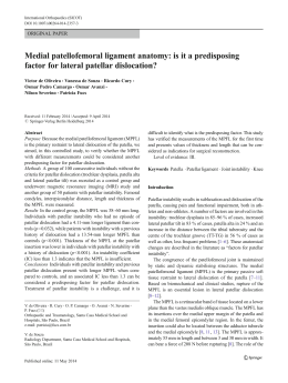
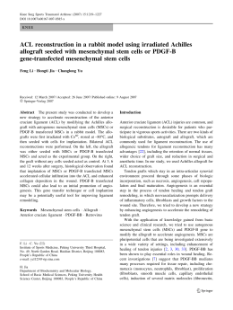
Knee Surg Sports Traumatol Arthrosc
DOI 10.1007/s00167-008-0527-9
SHOULDER
Sternoclavicular dislocation—reconstruction with semitendinosus
tendon autograft: a case report
Wagner Castropil Æ Lucas Busnardo Ramadan Æ
Alexandre Carneiro Bitar Æ Breno Schor Æ
Caio de Oliveira D’Elia
Received: 28 January 2008 / Accepted: 10 March 2008
Ó Springer-Verlag 2008
Abstract Traumatic sternoclavicular dislocation is a rare
injury corresponding to less than 5% of all injuries of the
scapular belt. It is preferentially treated through reduction
of the sternoclavicular joint, symptom relief, a brief period
of immobilization and rehabilitation, with the aim of
gaining strength and range of motion. In some patients,
however, this type of injury may progress with instability
and pain, thus causing discomfort and pain. On such
occasions, surgical treatment is chosen. The objective of
this study was to report the clinical case of a sports player
who progressed with chronic traumatic anterior instability
of the sternoclavicular joint and underwent reconstruction
using the ipsilateral semitendinosus tendon. This was a
16-year-old male patient who was a state-level judo player.
Following a fall during a fight, he presented pain, slight
deformity and edema in the right sternoclavicular joint,
and he underwent conservative treatment for 12 months,
without success. In the end, reconstruction of the
W. Castropil (&) L. B. Ramadan A. C. Bitar B. Schor C. de Oliveira D’Elia
Instituto Vita, Rua Mato Grosso, 306, 1st Floor, Higienópolis,
CEP: 01239-040 São Paulo, SP, Brazil
e-mail: [email protected]; [email protected]
L. B. Ramadan
e-mail: [email protected]
A. C. Bitar
e-mail: [email protected]
B. Schor
e-mail: [email protected]
C. de Oliveira D’Elia
e-mail: [email protected]
sternoclavicular joint was carried out using the ipsilateral
autologous semitendinosus, with resection of the intraarticular disc and suturing of the costoclavicular ligaments.
We have presented a case of dislocation of the sternoclavicular joint in a high-performance judo player who
underwent reconstruction using the semitendinosus, with
excellent functional results after 1 year of follow-up.
Keywords Orthopedics Trauma in sports players Sternoclavicular joint Autologous transplantation Treatment results
Introduction
Traumatic sternoclavicular dislocation is a rare injury
corresponding to less than 5% of all injuries of the scapular
belt [4, 8, 10]. It usually results from direct high-energy
trauma or indirect trauma following a fall. The culmination
is dislocation or subdislocation of the sternoclavicular
joint, or epiphyseal lesion of the medial clavicle in
immature skeletons [10, 11, 16].
Although rare, such injuries deserve rapid diagnosis and
effective treatment to avoid future complications. Anterior
dislocation is three to twenty times more common than
posterior dislocation [10, 12], and normally it presents few
complications.
Posterior dislocation, which is rare, may cause serious
complications due to compression of prime central structures by the medial clavicle. Respiratory discomfort,
lesions of the brachial plexus and arterial insufficiency are
some of the harmful consequences of this type of dislocation [3, 6, 7, 9].
Acute or chronic anterior dislocation of the sternoclavicular joint is mostly treated by means of symptom relief, a
123
Knee Surg Sports Traumatol Arthrosc
short period of immobilization and rehabilitation, with the
aim of gaining strength and range of motion. There are cases
in which reduction of the sternoclavicular joint becomes
necessary and, even if it remains unstable, the deformity is
accepted because of the low risk of sequelae [5].
Surgical treatment is chosen when this type of injury
progresses with recurrent instability and pain, thereby
causing great discomfort and incapacity [1, 5, 13]. A
variety of surgical techniques have been used to keep the
medial clavicle and the sternum together. Some examples
include intramedullary suture, medial resection of the
clavicle, use of a plate, reinforcement with the subclavian
tendon and reinforcement with the semitendinosus tendon
[1, 13].
The objective of this study was to demonstrate the
clinical case of a high-performance sports player who
progressed with chronic traumatic anterior instability of the
sternoclavicular joint and underwent reconstruction using
the ipsilateral semitendinosus tendon. What stimulated our
group to publish this report was that it was an unusual type
of case in our daily practice, with a precise surgical indication that was also unusual, and with a result from the
present case that was very good, in line with what we have
found in the literature.
Fig. 1 Computed tomography image at rest, showing stable sternoclavicular joint
Case report
This was a 16-year-old male patient who was a state-level
judo player. Following a fall during a fight, he presented
pain, slight deformity and edema in the right sternoclavicular joint. Following an initial evaluation, anterior
dislocation of the sternoclavicular joint was diagnosed.
It was initially decided to administer conservative
treatment consisting of immobilization for 10 days, followed by physiotherapy for rehabilitation. After 4 months
of muscle strengthening, the patient returned to his highperformance sport, while maintaining the symptomatology.
The rehabilitation process was continued, with muscle
strengthening and analgesia.
After 12 months, the symptoms presented by the patient
at a physical examination were pain, slight anterior
deformity of the sternoclavicular joint and muscle weakness. In training sessions, he was complaining of joint
instability.
Dynamic computed tomography was performed. This
showed that the sternoclavicular joint was stable at rest
(Fig. 1), but that dynamic maneuvers provoked anterior
dislocation (Fig. 2). Chronic degeneration of the intraarticular disc was also found. We decided to carry out
reconstruction surgery with reinforcement using the semitendinosus tendon, since this is a biological technique with
good functional results, according to the literature.
123
Fig. 2 Computed tomography image with dynamic maneuver,
showing anterior dislocation of the clavicle
Surgical technique
The patient underwent general anesthesia, positioned in
horizontal dorsal decubitus with the dorsum inclined at
around 30°. The surgical fields were laid out so as to
expose the right sternoclavicular joint, and a transversal
incision was made above the joint.
After accessing the joint, subperiosteal dissection was
performed laterally to medially, while protecting the deep
vascular structures. This was followed by debridement and
local scraping until bleeding, excision of the degenerated
intra-articular disc and partial resection of the medial
clavicle, which showed signals of degenerative cartilage
and bone spurs.
The graft removed was from the semitendinosus tendon
of the patient’s ipsilateral knee. The surgeon who did this
was the same surgeon operating on the sternoclavicular
joint, who was already qualified to carry out knee surgery.
Krakow-type sutures were performed at the extremities of
the graft, thus enabling good attachment to the sternoclavicular joint.
Knee Surg Sports Traumatol Arthrosc
Fig. 4 Elevation without pain
Fig. 3 Surgical technique—autologous graft from the semitendinosus through in a figure-of-eight
Holes were drilled 1.5 cm from the sternum and from
the medial clavicle, to pass the autologous graft from the
semitendinosus through in a figure-of-eight, and the
reduction was reinforced with nonabsorbable thread
(Ethibond 5) through the tendon (Fig. 3). After observing
that stability had been achieved, by means of an adduction
maneuver, we closed the remainder of the periosteum and
repaired the costoclavicular ligaments.
Postoperative period
During the postoperative period, the patient continued to be
immobilized using an arm sling for 3 weeks. After this
period, he was sent for physiotherapeutic treatment, starting with passive movement to gain range of motion.
The immobilization was withdrawn in the fifth week,
and the patient was allowed to start performing active
aerobic exercises with restriction on shoulder extension.
Thereafter, he progressed to strengthening exercises and
sports action training. In the fourth month, the patient
returned for assessment, and was found to present stability
of the sternoclavicular joint and absence of pain at the site
of the surgery, on physical examination. The patient
reported that he was not suffering any pain during training
sessions, and he was given clearance to return to practicing
judo.
After 1 year of evolution, the patient presented normal
range of motion, absence of pain, muscle strength without
deficit, as proven by an isokinetic test, both in the
shoulder and in the knee. An imaging examination
showed reduction of the sternoclavicular joint (Figs. 4, 5).
The patient went back to competing at the same level as
before the injury, and has even won a state competition
recently.
Fig. 5 Computed tomography image following surgery, showing the
sternoclavicular joint together
Discussion
The recommendations for treating anterior sternoclavicular
dislocation range from pain relief and immobilization to
closed reduction and surgical reconstruction [1, 5, 13, 17].
Few studies in the literature show objective data for precise
indications of surgery in cases of acute or chronic traumatic
anterior dislocation, or show which would be the best
method [1].
The objective of this study was to demonstrate the case
report on a sports player with the sequela of traumatic
anterior dislocation that progressed with pain and instability. In most cases, anterior dislocation receives
conservative treatment, with a good response from most
patients. Even, cases in which deformity continues, with
slight instability, do not end up having repercussions on
these patients’ ways of life. However, because our patient
was a high-performance judo player who constantly needed
to use adduction force and elevation of the arm, his chronic
instability was leading to pain and weakness, thereby
causing decreased performance.
123
Knee Surg Sports Traumatol Arthrosc
These cases of chronic traumatic anterior instability that
respond poorly to conservative treatment and give rise to
clinical repercussions are indicated for surgery [1, 2]. Our
patient underwent 12 months of conservative treatment
without success, which led us to indicate surgery.
Studies in the literature have shown good functional
results from the use of autologous grafts from the semitendinosus for reinforcing the sternoclavicular joint [1].
This is a biological reconstruction and therefore has fewer
complications than that with the use of plates or other
implants. Even though plates bring good results, they may
cause the complications that are inherent to all nonbiological implants, as well as needing to be removed later on [15].
A study on cadavers comparing reinforcement using the
semitendinosus tendon in a figure-of-eight, reinforcement
using the subclavian tendon and reconstruction of the
intramedullary ligament showed that the first of these
techniques is more resistant and confers greater biomechanical stability to the sternoclavicular joint than what the
other two do [14].
One criticism of the use of the autologous semitendinosus is the possible damage to the flexion strength of the
knee. However, this tendon is constantly used in ligament
reconstructions in the knee. Another option would be the
use of a graft from a tissue bank, for biological reconstruction without harming a donor area.
Rockwood and Wirth [13] showed that the continuing
presence of the costoclavicular ligaments is fundamental
for stability of the sternoclavicular joint. Therefore, their
reconstruction in open surgery has high priority, and this
was done in our technique.
In the present case, all our steps were based on data in
the literature, from the best time for indicating surgery and
the reconstruction technique to the care taken during the
operation, such as repair of the costoclavicular ligaments.
Today, our patient is satisfied, asymptomatic and pursuing
his competitive sports activities at a high level.
Conclusion
We have presented a case of dislocation of the sternoclavicular joint in a high-performance judo player who
123
underwent reconstruction using the semitendinosus, with
excellent functional results after 1 year of follow-up.
References
1. Bae DS, Kocher MS (2006) Chronic recurrent anterior sternoclavicular joint instability: results of surgical management.
J Pediatr Orthop 26:71–74
2. Bicos J, Nicholson GP (2003) Treatment and results of sternoclavicular joint injuries. Clin Sports Med 22:359–370
3. Borrero E (1987) Traumatic posterior displacement of the left
clavicular head causing chronic extrinsic compression of the
subclavian artery. Phys Sportsmed 15:87–89
4. Cave EF (1958) Fractures and other injuries. Year Book Medical
Publishers, Chicago
5. De Jong KP, Sukul DMKS (1990) Anterior sternoclavicular: a
longterm follow-up study. J Orthop Trauma 4:420–423
6. Gangahar DM, Flogaites T (1978) Retrosternal dislocation of the
clavicle producing thoracic outlet syndrome. J Trauma 18:369–
372
7. Homdahl HC (1953) A case of posterior sternoclavicular dislocation. Acta Orthop Scand 23:218–222
8. Kocher MS, Waters PM, Micheli LJ (2000) Upper extremity
injuries in the paediatric athlete. Sports Med 30:117–135
9. Louw JA, Louw JA (1987) Posterior dislocation of the sternoclavicular joint associated with major spinal injury: a case report.
S Afr Med J 71:791–792
10. Nettles JL, Linscheid RL (1968) Sternoclavicular dislocations.
J Trauma 8:158–164
11. Omer GE (1967) Osteotomy of the clavicle in surgical reduction
of anterior sternoclavicular dislocation. J Trauma 7:584–590
12. Rockwood CA Jr, Green DP, Bucholz RW (1991) Fractures in
adults. JB Lippincott, Philadelphia, pp 1253–1307
13. Rockwood CA, Wirth MA (1996) Injuries to the sternoclavicular
joint. In: Rockwood CA, Green DP, Bucholz RW (eds). Rockwood and Green’s fractures in adults, 4th edn. Lippincott-Raven,
Philadelphia, pp 1415–1471
14. Spencer EE, Kuhn JE (2004) Biomechanical analysis of reconstructions for sternoclavicular joint instability. J Bone Joint Surg
Am 86:98–105
15. Thacker MM, Patankar JV, Goregaonkar AB (2006) A safe
technique for sternoclavicular stabilization. Am J Orthop 35:64–
66
16. Waskowitz WJ (1961) Disruption of the sternoclavicular joint: an
analysis and review. Am J Orthop 3:176–179
17. Wirth MA, Rockwood CA (1996) Acute and chronic traumatic
injuries of the sternoclavicular joint. J Am Acad Orthop Surg
4:268–278
Baixar