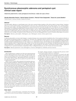
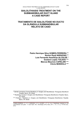
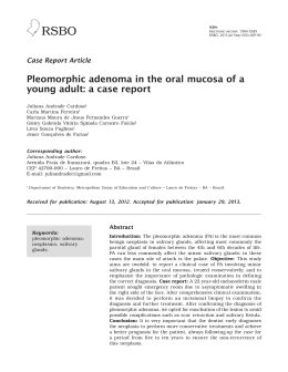
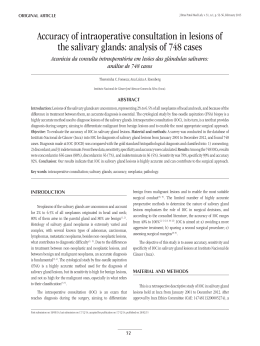
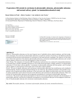
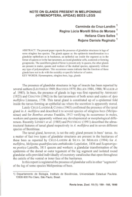
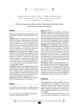
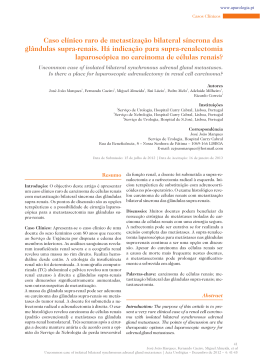
The Journal of Craniofacial Surgery
Brief Clinical Studies
10. Leverstein H, Tiwari RM, Snow GB, et al. The surgical management
of recurrent or residual pleomorphic adenomas of the parotid gland.
Analysis and results in 40 patients. Eur Arch Otorhinolaryngol
1997;254:313Y317
11. Witt RL. The significance of the margin in parotid surgery for
pleomorphic adenoma. Laryngoscope 2002;112:2141Y2154
12. Lewis JE, Olsen KD, Sebo TJ. Carcinoma expleomorphic adenoma:
pathologic analysis of 73 cases. Hum Pathol 2001;32:596Y604
13. DeRoche TC, Hoschar AP, Hunt JL. Immunohistochemical evaluation
of androgen receptor, HER-2/neu, and p53 in benign pleomorphic
adenomas. Arch Pathol Lab Med 2008;132:1907Y1911
14. Hamada T, Matsukita S, Goto M, et al. Mucin expression in pleomorphic
adenoma of salivary gland: a potential role for MUC1 as a marker
to predict recurrence. J Clin Pathol 2004;57:813Y821
15. Carrau RL, Myers EN, Johnson JT. Management of tumors arising in
the parapharyngeal space. Laryngoscope 1990;100:583Y589
16. Buchman C, Stringer SP, Mendenhall WM, et al. Pleomorphic adenoma:
effect of tumor spill and inadequate resection on tumor recurrence.
Laryngoscope 1994;104:1231Y1234
17. Natvig K, SLberg R. Relationship of intraoperative rupture of
pleomorphic adenomas to recurrence: an 11Y25 year follow-up study.
Head Neck 1994;16:213Y217
18. Henriksson G, Westrin KM, Carlsöö B, et al. Recurrent primary
pleomorphic adenomas of salivary gland origin: intrasurgical rupture,
histopathologic features, and pseudopodia. Cancer 1998;82:617Y620
19. Shahab R, Heliwell T, Jones AS. How we do it: a series of 114 primary
pharyngeal space neoplasms. Clin Otolaryngol 2005;30:364Y367
20. Douglas JG, Einck J, Austin-Seymour M, et al. Neutron radiotherapy
for recurrent pleomorphic adenomas of major salivary glands.
Head Neck 2001;23:1037Y1042
Sialolithiasis of the
Submandibular Gland
Ellen Cristina Gaetti Jardim, DDS,* Daniela Ponzoni, MD,*
Paulo Sérgio Perri de Carvalho, MD,*Þ
Marcos Rogério Demétrio, DDS,
Alessandra Marcondes Aranega, MD*
Abstract: Sialolithiasis of the salivary gland is a benign pathology
that occurs most frequently in the submandibular gland because
of its anatomic features. Depending on the sialolith size and calcification degree, it can be visible in radiographic examinations.
Commonly, patients may experience pain and/or edema, when the
ducts are obstructed. The authors report the case of sialolithiasis
of the submandibular gland in a 42-year-old, female, white-skinned
patient, noticed during routine dental examination. Following diagnosis confirmed by clinical and radiographic examinations, the
From the *Department of Surgery and Integrated Clinics, School of Dentistry
of Araçatuba, Univ Estadual PaulistaYUNESP; and †Stomatology Department, School of Dentistry of Bauru, University of São PauloYUSP,
São Paulo, Brazil.
Received July 29, 2010.
Accepted for publication September 25, 2010.
Address correspondence and reprint requests to Ellen Cristina Gaetti Jardim,
DDS, Departamento de Cirurgia e Clinica Integrada, Faculdade de
Odontologia de AracatubaYUNESP, Rua José Bonifácio, 1193 Araçatuba,
São Paulo, Brasil, CEP 16015-050; E-mail: [email protected]
The authors report no conflicts of interest.
Copyright * 2011 by Mutaz B. Habal, MD
ISSN: 1049-2275
DOI: 10.1097/SCS.0b013e3182108f4f
1128
& Volume 22, Number 3, May 2011
treatment plan consisted of surgery for removal of the calcified
mass. The prognosis is often good, and generally there is no
recurrence.
Key Words: Sialolithiasis, submandibular gland, oral surgery
S
ialolithiasis is the presence of stone or calculi in the duct of major
salivary glands. Although their pathogenesis remains unknown,
sialoliths are constituted by the deposition of calcium-rich salts
around a central nidus, which may consist of desquamated epithelial
cells, foreign bodies, or bacteria and their decomposition products.1
Its development is not related to any systemic disorder in calcium or
phosphate metabolism.
Sialolithiasis is the most common disease of the salivary glands.
It is estimated that its incidence is 12 per 1000 adult individuals,2,3
occurring more frequently in white women aged 41 to 60 years.4
They may occur in the interior of the gland, although they are most
commonly found in the main duct of the submandibular gland, at
times in the parotid gland, and rarely in the sublingual gland and
minor salivary glands.
The submandibular gland system is more susceptible to sialolithiasis because of anatomic and physiological features of the gland
itself, where saliva is more alkaline and presents higher concentration of calcium and phosphate in the form of carbonates, besides
higher amounts of mucin, providing higher viscosity and favoring
adherence around foreign bodies.5 Siddiqui3 states that the submandibular gland duct is the most frequent site to form salivary
calculi where the main duct contours the distal border of the
mylohyoid muscle.
Capaccio et al6 relate the occurrence of salivary calculi in HIVpositive patients. These patients show significant reduction in the
salivary flow of the parotid, submandibular, and sublingual glands as
well as alterations in the saliva chemical composition, such as increased sodium, chloride, and immunoglobulin, among others.
Patients with sialolithiasis involving the duct of a major salivary
gland may complain of moderate to intense pain, particularly at
mealtimes, due to the increased salivary flow rate, associated with
enlargement of the gland. The duct occlusion blocks or decreases the
free flow, leading to saliva accumulation in the gland, which under
pressure causes pain and swelling. In certain situations, it may remain asymptomatic, and its only evidence is the presence of a palpable mineralized mass in the duct or gland.1
Depending on the calcification degree of the calculi, it can be
visible in conventional radiographs, being radiopaque and observable at any point of the duct or inside the gland itself. Calculi in the
terminal portion of the submandibular main duct are better visualized through occlusal radiography. In panoramic or periapical
radiographs, the calcification image may appear superimposed on
the mandible; therefore, it may be mistaken by an intrabone lesion.7
Periapical radiography can detect sialoliths in salivary glands with
an x-ray film positioned in the vestibule, similar to the occlusal
radiograph, which is reported in literature as one of the complementary examinations for the diagnosis of lesions in the submandibular region.8,9 During clinical inspection of the lower lip, the
mucosa can be reddish and edematous, suggesting the existence of a
sialolith.10
On the other hand, because of the calcification degree of certain
lesions, not all calculi can be visualized in conventional radiographic
examinations; in this case, other imaging examinations may be
necessary such as sialography, ultrasound, computed tomographic
scan, and magnetic resonance imaging.11Y13
In the presence of sialolithiasis, the sialography examination
shows interruption in the contrast image or image straightening.
* 2011 Mutaz B. Habal, MD
Copyright © 2011 Mutaz B. Habal, MD. Unauthorized reproduction of this article is prohibited.
The Journal of Craniofacial Surgery
& Volume 22, Number 3, May 2011
FIGURE 1. Initial intraoral examination. Volume increase on
the floor of the mouth.
FIGURE 2. Occlusal radiograph showing the lesion (arrow).
FIGURE 3. Linear incision.
Brief Clinical Studies
FIGURE 5. Lesion removal.
Sialography is indicated when it is necessary to differentiate salivary
stones from vascular calcified formations (phleboliths) or from
calcified lymphatic nodes. However, it is contraindicated in cases of
infection or allergy to iodinated contrast material.14 Sialography is
the retrograde injection of radiopaque material in the duct system of
a salivary gland to investigate its radiographic distribution.11
Yu et al15 reported microendoscopy as a noninvasive method for
the diagnosis and treatment (lithotripsy) of the diseases of major
salivary glands as described by Baek and Jeong.12
In the macroscopic examination, sialoliths are seen as hard
masses that may be round, oval, or cylindrical. They are characteristically yellow, although they may be white or yellow-brownish.
The calcified masses present concentric laminations that may
encircle a nidus of irregular organic remains. If the associated
duct is also removed, there will commonly be squamous metaplastic, oncocytic, or mucous cells. Periductal inflammation is also
evident.16
Sialolithiasis treatment depends on the localization of the salivary calculus; for those closer to the ostium, duct catheterization and
dilatation facilitate and allow retrieval of the sialolith. For those
located in the anterior half of the duct, surgical intervention is the
best choice. Finally, the ones located in the posterior region of the
duct or within the gland may require total gland removal. Despite
the latter having a favorable prognosis; it may cause salivary fistula,
which is of difficult resolution.1,17,18
Another treatment option for sialolithiasis is the gland handling
by liquid ingestion and saliva stimulation, with the purpose of displacing the salivary calculus to be spontaneously expelled. Nevertheless, when this method fails, its removal must be done through
surgical methods.
Sialoliths of minor salivary glands are better treated through
surgical removal, including the associated gland.7,19
CLINICAL REPORT
A 42-year-old female patient presented to routine dental treatment
at a private dental office and was referred to the Department of
FIGURE 4. Divulsion of the surgical area.
FIGURE 6. The sialolith.
* 2011 Mutaz B. Habal, MD
Copyright © 2011 Mutaz B. Habal, MD. Unauthorized reproduction of this article is prohibited.
1129
The Journal of Craniofacial Surgery
Brief Clinical Studies
& Volume 22, Number 3, May 2011
with the great incidence of this lesion.22 The frequent incidence of
sialoliths in the submandibular gland can be corroborated by physicalchemical characteristics of the saliva produced by this gland.
Because of the sialolith size as well as its localization in the gland
duct superficially to the mucosa, the first treatment option was
surgery, with favorable prognosis and low recurrence rate. The conventional treatment would be impractical in this case, because retrieving the sialolith through the duct orifice by manipulation of the
gland would be impossible. In such cases, the retrieval of the sialolith
alone is sufficient. In the present case, there was no need to remove
the gland, because there was no inflammation in its interior.
FIGURE 7. Suture on the area.
Surgery at the School of Dentistry of Araçatuba (UNESP). During
clinical intraoral examination, asymmetry on the right side of the
floor of the mouth was observed, caused by increased hard volume
that moved with palpation (Fig. 1). This increased volume was
asymptomatic and unnoticed by the patient. The intraoral examination revealed no volume increase or pain in the major salivary
gland region.
Panoramic and occlusal radiographs demonstrated an oval radiopaque area in the floor of the mouth, close to the premolars and
lower-first-molar region (Fig. 2).
Diagnosis of calculus in the submandibular gland duct on the
right side of the mouth was made. Because of the size and localization of the calculus, surgical removal under local anesthesia was
the treatment option. A linear incision over the most prominent
area of the mucosa was done, followed by dissection and removal of
the calcified mass (Figs. 3Y5). The calculus was 20 mm long and
15 mm wide (Fig. 6). Absorbable interrupted sutures with polyglactin 910 were performed (Fig. 7). The patient underwent postoperative follow-up, and the functional integrity of the gland was
confirmed (Fig. 8).
DISCUSSION
The signs presented by the patient (volume increase, absence of
pain, and radiopacity to radiographic examination) are in agreement
with the clinical characteristics observed in the literature.3,10,11,20
Most sialoliths are diagnosed because of patients’ complaints of
pain or swelling of the involved gland. In the present case report, the
patient was asymptomatic, and the volume produced by the calcified
mass in the floor of the mouth was until then unnoticed by the
patient. Probably, the calculus, although very large, was not completely obstructing the salivary flow of that duct.1,5
Diagnosis was made from the radiopaque image seen in the
occlusal radiograph, which allows adequate visualization of calcified masses in the floor of the mouth.8,9,21 The fact that the sialolith
was located in the duct of the submandibular gland is in agreement
FIGURE 8. Six months after surgery.
1130
CONCLUSIONS
Diagnosis of sialolithiasis is made both clinically and radiographically. The earlier the diagnosis, the more conventional is the treatment, with lower possibilities of duct obstruction and inflammatory
alterations in the interior of the glands.
REFERENCES
1. Ord RA, Pazoki AE. Salivary gland disorders. In: Miloro M, ed.
Peterson’s Principles of Oral and Maxillofacial Surgery. 2nd ed. Vol 1.
London: BC Decker Inc, 2004:674
2. Leung AK, Choi MC, Wagner GA. Multiple sialoliths and a sialolith
of unusual size in the submandibular duct: a case report. Oral Surg
Oral Med Oral Pathol Oral Radiol Endod 1999;87:331Y333
3. Siddiqui SJ. Sialolithiasis: an unusually large submandibular salivary
stone. Br Dent J 2002;193:89Y91
4. Ngu RK, Brown JE, Whaites EJ, et al. Salivary duct strictures:
nature and incidence in benign salivary obstruction.
Dentomaxillofac Radiol 2007;36:63Y67
5. Sherman JA, Mcgurk M. Lack of correlation between water hardness
and salivary calculi in England. Br J Oral Maxillofac Surg
2000;38:50Y53
6. Capaccio P, Monforte A, Moroni M, et al. Salivary stone lithotripsy
in the HIV patient. Oral Surg Oral Med Oral Pathol Oral Radiol Endod
2002;93:525Y527
7. Alkurt MT, Peker I. Unusually large submandibular sialoliths: report
of two cases. Eur J Dent 2009;3:135Y139
8. Ledesma-Montes C, Garcés-Ortı́z M, Salcido-Garcı́a JF, et al. Giant
sialolith: case report and review of the literature. J Oral Maxillofac
Surg 2007;65:128Y130
9. Rai M, Burman R. Giant submandibular sialolith of remarkable size
in the comma area of Wharton’s duct: a case report. J Oral Maxillofac
Surg 2009;67:1329Y1332
10. Kasaboglu O, Er N, Tumer C, et al. Micromorphology of sialoliths
in submandibular salivary gland: a scanning electron microscope
and x-ray diffraction analysis. J Oral Maxillofac Surg
2004;62:1253Y1258
11. Uluc ME, Vidinli BD, Erdogan N, et al. Giant cystic dilatation that
includes multiple sialolithiasis of submandibular gland.
Otolaryngol Head Neck Surg 2006;134:533Y534
12. Baek CH, Jeong HS. Endoscope-assisted submandibular
sialadenectomy: a new minimally invasive approach to the
submandibular gland. Am J Otolaryngol 2006;27:306Y309
13. Park JS, Sohn JH, Kim JK. Factors influencing intraoral removal of
submandibular calculi. Otolaryngol Head Neck Surg 2006;135:704Y709
14. Teymoortash A, Buck P, Jepsen H, et al. Sialolith crystals localized
intraglandularly and in the Wharton’s duct of the human submandibular
gland: an x-ray diffraction analysis. Arch Oral Biol 2003;48:233Y236
15. Yu C, Yang C, Zheng L, et al. Endoscopic observation and strategic
management of obstructive submandibular sialadenitis. J Oral
Maxillofac Surg 2010;68:1770Y1775
16. Mimura M, Tanaka N, Ichinose S, et al. Possible etiology of calculi
formation in salivary glands: biophysical analysis of calculus.
Med Mol Morphol 2005;38:189Y195
* 2011 Mutaz B. Habal, MD
Copyright © 2011 Mutaz B. Habal, MD. Unauthorized reproduction of this article is prohibited.
The Journal of Craniofacial Surgery
& Volume 22, Number 3, May 2011
Brief Clinical Studies
17. Ziegler CM, Steveling H, Seubert M, et al. Endoscopy: a minimally
invasive procedure for diagnosis and treatment of diseases of the salivary
glands. Six years of practical experience. Br J Oral Maxillofac Surg
2004;42:1Y7
18. Austin T, Davis J, Chan T. Sialolithiasis of submandibular gland.
J Emerg Med 2004;26:221Y223
19. McGurk M, Escudier MP, Brown JE. Modern management of
salivary calculi. Br J Surg 2005;92:107Y112
20. Torroni AA, Mustazza MC, Bartoli DD, et al. Transcervical
submandibular sialoadenectomy. J Craniofac Surg 2007;18:613Y621
21. Bodner L. Giant salivary gland calculi: diagnostic imaging and surgical
management. Oral Surg Oral Med Oral Pathol Oral Radiol Endod
2002;94:320Y323
22. Makdissi J, Escudier MP, Brown JE, et al. Glandular function after
intraoral removal of salivary calculi from the hilum of submandibular
gland. Br J Oral Maxillofac Surg 2005;42:538Y541
Pleomorphic Adenoma of
the Palate
Mehmet Ali Erdem, DDS, PhD,*
Abdulkadir Burak Çankaya, DDS, PhD,* Gülzah Güven, DDS,*
Vakur Olgaç, DDS,Þ Çetin Kasapoğlu, DDS*
Abstract: Pleomorphic adenoma is the most common mixed benign
tumor of major salivary glands. Approximately 80% of these tumors
arise in the parotid gland, whereas 7% arise in the minor salivary
glands. The most common sites for minor salivary gland where
pleomorphic adenoma arises are the palates followed by lips and
cheek. We report a palate mass in a 46-year-old male patient. The
initial cytologic diagnosis by fine-needle aspiration biopsy was
pleomorphic adenoma. This report describes a case of pleomorphic
adenoma regarding all distinctive diagnoses with the review of the
literature.
Key Words: Pleomorphic adenoma, palate, fine-needle aspiration
biopsy
P
leomorphic adenoma is the most common mixed benign tumor
of the major salivary glands, especially the parotid gland. Approximately 80% of these tumors arise in the parotid gland, whereas
7% arise in the minor salivary glands.1Y3 Palate is the most commonly affected site in the oral cavity. Other intraoral affected sites
include the upper lip, buccal mucosa, tongue, and gingiva. Pleomorphic adenoma is mostly seen in women and is most prevalent in
the fourth through sixth decades of life. It usually appears as a
solitary, painless mass on oral mucosa.4,5
The term pleomorphic shows the diversity of the histology of the
tumor. The essential components are the capsule, epithelial and
myoepithelial cells, and mesenchymal or stromal elements. The
From the Departments of *Oral Surgery, Faculty of Dentistry, and †Department
of Pathology, Institute of Oncology, Istanbul University, Istanbul, Turkey.
Received July 30, 2010.
Accepted for publication September 25, 2010.
Address correspondence and reprint requests to Gülzah Güven, DDS,
Department of Oral Surgery, Faculty of Dentistry, Istanbul University,
Istanbul, Turkey; E-mail: [email protected]
The authors report no conflicts of interest.
Copyright * 2011 by Mutaz B. Habal, MD
ISSN: 1049-2275
DOI: 10.1097/SCS.0b013e3182108f8b
FIGURE 1. Clinical examination revealed a firm, mobile
mass at the right side of the hard palate.
capsule varies in thickness and presence. Cells of epithelial origin
give rise to ductal structures and are intermixed with mesenchymal
component that is mucoid/myxoid, cartilaginous, or hyalinized.4,6
The treatment of pleomorphic adenoma is surgical excision.
Enucleation of pleomorphic adenoma is not suitable because of the
tumor relapse, which is due to incomplete surgical resection of the
lesion.7Y9
We report a case of pleomorphic adenoma in the palate that was
excised under local anesthesia. The relevant studies are discussed.
CLINICAL REPORT
A 46-year-old man with a painless mass in the hard palate was
referred to the Department of Oral Surgery of Istanbul University
Faculty of Dentistry. The patient had complaints of disturbance of
his speech due to the mass. Clinical examination revealed a firm,
mobile mass at the right side of the hard palate. The oral mucosa
covering the lesion was intact (Fig. 1). Examination of panoramic
radiography showed no abnormality. A computed tomography scan
revealed a 2 2-cm, round soft-tissue mass that was well delineated
between the surrounding tissues (Fig. 2). His medical history was
noncontributory. Fine-needle aspiration cytology was performed.
Evaluation of the fine-needle aspiration biopsy (FNAB) showed
spindle-shaped or plasma cytoid morphology epithelioid myoepithelial cells in the chondromyxoid stroma (Figs. 3 and 4).
FIGURE 2. A computed tomography scan revealed a
2 2-cm, round soft-tissue mass that was well delineated
between the surrounding tissues.
* 2011 Mutaz B. Habal, MD
Copyright © 2011 Mutaz B. Habal, MD. Unauthorized reproduction of this article is prohibited.
1131
Baixar