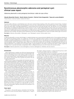
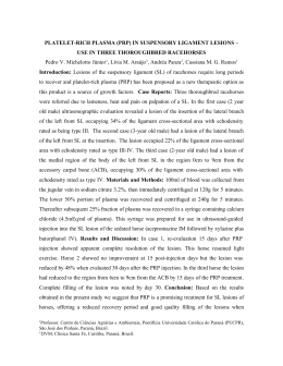
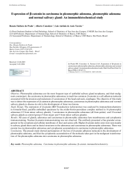
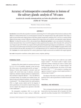
ISSN:
Electronic version: 1984-5685
RSBO. 2013 Jul-Sep;10(3):289-94
Case Report Article
Pleomorphic adenoma in the oral mucosa of a
young adult: a case report
Juliana Andrade Cardoso1
Carla Martins Ferreira1
Mariana Moura de Jesus Fernandes Guerra1
Gleicy Gabriela Vitória Spinola Carneiro Falcão1
Lívia Souza Pugliese1
Jener Gonçalves de Farias1
Corresponding author:
Juliana Andrade Cardoso
Avenida Praia de Itamaracá, quadra B3, lote 24 – Vilas do Atlântico
CEP 42700-000 – Lauro de Freitas – BA – Brasil
E-mail: [email protected]
1
Department of Dentistry, Metropolitan Union of Education and Culture – Lauro de Freitas – BA – Brazil.
Received for publication: August 13, 2012. Accepted for publication: January 29, 2013.
Keywords:
pleomorphic adenoma;
neoplasms; salivary
glands.
Abstract
Introduction: The pleomorphic adenoma (PA) is the most common
benign neoplasia in salivary glands, affecting most commonly the
parotid gland of females between the 4th and 6th decades of life.
PA can less commonly affect the minor salivary glands; in these
cases the main site of attack is the palate. Objective: This study
aims are twofold: to report a clinical case of PA involving minor
salivary glands in the oral mucosa, treated conservatively; and to
emphasize the importance of pathologic examination in defining
the correct diagnosis. Case report: A 22 year-old melanoderm male
patient sought emergency room due to asymptomatic swelling in
the right side of the face. After comprehensive clinical examination,
it was decided to perform an incisional biopsy to confirm the
diagnosis and further treatment. After confirming the diagnosis of
pleomorphic adenoma, we opted for enucleation of the lesion to avoid
possible complications such as scar retraction and salivary fistula.
Conclusion: It is very important that the dentist early diagnoses
the neoplasia to perform more conservative treatments and achieve
a better prognosis for the patient, always following-up the case for
a period from five to ten years to ensure the non-recurrence of
this neoplasia.
290 – RSBO. 2013 Jul-Sep;10(3):289-94
Cardoso� et al. – ��������������������������������
Pleomorphic adenoma in the oral ����������
mucosa of ��
a ������
young ��������������������
adult: a case report
Introduction
The pleomorphic adenoma (PA) is the most
common tumor affection the salivary glands [7, 8],
accounting for 33.2% to 89.9% of the cases, followed
by carcinomas. [7]. The parotid gland is the most
affected [8, 11], followed by the submandibular
and minor salivary glands, which have been lesser
affected [8]. Considering the minor salivary glands,
the palate is the place mostly affected [11].
Clinically, PA of minor salivary glands is a
single, solid, smooth, well-delimited structure.
This tumor is composed of a single mass that is
encapsulated and consequently shows mobility,
except when the lesion occurs in the palate.
Generally, PA is asymptomatic and has a slow
development [2, 5, 6, 10].
Some lesions of the salivary glands exhibit
similar clinical, histopathological and imaging
aspects, which can compose a differential diagnosis
of PA [6]. PA may affect the hard palate, which
has been the site of most of the malign neoplasias
such as adenocarcinoma; and it may affect the
parapharyngeal space, a common site for lipoma,
therefore limiting and making difficult the diagnosis
by clinical aspects. Thus, to obtain a correct
differential diagnosis, complementary examinations
are required [9, 11].
The ima g ing exa minat ions, such as
ultrasonography, computed tomography and magnetic
resonance, help in the diagnosis of the lesions because
they established their origin, location and limits
[9]. Notwithstanding, microscopic examinations are
essential to define the diagnosis because they show
the histopathological aspects of the lesions differing
the sound cells from the tumoral ones [2, 9, 12].
PA early diagnosis enables a more conservative
treatment by preventing the bone and tissue
infiltration of the lesion l [5]. The election treatment is
the surgical excision by resection with a safe margin
of 3 to 5 mm [10], because the lesion may relapse
frequently according to its location and surgical
technique applied. Normally, the post-operative
period is uneventful with complete healing by 15
days. The patient must be followed-up for five years.
The appropriate treatment is of extreme importance
to reach a good prognosis, therefore avoiding the
lesion relapse. A good surgical technique shows a
healing rate of 95% [6].
The aim of this study was to report a clinical
case of pleomorphic adenoma affecting the minor
salivary gland and to discuss its characteristics,
emphasizing the important role of the dentist in
the correct and early diagnosis of this lesion,
resulting in a more conservative treatment and
better prognosis for the patient.
Case ������
report
A 22-year-old melanoderm, male patient was referred to the clinic of Specialization in Stomatology
of the Metropolitan Union of Education and Culture with chief complaint of a swelling in the right side
of the face. The patient was accompanied by his parents.
At extraoral physical examination, a facial asymmetry in the right side of the face was observed
so that the nasolabial fold disappeared (figure 1).
Figure 1 – A) Extraoral clinical aspect; B) Skull caudal view demonstrating the volume increasing at the right side
291 – RSBO. 2013 Jul-Sep;10(3):289-94
Cardoso� et al. – ��������������������������������
Pleomorphic adenoma in the oral mucosa
����������
of ��
a young
������ adult:
��������������������
a case report
During intraoral physical examination, a tumor lesion could be seen in the buccal mucosa covered
by sound tissue, well delimited, with a firm consistency and mobility to palpation. The patient signed
a free and clarified consent form. An incisional biopsy under local anesthesia was chosen to define the
lesion histopathologically. Firstly, an aspiration puncture was executed aiming to evaluate the lesion
type: liquid or solid. No liquids were aspired, so that according to the lesion location and solid aspect
the clinical diagnosis proposed was of lipoma (figure 2).
Figure 2 – A) Intraoral clinical aspect; B) Aspiration puncture negative for liquid; C) Incision and divulsion to access
the lesion; D) Fragments of the lesion obtained through incisional biopsy
However, the histopathological examination concluded that lesion was a pleomorphic adenoma.
Then, the patient was admitted in the Service of Stomatology and Buccomaxillofacial Surgery of
the Holly House Hospital of São Felix, Bahia, Brazil to be submitted to the lesion enucleation under
general anesthesia (figure 3).
Figure 3 – A) Incision; B) Divulsion; C) Enucleation of the lesion; D) Surgical sample of 10 x 4 x 7 cm
292 – RSBO. 2013 Jul-Sep;10(3):289-94
Cardoso� et al. – ��������������������������������
Pleomorphic adenoma in the oral mucosa
����������
of ��
a young
������ adult:
��������������������
a case report
The sample was referred to the service of pathological anatomy of the Metropolitan Union of
Education and Culture which confirmed the diagnosis of pleomorphic adenoma (figure 4).
Figure 4 – A) Lesion encapsulated (arrow), presenting architectural heterogeneity ; B) Proliferation of the ductal
epithelial cells ( white asterisks), involved by myoepithelial cells; C) Presence of abundant myxoid stroma; D)
lipomatous differentiation (empty triangles)
Images acquired from a digital lamina by using ImageScope software version 11.0.2.725 – Aperio Tecnologies. Hematoxylineosin staining
After fifteen days, the post-operative period was uneventful and the sutures were removed, but there
was still a little face asymmetry due to the residual swelling (figure 5).
Figure 5 – Clinical aspect after 15 days
293 – RSBO. 2013 Jul-Sep;10(3):289-94
Cardoso� et al. – ��������������������������������
Pleomorphic adenoma in the oral mucosa
����������
of ��
a young
������ adult:
��������������������
a case report
After 8 post-surgery months, the patient exhibited no signs of lesion relapse and good face symmetry
(figure 6). The patient is still undergoing 1-year follow-up in the clinic of Specialization in Stomatology
for at least 5 years.
Figure 6 – A) Extraoral clinical aspect after 8 months; B) Skull caudal view after 8 months; C) Intraoral clinical aspect
after 8 months
Discussion
According to previous studies, PA occurs
most frequently in the parotid gland, followed by
submandibular glands and less frequently the minor
salivary glands [8, 11]. The area main affected by
PA of minor salivary glands is the hard and soft
palate, corresponding from 42.2% to 55% of the
cases, followed by the lips with 14% to 18.5% of the
cases and oral mucosa in 15% of the cases [1, 2, 7,
9]. PA predominantly occurs in Caucasian females
between the 4th and 6th decades of life aging from
43 to 46 years in average [1, 2, 4, 7, 9, 13]. This
aspect differs from those of this present case report
in which PA occurred in a melanoderm male patient
at the third decade of life. During the intraoral
physical examination, clinical aspects similar to
those reported in the literature were observed, such
as single submucosal tumor lesion of firm consistency
and with mobility to palpation [2, 5, 6, 10].
The histopathological examination confirmed
the diagnosis of PA because of the abundant and
heterogeneous presence of myoepithelial cells in
tubular arrangement, glandular epithelium with
ductiform structures, and with transformation of the
stroma by exhibiting a myxoid and adipose tissue,
which is in agreement with the aspects previous
reported in the literature [2, 9, 12].
Because the buccal mucosa is not a very
common site of PA appearance [3], and the patient
was a young melanoderm male adult, facts that
disagree with the literature [1, 2, 4, 7, 9, 13], the
first diagnosis option suggested in this present
case report was of lipoma.
By reviewing the literature, the treatment
recommended is the surgical excision by resection
with a safe margin from 3 to 5 mm [5, 6, 10].
Notwithstanding, in this case report, an enucleation
was chosen because the lesion showed great
dimensions, therefore avoiding some complications
as scar retraction and salivary fistula. Thus, it
is necessary to perform a careful following-up of
at least five years because of the probability of
relapse.
Conclusion
Because PA is a neoplasia with great diversity
of cells in constant process of transformation, it
demands a correct surgical procedure with safe
margin to assure that the surgical site is free of
tumoral cells.
It is worth emphasizing the important role of
the dentist in executing the early diagnosis of the
neoplasia to conduct more conservative treatments
and obtain a better prognosis for the patient,
always following-up the case for a period from
five to ten years to ensure the non-recurrence of
this neoplasia.
References
1. Bettio A, Salgado G, Azevedo-Alanis LR, Machado
MAN, Grégio AMT, Lima AAS. Prevalência das lesões
de glândulas salivares em laudos histopatológicos
do Laboratório de Patologia Experimental da
PUCPR. RSBO. 2009 Sep;6(3):231-6.
294 – RSBO. 2013 Jul-Sep;10(3):289-94
Cardoso� et al. – ��������������������������������
Pleomorphic adenoma in the oral mucosa
����������
of ��
a young
������ adult:
��������������������
a case report
2. Boros FL, Júnior JB, Boros FL, Boros LH, Silva
PA. Adenoma pleomórfico de glândula salivar
menor do palato. Odontologia Clín-Científ. 2004
Jan-Apr;3(1):67-72.
3. Dalati T, Hussein MR. Juvenile pleomorphic
adenoma of the cheek: a case report and review
of the literature. Diagn Pathol [serial online] 2009
Sep;4(32):[5 screens] [cited 2012 Oct 19]. Available
from: URL:http://www.diagnosticpathology.org/
content/4/1/32.
4. Jorge J, Pires FR, Alves FA, Perez DE, Kowalski
LP, Lopes MA et al. Juvenile intraoral pleomorphic
adenoma: report of five cases and review of
the literature. Int
���������������������������������
J Oral Maxillofac Surg. 2002
Jun;31(3):273-5.
5. Lacerda SA, Socolowski F, Rosa AL, Ferraz
MP, Brentegani LG. Adenoma pleomórfico intraósseo no maxilar: relato de caso clínico. Rev Bras
Estomatol. 2005;2(4):5-8.
6. Lawall MA, Simonato LE, Ribeiro ACP,
Crivelini MM, Moraes NP. Adenoma pleomórfico:
relato de caso clínico. Rev Odontol. 2007 SepDec;19(3):336-40.
7. Loiola RS, Matos FR, Nonaka CFW, Lopes FF,
Cruz MCFN. Perfil epidemiológico das neoplasias de
glândulas salivares diagnosticadas em São Luís-MA.
J Bras Patol Med Lab. 2009 Oct;45(5):413-20.
8. Rao PK, Shetty SR, Hegde D. Ectopic pleomorphic
adenoma. N Am J Med Sci. 2012 Apr;4(4):190-2.
9. Ribeiro-Rotta RF, Cruz ML, Paiva RR, Mendonça
EF, Spini TH, Mendonça AR. O papel da ressonância
magnética no diagnóstico do adenoma pleomórfico:
revisão da literatura e relato de casos. Rev Bras
Otorrinolaringol. 2003 Sep-Oct;69(5):699-707.
10. Santiago LM, Torres BCA, Andrade MC, Silva
UH, Santana AF, Santos EV. Adenoma pleomórfico
em lábio superior. Odontologia Clín-Científ. 2005
Jan–Apr;4(1):63-70.
11. Sharma Y, Maria A, Chhabria A. Pleomorphic
adenoma of the palate. Natl J Maxillofac Surg. 2011
Jul;2(2):169-71.
12. Teixeira JM, Cornélio SS, Grillo JPF,
Pontes JRM, Ramos MEB, Israel MS. Adenoma
pleomórfico en labio superior: relato de caso. �����
Acta
Odontol Venez. 2007 Sep;45(3):440-2.
13. Wang D, Li Y, He H, Liu L, Wu L, He Z.
Intraoral minor salivary gland tumors in a Chinese
population: a retrospective study on 737 cases.
Oral Surg Oral Med Oral Pathol Oral Radiol Endod.
2007 Jul;104(1):94-100.
Baixar