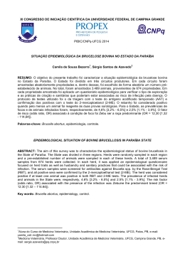
ISSN 0004-2749
versão impressa
Arquivos Brasileiros
PUBLICAÇÃO OFICIAL DO CONSELHO BRASILEIRO DE OFTALMOLOGIA
MAIO/JUNHO 2011
d e
74 03
Surgical weakening of the
oblique muscles
Pediatric cataract in
developing country
Microdevice for aqueous
humor drainage
Maldonado Bas - Pförtner
Protocols for the management
of impaired vision in the
public health care network
Prevention of retinopathy
of prematurity
INDEXADA NAS BASES DE DADOS
MEDLINE | EMBASE | ISI | SciELO
Capa Oftal 74.3.indd 1
17/08/2011 15:32:54
100
95
75
25
5
0
anuncio ABO_LIO_maio_2011
quarta-feira, 25 de maio de 2011 14:32:36
Junho’11
UM RELACIONAMENTO
CONSTRUÍDO OLHO NO OLHO.
COM O CUIDADO QUE VOCÊ
E SEUS PACIENTES MERECEM.
Descubra o que a JOHNSON & JOHNSON VISION CARE oferece:
Portfólio amplo
de produtos
Distribuição e reposição
gratuitas de lentes diagnósticas
Equipe de atendimento
exclusiva
Kit Saúde Visual: um programa
exclusivo de incentivo ao uso
correto de lentes de contato
Apoio à Classe Oftalmológica
através da participação nos
principais congressos do país
Patrono CBO
Patrocinador SOBLEC
(Portal SOBLEC e Cursos SOBLEC de Educação)
Acesse o website exclusivo
para Oftalmologistas:
www.jnjvisioncare.com.br
Para mais informações, ligue para 0800 7288281
ou envie e-mail para [email protected]
Johnson & Johnson Vision Care © JJVC Junho’11. Todos os direitos reservados.
100
95
75
25
5
0
anuncio ABO_HH_Adaptis_maio_2011
quarta-feira, 25 de maio de 2011 14:24:15
5150 - Anuncio pagina simples zylet_NOVO:Layout 2
13.05.11
11:48
Página 1
Indicado para os casos
de inflamação da superfície ocular
associados à profilaxia ou tratamento
da inflamação ocular bacteriana.
INDICAÇÕES: Zylet é indicado para condições oculares inflamatórias sensíveis a esteróides para as quais um corticosteróide é indicado
e quando exista infecção ocular bacteriana superficial ou risco de infecção ocular bacteriana. Os esteróides oculares são indicados em
condições inflamatórias da conjuntiva bulbar e palpebral, córnea e segmento anterior do globo ocular como conjuntivite alérgica, acne
rosácea, ceratite puntata superficial, ceratite por herpes zoster, irite, ciclite e quando o risco inerente do uso de esteróides em
determinadas conjuntivites infecciosas é aceito para se obter a diminuição do edema e da inflamação. Também é indicado em uveíte
anterior crônica e ferimentos da córnea por queimaduras térmicas, por radiação ou químicas, ou penetração de corpos estranhos. O uso
de um medicamento combinado com um componente antibiótico é indicado quando o risco de infecção ocular superficial é alto ou
quando há uma expectativa que um potencial número de bactérias perigosas estará presente no olho. O antibiótico presente neste
produto (tobramicina) é ativo contra os seguintes patógenos bacterianos comuns: estafilococos, incluindo S. aureus e S. epidermidis
(coagulase positiva e coagulase negativa), incluindo cepas penicilina-resistentes; estreptococos, incluindo algumas espécies do Grupo
A-beta-hemolítico, algumas espécies não hemolíticas e alguns Streptococus pneumoniae; Pseudomonas aeruginosa, Escherichia coli,
Klebisella pneumoniae, Enterobacter aerogenes, Proteus mirabilis, Morganella morganii, a maioria das cepas de Proteus vulgaris,
Haemophilus influenzae e H. aegyptius, Moraxella lacunata, Acinetobacter calcoacetius e algumas espécies de Neisseria. CUIDADOS E
ADVERTÊNCIAS: Conservar o produto em temperatura ambiente. Não congelar. Consumir em 28 dias após aberto. Não deixar o gotejador
tocar qualquer superfície, pois isto pode contaminar a suspensão. Não usar lentes de contato gelatinosas. O uso prolongado de
corticosteróides pode resultar em glaucoma com dano ao nervo óptico, problemas na acuidade visual e nos campos de visão, formação
de catarata subcapsular posterior, pode suprimir a resposta imunológica e assim aumentar o risco de infecções oculares secundárias.
Os esteróides devem ser usados com cautela na presença de glaucoma. Sensibilidade à aplicação tópica de aminoglicosídeos pode
ocorrer em alguns pacientes. Se ocorrerem reações de sensibilidade, descontinuar o uso. Nas doenças com afilamento da esclera ou
da córnea podem ocorrer perfurações com o uso de esteróides tópicos. Em condições purulentas agudas do olho, os esteróides podem
mascarar o quadro ou agravar a infecção pré-existente. O uso de esteróides oculares pode prolongar o curso e exacerbar a severidade
de diversas infecções virais do olho (incluindo herpes simples). A utilização de corticosteróides no tratamento de pacientes com história
de herpes simples requer muita cautela. O uso de esteróides após cirurgia da catarata pode retardar a cicatrização e aumentar a
incidência de formação de pústulas. REAÇÕES ADVERSAS: Incluem ceratite puntata superficial, aumento da pressão intra-ocular,
queimação e ardência após instilação, distúrbios da visão, erupção, prurido, distúrbios de lacrimejamento, fotofobia, depósitos corneanos,
desconforto ocular, distúrbios na pálpebra e outros distúrbios inespecíficos do olho. O evento adverso não ocular relatado foi dor de
cabeça. POSOLOGIA: Aplique uma ou duas gotas de Zylet no saco conjuntival do(s) olho(s) afetado(s) a cada 4 a 6 horas. Durante as
primeiras 24 a 48 horas, a dosagem deve ser aumentada para cada 1 ou 2 horas. A freqüência deve ser gradualmente diminuída quando
houver garantia de melhora dos sintomas clínicos. Reg. MS - 1.1961.0016 - VENDA SOB PRESCRIÇÃO MÉDICA - Só pode ser vendido
com retenção de receita.
CONTRA-INDICAÇÕES: Na maioria das doenças virais da córnea e conjuntiva incluindo ceratite epitelial por herpes simples
(ceratite dendrítica), vacínia e varicela, infecções micobacterianas e doenças fúngicas das estruturas oculares e também em
indivíduos com hipersensibilidade conhecida ou suspeita a algum dos ingredientes da fórmula ou a outros corticosteróides.
INTERAÇÕES MEDICAMENTOSAS: Não são conhecidas no uso indicado (tópico ocular). ATENÇÃO: Este é um medicamento
novo e, embora as pesquisas tenham indicado eficácia e segurança aceitáveis para comercialização, efeitos indesejáveis e
não conhecidos podem ocorrer. Neste caso, informe ao seu médico.
Material de orientação exclusiva para profissionais habilitados a prescrever ou dispensar medicamentos. Informações adicionais disponíveis à classe médica mediante solicitação a
Bausch + Lomb: Rua Surubim, 577 / 11º andar, Itaim Bibi, CEP 04571-050.
Maio/2011
an.Lacrifilm 21X28_mau.pdf
C
M
Y
CM
MY
CY
CMY
K
6/19/09
6:44:10 PM
!
u
o
g
e
h
C
A monoterapia de 1ª escolha1...
A gota
que permanece
3
Frasco contendo
para
3mL
8 semanas
de tratamento
Posologia: uma gota, uma vez
ao dia, no(s) olho(s) afetado(s).
(114 gotas)2
Contraindicações: Este produto é contraindicado para pessoas que apresentam hipersensibilidade (alergia)
a bimatoprosta ou a outros componentes da fórmula.
Interações medicamentosas: Não são previstas interações entre LUMIGAN® RC e outros medicamentos.
Referências Bibliográficas: 1. EGS Guidelines, III Edition, 2008. 2. LUMIGAN® RC 0,01% - Bula do produto. Allergan Produtos Farmacêuticos Ltda. - 02/2011. 3. Katz LJ, et al. Twelve-Month,
Randomized, Controlled Trial of Bimatoprost 0.01%, 0.0125%, and 0.03% in Patients with Glaucoma or Ocular Hypertension. American Journal of Ophthalmology 2009; 149(4):661-671.
LUMIGAN® RC (bimatoprosta 0,01%) USO ADULTO. Indicações: LUMIGAN® RC é indicado para o tratamento e prevenção do aumento da pressão dentro dos olhos em pacientes com
glaucoma de ângulo aberto, glaucoma de ângulo fechado em pacientes submetidos previamente a iridotomia e hipertensão ocular. Advertências/Precauções: tem sido relatadas
alterações de pigmentos dos tecidos com a utilização de solução oftálmica de bimatoprosta. Os relatos mais freqüentes têm sido os escurecimentos da íris, das pálpebras e cílios.
Houve relatos de ceratite bacteriana associada com o uso de recipientes de doses múltiplas de produtos oftálmicos de uso tópico. Gravidez e Lactação: não foram realizados estudos
controlados em gestantes. LUMIGAN® RC apenas deve ser utilizado em gestantes se os potenciais benefícios para a mãe justificarem os potenciais riscos para o feto. Posologia e
modo de usar: você deve aplicar o número de gotas da dose recomendada pelo seu médico em um ou ambos os olhos. A dose usual é de 1 gota aplicada no(s) olho(s) afetado(s),
uma vez ao dia, (de preferência à noite), com intervalo de aproximadamente 24 horas entre as doses. A dose não deve exceder a uma dose única diária, pois foi demonstrado que
administração mais freqüente pode diminuir o efeito do medicamento sobre a pressão intra-ocular elevada. Reações adversas oculares relatadas mais comumente com LUMIGAN®
RC por ordem de freqüência foram: Reação muito comum (> 10%): hiperemia conjuntival. A hiperemia conjuntival ocorre geralmente nos primeiros dias de tratamento, sendo
transitória. Reação comum (>1% e < 10%): coceira nos olhos, dor ocular, irritação ocular, crescimento e escurecimento dos cílios, escurecimento da pele ao redor dos olhos ente
outros. Reg. ANVISA/MS - 1.0147.0155 - Farm. Resp.: Dra. Flávia Regina Pegorer CRF-SP nº 18.150 VENDA SOB PRESCRIÇÃO MÉDICA. Para maiores informações, consultar a
bula completa do produto. Fabricado por ALLERGAN PRODUTOS FARMACÊUTICOS LTDA: Av. Guarulhos, 3272 - CEP 07030-000 – Guarulhos/SP - CNPJ nº 43.426.626/0009-24
- Indústria Brasileira - ® Marca Registrada.
VENDA SOB PRESCRIÇÃO MÉDICA.
www.allergan.com.br
BR/0032/2011 - 14/MAR/2011
anuncio-lumiganRC-21x28-BR.indd 1
3/21/11 10:39 AM
5483 - Anuncio Epitegel 2011:Layout 1
30.03.11
10:37
Página 1
Lesões corneanas
Queimaduras oculares
Prevenção e tratamento
de lesões causadas por
lentes de contato.
Doenças distróficas
da córnea.
Regeneração com conforto para o paciente1
EPITEGEL™ dexpantenol 50,0 mg/g INDICAÇÕES: É indicado para o tratamento de suporte e posterior de todos os tipos de queratite como a queratite dendrítica, cauterizações,
queimaduras, doenças distróficas da córnea, prevenção e tratamento de lesões corneais causadas pelo uso de lentes de contato. CUIDADOS E ADVERTÊNCIAS: Manter em local
seco em temperatura ambiente (entre 15 e 30ºC). Depois de aberto, deverá ser consumido em 04 semanas. Produto exclusivo para uso oftálmico. Devido à natureza, em gel,
a visão pode se apresentar embaçada por alguns minutos, imediatamente após a administração, e pode prejudicar a habilidade do paciente em dirigir veículos ou operar
máquinas. Não deve ser usado durante a gravidez e lactação, exceto sob orientação médica. REAÇÕES ADVERSAS: Podem ocorrer reações de hipersensibilidade em casos
isolados. Não existem registros de alterações de exames laboratoriais. POSOLOGIA: Dependendo da gravidade e intensidade das lesões, instilar uma gota no saco conjuntival
de três a cinco vezes ao dia ou com maior freqüência, de acordo com a prescrição médica. Durante a aplicação, não devem ser usadas lentes de contato. Reg. MS 1.1961.0012 VENDA SOB PRESCRIÇÃO MÉDICA.
CONTRA-INDICAÇÕES: Hipersensibilidade a um dos componentes da fórmula. INTERAÇÕES MEDICAMENTOSAS: Não são conhecidas interações medicamentosas.
Quando usado com outros agentes oftálmicos tópicos, preferencialmente deve ser a última medicação administrada, após um intervalo de cerca de cinco minutos.
REFERÊNCIA BIBLIOGRÁFICA: 1. Christ T: Treatment of corneal erosion with a new ophthalmic gel containing panthenol. Spektrum Augenheilkunde (1994) 8/5: 224-226.
Abr/2010
PUBLICAÇÃO OFICIAL DO CONSELHO BRASILEIRO DE OFTALMOLOGIA
PUBLICAÇÃO OFICIAL DO
CONSELHO BRASILEIRO
DE OFTALMOLOGIA
ISSN 0004-2749
(Versão impressa)
Publicação ininterrupta desde 1938
ISSN 1678-2925
(Versão eletrônica)
CODEN - AQBOAP
Periodicidade: bimestral
Arq Bras Oftalmol. São Paulo, v. 74, n. 3, p. 153-236, mai./jun. 2011
C ONSELHO A DMINISTRATIVO
EDITOR -C HEFE
Paulo Augusto de Arruda Mello
Harley E. A. Bicas
Roberto Lorens Marback
Rubens Belfort Jr.
Wallace Chamon
Wallace Chamon
E DITORES A SSOCIADOS
E DITORES A NTERIORES
Waldemar Belfort Mattos
Rubens Belfort Mattos
Rubens Belfort Jr.
Harley E. A. Bicas
Augusto Paranhos Jr.
Carlos Ramos de Souza Dias
Eduardo Melani Rocha
Eduardo Sone Soriano
Galton Carvalho Vasconcelos
Haroldo Vieira de Moraes Jr.
José Álvaro Pereira Gomes
Luiz Alberto S. Melo Jr.
Mário Luiz Ribeiro Monteiro
Michel Eid Farah
Norma Allemann
Paulo Schor
Rodrigo Pessoa Cavalcanti Lira
Sérgio Felberg
Suzana Matayoshi
C ONSELHO E DITORIAL
N ACIONAL
Áisa Haidar Lani (Campo Grande-MS)
Ana Luísa Höfling-Lima (São Paulo-SP)
André Augusto Homsi Jorge (Ribeirão Preto-SP)
André Messias (Ribeirão Preto-SP)
Antonio Augusto Velasco e Cruz (Ribeirão Preto-SP)
Arnaldo Furman Bordon (São Paulo-SP)
Ayrton Roberto B. Ramos (Florianópolis-SC)
Breno Barth (Natal-RN)
Carlos Roberto Neufeld (São Paulo-SP)
Carlos Teixeira Brandt (Recife-PE)
Cristina Muccioli (São Paulo-SP)
Denise de Freitas (São Paulo-SP)
Eduardo Cunha de Souza (São Paulo-SP)
Eduardo Ferrari Marback (Salvador-BA)
Enyr Saran Arcieri (Uberlândia-MG)
Érika Hoyama (Londrina-PR)
Fábio Ejzenbaum (São Paulo-SP)
Fábio Henrique C. Casanova (São Paulo-SP)
Fausto Uno (São Paulo-SP)
Flávio Jaime da Rocha (Uberlândia-MG)
Ivan Maynart Tavares (São Paulo-SP)
Jair Giampani Jr. (Cuiabá-MT)
Jayter Silva de Paula (Ribeirão Preto-SP)
João Borges Fortes Filho (Porto Alegre-RS)
João Carlos de Miranda Gonçalves (São Paulo-SP)
João J. Nassaralla Jr. (Goiânia-GO)
João Luiz Lobo Ferreira (Florianópolis-SC)
José Américo Bonatti (São Paulo-SP)
José Augusto Alves Ottaiano (Marília-SP)
José Beniz Neto (Goiânia-GO)
José Paulo Cabral Vasconcellos (Campinas-SP)
Keila Miriam Monteiro de Carvalho (Campinas-SP)
Luís Paves (São Paulo-SP)
Luiz V. Rizzo (São Paulo-SP)
Marcelo Francisco Gaal Vadas (São Paulo-SP)
Marcelo Jordão Lopes da Silva (Ribeirão Preto-SP)
Marcelo Vieira Netto (São Paulo-SP)
Maria Cristina Nishiwaki Dantas (São Paulo-SP)
Maria de Lourdes V. Rodrigues (Ribeirão Preto-SP)
Maria Rosa Bet de Moraes e Silva (Botucatu-SP)
Marinho Jorge Scarpi (São Paulo-SP)
Marlon Moraes Ibrahim (Franca-SP)
Martha Maria Motono Chojniak (São Paulo-SP)
Maurício Maia (Assis-SP)
Mauro Campos (São Paulo-SP)
Mauro Goldchmit (São Paulo-SP)
Mauro Waiswol (São Paulo-SP)
Midori Hentona Osaki (São Paulo-SP)
Milton Ruiz Alves (São Paulo-SP)
Mônica Fialho Cronemberger (São Paulo-SP)
Moysés Eduardo Zajdenweber (Rio de Janeiro-RJ)
Newton Kara-José Júnior (São Paulo-SP)
Norma Helen Medina (São Paulo-SP)
Paulo E. Correa Dantas (São Paulo-SP)
Paulo Ricardo de Oliveira (Goiânia-GO)
Procópio Miguel dos Santos (Brasília-DF)
Renato Curi (Rio de Janeiro-RJ)
Roberto L. Marback (Salvador-BA)
Roberto Pedrosa Galvão Fº (Recife-PE)
Roberto Pinto Coelho (Ribeirão Preto-SP)
Rosane da Cruz Ferreira (Porto Alegre-RS)
Rubens Belfort Jr. (São Paulo-SP)
Sérgio Kwitko (Porto Alegre-RS)
Sidney Júlio de Faria e Souza (Ribeirão Preto-SP)
Silvana Artioli Schellini (Botucatu-SP)
Suel Abujamra (São Paulo-SP)
Tomás Fernando S. Mendonça (São Paulo-SP)
Vera Lúcia D. Monte Mascaro (São Paulo-SP)
Walter Yukihiko Takahashi (São Paulo-SP)
I NTERNACIONAL
Alan B. Scott (E.U.A.)
Andrew Lee (E.U.A.)
Baruch D. Kuppermann (E.U.A.)
Bradley Straatsma (E.U.A.)
Careen Lowder (E.U.A.)
Cristian Luco (Chile)
Emílio Dodds (Argentina)
Fernando M. M. Falcão-Reis (Portugal)
Fernando Prieto Díaz (Argentina)
James Augsburger (E.U.A.)
José Carlos Cunha Vaz (Portugal)
José C. Pastor Jimeno (Espanha)
Marcelo Teixeira Nicolela (Canadá)
Maria Amélia Ferreira (Portugal)
Maria Estela Arroyo-Illanes (México)
Miguel N. Burnier Jr. (Canadá)
Pilar Gomez de Liaño (Espanha)
Richard L. Abbott (E.U.A.)
Zélia Maria da Silva Corrêa (E.U.A.)
ABO – ARQUIVOS BRASILEIROS DE OFTALMOLOGIA • PUBLICAÇÃO BIMESTRAL DO CONSELHO BRASILEIRO DE OFTALMOLOGIA (CBO)
Redação: R. Casa do Ator, 1.117 - 2º andar - Vila Olímpia - São Paulo - SP - CEP 04546-004
Fone: (55 11) 3266-4000 - Fax: (55 11) 3171-0953 - E-mail: [email protected] - Home-page: www.scielo.br/abo
A SSINATURAS - B RASIL :
Membros do CBO: Distribuição gratuita.
Não Membros: Assinatura anual: R$ 500,00
Fascículos avulsos: R$ 80,00
Foreign: Annual subscription: US$ 200.00
Single issue: US$ 40.00
Publicação:
Divulgação:
Tiragem:
Ipsis Gráfica e Editora S.A.
Conselho Brasileiro de Oftalmologia
7.500 exemplares
Editor: Wallace Chamon
Gerente Comercial: Mauro Nishi
Secretaria Executiva: Claudete N. Moral
Claudia Moral
Revisão Final: Paulo Mitsuru Imamura
Editoria Técnica: Edna Terezinha Rother
Maria Elisa Rangel Braga
Capa: Ipsis
Imagem da capa: Fotografia retirada em lâmpada de fenda, com iluminação difusa, de segmento anterior de
paciente com catarata coronal (também conhecida como catarata de Vogt). Autor da Fotografia: Laércio da Silva
Gonçalves (fotógrafo do Departamento de Oftalmologia da UNIFESP).
PUBLICAÇÃO OFICIAL DO
CONSELHO BRASILEIRO
DE OFTALMOLOGIA
PUBLICAÇÃO OFICIAL DO CONSELHO BRASILEIRO DE OFTALMOLOGIA
ISSN 0004-2749
(Versão impressa)
ISSN 1678-2925
(Versão eletrônica)
• ABO
Arquivos Brasileiros de Oftalmologia
www.abonet.com.br
www.freemedicaljournals.com
www.scielo.org
• Copernicus
www.copernicusmarketing.com
www.periodicos.capes.gov.br
• LILACS
Literatura Latino-americana
em Ciências da Saúde
• ISI Web of Knowledge (SM)
• MEDLINE
D IRETORIA
DO
CBO - 2009-2011
Paulo Augusto de Arruda Mello (Presidente)
Marco Antônio Rey de Faria (Vice-Presidente)
Fabíola Mansur de Carvalho (1º Secretário)
Nilo Holzchuh (Secretário Geral)
Mauro Nishi (Tesoureiro)
S OCIEDADES F ILIADAS
AO
C ONSELHO B RASILEIRO DE O FTALMOLOGIA
P RESIDENTES
E SEUS RESPECTIVOS
Apoio:
Centro Brasileiro de Estrabismo
Galton Carvalho Vasconcelos
Sociedade Brasileira de Administração em Oftalmologia
Mário Ursulino M. Carvalho
Sociedade Brasileira de Catarata e Implantes Intra-Oculares
Leonardo Akaishi
Sociedade Brasileira de Cirurgia Plástica Ocular
Suzana Matayoshi
Sociedade Brasileira de Cirurgia Refrativa
Newton Leitão de Andrade
Sociedade Brasileira de Ecografia em Oftalmologia
Norma Allemann
Sociedade Brasileira de Glaucoma
João Antônio Prata Junior
Sociedade Brasileira de Laser e Cirurgia em Oftalmologia
Maria Regina Catai Chalita
Sociedade Brasileira de Lentes de Contato, Córnea e Refratometria
Tania Mara Cunha Schaefer
Sociedade Brasileira de Oftalmologia Pediátrica
Célia Regina Nakanami
Sociedade Brasileira de Oncologia em Oftalmologia
Renato Luiz Gonzaga
Sociedade Brasileira de Retina e Vítreo
Mario Martins dos Santos Motta
Sociedade Brasileira de Trauma Ocular
Nilva Simeren Bueno Moraes
Sociedade Brasileira de Uveítes
Moyses Eduardo Zajdenweber
Sociedade Brasileira de Visão Subnormal
Alexandre Costa Lima Azevedo
www.scirus.com
PUBLICAÇÃO OFICIAL DO
CONSELHO BRASILEIRO
DE OFTALMOLOGIA
PUBLICAÇÃO OFICIAL DO CONSELHO BRASILEIRO DE OFTALMOLOGIA
ISSN 0004-2749
(Versão impressa)
ISSN 1678-2925
(Versão eletrônica)
Periodicidade: bimestral
Arq Bras Oftalmol. São Paulo, v. 74, n. 3, p. 153-236, maio./jun. 2011
SUMÁRIO | CONTENTS
E DITORIAL | EDITORIAL
157
“Workshop” sobre disfunção das glândulas de Meibomius
Workshop on meibomian gland dysfunction
José Álvaro Pereira Gomes, Eduardo M. Rocha, Denise de Freitas, David A. Sullivan
A RTIGOS O RIGINAIS
161
| ORIGINAL A RTICLES
Intraocular pressure, corneal thickness and corneal hysteresis in Steinert’s myotonic dystrophy
Pressão intraocular, espessura corneal e histerese corneal em distrofia miotônica de Steinert
Carlos Alexandre de A. Garcia Filho, Tiago Santos Prata, Aline Katia Siqueira Sousa, Larissa Morimoto Doi, Luiz Alberto Soares Melo Jr.
163
Catarata pediátrica en un país en vías de desarrollo: revisión retrospectiva de 328 casos
Pediatric cataract in a developing country: retrospective review of 328 cases
Martin A. Zimmermann-Paiz, Carlos R. Quiroga-Reyes
166
Intraocular lens power calculation by measuring axial length with partial optical coherence and ultrasonic biometry
Cálculo da dioptria da lente intraocular medindo o comprimento axial através de interferometria de coerência parcial ou biometria ultrassônica
Beatriz Machado Fontes, Bruno Machado Fontes, Elaine Castro
171
Reoperação nas esotropias congênita e essencial adquirida não acomodativa
Reoperation in congenital and essential acquired non acomodative esotropia
Fábio Ejzenbaum, Adele Christina Manso Marques, Janine Radd Ferreira Pinto, Carlos Ramos Souza-Dias, Mauro Goldchmit
175
Clinical and regulatory protocols for the management of impaired vision in the public health care network
Protocolos clínicos e de regulação para condução da dificuldade visual na rede pública de saúde
Jayter Silva Paula, João Marcello Fortes Furtado, Fabiano Cade Jorge, Eduardo Melani Rocha, Ingrid U. Scott, Luciane Loures dos Santos,
Maria de Lourdes Veronese Rodrigues, José Sebastião dos Santos
180
Horizontal effect of the surgical weakening of the oblique muscles
Efeito horizontal do debilitamento cirúrgico dos músculos oblíquos
Carlos Souza-Dias
184
Ceratoplastia autóloga ipsilateral rotacional em ceratocone após 24 meses de seguimento
Refractive and topographic changes of ipsilateral rotational autokeratoplasty with a crescent-shape 0.5 mm resection of the inferior
cornea in keratoconus after a 24 months follow-up
Fernando J. M. Cunha, Wagner Lira, Carlos Teixeira Brandt
190
Alterações oculares e eletrorretinográficas na doença falciforme
Ocular and electroretinographic changes in sickle cell disease
Renato Cunha David, Haroldo Vieira de Moraes Júnior, Márcio Penha Morterá Rodrigues
195
Ceratoplastia endotelial com desnudamento da Descemet (DSEK) utilizando o dispositivo
TAN EndoGlideTM: série de casos
Endothelial keratoplasty: Descemet stripping (DSEK) using TAN EndoGlideTM device: case series
Henrique Santiago Baltar Pazos, Paula Fernanda Morais Ramalho Baltar Pazos, Pedro Antônio Nogueira Filho, Ana Beatriz Diniz Grisolia,
André Berger Emiliano Silva, José Álvaro Pereira Gomes
201
Microdevice for aqueous humor drainage Maldonado Bas - Pförtner
Microdispositivo para drenaje de humor acuoso Maldonado Bas - Pförtner
Arturo Maldonado-Bas, Ana Maldonado-Junyent, Arturo Maldonado-Junyent
R ELATOS
207
DE
C ASOS
| CASE R EPORTS
Oral propranolol as a new treatment for facial infantile hemangioma: case report
Propranolol oral: novo tratamento para hemangioma facial em bebês: relato de caso
Rosane da Cruz Ferreira, Francisco Romeu Locatelli Wolff, Ricardo Mörschbächer
209
Síndrome de Waardenburg tipo I: relato de caso
Waardenburg syndrome type I: case report
Patricia Capua Vieira da Silva, Paula Rangel, Abelardo Couto Jr.
211
Acquired oculomotor muscle fibrosis in infant: case report
Fibrose adquirida de músculo oculomotor em criança: relato de caso
Carlos Ramos de Souza-Dias, Mauro Goldchmit, Carlos Fumiaki Uesugui
214
Oclusão de ramo venoso da retina associado ao uso de infliximabe: relato de caso
Branch retinal vein occlusion following infliximab treatment: case report
Bruno Diniz, Carolina Pelegrini Barbosa, Caio Vinicius Regatieri, Eduardo B. Rodrigues
A RTIGOS
217
DE
R EVISÃO | R EVIEW A RTICLES
Prevention of retinopathy of prematurity
Prevenção da retinopatia da prematuridade
João Borges Fortes Filho, Gabriela Unchalo Eckert, Marcia Beatriz Tartarella, Renato Soibelmann Procianoy
222
Pattern-reversal electroretinograms for the diagnosis and management of disorders
of the anterior visual pathway
Eletroretinograma de padrão reverso no diagnóstico e acompanhamento das afecções da via óptica anterior
Kenzo Hokazono, Maria Kiyoko Oyamada, Mário Luiz Ribeiro Monteiro
C ARTAS
227
AO
E DITOR | LETTERS TO THE E DITOR
Susceptibilidade a retinocoroidite toxoplásmica e poliformismo genético
Retinochoroiditis toxoplasmosis susceptibility and gene polymorphism
Maíra Cavalcanti de Albuquerque, Maria da Glória Bonecini Almeida, Maria Regina Reis Amendoeira
227
Expressão de preocupação
Expression of concern
Editores chefe e associados dos Arquivos Brasileiros de Oftalmologia
228
Estudo epidemiológico de tracoma
Epidemiological study of trachoma
Ricardo Mörschbächer, Norma Helen Medina, Expedito J. A. Luna
229
Resposta
Reply
Paulo E. C. Dantas
2 3 3 I NSTRUÇÕES
PARA OS
A UTORES
| I NSTRUCTIONS
TO
A UTHORS
E DITORIAL |
EDITORIAL
“Workshop” sobre disfunção das glândulas de Meibomius
Workshop on meibomian gland dysfunction
JOSÉ ÁLVARO PEREIRA GOMES1, EDUARDO M. ROCHA2, DENISE DE FREITAS3, DAVID A. SULLIVAN4
A disfunção das glândulas meibomianas (DGM) é uma das doenças mais comuns da prática oftalmológica e está
associada a várias alterações da superfície ocular e ao olho seco. Apesar da sua importância no dia-a-dia do
oftalmologista, DGM não havia recebido a atenção merecida na literatura médica nacional e internacional. Nesse
contexto, a “Tear Filme & Ocular Surface Society” (TFOS) recrutou mais de 50 especialistas no assunto e organizou um
“workshop” Internacional sobre DGM(1). Esse “workshop”, que teve mais de dois anos de duração, apresentou como
objetivo principal estabelecer um consenso em relação à definição, classificação, fisiopatologia, epidemiologia,
diagnóstico e terapia para esta doença, permitindo uma padronização de condutas necessária e bem-vinda para
todos os oftalmologistas. Pela sua importância e valor, o “workshop” foi publicado num suplemento na prestigiosa
revista Investigative Ophthalmology and Visual Science (IOVS) nesse ano de 2011(2) e teve o seu resumo traduzido
para o português e publicado em anexo a este editorial do ABO.
A DGM é uma anormalidade crônica e difusa das glândulas meibomianas, caracterizada por obstrução do ducto
terminal e/ou mudança quantitativa/qualitativa da secreção glandular, que consequentemente resulta em alteração
do filme lacrimal. Essa alteração traduz-se em inflamação clínica aparente da superfície ocular e sintomas como
irritação ocular e embaçamento visual, que podem limitar as atividades dos pacientes(3).
A classificação da DGM baseia-se na secreção das glândulas meibomianas e pode ser dividida em dois grupos:
baixa secreção e alta secreção. O estado de baixa secreção pode ser subclassificado em hiposecretor e em obstrutivo,
podendo ser este último cicatricial e não-cicatricial (4).
Primariamente, a DGM de baixa secreção é causada por obstrução do ducto terminal devido à hiperceratinização do epitélio ductal e à aumentada viscosidade meibomiana, processo influenciado por fatores como idade,
sexo, distúrbios hormonais, medicação tópica, dentre outros. A obstrução meibomiana pode originar dilatação
cística intraglandular, atrofia e diminuição da secreção lipídica. Como resultado, há aumento da evaporação da
lágrima, hiperosmolaridade, instabilidade lacrimal, crescimento bacteriano na margem palpebral e inflamação
da superfície ocular(5).
A prevalência da DGM parece ser maior na população asiática (mais de 60%), enquanto varia de 3,5 a 19,9% entre
os caucasianos. Inúmeros fatores podem ser associados à DGM, coexistindo ou participando da patogênese desta
doença, tais como blefarite anterior, uso de lente de contato (6,7), infestação por Demodex folliculorum(8-11), olho seco(12,13),
deficiência de andrógeno, menopausa, idade avançada, síndrome de Sjögren, dislipidemia, psoríase (14), atopia(15),
rosácea(16,17), hipertensão, hiperplasia prostática benigna(18) (HPB), uso de antiandrógeno, terapia hormonal pós-menopausa,
uso de anti-histamínicos, antidepressivos e retinóide. O uso de ômega-3 parece ser um fator de proteção(19).
Para o diagnóstico de DGM, há a recomendação de que sejam feitos os seguintes exames: 1) administração de um
questionário de sintomas; 2) medida da frequência e do intervalo do piscar; 3) medida da altura do menisco lacrimal
inferior; 4) medida da osmolaridade do filme lacrimal; 5) medida do tempo de ruptura do filme lacrimal (BUT) e do
Índice de Proteção Ocular; 6) classificação da coloração conjuntival e corneana com corante vital; 7) teste de Schirmer
ou teste vermelho de fenol. Caso haja anormalidade nos testes 1, 4, 5 e 6, há evidência parcial da presença de olho
seco genérico. Evidência de olho seco por deficiência aquosa pode ser obtida pela medida do fluxo de filme lacrimal
ou pela altura do menisco lacrimal ou pelo teste de Schirmer. Caso não se tenha caracterizado DGM, a sequência
diagnóstica pode ser continuada da seguinte forma: 8) a- quantificação das características morfológicas da pálpebra,
b- quantificação da expressão meibomiana e avaliação da qualidade da secreção e c- quantificação da saída de
secreção(20) (meibografia). Se os testes sugerirem olho seco genérico e os testes de fluxo e de volume lacrimal forem
normais, olho seco evaporativo torna-se evidente e a quantificação da DGM indicará a contribuição das glândulas
meibomianas na alteração do filme lacrimal e da superfície ocular (21).
O tratamento da DGM foi padronizado para cada estágio: 1- higiene palpebral, compressas mornas e expressão
das glândulas com massagem; 2- os anteriores, adicionados de uso de ômega-3(22), lubrificante artificial, azitromicina
Submitted for publication: July 26, 2011
Accepted for publication: July 26, 2011
1
2
3
4
Physician, Department of Ophthalmology of Federal University of São Paulo - UNIFESP - São Paulo
(SP), Brazil.
Physician, Department of Ophthalmology, Otorhinolaryngology, and Head and Neck Surgery Universidade de São Paulo, Ribeirão Preto (SP), Brazil.
Associate Professor, Chief of Department of Ophthalmology of Federal University of São Paulo UNIFESP - São Paulo (SP), Brazil.
Senior Scientist of Schepens Eye Research Institute, Associate Professor, Department of Ophthalmology, Harvard Medical School, President, Tear Film & Ocular Surface Society.
Funding: No specific financial support was available for this study.
Disclosure of potential conflicts of interest: Gomes JAP, Consultant (Allergan), Recipient (Alcon,
Natura); Rocha EM, None; de Freitas D, None; Sullivan DA, Consultant (Pfizer), Financial Support
(Alcon), Board Member (Singularis, TearLab), Personal Financial Interest (Singularis, TearLab).
Arq Bras Oftalmol. 2011;74( 3): 157-60
157
“W O R K S H O P ”
158
SOBRE
DISFUNÇÃO
DAS
GLÂNDULAS
DE
Arq Bras Oftalmol. 2011;74( 3): 157-60
MEIBOMIUS
G OMES JAP,
ET AL .
Laboratórios Théa (www.laboratoires-thea.com) patrocinaram a tradução deste anexo, ARVO (www.arvo.org) publicou o relatório e TFOS (www.TearFilm.org)
patrocinou o encontro sobre DGM.
Arq Bras Oftalmol. 2011;74( 3): 157-60
159
“W O R K S H O P ”
SOBRE
DISFUNÇÃO
DAS
GLÂNDULAS
DE
MEIBOMIUS
tópica(23,24), lubrificante tópico emoliente ou “spray” lipossomal e derivados de tetraciclina orais (25); 3 - os anteriores + pomada lubrificante ao
dormir; 4- os anteriores e terapia anti-inflamatória para olho seco. Para a doença plus, recomendam-se os seguintes tratamentos: 1) inflamação
exacerbada da superfície ocular - pulso de esteróide fraco; 2) ceratinização mucosa - lente de contato terapêutica; 3) ceratite flictenular esteróide; 4) triquíase - epilação, crioterapia; 5) calázio - esteróide intralesional ou exérese; 6) blefarite anterior - antibiótico tópico (com ou sem
esteróide); 7) blefarite anterior associada ao Demodex - “tea tree oil”(26).
Com este editorial, o ABO e a TFOS, decidiram introduzir esses importantes resultados e compartilhar com todos os oftalmologistas
estas informações.
REFERÊNCIAS
1. MGD Redefined: International Workshop on Meibomian gland dysfunction report [Internet]. Boston: The Tear Film & Ocular Surface Society (TFOS); March 31, 2011. [cited 2011
May 31]. Available from: www.tearfilm.org/mgdworkshop/index.html
2. Nichols KK. The international workshop on meibomian gland dysfunction: introduction.
Invest Ophthalmol Vis Sci. 2011;52(4):1917-21.
3. Nichols KK, Foulks GN, Bron AJ, Glasgow BJ, Dogru M, Tsubota K, et al. The international
workshop on meibomian gland dysfunction: executive summary. Invest Ophthalmol Vis
Sci. 2011;52(4):1922-9.
4. Nelson JD, Shimazaki J, Benitez-del-Castilo JM, Craig JP, McCulley JP, Den S, et al. The
international workshop on meibomian gland dysfunction: report of the definition and
classification subcommittee. Invest Ophthalmol Vis Sci. 2011; 52(4):1930-7.
5. Knop E, Knop N, Millar T, Obata H, Sullivan DA. The international workshop on meibomian
gland dysfunction: report of the subcommittee on anatomy, physiology, and pathophysiology of the meibomian gland. Invest Ophthalmol Vis Sci. 2011; 52(4):1938-78.
6. McCulley J P, Shine W E. Eyelid disorders: the meibomian gland, blepharitis, and contact
lenses. Eye Contact Lens. 2003; 29(1 Suppl): S93-5; discussion S115-8, S192-4.
7. Lemp MA, Bielory L. Contact lenses and associated anterior segment disorders: dry eye
disease, blepharitis, and allergy. Immunol Allergy Clin North Am. 2008;28(1):105-17, vi-vii.
8. Liu J, Sheha H, Tseng SC. Pathogenic role of Demodex mites in blepharitis. Curr Opin
Allergy Clin Immunol. 2010;10(5):505-10.
9. Kim JT, Lee SH, Chun YS, Kim JC. Tear cytokines and chemokines in patients with Demodex
blepharitis. Cytokine. 2011;53(1):94-9.
10. Filho PA, Hazarbassanov RM, Grisolia AB, Pazos HB, Kaiserman I, Gomes JA. The efficacy of
oral ivermectin for the treatment of chronic blepharitis in patients tested positive for
Demodex spp. Br J Ophthalmol. 2011;95(6):893-5.
11. Divani S, Barpakis K, Kapsalas D. Chronic blepharitis caused by Demodex folliculorum
mites. Cytopathology. 2009;20(5):343-4.
12. Bernardes T F, Bonfioli A A. Blepharitis. Semin Ophthalmol. 2010;25(3):79-83.
13. Brewitt H, Kaercher T, Rufer F. [Dry eye and blepharitis]. Klin Monbl Augenheilkd. 2008;
225(2):R15-32, quiz R33. German.
14. Zhu F, Tao JP. Bilateral upper and lower eyelid severe psoriasiform blepharitis: case report
and review of literature. Ophthal Plast Reconstr Surg. 2011 April 1.[Epub ahead of print].
160
Arq Bras Oftalmol. 2011;74( 3): 157-60
15. Asano-Kato N, Fukagawa K, Tsubota K, Urayama K, Takahashi S, Fujishima H. Quantitative
evaluation of atopic blepharitis by scoring of eyelid conditions and measuring the
water content of the skin and evaporation from the eyelid surface. Cornea. 2001;20(3):
255-9.
16. Neiberg M N, Sowka J. Phlyctenular keratoconjunctivitis in a patient with Staphylococcal
blepharitis and ocular rosacea. Optometry. 2008;79(3):133-7.
17. Jackson WB. Blepharitis: current strategies for diagnosis and management. Can J Ophthalmol. 2008;43(2):170-9. Comment in: Can J Ophthalmol. 2008;43(4):485.
18. Nemet AY, Vinker S, Kaiserman I. Associated morbidity of blepharitis. Ophthalmology.
2011;118(6):1062-8.
19. Schaumberg DA, Nichols JJ, Papas EB, Tong L, Uchino M, Nichols KK. The international
workshop on meibomian gland dysfunction: report of the subcommittee on the epidemiology of, and associated risk factors for, MGD. Invest Ophthalmol Vis Sci. 2011;
52(4):1994-2005.
20. McCann LC, Tomlinson A, Pearce EI, Diaper C. Tear and meibomian gland function in
blepharitis and normals. Eye Contact Lens. 2009;35(4): 203-8.
21. Tomlinson A, Bron AJ, Korb DR, Amano S, Paugh JR, Pearce EI, Yee R, et al. The
international workshop on meibomian gland dysfunction: report of the diagnosis subcommittee. Invest Ophthalmol Vis Sci. 2011;52(4):2006-49.
22. Macsai MS. The role of omega-3 dietary supplementation in blepharitis and meibomian
gland dysfunction (an AOS thesis). Trans Am Ophthalmol Soc. 2008;106:336-56.
23. Haque R, Torkidsen GL, Brubaker K, Zink RC, Kowalski RP, Mah FS, et al. Multicenter openlabel study evaluating the efficacy of azithromycin ophthalmic solution 1% on the signs
and symptoms of subjects with blepharitis. Cornea. 2010;29(8): 871-7.
24. Luchs J. Efficacy of topical azithromycin ophthalmic solution 1% in the treatment of
posterior blepharitis. Adv Ther. 2008;25(9):858-70.
25. Ta CN, Shine WE, McCulley JP, Pandya A, Trattler W, Norbury JW. Effects of minocycline on
the ocular flora of patients with acne rosacea or seborrheic blepharitis. Cornea. 2003;
22(6):545-8.
26. Geerling G, Tauber J, Baudouin C, Goto E, Matsumoto Y, O´Brien T, et al. The international
workshop on meibomian gland dysfunction: report of the subcommittee on management and treatment of meibomian gland dysfunction. Invest Ophthalmol Vis Sci. 2011;
52(4):2050-64.
A RTIGO O RIGINAL |
O RI G I N A L A R T I C L E
Intraocular pressure, corneal thickness, and corneal hysteresis in
Steinert’s myotonic dystrophy
Pressão intraocular, espessura corneal e histerese corneal em distrofia miotônica de Steinert
CARLOS ALEXANDRE DE A. GARCIA FILHO1, TIAGO SANTOS PRATA1, ALINE KATIA SIQUEIRA SOUSA1, LARISSA MORIMOTO DOI1, LUIZ ALBERTO SOARES MELO JR.1
ABSTRACT
RESUMO
Purpose: Low intraocular pressure (IOP) measured by Goldmann applanation tonometry (GAT) is one of the ocular manifestations of Steinert’s myotonic dystrophy. The
goal of this study was to evaluate the corneal-compensated IOP as well as corneal
properties (central corneal thickness and corneal hysteresis) in patients with myotonic
dystrophy.
Methods: A total of 12 eyes of 6 patients with Steinert’s myotonic dystrophy (dystrophy
group) and 12 eyes of 6 age-, race-, and gender-matched healthy volunteers (control
group) were included in the study. GAT, Dynamic Contour Tonometry (DCT-Pascal)
and Ocular Response Analyzer (ORA) were used to assess the IOP. Central corneal
thickness was obtained by ultrasound pachymetry, and corneal hysteresis was analyzed
using the ORA device. In light of the multiplicity of tests performed, the significance
level was set at 0.01 rather than 0.05.
Results: The mean (standard deviation [SD]) GAT, DCT, and corneal-compensated
ORA IOP in the dystrophy group were 5.4 (1.4) mmHg, 9.7 (1.5) mmHg, and 10.1
(2.6) mmHg, respectively. The mean (SD) GAT, DCT, and corneal-compensated ORA
IOP in the control group was 12.6 (2.9) mmHg, 15.5 (2.7) mmHg, and 15.8 (3.4) mmHg,
respectively. There were significant differences in IOP values between dystrophy
and control groups obtained by GAT (mean, -7.2 mmHg; 99% confidence interval [CI],
-10.5 to -3.9 mmHg; P<0.001), DCT (mean, -5.9 mmHg; 99% CI, -8.9 to -2.8 mmHg;
P<0.001), and corneal-compensated ORA measurements (mean, -5.7 mmHg; 99%
CI, -10.4 to -1.0 mmHg; P=0.003). The mean (SD) central corneal thickness was similar
in the dystrophy (542 [31] μm) and control (537 [11] μm) groups (P=0.65). The mean
(SD) corneal hysteresis in the dystrophy and control groups were 11.2 (1.5) mmHg and
9.7 (1.2) mmHg, respectively (P=0.04).
Conclusions: Patients with Steinert’s myotonic dystrophy showed lower Goldmann
and corneal-compensated IOP in comparison with healthy individuals. Since central
corneal thickness and corneal hysteresis did not differ significantly between groups,
the lower IOP readings documented in this dystrophy seem not to be related to changes
in corneal properties.
Objetivos: Pressão intraocular (PIO) baixa medida por meio da tonometria de aplanação
de Goldmann (TAG) é uma das manifestações oculares da distrofia miotônica de Steinert.
O objetivo deste estudo foi avaliar a pressão intraocular compensada para as propriedades corneais (espessura corneal central e histerese corneal) em pacientes com distrofia
miotônica.
Métodos: Um total de 12 olhos de 6 pacientes com distrofia miotônica de Steinert (grupo
distrofia) e 12 olhos de 6 voluntários sadios (grupo controle) pareados para idade, raça
e sexo foram incluídos no estudo. Tonometria de aplanação de Goldmann, tonometria
de contorno dinâmico (TCD-Pascal) e analisador de resposta ocular (ORA) foram usados
para medir a pressão intraocular. A espessura corneal central foi obtida por meio da
paquimetria ultrassônica e a histerese corneal foi analizada usando o aparelho ORA.
Resultados: A pressão intraocular média (desvio-padrão) da TAG, TCD e compensada
para a córnea do ORA no grupo distrofia foram 5,4 (1,4) mmHg, 9,7 (1,5) mmHg e 10,1
(2,6) mmHg, respectivamente. A pressão intraocular média (desvio-padrão) da TAG, TCD
e compensada para a córnea do ORA no grupo controle foram 12,6 (2,9) mmHg, 15,5
(2,7) mmHg e 15,8 (3,4) mmHg, respectivamente. Houve diferença significativa nos valores
da pressão intraocular entre os grupos distrofia e controle obtidas pela TAG (média,
-7,2 mmHg; intervalo de confiança (IC) de 99%, -10,5 a -3,9 mmHg; P<0,001), TCD (média,
-5,9 mmHg; IC de 99%, -8,9 a -2,8 mmHg; P<0,001) e ORA compensada para córnea
(média, -5,7 mmHg; IC de 99%, -10,4 a -1,0 mmHg; P=0,003). A espessura corneal média
(desvio-padrão) foi similar nos grupos distrofia (542 [31] μm) e controle (537 [11] μm)
(P=0,65). A histerese corneal média (desvio-padrão) nos grupos distrofia e controle
foram de 11,2 (1,5) mmHg e 9,7 (1,2) mmHg, respectivamente (P=0,04).
Conclusão: Os pacientes com distrofia miotônica de Steinert apresentaram valores
menores de pressão intraocular medidas tanto com Goldmann quanto compensadas
para a córnea em comparação com indivíduos sadios. Uma vez que os valores da espessura
corneal central e histerese corneal não diferiram significantemente entre os grupos, os
valores baixos da pressão intraocular encontrados nos pacientes com distrofia miotônica
não parecem estar relacionados com as propriedades corneais.
Keywords: Intraocular pressure; Tonometry, ocular; Myotonic dystrophy; Ocular hypotension; Cornea; Corneal topography
Descritores: Pressão intraocular; Tonometria ocular; Distrofia miotônica; Hipotensão
ocular; Córnea; Topografia da córnea
INTRODUCTION
Steinert´s myotonic dystrophy is a multisystemic disease inherited
in an autossomal dominant pattern that affects squeletal musculature, cardiac conduction system and endocrine system, and has
neurologic and ocular alterations. It is the most common muscular
dystrophy with a prevalence of 3-5-cases/100.000 births(1).
Ocular involvement includes cataract, ptosis, retinal abnormalities
and ocular hypotony(2-4). Low intraocular pressure (IOP) is a common
finding in myotonic dystrophy patients, but its pathophysiologic
mechanism remains unclear. Jung(5) suggested that the hypotony
was secondary to a high uveal-scleral outflow. Other authors(6,7) suggested an association of the high uveal-scleral outflow with a reduced
production of aqueous humor secondary to ciliary body atrophy. In
these studies, Goldmann applanation tonometry (GAT) was used to
measure the IOP.
GAT is considered the gold standard method to evaluate IOP,
but it is influenced by central corneal thickness (CCT) and curvature
as well as axial length of the eye(8,9). More recently, corneal biomechanical properties have been evaluated using the Ocular Response Analyzer (ORA), which measures the corneal hysteresis (CH) - an
Submitted for publication: December 22, 2010
Accepted for publication: April 8, 2011
1
Funding: No specific financial support was available for this study.
Study carried out at the Departamento de Oftalmologia, Universidade Federal de São Paulo (UNIFESP).
Disclosure of potential conflicts of interest: C.A.A.Garcia Filho, None; T.S.Prata, None;
A.K.S.Sousa, None; L.A.S Melo Jr., None.
Physician, Departamento de Oftalmologia, Universidade Federal de São Paulo - UNIFESP - São
Paulo (SP), Brasil.
Correspondence address: Carlos Alexandre de Amorim Garcia Filho, 4747 Collins Avenue, Ap. 1105,
Miami Beach, FL, USA. - E-mail: [email protected]
Arq Bras Oftalmol. 2011;74( 3): 161-2
161
INTRAOCULAR
PRESSURE , CORNEAL
THICKNESS, AND
CORNEAL
HYSTERESIS
IN
STEINERT’S
MYOTONIC
DYSTROPHY
estimation of the viscous damping properties of the cornea(10,11). The
ORA and the dynamic contour tonometry (DCT) can provide cornealcompensated IOP measurements.
The goal of this study was to evaluate the Goldmann and cornealcompensated IOP, CCT and CH in patients with myotonic dystrophy
and healthy individuals and to analyze whether corneal thickness and
biomechanical properties were associated with the low IOP found
by GAT in this disease.
groups for GAT (mean, -7.2 mmHg; 99% CI, -10.5 to -3.9 mmHg;
P<0.001), DCT (mean: -5.9 mmHg; 99% CI, -8.9 to -2.8 mmHg; P<0.001),
and corneal-compensated ORA IOP measurements (mean, -5.7 mmHg;
99% CI, -10.4 to -1.0 mmHg; P=0.003) were statistically significant.
The mean (SD) CCT was similar between dystrophy (542 [31] μm)
and control groups (537 [11] μm) (P=0.65). The mean (SD) corneal
hysteresis in the dystrophy and control groups were 11.2 (1.5) mmHg
and 9.7 (1.2) mmHg, respectively (P=0.04).
METHODS
This cross-sectional study was carried out at the Department of
Ophthalmology of the Federal University of São Paulo. The study protocol adhered to the tenets of the Declaration of Helsinki and was
approved by the Institutional Ethics Committee. Written informed
consent was obtained from all subjects.
DISCUSSION
In this study, the IOP was significantly lower in patients with myotonic dystrophy compared with healthy subjects when measured by GAT
as well as by instruments that compensate for corneal properties (ORA
and DCT). This finding is in agreement with several other studies that
reported low IOP using GAT(2-6,12,13). One study, conducted by Rosa et al.(13),
also found lower IOP in dystrophy patients using ORA. No publication
using DCT to measure IOP in myotonic dystrophy was found.
No significant difference in CCT values between patients with
and without myotonic dystrophy was found in this study. Kesler et al.(12),
also found similar CCT in patients with myotonic dystrophy and healthy
subjects. In contrast, Rosa et al.(13), found thicker cornea in myotonic
dystrophy patients in comparison with healthy individuals, but the
difference of 20 μm between the groups was not clinically important.
The difference in corneal hysteresis was not statistically significant in the dystrophy group compared with control group. This finding
is consistent with those recently published by Rosa et al.(13).
A limitation of this study includes the small sample size and, therefore, it should be replicated with larger sample. In addition, other
IOP-related factors such as ciliary body structural and functional integrity could also be evaluated in future studies.
PARTICIPANTS
A total of 12 eyes of 6 patients followed at the Neurology Department of the Federal University of São Paulo with clinical diagnosis of
Steinert’s myotonic dystrophy were included in the dystrophy group.
The diagnosis was based on family history, typical muscle findings
and electromyography, and confirmed by genetic analysis. Twelve
eyes of 6 age (within 5 years difference), race, and gender-matched
healthy volunteers were included in the control group.
All participants underwent a complete ophthalmological examination including biomicroscopy and fundus examination using a
78-Diopter lens. Patients with any significant ocular disease, history of
contact lenses or ocular medication use, previous eye trauma or intraocular surgery were excluded.
PROCEDURES
IOP was recorded with Goldmann applanation tonometry (GAT;
Haag-Streit, Bern, Switzerland), DCT (PASCAL®, Ziemer Ophthalmic
System, Port, Switzerland) and ORA (Reichert Inc., Depew, USA) in
random order for each participant. For the ORA device, the cornealcompensated IOP reading, which is a pressure measurement that compensates the biomechanical properties of the cornea, was evaluated.
Central corneal thickness was obtained by ultrasound pachymetry
(Corneo gage plus, Sonogage, Cleveland, Ohio) and CH was evaluated using the ORA. Three good quality measurement of each device
were taken and the mean measurements were used for the analysis.
For the DCT measurements, readings with a quality score ≤3 were
excluded. Readings from the ORA required consistent and smooth
raw signal morphology (clean, sharp, well-defined raw signal peaks,
with repeatable characteristics in multiple measurements).
STATISTICAL
ANALYSIS
Corneal parameters and IOP values were compared using paired
t-test. In light of the multiplicity of tests performed, the significance
level was set at 0.01 rather than 0.05, and, accordingly, 99% rather
than 95% confidence interval (CI) was provided.
RESULTS
The mean (range) age was 41.2 (31 to 53) years in the dystrophy
group and 44.5 (27 to 55) years in the control group. There were 3
males and 3 females in each group.
Anterior biomicroscopy and fundus examination were normal
in 5 out of 6 patients in the study group. One patient presented a
mild nuclear cataract that did not require any further intervention
and did not interfere with IOP measurements.
The mean (standard deviation [SD]) GAT, DCT, and corneal-compensated ORA IOP in the dystrophy group were 5.4 (1.4) mmHg,
9.7 (1.5) mmHg, and 10.1 (2.6) mmHg, respectively. The mean (SD) GAT,
DCT, and corneal-compensated ORA IOP in the control group was
12.6 (2.9) mmHg, 15.5 (2.7) mmHg, and 15.8 (3.4) mmHg, respectively. The differences in IOP values between dystrophy and control
162
Arq Bras Oftalmol. 2011;74( 3): 161-2
CONCLUSION
In summary, patients with Steinert’s myotonic dystrophy had
lower Goldmann and corneal-compensated IOP than healthy individuals. Since corneal parameters (CCT and CH) did not differ significantly between groups, the lower IOP readings documented in these
patients seem not to be related to changes in corneal properties.
Other factors not evaluated in this study could be implicated in the
lower IOP observed in this disease.
REFERENCES
1. Victor M, Ropper AH, editors. Adams and Victor’s principles of neurology. 7th ed. New
York: McGraw-Hill; 2001. Chapter 50, The muscular dystrophies; p.1493-511.
2. Burian HM, Burns CA. Ocular changes in myotonic dystrophy. Trans Am Ophthalmol Soc.
1966;64:250-73.
3. Walker SD, Brubaker RF, Nagataki S. Hypotony and aqueous humor dynamics in myotonic
dystrophy. Invest Ophthalmol Vis Sci. 1982;22(6):744-51.
4. Khan AR, Brubaker RF. Aqueous humor flow and flare in patients with myotonic dystrophy.
Invest Ophthalmol Vis Sci. 1993;34(11):3131-9.
5. Junge J. Ocular changes in dystrophia myotonica. Ophthalmologica. 1968;155:291-3.
6. Dreyer RF. Ocular hypotony in myotonic dystrophy. Int Ophthalmol.1983; 6(3):221-3.
7. Nappi G, Savoldi F, Sandrini G, Poloni M. Tonographic evaluation of intraocular pressure in
myotonic dystrophy. Boll Soc Ital Biol Sper. 1978;54(2):180-3.
8. Dielemans I, Vingerling JR, Hofman A, Grobbee DE, de Jong PT. Reliability of intraocular
pressure measurement with the Goldmann applanation tonometer in epidemiological
studies. Graefes Arch Clin Exp Ophthalmol. 1994;232(3):141-4.
9. Whitacre MM, Stein R. Sources of error with use of Goldmann-type tonometers. Surv
Ophthalmol. 1993;38(1):1-30.
10. Luce DA. Determining in vivo biomechanical properties of the cornea with an ocular
response analyzer. J Cataract Refract Surg. 2005;31(1):156-62.
11. Touboul D, Roberts C, Kérautret J, Garra C, Maurice-Tison S, Saubusse E, et al. Correlations
between corneal hysteresis, intraocular pressure, and corneal central pachymetry. J Cataract
Refract Surg. 2008;34(4):616-22.
12. Kesler A, Berkner L, Sadeh M, Levite R, Varssano D. Is intraocular pressure in myotonic
dystrophy patients spuriously low? Isr Med Assoc J. 2008;10(10):699-701.
13. Rosa N, Lanza M, Borrelli M, Palladino A, Di Gregorio MG, Politano L. Intraocular pressure
and corneal biomechanical properties in patients with myotonic dystrophy. Ophthalmology. 2009;116(2):231-4.
A RTIGO O RIGINAL |
O RI G I N A L A R T I C L E
Catarata pediátrica en un país en vías de desarrollo: revisión retrospectiva de 328 casos
Pediatric cataract in a developing country: retrospective review of 328 cases
MARTIN A. ZIMMERMANN-PAIZ1, CARLOS R. QUIROGA-REYES1
RESUMEN
ABSTRACT
Introducción: La catarata en edad pediátrica constituye una causa importante de
disminución de la agudeza visual. El objetivo del presente trabajo es conocer las
características de esta patología en una unidad de atención ocular de un país en vías
de desarrollo.
Métodos: Se realizó un estudio retrospectivo, descriptivo, transversal de 328 casos
de catarata en edad pediátrica (niños entre 0 y 14 años).
Resultados: Se encontró 190 (57.9%) casos de sexo masculino y 138 (42.1%) femenino, 145 (44.2%) casos de catarata congénita y 183 (55.8%) de adquirida. 160 (48.8%)
presentaron afección unilateral y 168 (51.2%) bilateral. El tratamiento fue quirúrgico
en 98.2% de los casos. La edad media de diagnóstico en las cataratas congénitas fue
34.9 meses. La edad media de presentación en las adquiridas fue 76.6 meses. Se
encontró abandono de tratamiento en 70.58% de cataratas congénitas y 64.1% de
adquiridas. En las congénitas el 44% presentó agudeza visual en el rango de 20/200
a 20/70 y en las adquiridas el 32.5% mayor a 20/30.
Conclusiones: Se encontró datos poco alentadores como la detección tardía y el
abandono del seguimiento, los cuales son factores de mal pronóstico.
Introduction: Pediatric cataracts constitute an important cause of low visual acuity.
Purpose: The objective of the present work is to know the characteristics of this pathology
in an ophthalmic attention unit of a developing country.
Methods: A retrospective, descriptive, transversal study was carried out in 328 cases of
pediatric cataracts (children between 0 and 14 years).
Results: 190 (57.9%) patients were male and 138 (42.1%) female, 145 (44.2%) cases
were congenital cataracts and 183 (55.8%) acquired cataracts; 160 (48.8%) presented
unilateral affection and 168 (51.2%) bilateral. The treatment was surgical in 98.2% of the
cases. The mean age at diagnosis of congenital cataract was 34.9 months. The mean age
of presentation of the acquired ones was 76.6 months. The treatment was abandoned
in 70.58% cases of congenital cataracts and 64.10% of acquired cataracts. In the congenital ones, 44% presented visual acuity in the range of 20/200 and 20/70 and in the
acquired ones, 32.5% were better than 20/30.
Conclusions: The data found was not encouraging due to the late detection and the
abandon of the treatment, which are bad prognosis factors.
Descriptores: Enfermedades de los ojos; Catarata; Niño; País en desarrollo
Keywords: Eye diseases; Cataracts; Child; Developing countries
INTRODUCCIÓN
La catarata (Figura 1) se define como una opacidad del cristalino.
En la edad pediátrica constituye una causa importante de disminución de la agudeza visual. En la actualidad se estima una incidencia de 1 a 13.6 por 10,000 nacimientos(1-4). En países en desarrollo la tasa de ceguera puede ser tan alta como 1.5 por 1000 habitantes, y la relacionada a la catarata pediátrica representa un
enorme problema con relación a morbilidad, pérdida económica y
aislamiento social(5). Debido a la expectativa de vida de la población pediátrica, restablecer la visión en un niño ciego por catarata
equivaldría a hacerlo en 10 pacientes adultos(6). Con el objeto de
conocer las características de esta patología en una unidad de
atención ocular de un país en vías de desarrollo, en el presente
trabajo se hace una revisión retrospectiva de 328 casos atendidos
en un período de 7 años.
cataratas congénitas y adquiridas en niños con edad comprendida
entre 0 y 14 años) atendidos en el período comprendido de 2000
a 2007 en la Clínica de oftalmología pediátrica estrabismo y neurooftalmología “Dra. Amaría Illescas Putzeys”, Hospital de ojos y oídos
“Dr. Rodolfo Robles V”. Instituto de ciencias de la visión, Benemérito Comité pro Ciegos y Sordos de Guatemala.
Se revisó sistemáticamente cada expediente, y en cada caso se
recaudaron los siguientes datos: edad de presentación, edad del
diagnóstico, sexo, etnia (mestizo, indígena, otro), procedencia (capital, interior de la república), etiología (Congénitas: idiopática, hereditaria, infecciosa, otros; Adquiridas: traumática, metabólica, imflamatoria, del desarrollo, otros), afectación uni o bilateral, anomalías asociadas (microftalmos, glaucoma, vítreo primario persistente,
otros), presencia de signos de mal pronóstico en las cataratas congénitas (nistagmus, estrabismo, unilateral, otros). Abandono o no
del tratamiento en los casos con un seguimiento potencial mínimo
de 4 años (abandono se definió como la no asistencia a la consulta
durante 3 años consecutivos). Agudeza visual en la última evaluación (se utiliza la notación de Snellen) en los casos que no abandonaron el tratamiento (en casos bilaterales se documentó la del
METODOS
Se realizó un estudio retrospectivo, descriptivo, transversal en
una serie de 328 casos de catarata en edad pediátrica (se incluyó
Submitted for publication: September 10, 2010
Accepted for publication: April 17, 2011
Study carried out at the Pediatric Ophthalmology, Strabismus and Neuro-Ophthalmology Clinic
“Dra. Amaría Illescas Putzeys”, Eye and Ear Hospital “Dr. Rodolfo Robles V.” Vision Science
Institute, Benemérito Comité pro Ciegos y Sordos de Guatemala - Guatemala.
1
Funding: No specific financial support was available for this study.
Disclosure of potential conflicts of interest: M.A.Zimmermann-Paiz, None; C.R.Quiroga-Reyes,
None.
Correspondence address: Martin A. Zimmermann-Paiz. Diagonal 21, 19-19 Anillo Periférico
Zona 11 Guatemala - Guatemala C.A. - E-mail: [email protected]
Clínica de Oftalmología Pediátrica Estrabismo y Neuro-oftalmología “Dra. Amaría Illescas Putzeys”,
Hospital de ojos y oídos “Dr. Rodolfo Robles V.” Instituto de Ciencias de la Visión, Benemérito
Comité pro Ciegos y Sordos de Guatemala - Guatemala.
Arq Bras Oftalmol. 2011;74( 3): 163-5
163
CATARATA
P E D I Á T R I C A EN U N P A Í S EN V Í A S DE D E S A R R O L L O : R E V I S I Ó N R E T R O S P E C T I V A D E
A
328
CASOS
B
Figura 1. A) Catarata adquirida en el ojo izquierdo de un niño de 7 años. La causa fue un trauma penetrante. Nótese la opacidad lineal en
la cornea secundaria a la herida; B) Catarata congénita bilateral (se muestra el ojo derecho) de un niño de 9 meses de edad. La causa fue
desconocida.
mejor ojo). Para el análisis estadístico, los datos se consignaron a una
hoja electrónica diseñada para el efecto utilizando el programa
EPI-INFO (Database and stadistics software for public health professionals, Centers for Disease Control and Prevention 2008).
RESULTADOS
Se estudiaron 328 casos, 190 (57.9%) de sexo masculino y 138
(42.1%) femenino. De los pacientes estudiados 270 (82.3%) se
catalogaron como mestizos, 40 (12.2%) indígenas y 18 (5.5%) con
etnia desconocida. Con respecto a la procedencia 113 (34.5%) casos
fueron originarios de la ciudad capital y 215 (65.5%) del área rural
de la república. En cuanto a la etiología se encontró 145 (44.2%)
casos de catarata congénita y 183 (55.8%) de adquirida. En la tabla 1
se detalla lo encontrado respecto a la etiología. Del total de casos
160 (48.8%) presentaron afección unilateral y 168 (51.2%) bilateral.
El tratamiento fue quirúrgico en 322 (98.2%) de los casos y conservador en 6 (1.8%).
Con respecto al sub grupo de cataratas congénitas se encontró:
117 (80.7%) casos con afección bilateral y 28 (19.3%) unilateral. 11
casos (7.6%) presentaron alguna anomalía ocular asociada. En 74
(51%) casos se documentó uno o más signos clínicos de mal
pronóstico (afección unilateral, nistagmos, estrabismo). La edad
media de diagnóstico en las cataratas congénitas fue 34.9 meses
(Desviación estándar DS: 41.74). El 10.6% de los pacientes con catarata congénita fueron diagnosticados en los primeros 2 meses de
vida, el 40.8% entre los 3 a 12 meses y el resto (48.6%) luego del
primer año. En 141 (97.2%) de los casos el tratamiento fue quirúrgico. En 51 (35.2%) de los casos de catarata congénita se realizo
implante de lente intraocular. Se identificó que 85 (58.62%) casos
de catarata congénita tuvieron potencialmente entre 4 y 7 años de
seguimiento. De estos, 60 (70.58%) casos se catalogaron como
abandono de tratamiento. En la tabla 2 se puede observar lo encontrado con respecto a la agudeza visual del mejor ojo en la última
consulta registrada.
Con respecto al sub grupo de catarata adquirida se encontró: La
edad media de presentación en las cataratas adquiridas fue 76.6
meses (DS: 37.7), y la edad media de diagnóstico 84.5 meses (DS:
36.6). El 42.6% de las cataratas adquiridas se presentaron en los
primeros 5 años (60 meses) de vida, 45.4% en los segundos 5 años
(61 a 120 meses) de vida y el resto en los siguientes 5 años (121 a
164
Arq Bras Oftalmol. 2011;74( 3): 163-5
Tabla 1. Etiologia encontrada según cada subgrupo
Congénita
Causa no determinada
Infecciosa
Hereditaria
Sindromes
Otros
N (%)
145 (44.2)
N (%)
183 (55.8)
Adquirida
127 (87.6)
11 (07.6)
0 (000)
6 (04.1)
1 (00.7)
Traumática
Causa no determinada
Catarata del desarrollo
Imflamatoria
Metabolica
Otros
89 (48.6)
47 (25.7)
29 (15.8)
16 (08.7)
0 (000)
2 (01.1)
Tabla 2. Agudeza visual según cada subgrupo
Rango de agudeza visual
(notación de Snellen)
Menor a 20/200
20/200 a 20/70
20/60 a 20/30
Mayor a 20/30
No cuantificable
Catarata congénita
N (%)
07
11
01
00
06
(28.0)
(44.0)
(04.0)
( 0 )
(24.0)
Catarata adquirida
N (%)
35
20
19
38
05
(29.9)
(17.1)
(16.2)
(32.5)
(04.3)
180 meses). Se encontró 131 (71.6%) casos de afección unilateral y
52 (28.4%) bilateral. En 181 (98.9%) casos el tratamiento fue quirúrgico. En 133 casos (72.7%) se realizo implante de lente intra ocular.
Se identificó 117 (63.9%) casos de catarata adquirida tuvieron potencialmente entre 4 y 7 años de seguimiento. De estos, 75 (64.1%)
casos se catalogaron como abandono de tratamiento. En la tabla 2
se puede observar lo encontrado con respecto a la agudeza visual
del mejor ojo en la última consulta registrada.
DISCUSIÓN
El desarrollo visual tiene un período crítico, durante el cual
cualquier insulto que provoque una alteración del estimulo visual
normal (uni o bilateral), tendrá como resultado el aparecimiento
de ambliopía. El período más sensible se presenta en los primeros
3 años de vida(7), pero puede extenderse. Por lo tanto el propósito
Z IMMERMANN -P AIZ MA, Q UIROGA -R EYES CR
principal del diagnóstico precoz de la catarata es evitar las complicaciones irreversibles, principalmente la ambliopía, que influye
directamente en el pronóstico visual del niño. En el Continente
Americano se estima unos 100,000 niños ciegos(8), con un porcentaje de ceguera secundaria a problemas del cristalino de un
7%(9), por lo que constituye una causa de ceguera prevenible muy
importante. La presente serie mostró que en nuestro medio, la
catarata se presenta en similar proporción tanto en etiología congénita como adquirida, datos que difieren de reportes similares en
países industrializados(10,11), en donde predomina la causa congénita
y se presenta con poca frecuencia, especialmente en niños con
retardo del crecimiento intrauterino (12). En nuestro medio no tenemos datos sobre la prevalencia de esta patología, pero sospechamos que es alta. Los datos encontrados mostraron que en la mayoría
de casos de catarata congénita no se identifico la causa, dato similar
al encontrado en otros reportes(10,11,13,14). En estos pacientes podría
ser importantes insultos hacia el feto no determinados como toxinas, infecciones, abortivos, factores ambientales, factores genéticos
y en nuestro medio en particular, casos en los que no se logra
estudiar al paciente por múltiples factores. Contrario a estos informes(10,11,13,14), nuestros datos no mostraron casos de cataratas hereditarias, a pesar que principalmente se trataron de casos con problema bilateral. Este dato parece interesante, pero podría tratarse
simplemente de ausencia de estos datos debido a la naturaleza
retrospectiva del trabajo. La presente serie mostro una edad promedio de diagnóstico en las cataratas congénitas sumamente elevada, solo 10.6% se diagnosticaron en los primeros 2 meses de vida
lo que representa un factor de mal pronóstico visual. En nuestro
medio existe una gran cantidad de partos extra hospitalarios, lo
cual limita probablemente la detección temprana; aunado a esto
una buena cantidad de casos provienen del interior de la república,
en donde el acceso a cuidados de salud es más limitada. Por
factores que desconocemos el abandono de seguimiento es elevado, siendo otro factor de mal pronóstico debido a la importancia
que tiene la identificación y tratamiento de la ambliopía. En los
pacientes con catarata congénita que no abandonaron el seguimiento, la agudeza visual en el mejor ojo se presentó principalmente en el rango de 20/200 a 20/70, el cual corresponde a un
impedimento visual moderado. Este dato es similar al de otros
reportes(15,16), pero solo son los datos de los pacientes que no
abandonaron el seguimiento. Desconocemos que factores estén
implicados de manera directa con este resultado. En cuanto a las
cataratas adquiridas, la presente serie mostró que la principal causa
es el trauma, lo cual concuerda con otros reportes (10). No se estudió
el tipo de trauma ni el lugar en donde ocurrió, interrogantes que
implican futuras investigaciones que den información útil para
tomar medidas preventivas. Se encontró un porcentaje de casos en
el cuál no se identificó la causa. Entre estos pacientes probablemente están incluidos traumatismos no identificados, cataratas del
desarrollo, problemas inflamatorios y otros en donde no se encontró una historia clara o evidencias del causante. El 88% de los casos
de catarata adquirida se presentó en los primeros 10 años de vida,
con un promedio de 6.4 años al diagnóstico y tiempo promedio de
retardo en la consulta de 7.9 meses. Estos pacientes están en
riesgo de ambliopía tanto por la edad como por el retardo en la
consulta. De manera similar a la catarata congénita, por causas que
desconocemos también se observó un alto porcentaje de abandono del seguimiento. En estos pacientes se encontró mejor pronóstico visual, con agudeza visual que está entre impedimento
visual leve y rango normal de visión (pacientes que no abandonaron
el seguimiento).
Para terminar, meditamos en el hecho de encontrar características poco alentadoras como la detección tardía y el abandono del
seguimiento, los cuales consideramos que son factores de mal
pronóstico que deben tomarse en cuenta. De los presentes datos
surge el reto establecer estrategias para la educación de la población y creación de programas de detección temprana como los
recomendados en otros países(17). Concordamos con otros autores(18)
en cuanto a que promover la consulta temprana incidirá en el
mejoramiento del pronóstico visual, especialmente en las cataratas
congénitas.
REFERENCIAS
1. Zetterström C, Kugelberg M. Paediatric cataract surgery. Acta Ophthalmol Scand. 2007;
85(7):698-710.
2. Lloyd IC, Ashworth J, Biswas S, Abadi RV. Advances in the management of congenital and
infantile cataract. Eye (Lond). 2007;21(10):1301-9.
3. Fan DS, Yip WW, Yu CB, Rao SK, Lam DS. Updates on the surgical management of
paediatric cataract with primary intraocular lens implantation. Ann Acad Med Singapore.
2006;35(8):564-70.
4. SanGiovanni JP, Chew EY, Reed GF, Remaley NA, Bateman JB, Sugimoto TA, Klebanoff MA.
Infantile cataract in the collaborative perinatal project: prevalence and risk factors. Arch
Ophthalmol. 2002;120(11):1559-65.
5. Wilson ME, Pandey SK, Thakur J. Paediatric cataract blindness in the developing world:
surgical techniques and intraocular lenses in the new millennium. Br J Ophthalmol.
2003;87(1):14-9.
6. Foster A. Gilbert C, Rahi J. Epidemiology of cataract in childhood: a global perspective. J
Cataract Refract Surg. 1997;23 Suppl 1:601-4.
7. Von Noorden GK, Campos EC. Binocular vision and ocular motility: theory and management of strabismus. 6th ed. St. Louis: Mosby; 2002.
8. Muñoz B, West SK. Blindness and visual impairment in the Americas and the Caribbean. Br
J Ophthalmol. 2002;86(5):498-504.
9. Gilbert C, Foster A. Childhood blindness in the context of VISION 2020-the right to sight.
Bull World Health Organ. 2001;79(3):227-32.
10. Perucho-Martínez S, De-la-Cruz-Bertolo J, Tejada-Palacios P. Cataratas pediátricas: estudio epidemiológico y diagnóstico. Análisis retrospectivo de 79 casos. Arch Soc Esp
Oftalmol. 2007;82(1):37-42.
11. González Viejo I, Ferrer Novella C, Pueyo Subias M, Melcón Sánchez B, Cuevas R, Bada T,
Honrubia Lopez FM. Cataratas congénitas y adquiridas infantiles en nuestro medio. Arch
Soc Esp Oftalmol. 1999;74:627-30.
12. Haargard B, Wohlfarht J, Rosemberg T, Fledelius HC, Melbye M. Risk Factors for idiopatic
Congenital/Infantile Cataract. Invest Ophthalmol Vis Sci. 2005;46(9):3067-73.
13. Rahi JS, Dezateux C. Congenital and infantile cataract in the United Kingdom: underlying
or associated factors. British Congenital Cataract Interest Group. Invest Ophthalmol Vis
Sci. 2000;41(8):2108-14.
14. Wirth MG, Russell-Eggitt IM, Craig JE, Elder JE, Mackey DA. Aetiology of congenital and
paediatric cataract in an Australian population. Br J Ophthalmol. 2002;86(7):782-6.
15. Chak M, Wade A, Rahi JS; British Congenital Cataract Interest Group. Long-term visual
acuity and its predictors after surgery for congenital cataract: findings of the British
congenital cataract study. Invest Ophthalmol Vis Sci. 2006;47(10):4262-9.
16. Yorston D, Wood M, Foster A. Results of cataract surgery in young children in east Africa.
Br J Ophthalmol. 2001;85(3):267-71.
17. Magnusson G, Jakobsson P, Kugelberg U, Lundvall A, Maly E, Tornqvist K, Abrahamsson
M, et al. Evaluation of screening procedures for congenital cataracts. Acta Paediatr.
2003;92(12):1468-73.
18. Ye HH, Deng DM, Qian YY, Lin Z, Chen WR. Long-term visual outcome of dense bilateral
congenital cataract. Chin Med J (Engl). 2007;120(17):1494-7.
Arq Bras Oftalmol. 2011;74( 3): 163-5
165
A RTIGO O RIGINAL |
O RI G I N A L A R T I C L E
Intraocular lens power calculation by measuring axial length with partial optical
coherence and ultrasonic biometry
Cálculo da dioptria da lente intraocular medindo o comprimento axial através de
interferometria de coerência parcial ou biometria ultrassônica
BEATRIZ MACHADO FONTES1, BRUNO MACHADO FONTES2,3, ELAINE CASTRO3
ABSTRACT
RESUMO
Purpose: To compare the achieved refractive outcomes of patients undergoing
cataract surgery with intraocular lens (IOL) power calculation performed by conventional immersion ultrasound (US) or partial coherence interferometry (PCI).
Methods: Prospective, comparative case series. Patients undergoing cataract surgery
were randomly divided in two groups with regard to the IOL power calculation
method. Group 1 had calculations performed by PCI (IOL Master; Carl Zeiss Meditec),
while US was used in Group 2 (Ultrascan; Alcon), using the Holladay 1 formula. Differences between target and achieved refractions were then compared.
Results: The study comprised 120 eyes from 79 patients. Biometry with PCI was used
in 50 eyes of 33 patients, and US was used in 70 eyes of 46 patients. Mean age of
patients in the PCI Group was 69.8 ± 13.1 years (range 11 - 85) and 70.0 ± 9.3 (45 - 86)
in the US Group (P=0.7165). Mean axial length measured by PCI was 23.22 ± 1.00 mm
(range 21.01 - 25.45) and that by US was 23.22 ± 1.06 mm (20.05 - 25.78) (P=0.9110).
Mean absolute error in the PCI group was 0.15 ± 0.33 D (range -0.65 - 0.9) and that
in the US group was 0.26 ± 0.48 D (-1.05 - 1.76). All eyes in the PCI group and 94.3%
of those in the US group were within 1.00 D of the planned refraction.
Conclusion: Although both PCI and US yielded good prediction in IOL power calculation, the PCI group tended to show better accuracy and improved refractive
outcome.
Keywords: Diagnostic techniques, ophthalmological; Preoperative care; Cataract extraction; Lens implantation, intraocular; Refractive errors
Objetivo: Comparar os resultados refracionais obtidos em pacientes submetidos à
cirurgia de catarata com cálculo da lente intraocular realizado por meio de biometria
ultrassônica de imersão (US) ou por interferometria de coerência parcial (ICP).
Métodos: Série de casos comparativa, prospectiva. Os pacientes submetidos à cirurgia de
catarata foram aleatoriamente divididos em dois grupos em relação ao método de
cálculo do poder da lente intraocular (LIO) a ser implantada. O grupo 1 teve seu cálculo
efetuado por ICP (IOL Master; Carl Zeiss Meditec), enquanto o grupo 2 usou-se US (Ultrascan;
Alcon). A fórmula Holladay 1 foi utilizada nos dois grupos. As diferenças entre os
resultados refracionais planejados e obtidos foram então comparadas.
Resultados: O estudo contou com 120 olhos de 79 pacientes. Biometria por meio de ICP
foi utilizada em 50 olhos de 33 pacientes e com US em 70 olhos de 46 pacientes. A idade
média dos pacientes no grupo 1 foi de 69,8 ± 13,1 anos e de 70,0 ± 9,3 anos no grupo
2 (P=0,7165). O comprimento axial médio aferido pela ICP foi de 23,22 ± 1,00 mm
(variando de 21,01 a 25,45 mm) e no grupo US foi de 23,22 ± 1,06 mm (variando de
20,05 a 25,78 mm) (P=0,9110). O erro absoluto médio (diferença entre o planejado e o
obtido) no grupo 1 foi de 0,15 ± 0,33 D (variando de -0,65 a 0,9 D) e no grupo 2 de
0,26 ± 0,48 D (variando de -1,05 a 1,76 D). Todos os olhos do grupo 1 e 94,3% dos olhos
do grupo 2 obtiveram resultados refracionais de até 1,00 D de diferença do resultado
refracional inicialmente planejado.
Conclusão: Os dois métodos estudados mostraram boa previsibilidade refracional quando utilizados para o cálculo do poder da LIO. No entanto, o grupo que utilizou ICP
mostrou uma tendência a melhor acurácia, com menos dispersão de resultados.
Descritores: Técnicas de diagnóstico oftalmológico; Cuidados pré-operatórios; Extração
de catarata; Implante de lente intraocular; Erros de refração
INTRODUCTION
Innovative techniques and advanced technology have greatly
improved cataract surgery over the past few years. Modern techniques utilize small-incision phacoemulsification and foldable lens implantation(1). Premium accommodative, toric, aspheric, and multifocal intraocular lenses (IOLs) are now widely available. There is an
increased quest for accuracy, and patients are now seeking better
results.
As a result, accurate biometry and power precision of IOLs have
gained greater importance. Several factors influence the refractive
outcome. Keratometry measurements, axial length, IOL power formulas, and anterior chamber depth are all related to better accuracy and refractive success(2,3). Axial length (AL) measurements are
essential for determining the accuracy of the IOL calculation and are
probably the element with the largest potential for error. Inaccurate
measurements can limit the predictability of refractive outcomes.
Methods are still evolving, but ultrasound (US) biometry and partial
coherence interferometry (PCI) are the most commonly used methods for determining the IOL power.
Traditionally, ALs have been measured using ultrasound biometry,
a time consuming exam that requires skilled biometrists. Contact Ascan ultrasonography is a well established method for measuring AL
but immersion A-scan technique is potentially more accurate, since it
does not require indentation of the cornea. More recently, PCI has
emerged as a new modality for biometry with the advantages of
being fast, noninvasive and less dependent on technician expertise.
1
2
3
Submitted for publication: November 18, 2010
Accepted for publication: April 17, 2011
Funding: No specific financial support was available for this study.
Study carried out at the Centro de Microcirurgia e Diagnóstico - Rio de Janeiro (RJ), Brazil.
Correspondence address: Beatriz M. Fontes. Av. das Américas 2.300 B casa 27 - Rio de Janeiro - RJ 22640-101 - Brazil - E-mail: [email protected]
Physician, Centro de Microcirurgia e Diagnóstico, Rio de Janeiro (RJ), Brazil.
Physician, Ophthalmology Department, Federal University of São Paulo - UNIFESP - São Paulo
(SP), Brazil.
Physician, Oftalmoclínica Méier, Rio de Janeiro (RJ), Brazil.
166
Arq Bras Oftalmol. 2011;74( 3): 166-70
Disclosure of potential conflicts of interest: B.M.Fontes, None; B.M.Fontes, None; E.Castro, None.
F ONTES BM,
In this study, we performed IOL power calculations by conventional immersion US biometry or PCI and compared the refractive
outcomes in patients who presented for cataract surgery.
ET AL.
compared. Spherical equivalent in diopters was used for programmed final refraction, final refractive outcome and mean absolute error.
The Wilcoxon rank-sum test was used to compare numerical
variables between the two groups, and the chi-square test was performed to compare frequencies of categorical variables within the same
group. The level of significance was set at 0.05, and the S-Plus 8.0
program was used for the statistical analysis.
METHODS
We employed a prospective, randomized, comparative case series
study design. Consecutive patients were randomly separated into
two groups based on the method used to calculate the IOL power,
creating comparable groups. In Group 1, 50 eyes from 33 patients
underwent biometry with PCI (IOL Master Carl Zeiss Meditec, Dublin, CA, USA) to calculate the IOL power. In Group 2, 70 eyes from 46
patients underwent immersion US (Ultrascan, Alcon, Fort Worth,
TX, USA) for IOL power calculation. An experienced ophthalmologist performed the immersion US and PCI measurements in all
cases. The Holladay 1 formula was also used to calculate the IOL power
in all patients.
The same surgeon performed small-incision phacoemulsification with the standard phaco-chop technique and in-the-bag implantation using an Acrysoft IQ model (Alcon) IOL in all cases. The
final manifest refraction was assessed by the same examiner at least
4 weeks after the procedure.
Since certain calculation formulas for intraocular lens may be more
precise depending on the axial length of the eye and the Holladay
1 formula was used in the study, eyes with axial length <20 mm or
>25.8 mm were excluded. Other exclusion criteria were corneal
astigmatism >2.5 diopters (D), complications during surgery, and
patients with poor visual prognosis (eg, macular scar, amblyopia).
The desired final refraction was determined prior to surgery, and
the final refractive outcome was compared between the two groups.
The differences between the programmed final refraction and the
achieved final refractive outcome for the two methodologies were
RESULTS
We enrolled 120 eyes from 79 patients undergoing cataract surgery. The mean age of patients was 69.8 ± 13.1 years (range, 11 - 85
years) in the PCI Group and 70.0 ± 9.3 (range, 45 - 86 years) in the
US Group (P=0.7165). The mean AL measured by the PCI was 23.22
± 1.00 mm (range, 21.01 - 25.45 mm) and that measured by US was
23.22 ± 1.06 mm (range, 20.05 - 25.78 mm) (P=0.9110) (Table 1).
Table 2 shows the mean preoperative planned refractive outcome, the mean final achieved refraction, and the difference between the two values (mean absolute error, MAE). The MAE was 0.15
± 0.33 D (upper and lower of -0.65 and 0.9, respectively) in the PCI
group and 0.26 ± 0.48 D (upper and lower of -1.05 and 1.76, respectively) in the US group (P=0.0836). Although there was no statistical
difference in the MAE between the two groups, figure 1 suggests
that the variability of these differences was higher for the US group
than for the PCI group.
In the PCI group, 68% of the eyes achieved a postoperative
refraction that differed by <0.25 D from the predicted value, as
compared with 45.7% of the eyes in the US group. All eyes in the
PCI group were within 1.00 D of the planned refraction, and 94.3%
of the eyes in the US group met this criterion. The accuracies of the
predictions are given in table 3.
Table 1. Patients demographics and axial length measurements
Gender
Female
Male
Age
Mean ± SD
(range in years-old)
AL
Mean ± SD
(range in mm)
Group 1
(interferometry)
Group 2
(ultrasound)
18 (55%)
15 (45%)
30 (65%)
16 (35%)
69.8 ± 13.1
(11 - 85)
70.0 ± 9.3
(45 - 86)
23.22 ± 1.00
(21.01 - 25.45)
23.22 ± 1.06
(20.05 - 25.78)
AL= axial length; SD= standard deviation
Table 2. Programmed and achieved refraction
Programmed refractive outcome
Mean ± SD
(range in diopters)
Final achieved refraction
Mean ± SD
(range in diopters)
Mean absolute error
(programmed - achieved)
Mean ± SD
(range in diopters)
Group 1
(interferometry)
Group 2
(ultrasound)
P
value
-0.47 ± 0.43 D
(-2.15 – 0.75)
-0.76 ± 0.26 D
(-1.59 – -0.33)
<0.0001
-0.32 ± 0.54 D
(-2.00 – 1.00)
-0.50 ± 0.50 D
(-1.75 – 1.00)
0.0313
0.15 ± 0.33 D
0.26 ± 0.48 D
0.0836
(-0.65 – 0.9)
(-1.05 – 1.76)
D= diopter; SD= standard deviation
Arq Bras Oftalmol. 2011;74( 3): 166-70
167
INTRAOCULAR
LENS POWER CALCULATION BY MEASURING AXIAL LENGTH WITH PARTIAL OPTICAL COHERENCE AND ULTRASONIC BIOMETRY
The values for the final and programmed refractions showed a
weak (r=0.3), but significant (P=0.0127), correlation in the US group
(Figure 2 A). This weak correlation is seen in the Bland-Altman graphic
(Figure 2 B). The mean refraction is represented on the horizontal
axis, and the difference between the final and programmed refractions is indicated on the vertical axis. The chart demonstrates that
the final values were generally higher, more hyperopic, than the
programmed values (P<0.0001; Figure 2 C).
In the PCI group, the final and programmed refractions were
strongly (r=0.73) and significantly correlated (P<0.0001). Figure 3 A
indicates a better match between the final and programmed refractions in the PCI group than that shown in the corresponding US
figure. The differences had less variation in the PCI group (Figure 3 B),
and the programmed and final refractions were significantly
different (P=0.0041; Figure 4).
DISCUSSION
Younger patients with less visual disability are now undergoing
surgery with higher expectations and demands for an optimal final
result. As a consequence, cataract surgery is no longer just for visual
rehabilitation but has also become a form of refractive surgery in
which the final refractive result can define visual outcome. With the
emergence of the so-called “premium IOLs,” it became clear that
an IOL power calculation was essential for determining the success
of cataract surgery. Small biometric errors can limit IOL performan-
ce and cause patient dissatisfaction and frustration. An incorrect lens
power calculation is the main cause for dissatisfaction and lens
exchange in modern cataract surgery(1-3). This led to several studies
with the objective of improving the accuracy and precision of the
IOL power calculation(4-8).
Classical ultrasound biometry is still used for IOL assortment,
but the emergence of new technology using optical biometry has
caused a great change in IOL selection. The advantages of the new
technology include high precision, noncontact and noninvasive
measurements, speed, and superior patient comfort. Among the
disadvantages are the high cost of the equipment and the inability
to measure dense cataracts, some serious corneal pathologies, lid
abnormalities, and eyes with poor fixation.
Recent studies have compared the two methods(8-21). In our study,
we found high precision and reproducibility with both methods.
The high accuracy level of both technologies was also demonstrated by Packer et al.(14), Kiss et al.(11), and Haigis et al.(15).
The design of our study offered a limitation since axial length
measurements were obtained with only one of the two methods
used. We suggest and encourage other researchers to do further
studies measuring axial length preoperatively with both methods
in our population.
In conclusion, we found that PCI is directly comparable to US
with regard to the accuracy and reproducibility of the IOL power
calculation. There was a trend toward a subtle improvement in the
prediction for postoperative refraction with PCI, especially within
close ranges. This finding has also been reported by Bhatt et al.(16)
Table 3. Final Outcome
Difference between final spherical
equivalent and preoperative prediction
≤0.25 D
0.25 to ≤0.50 D
0.50 to ≤0.75 D
0.75 to ≤1.00 D
>1.00 D
Interferometry
Ultrasound
34 (68%)
07 (14%)
06 (12%)
3 (06%)
0 (00%)
32 (45.7%)
21 (30.0%)
7 (10.0%)
6 ( 8.6%)
4 ( 5.7%)
D= Diopter
Figure 1. Variability of mean numerical error (difference between achieved and programmed refraction)
in diopters between studied groups.
168
Arq Bras Oftalmol. 2011;74( 3): 166-70
Total
66 (55.0%)
28 (23.3%)
13 (10.9%)
9 ( 7.5%)
4 ( 3.3%)
F ONTES BM,
A
ET AL.
B
C
Figure 2. A) Comparison of the prediction accuracies of programmed and final refraction by US; B) Bland-Altman analysis demonstrating the difference between final and programmed
refraction and mean refraction in the US group; C) Hyperopic shift in the US group.
A
B
Figure 3. A) Comparison of the prediction accuracies of programmed and final refraction by PCI; B) Bland-Altman analysis demonstrating the difference between final and
programmed refraction and mean refraction in the PCI group.
Arq Bras Oftalmol. 2011;74( 3): 166-70
169
INTRAOCULAR
LENS POWER CALCULATION BY MEASURING AXIAL LENGTH WITH PARTIAL OPTICAL COHERENCE AND ULTRASONIC BIOMETRY
Figure 4. Differences between refractive outcomes in eyes that underwent PCI and US.
and, more recently, by Landers et al.(22). As surgical methods and
materials continue to evolve and as patients’ expectations become
greater, we should consider the pursuit of excellence with methods
that yield superior precision.
REFERENCES
1. Mamalis N. Intraocular lens power accuracy: how are we doing? J Cataract Refract Surg.
2003;29(1):1-3. Comment on J Cataract Refract Surg. 2003;29(1):85-8.
2. Kora Y, Shimizu K, Yoshida M, Inatomi M, Ozawa T. Intraocular lens power calculation for
lens exchange. J Cataract Refract Surg. 2001;27(4):543-8.
3. Jin GJ, Crandall AS, Jones JJ. Intraocular lens exchange due to incorrect lens power.
Ophthalmology. 2007;114(3):417-24.
4. Olsen T. Calculation of intraocular lens power: a review. Acta Ophthalmol Scand. 2007;
85(5):472-85.
5. Narváez J, Zimmerman G, Stulting RD, Chang DH. Accuracy of intraocular lens power
prediction using the Hoffer Q, Holladay 1, Holladay 2, and SRK/T formulas. J Cataract Refract
Surg. 2006;32(12):2050-3.
6. Prager TC, Hardten DR, Fogal BJ. Enhancing intraocular lens outcome precision: an
evaluation of axial length determinations, keratometry, and IOL formulas. Ophthalmol
Clin North Am. 2006;19(4):435-48.
7. Olsen T, Thorwest M. Calibration of axial length measurements with the Zeiss IOLMaster.
J Cataract Refract Surg. 2005;31(7):1345-50.
8. Olsen T. Improved accuracy of intraocular lens power calculation with the Zeiss
IOLMaster. Acta Ophthalmol Scand. 2007;85(1):84-7.
9. Rajan MS, Keilhorn I, Bell JA. Partial coherence laser interferometry vs conventional ultrasound biometry in intraocular lens power calculations. Eye (Lond). 2002;16(5):552-6.
10. Kiss B, Findl O, Menapace R, Wirtitsch M, Drexler W, Hitzenberger CK, Fercher AF. Biometry
of cataractous eyes using partial coherence interferometry: clinical feasibility study of a
commercial prototype I. J Cataract Refract Surg. 2002;28(2):224-9.
170
Arq Bras Oftalmol. 2011;74( 3): 166-70
11. Kiss B, Findl O, Menapace R, Wirtitsch M, Petternel V, Drexler W, et al. Refractive outcome of
cataract surgery using partial coherence interferometry and ultrasound biometry: clinical
feasibility study of a commercial prototype II. J Cataract Refract Surg. 2002;28(2): 230-4.
12. Connors R 3rd, Boseman P 3rd, Olson RJ. Accuracy and reproducibility of biometry using
partial coherence interferometry. J Cataract Refract Surg. 2002;28(2):235-8.
13. Verhulst E, Vrijghem JC. Accuracy of intraocular lens power calculations using the Zeiss
IOLMaster. A prospective study. Bull Soc Belge Ophtalmol. 2001;(281):61-5.
14. Packer M, Fine IH, Hoffman RS, Coffman PG, Brown LK. Immersion A-scan compared
with partial coherence interferometry: outcomes analysis. J Cataract Refract Surg. 2002;
28(2): 239-42.
15. Haigis W, Lege B, Miller N, Schneider B. Comparison of immersion ultrasound biometry
and partial coherence interferometry for intraocular lens calculation according to Haigis.
Graefes Arch Clin Exp Ophthalmol. 2000;238(9):765-73.
16. Bhatt AB, Schefler AC, Feuer WJ, Yoo SH, Murray TG. Comparison of predictions made by
the intraocular lens master and ultrasound biometry. Arch Ophthalmol. 2008;126(7):929-33.
17. Remsch H, Kampmeier J, Muche R, Lang GE, Lang GK. [Comparison of the optical
coherence method (Zeiss IOL-Master) with two ultrasonographic biometric methods for
the calculation of posterior chamber intraocular lenses after phacoemulsification as part
of clinical routine]. Klin Monbl Augenheilkd. 2004;221(10):837-42. German.
18. Gantenbein CP, Ruprecht KW. [Comparison between optical and acoustical biometry]. J
Fr Ophtalmol. 2004;27(10):1121-7. French.
19. Siahmed K, Muraine M, Brasseur G. [Optic biometry in intraocular lense calculation for
cataract surgery. Comparison with usual methods]. J Fr Ophtalmol. 2001;24(9):922-6. French.
20. Gantenbein C, Lang HM, Ruprecht KW, Georg T. [First steps with the Zeiss IOLMaster: A
comparison between acoustic contact biometry and non-contact optical biometry]. Klin
Monbl Augenheilkd. 2003;220(5):309-14. German.
21. Kutschan A, Wiegand W. [Individual postoperative refraction after cataract surgery-a
comparison of optical and acoustical biometry]. Klin Monbl Augenheilkd. 2004;221(9):743-8.
German.
22. Landers J, Goggin M. Comparison of refractive outcomes using immersion ultrasound
biometry and IOLMaster biometry. Clin Experiment Ophthalmol. 2009;37(6):566-9.
A RTIGO O RIGINAL |
O RI G I N A L A R T I C L E
Reoperação nas esotropias congênita e essencial adquirida não acomodativa
Reoperation in congenital and essential acquired non acomodative esotropia
FÁBIO EJZENBAUM1, ADELE CHRISTINA MANSO MARQUES2, JANINE RADD FERREIRA PINTO3, CARLOS RAMOS SOUZA-DIAS3, MAURO GOLDCHMIT2
RESUMO
ABSTRACT
Objetivo: Analisar os resultados das reoperações nas esotropias congênita e essencial adquirida não acomodativa.
Métodos: Foram avaliados retrospectivamente 393 prontuários de pacientes com
diagnóstico de esotropia (91 esotropias congênitas e 302 adquiridas) no Departamento de Oftalmologia da Santa Casa de São Paulo, operados entre os anos de 2000 e
2004.
Resultados: No grupo dos portadores de esotropia congênita, 9 pacientes foram
reoperados (9,9%). As indicações para a nova intervenção foram: subcorreções (3,3%),
supercorreções (2,2%), anisotropia (V) (1,1%), hipotropia (1,1%) e divergências visuais dissociadas (2,2%). No grupo dos portadores de esotropia essencial adquirida não
acomodativa 31 pacientes foram reoperados (10,3%). As indicações para a nova
intervenção foram: subcorreções (n=6,6%), supercorreções (n=2%) e hipertropias
(n=1,7%).
Conclusões: A porcentagem de reoperação nos casos de esotropia congênita e
essencial adquirida não acomodativa foram 9,9% e 10,2% respectivamente, com
predominância de subcorreções nas indicações para a realização de nova cirurgia. A
presença de ambliopia e desvios maiores que 50Δ na esotropia essencial adquirida não
acomodativa (EEANA) foram os mais importantes fatores para maus resultados.
Purpose: To analyze the results in patients reoperated from congenital and essential
esotropia.
Methods: A retrospective chart review of 393 patients who underwent surgery from
2000-2004 was performed. Subjects were divided into two groups: Congenital esotropia
(91patients) and essential esotropia (302 cases).
Results: Among congenital cases we had 9 reoperations (9.9%). There were undercorrections (3.3%), overcorrections (2.2%), anisotropia (V) (1.1%), hypotropia (1.1%) and
dissociatd vertical divergences (2.2%). Among the essential cases, there were 31 (10.3%)
reoperations due to undercorrections (n=6.6%), overcorrections (n=2%) and hypotropias (1.7%).
Conclusions: Outcomes reoperations rates were 9.9% and 10.2% between congenital
and essential esotropias with a higher rate of undercorrections. Amblyopia in both groups
and deviations higher than 50Δ in essential esotropias seems to be the most important
factors for poor results.
Descritores: Esotropia/diagnóstico; Esotropia/cirurgia; Músculos oculomotores/cirurgia; Procedimentos cirúrgicos oftalmológicos; Visão binocular; Reoperação
Keywords: Esotropia/diagnosis; Esotropia/surgery; Oculomotor muscles/surgery; Ophthalmologic surgical procedures; Vision, binocular; Reoperation
INTRODUÇÃO
As reoperações são muito comuns nas esotropias (ET) congênita
(EC) e essencial adquirida não acomodativa (EEANA) com incidências muito variáveis entre os estudos.
Aparentemente, as taxas de reintervenção cirúrgica são mais altas
na EC em comparação com a EEANA, pois como os pacientes são
operados em média até os 18 meses de vida existem limitações do
exame pré-operatório, bem como podem ocorrer o aparecimento
tardio de complicações como o desvio vertical dissociado (DVD),
hiperfunção de músculos oblíquos e desvios verticais(1). Dentre os
fatores que influenciam no resultado cirúrgico, a magnitude do ângulo
de desvio pré-operatório, e a precocidade da cirurgia parecem ser
os fatores mais importantes nestes pacientes(2).
A ET residual é o desvio mais comum nas indicações de reoperações, ela ocorre de acordo com a literatura, entre 20% e 40% dos
casos, independentemente do ângulo de desvio pré-operatório e
do planejamento cirúrgico(3). Os fatores associados mais comuns se
devem à falta de colaboração do paciente à semiologia, ao planejamento inadequado, à acomodação residual e à presença de ambliopia(4).
A exotropia consecutiva é observada em10% a 27% dos casos,
principalmente na EC(5-7). Dentre as principais causas relatadas, estão
a ambliopia, a limitação pós-operatória de adução após grandes
retrocessos do reto medial, a ausência de estereopsia e a cirurgia
simultânea em 3 ou 4 músculos retos horizontais(5,7,8).
O objetivo deste estudo foi observar a incidência de reoperações, e possíveis fatores associados às reintervenções cirúrgicas nos
casos de EC e EEANA.
Submitted for publication: January 30, 2010
Accepted for publication: June 10, 2011
Study carried out at the Departamento de Oftalmologia, Santa Casa de Misericórdia de São Paulo.
1
2
3
Physician, Universidade Federal de São Paulo - UNIFESP - São Paulo (SP), Brasil. Setor de
Estrabismo, Departamento de Oftalmologia, Faculdade de Ciências Médicas da Santa Casa de São
Paulo, São Paulo (SP), Brasil.
Physician, Setor de Estrabismo, Departamento de Oftalmologia, Faculdade de Ciências Médicas da
Santa Casa de São Paulo, São Paulo (SP), Brasil.
Physician, Departamento de Oftalmologia, Faculdade de Ciências Médicas da Santa Casa de São
Paulo, São Paulo (SP), Brasil.
MÉTODOS
Foram analisados retrospectivamente os prontuários de 393
casos de esotropias operadas no Departamento de Oftalmologia da
Funding: No specific financial support was available for this study.
Disclosure of potential conflicts of interest: F. Ejzenbaum, None; A.C.M. Marques, None; J.F.R.
Pinto, None; C.R.Souza-Dias, None; M.Goldchmit, None.
Correspondence address: Dr. Fábio Ejzenbaum. Rua Albuquerque Lins, 1.144 - Apto. 82 - São Paulo SP - 01230-000 - Brazil - Email: [email protected]
Editorial Note: After completing the confidential analysis of the manuscript, ABO discloses, with
her agreement, the name Dr. Patricia Grativol Costa as a reviewer. We thank her effort and expertise
in participating in this process.
Arq Bras Oftalmol. 2011;74( 3): 171-4
171
REOPERAÇÃO
N A S E S O T R O P I A S C O N G Ê N I T A E ESS E N C I A L A D Q U I R I D A N Ã O A C O M O D A T I V A
Santa Casa de São Paulo, entre os anos de 2000 e 2004. Destas, 91
eram EC e 302 EEANA.
Os casos foram analisados com relação ao resultado pós-operatório, enfocando a presença ou não de ambliopia, o número de reoperações, as medidas dos desvios pré e pós-operatórios, a presença
de anisotropias, DVD e hipertropias.
Foram estabelecidos como critérios de exclusão os casos que
não tiveram todos os procedimentos cirúrgicos realizados na Santa
Casa de SP, pacientes com seguimento pós-operatório inferior a
cinco meses, casos com fator acomodativo associado e presença de
outras doenças oftalmológicas que pudessem influenciar no resultado cirúrgico.
Todos os pacientes submeteram-se ao exame oftalmológico
pré-operatório, que consistiu da medida da acuidade visual (quando possível), refratrometria sob cicloplegia e fundoscopia. O desvio
pré-operatório foi medido com o uso de prismas com os métodos
de prisma e cover ou Krimsky, para longe e perto. Ambliopia foi
definida como diferença de duas linhas na medida da acuidade
visual com a melhor correção óptica, ou forte preferência por um
dos olhos, sem alternância, em crianças pré-verbais.
Quanto ao planejamento cirúrgico das reoperações, foram levadas em consideração as seguintes variáveis: quantidade de milímetros na primeira cirurgia, número de músculos operados, desvio residual e presença de limitação de adução/abdução. Com base nesses
achados foram programadas as reintervenções, tentando preservar
o maior número possível de artérias ciliares.
O estudo teve aprovação do Comitê de Ética da Santa Casa de
São Paulo (059/09).
No grupo de EEANA, a taxa de reoperação foi de 10,3% com
seguimento de 20 ± 17 meses (5 a 72 meses). A idade média da
primeira cirurgia foi de 19,12 ± 1 2,64 anos (1 a 51anos). O desvio
pré-operatório antes da primeira cirurgia nos pacientes reoperados foi de ET 46,1Δ ± 17,2Δ (20Δ a 80Δ), sendo que 17 casos tinham
desvio menor que 50Δ e 14 tinham desvio maior que 50Δ.
Dos 31 casos, as indicações para reoperação foram: subcorreção
(6,6%), supercorreção (2%), hipertropia isolada (1,7%). Três casos
tiveram terceira intervenção, uma supercorreção que após a segunda cirurgia ficou subcorrigida, uma anisotropia em A e uma subcorreção que permaneceu com ET.
As características principais dos pacientes reoperados de
EEANA podem ser observadas no quadro 3.
Entre os 302 casos com EEANA, 101 eram amblíopes, destes 20
(19,8% dos amblíopes) foram reoperados; quanto à disfunção de
músculos oblíquos, 10 dos 94 casos (10,6%) tiveram segunda intervenção. Quatorze pacientes tinham hipertropias pré-operatórias, destes, 5 casos (36% das hipertropias) foram reoperados, as características
principais dos casos reoperados estão no quadro 4.
As comparações entre as frequências de reoperações dos pacientes com desvios pré-operatórios maior ou menor de 50Δ, presença
de ambliopia, hipertropias e disfunção de oblíquos pode ser observada no quadro 5.
Quanto à quantidade de re-retrocesso ou re-ressecção necessários para corrigir o desvio, foram selecionados apenas 6 casos que
tiveram re-retrocessos puros do músculo reto medial, com variação
de 3 a 5 mm( 4 ± 1 mm). A quantidade média de correção foi de 12
± 4,2Δ, ou seja, 3Δ/ mm de retrocesso.
RESULTADOS
No grupo de EC, a taxa de reoperação foi de 9,9%, com seguimento de 19,3 ± 15 meses (5 a 48 meses). A idade média da
primeira cirurgia foi de 46,5 ± 27,2 meses (8 a 80 meses). O desvio
pré-operatório antes da primeira cirurgia nos pacientes reoperados foi de ET 45,6Δ ± 10,5Δ (30Δ - 60Δ).
Os nove casos tiveram como indicações para reoperação:
subcorreção (3,3%), supercorreção (2,2%), DVD tardia (2,2%), anisotropia em V (1,1%) e hipotropia (1,1%).
As características principais dos casos reoperados de EC podem
ser observadas no quadro 1.
Entre os 91 casos com EC, 10 eram amblíopes, destes 5 (50%
dos amblíopes) foram reoperados; quanto à disfunção de músculos
oblíquos, dentre o total de 32 casos, 4 (12,5%) tiveram segunda
intervenção (1 caso como motivo principal da reoperação, 1 associado à DVD e 2 casos associados a subcorreção); entre os 14 casos
de DVD, 2 (14,2%) foram reoperados .
As comparações entre as frequências de reoperações dos pacientes com desvios pré-operatórios maior ou menor de 50Δ, presença de ambliopia, DVD e disfunção de oblíquos pode ser observada no quadro 2.
DISCUSSÃO
As principais indicações de reintervenção cirúrgica nas EC foram subcorreções, supercorreções e DVD.
Alguns estudos identificaram alguns fatores responsáveis pela
reoperação na EC, como ângulo de desvio pré-operatório(9), ambliopia(9), presença de nistagmo(9), disfunção de músculos oblíquos
inferiores(10) e DVD(10).
Trigler, Siatkowski(2) observaram 149 pacientes com EC. Desses,
34% (51 casos) precisaram ser reoperados. Algumas características
contribuíram para a reintervenção cirúrgica, como a ambliopia,
cirurgia realizada antes dos 15 meses de vida e desvio maior que
30Δ. Observou-se em nosso estudo, alta frequência de reoperação
entre os amblíopes (50% dos casos), inclusive com teste de Fisher
significante.
Ainda quanto ao tamanho do desvio pré-operatório, alguns
autores(11) observaram que desvios maiores que 50Δ têm tendência
a subcorreção. As conclusões desses autores não corroboraram com
nossos resultados.
Outros autores(1) acompanharam 10 crianças com ET congênita
e cirurgia precoce (entre 83 e 159 dias de vida); quatro tiveram
Quadro 1. Características principais dos pacientes reoperados de esotropia congênita (EC)
ID
Sexo
Idade
(meses)
Ambliopia
Desvio PPO
pré-operatório
RH
Motivo principal
da reoperação
1
2
3
4
5
6
7
8
9
F
M
M
F
M
F
M
F
M
40
08
24
70
80
80
40
17
60
S
S
S
N
S
N
S
N
N
ET 60Δ + DVD
ET 40Δ
ET 40Δ +20Δ
ET 40Δ +40Δ
ET 60Δ
ET 50Δ
ET 45Δ +20Δ
ET 30Δ
ET 70Δ
4
2
2
2
2
2
3
2
3
DVD
Anisotropia em V
Subcorreção
DVD
Subcorreção
Subcorreção
Hipertropia
Supercorreção
Supercorreção
Posição
final
ET
ET
ET
ET
ET
ET
ET
ET
ET
10 Δ
15 Δ
2Δ
5Δ0
12 Δ
8Δ0
5Δ
4Δ
4Δ
Seguimento
(meses)
06
05
16
06
12
09
48
36
24
ID= identificação; F= feminino; M= masculino; S= sim; N= não; ET= esotropia; DVD= desvio vertical dissociado; PPO= posição primária do olhar; RH= número de retos horizontais
operados na 1ª cirurgia; posição final= desvio após reoperação; + = direito sobre esquerdo; - = esquerdo sobre direito
172
Arq Bras Oftalmol. 2011;74( 3): 171-4
E JZENBAUM F,
E T AL .
Quadro 2. Teste de Fisher comparando as frequências entre os amblíopes, pacientes com desvio
vertical dissociada (DVD), reoperações de músculos oblíquos e desvios maiores ou menores que
50Δ dos pacientes com esotropia congênita (EC)
Variável
Pacientes reoperados
Pacientes não reoperados
P
5
4
2
7
4
5
5
4
05
07
12
70
28
54
22
60
0,001
Amblíopes
Não amblíopes
DVD
Sem DVD
Hiperfunção de oblíquos
Sem hiperfunção de oblíquos
Desvio < 50Δ
Desvio > 50Δ
0,624
0,715
0,282
Quadro 3. Características principais dos pacientes reoperados de esotropia essencial adquirida não acomodativa (EEANA)
ID
Sexo
Idade
(anos)
Ambliopia
01
02
03
04
05
06
07
08
09
10
11
12
13
14
15
16
17
18
19
20
21
22
23
24
25
26
27
28
29
30
31
F
F
M
F
F
F
M
F
F
M
F
F
F
M
F
F
M
M
F
M
F
M
F
F
M
F
M
M
F
F
M
31
31
13
10
32
18
25
19
14
08
32
20
01
20
06
02
18
26
13
12
41
51
32
26
16
43
23
16
01
06
07
S
S
N
N
S
S
S
S
S
S
S
S
S
N
N
N
N
S
N
S
N
S
S
S
S
N
S
S
S
N
N
Desvio em PPO
pré-operatório
ET
ET
ET
ET
ET
ET
ET
ET
ET
ET
ET
ET
ET
ET
ET
ET
ET
ET
ET
ET
ET
ET
ET
ET
ET
ET
ET
ET
ET
ET
ET
RH
Motivo principal
da reoperação
2
3
3
2
3
2
4
3
2
3
2
2
2
3
2
2
1
2
2
2
2
2
2
2
2
2
2
2
2
2
3
Subcorreção
Subcorreção
Subcorreção
Subcorreção
Subcorreção
Subcorreção
Subcorreção
Subcorreção
Subcorreção
Subcorreção
Subcorreção
HT
Subcorreção
Subcorreção
HT
HT
Subcorreção
Subcorreção
HT
Subcorreção
HT
Subcorreção
Subcorreção
Subcorreção
Subcorreção
Supercorreção
Supercorreção
Supercorreção
Supercorreção
Supercorreção
Supercorreção
45Δ
55Δ
52Δ
30Δ
75Δ
45Δ +15Δ
95Δ
50Δ
50Δ
80Δ
40Δ
40Δ +8Δ
45Δ +15Δ
50Δ
50Δ
35Δ +10Δ
20Δ +25Δ
30Δ +8Δ
50Δ
20Δ
35Δ +15Δ
45Δ
45Δ
50Δ
40Δ
30Δ
70Δ
25Δ
30Δ
50Δ
40Δ
Motivo segunda
reoperação
Subcorreção
Subcorreção
Posição
final
Seguimento
(meses)
ET 15 Δ
ET 15 Δ
ET 10 Δ
0
0
ET 12 Δ
00
00
ET 5 Δ
0
ET 8 Δ
0
0
00
ET 15 Δ
00
ET 10 Δ
0
ET 15 Δ
0
00
ET 15 Δ
0
00
ET 12 Δ
ET 8 Δ
00
ET 15 Δ
ET 6 Δ
ET 4 Δ
ET 6 Δ
31
6
5
7
8
48
07
06
08
10
36
36
14
09
06
05
06
06
06
06
05
72
05
07
13
15
05
15
06
58
15
ID= identificação; F= feminino; M= masculino; S= sim; N= não; ET= esotropia; PPO= posição primária do olhar; RH= número de retos horizontais operados na 1ª cirurgia; posição final=
desvio após reoperação; + = direito sobre esquerdo; - = esquerdo sobre direito
segunda intervenção e três foram submetidas a duas reoperações.
Prieto-Díaz (10) estudou 60 crianças operadas de ET congênita e
observou indicação de reoperação em 21 delas: nove por recidiva
da esotropia, 10 por exotropia consecutiva e duas por apresentarem DVD descompensada. Nesse mesmo estudo o autor observou
que a cirurgia até os 18 meses de vida permitia algum grau de visão
binocular. Alguns autores acreditam que a estereopsia pode ser
atingida com cirurgia precoce(12,13), contribuindo para um melhor
alinhamento pós-operatório.
É importante ressaltar que a média da idade dos pacientes na
primeira cirurgia em nosso estudo foi bem maior que outros centros,
provavelmente porque os pacientes demoram a chegar ao nosso
serviço, devido ao baixo poder econômico. Esse fato cria viés quanto
ao aparecimento tardio de desvios associados à EC. Provavelmente
devido a essa alta média, nossa taxa de reoperação seja baixa comparada a outros trabalhos.
A esotropia residual é o tipo de desvio pós-operatório mais
comum nas reoperações de EC, alguns autores observaram até 40%
de reincidência da ET(14).
Simonsz et al., e Prieto-Díaz observaram que a DVD representa
achado importante na reintervenção tardia da EC. Porém em nosso
estudo foi observado que a DVD não foi fator preponderante entre
nossas taxas de reoperação.
Dentre os casos de EEANA, houve predominância de subcorreções, supercorreções e hipertropias tardias(10,15).
Características como presença de ambliopia, anisotropia em “A”
ou “V” e cirurgia em 3 ou 4 músculos retos parecem ser fatores que
contribuem para a necessidade de reoperação(5,16,17).
Arq Bras Oftalmol. 2011;74( 3): 171-4
173
REOPERAÇÃO
N A S E S O T R O P I A S C O N G Ê N I T A E ESS E N C I A L A D Q U I R I D A N Ã O A C O M O D A T I V A
Quadro 4. Características principais dos pacientes com esotropia essencial adquirida não
acomodativa (EEANA) reoperados de hipertropia
ID
12
15
16
19
21
Desvio em PPO
pré-operatório
ET
ET
ET
ET
ET
40Δ +8 Δ
50Δ
35Δ +10Δ
50Δ
35Δ +15Δ
Desvio PPO
pós 1ª cirurgia
Causa da hipertropia
pós-operatória
Posição
final
ET 20 Δ +20 Δ
ET 15 Δ +20 Δ
+25 Δ
ET 15 Δ -12 Δ
ET 4 Δ +12 Δ
Hiperfunção OI direito pós retrocesso
Hiperfunção OI direito tardia
Hiperfunção OS esquerdo (pós Berke)
DVD
Hiperfunção OS esquerdo tardia
0
ET 15Δ
0
ET 15Δ
0
ID= identificação; PPO= posição primária do olhar; + = direito sobre esquerdo; - = esquerdo sobre direito
Quadro 5. Teste de Fisher comparando as frequências entre os amblíopes, pacientes com
hipertropias, reoperações de músculos oblíquos e desvios maiores ou menores que 50Δ no
grupo esotropia essencial adquirida não acomodativa (EEANA)
Variável
Amblíopes
Não amblíopes
Hipertropia
Sem hipertropia
Hiperfunção de oblíquos
Sem hiperfunção de oblíquos
Desvio < 50Δ
Desvio > 50Δ
Pacientes reoperados
Pacientes não reoperados
P
20
11
05
26
10
21
17
14
008
190
009
262
084
187
201
70
<0,001
Acreditamos que o grande número de amblíopes reoperados
(19,8%) reflita o achado primordial para a indicação de reintervenção cirúrgica no nosso estudo. Nas exotropias consecutivas, segundo outros autores, bem como nas subcorreções como observado no
estudo de Simonsz et al., o principal fator causal parece ser a
ambliopia(16-18).
A magnitude do ângulo inicial de desvio também foi fator que
pode ter contribuído para o mau resultado em alguns casos de
EEANA. Dos 31 pacientes reoperados, apenas sete tinham desvio
menor ou igual a 30Δ, sendo que14 tinham desvio maior que 50Δ.
Obtivemos resultado significante quando comparamos desvios
maiores ou menores que 50Δ anteriores a primeira cirurgia.
As hipertropias também representaram importante motivo de
reoperações (36%, p=0,001). O principal fator causal para o mau
resultado nas hipertropias parece ser a intervenção sobre os músculos oblíquos, pois dos cinco casos em que a hipertropia isolada foi a
causa de reoperação, apenas um paciente não tinha disfunção de
músculos oblíquos.
Assim como nas EC, as subcorreções responderam pelo maior
número de reintervenções. Acreditamos que esse fato se deva à
tendência de nosso serviço de planejamentos cirúrgicos mais moderados, visando caso haja necessidade de reoperação, agir sobre
as subcorreções.
Analisando os seis re-retrocessos de músculos retos mediais,
observamos que cada milímetro correspondeu a correção de aproximadamente 3Δ. Outros autores observaram em 12 pacientes que
foram submetidos a re-retrocessos dos retos mediais, correção de
aproximadamente 2,5Δ para cada milímetro de retrocesso(19).
CONCLUSÕES
A porcentagem de reoperações nos casos de EC e de EEANA foi
de 9,9% e 10,2% respectivamente.
Houve predominância das subcorreções, e a presença de ambliopia foi a principal causa responsável pelos insucessos cirúrgicos.
Observou-se também que desvios de maior magnitude (maior
que 50Δ) representam fator de risco para reoperações nos casos de
EEANA.
174
Arq Bras Oftalmol. 2011;74( 3): 171-4
0,001
0,601
0,033
REFERÊNCIAS
1. Helveston EM, Neely DF, Stidham DB, Wallace DK, Plager DA, Sprunger DT. Results of early
alignment of congenital esotropia. Ophthalmology. 1999;106(9):1716-26. Comment in:
Ophthalmology. 2001;108(1):5-7. Ophthalmology. 2001;108(1):7.
2. Trigler L, Siatkowski RM. Factors associated with horizontal reoperation in infantile esotropia. JAAPOS. 2002;6(1):15-20.
3. Vroman DT, Hutchinson AK, Saunders RA, Wilson ME. Two muscle surgery for congenital
esotropia: rate of reoperation in patients with small versus large angles of deviation.
JAAPOS. 2000;4(5):267-70.
4. Felius J, Stager DR Jr, Beauchamp GR, Stager DR. Re-recession of the medial rectus muscles
in patients with recurrent esotropia. JAAPOS. 2001;5(6):381-7.
5. Bradbury JA, Doran RM. Secondary exotropia: a retrospective analysis of matched cases.
J Pediatr Ophthalmol Strabismus. 1993;30(3):163-6. Comment in: J Pediatr Ophthalmol
Strabismus. 1994;31(4):212-3.
6. Stager DR, Weakley DR Jr, Everette M, Birch EE. Delayed consecutive exotropia following 7milimeter bilateral medial rectus recession for congenital esotropia. J Pediatr Ophthalmol
Strabismus. 1994;31(3):147-50.
7. Oguz V, Arvas S, Yolar M, Kizilkaya M, Tolun H. Consecutive exotropia following strabismus surgery. Ophthalmologica. 2002;216(4):246-8.
8. Spaeth EB. Factors related to postoperative exotropia. J Pediatr Ophthalmol 1972;9:47-51.
9. Hiles DA, Watson BA, Biglan AW. Characteristics of infantile esotropia following early
bimedial rectus recession. Arch Ophthalmol. 1980;98(4):697-703.
10. Prieto-Díaz J. Surgical management of congenital (or infantile esotropia) with Ciancia
Syndrome. In: 2nd International Symposium on Strabismus 1988: Barcelona: Jims. p. 303.
11. Robb RM, Rodier DW. The variable clinical characteristics and course of early infantile
esotropia. J Pediatr Ophthalmol Strabismus. 1987;24(6):276-81.
12. O’ Keefe M, Abdulla N, Bowell R, Lanigan B. Binocular function and amblyopia after early
surgery in infantile esotropia. Acta Ophthalmol Scand. 1996;74(5):461-2.
13. Wright K, Edelman PM, McVey JH, Terry AP, Lin M. High grade stereoacuity after early
surgery for congenital esotropia. Arch Ophthalmol. 1994;112(7):913-9.
14. Bietti GB, Bagolini B. Problems related to surgical overcorrection in strabismus surgery.
J Pediatr Ophthalmol Strabismus. 1965;2:11-4.
15. Simonsz HJ, Kolling GH, Unnebrink K. Final report of the early vs.late infantile strabismus
surgery study (ELISSS), a controlled, prospective, multicenter study. Strabismus. 2005;
13(4):169-99. Erratum in: Strabismus. 2006;14(2):127-8.
16. Mittelman D, Folk ER. The surgical treatment of overcorrected esotropia. J Pediatr
Ophthalmol Strabismus. 1979;16(3):156-9.
17. Patel AS, Simon JW, Lininger LL. Bilateral lateral rectus recession for consecutive exotropia. JAAPOS. 2004;4(5):291-4.
18. Donaldson M J, Forrest M P, Gole G A. The surgical management of consecutive exotropia.
J AAPOS. 2004;8(3):230-6. Comment in: JAAPOS. 2006;10(3):287; author reply 287-8.
JAAPOS. 2006;10(3):287.
19. Zhale Rajavi Z, Ghadim H, Ramezani A, Mansoor A, Daneshvar F. Lateral rectus resection
versus medial rectus re-recession for residual esotropia: early results of a randomized
clinical trial. Clin Experiment Ophthalmol. 2007;35(6):520-6. Comment in: Clin Experiment
Ophthalmol. 2008:36(5):489-90; author reply 490-1.
A RTIGO O RIGINAL |
O RI G I N A L A R T I C L E
Clinical and regulatory protocols for the management of impaired vision in the
public health care network
Protocolos clínicos e de regulação para condução da dificuldade visual na rede pública de saúde
JAYTER SILVA PAULA1, JOÃO MARCELLO FORTES FURTADO2, FABIANO CADE JORGE2, EDUARDO MELANI ROCHA1, INGRID U. SCOTT3, LUCIANE LOURES DOS SANTOS4,
MARIA DE LOURDES VERONESE RODRIGUES1, JOSÉ SEBASTIÃO DOS SANTOS5
ABSTRACT
RESUMO
Purpose: To describe the procedures used in developing Clinical and Regulatory Protocols for primary care teams to use in the management of the most common
scenarios of impaired vision in Southern Brazil.
Methods: A retrospective review of 1.333 referral forms from all primary care practitioners was performed in Ribeirão Preto city, during a 30-day period. The major
ophthalmic diagnostic categories were evaluated from those referrals forms. The
Clinical and Regulatory Protocols development process was held afterwards and
involved scientific cooperation between a university and the health care system, in
the form of workshops attended by primary care practitioners and regulatory system
team members composed of health care administrators, ophthalmologists, and
professors of ophthalmology and social medicine.
Results: The management of impaired vision was chosen as the theme, since it accounted for 43.6% of the ophthalmology-related referrals from primary care providers of
Ribeirão Preto. The Clinical and Regulatory Protocols developed involve distinctive
diagnostic and therapeutic interventions that can be performed at the primary care
level and in different health care settings. The most relevant clinical and regulatory
interventions were expressed as algorithms in order to facilitate the use of the Clinical
and Regulatory Protocols by health care practitioners.
Conclusions: These Clinical and Regulatory Protocols could represent a useful tool
for health systems with universal access, as well as for health care networks based on
primary care and for regulatory system teams. Implementation of these Clinical and
Regulatory Protocols can minimize the disparity between the needs of patients with
impaired vision and the treatment modalities offered, resulting in a more cooperative
health care network.
Objetivo: Descrever os procedimentos utilizados no desenvolvimento de Protocolos
Clínicos e de Regulação, para equipes de atenção primária à saúde, voltados à condução
dos cenários clínicos mais comuns de dificuldade visual observados na região sudeste
do Brasil.
Métodos: Realizou-se a revisão retrospectiva de 1.333 guias de encaminhamento advindas de todos os profissionais da atenção primária da cidade de Ribeirão Preto, durante
um período de 30 dias. As principais categorias diagnósticas oftalmológicas foram
avaliadas nessas guias de referência. O processo de desenvolvimento dos Protocolos
Clínicos e de Regulação ocorreu na sequência e envolveu a cooperação científica entre a
universidade e o sistema de saúde, sob a forma de oficinas com médicos da atenção
primária e membros da equipe do sistema de regulação, composto por gestores de saúde,
oftalmologistas, além de professores de oftalmologia e medicina social.
Resultados: A dificuldade visual foi escolhida como tema central, uma vez que representou
43,6% dos encaminhamentos oftalmológicos advindos de serviços de atenção primária
de Ribeirão Preto. Os Protocolos Clínicos e de Regulação desenvolvidos envolveram diferentes procedimentos diagnósticos e terapêuticos que podem ser executados na atenção primária e outros níveis ou contextos de cuidados à saúde. As intervenções clínicas e
de encaminhamento mais relevantes foram expressas como algoritmos, a fim de facilitar
a utilização do protocolo pelos profissionais da saúde.
Conclusões: Os Protocolos Clínicos e de Regulação poderão representar uma ferramenta
útil para os sistemas de saúde que contam com acesso universal, bem como para as redes
de cuidados de saúde baseadas na atenção primária e nos sistemas de regulação. A
implementação de Protocolos Clínicos e de Regulação poderá minimizar a disparidade
entre as necessidades dos pacientes com dificuldade visual e as formas de condução de
casos oftalmológicos, resultando em uma rede de saúde mais eficiente.
Keywords: Vision disorders; Delivery of health care; Public Health; Health Services;
Primary health care; Health systems; Clinical protocols
Descritores: Transtornos da visão; Assistência à saúde; Saúde Pública; Serviços de Saúde;
Atenção primária à saúde; Sistema de saúde; Protocolos clínicos
INTRODUCTION
The Brazilian Sistema Único de Saúde (SUS, Unified Health Care
System), established in 1988, provides universal, decentralized access
to health care and has increasingly become a hierarchically complex network based on primary health care(1,2).
Although implementation of the SUS has amplified the primary health care structure, inefficient clinical interventions and poor
primary care coordination have resulted in excessive demand for
specialized and emergency care services(3). Measures adopted in
order to streamline access to various health care services include
the implementation of the Regulatory System (RS), which is aimed
at mapping the allocation of health care resources and structuring
the various regulatory bodies. Non-emergency cases that require
specialty care are referred to a RS team via the heath care network.
Submitted for publication: March 10, 2011
Accepted for publication: April 21, 2011
Study was carried out at the Ribeirão Preto School of Medicine - University of São Paulo
1
2
3
4
5
Professor, Department of Ophthalmology, Otorhinolaryngology and Head and Neck Surgery.
Ribeirão Preto School of Medicine, Universidade de São Paulo - USP - Ribeirão Preto (SP), Brazil.
Physician, Department of Ophthalmology, Otorhinolaryngology and Head and Neck Surgery.
Ribeirão Preto School of Medicine, Universidade de São Paulo - USP - Ribeirão Preto (SP), Brazil.
Professor, Departments of Ophthalmology and Public Health Sciences, Penn State College of
Medicine, Hershey, Pennsylvania, USA.
Professor, Department of Social Medicine, Ribeirão Preto School of Medicine, Universidade de São
Paulo - USP - Ribeirão Preto (SP), Brazil.
Professor, Department of Surgery and Anatomy, Ribeirão Preto, School of Medicine Universidade
de São Paulo - USP - Ribeirão Preto (SP), Brazil.
Funding: This study was supported by CNPq, FAEPA and Fundação Waldemar Barnsley Pessoa.
Disclosure of potential conflicts of interest: Paula JS, None; Furtado JMF, None; Jorge FC,
None; Rocha EM, None; Scott IU, None; dos Santos LL, None; Rodrigues MLV, None; dos Santos
JS, None.
Correspondence address: Jayter Silva Paula. Department of Ophthalmology, Otorhinolaryngology and Head and Neck Surgery. Ribeirão Preto School of Medicine - University of São Paulo Av. Bandeirantes, 3900 - 12º. Andar - Ribeirão Preto - SP - 14049-900 - Brazil.
E-mail: [email protected]
Arq Bras Oftalmol. 2011;74( 3): 175-9
175
CLINICAL
AND REGULATORY PROTOCOLS FOR THE MANAGEMENT OF IMPAIRED VISION IN THE PUBLIC HEALTH CARE NETWORK
Medical teams and users can also consult the various RS units, the
emergency unit in particular, by telephone, internet and fax(3-5).
Ineffective ophthalmologic interventions and limited knowledge of community eye health on the part of primary care providers
have led to various problems related to local treatment and referral
of ophthalmologic cases. The objective of the present study was to
evaluate the major ocular conditions in patients referred to health
care clinics, as well as to present Clinical and Regulatory Protocols
(CRPs) for the treatment and management of those conditions,
developed through scientific cooperation between the university
and the health care system.
METHODS
SELECTION
OF THEMES FOR DEVELOPING
CRPS
The CRPs presented here were developed by ophthalmologists, general practitioners, nurses and representatives of the Municipal Health Department of the city of Ribeirão Preto, Brazil. The
CRP themes were categorized as “mixed” (when no specified ocular
diagnosis was mentioned) or “authentic” (when a specific ophthalmologic problem/diagnosis was described or suspected) based on
ophthalmic cases included on the referral forms reviewed. Treatment, conduction or any kind of clinical resolution proposed for a
specific theme implied in the use of several health care network
elements and other social resources.
The themes were analyzed and selected, in workshops, by a team
composed of two primary care practitioners (family physicians) and
two specialized health practitioners (ophthalmologists) associated
with the University of São Paulo (a matrix care team) and an RS team,
composed of three health care administrators. In order to identify
the major ocular problems, we surveyed all ophthalmic patients
referred to Ribeirão Preto Municipal Health Department clinics in
August of 2007. The themes selected are representative of the
most prevalent problems in the health care network.
GUIDELINES
FOR
CRP
DEVELOPMENT
Each CRP defines the clinical dimension of a given theme in
terms of the various health care network elements involved, as well
as stating whether and why specialized services of moderate or high
complexity must be provided. The CRP format includes a brief
introduction of the theme, including clinical findings setting limits
for each service provided. Relevant observations can be attached
together with bibliographic references. In accordance with the
recommendations of the World Health Organization(6) and the Pan
American Health Organization, the guidelines used for CRP development are based on scientific evidence, collected from the systematic reviews published by the Brazilian Cochrane Centre and PubMed
citations, as well as from publications describing Brazilian health
care policies related to treatment allocation and regulation of user
access to the SUS(7-11).
The CRPs were developed by the matrix team under the supervision of a technical group (composed of two professors of ophthalmology and one professor of social medicine) designated to monitor the methodological aspects. The content of each protocol mirrors
the multidisciplinary intervention undertaken to handle a specific
health-related problem and is in accordance with the policies of the
national and local health care networks.
The rationale for regulating access to health care services is
based on using primary care as a portal of entry, both in terms of
elective needs and referral to other health providers(2-11). Emergency cases of low complexity can be addressed at the primary care
level, whereas severe cases must be referred to emergency care
facilities as required by the regulatory guidelines(3-5).
The technical and regulatory components of the CRPs reflect
the interactions among the facilities (hospitals, health care clinics,
176
Arq Bras Oftalmol. 2011;74( 3): 175-9
ambulance services, etc.) available in the community under study,
taking into consideration their complexity and capacity, which may
be useful as an example of the health system as a whole. Clinically,
CRPs for impaired vision are grounded in the description of
ophthalmic signs and symptoms, or of the disease itself. The CRPs
presented here were revised by the technical group linked to the
Ribeirão Preto Municipal Health Department and services provided by the Department itself.
DEFINITION
OF IMPAIRED VISION
Impaired or blurred vision, in one or both eyes, should be preferentially measured using a logMAR chart (visual acuity of a normal
eye was then recorded as decimals, for example, 1.0). Normal visual
acuity can vary according to patient age, as follows: 6 months to 2
years - fixate and maintain steady fixation on light and objects; 2 to
5 years - 0.5 or better; and older than 5 years - 0.7 or better and a
difference of less than two lines between eyes. Patients who do not
reach these levels of visual acuity are classified as having impaired
vision. The findings of the clinical history, examination parameters
and results of visual acuity measurement, should all be addressed on
the referral form, together with individual patient data, prior to the
proposal of diagnoses. The RS team uses these forms to refer patients
to a higher level of the SUS for an ophthalmologic consultation.
CRPS
FOR VISUAL IMPAIRMENT
Considering the SUS administrative planning and the multidisciplinary research goals, CRPs for visual impairment were developed according to the methods proposed for the five most common clinical scenarios.
RESULTS
PRINCIPAL OPHTHALMOLOGICAL CONDITIONS IN CASES
REFERRED FROM HEALTH CARE CLINICS
In evaluating the records related to all cases referred for ophthalmological treatment during the study period, we found the principal conditions to be suspected are refractive error (in 23.1%) and
undiagnosed impaired vision (in 20.5 %), although 39.7 % of the case
files were incomplete (Table 1). Since the majority of refractive error
suspects are accompanied by some degree of impaired or blurred
vision, 43.6 percent of the cases could be said to be associated with
a diagnosis of impaired vision. Based on these findings, we decided
to create CRPs for impaired vision and the various conditions with
which it is associated. Table 2 details the CRP for each scenario, and
Figure 1 provides an overview.
Table 1. Principal vision problems observed on referral forms
from primary care physicians for ophthalmologic services
during a one-month period in the city of Ribeirão Preto, Brazil
Diagnosis
Referrals n (%)
Missing data
Refractive errors
Visual impairment
Retinal problems
Cataract
Other (not specified)
Pterygium
Headache
Conjunctivitis
Strabismus
Eyelids and adnexa problems
Glaucoma
Ocular pain
Red eye
Ocular trauma
Total
529
308
273
55
31
29
27
18
17
16
10
7
5
5
3
1333
(39.7%)
(23.1%)
(20.5%)
(4.1%)
( 2.3%)
(2.2%)
(2.0%)
(1.4%)
(1.3%)
(1.2%)
(0.8%)
(0.5%)
(0.4%)
(0.4%)
(0.2%)
(100.0%)
P AULA JS,
ET AL.
Table 2. Clinical and regulatory protocol for ophthalmological scenarios in the primary health care
Scenario I
Syndromic and etiological diagnosis
Insidious (occasionally progressive) blurred vision, without pain or ocular hyperemia
Clinical and regulatory measures
No other relevant ocular findings = refractive
errors (e.g., myopia)
Non-black pupil and altered red reflex
test result = cataract
Patient informed that the condition does not require immediate treatment
Complementary investigation and treatment performed at
the secondary level (ophthalmic referral center)
Comments
This information must be shared
with all primary care practitioners,
such as nurses and community
heath agents, in order to increase
detection of vision problems.
Primary care physician-completed referral form sent to the RS unit
Patient informed of the date, time, and locale of the appointment
(by the RS team member, primary care practitioner or community health agent)
Cataract only: Secondary or tertiary level physician-guided counseling
and assistance given in the postoperative period.
If children: Patient immediately referred to a tertiary care facility
to confirm the diagnosis and rule out ocular tumors.
If vision correction is prescribed, patient advised to undergo
ophthalmological evaluation once a year (or, in some cases, every 2 years)
Scenario II
Blurred vision, with an acute onset (less than 3 days), usually mild, with diffuse ocular hyperemia, with or without secretion, mild pain or foreign body sensation
Syndromic and etiological diagnosis
Clinical and regulatory measures
Comments
Purulent discharge, occasionally with eyelid
edema = (possible) bacterial conjunctivitis
Bacterial conjunctivitis: ocular hygiene using saline solution,
cold wet compresses and antibiotic eye drops
Signs of viral upper respiratory infection,
history of contact with conjunctivitis patients,
pre-auricular nodules, aqueous/mucous
discharge (worse in the morning) or
foreign body sensation = (possible) viral
conjunctivitis
Viral conjunctivitis: ocular hygiene using saline solution, cold
wet compresses and artificial tears (1 drop/2 h or as needed)
Aqueous discharge, occasionally
with conjunctival or eyelid edema
and excessive itching = (possible)
allergic conjunctivitis
Allergic conjunctivitis: cold wet compresses and artificial tears to relieve itching
Patients advised to return to the primary heath care facility if
worsening of blurred vision occurs, or if the symptoms do not
lessen within 4 days, at which point they are referred to a
secondary level facility (ophthalmic referral center)
Primary care physician-completed referral form sent to the RS unit
Patient informed of the date, time, and locale of the
appointment (by the RS team member, primary care
practitioner or community health agent)
Bacterial and viral conjunctivitis: Patient advised to avoid
contact with other people for 7 days, due to the risk of
transmission. If necessary, a statement regarding the clinical
condition is provided for professional purposes
Cases of decreased visual acuity due to
corneal opacities can be referred
for specialized (secondary level)
treatment, since corneal infection
following bacterial conjunctivitis
(keratitis), which presents as
severe pain and markedly
decreased visual acuity,
is a possibility. These cases
should be given
immediate attention via the RS.
Due to the risk of secondary
glaucoma and cataract,
corticosteroid eye drops are not
recommended in cases of allergic
conjunctivitis, although they can be
used after specialized evaluation
at a referral (secondary level)
facility. If other causes with different
presentations, such as Chlamydia
trachomatis (including trachoma),
herpes simplex virus, gonococcus,
etc., are suspected, they should be
investigated at the secondary level.
This information must be shared
with all primary care practitioners,
such as nurses and community
heath agents, in order to increase
detection of vision problems.
Scenario III
Moderate or severe decrease in the visual acuity, with ocular hyperemia around the cornea, accompanied by severe pain, typically unilateral;
altered pupillary light-reflex test results and altered corneal color/brightness, with or without proptosis
Syndromic and etiological diagnosis
Clinical and regulatory measures
Comments
Diffuse ocular or circumcorneal hyperemia
and excessive tearing; corneal haze with or
without a history of foreign body or contact
lens use; Positive (green) fluorescein corneal
staining = corneal ulcer (keratitis)
Severe unilateral ocular pain with periocular
radiation, accompanied by headache, nausea,
vomiting, pupillary dilation and perception
of colored halos around lights, without
healing when anesthetic is instilled, and
globe hard to palpation = acute glaucoma
Patient referred to an ophthalmologist
Patient immediately referred to a tertiary care facility to confirm the diagnosis
and prescribe appropriate treatment
Primary care physician completes the referral order manually or electronically and
makes phone contact with the RS
Patient advised that the condition can worsen if not treated appropriately
Patient followed up in accordance with instructions given by tertiary
level physicians
Due to the possibility of severe ocular
sequelae or neoplastic diseases,
none these diseases should be
managed at the primary care level;
nor should other rare diseases
that can present with these severe
signs and symptoms. Since such
cases can require the use of more
highly complex diagnostic
tools, available exclusively in
tertiary facilities, immediate
referral for ophthalmological
examination is imperative.
Severely blurred vision with mild pain and
photophobia, with or without circumcorneal
hyperemia = uveitis
Severe hyperemia and painful eye,
with history of ocular trauma
or previous ophthalmological
surgery = endophthalmitis
Arq Bras Oftalmol. 2011;74( 3): 175-9
177
CLINICAL
AND REGULATORY PROTOCOLS FOR THE MANAGEMENT OF IMPAIRED VISION IN THE PUBLIC HEALTH CARE NETWORK
Continuation table 2
Scenario IV
Mild blurred vision, focal ocular hyperemia and foreign body sensation, without secretion or significant ocular pain
Syndromic and etiological diagnosis
Clinical and regulatory measures
Reddish fibrovascular tissue, often in the
medial quadrant of the globe, without
corneal involvement = pinguecula
Patient informed that the condition does not require immediate treatment
Reddish fibrovascular tissue, often
in the medial quadrant of the globe,
impinging upon the cornea = pterygium
Spontaneous subconjunctival hemorrhage: a history of systemic hypertension,
vasculitis or other hematological anomalies should be ruled out.
Red spot, nonspecific in shape,
caused by bleeding; can be spontaneous =
subconjunctival hemorrhage
Cold compresses and artificial tears (1 drop/2 h or as needed) prescribed
If no major spontaneous remission occurs within 7 days,
patient referred to secondary level care
Primary care physician-completed referral form sent to the RS unit
Comments
Although pterygium can be surgically
removed, this should be reserved for
very specific cases, due to the risk of
postoperative recurrence. These cases
should be referred as described, and all
details should be explained to the
patient. It is important to instruct
patients with a pinguecula or a
pterygium to avoid exposure to
sunlight, dust and wind by using
sunglasses and hats.
Scenario V
Mild blurred vision, accompanied by diffuse ocular hyperemia, with increased foreign body sensation; history of foreign body or of high risk activity for
foreign body accident (e.g., motorcycle riding without eye protection)
Syndromic and etiological diagnosis
Clinical and regulatory measures
Comments
Foreign body in the bulbar
conjunctiva, superior fornix
or inferior fornix after
eyelid eversion = conjunctival foreign body
Foreign body on the
corneal surface = corneal foreign body
Patient investigated for ocular perforation, with leakage of intraocular liquid,
as well as history of car accident or severe decrease in visual acuity
If ocular perforation is suspected, the patient must be immediately referred
to a tertiary care facility
Conjunctival foreign body treated at the primary health care level in the following
sequence: foreign body localization; application of anesthetic eye drops
(proxymetacaine 1%, 1 drop); removal of the foreign body with cotton-swabs
or saline solution irrigation, after eyelid eversion, if necessary
Corneal foreign must be removed, with appropriate
instruments, by an ophthalmologist.
These cases are serious due to
the risk of contamination,
which can cause corneal ulcer
and/or severe scarring.
Finally, patients should be
instructed not to remove
the foreign body without
medical assistance. The
continuous use of anesthetic
eye drops has deleterious
effects and should therefore
be prohibited.
Conjunctival foreign body: foreign bodies that cannot be removed at the primary
care level must be managed by an ophthalmologist, prior to which the affected
eye is occluded, with ophthalmological (antibiotic) ointment, to comfort and protect
Primary care physician completes the referral order manually or electronically
and makes phone contact with the RS
Patient advised that the condition can worsen if not treated appropriately
DISCUSSION
The reality of health care in Brazil is far from the SUS concepts
theoretically ensured by the laws, policies and rules. According to
new concepts, norms and governmental decrees, primary health
care is the main portal for clinical interventions and is expected to
coordinate access to all levels of treatment(2,7,8,10) whereas the RS is
charged with the evaluation of all referrals and with the coordination of patient flow to specific components of the health care network depending on the health needs of the user(3-5,9,11). Difficulties in
this coordination have contributed to the decision to amplify and
strengthen primary health care, as well as to implement the RS, at
the local level. Despite such difficulties, these objectives have been
met and the RS implementation process has been discussed (3-5,9,12).
The development, validation, and dissemination of the CRPs, which
involve efforts by primary care practitioners, academicians and RS
team members, might contribute to the development of the bases
underlying the health care networks in various specialties(3,8,12).
In general, protocols described in the literature address only
clinical elements and are designed to coordinate specific interventions, such as outpatient, inpatient and intensive care. Therefore,
the challenge resides not only in organizing clinical and regulatory
interventions using CRPs but also in validating these CRPs in cooperation with primary care practitioners and managers in order to
ensure integrity and equity(3,13).
Impaired vision was chosen as the major theme here because we
observed that a considerable portion of the cases referred for
ophthalmology consults were in patients who presented with refrac-
178
Arq Bras Oftalmol. 2011;74( 3): 175-9
tive errors or with some degree of undiagnosed impaired vision,
both of which are frequently associated with blurred vision(14). In
our region, forms that are missing data might account for many of
the difficulties encountered by primary health care physicians in
understanding ocular conditions. It is likely that many of these forms
were related to patients with some kind of impaired vision. These
difficulties might be associated with a lack of knowledge and experience in ophthalmology on the part of general practitioners, which
probably dates back to their university education. However, the
present CRP, which was based on simple ophthalmologic details,
could help general practitioners in solving ocular problems adequately through the health care system.
The development of a CRP requires knowledge of scientific, operational and administrative processes, making it a potential tool for
qualifying health care delivery, improving professional skills and facilitating treatment access. One limitation of the present study was the
failure to consider other potential causes of impaired vision. However,
the primordial objective here was to organize relevant ophthalmic
scenarios based on the reality of primary health care.
The management of impaired vision involves various clinical forms,
ranging from mild to severe. It is common to see patients with mild
blurred vision, due to acute conjunctivitis, referred to tertiary emergency hospitals for specialized investigation. In contrast, it is likely that
there are patients with severe uveitis who encounter difficulties in
gaining access to such treatment.
In general, primary care teams have immediately referred the
majority of simple cases, such as cases of conjunctivitis, for tertiary care.
We believe that the treatment of such cases can be initiated at the
P AULA JS,
ET AL.
SS= saline solution; Secondary= secondary-care ophthalmological clinic; Tertiary= tertiary-care ophthalmological clinic
Figure 1. Five visual impairment scenarios.
primary care level, being referred for specialized attention only if
there is no healing. However, patients presenting with signs and
symptoms of cataracts should be sent to secondary or tertiary care for
surgery. Therefore, correct diagnoses made at the primary care level
could reduce treatment delays.
The inequities between clinical and social conditions, embodied
by the inability of some patients to undergo clinical evaluations, exams
and procedures, are imposed by a fragmented health care system
that lacks coordination and organization. These inequities result in
generalized dissatisfaction, treatment delay and wasted resources.
Once developed, CRPs for impaired vision need to be validated in
order to promote appropriate access to the best quality vision care,
defined as receiving clinical eye care from the right professional, at
the right time and in the right place.
The CRPs described here can further the understanding of ophthalmological issues by general physicians, promoting accurate diagnosis
and rapid movement of patients through the health care system.
CONCLUSIONS
Impaired vision was a common ophthalmic reason of referrals
in the primary care services and the presented CRPs could improve
the ophthalmological knowledge in those settings as well as the
efficacy of this hierarchical-based referral system.
REFERENCES
1. Brasil. Ministério da Saúde. Lei nº 8142/90 de 28 de dezembro de 1990. Dispõe sobre a
participação da comunidade na gestão do Sistema Único de Sáude (SUS) e sobre as
transferências intergovernamentais de recursos financeiros na área da saúde e dá outras
providências [Internet]. Brasília; MS; 1990. [citado 2010 Dez 12]. Disponível em: http://
www.jusbrasil.com.br/legislacao/109610/lei-8142-90
2. Starfield B. Primary care: concept, evaluation and policy. New York: Oxford University
Press; 1992.
3. Santos JS, Kemp R, Sankarankutty AK, Salgado Jr W, Souza FF, Teixeira AC, et al. Clinical and
regulatory protocol for treatment of jaundice in adults and elderly: a support for health
care network and regulatory system. Acta Cir Bras. 2008;23 Suppl 1:133-42.
4. Lopes SL, Santos JS, Scarpelini S. The implementation of the medical regulation office and
mobile emergency attendance system and its impact on the gravity profile of non-traumatic
afflictions treated in a University Hospital: a research study. BMC Health Serv Res. 2007;7:173.
5. Santos JS, Scarpelini S, Brasileiro SL, Ferraz CA, Dalloro ME, Sá MF. Avaliação do modelo
de organização da Unidade de Emergência do HCFMRP-USP, adotando, como referência
as políticas nacionais de atenção às urgências e de humanização. Medicina (Ribeirão
Preto). 2003; 36(2/4):498-515.
6. World Health Organization. Global Programme on Evidence for Health Policy. Guidelines
for WHO Guidelines [Internet]. Geneva: WHO; 2003. [cited 2010 Jan 12]]. Available from:
http://whqlibdoc.who.int/HQ/2003/EIP_GPE_EQC_2003_1.pdf
7. Brazilian Heath Ministry´s. Law number 399; February 22, 2006. [Internet]. Disponível em:
http://dtr2001.saude.gov.br/sas/ PORTARIAS/Port2006/GM/GM-399.htm
8. Brasil. Ministério da Saúde. Portaria nº. 399/GM de 22 de fevereiro de 2006. Divulga o pacto
pela saúde 2006- Consolidação do SUS e aprova as diretrizes operacionais do referido
pacto [Internet]. Brasília: MS; 2006. [citado 2009 Nov 20]. Disponível em: em: http://
www.mp.go.gov.br/portalweb/hp/2/docs/ma_pga2008_por._399_colegiado
_de_gestao.pdf
9. Ribeirão Preto. Prefeitura Municipal de Ribeirão Preto. Secretaria Municipal da Saúde.
Plano de saúde de Ribeirão Preto: período 2005-2008. Ribeirão Preto (SP); 2005. [citado em
2006 Fev 24]. Disponível em: http://www.ribeiraopreto.sp.gov.br/ssaude/vigilancia/ planeja/plano05-08. Pdf
10. Brasil. Ministério da Saúde. Portaria nº. 1101 de 12 de junho de 2002. Estabelece os
parâmetros de cobertura assistencial sejam estabelecidos pela Direção Nacional do Sistema
Único de Saúde - SUS [Internet]. Brasília: MS; 2002. [citado em 2006 Jul 27]. Disponível em:
http://dtr2001.saude.gov.br/sas/ PORTARIAS/Port2002/Gm/GM-1101.htm
11. Persaud DD, Jreige S, LeBlanc RP. Enhancing vision care integration: 1. Development of
practice algorithms. Can J Ophtalmol. 2004;39(3):219-24.
12. Persaud DD, Jreige S, LeBlanc RP. Enhancing vision care integration: 2. Implementation
of practice algorithms. Can J Ophtalmol. 2004; 39(3):225-33.
13. Hartz ZM, Contandripolus AP. [Comprehensive health care and integrated health
services: challenges for evaluating the implementation of a “system without walls”]. Cad
Saude Publica. 2004;20 Suppl 2:331-6. Portuguese.
14. Resnikoff S, Pascolini D, Mariotti SP, Pokharel GP. Global magnitude of visual impairment
caused by uncorrected refractive errors in 2004. Bull World Health Organ. 2008;86(1):63-70.
Comment in: Bull World Health Organ. 2008;86(8): B-C; author reply C.
Arq Bras Oftalmol. 2011;74( 3): 175-9
179
A RTIGO O RIGINAL |
O RI G I N A L A R T I C L E
Horizontal effect of the surgical weakening of the oblique muscles
Efeito horizontal do debilitamento cirúrgico dos músculos oblíquos
CARLOS SOUZA-DIAS1
ABSTRACT
RESUMO
Purpose: To evaluate the influence of the oblique muscles surgical weakening on
the horizontal alignment in the primary position (PP) and its efficacy on the correction
of the “A” and “V” anisotropies.
Methods: In order to study the influence of bilateral superior oblique muscles (SO)
weakening on the horizontal alignment in PP, we analyzed the files of 12 patients who
underwent only that operation; no other muscle was operated on. We took the
opportunity of those 12 patients to analyze the effect of their operation on the
correction of “A” incomitance. For evaluating the effect of the inferior oblique muscles
(IO) weakening on the correction of the “V” pattern, we analyzed retrospectively the
files of 67 anisotropic patients who underwent a bilateral SO weakening. In 10 of
them, the only operation was the oblique muscles weakening and, in 57 patients, the
horizontal recti were also operated on for the horizontal deviations in primary
position. These patients were divided into two groups: 50 were esotropic and 17
exotropic. There was not any mixed anisotropy.
Results: The mean value of the preoperative “V” incomitance of the 50 esotropic
patients was 24.25Δ ± 10.15Δ; the mean postoperative correction was 15.56 Δ ± 8.74Δ.
The mean correction between the PP and upgaze was 7.52Δ ± 7.47Δ and from the PP
to downgaze was 8.56Δ ± 9.21Δ. The same values of the 17 exotropic patients was:
preoperative 31.88Δ ± 9.4Δ; primary position to upgaze was 13.11Δ ± 4.9Δ and primary
position to downgaze 14.11Δ ± 12.48 Δ. The mean preoperative value of the “A”
incomitance among the 12 patients who underwent isolated SO weakening was
30.50Δ ± 19.25Δ and the postoperative was of 9,92Δ, therefore a mean correction of
22.58Δ ± 17.54Δ. Among these ones, in 5 there was no alteration of the deviation in
primary position, in 4 there was an exo-effect and in 3 there was an eso-effect. The
mean alteration of the deviation in PP was an exo-shift of 2.25Δ.
Conclusions: 1) There was no difference in the relative correction of “V” pattern
between the esotropic and exotropic patients, although the absolute numbers were
greater in the exotropic ones. 2) The correction of the “V” pattern was between 65 and
75 % from the initial values. 3) The IO weakening corrected similarly the anisotropia
between the superior and inferior components among the esotropic patients, but
among the exotropic ones the correction was greater in the inferior component. 4)
The SO weakening did not cause an eso-effect in the deviation in primary position.
Keywords: Oculomotor muscles/surgery; Ophthalmologic surgical procedures; Strabismus/surgery
Objetivos: Avaliar a influência do debilitamento cirúrgico dos músculos oblíquos sobre
o alinhamento horizontal na posição primária do olhar (PPO) e na correção das anisotropias em “A” e “V”.
Métodos: Para estudar a influência do debilitamento bilateral dos músculos oblíquos
superiores (OS) no alinhamento horizontal na posição primária do olhar, analisamos
prontuários de 12 pacientes com debilitamento isolado desses músculos. Aproveitamos
para verificar o seu efeito na correção da anisotropia em “A”. Para avaliar eficácia do
debilitamento dos oblíquos inferiores (OI) na correção da incomitância em “V”, analisamos retrospectivamente os prontuários de 67 pacientes com debilitamento bilateral
desses músculos. Em 10 pacientes, a única operação foi o debilitamento dos OI e em 57
operaram-se também os retos horizontais, para o desvio na PPO. Destes, 50 tinham
esotropia e 17 exotropia. Nenhum tinha anisotropia mista.
Resultados: O valor médio da incomitância em “V” pré-operatória dos pacientes
esotrópicos era 24.25Δ ± 10,15Δ e a correção média pós-operatória foi 15.56 Δ ± 8,74Δ
(68,0 %). O componente superior era de 10,98Δ e a correção foi de 7,52Δ ± 7,47Δ (68,5 %)
e, entre a PPO e a infraversão, o valor era de 13,28 Δ e a correção foi de de 8,56Δ ± 9,21 Δ
(64,5 %). Esses valores nos 17 exotrópicos foram: pré-operatório 31.88 Δ ± 9.4Δ e correção
de 20,93Δ; no componente superior a incomitância era de 13.11 Δ ± 4.9 Δ e a correção
foi de 6,82Δ (52,0 %); da PPO à infraversão, o incomitância era de 18,76Δ e a correção
foi de 14.11 Δ ± 12.48 Δ (75,2 %). A incomitância em “A” dos 12 pacientes com debilitamento isolado dos OS era 32,17 Δ ± 19,25Δ e a pós-operatória era de 9,92Δ, correção,
portanto, de 22,25 Δ ± 17,54 Δ. Entre estes, em 5 não houve alteração do desvio na PPO,
em 4 houve exo-efeito e em 3 houve eso-efeito nessa posição; a alteração média foi um
exo-efeito de 2,25Δ.
Conclusões: 1) Não houve diferença na correção relativa da incomitância em “V” entre
esotrópicos e exotrópicos; embora os números absolutos tenham sido maiores entre estes.
2) A correção da forma em “V” foi entre 65 e 75% dos valores iniciais. 3) O debilitamento
dos OIs corrigiu similarmente a anisotropia entre os componentes superior (da PPO à
supraversão) e inferior (da PPO à infraversão) entre esotrópicos, mas entre exotrópicos, a
correção foi maior no componente inferior. 4) O debilitamento dos OS não provocou esoefeito do desvio na PPO.
INTRODUCTION
Until the decade of 1950, strabismologists used to measure horizontal strabismus only in the primary position when planning surgeries. After the observations of White & Brown in 1939(1) and UrretsZavalia in 1948 (2), who pointed out the differences between the
magnitudes of the horizontal deviations in up and downgaze observed in some patients, there is no doubt about the fact that the
dysfunctions of the oblique muscles are the main pathogenic factors
of the alphabetic anisotropies. Urretz-Zavalia also demonstrated that
the inferior oblique overaction causes some relative divergence in
upgaze(2). In 1959, Knapp confirmed the theory that implicates the
oblique muscles dysfunction as the main cause of “A” and “V” anisotropies(3), which led the strabismologists to consider more seriously
the horizontal action of the oblique muscles when planning their
surgeries.
But there are certainly other unknown factors in the pathogenesis of the anisotropies, recognized by the fact that there are some
cases of this incomitance without oblique muscles dysfunctions and
some cases of oblique muscles dysfunctions without anisotropy. In
other cases, there is a sum of both factors, which is probably the
Submitted for publication: June 21, 2010
Accepted for publication: June 10, 2011
Study carried out at the Department of Ophthalmology, Santa Casa de Misericórdia de São Paulo São Paulo (SP), Brazil.
1
Physician, Department of Ophthalmology, Santa Casa de Misericórdia de São Paulo - São Paulo
(SP), Brazil.
180
Arq Bras Oftalmol. 2011;74( 3): 180-3
Descritores: Músculos oculomotores/cirurgia; Procedimentos cirúrgicos oftalmológicos;
Estrabismo/cirurgia
Funding: No specific financial support was available for this study.
Disclosure of potential conflicts of interest: C.R.Souza-Dias, None.
Correspondence address: Carlos Souza Dias. Rua Cincinato Braga, 59 - Cj. 5 B2 - São Paulo - SP 01333-011 - Brazil - E-mail: [email protected]
Editorial Note: After completing the confidential analysis of the manuscript, ABO discloses, with his
agreement, the name Dr. Marcelo Francisco Gaal Vadas as a reviewer. We thank his effort and
expertise in participating in this process.
S OUZA -D IAS C.
explanation to the fact that the horizontal effect of the oblique muscles
weakening surgery is so variable on these incomitances. There are
some cases in which the strabismologist predicts that a so low overaction of the oblique muscles cannot be the only pathogenic factor
of a so large anisotropy; in those cases one must associate to the
oblique muscles weakening a vertical transposition of the horizontal
rectus muscles, or a horizontal transposition of the vertical recti, in
order to eliminate all the incomitance, a blind (but effective) surgery
since it does not act upon the real unknown mechanic cause. There
are some theories that try to point out that factor (4,5) but they are still
lacking evidence.
However, in spite of the variability of the results, it is important
to have an idea about the magnitude of the anisotropies´ correction
caused by the oblique muscles weakening surgery. Besides, it is
important to know if the weakening of these muscles has any influence on the horizontal alignment in the primary position. There is
a classic concept that the inferior oblique muscle weakening causes
an “eso shift” of 5Δ to 10Δ and the superior oblique muscles weakening
causes an “eso shift” of 10Δ to 15Δ on the primary position alignment(6).
That knowledge would oblige the surgeon to take it in consideration during the planning of the horizontal surgery.
The object of this work was to evaluate the influence of the
superior oblique muscles weakening on the horizontal alignment
of the eyes in the primary position and secondarily the efficacy of
the oblique muscles surgical weakening on the correction of the
“A” and “V” anisotropies.
METHODS
In order to study the influence of bilateral superior oblique muscles weakening surgery on the horizontal alignment in primary
position, we analyzed the files of 12 patients who underwent only
that operation for the correction of “A” anisotropy; no other extraocular muscle was operated on. The mean age of these patients was
11.5 ± 8.44 years, ranging from 2 to 29 years. The follow-up time
varied from 1 month to 3 years (average 14.41 ± 11.12 months). The
weakening techniques employed for weakening the superior oblique muscles were: disinsertion (Gobin´s technique(7,8)), tenectomy
close to the insertion (technique that we proposed in 1986(9,10)), tenotomy medially to the superior rectus (Berke´s technique(11)) and
elongation of the tendon (Crawford´s technique(12)), according to
the magnitude of the overaction. The details of age, preoperative
deviations, surgical procedure and follow-up time can be seen in
the table 3.
Utilizing the files of those 12 patients, we analyzed the effect of
their operation on the “A” anisotropy. The preoperative data are exposed in the table 4.
For evaluating the effect of the inferior oblique muscles surgical weakening on the correction of the “V” anisotropy, we analyzed
retrospectively the files of 67 anisotropic patients of our clinical
practice who underwent a bilateral inferior oblique muscle weakening. In ten of them the only operation was the oblique muscles
weakening and, in the other 57 patients, the horizontal recti were
also operated on in order to correct horizontal deviations in the
primary position. These 67 patients were divided into two groups:
50 of them had originally esotropic anisotropy and 17 had exotropic anisotropy. There was not any mixed anisotropy.
The mean age of the 67 patients was 8.0 ± 4.7 years, from one to
20 years. The average follow-up time was 14.98 ± 16.28 months,
from 2 to 65 months.
We analyzed not only the correction of the full anisotropy (the
difference of the values of the horizontal deviations between up
and downgaze), but also the correction of the horizontal incomitance between upgaze and primary position (superior component
of the anisotropy) and between primary position and downgaze
(inferior component).
The inferior oblique muscles weakening techniques were recessions from 6 mm to 13 mm and myectomy with cauterization of
the stump, according to the magnitude of the muscles overaction.
The 6 mm recession was performed in a few cases in which the
inferior oblique overaction was very mild, which otherwise would
not require surgery, and the opposite inferior oblique presented a
severe overaction, for averting the risk of postoperative increase of
the overaction of that one.
Patients who had been operated for correction of strabismus
associated to any oculomotor syndrome, as Duane’s, Brown’s, Graves’ orbitopathy, congenital muscle fibrosis etc. were excluded from
both the samples. Gender or race were not exclusion factors .
RESULTS
Concerning the efficacy of the inferior oblique weakening in
correcting the “V” anisotropy, the results are summarized in the tables
1 and 2. The table 1 shows the mean magnitude of the preoperative
anisotropy of the 50 esotropic patients (X), the average surgical
reduction of the incomitance (Y), and the coefficient of correlation
between these data (r). The left column contains these data in
relation to the full anisotropy (from up to downgaze); the central
column refers to the superior component and the right column
refers to the inferior component of the anisotropy.
The table 2 shows the same aforementioned data referent to
the 17 exotropic patients.
The table 3 exhibits analytically the reduction of the “A” anisotropy in those 12 patients who underwent isolated bilateral superior oblique weakening surgery, with special reference to the change of the alignment in the primary position.
The table 4 shows the mean effect of the isolated bilateral superior
oblique weakening surgery on the correction of the “A” anisotropy in
those 12 patients.
The table 4 also shows that in 4 patients there was an exo shift
of the deviation in primary position (mean 10.5Δ in 3 patients there
was an eso shift (mean 4.5Δ and in 5 patients there was no change
on the deviation in the primary position.
DISCUSSION
The reason for separating the results between esotropic and
exotropic patients in the first phase of this paper was the clinical
impression that the “V” anisotropy is generally larger in exotropic
than in esotropic patients, which was confirmed by the data of this
work (31.88 ± 9.44 and 24.25 ± 10.15 prism diopters respectively).
The reduction of the anisotropy in absolute numbers was larger in
the exotropic than in esotropic patients with the same operation
(20.94 ± 15.0 and 16.56 ± 8.74 prism diopters respectively), but it
was almost identical in relative numbers (68.0% and 65.7% respectively).
Table 1. Mean correction, in prism diopters, of “V” anisotropia
in 50 esotropic patients who underwent bilateral weakening
surgery of the inferior oblique muscles, with especial reference
of the correction in its superior and inferior components
X=24.25 ± 10.15
Y=15.56 ± 08.74
68.0% (2 to 40)
r=0.65
p=1.60
X=10.98 ± 8.60
Y=07.52 ± 7.47
68.5%
r=0.74
p=1.36
X=13.28 ± 8.77
Y=08.56 ± 9.21
64.5%
r=0.77
p=1.46
Arq Bras Oftalmol. 2011;74( 3): 180-3
181
HORIZONTAL
EFFECT OF THE SURGICAL WEAKENING OF THE OBLIQUE MUSCLES
It is worth noting the fact that the horizontal incomitance in the
inferior component of the “V” anisotropy in exotropic patients was
larger than the superior component (16.76 ± 12.48 and 13.11 ± 4.9),
an unexpected fact because it is known that the inferior oblique
muscles exert their action mainly in upgaze. The probable explanation of this fact is that, once eliminated the inferior oblique overaction, with probable contracture, the superior oblique becomes released to exert its normal function.
Table 2. Mean correction, in prism diopters, of “V” anisotropia
in 17 exotropic patients who underwent bilateral weakening
surgery of the inferior oblique muscles, with especial reference of
the correction in its superior and inferior components
X=31.88 ± 09.4
Y=20.94 ± 15.0
65.7% (0 to 50)
r=0.92
p=1.32
X=13.11 ± 4.9
Y=06.82 ± 8.0
52.0%
r=0.63
p=1.37
X=18.76 ± 10.10
Y=14.11 ± 12.48
75.3%
r=0.92
p=1.44
CONCLUSION
The conclusions of these data are:
1) There is no difference between the relative magnitude of
correction of the “V” anisotropy, by inferior oblique weakening, in
esotropic and exotropic patients, although in absolute numbers
the correction is larger in the exotropic patients.
2) The mean reduction of the “V” anisotropy by inferior oblique
weakening is between 65 and 70% of the initial incomitance.
3) The bilateral inferior oblique muscles weakening corrects
similar amounts of “V” anisotropy in its superior (eso shift) and
inferior (exo shift) components, except in the exotropic patients, in
whom the correction in the inferior component was larger (superior = 6.82Δ ± 8.0Δ and inferior = 14.11 Δ ± 12.48Δ).
Regarding the influence of the isolated bilateral superior oblique muscle weakening on the horizontal alignment in the primary
position, the results observed in those 12 patients show that the
classic concept of the “eso shift” is not true. Though in 3 patients
there was such effect (patients 3, 9 and 10), in 5 patients the alignment
in primary position did not change (patients 1, 2, 4, 7, 8) and in 4
patients there was an “exo shift” (patients 5, 6, 11 and 12). Diamond
and Parks had already shown that in 1981(6). Our result shows that
the mean effect of this operation on the horizontal alignment in
Table 3. Amount of correction, in prism diopters, of the “A” anisotropia in 12 patients who underwent isolated weakening surgery of the
superior oblique muscles, with especial reference of its influence on the alignment in the primary position (bold-face)
Nº
Age
Preoperative
Surgery
Immed. postop.
Late postop.
Follow-up
01
29
0
XT 20
XT 30
Tenectomy at insertion
XT 15
XT 20
XT 25
XT 10
XT 20 + 10
XT 30 + 7
24 m
02
11
XT 15
XT 25 - 4
XT 40
Tenectomy at insertion
XT 22 - 3
XT 27 - 3
XT 32
XT 25
XT 25 - 2
XT 35 - 6
01 m
03
07
XT 6
XT 25 + 10
XT > 80
Berke´s tenotomy
XT 2
XT 18 - 7
XT 20 - 10
XT 12 - 3
XT 20 - 8
XT 22 – 10
22 m
04
25
ET 30
ET 15
XT 20
Berke´s tenotomy
ET 7
0
XT 7
ET 10 + 5
ET 15 + 10
XT 15 + 15
24 m
05
04
XT 15
XT 20
XT 40
Ressection
XT 15
XT 15
XT 15
XT 40
XT 35
XT 40
12 m
06
10
XT 20
XT 25
XT 50
Elongation
XT 25
XT 15
XT 20
XT 35
XT 40
XT 35
36 m
07
02
ET 25
ET 20
0
Disinsertion
ET 20
ET 20
ET 20 - 4
08 m
08
10
ET 2 + 1
+3
+8
Tenectomy at insertion
-15
0
ET 4 – 3
02 m
09
19
XT 4 + 2
XT 12 + 4
XT 22 + 2
Disinsertion
XT 6
XT 6 + 2
XT 10 + 2
7m
10
08
XT 8
XT 22 – 2
XT 40 + 4
Berke´s tenotomy
XT 8 - 4
XT 20 - 6
XT 35 - 8
6m
11
05
ET 2
ET 2
XT 30
Tenectomy at insertion
0
0
XT 4
7m
12
08
ET 30 + 3
ET 25 + 5
ET 13+ 5
Elongation
ET 25
ET 15 + 10
ET 2 + 4
24 m
182
Arq Bras Oftalmol. 2011;74( 3): 180-3
XT 12 - 4
XT 20 - 4
XT 35 - 4
ET 20 + 4
ET 12 + 4
ET 3 + 4
S OUZA -D IAS C.
Table 4. Mean correction, in prism diopters, of “A” anisotropia
in 12 patients who underwent isolated superior oblique
weakening surgery, with especial reference to its horizontal
influence on the alignment in the primary position
Supraversion
Primary position
Infraversion
Preoperative
Postoperative
Correction
ET 01.75 ± 17.48
XT 07.42 ± 18.81
XT 30.42 ± 22.26
XT 06.75 ± 19.94
XT0 9.67 ± 20.03
XT 16.67 ± 19.27
Exo shift 8.5
Exo shift 2.25
Eso shift 13.75
in upgaze and practically no horizontal effect in primary position
(Figure 1). The conclusion is that one does not have to include this
factor when planning the surgery for the horizontal deviation in
primary position.
As to the effect of the isolated bilateral superior oblique muscle
weakening surgery on the “A” anisotropy in those 12 patients, the
result shows that the decrease of the incomitance was similar to the
decrease of the “V” anisotropy by weakening the inferior oblique
muscles (22.25Δ and 20.94Δ respectively).
REFERENCES
Figure 1. Shematic representation of the effect of the surgical weakening of the superior
oblique muscles on the correction of the “A” anisotropia.
the primary position was an “exo shift” of 2.25Δ a clinically negligible
amount. This datum shows that the fulcrum of the correction of the
“A” anisotropy by superior oblique muscle weakening is the primary position, i.e., there was an “eso-shift” in downgaze, an “exo-shift”
1. White JW, Brown HW. Occurrence of vertical anomalies associated with convergent and
divergent anomalies. Arch Ophthalmol. 1939;21:999-1009.
2. Urrets-Zavalia A. Inducción en la elevación. Arch Oftalmol B Aires. 1948;22:125.
3. Knapp P. Vertically incomitant horizontal strabismus: the so-called “A” & “V” syndromes. Trans
Am Ophthalmol Soc. 1959;57:688-99.
4. Guyton D, Weingarten P. Sensory torsion as the cause of primary oblique muscle overaction/underaction and A- and V- pattern strabismus. Binocular Vis Eye Muscle Surg Q.
1994;9:209-36.
5. Demer JL. The orbital pulley system: a revolution in concepts of orbital anatomy. Ann N
Y Acad Sci. 2002;956:17-32.
6. Diamond GR, Parks MM. The effect of superior oblique weakening procedure on primary
position horizontal alignment. J Ped Ophthalmol Strabismus. 1981;18(1):35-8.
7. Gobin M. Desinsertion of the superior oblique in A- patterns. Doc Ophthalmol. 1977;44(1):
193-202.
8. Prieto-Díaz J. Disinsertion of the superior oblique for A-pattern anisotropies. Binocul Vis.
1987;2:7.
9. Souza-Dias C. Full tenectomy of the superior oblique muscle close to the scleral insertion
for the correction of “A” anisotropy. In: 5th Proceedings of the International Strabismological Association (ISA). Meeting. Rome: Campos; 1986. p.429-36.
10. Prieto-Díaz J, Souza-Dias C. Estrabismo. 5ª ed. Buenos Aires: Ediciones Científicas Argentinas; 2005. p.541.
11. Berke RN. Tenotomy of the superior oblique for hypertropia. Trans Am Ohthalmol Soc.
1946;44:304-42.
12. Crawford JS. Surgical treatment of true Brown syndrome. Am J Ophthalmol. 1976;81(3):
286-95.
SIMBOS
Simpósio Internacional de Córnea
17 a 19 de novembro de 2011
Banco de Olhos de Sorocaba
Sorocaba - SP
Informações:
Tel.: (15) 3212-7838
Arq Bras Oftalmol. 2011;74( 3): 180-3
183
A RTIGO O RIGINAL |
O RI G I N A L A R T I C L E
Ceratoplastia autóloga ipsilateral rotacional em ceratocone
após 24 meses de seguimento
Refractive and topographic changes of ipsilateral rotational autokeratoplasty with a crescent-shape
0.5 mm resection of the inferior cornea in keratoconus after a 24 months follow-up
FERNANDO J. M. CUNHA1, WAGNER LIRA2, CARLOS TEIXEIRA BRANDT3
RESUMO
ABSTRACT
Introdução: A ceratoplastia penetrante autóloga ipsilateral rotacional pode ser uma
alternativa para minimizar o problema de longas filas de espera para transplante por
ceratocone.
Objetivo: Apresentar os resultados visuais em portadores de ceratocone após ceratoplastia penetrante autóloga ipsilateral rotacional associada à ressecção de 0,5 mm
de crescente corneana inferior.
Métodos: Estudo prospectivo, longitudinal, de intervenção, analítico, investigando
os resultados visuais de 15 pacientes (18 olhos), com diagnóstico de ceratocone,
submetidos a ceratoplastia penetrante autóloga ipsilateral rotacional (CPAIR) por
meio de duas trepanações, coincidentes superiormente e com diferença de 0,5 mm
inferiormente. Após ressecção da cunha resultante das duas trepanações inferiores, o
disco corneano foi rotado de 180° e suturado com 24 pontos diametralmente opostos.
Os pacientes foram acompanhados por um período de 24 meses.
Resultados: Ao comparar os dados de 1º e 2º anos de pós-operatório aos do préoperatório, observou-se melhora significante de: acuidade visual, linhas de visão,
equivalente esférico, diminuição do astigmatismo topográfico e das curvaturas
corneais máxima e média. Houve adelgaçamento corneano, com diminuição do
número de células endoteliais da córnea.
Conclusão: Observou-se melhora dos parâmetros oftalmológicos decorridos 6,
12 e 24 meses da ceratoplastia penetrante autóloga ipsilateral rotacional associada
à ressecção de 0,5 mm na porção inferior corneana, mas ao longo do seguimento
houve redução da acuidade visual com aumento do astigmatismo.
Introduction: Ipsilateral rotational penetrating autokeratoplasty technique may be an
alternative to minimize the problem of long waiting lists for transplantation due to
keratoconus.
Purpose: To report the visual outcomes in keratoconus patients after ipsilateral rotational
penetrating autokeratoplasty technique (IRPAK) associated with a crescent-shape resection
of 0.5 mm of the inferior cornea.
Methods: According to a prospective, longitudinal, interventional, analytic study, the
authors investigated the visual results of 15 patients (18 eyes) with keratoconus, who
underwent an ipsilateral rotational penetrating autokeratoplasty, by two coincident
trepanations superiorly and with a difference of 0.5 mm inferiorly. After resection of this
corneal crescent, a 180° rotation of the corneal disk was performed, which was fixed with
24 sutures diametrically opposed. The patients were followed-up for a period of 24
months.
Results: Comparing data of the first and second postoperative years to preoperative
data, there were: significant improvements in visual acuity, vision lines and spherical
equivalent; reduction of topographic astigmatism and maximum and mean corneal
curvatures. There was also a decrease in corneal thickness associated to a reduction of
corneal endothelial cell count.
Conclusion: There was an improvement of ophthalmologic parameters 6, 12 and 24
months after ipsilateral rotational penetrating autokeratoplasty associated with a
crescent-shape resection of 0.5 mm of the inferior cornea, but along the follow-up there
was reduction of visual acuity with increased astigmatism.
Descritores: Ceratocone/diagnóstico; Ceratoplastia penetrante/métodos; Córnea/
fisiopatologia; Visão binocular/fisiologia. Transplante autólogo; Acuidade visual; Seguimentos
Keywords: Keratoconus/diagnosis; Keratoplasty, penetrating/methods; Cornea/physiopathology; Vision, binocular/physiology; Transplantation, autologous;. Visual acuity; Follow-up
studies
INTRODUÇÃO
O ceratocone tem sido a indicação mais frequente para ceratoplastia penetrante em centros de referência no mundo e no Brasil,
incluindo a Fundação Altino Ventura na cidade de Recife - Pernambuco(1).
A doença inicia-se, geralmente, na segunda ou terceira década
de vida, incapacitando indivíduos, devido à deficiência visual secundária à deformidade corneana(2). Pode evoluir para um astigmatismo irregular, passível de correção e permitindo boa acuidade
visual com uso de óculos ou lente de contato rígida, ou evoluir para
uma situação de adelgaçamento e protrusão avançados, inviabilizando a correção pelos recursos ópticos existentes. Nesses casos,
está indicada a realização de transplante de córnea, o que ocorre
em 10% a 20% dos pacientes (3,4).
A espera por um transplante em países em desenvolvimento é
prolongada(4). Apesar de em algumas regiões brasileiras esse problema estar solucionado, segundo os dados do Banco de Olhos do
Recife, em outubro de 2010, havia 1.139 inscritos na lista de espera
para transplante de córnea, a serem atendidos num tempo médio
de quatro a cinco anos (5).
O transplante rotacional autólogo tem sido descrito como boa
alternativa no manejo de opacidades corneanas centrais, principalmente em países onde não se encontra disponibilidade de tecido
corneano para doação. Apresenta ainda, a vantagem de não induzir
Submitted for publication: December 22, 2010
Accepted for publication: June 11, 2011
Study carried out at the Fundação Altino Ventura (FAV) - Recife - PE.
1
2
3
Physician, Departamento de Córnea e Doenças Externas do Hospital da Visão, Recife (PE), Brazil.
Physician, Universidade Federal de Pernambuco - UFPE- Recife (PE), Brazil.
Physician, Departamento de Córnea e Doenças Externas, Fundação Altino Ventura, Recife (PE),
Brazil.
184
Arq Bras Oftalmol. 2011;74( 3): 184-9
Funding: No specific financial support was available for this study.
Disclosure of potential conflicts of interest: Cunha FJM, None; Lira W, None; Brandt CT, None.
Correspondence address: Fernando J. M. Cunha. Av. Boa Viagem, 2.080 - Apto. 1.901 - Recife PE - 51011-000 - Brazil - E-mail: [email protected]
C UNHA FJM,
ET AL.
rejeição, que é uma das principais causas de insucesso nos transplantes de córnea(6,7).
O uso da autoceratoplastia ipsilateral rotacional para opacidades corneanas tem produzido resultados bons e regulares, com
média de tempos de seguimento de cerca de 12 meses (4,6,8,9).
O objetivo do estudo foi apresentar os resultados de acuidade
visual, dados refracionais e topográficos de portadores de ceratocone submetidos a ceratoplastia penetrante autóloga ipsilateral
rotacional associada à ressecção de 0,5 mm de crescente corneana
inferior, após seguimento de dois anos.
MÉTODOS
Foi realizado estudo prospectivo, longitudinal, de intervenção,
analítico, envolvendo 15 pacientes (18 olhos) com ceratocone, submetidos a ceratoplastia penetrante autóloga ipsilateral rotacional
(CPAIR) na Fundação Altino Ventura, no período de agosto de 2004
a junho de 2009.
Foram incluídos os pacientes com ceratocone, inscritos no Banco de Olhos, que apresentavam contraindicação para o uso de
lentes de contato, por alergia ocular, má adaptação ou condições
sociais desfavoráveis. Esses pacientes apresentavam acuidade visual
corrigida por uso de óculos pior ou igual que 20/200 (logMAR 1,0)
ou corrigida por lente de contato rígida pior ou igual a 20/80
(logMAR 0,602).
Foram excluídos os pacientes com ceratocone do tipo globoso
ou com cicatrizes estromais secundárias a hidropsia corneana, assim
como os portadores de glaucoma ou doenças do segmento posterior. Foram também excluídos pacientes com doença de base que
pudesse comprometer o processo cicatricial e aqueles com história
de cirurgias oculares prévias.
Dez (66,7%) pacientes do sexo feminino e 5 (33,3%) do masculino, eram procedentes do estado de Pernambuco, inscritos no
Banco de Olhos de Pernambuco entre março de 2002 e março de
2006. A média de idades foi de 25,7 ± 2,3 anos, variando entre 13,9
anos e 50,9 anos, com mediana de 24 anos.
Os pacientes foram submetidos à ceratoplastia penetrante
autóloga ipsilateral rotacional, sendo nove olhos direitos e nove
esquerdos. Doze (80%) pacientes tiveram um olho operado e 3
(20%) pacientes tiveram ambos os olhos submetidos ao procedimento cirúrgico.
Os pacientes foram submetidos a exame oftalmológico composto por: aferição da acuidade visual com e sem correção, utilizando tabela de Snellen a seis metros de distância, refratometria, biomicroscopia, tonometria de aplanação, fundoscopia e ceratoscopia
computadorizada, em topógrafo Eye Sys (versão 3.10).
Nos pacientes com indicação de CPAIR, procedeu-se ao planejamento cirúrgico em lâmpada de fenda para determinar o diâmetro dos trépanos e localização da trepanação, guiada também pela
topografia. Realizou-se trepanação centrada para o ceratocone mamilar e trepanação abrangendo a periferia superior, para ceratocone descentrado temporal inferior. Utilizaram-se dois trépanos de
diâmetros diferentes: um de diâmetro capaz de abranger todo o
ceratocone e a periferia corneana superior, e outro, capaz de poupar
0,5 mm do limbo inferior.
Após anestesia peribulbar, foram realizadas duas trepanações
coincidentes superiormente e com diferença de 0,5 mm inferiormente, o que permitiu a ressecção de um segmento corneano, em
forma de crescente, na região inferior mais próxima ao afinamento,
com a finalidade de promover maior aplanação do disco corneano.
Foi realizada rotação do disco corneano a 180° deslocando o ápice
do cone para a região superior e a região mais espessa da córnea,
para o eixo visual. O botão foi suturado utilizando fio mononylon 10.0
com 24 pontos diametralmente separados. Ao final da cirurgia, foi
realizada ceratoscopia com disco de Plácido e substituição dos
pontos frouxos ou apertados (Figura 1).
Figura 1. Esquema da aplanação corneana empregada na CPAIR. À esquerda, demonstrativo da dupla trepanação corneana, com segmento semilunar de 0,5 mm. À direita,
demonstração da rotação do botão corneano, posicionando o ápice do ceratocone
superiormente.
Foram prescritos, no pós-operatório, colírios de ofloxacino a 0,3%
por um período de 21 dias e acetato de prednisolona a 1%, por seis
semanas. Em ambulatório, no acompanhamento pós-operatório,
procedeu-se ao exame oftalmológico mensal composto por aferição da acuidade visual, utilizando tabela de Snellen, biomicroscopia e tonometria de aplanação.
A partir do terceiro mês pós-operatório, foram realizadas refração e topografia corneal de todos os pacientes, a cada três meses,
cujos resultados serviram de orientação para a remoção seletiva de
suturas indutoras de astigmatismo. Após a retirada das suturas, os
pacientes foram acompanhados por 24 meses.
Os resultados foram expressos por médias. Para avaliação dos
resultados obtidos com a CPAIR, foram comparados os parâmetros
oftalmológicos iniciais com aqueles decorridos seis, 12 e 24 meses.
Foi usado o teste de Wilcoxon pareado de diferenças em amostras
relacionadas. O nível de significância foi estabelecido em p<0,050.
O trabalho foi aprovado pelo Comitê de Ética da Fundação Altino
Ventura e os pacientes assinaram o Termo de Consentimento Livre
Esclarecido.
RESULTADOS
Na tabela 1, estão expressas as avaliações médias iniciais e
decorridos seis, 12 e 24 meses da CPAIR, constatando-se melhora
da acuidade visual (expressa em logMAR), ganho de linhas de visão,
melhora do equivalente esférico, diminuição do astigmatismo topográfico e das curvaturas corneanas máxima e média, assim como
adelgaçamento corneano. Também se verificou diminuição do
número de células endoteliais corneanas, decorridos seis, 12 e 24
meses da CPAIR, quando comparado ao inicial.
COMPARAÇÃO DOS PARÂMETROS OFTALMOLÓGICOS DECORRIDOS
24 MESES DA CPAIR
Considerando a acuidade visual inicial de cada um dos 18 olhos
como seu padrão, houve melhora significante em cada uma das
avaliações, em relação à AVL inicial, conforme demonstrado nas
tabelas 2 a 4. Constatou-se que nenhum paciente apresentou piora
da acuidade visual, decorridos seis meses da CPAIR e o ganho de
linhas de visão variou de quatro (passando a AVL de conta-dedos a
1,0 m para 20/200) a 16 (com variação da AVL de conta-dedos a 2 m
para 20/25). Após 12 meses, 17 (94,4%) pacientes tiveram ganho de
linhas de visão, variando de duas linhas (com AVL passando de
20/200 para 20/100) a 16 linhas (em um paciente que manteve AVL
igual a 20/25). Nessa avaliação, um paciente apresentou piora da
acuidade visual (20/400) em relação à inicial (20/200). Aos 24 meses,
mais dois pacientes apresentavam acuidade visual igual à inicial
(mantendo 20/400) (Tabela 2).
Arq Bras Oftalmol. 2011;74( 3): 184-9
185
CERATOPLASTIA
A U T Ó L O G A I P S I L A T E R A L R O T A C I O N A L EM C E R A T O C O N E A P Ó S
24
M E S E S DE S E G U I M E N T O
Tabela 1. Distribuição das avaliações de 18 olhos submetidos a CPAIR, segundo época das aferições - Recife - agosto 2004 - junho 2009
Média segundo períodos de avaliação
Variáveis
oftalmológicas
1ª avaliação
Inicial
Média ± EPM
2ª avaliação
após 6 meses
Média ± EPM
3ª avaliação
após 12 meses
Média ± EPM
4ª avaliação
após 24 meses
Média ± EPM
Acuidade visual sem correção (logMAR)
1,63 ± 0,08
0,86 ± 0,10
0,96 ± 0,12
0,98 ± 0,12
Acuidade visual com correção (logMAR)
1,13 ± 0,09
0,40 ± 0,06
0,48 ± 0,07
0,51 ± 0,08
Variação de linhas de visão com correção
-
+7,00 ± 2,00
+6,00 ± 3,00
+ 5,00 ± 2,00
Equivalente esférico (Dp)
-8,83 ± 2,40
-3,40 ± 1,25
-4,94 ± 1,28
-3,60 ± 0,88
Astigmatismo refracional (Dp)
-4,93 ± 0,42
-3,41 ± 0,47
-3,45 ± 0,55
-4,70 ± 0,64
Astigmatismo topográfico (DK) (Dp)
7,73 ± 1,03
4,80 ± 0,60
5,12 ± 0,72
5,63 ± 0,87
K máximo (PWR)
66,41 ± 1,92
55,91 ± 2,52
57,45 ± 2,39
57,60 ± 4,73
K1
61,26 ± 1,67
50,67 ± 2,46
52,38 ± 2,08
54,40 ± 2,18
K2
53,53 ± 1,45
45,92 ± 1,90
47,26 ± 1,89
48,67 ± 1,99
Kmédio
57,39 ± 1,48
48,27 ± 2,28
49,82 ± 1,95
51,53 ± 2,04
Espessura corneana (μ)
444,47 ± 9,21
439,12 ± 10,86
416,72 ± 9,87
438,33 ± 19,89
2459 ± 124
2364 ± 115
2136 ± 132
2502 ± 513
Número de células corneanas
Nota: Um paciente não pode ter o equivalente esférico aferido ao primeiro exame devido à irregularidade corneana. Essa irregularidade acentuou-se para dois pacientes na 3ª avaliação
e em três casos, na 4ª avaliação. Não puderam ser aferidos após a CPAIR= DK de dois pacientes na 2ª avaliação e um paciente na 4ª avaliação; Kmáximo de um paciente, na 2ª, 3ª e 4ª avaliações;
PIO de três pacientes, na 2ª avaliação e de um paciente na 3ª e 4ª avaliações; K1 de dois pacientes, na 2ª avaliação e um paciente na 3ª e 4ª avaliações, K2, de um paciente na 4ª avaliação;
espessura corneana de um paciente, na 2ª e 3ª avaliações e de três pacientes na 4ª; número de células de seis pacientes na 2ª avaliação, quatro casos, na 3ª. e cinco casos na 4ª, devido à
irregularidade corneana
Ao comparar cada avaliação com a subsequente, observou-se
que na avaliação após 12 meses da cirurgia, 8 (44,4%) olhos tiveram
perda de linhas de visão, variando de uma a oito linhas. Um
paciente alcançara AVL de 20/50, na avaliação após seis meses, a
qual se reduziu a 20/400, após 12 meses da cirurgia, o que o caracterizou como o único resultado do estudo com AVL final menor que o
inicial.
Quanto ao astigmatismo topográfico, decorridos seis meses da
CPAIR, em 14 dos 16 olhos aferidos (87,5%) houve redução do parâmetro, variando de -0,01 a -13,0 Dp, em relação à avaliação préoperatória, enquanto que 2 (12,5%) apresentaram aumento desse
parâmetro. As reduções medianas alcançaram significância estatística, decorridos seis e 12 meses da CPAIR, em relação ao valor
inicial. No entanto, ao longo do tempo, um número crescente de
olhos teve aumento do astigmatismo, de tal forma que, após 24
meses da cirurgia, 11 dos 17 (64,7%) olhos aferidos apresentavam
maior astigmatismo que o valor determinado após 12 meses da
cirurgia (Tabela 2). Mesmo assim, em nenhum dos olhos a aferição
após 24 meses atingiu o valor pré-operatório.
Houve melhora significante do equivalente esférico entre a
primeira avaliação e as demais, com variação predominante entre
-10,55 Dp e -0,50 Dp. Também foi significante o aumento do
equivalente esférico entre a aferição após seis meses da CPAIR e a
realizada após 12 meses da cirurgia, porém a valores maiores que o
inicial (Tabela 1). A redução do equivalente esférico ocorreu em 16
(94,1%) dos 17 olhos aferidos após seis meses da cirurgia, em 15
(93,8%) dos 16 olhos após 12 meses da CPAIR e em 100% dos 15
olhos, na última avaliação. Ao comparar cada aferição à subsequente, constatou-se aumento do equivalente esférico em 9 (69,2%) de
13 olhos entre seis e 12 meses após a cirurgia, porcentual que se
reduziu a 26,7% entre esta avaliação e a última, após 24 meses
(Tabela 2).
Comparando-se as aferições do valor ceratométrico máximo
simulado (Kmáximo) após seis meses da cirurgia, com o valor inicial,
constatou-se que 16 dos 17 olhos aferidos (94,1%) apresentavam
redução do Kmáximo, porcentual que diminuiu apenas no segundo
ano da CPAIR, para 14 dos 16 olhos (87,5%). No entanto, ao comparar cada aferição à subsequente, identificou-se que o ganho inicial
foi se reduzindo, de tal forma que aos 12 meses, 10 dos 16 olhos
186
Arq Bras Oftalmol. 2011;74( 3): 184-9
(62,5%) apresentavam aumento do valor ceratométrico máximo
simulado de até 10 Dp, acentuando essa diferença, decorridos 24
meses (Tabela 2).
Em relação aos parâmetros ceratométricos, conforme apresentado na Tabela 3, mais frequentemente se observou redução em
porcentuais próximos ou iguais a 100% dos olhos, nas três avaliações, quando comparadas à aferição pré-operatória, em valores
variando de -30,00 a -0,01. Todas essas diferenças alcançaram significância estatística. Apesar disso, quando se determinaram as variações de uma avaliação para outra, constatou-se aumento gradual
desses valores. Da comparação entre a aferição após 12 meses com
aquela realizada após seis meses da CPAIR, observou-se que em 11
dos 16 olhos aferidos (68,8%) houve aumento de K1, o qual se
manteve na comparação feita entre esta e a avaliação subsequente.
Comportamento semelhante foi verificado para a aferição de K2.
Foram significantes as variações de K1 e Kmédio entre seis meses e 12
meses, enquanto que a variação de K2 foi significante no período
entre 12 e 24 meses (Tabela 3).
Foram significantes as reduções da espessura corneana ocorridas após 12 meses da cirurgia, quando comparadas aos valores
iniciais e depois de seis meses da CPAIR, assim como foi significante o aumento da espessura que se verificou após 24 meses, quando
comparada àquela dos 12 meses, como demonstrado nas tabelas 1
e 4. Decorridos seis meses da cirurgia, oito dos 17 olhos (47%)
tinham aumento da espessura corneana, mas, após 12 meses, esse
porcentual reduziu-se a 17,6%, voltando a alcançar o mesmo porcentual de 47%, na comparação entre a terceira e a quarta avaliações.
A esse fato, associou-se redução significante do número de células endoteliais corneanas decorridos seis meses, verificada em oito
dos 12 olhos aferidos (75%), como também após 12 meses de cirurgia,
em 10 dos 14 olhos aferidos (71,4%) e 12 dos 13 olhos aferidos
(92,3%), na avaliação após 24 meses da cirurgia. Essas variações
embora tenham ocorrido com frequência, não alcançaram significância estatística. É importante ressaltar que em seis dos 12 olhos
aferidos, na comparação entre a avaliação de seis meses e a subsequente, foi diagnosticado aumento do número de células (Tabela 4).
No quadro 1, estão resumidas as alterações significantes, apresentadas com o intuito de facilitar a análise dos resultados.
Foram observadas complicações em três pacientes. Um caso
apresentou ruptura de dois pontos por trauma ocular e Seidel, que
C UNHA FJM,
ET AL.
Tabela 2. Distribuição das variações dos parâmetros de 18 olhos submetidos a CPAIR, segundo época das aferições - Recife agosto 2004 - junho 2009
Variações dos parâmetros
nas avaliações
Variação da AVL em linhas de visão
Redução
-8 a -3
- 2 a -1
Manutenção 0
Ganho
+1 a +3
+4 a +7
+8 a +16
Variação do astigmatismo refracional (Dp)
Aumento
-6,00 a -4,00
-3,00 a -0,25
Manutenção 0
Redução
+0,50 a +1,75
+2,00 a +4,75
+5,00 a +6,00
Variação do astigmatismo topográfico (Dp)1
Redução
-13,00 a -6,00
-5,99 a -2,00
-1,99 a -0,01
Aumento
+0,01 a +1,99
+2,00 a +7,00
Variação do equivalente esférico (Dp)
Redução
-17,00 a -8,00
-7,99 a -0,01
Manutenção
0
Aumento
+0,01 a +7,99
+8,00 a +17,00
Variação do valor ceratométrico máximo (PWR) (Dp)
Redução
-60,00 a -10,00
-9,99 a -0,01
Aumento
+0,01 a +10,00
+10,01 a +20,00
de 6 meses inicial
de 12 meses inicial
de 24 meses inicial
de 12 meses 6 meses
de 24 meses 12 meses
0
0
0
0
12
6
0
1
0
3
8
6
0
1
0
4
8
5
4
4
6
3
1
0
3
3
6
5
1
0
0
2
5
6
3
2
0
5
5
2
4
2
0
4
9
2
3
0
0
8
7
5
0
0
3
4
6
3
2
0
4
4
6
2
0
6
5
2
2
3
4
5
3
2
3
0
3
5
6
2
0
3
3
7
4
8
8
0
1
0
6
9
0
1
0
8
6
0
0
0
0
3
1
8
1
0
11
0
4
0
7
9
1
0
8
8
1
0
3
11
1
1
0
6
10
0
1
4
9
2
Nota: Um paciente não pode ter o equivalente esférico aferido ao primeiro exame devido à irregularidade corneana. Essa irregularidade acentuou-se para dois pacientes na 3ª avaliação
e em três casos, na 4ª avaliação. Não puderam ser aferidos após a CPAIR= DK de dois pacientes na 2ª avaliação e um paciente na 4ª avaliação; Kmáximo de um paciente, na 2ª , 3ª e 4ª avaliações
foram repostos. Em outro, foi diagnosticado Seidel passivo inferior,
no sétimo dia de pós-operatório, tendo sido submetido a nova
sutura. O terceiro paciente apresentou sinéquia anterior localizada
às 12 horas.
DISCUSSÃO
A técnica de autoceratoplastia ipsilateral tem sido esporadicamente referida na literatura, embora tenha sido descrita desde 1967
para correção de cicatrizes corneanas e desde 1978(10), para ceratocone. No entanto os estudos relativos à técnica para ceratocone oferecem poucos detalhes sobre os resultados.
O presente estudo permitiu maior detalhamento dos resultados pelo fato de se ter adotado tempo total de seguimento de 24
meses para todos os pacientes, com avaliações trimestrais sistemáticas permitindo identificação de modificações corneanas pela retirada das suturas e renovação do epitélio corneal, já que as modificações fisiológicas são observáveis tardiamente(11). Evitou-se o uso de
melhor acuidade visual ou acuidade visual final, pela possibilidade
de superestimar o número de pacientes com bons resultados e
subestimar aqueles com baixa acuidade visual, em relação à possibilidade de modificação desse parâmetro oftalmológico, ao longo
do tempo(12).
Os aspectos mais relevantes de nossos resultados foram a redução do astigmatismo com consequente melhora da acuidade visual,
representada pelo ganho de linhas de visão, e redução do equivalente esférico, devido ao aplanamento corneano cirúrgico, man-
tendo estável a celularidade. No entanto, à medida que houve a
acomodação do tecido corneano após a retirada das suturas, ocorreu
piora dos parâmetros verificados após seis meses da cirurgia.
Verificou-se redução da acuidade visual, embora não significante, no período de 12 para 24 meses, com piora do equivalente esférico,
consoante com a literatura, que demonstra modificações até mesmo imprevisíveis com a retirada das suturas(11). Dentre os fatores a
serem considerados para explicar os resultados da pesquisa em foco
estão o processo incisional na córnea, as características das suturas e
sua relação com o processo cicatricial de uma córnea com ceratocone(13).
Admitindo que a córnea normal tem a conformação de uma
hemiesfera, discretamente elíptica(14), é possível comprovar que as
forças se distribuem de maneira homogênea tridimensionalmente,
devido a um comportamento mecânico elástico aproximadamente
linear(15). Esse efeito é determinado pela aparente inextensibilidade do limbo corneal, devido à orientação circunferencial das
fibras colágenas(13).
No entanto, no ceratocone, as regiões afinadas contêm menor
número de lamelas colágenas, que deslizam entre si, e falha mecânica das fibras, o que promove alteração das propriedades mecânicas do tecido. Com isso, o poder dióptrico é alterado. Como a combinação do efeito de afinamento tecidual com a degradação das
propriedades sobre o poder dióptrico não é linear, com o passar do
tempo as interações entre esses fatores exacerbam a falha local
progressiva das fibras colágenas, o que causa aumento das deformações na região corneal afinada, de forma imprevisível(13).
Arq Bras Oftalmol. 2011;74( 3): 184-9
187
CERATOPLASTIA
A U T Ó L O G A I P S I L A T E R A L R O T A C I O N A L EM C E R A T O C O N E A P Ó S
24
M E S E S DE S E G U I M E N T O
Tabela 3. Distribuição das variações dos valores ceratométricos de 18 olhos submetidos a CPAIR, segundo época das aferições Recife - agosto 2004 - junho 2009
Variação dos valores
ceratométricos
de 6 meses inicial
de 12 meses inicial
de 24 meses inicial
de 12 meses 6 meses
de 24 meses 12 meses
08
08
00
00
06
09
01
00
03
14
00
00
00
05
10
01
00
05
12
00
05
12
01
00
06
11
00
01
02
14
01
00
00
05
13
00
00
05
12
00
08
07
01
06
11
01
02
14
01
00
04
12
00
04
13
Variação do K1 (Dp)
Redução
-30,00 a -10,00
-9,99 a -0,01
Aumento
+0,01 a +10,00
+10,01 a +20,00
Variação do K2 (Dp)
Redução
-30,00 a -10,00
-9,99 a -0,01
Aumento
+0,01 a +10,00
+10,01 a +20,00
Variação do K médio (Dp)
Redução
-30,00 a -10,00
-9,99 a -0,01
Aumento
+0,01 a +10,00
Nota: Não se pode aferir K1 de dois pacientes, na 2ª avaliação e um paciente na 3ª e 4ª avaliações, K2, de um paciente na 4ª avaliação
Tabela 4. Distribuição das variações da espessura e do número de células corneana de 18 olhos submetidos a CPAIR, segundo época
das aferições - Recife - agosto 2004 - junho 2009
Diferenças
corneanas
de 6 meses inicial
de 12 meses inicial
de 24 meses inicial
de 12 meses 6 meses
de 24 meses 12 meses
0
9
0
8
0
00
14
00
03
00
01
06
00
06
02
00
10
01
06
00
0
5
1
8
2
6
2
2
1
1
08
02
00
02
02
12
00
00
00
01
03
02
01
03
03
7
3
0
3
4
Variação da espessura corneana (μ)
Redução
-150,00 a -100,00
-99,99 a -1,00
Manutenção 0
Aumento
+1,00 a +100,00
+100,01 a +160,00
Variação do número de células corneanas
Redução
-1500,00 a -200,00
-199,00 a -1,00
Manutenção 0
Aumento
+1,00 a +200,00
+201,00 a +8500,00
Nota: Não se pode aferir espessura corneana de um paciente, na 2ª e 3ª avaliações e de três pacientes na 4ª; como também o número de células de seis pacientes na 2ª avaliação, quatro
casos, na 3ª e cinco casos na 4ª, devido à irregularidade corneana
Esse fato pareceu explicar as alternâncias entre o aumento e a
redução nos parâmetros ceratométricos, quando foi comparada
cada aferição à subsequente, demonstrando que, durante o processo cicatricial, a alteração das propriedades mecânicas ocorre de
forma imprevisível, diferindo de uma córnea normal, sem ceratocone(16). O aplanamento corneano cirúrgico possivelmente promove um rearranjo das lamelas colágenas na região do ceratocone,
cuja associação com a tensão das suturas, acarreta a melhora dos
parâmetros ceratométricos. Dessa forma, a retirada das suturas, reduzindo a força de tração, permite que as lamelas colágenas deslizem
livremente e, à medida que a região corneal afinada se reorganiza,
determina novos parâmetros ceratométricos. É plausível supor que
decorridos 24 meses, esse processo de reorganização se estabilize
ou sofra apenas pequenas modificações(17).
Poder-se-ia argumentar que a técnica de sutura empregada,
com pontos separados, poderia ter contribuído também para os
resultados. No entanto Javadi et al.(18), comprovaram que a sutura
contínua, interrompida, dupla ou a combinação da sutura contínua
e interrompida são comparáveis quanto ao astigmatismo resultante após 12 meses da cirurgia e dois meses após a retirada completa
das suturas.
A variabilidade das mensurações paquimétricas provavelmente se deveu ao fato da modificação do local de aferição. A paquime-
188
Arq Bras Oftalmol. 2011;74( 3): 184-9
tria é avaliada na região central da córnea. Após a cirurgia, o ponto
de aferição foi modificado pela rotação a 180º do botão corneano,
bem como houve o aplanamento corneano pela retirada do crescente de 0,5 mm. Esses fatores contribuíram para as diferenças das
médias da curvatura, mas também podem ter atuado na maior
variabilidade do desvio padrão das quatro aferições.
Diferente da crítica de Panda et al.(19), relativa ao maior risco de
desenvolvimento de glaucoma secundário derivado do “stress” das
estruturas angulares, maior mudança da vascularização devido a
alteração limbal, a qual pode induzir defeitos epiteliais ou infecção
como resultado da persistência das suturas por um longo período,
é importante ressaltar que nenhum dos olhos operados apresentou
qualquer dessas alterações.
Apesar do astigmatismo verificado nos pacientes após 24 meses
da cirurgia, deve-se considerar o ganho de linhas de visão identificado no seguimento de 24 meses, posto que esse astigmatismo
que não comprometeu, de maneira significante, a qualidade de
vida dos pacientes. Além disso, é importante ressaltar os relatos de
satisfação feitos pelos pacientes com os resultados obtidos já que,
comparativamente à acuidade visual inicial, apenas dois não apresentaram ganho com a cirurgia e possibilidade de retorno ao trabalho.
Durante os primeiros seis meses após a CPAIR, a avaliação oftalmológica coincidiu com o relato de ganho de qualidade de vida
C UNHA FJM,
ET AL.
Quadro 1. Demonstrativo das alterações significantes nos parâmetros oftalmológicos de 18 olhos submetidos a CPAIR, segundo época
das aferições - Recife - agosto 2004 - junho 2009
Diferenças significantes dos
parâmetros avaliados
6 meses x
inicial
12 meses x
inicial
24 meses x
inicial
Acuidade visual (logMAR)
Equivalente esférico
Astigmatismo topográfico
Valor ceratométrico máximo simulado
K1
K2
Kmédio
Número de células endoteliais corneanas
24 meses x
12 meses
Espessura corneana
12 meses x
6 meses
= aumento significante; = redução significante
feito pelos pacientes, cuja intensidade era de tal ordem que três
deles solicitaram a realização da CPAIR no olho contralateral. No
entanto, o seguimento prolongado para 12 e 24 meses permitiu a
verificação de uma preocupante redução da acuidade visual, período em que a discordância entre a avaliação clínica e a do paciente
se tornou evidente e a decisão da suspensão de novos procedimentos foi adotada, apesar dos resultados encorajadores obtidos com
poucas complicações derivadas do procedimento cirúrgico, diferindo dos relatos de resultados com ceratoplastia homóloga (20).
No Colaborative Longitudinal Evaluation of Keratoconus Study
(CLEK), o qual envolveu 1.209 pacientes acompanhados por oito anos
após cirurgia para ceratocone realizada por várias técnicas, a avaliação de qualidade de vida desses pacientes demonstrou que seu
parâmetro para classificar a modificação da visão foi a lembrança da
acuidade visual antes do procedimento cirúrgico, do que derivou
uma diferença entre a avaliação oftalmológica e a do paciente. Os
autores concluíram que as avaliações clínicas tradicionais, tais como a
acuidade visual, não traduzem adequadamente o impacto antes e
após a cirurgia na qualidade de vida do paciente, podendo gerar
aflição ao oftalmologista, em face de uma acuidade visual diferente
daquela que seria o ideal, e a satisfação do paciente com o resultado
obtido(21,22).
A presente pesquisa apresentou limitações que foram ressaltadas por Panda et al.(19) A ceratoplastia eletiva deve oferecer tanto
resultados ópticos quanto cosméticos e, na CPAIR, os resultados
cosméticos não foram avaliados, porém a distorção palpebral secundária à protrusão corneal é minimizada já que a técnica promove aplanação.
CONCLUSÃO
A ceratoplastia penetrante autóloga ipsilateral rotacional mostrou-se uma técnica segura, com possibilidade de melhorar a acuidade visual dos pacientes com ceratocone, pela redução do astigmatismo, sem perda significante do número de células corneanas.
No entanto não se puderam explicar resultados tão díspares entre
os olhos operados, em relação aos parâmetros aferidos.
Parece plausível supor que novos estudos poderão permitir a
identificação das características dos ceratocones, responsáveis por
perda da acuidade visual ao longo do tempo, diferenciando pacientes para os quais essa técnica cirúrgica poderá ser uma opção terapêutica válida.
REFERÊNCIAS
1. Endriss D, Cunha F, Ribeiro MP, Toscano J. Ceratoplastias penetrantes realizadas na
Fundação Altino Ventura: revisão dos resultados e complicações. Arq Bras Oftalmol. 2003;
66(3):273-7.
2. Langenbucher A, Gusek-Schneider GC, Kus MM, Seitz B. [Topography-based calculation
of Keratoconus dimensions]. Klin Monatsbi Augenheilkd. 1999;214(6):372-7. German.
3. Colin J, Velou S. Current surgical options for keratoconus. J Cataract Refract Surg. 2003;
29(2):379-86.
4. Tanure MA, Trindade FC. Ceratoplastia penetrante autóloga ipsilateral com rotação do
disco corneano em casos de ceratocone: resultados iniciais. Rev Bras Oftalmol. 2001;60(10):
719-29.
5. Pernambuco. Secretaria Estadual de Saúde. Central de transplantes. Lista única de Pernambuco. Pacientes ativos e inativos a espera de órgãos e tecidos no Estado de Pernambuco
[Internet]. Recife: Secretaria Estadual de Saúde; 2011. [citado 2011 abril 30]. Disponível em:
http://www.transplantes.pe.gov.br/estatistica_lista.htm
6. Jonas JB, Rank RM, Budde WM. Autologous ipsilateral rotating penetrating keratoplasty.
Am J Ophthalmol. 2001;131(4):427-30. Comment in: Am J Ophthalmol. 2002;133(4):590; author
reply 590-1.
7. Melo Filho A, Palmeira FC, Machado MA. Transplante autólogo de córnea: relato de 3 casos.
An Paul Med Cir. 2000;127(4):280-3.
8. Verma N, Melengas S, Garap JA. Ipsilateral rotation autokeratoplasty for management of
corneal opacities. Aust N Z J Ophthalmol. 1999;27(1):21-5.
9. Montenegro WT. Ceratoplastia penetrante autóloga ipsilateral com ressecção em crescente
e rotação do disco corneal em ceratocone anterior [tese]. Belo Horizonte: Universidade
Federal de Minas Gerais; 2003.
10. Bourne WM, Brubaker RF. A method for ipsilateral rotational autokeratoplasty. Ophthalmology. 1978;85(12):1312-6.
11. Langenbucher A, Naumann GOH, Seitz B. Spontaneous long-term changes of corneal
power and astigmatism after suture removal after penetrating keratoplasty using a regression model. Am J Ophthalmol. 2005;140(1):29-34.
12. DiLoreto DA Jr, Bressler NM, Bressler SB, Schachat AP. Use of best and final visual acuity
outcomes in ophthalmological research. Arch Ophthalmol. 2003;121(11):1586-90.
13. Gefen A, Shalom R, Elad D, Mandel Y. Biomechanical analysis of the keratoconic cornea.
J Mech Behavior Biomed Mater. 2009:2|(3):224-36.
14. Calossi A. Corneal asphericity and spherical aberration. J Refract Surg. 2007;23(5):505-14.
Comment in: J Refract SUrg. 2008;24(2):119-21; author reply 121.
15. Pierscionek BK, Asejcyk-Widlicka M, Schachar RA. The effect o changing intraocular
pressure on the corneal and scleral curvatures in the fresh porcine eye. Br J Ophthalmol.
2007;91(6):801-3.
16. Joseph R, Srivastava OP, Pfister RR. Differential epithelial and stromal protein profiles in
keratoconus and normal human corneas. Exp Eye Res. 2011;92(4):282-98.
17. Akhtar S, Bron AJ, Salvi SM, Hawksworth NR, Tuft SJ, Meek KM. Ultrastructural analysis of
collagen fibrils and proteoglycans in keratoconus. Acta Ophthalmol. 2008;86(7):764-72.
18. Javadi MA, NaderI M, Zare M, Jenaban A, Rabei HM, Anissian A. Comparison of the effect
of three suturing techniques on postkeratoplasty astigmatism in keratoconus. Cornea.
2006;25(9):1029-33.
19. Panda A, Sudan R, Nainiwal S. Autologous ipsilateral rotating penetrating keratoplasty.
Am J Ophthalmol. 2002;133(4):590.; author reply 590-1. Comment in: Am J Ophthalmol.
2001;131(4):427-30.
20. Bahar I, Kaiserman I, Srinivasan S, Ya-Ping J, Slomovic AR, Rootman DS. Comparison of
three different techniques of corneal transplantation for keratoconus. Am J Ophthalmol.
2008;146(6):905-12.
21. Kymes SM, Walline JJ, Zadnik K, Sterling J, Gordon MO; Collaborative Longitudinal
Evaluation of Keratoconus Study Group. Changes in the quality-of life of people with
keratoconus. Am J Ophthalmol. 2008;145(4):611-7.
22. Kymes SM, Walline JJ, Zadnik K, Gordon MO; Collaborative Longitudinal Evaluation of
Keratoconus Study Group. Quality of life in keratoconus. Am J Ophthalmol. 2004;138(4):
527-35. Comment in: Am J Ophthalmol. 2004;138(4):63-8.
Arq Bras Oftalmol. 2011;74( 3): 184-9
189
A RTIGO O RIGINAL |
O RIGINAL A R T I C L E
Alterações oculares e eletrorretinográficas na doença falciforme
Ocular and electroretinographic changes in sickle cell disease
RENATO CUNHA DAVID1, HAROLDO VIEIRA DE MORAES JÚNIOR2, MÁRCIO PENHA MORTERÁ RODRIGUES2
RESUMO
ABSTRACT
Objetivo: O objetivo principal desta série de casos foi obter dados eletrorretinográficos em indivíduos portadores de síndrome falcêmica. O objetivo secundário foi
identificar alterações oculares, sua frequência e o estágio de evolução.
Métodos: Foram avaliados 90 portadores de doença falciforme, com idade entre 8
e 75 anos, submetidos a exame oftalmológico completo, mapeamento de retina,
retinografia, angiografia fluoresceínica e eletrorretinografia. Eletrorretinogramas
com amplitude da onda b inferior a 400 mW foram considerados subnormais. Foi
realizado estudo de prevalência, utilizando o teste do Chi-quadrado, sendo P<0,001.
Resultados: O eletrorretinograma se mostrou subnormal em 27 (30%) dos 90 pacientes, mas destes, apenas 4 (4,4%) apresentavam retinopatia proliferativa. O sinal da
vírgula em 95% dos indivíduos, a tortuosidade vascular retiniana em 36,6% e o “black
sunburst” em 24,4% foram as alterações mais encontradas.
Conclusão: Não foi observada correlação entre a doença falciforme e alterações eletrorretinográficas, como descrito em outros estudos. Por outro lado, os achados oculares corroboram pesquisas anteriores.
Purpose: The primary objective of this case series was to get electroretinographic data in
subjects with sickle cell disease. The secondary purpose was to identify ocular changes,
their frequency and stage of evolution.
Methods: 90 patients aged between 8 and 75 years were evaluated. They were
subjected to complete ophthalmological examination, indirect ophthalmoscopy,
retinography, angiography and electroretinography. Electroretinograms with amplitude of wave b less than 400 mW were considered subnormal. Prevalence study was
conducted using the Chi-square test, P<0.001.
Results: The electroretinogram was subnormal in 27 (30%) of 90 patients, but of these,
only 4 (4.4%) had proliferative retinopathy. The sign of comma in 95% of individuals, the
retinal vascular tortuosity in 36.6% and black sunburst in 24.4% were the most
frequent changes.
Conclusion: No correlation between sickle cell disease and electroretinographics changes,
as described in other studies, were noted. On the other hand, the ocular findings corroborate
the previous researches.
Descritores: Doença da hemoglobina SC; Retina; Doenças retinianas; Vasos retinianos; Eletrorretinografia
Keywords: Hemoglobin SC disease; Retina; Retinal diseases; Retinal vessels; Electroretinography
INTRODUÇÃO
Dentre as anemias hemolíticas crônicas, a doença falciforme é
a mais frequente e incapacitante em nosso meio. Aproximadamente 0,3% a 3% dos nascidos vivos no Estado do Rio de Janeiro,
apresentam anemia falciforme e traço falcêmico, respectivamente(1); cerca de 0,3% das crianças negras nos Estados Unidos são
homozigotos SS(2).
A anemia falciforme é caracterizada pela produção de hemoglobinas anormais que deformam e tornam rígidas as hemácias, causando o aumento da viscosidade sanguínea e oclusão da microcirculação em graus variáveis(3). As limitações físicas por todo o organismo aumentam em frequência e intensidade com o passar dos
anos. Sabe-se que suas alterações oculares têm grande incidência e
podem levar à amaurose irreversível. Estas possibilitam excepcional
visualização e documentação, viabilizando sua análise precisa e
bem documentada. Deve-se ressaltar que os quadros oculares são
de início assintomático, o que torna imperioso o exame oftalmológico completo e sob midríase dos portadores de doença falciforme.
O genótipo SS possui 90 a 100% de hemoglobina S e apresenta
maior frequência e intensidade de manifestações clínicas. Os indivíduos portadores de genótipo SC, SD e S-Tal evoluem com anemia moderada e usualmente apresentam curso clínico sistêmico
benigno, com poucas crises por ano (4). Entretanto, essa população
apresenta as alterações oculares mais graves dentre as síndromes
falciformes(5). A menor ocorrência de retinopatia falciforme proliferativa em portadores de genótipo SS pode ser justificada pela anemia
hemolítica grave sempre presente nestes indivíduos e assim os
baixos valores de hematócrito evitariam os fenômenos vaso-oclusivos por diminuir a viscosidade sanguínea. Já nos genótipos SC, SD
e S-Tal, isto não ocorre, pois a quantidade de hemácias circulantes
está frequentemente próxima do normal(6). Os indivíduos com genótipo AS, AC, AD e A-Tal são considerados portadores de traço falcêmico ou transmissores, sendo de grande interesse sob o aspecto
genético.
Numerosos estudos sobre a retinopatia falciforme foram realizados, principalmente após sua classificação proposta por Goldberg
em 1971(7):
Submitted for publication: June 15, 2009
Accepted for publication: November 9, 2010
Study carried out at the Departamento de Oftalmologia do Hospital Universitário Clementino Fraga
Filho - Universidade Federal do Rio de Janeiro (RJ) - Brazil.
1
2
Physician, Serviço de Oftalmologia, Hospital Municipal da Piedade; Serviço de Oftalmologia do
Hospital Universitário Clementino Fraga Filho, Universidade Federal do Rio de Janeiro - UFRJ Rio de Janeiro (RJ) - Brazil.
Physician, Serviço de Oftalmologia, Hospital Universitário Clementino Fraga Filho da Universidade Federal do Rio de Janeiro - UFRJ - Rio de Janeiro (RJ). Brazil.
Funding: No specific financial support was available for this study.
Disclosure of potential conflicts of interest: R.C.David, None; H.V.Moraes Junior, None; M.P.M.
Rodrigues, None.
Correspondence address: Renato Cunha David. Rua Henrique Cordeiro, 120 - Bloco 1 - Apto.
1.801 - Rio de Janeiro - RJ - 22631-450 - Brazil - E- mail: [email protected]
Summary of the Master’s Dissertion submitted to and approved by the Postgraduate Program in
General Surgery, Faculty of Medicine, Federal University of Rio de Janeiro - UFRJ, Brazil, as part
of the requirements to obtain the Master’s Degree, area of focus General Surgery, Ophthalmology
section.
Editorial Note: After completing the confidential analysis of the manuscript, ABO discloses, with
his agreement, the name Dr. Luiz Carlos Molinari Gomes as a reviewer. We thank his effort and
expertise in participating in this process.
190
Arq Bras Oftalmol. 2011;74( 3): 190-4
D AVID RC,
RETINOPATIA FALCIFORME NÃO PROLIFERANTE
ET AL .
O exame consistiu de: anamnese, leitura do termo de livre consentimento e avaliação oftalmológica, incluindo acuidade visual corrigida, biomicroscopia, tonometria de aplanação, fundoscopia e mapeamento de retina realizado sob midríase, obtida com a instilação
de tropicamida a 1%, com oftalmoscópio binocular indireto Keeler®
e lente asférica de +20 D. A avaliação oftalmológica por imagem
consistiu de retinografia e angiografia fluoresceínica, com retinógrafo Topcon TRC50-VT®, sob midríase, utilizando 2 ml de fluoresceína
sódica a 20% por via intravenosa (veia cubital). A avaliação eletrorretinográfica foi realizada com aparelho Neuropack II da empresa
Nihon Koden® segundo o protocolo definido pela Sociedade Internacional de Eletrofisiologia Clínica da Visão Ocular (ISCEV) de 2004,
com exceção da utilização da cúpula de Ganzfield (full field) que não
está disponível no Hospital Universitário Clementino Fraga Filho.
Exames com amplitude de onda b superior a 400 mW foram considerados normais. Os restantes foram divididos em subnormais e extintos.
Foi realizado tratamento dos estágios proliferativos iniciais por meio
de fotocoagulação a laser de argônio, com aparelho Zeiss Visulas
Argon II®. Intervenção cirúrgica quando necessária, foi realizada no
Centro Cirúrgico do Hospital Universtário Clementino Fraga Filho,
com vitreófago Alcon MVS e endolaser Alcon Eye lite®.
Foi realizado estudo de prevalência, utilizando o teste do Chiquadrado, sendo P<0,001.
Os seguintes critérios de exclusão foram usados: pacientes com
visão 20/200 ou menor nos dois olhos, com distúrbios psiquiátricos,
com trauma ocular prévio, portadores de diabetes mellitus ou de
hipertensão arterial sistêmica, portadores de qualquer retinopatia
que não fosse decorrente da doença falciforme e menores de 18
anos sem a autorização dos pais.
• Presença de branco sem pressão próximo à ora serrata.
• Presença de manchas pigmentares escuras (intermediário
entre hemorragia e “black sunburst spots”).
• Presença de “black sunburst spots”.
RETINOPATIA FALCIFORME NÃO PROLIFERANTE
GRAU I: Oclusão de arteríolas periféricas.
GRAU II: Anastomoses arteríolo-venulares na extrema periferia
retiniana.
GRAU III: Alterações neovasculares - presença de “sea fans”.
GRAU IV: Hemorragia vítrea.
GRAU V: Descolamento tracional da retina.
Entretanto, poucos alcançaram população próxima de 100 pacientes e registros associados de eletrorretinograma são ainda mais
raros. A valiosa oportunidade de investigar um número significativo de portadores de doença falciforme, de várias faixas etárias,
motivou o presente estudo.
O desenho de estudo realizado foi observacional transversal, de
achados oculares e registros eletrorretinográficos em 90 pacientes
portadores de doença falciforme (genótipos SS, SC e S-Tal). Seu objetivo principal foi avaliar indivíduos portadores de síndrome falcêmica a
fim de obter dados eletrorretinográficos e o objetivo secundário foi
identificar alterações oculares, a frequência e o estágio de evolução das
mesmas em pacientes acometidos pela doença falciforme.
MÉTODOS
Foram avaliados 90 portadores de doença falciforme, que cadastrados com genótipo confirmado no Instituto Estadual de Hematologia Arthur de Siqueira Cavalcanti (HEMORIO) manifestaram
interesse em participar da pesquisa por livre e espontânea vontade.
RESULTADOS
As características da população estudada em relação ao sexo
mostraram forte predominância de mulheres, com 63 (70%) mu-
Tabela 1. Pacientes com traçado eletrorretinográfico subnormal associado à fundoscopia, genótipo, sexo, cor e idade
Paciente Nº
004
08
13
14
21
24
30
35
36
38
40
42
50
53
54
55
60
61
73
76
78
79
88
89
03
64
87
Genótipo
Sexo
Cor
Idade
ERG OD
ERG OE
FO OD
FO OE
SS
SS
SS
SS
SS
SS
SS
SS
SS
SS
SS
SS
SS
SS
SS
SS
SS
SS
SS
SS
SS
SS
SS
SS
SC
SC
SC
Feminino
Feminino
Masculino
Feminino
Masculino
Feminino
Feminino
Masculino
Feminino
Feminino
Feminino
Masculino
Feminino
Feminino
Feminino
Feminino
Feminino
Feminino
Masculino
Feminino
Feminino
Feminino
Masculino
Feminino
Masculino
Masculino
Masculino
Negra
Negra
Negra
Negra
Negra
Negra
Negra
Negra
Negra
Negra
Negra
Negra
Negra
Negra
Branca
Negra
Negra
Parda
Parda
Negra
Negra
Negra
Negra
Parda
Negra
Negra
Branca
28
33
28
49
30
44
28
23
17
10
35
41
50
42
19
38
41
39
30
34
48
45
18
20
47
28
33
Subnormal
Subnormal
Subnormal
Subnormal
Subnormal
Subnormal
Subnormal
Subnormal
Subnormal
Subnormal
Subnormal
Subnormal
Subnormal
Subnormal
Subnormal
Subnormal
Subnormal
Subnormal
Subnormal
Subnormal
Subnormal
Subnormal
Normal
Subnormal
Subnormal
Normal
Subnormal
Subnormal
Normal
Subnormal
Subnormal
Subnormal
Subnormal
Subnormal
Subnormal
Subnormal
Subnormal
Subnormal
Subnormal
Subnormal
Subnormal
Subnormal
Normal
Subnormal
Subnormal
Subnormal
Subnormal
Extinto
Normal
Subnormal
Normal
Subnormal
Subnormal
Normal
RFNP
RFNP
RFNP
Normal
RFNP
RFNP
RFNP
RFNP
RFNP
RFNP
Normal
Normal
Normal
RFP
RFNP
Normal
Normal
Normal
RFNP
RFNP
RFP
RFNP
Normal
Normal
RFNP
RFNP
RFP
RFNP
RFNP
RFNP
Normal
RFNP
RFNP
RFNP
RFNP
RFNP
RFNP
Normal
Normal
Normal
RFP
RFNP
Normal
Normal
Normal
RFNP
RFP
RFP
RFNP
Normal
Normal
RFNP
RFNP
RFP
RFNP= retinopatia falciforme não proliferativa; RFP= retinopatia falciforme proliferativa; ERG= eletrorretinograma; FO= fundoscopia; OD= olho direito; OE= olho esquerdo
Arq Bras Oftalmol. 2011;74( 3): 190-4
191
ALTERAÇÕES
OCULARES
E
E L E T R O R R E T I N O G R Á F I C A S NA
DOENÇA
FALCIFORME
lheres e 27 (30%) homens. A idade variou de 8 a 75 anos, com predominância de pacientes entre 16 e 45 anos (73,3%) e média de 31,4
anos. Como a doença falciforme atinge predominantemente negros e seus descendentes, apenas 4 (4,4%) pacientes eram de cor
branca, 20 (22,2%) de cor parda e 66 (73,3%) negros.
Sob o aspecto genotípico, o grupo SS contribuiu com 66 (73,3%)
pacientes, enquanto que 20 (22,2%) pacientes portavam o genótipo SC e 4 (4,4%) o S-Tal.
A tabela 1 expõe os pacientes com traçado subnormal correlacionados com as outras variáveis. Os pacientes cujo eletrorretinograma se mostrou subnormal formaram um grupo de 27 pacientes
(30%), com idade entre 10 e 50 anos, média de 33,2 anos. Nestes, a
alteração foi evidenciada em ambos os olhos de 19 pacientes (21,1%),
enquanto 8 (8,8%) apresentaram baixa amplitude de onda b em
apenas um olho.
A frequência de retinopatia falciforme foi semelhante nos três
grupos, assim como na população total - mais da metade dos pacientes (Tabela 2). Com relação à forma proliferativa, 17 olhos de 14
pacientes apresentaram alterações periféricas desde anastomoses
arteríolo-venulares com retina isquêmica, “sea fans” (Figuras 4 a e 4 b),
até hemorragia vítrea moderada. Esses pacientes foram submetidos à laserterapia com resultado satisfatório, sem comprometimento da acuidade visual. Apenas três olhos de dois pacientes - um SS
e um SC - encontravam-se em fase terminal, com descolamento de
retina tracional antigo e sem possibilidade de tratamento mesmo
que cirúrgico.
Como a doença falciforme é caracterizada por diminuição do
fluxo sanguíneo e fenômenos oclusivos da microcirculação em
graus variados, toda a vasculatura do olho pode ser afetada. Na
conjuntiva, segmentos vasculares tortuosos em forma de vírgula,
aparentemente isolados, revelam o sinal da vírgula (Figura 1). Este,
muito frequente, mostrou-se presente em 95% dos olhos examinados e a tortuosidade vascular retiniana (Figura 2) em 36,6%. Os
“black sunburst” (Figura 3), que correspondem à hiperplasia e hipertrofia do epitélio pigmentado da retina, secundários às hemorragias,
foram encontrados em 24,4% dos exames, sendo a terceira alteração mais frequente, ratificando estudos anteriores(8-15). A presença
Tabela 2. Frequência de retinopatia falciforme nos genótipos
SS, SC e STal e na população total
Sem RF
RFNP
RFP
SS
SC
S-Tal
Total
28 (42,4%)
31 (47,0%)
07 (10,6%)
9 (45%)
6 (30%)
5 (25%)
1 (25%)
1 (25%)
2 (50%)
40 (44,4%)
36 (40,0%)
14 (15,6%)
de estrias angióides em 5,5% dos casos merece ênfase em função
de sua evolução natural que somada à retinopatia falciforme, aumenta o risco de seus portadores ao longo do tempo de desenvolver perda visual grave(16). A tabela 3 relaciona as alterações oculares
encontradas com os respectivos genótipos.
Os pacientes portadores de retinopatia falciforme proliferativa
formam um grupo de 13 indivíduos (14,4%) com idade entre 31 e
58 anos, média de 43 anos. O quadro se apresentou em ambos os
olhos de 5 pacientes (5,5%), enquanto 8 (8,8%) tiveram um olho
acometido.
Apenas os dois pacientes, já mencionados, com quadro avançado apresentaram perda visual significativa. O primeiro (genótipo
SC), com visão no olho direito (OD) de 20/400 e no olho esquerdo
(OE) de vultos. O segundo (genótipo SS) com acuidade visual em
OD de 20/40 e sem percepção luminosa em OE. Os outros 88
indivíduos (97,7%) apresentaram acuidade visual igual ou maior
que 20/30 no melhor olho com correção máxima.
DISCUSSÃO
Dentre os vários estudos sobre a retinopatia falciforme, dois são
os principais:
O Jamaica Sickle Cohort Study e outro realizado no Sickle Cell
Center, Miami.
O primeiro estudou 100.000 recém-nascidos consecutivos entre 1973 e 1981. A coorte reuniu 516 falcêmicos que foram avaliados durante vinte anos. Os resultados publicados em 2005(17) revelaram que a retinopatia falciforme proliferativa se desenvolveu em
14% dos homozigotos SS e em 43% dos genótipos SC. Além disso,
a incidência da retinopatia falciforme proliferativa aumenta com a
idade e sua regressão espontânea foi observada em 32% dos casos.
O segundo foi iniciado em 1979 no Sickle Cell Center, Miami,
no qual 85 pacientes completaram 6,7 anos de avaliação. Os resultados publicados em 1992 concluíram que(18):
1) A retinopatia falciforme apresenta agravamento com o passar dos anos em todos os genótipos.
2) Os portadores de genótipo SC, com idade entre 15 a 30 anos,
merecem atenção especial em virtude de seu maior risco em
desenvolver quadros oculares mais avançados.
3) A laserterapia profilática das neovascularizações periféricas
deve ser bem avaliada, devido a certa tendência à involução espontânea observada nestas lesões.
Em 1987 e 1990, foram publicados os dois únicos estudos sobre
a correlação entre eletrorretinografia e retinopatia falciforme(19,20).
No primeiro, a avaliação de oito indivíduos evidenciou uma dimi-
RF= retinopatia falciforme; RFNP= retinopatia falciforme não proliferativa;
RFP= retinopatia falciforme proliferativa, P<0,001
Tabela 3. Alterações oculares encontradas nos genótipos SS, SC
e STal e no total de indivíduo
SS
Sinal da vírgula
Atrofia iriana
Tortuosidade vascular
Sinal do disco
Estrias angióides
Depressão macular
“Black sunburst”
Pontos iridescentes
Proliferação neovascular
192
Nº
%
63
11
24
04
04
09
15
07
07
95,4
16,6
36,4
06,0
06,0
13,6
22,7
10,6
10,6
SC
S tal
Nº %
Nº %
20
03
06
03
02
03
06
03
05
100
015
030
015
010
015
030
015
025
Arq Bras Oftalmol. 2011;74( 3): 190-4
3
2
3
0
0
1
2
2
2
75
50
75
00
00
25
50
50
50
Total
Nº
%
86
15
33
07
06
13
23
12
14
95,5
16,6
36,6
07,7
06,6
14,4
25,5
13,3
15,5
Figura 1. Sinal da vírgula (paciente 36).
D AVID RC,
nuição da amplitude da onda b nos pacientes que apresentavam
retinopatia proliferativa. Foi sugerido que o eletrorretinograma poderia ter valor no monitoramento da evolução dos pacientes falcêmicos. No segundo, com 44 pacientes, o autor obteve resultado
semelhante e ratificou sua proposta.
Figura 2. Tortuosidade vascular aumentada (paciente 29).
Figura 3. “Black sunburst” (paciente 7).
A
ET AL .
Sobre os resultados obtidos no presente estudo, seria esperada
uma distribuição igualitária entre homens e mulheres, por se tratar
de doença hereditária não ligada ao sexo. O fato de haver predominância de mulheres nesta amostra, talvez possa ser explicado pela
ausência de sintomas oculares na maioria dos indivíduos, aliada à
forma espontânea de inclusão na pesquisa. Sabe-se que culturalmente as mulheres têm maior costume de exames periódicos.
Entretanto, as alterações oculares e eletrorretinográficas não demonstraram preferência por nenhum dos sexos.
A variável idade deve ser valorizada, uma vez que considerável
quantidade de trabalhos anteriores examinou crianças(21-25). Os achados mais frequentes por estes autores foram o embainhamento
arteriolar e aumento da tortuosidade vascular. Estes estudos ratificam o fato de que a retinopatia falciforme é agravada com o passar
do tempo, e por isso, seus resultados serviram apenas como referência para o presente estudo.
A variável genótipo mostrou maior número de homozigotos
SS, seguidos dos SC e S-Tal respectivamente, conforme todos os
estudos relacionados(8-15).
Sobre os estudos que concluíram que o eletrorretinograma
seria útil no monitoramento da evolução da retinopatia falciforme(19,20),
o presente estudo obteve dados opostos. Entre os 13 pacientes
portadores de proliferação sem eletrorretinograma (ERG) extinto,
apenas 4 apresentaram diminuição da amplitude da onda b. O grande
número de exames normais, independente do estágio de retinopatia falciforme, nos leva a supor também que a diminuição da
amplitude ocorre apenas naqueles casos de comprometimento de
áreas retinianas extensas.
O estudo baseado no Jamaica Sickle Cohort Study contribuiu com
dados importantes em relação à incidência de retinopatia proliferativa. Entretanto, por acompanhar indivíduos dos 5 aos 26 anos,
obteve idade média de 21,3 anos na data da última avaliação. Além
disso, enfatizou a ocorrência ou não de proliferação, uni ou bilateralmente. Seus resultados, inegavelmente expressivos, serviram
como referência ao nosso estudo e são eles(17):
- A retinopatia proliferativa foi evidenciada em 43% dos indivíduos SC e 14% dos SS. Destes, 28% foram acometidos bilateralmente e 62% unilateralmente.
- A proliferação, quando unilateral, apresenta 16% de probabilidade de regressão espontânea; quando bilateral, a probabilidade diminui para 8%.
- Baixa acuidade visual permanente é ocorrência incomum até
os 26 anos.
O estudo realizado no Sickle Cell Center é sem duvida o que
mais se aproxima do nosso. Ao acompanhar 150 pacientes periodicamente durante dez anos, obteve dados expressivos em popula-
B
Figura 4. A) “Sea fan” (paciente 50); B) Aspecto angiográfico.
Arq Bras Oftalmol. 2011;74( 3): 190-4
193
ALTERAÇÕES
OCULARES
E
E L E T R O R R E T I N O G R Á F I C A S NA
DOENÇA
FALCIFORME
ção com idade entre 5 e 66 anos na data do primeiro exame. Ao
final da pesquisa, 85 pacientes (60 SS, 20 SC e 5 S-Tal) com idade
média de 30,2 anos formaram uma amostra semelhante à nossa em
todas as variáveis, permitindo assim uma comparação direta entre
os dois estudos. Os resultados foram semelhantes em todos os
achados, inclusive no número de olhos acometidos pelos estágios
IV e V da retinopatia falciforme proliferativa(18).
A alteração retiniana mais encontrada neste estudo foi o aumento da tortuosidade vascular, seguida de “black sunburst”, assim
como em nosso estudo.
A presença de estrias angióides em ambos os estudos foi semelhante, em torno de 7%. Este resultado difere do obtido por Condon
e Serjean em 1976, quando os autores encontraram 22% de 60
indivíduos homozigóticos SS com tal alteração associada. A diferença pode ser explicada pela idade da população estudada: todos
os pacientes apresentavam idade de 40 anos ou mais(11).
Em 1981, alguns autores encontraram 8,6% de estrias angióides
em 241 pacientes de todas as idades, como em nosso estudo(16).
CONCLUSÃO
O estudo dos registros eletrorretinográficos nos permite concluir que o exame não fornece dados úteis para o acompanhamento
de pacientes portadores de retinopatia falciforme, contrariando os
dois estudos anteriores.
Por outro lado, foram confirmadas:
- A maior incidência de retinopatia falciforme proliferativa em
indivíduos portadores de genótipo SC.
- A tendência evolutiva das alterações oculares.
- A frequencia de estrias angióides em 7% dos indivíduos
avaliados.
- A importância do acompanhamento dos pacientes falcêmicos com o objetivo de evitar o estabelecimento de quadros
avançados.
REFERÊNCIAS
1. Marra V, Moura P, Lobo P. Anemia hemolítica hereditária: I Doença falciforme. Orientações
básicas aos pacientes e familiares [Internet]. Rio de Janeiro; HEMORIO; 2010. [citado em 2010
Dez 12]. Disponível em: http://www.hemorio.rj.gov.br/Html/pdf/manuais_2011/
Anemia_Hemolítica_Doenca_Falciforme.pdf
2. Popma SE. Ocular manifestations of sickle hemoglobinopathies. Clin Eye Vis Care. 1996;
8(2):111-7.
194
Arq Bras Oftalmol. 2011;74( 3): 190-4
3. Braunwald E, Fauci AS, Dasper DL, Hauser SL, Longo DL, Jameson JL. Distúrbios da
Hemoglobina. In: Isselbacher K, Braunwald E, Wilson J, Martin A, Fauci AS, Kasper D,
editors. Harrison Medicina Interna. 13ª ed. México: Interamericana; 1995. p.1818-27.
4. Beutler E, Coller BS, Kipps TJ. The sickle cell diseases and related disorders. In: Lichtman MA,
editor. Williams Hematology. 15th ed. Philadelphia: Mac Graw-Hill, 1995. p.616-50.
5. Cohen SB, Van Houten PA. Hemoglobinopaties. In: Ryan SJ, editor. Retina. 2ª ed. St. Louis:
Mosby; 1994. Vol. 2. Chap.86, p.1465- 72.
6. Lee GR, Foerester J, Lukens J, Paraskevas F, Greer JP, Rodgers GM. Sickle cell anemia and other
sickling syndromes. In: Lee GR, editor. Wintrobe’s Clinical Hematology. 10th ed. New York:
Williams & Wilkins, 1999. Chap. 51, p. 1346-97.
7. Goldberg MF. Classification and pathogenesis of proliferative sickle retinopathy. Am J
Ophthalmol. 1971;71(3):649–65.
8. Bonamomi MT, Cunha SL, de Araújo JT. Funduscopic alterations in SS and SC hemoglobinopathies. Study of a Brazilian population. Ophthalmologica. 1988;197(1):26-33.
9. Condon PI, Serjeant GR. Ocular findings in homozygous sickle cell anemia in Jamaica. Am
J Ophthalmol. 1972;73(4):533-43.
10. Condon PI, Serjeant GR. Behavior of untreated sickle retinopathy. Br J Ophthalmol. 1980;
64(6):404-11.
11. Condon PI, Serjeant GR. Ocular findings of eldery cases of homozygous sickle cell disease
in Jamaica. Br J Ophthalmol. 1976;60(5):361-4.
12. Condon PI, Serjeant GR. Ocular findings in hemoglobin SC disease in Jamaica. Am J Ophthalmol. 1972;74(5):921-31.
13. Condon PI, Serjeant GR. Ocular findings in sickle- cell thalassemia in Jamaica. Am J Ophthalmol.
1972;74(6):1105-9.
14. Condon PI, Hayes RJ, Serjeant GR. Retinal and choroidal neovascularization in sickle cell
disease. Trans Ophthalmol Soc UK. 1980;100(3):434-9.
15. Moraes HV Jr, Mello PC, Brasil OF, Oliveira MV, Cerqueira V, Turchetti R. Achados oculares
em doença falciforme. Rev Bras Oftalmol. 2004;63(5/6): 299-302.
16. Hamilton AM, Pope FM, Condom PI, Slavin G, Sowter C, Ford S, et. al. Angioid streaks in
Jamaican patients with homozigous sickle cell disease. Br J Ophthalmol. 1981;65(5):341-7.
17. Downes SM, Hambleton IR, Chuang EL, Lois N, Serjeant GR, Bird AC. Incidence and natural
history of proliferative sickle cell retinopathy: observations from a cohort study. Ophthalmology. 2005;112(11):1869-75.
18. Clarkson JG. The ocular manifestations of sickle-cell disease: a prevalence and natural history
study. Trans Am Ophthalmol Soc. 1992;90:481-504.
19. Peachey NS, Charles HC, Lee CM, Fishman GA, Cunha-Vaz JG, Smith RT. Electroretinographic findings in sickle cell retinopathy. Arch Ophthalmol. 1987; 105(7):934-8.
20. Peachey NS, Gagliano DA, Jacobson MS, Derlacki DJ, Fishman GA, Cohen SB. Correlation
of electroretinographic findings and peripheral retinal nonperfusion in patients with
sickle cell retinopathy. Arch Ophthalmol. 1990; 108(8):1106-9.
21. Talbot JF, Bird AC, Maude GH, Acherson RW, Moriarty BJ, Serjeant GR. Sickle cell retinopathy in
Jamaica children: further observations from a cohort study. Br J Ophthalmol. 1988; 72(10):727-32.
22. Talbot JF, Bird AC, Serjeant GR, Hayes RJ. Sickle cell retinopathy in young children in Jamaica.
Br J Ophthalmol. 1982;66(3):149-54.
23. Garcia CA, Fernandes MZ, Uchôa UB, Cavalcante BM, Uchôa RA. Achados fundoscópicos
em crianças portadoras de anemia falciforme no Estado do Rio Grande do Norte. Arq Bras
Oftalmol. 2002;65(6):615-8.
24. Gonçalves JC, Braga JA, Nione AS, Simoceli RA, Yamamoto M. Retinopatia falciforme em
Crianças. Arq Bras Oftalmol. 1990;53(4):158-61.
25. Eruchalu UV, Pam VA, Akuse RM. Ocular findings in children with severe clinical symptoms
of homozygous sickle cell anaemia in Kaduna, Nigeria. West Afr J Med. 2006;25(2):88-91.
A RTIGO O RIGINAL |
O RI G I N A L A R T I C L E
Ceratoplastia endotelial com desnudamento da Descemet (DSEK) utilizando
o dispositivo TAN EndoGlideTM: série de casos
Endothelial keratoplasty: Descemet stripping (DSEK) using TAN EndoGlideTM device: case series
HENRIQUE SANTIAGO BALTAR PAZOS1, PAULA FERNANDA MORAIS RAMALHO BALTAR PAZOS1, PEDRO ANTÔNIO NOGUEIRA FILHO1, ANA BEATRIZ DINIZ GRISOLIA1,
ANDRÉ BERGER EMILIANO SILVA2, JOSÉ ÁLVARO PEREIRA GOMES3
RESUMO
ABSTRACT
Objetivo: Relatar os resultados da ceratoplastia endotelial com desnudamento da
Descemet (DSEK) utilizando o dispositivo TAN EndoGlide TM para facilitar a introdução
da membrana endotelial.
Métodos: Série de casos consecutivos, prospectiva. Foram incluídos 9 pacientes
com edema corneano secundário à disfunção endotelial. Melhor acuidade visual
corrigida, refração, astigmatismo ceratométrico, espessura corneana central, densidade das células endoteliais e complicações foram analisadas após seguimento de
seis meses.
Resultados: Houve melhora do edema de córnea e da visão em 7 pacientes (77,78%).
A melhor acuidade visual corrigida ficou entre 20/40 e 20/200. A densidade endotelial
média após 6 meses variou entre 1.305 céls/mm² e 2.346 céls/mm² com média de
perda de 33,14%. Desprendimento de parte do enxerto ocorreu em 1 olho (11,11%),
falência primária do transplante endotelial em 2 olhos (22,22%).
Conclusão: O dispositivo TAN EndoGlideTM facilita a introdução do enxerto na
ceratoplastia endotelial com desnudamento da Descemet.
Purpose: To report the results of Descemet stripping endothelial keratoplasty (DSEK) using
the TAN EndoGlideTM device to facilitate the insertion of the endothelial membrane.
Methods: Prospective clinical study that included nine patients presenting corneal edema
secondary to endothelial dysfunction. Best corrected visual acuity, refraction, central
corneal thickness, endothelial cell density and complications were analyzed after a
six-month follow-up.
Results: There was a significant improvement in the corneal edema and visual acuity in
7 patients (77.78%). The best corrected visual acuity ranged between 20/40 and 20/200.
The average density of endothelial cells in six months varied between 1,305 cells/mm² and
2,346 cells/mm² with an average loss of 33.14% cells. Detachment of part of the graft
was observed in one eye (11.11%) and primary failure of the endothelial transplantation
occurred in 2 eyes (22.22%).
Conclusion: The device TAN EndoGlideTM facilitates the introduction of the graft in
Descemet stripping endothelial keratoplasty.
Descritores: Transplante de córnea; Edema de córnea; Perda de células endoteliais
da córnea; Lâmina limitante posterior da córnea; Ceratoplastia endotelial com remoção da membrana de Descemet
Keywords: Corneal transplantation; Corneal edema; Corneal endothelial cell loss; Descemet membrane; Descemet stripping endothelial keratoplasty
INTRODUÇÃO
A córnea é um tecido transparente que contribui com 74% do
poder dióptrico do olho. Esta transparência ocorre, principalmente,
devido ao controle da hidratação pelo endotélio, realizado pelo
sistema de transporte iônico controlado pela enzima Na+/K+ - ATPase.
A disfunção endotelial, encontrada em afecções como distrofia de
Fuchs e ceratopatia bolhosa do afácico ou pseudofácico, reduzem a
eficácia da bomba metabólica endotelial resultando em edema e
perda de visão(1).
As células do endotélio humano raramente se proliferam in vivo.
Devido a esta falta de capacidade proliferativa, o tratamento mais
comumente preconizado nos casos de disfunção endotelial com
perda de transparência da córnea é a ceratoplastia penetrante (PK)(2,3).
Esta técnica, realizada com o olho aberto, tem como desvantagem
maior risco de complicação intraoperatória, maior chance de rejeição ou infecção e pode induzir astigmatismo alto e irregular comprometendo a qualidade visual final(4).
Em 1998, Melles et al., descreveram técnica de transplante de
disco lamelar composto por estroma profundo e endotélio (PLK) em
humanos, implantado por uma incisão escleral de 9,0 mm, que é
suturada posteriormente(5-7). Esta técnica se popularizou nos Estados
Unidos a partir de 2001 como ceratoplastia endotelial lamelar profunda (DLEK)(5-8). As vantagens do DLEK incluem: recuperação rápida
da visão, indução mínima de astigmatismo e boa acuidade visual
final mesmo com a permanência do estroma do receptor. No entanto, o DLEK necessita de instrumental cirúrgico específico e requer
grande habilidade cirúrgica(9,10). Em 2003, Melles et al., introduziram
um avanço na técnica de transplante endotelial: a descemetorrexis,
que consistia no desnudamento da membrana de Descemet e endotélio do receptor, preservando o estroma profundo (Descemet
stripping endothelial keratoplasty - DSEK)(6,11,12).
Esse avanço tornou a técnica mais acessível e diminuiu o tempo
cirúrgico. Após estudo publicado por Price em 2006, que incluiu
200 olhos submetidos a DSEK, esta técnica ganhou muitos adeptos
e praticamente substituiu o DLEK (2,12).
Submitted for publication: August 9, 2010
Accepted for publication: May 9, 2011
Study carried out at the Instituto Suel Abujamra - ISA - São Paulo (SP), Brasil.
1
2
3
Médico, Setor de Córnea e Doenças Externas, Instituto Suel Abujamra - ISA - São Paulo (SP), Brasil.
Médico, Setor de Catarata, Universidade de Santo Amaro - UNISA - São Paulo (SP), Brasil. Instituto
Suel Abujamra - ISA - São Paulo (SP), Brazil.
Médico, Setor de Córnea e Doenças Externas, Universidade Federal de São Paulo - UNIFESP - São
Paulo (SP), Brasil; Setor de Córnea e Doenças Externas, Instituto Suel Abujamra - ISA - São Paulo
(SP), Brazil.
Funding: No specific financial support was available for this study.
Disclosure of potential conflicts of interest: H.S.B.Pazos, None; P.F.M.R.B.Pazos, None; P.A.
Nogueira Filho, None; A.B.D.Grisolia, None; A.B.E.Silva, None; J.A.P.Gomes Consultant (Alcon,
Allergan, Bausch & Lomb), Speaker (Alcon, Allergan, Bausch & Lomb).
Correspondence address: José Álvaro Pereira Gomes. Rua Sabará, 566 - Cj. 43 - São Paulo - SP 01239-010 - Brazil - E-mail: [email protected]
Arq Bras Oftalmol. 2011;74( 3):195-200
195
CERATOPLASTIA
E N D O T E L I A L C O M D E S N U D A M E N T O DA
D E S C E M E T (DSEK)
A preparação do tecido doador foi facilitada com a utilização do
microcerátomo. Esta variação da técnica foi denominada de ceratoplastia endotelial com desnudamento automatizado da Descemet
(DSAEK)(2). Mais recentemente, alguns serviços têm utilizado o laser
de femtosegundo para a confecção do tecido doador(13).
Um dos maiores desafios dos procedimentos de transplante
endotelial é a introdução do enxerto de maneira mais atraumática
possível para não haver perda do número de células endoteliais(14,15).
Mais recentemente, uma série de injetores e insertores têm sido
desenvolvidos para facilitar a introdução do enxerto(14-18).
DSAEK ou DSEK são considerados em muitos países como tratamento de escolha para disfunção endotelial devido à rápida recuperação visual, maior estabilidade de refração e menor risco cirúrgico quando comparado ao transplante penetrante (PK)(2,12).
Uma das desvantagens desta técnica é a perda endotelial significantemente maior em um a dois anos em comparação ao PK(12).
Alguns autores, em estudos prospectivos sobre DSEK/DSAEK (n=80
olhos e n=263 olhos, respectivamente), demonstraram que houve
34% de perda de células do endotélio em seis meses e 35% em um
ano(17,19). Essa perda de células endoteliais pode ser explicada possivelmente pela curva aprendizado do DSEK/DSAEK e principalmente por fatores relacionados ao preparo e manipulação do botão
doador(17,19,20).
Atualmente, para solucionar estes problemas, cirurgiões e empresas de equipamento cirúrgico oftalmológico, em conjunto, têm
desenvolvido dispositivos para inserção do tecido doador, com o
intuito de evitar a compressão exercida pela ferida cirúrgica ao
enxerto e minimizar o dano endotelial(2). Entre os mais utilizados
atualmente estão os insertores: Busin Glide (DSAEK Busin Glide
Spatula, Moria SA, Antony, Fr) e TAN EndoGlide TM. Entre os injetores
temos: Endosaver (Endosaver, Ocular Systems, INC., Winston-Salem,
EUA) e o Neusidl Corneal Inserter (Neusidl Corneal Inserter, Fischer
Surgical, Arnold, EUA).
Outros autores em 2008 comprovaram que ao dobrar o enxerto
em forma de taco há uma perda de 32% de células endoteliais.
Entretanto, ao enrolá-lo sem que ocorra contato endotélio-endotélio, a perda endotelial reduz-se a um terço, chegando a 8%(17).
O objetivo deste trabalho é relatar os resultados do DSEK utilizando o dispositivo TAN EndoGlideTM (Coronet Tan EndoGlide,
Network Medical Products Ltd., North Yorkshire, GB) para facilitar a
introdução da membrana endotelial (Figura 1).
MÉTODOS
Este estudo foi aprovado pelo Comitê de Ética do Instituto Suel
Abujamra. Foram selecionados para esse estudo pacientes apresentando edema corneano secundário à disfunção endotelial devido à
A
Figura 1. A e B) Dispositivo TAN EndoGlideTM.
196
Arq Bras Oftalmol. 2011;74( 3): 195-200
UTILIZANDO O DISPOSITIVO
TAN E N D O G L I D E TM :
S É R I E DE C A S O S
distrofia de Fuchs ou ceratopatia bolhosa do pseudofácico ou
afácico, com edema persistente, sem melhora com o tratamento
clínico, por mais de seis meses. Todos os pacientes assinaram o
termo de consentimento para serem submetidos à DSEK com o uso
do dispositivo TAN EndoGlideTM. Critérios de exclusão incluíram olhos
com inflamação ativa, óleo de silicone na cavidade vítrea, sinéquias
anteriores extensas e glaucoma não controlado.
Na avaliação pré-operatória foram avaliadas: melhor acuidade
visual corrigida utilizando a Tabela de Snellen, pressão intraocular,
biomicroscopia e paquimetria. Dados da córnea doadora como a
densidade das células endoteliais, idade do doador, tempo de
enucleação e preservação foram fornecidos pela Central Única de
Transplantes.
Durante o período pós-operatório (PO), os pacientes foram
avaliados nos seguintes dias: 1º PO, 7º PO, 15º PO, 30º PO, 90º PO e
180º PO. Nessas avaliações, foram realizadas: medida da melhor
acuidade visual corrigida, biomicroscopia e tonometria. No 30º PO,
90º PO e 180º PO também foram realizados topografia corneana
computadorizada (Eye Sys system 2000®, Eye Sys Technologies, Houston, EUA), paquimetria ultrassônica (AccuPachV®, Accutome Inc.Malvern, Filadélfia, EUA) e microscopia especular (Konan®; Noncon Robo-CA-ICO-NAN Inc,. Hyogo, Japão).
Preparo do enxerto endotélio do doador: A córnea doadora
foi montada em uma câmara anterior artificial (Barron Artificial Anterior Chamber; Katena Products, Inc. Nova Jersey, EUA). Foi feita
marcação da trepanação central, variando de 8,0 a 8,25 mm. Em
seguida, foi realizada incisão límbica de 5,0 mm com bisturi de
diamante. Foi realizada dissecção lamelar manual, com lâmina
crescente e espátula, em aproximadamente dois terços de profundidade da espessura total da córnea. Após a completa dissecção da
córnea, o tecido doador, com o endotélio voltado para cima, foi
transferido para a base de teflon para ser trepanada. Após a trepanação, a córnea doadora foi submergida em Optisol GS até o preparo
do leito receptor.
Preparação do enxerto no injetor TAN EndoGlideTM: Primeiramente, o enxerto foi colocado, na base do dispositivo, com o
endotélio para cima. Uma pequena quantidade de viscoelástico
dispersivo foi aplicada na superfície endotelial. O injetor foi posicionado na base de preparação e algumas gotas de solução salina
balanceada (BSS) foram injetadas para lubrificação. Uma pinça específica, atraumática, desenvolvida por Tan (Coronet Tan EndoGlide Loading Forceps, Network Medical Products Ltd., North Yorkshire, GB.), foi introduzida através do cartucho para segurar a borda do
estroma posterior da lentícula doadora. Ao puxar o enxerto para
dentro do dispositivo, as bordas se enrolam simetricamente em
uma configuração sem que as superfícies endoteliais se toquem. O
injetor foi removido da base, rodado para cima, ficando pronto
para inserção.
B
P AZOS HSB,
Técnica cirúrgica do DSEK: Peritomia conjuntival temporal foi
confeccionada e em seguida foi feito túnel corneoescleral temporal
de 5,0 mm. Foram utilizadas três paracenteses corneanas de aproximadamente 1,0 mm cada: uma para colocação de mantenedor de
câmara anterior (CA), outra para “Descemetorrexis” e a terceira para
colocação do TAN EndoForceps (Coronet TAN EndoGlide Placement
Forceps, Network Medical Products Ltd., North Yorkshire, GB.). Com o
mantenedor da CA funcionando, o EndoGlideTM foi cuidadosamente
inserido através do túnel escleral, garantindo sua introdução completa e oclusão da ferida cirúrgica. O EndoForcepsTM foi introduzido
através da paracentese nasal até a plataforma do injetor. Em seguida,
a pinça foi posicionada para apreender o estroma e puxar o enxerto.
Neste momento, o cartucho foi removido. Mantendo a pinça apreendida com o enxerto, uma pequena quantidade de ar foi introduzida
debaixo do botão para evitar a descida para a íris, e, em seguida o
botão foi liberado. A ferida escleral foi suturada com três pontos de
nylon 10.0, o mantenedor do CA foi removido e as paracenteses
foram suturadas, quando necessário. O enxerto foi reposicionado
deslizando repetidamente a espátula de íris sobre a córnea. Em
seguida, injetou-se mais ar na CA, fazendo com que o tecido doador
aderisse à superfície posterior da córnea. A compressão do ar foi
mantida por pelo menos 10 minutos, período durante o qual a
conjuntiva foi suturada e injeção subconjuntival de antibiótico e
esteróide foi aplicada. Finalmente, BSS foi injetado e um pouco de ar
foi removido para deixar para trás uma bolha de ar que se aproximasse do tamanho do botão doador. O procedimento foi concluído com
colocação de uma lente de contato terapêutica.
DADOS PRÉ-OPERATÓRIOS
Foram incluídos neste estudo 9 olhos de 9 pacientes, sendo 6
(66,67%) olhos direitos e 3 (33,33%) olhos esquerdos. Dos 9 pacientes estudados, 5 (55,56%) eram do sexo feminino e 4 (44,44%) do
sexo masculino. A idade média dos pacientes foi de 71,89 anos,
variando entre 51 e 87 anos (desvio-padrão 11,37).
Em relação à doença de base (Tabela 1), 7 (77,78%) eram pacientes com ceratopatia bolhosa do pseudofácico ou afácico, 1 (11,11%)
paciente apresentava distrofia de Fuchs e 1 (11,11%) paciente apre-
ET AL.
sentava descolamento de Descemet por iatrogenia no procedimento de facoemulsificação. Como outros diagnósticos associados ao
quadro, 1 paciente (11,11%) apresentava glaucoma, 1 (11,11%) buraco
de mácula, 1 (11,11%) retinopatia diabética, 1 (11,11%) sequela de
oclusão venosa do ramo temporal superior e 1 (11,11%) ambliopia.
RESULTADOS
No período pré-operatório, os exames realizados revelaram:
melhor acuidade visual de conta-dedos em 7 (77,78%) casos e de
percepção de movimento de mãos em 2 (22,22%). Os achados biomicroscópicos foram: edema de córnea em todos os casos (100%),
dobras de Descemet em todos os olhos (100%), bolhas epiteliais ou
subepiteliais em 6 (66,67%), guttata e catarata em 1 (11,11%) olho,
afacia em 1 (11,11%) olho, pseudofacia em 7 (77,78%) olhos e corectopia em 4 (44,44%) dos olhos.
Nesse período, em todos os casos, o aparelho de paquimetria
não conseguiu realizar a medida da espessura corneana. Da mesma
forma, não foi possível determinar a contagem de células endoteliais pelo aparelho de microscopia especular.
A maioria dos casos desse estudo apresentava no pré-operatório edema importante da córnea com irregularidade da superfície.
Sendo assim, apesar de ter sido realizada, ou pelo menos tentada,
não conseguimos dados confiáveis nas tonometrias. Com relação às
topografias pré-operatórias, em nenhum dos pacientes houve leitura da ceratometria.
Com relação aos pacientes doadores das córneas transplantadas, a idade média foi de 59 anos (variando de 48 a 65 anos,
desvio-padrão 6,51). A média da contagem de células endoteliais
das córneas doadoras foi 2587,81 céls/mm², variando entre 22622898 céls/mm², desvio-padrão 236,92. A média do tempo de preservação foi de 8,11 dias (variando de 6 a 11 dias, desvio-padrão 1,52).
DADOS PÓS-OPERATÓRIOS
No primeiro dia de pós-operatório, todos os olhos apresentaram edema corneano, variando de 2 a 3+ em 4+ e, aparentemente,
todos os enxertos estavam aderidos ao estroma posterior do receptor.
Tabela 1. Dados pré e pós-operatórios
Nº Sexo/Idade
Diagnóstico
AV pré
Outros
diagnósticos
Cirurgia
Complicações
Ceratometria
Refração
AV pós
Tempo
seguimento
1
F/80
Síndrome
Fuchs OE
CD a 30 cm
Catarata
Glaucoma
DSEK+
FACO+LIO OE
_
46,00 a 97° x
46,50 a 7°
-2,00 -2,25 x 130°
20/200
10 meses
2
M/65
CBP OD
CD a 30 cm
Buraco
macular
DSEK OD
Sinéquia anterior
atrofia de íris
42,13 a 116° x
43,60 a 26°
+2,00 -1,50 x 85°
20/100
9 meses
3
M/67
CBP OD
CD a 2 m
DSEK OD
Seidel provocado
39,00 a 147° x
39,50 a 57°
+0,50 -0,75 x 180°
20/60
9 meses
4
M/63
CBP OE
MM
DSEK OE
Falência
primária
Não lê
Não melhora
CD a 1 m
8 meses
5
F/73
CBP OD
CD a 1 m
DSEK OD
Sinéquia anterior
45,00 a 15° x
47,25 a 105°
+1,75 -2,25 x 5°
20/40
8 meses
6
F/51
CBA OD
MM
DSEK OD
Desprendimento
41,00 a 163° x
43,50 a 73°
+12,50 -0,75 x 10⁵°
20/40
parcial
6 meses
7
F/87
CBP OD
CD a 1/2 m
Sequela
OVRTS
DSEK OD
Falência
primária
Não lê
Não melhora
CD a 1/2 m
6 meses
8
M/79
Desc. Descemet
pós-FACO OD
CD a 1 m
Ambliopia
DSEK OD
_
44,40 a 88° x
40,85 a 178°
+1,00 -3,75 x 175°
20/60
parcial
6 meses
9
F/82
CBP OE
CD a 1,5 m
DSEK OE
_
42,00 a 89° x
45,75 a 179°
3,75 -1,25 x 90°
20/40
5 meses
Retinopatia
diabética
AV= acuidade visual; F= feminino; M= masculino; OD= olho direito; OE= olho esquerdo; Desc.= descolamento; FACO= facoemulsificação; CD= conta dedos; MM= movimento de mão;
OVRTS= oclusão venosa do ramo temporal superior; LIO= lente intraocular; DSEK= Descemet stripping endothelial keratoplasty; CBP= ceratopatia bolhosa do pseudofácico; CBA=
ceratopatia bolhosa do afácico; OVRTS= oclusão venosa do ramo temporal superior
Arq Bras Oftalmol. 2011;74( 3):195-200
197
CERATOPLASTIA
E N D O T E L I A L C O M D E S N U D A M E N T O DA
D E S C E M E T (DSEK)
Após uma semana, houve diminuição do edema corneano evidenciada na biomicroscopia pela melhora da transparência da córnea e
redução das dobras de Descemet, que possibilitaram melhor observação dos detalhes da CA. Somente no olho afácico notou-se o
desprendimento de parte do enxerto, que foi submetido a uma
reintervenção com aplicação de bolha de ar na CA, obtendo resultado satisfatório. No 42º pós-operatório, um dos pacientes permanecia com pequena quantidade de Seidel provocado e foi submetido à ressutura da incisão do túnel corneoescleral.
Durante o período de acompanhamento, 7 (77,78%) pacientes
apresentaram melhora da acuidade visual. A melhor acuidade visual corrigida ficou entre 20/40 e 20/200, conforme a tabela 1. Seis
pacientes tiveram como resultado refracional hipermetropia e, um
paciente apresentou miopia. O astigmatismo variou entre -0,75 e
-3,75 dioptrias. Falência do transplante endotelial ocorreu em 2
UTILIZANDO O DISPOSITIVO
TAN E N D O G L I D E TM :
S É R I E DE C A S O S
(22,22%), sendo impossível a avaliação do resultado refracional com
a técnica apresentada nesses pacientes.
Em relação aos aspectos biomicroscópicos, com exceção dos
dois pacientes que tiveram falência primaria, houve melhora significativa do edema de córnea com total transparência desta. Em 2
olhos (22,22%) houve a formação de pequena sinéquia anterior em
região temporal, sendo que um evoluiu com corectopia.
A paquimetria, no sexto mês de pós-operatório, variou entre
504 μm e 764 μm, com média de 638,14 μm.
A última contagem de células endoteliais (Tabela 2), realizada
antes da publicação desse estudo, variou entre 1.305 céls/mm² e 2.346
céls/mm², com média de perda de células endoteliais de 33,14%.
Os dois pacientes que apresentaram falência primária do transplante endotelial tiveram o diagnóstico confirmado por volta do
terceiro mês de pós-operatório e foram considerados como tendo
Tabela 2. Dados da córnea doadora pós-DSEK
Paciente
1
2
3
4
5
6
7
8
9
Contagem endotelial córnea doadora
2.853
2.785
2.267
2.262
2.403
2.577
2.444
2.898
2.777
céls/mm²
céls/mm²
céls/mm²
céls/mm²
céls/mm²
céls/mm²
céls/mm²
céls/mm²
céls/mm²
ME
Paquimetria
% Perda endotelial
2.346 céls/mm²
1.728 céls/mm²
2.082 céls/mm²
Não lê
1.305 céls/mm²
1.729 céls/mm²
Não lê
1.679 céls/mm²
1.459 céls/mm²
504 μm
625 μm
764 μm
Não lê
635 μm
620 μm
Não lê
689 μm
630 μm
17,77%
37,95%
08,16%
*
45,69%
32,90%
*
42,06%
47,46%
ME= microscopia especular
*= pacientes que apresentaram falência primária do enxerto
A
B
C
D
Figura 2. A e B) Ceratopatia bolhosa do pseudofácico. Paciente 2 da tabela; C e D) 6º mês de pós-operatório de DSEK. Paciente 2 da tabela.
198
Arq Bras Oftalmol. 2011;74( 3): 195-200
P AZOS HSB,
insucesso. Um deles foi submetido a transplante penetrante de
córnea. O outro paciente não quis realizar novo procedimento cirúrgico, estando apenas em acompanhamento clínico no setor de córnea.
DISCUSSÃO
Tan, no VI Congresso Mundial de Córnea (VI WCC Boston 2010),
atualizou seus resultados com o EndoGlide em 17 olhos submetidos a
DSAEK. Houve perda endotelial de 15,7% em seis meses. Outros
estudos revelaram perda de células endoteliais entre 24 a 28% com
o mesmo período de acompanhamento utilizando o Busin Glide.
No presente estudo, a perda endotelial dos pacientes que
foram acompanhados por um período maior ou igual a 6 meses,
foi de 33,14%. A curva de aprendizado com o procedimento e a
dissecção manual da córnea doadora podem explicar a maior
perda de células endoteliais em comparação com os resultados
obtidos por Tan. Além disso, diferentemente da casuística de Tan,
que é baseada principalmente em pacientes com distrofia de
Fuchs, 7 dos 9 casos por nós operados (77,78%) eram de ceratopatia bolhosa do pseudofácico ou afácico, muitos deles secundários às cirurgias complicadas e apresentando alteração do segmento anterior (Figuras 2 e 3).
Lee et al., ao revisarem a literatura científica sobre os resultados
obtidos com a técnica DSEK/DSAEK, verificaram que a complicação mais
comumente relatada é o desprendimento do enxerto na CA. (média
14%)(21). No nosso estudo, verificamos essa ocorrência em apenas um
olho (11,11%), que era afácico (Figura 4). O desprendimento foi parcial
e facilmente resolvido após nova aplicação de ar na CA. Segundo Rose
et al, olhos afácicos são mais sujeitos a este tipo de complicação devido
à migração da bolha de ar para o segmento posterior(6).
ET AL.
Segundo a literatura, cerca de 10% dos EK que utilizam a técnica
do desnudamento da Descemet cursam com rejeição do botão
doador(21). Em nosso estudo, até o momento, não se observou
nenhum caso de rejeição. Todavia, falência primária endotelial
ocorreu em dois olhos (22,2%), o que coincide com a literatura
(média 5%; variando 0%-29%)(2). Correlacionamos o ocorrido à dissecção lamelar manual do enxerto doador, que induz maior traumatismo do endotélio quando comparado à dissecção automatizada ou com o femtosegundo.
Nos procedimentos lamelares posteriores, devido à inserção
de tecido, a espessura da córnea aumenta, levando a redução do
espaço na CA e a indução de hipermetropia (3,21). No nosso estudo, 8
dos 9 pacientes (88,89%), apresentaram hipermetropia leve, o que
coincide com os dados da literatura. O único caso em que houve
miopia foi no olho submetido à cirurgia combinada [facoemulsificação com implante de lente intraocular (LIO) e DSEK]. Nesse caso,
devido às alterações importantes provocadas pela distrofia de
Fuchs na córnea, a biometria foi estimada através dos dados do olho
contralateral, previamente submetido a um transplante penetrante corneano.
A melhora da acuidade visual foi observada nos primeiros
meses após a realização do DSEK. Segundo a literatura, a melhora
da acuidade visual após o transplante penetrante de maneira semelhante a de um transplante endotelial ocorre de maneira mais
lenta, após cerca de 2 a 3 anos(23).
Dos 9 pacientes estudados, 5 apresentavam condições para limitação da recuperação visual, como ambliopia, buraco macular, retinopatia diabética, glaucoma e sequela de oclusão do ramo temporal
superior. Entre os 5, 2 evoluíram com falência primária e, portanto, as
limitações por eles apresentadas não interferiram nos resultados.
A
B
C
D
Figura 3. A e B) Síndrome de Fuchs e catarata. Paciente 1 da tabela; C e D) 3º mês de pós-operatório de DSEK e Faco com implante de LIO. Paciente
1 da tabela.
Arq Bras Oftalmol. 2011;74( 3):195-200
199
CERATOPLASTIA
E N D O T E L I A L C O M D E S N U D A M E N T O DA
D E S C E M E T (DSEK)
UTILIZANDO O DISPOSITIVO
A
B
C
D
TAN E N D O G L I D E TM :
S É R I E DE C A S O S
Figura 4. A e B) Ceratopatia bolhosa do afácico. Paciente 6 da tabela; C e D) 3º mês de pós-operatório de DSEK. Paciente 6 da tabela.
O transplante endotelial é uma mudança revolucionária no tratamento de distúrbios endoteliais, evitando a realização do transplante penetrante, que é mais invasivo.
Os cirurgiões têm desenvolvido melhorias na técnica do transplante endotelial nos últimos anos, mas não conseguiram chegar a um
consenso com relação ao método ideal para transferência do tecido.
O insertor TAN EndoGlideTM utilizado nesse estudo facilita a técnica cirúrgica do DSEK mesmo nos casos de ceratopatia bolhosa do
pseudofácico que apresentam alterações na CA.
Mais estudos comparativos são necessários para determinar se a
perda endotelial é menor com o dispositivo TAN EndoGlideTM em
comparação com outros insertores.
REFERÊNCIAS
1. Klyce SD, Beuerman RW. Structure and function of the cornea. In: Kaufman HE, Barron BA,
McDonald MB, editors. The cornea. 2th ed. New York: Butterworth-Heinemann; 1998. p.3-50.
2. Ide T. Descemet´s stripping automated endothelial keratoplasty injecting device. Expert
Rev Ophthalmol. 2009; 4(1):5-9.
3. Basak SK. Descemet stripping and endothelial keratoplasty in endothelial dysfunctions:
three-month results in 75 eyes. Indian J Ophthalmol. 2008g;56(4):291-6.
4. Shih CY, Ritterband DC, Rubino S, Palmiero PM, Jangi A, Liebmann J, et al. Visually
significant and nonsignificant complications arising from Descemet stripping automated
endothelial keratoplasty. Am J Ophthalmol. 2009;148(6):837-43.
5. Dapena I, Ham L, Lie J, Van-Der-Wess J, Melles GR. [Descement membrane endothelial
keratoplasty (DMEK): two-year results.] Arch Soc Esp Oftalmol. 2009;84(5):237-43.
Spanish.
6. Rose L, Kelliher C, Jun AS. Endothelial keratoplasty: historical perspectives, current
techniques, future directions. Can J Ophthalmol. 2009;44(4):401-5.
7. Melles GR, Eggink FA, Lander F, Pels E, Rietveld FJ, Beekhuis WH,, et al. A surgical
techinique for posterior lamellar keratoplasty. Cornea. 1998;17(6):618-26.
8. Terry MA, Ousley PJ. Small-incision deep lamellar endothelial keratoplasty (DLEK): sixmonth results in the first prospective clinical study. Cornea. 2005;24(1):59-65.
9. Terry MA, Ousley PJ. In pursuit of emmetropia: spherical equivalent refraction results with
deep lamellar endothelial keratoplasty (DLEK). Cornea. 2003;22(7):619-26.
200
Arq Bras Oftalmol. 2011;74( 3): 195-200
10. Terry MA, Ousley PJ. Rapid visual rehabilitation after endothelial transplants with deep
lamellar endothelial keratoplasty (DLEK). Cornea. 2004;23(2):143-53.
11. Melles GR, Wijdh RH, Nieuwendaal CP. A technique to excise the descemet membrane
from a recipient cornea (descemetorhexis). Cornea. 2004;23(3):286-8.
12. Price FW,Jr., Price MO. Descemet´s stripping with endothelial keratoplasty in 200 eyes:
early challenges and techniques to enhance donor adherence. J Cataract Refract Surg.
2006;32(3):411-8.
13. Cheng YY, Hendrikse F, Pels E, Wijdh RJ, van Cleynenbreugel H, Eggink CA, et al. Preliminary
results of femtosecond laser-assisted descemet stripping endothelial keratoplasty. Arch
Ophthalmol. 2008;126(10):1351-6.
14. Walter KA, Foster JB. Innovation in endothelial keratoplasty, early results from a no-fold,
small-incision, DSAEK graft injector. Cataract Refract Surg Today. 2009; Sept: 83-5.
15. Busin M, Bhatt PR, Scorcia V. A modified technique for Descemet membrane stripping
automated endothelial keratoplasty to minimize endothelial cell loss. Arch Ophthalmol.
2008;126(8):1133-7. Comment in: Arch Ophthalmol. 2008;126(8):1140.
16. Mehta JS, Por YM, Poh R, Beuerman RW, Tan D. Comparison of donor insertion techniques for descemet stripping automated endothelial keratoplasty. Arch Ophthalmol.
2008;126(10):1383-8.
17. Terry MA, Chen ES, Shamie N, Hoar KL, Friend DJ. Endothelial cell loss after Descemet’s
stripping endothelial keratoplasty in a large prospective series. Ophthalmology. 2008;
115(3):488-96.
18. Bahar I, Kaiserman I, Sansanayudh W, Levinger E, Rootman DS. Busin guide vs forceps for
the insertion of the donor lenticule in Descemet stripping automated endothelial
keratoplasty. Am J Ophthalmol. 2009;147(2):220-6. Comment in: Am J Ophthalmol.
2009;148(1):175; author reply 175-6.
19. Price MO, Price FW Jr. Endothelial cell loss after descemet stripping with endothelial
keratoplasty influencing factors and 2-year trend. Ophthalmology. 2008;115(5):857-65.
Comment in: Ophthalmology. 2009;116(3):367-8.
20. Lee WB, Jacobs DS, Musch DC, Kaufman SC, Reinhart WJ, Shtein RM. Descemet´s stripping
endothelial keratoplasty: safety and outcomes: a report by the American academy of
ophthalmology. Ophthalmology. 2009;116(9):1818-30.
21. Yoo SH, Kymionis GD, Deobhakta AA, Ide T, Manns F, Culbertson WW, et al. One-year
results and anterior segment optical coherence tomography findings of descemet
stripping automated endothelial keratoplasty combined with phacoemulsification. Arch
Ophthalmol. 2008;126(8):1052-5.
22. Hjortdal J, Ehlers N. Descemet’s stripping automated endothelial keratoplasty and penetrating keratoplasty for Fuchs’ endothelial dystrophy. Acta Ophthalmol. 2009;87(3):310-4.
A RTIGO O RIGINAL |
O RI G I N A L A R T I C L E
Microdevice for aqueous humor drainage Maldonado Bas - Pförtner
Microdispositivo para drenaje de humor acuoso Maldonado Bas - Pförtner
ARTURO MALDONADO-BAS1, ANA MALDONADO-JUNYENT2, ARTURO MALDONADO -JUNYENT3
ABSTRACT
RESUMEN
Purpose: A microdevice for the treatment of refractory glaucoma is presented. The
underlying concepts, its mechanisms of action and the surgical technique for implanting
are explained and the results are analyzed. The microdevice was developed and the
surgeries were performed at the Maldonado-Bas Eye Clinic (Cordoba, Argentina), under
the rules established in the protocol approved by the provisions of the National Administration of Drugs, Food and Medical Technology 430/7. File No.:1-47-25-649-07-1.
Methods: In a prospective study, following the protocol, 16 eyes with refractory
glaucoma were included and operated. Intraocular pressure ≤21mmHg with or
without additional medication was considered successful. The follow-up was one
year. Averages, percentages and their 95% confidence bands were calculated.
Analysis of variance for repeated measures was used to compare averages.
Results: The average preoperative intraocular pressure was 32.81 mmHg, SD
± 10.94 mmHg in a range of 14 to 50 mmHg. The average post-surgical intraocular
pressure at one year was 12.43 mmHg, SD ± 2.85 mmHg in a range of 7 to 19 mmHg.
The difference between the pre-and post-surgery average intraocular pressure was
20.38 mmHg. The number of successes was 14 eyes (87.5%, confidence interval (CI)
95% 61.6% - 98.6%). The number of failures was two eyes (12.5%, CI 95% 1.43% - 38.4%).
Conclusions: The results show that the microdevice is successful for the treatment of
refractory glaucoma.
Objetivo: Se presenta un microdispositivo para el tratamiento del glaucoma refractario.
Se explican los conceptos con los que fue desarrollado, su mecanismo de acción, la técnica
quirúrgica para implantarlo y se analizan los resultados obtenidos. Realizado en la Clínica
de ojos Maldonado-Bas (Córdoba- Argentina), bajo la reglamentación establecida en el
protocolo aprobado por disposición de la Administración Nacional de Medicamentos,
Alimentos y Tecnología Médica 430/7. Expediente:1-47-25-649-07-1.
Métodos: En un estudio prospectivo, según la reglamentación del protocolo se incluyeron
e intervinieron 16 ojos con glaucoma refractario. Se consideró éxito presión intraocular
≤21mmHg con o sin medicación adicional. El seguimiento fue de un año. Se calcularon
promedios, porcentajes y sus bandas de confianza del 95% según estuviera indicado. Para
comparar promedios se empleó Análisis de la Varianza para mediciones repetidas.
Resultados: El promedio de la presión intraocular pre-quirúrgica fue de 32,81 mmHg, SD
± 10,94 mmHg con un rango entre 14 y 50 mmHg. La presión intraocular postquirúrgica promedio al año fue de 12,43 mmHg, SD ± 2,85 mmHg con un rango entre
7 y 19 mmHg. La diferencia entre el promedio de la presión intraocular pre y posquirúrgica fue de 20,38 mmHg. El número de éxitos fue de 14 ojos (87,5%; IC95%
61,6% - 98,6%). El número de fracasos fue de dos ojos (12,5%; IC95% 1,43% - 38,4%).
Conclusiones: Los resultados demuestran que el microdispositivo es eficaz para el
tratamiento del glaucoma refractario.
Keywords: Glaucoma/surgery; Filtering surgery; Intraocular pressure; Trabeculectomy/methods; Aqueous humor; Optical devices; Sclera/surgery; Sclerostomy
Descriptores:. Glaucoma/cirugia; Cirugía filtrante; Presión intraocular; Trabeculectomia/
métodos; Humor acuoso; Dispositivos ópticos; Esclerótica/cirugia; Esclerostomía
INTRODUCTION
For intraocular pressure (IOP) control in patients with refractory
glaucoma, special devices called filter implants are used for glaucoma surgery(1). Molteno(2) with one or two plates The bestis the most
known of these is the Molteno(2) with one or two plates, but there
are also other models such as the Krupin(3,4), the Baerveldt (5) and the
Ahmed(6). All these implants, which can lead to surgical complications(1), are designed to drain the aqueous humor from the anterior
chamber towards the conjunctival sub-Tenon’s space, forming a filtering bleb around the plate at the height of the eye’s equator. The
differences are the surface drainage area of the devices and the
presence or absence of a valve mechanism.
Non-penetrating deep sclerectomy (NPDS) was described by
Fyodorov et al.(7), and Zimmerman et al.(8), as non-penetrating trabeculectomy in 1984 and by Arenas(9) as trabeculectomy ab-externo in 1991. NPDS was then modified by Demailly et al.(10), and Koslov
et al.(11), in 1996 as a non-penetrating filtering surgery in which the
aqueous humor percolates through the trabecular-Descemet’s membrane to the intrascleral space(12,13) (maintained or not with implants).
This technique represents an advance over the classical trabeculectomy which was described by Cairns(14), and by Vasco Posadas(15) as
protected filtering, in which after dissecting the superficial scleral
flap a piece of deep corneoscleral tissue is removed involving the
trabeculae and Schlemm’s canal channel and completed with an
iridectomy.
The microdevice was developed by taking into account the
concept of filtering implants, non-penetrating deep sclerectomy
and trabeculectomy, which reduce the intraocular pressure by connecting the anterior chamber with a surgically created intrascleral
space.
In the case of the microdevice, the tube connecting the anterior chamber with the intrascleral space ensures filtration of the
aqueous humor between the two spaces, while the body of the
microdevice, as well as fixing the tube and protecting its distal end
from blockage, acts as a permanent space maintainer.
The microdevice is not a filter implant in the classic sense. It
partially combines trabeculectomy and NPDS techniques with its
own function and location.
Submitted for publication: March 11, 2011
Accepted for publication: May 17, 2011
Study carried out at the Clinica de Ojos Maldonado Bas, Cardoba, Argentina.
1
2
3
Physician, Director Médico, Clínica de Ojos Maldonado Bas, Cordoba, Argentina.
Physician, Sub-Jefe del Banco de Córneas, Clínica de Ojos Maldonado Bas. Cordoba, Argentina.
Physician, Departamento de segmento anterior, Clínica de Ojos Maldonado Bas, Cordoba, Argentina.
Funding: No specific financial support was available for this study.
Disclosure of potential conflicts of interest: A.Maldonado-Bas, None; A.Maldonado-Junyent,
None; A.Maldonado-Junyent, None.
Correspondence address: Clinica de Ojos Maldonado Bas. Achával Rodrígues - 544 - Cordoba Argentina - E-mail: [email protected]
Arq Bras Oftalmol. 2011;74( 3): 201-6
201
MICRODEVICE
FOR AQUEOUS
HUMOR DRAINAGE
MALDONADO B AS - PFÖRTNER
The present work reflects the final report submitted to
National Administration of Drugs, Food and Medical Technology
(ANMAT) in June, 2009, file number: 1-47-9227-09-9.
METHODS
According to the rules established in the protocol entitled “National open-label, multi-centermulticenter trial to evaluate the safety and
efficacy of a microdevice for drainage of aqueous humor in refractory glaucoma”, approved by ANMAT provision 430/7 - File: 1-47-25 -649-07-1,
sixteen eyes presenting with refractory glaucoma were included
and operated with the microdevice (Table 1). Two eyes (12.5%) were
presented neovascular glaucoma and three ones (18.75%) were
glaucoma associated with uveitis. IOP ≤21 mmHg with or without
additional medication was considered successful. According to the
rules established in the protocol the follow-up was one year.
This implant was designed for the treatment of refractory
glaucoma, defined as that which does not respond to medical
treatment and in which the “Gold Standard” technique (trabeculectomy) cannot be performed, because of the nature of the glaucoma (uveitic, neovascular or chronic congestive glaucoma) or because there are previous failed filtering surgeries, and therefore
the evolution of the disease leads inexorably to blindness.
The protocol was approved by the Independent Ethics Committee for Clinical Pharmacology Trials, and the Institutional Health
Research Ethics Committee (CIEIS) of the Hospital Nacional de
Clínicas of the Universidad Nacional de Córdoba.
PATIENT INCLUSION CRITERIA:
1. 21 or older.
2. Diagnosis of refractory glaucoma.
3. Failed prior conventional surgery
4. Patients who could not be treated medically or with conventional techniques (trabeculectomy) and/or
5. With neovascular glaucoma, and
6. Patients who have signed the Informed Consent Form (ICF).
EXCLUSION CRITERIA:
1. Patients who have previously received other valve implants.
2. Subjects with current diagnosis of cancer (although the
subjects who have previously had cancer and have proven to
be without disease for more than five years are eligible).
3. Subjects who have known hypersensitivity or allergy to multiple antibiotics.
4. Subjects with a history or presence of significant systemic
disease that is capable of interfering with study assessments or
patient safety.
5. Subjects who have received any other experimental treatment
within the past eight weeks, and
6. Patients who present incurable retinal detachment generating hypotonia or suffering from any other condition other
than glaucoma whose presence and/or evolution could affect
the evaluation of the results.
All patients underwent medical ophthalmological checks including the procedures detailed in table 2.
For IOP analysis, the averages, percentages and their 95% confidence bands were calculated. Analysis of variance for repeated measures was used to compare means.
CHARACTERISTICS OF THE MICRODEVICE:
It consists of two parts assembled together: (Figure 1A, 1B)
1. A square elasthane™(16) body 5x5 mm in diameter and 0.3 mm
thick. Each corner has a hole for attachment to the sclera.
2. A portion of silicone tube 10 mm long, 0.5 mm in external
diameter and 0.3 mm in internal diameter.
SURGICAL PROCEDURE:
1. Dissection of fornix-based conjunctival flap.
2. Dissection of the scleral flap by half its thickness, not less than
6x6 mm in diameter (Figure 2-A).
3. Paracentesis under the scleral flap using a V-Lance knife, and
injection of viscoelastic substance. (Figure 2-B)
4. Placement of the tubular portion of the microdevice which is
inserted into the anterior chamber through the paracentesis
as performed in Section 3. (Figure 3-A, 3B).
5. The device is sutured to the scleral bed with prolene 10/0 or 9/0
prolene (Figure 4A).
6. Replacement and suture of the scleral flap with 9.0 nylon covering
the device with 5 stitches, whether removable or not (Figure 4B).
7. The conjunctival flap is sutured with separate 8.0 silk sutures.
Post-surgical medication consisted of topical antibiotics and
steroids (moxifloxacin and dexamethasone) were used for fifteen
days in all cases.
RESULTS
The average preoperative IOP was 32.81 mmHg, SD ± 10.94 with
a range between 14 and 50 mmHg. The average post-surgical IOP
Table 1. Patients enrolled in the protocol and their background
Case
01
02
03
04
05
06
07
08
09
10
11
12
13
14
15
16
History prior to implant of the microdevice
POAG end stage, pseudophakia, previous failed trabeculectomy.
End-stage POAG, malignant myopia, retinal detachment with circular implant, uveitis.
End-stage POAG, previous failed trabeculectomy, pseudophakic.
Chronic congestive glaucoma.
Late congenital glaucoma, end stage. Two previous failed trabeculectomies.
Pseudoexfoliative glaucoma, pseudophakia, two previous failed filtering surgeries.
Traumatic glaucoma, aphakia, band keratopathy, previous failed trabeculectomy.
Uveitis, pseudophakia, two previous failed trabeculectomies, band keratopathy, bullous keratopathy. PainfullPainful blind eye.
End-stage POAG, myopia magna, pseudophakia, corneal transplant, failed previous filtering surgery
POAG end-stage, prior failed trabeculectomy.
Terminal cortisone glaucoma, uveitis, previous failed trabeculectomy.
CRVT, NVG. bullous keratopathy. Painful blind eye.
CRVT, hemovitreous, NVG. PainfullPainful blind eye.
CRVT, previous failed trabeculectomy, pseudophakia. Painful blind eye.
End-stage POAG, previous failed trabeculectomy.
End-stage POAG, previous failed trabeculectomy, uveitis.
POAG= primary open-angle glaucoma; CRVT= thrombosis of central retinal vein; NVG= neovascular glaucoma
202
Arq Bras Oftalmol. 2011;74( 3): 201-6
M ALDONADO -B AS A,
ET AL.
Table 2. Procedure schedule
Day *
Visit
Informed consent
Demographic data
Inclusion and exclusion criteria
Medical history
Physical examination
Visual acuity
IOP
Fundus
Refractometry
Biomicroscopy
Computerized visual field
Pachymetry
Handover patient diary
Concomitant medication
Implant placement
Adverse events
Daily monitoring of patient
Collection of patient diary
Thanks to the patient
-7 to -1
0
2
30
60
90
120
180
360
1
x
x
x
x
x
x
x
x
x
x
x
x
x
x
2
3
4
5
6
7
8
9
x
x
x
x
x
x
x
x
x
x
x
x
x
x
x
x
x
x
x
x
x
x
x
x
x
x
x
x
x
x
x
x
x
x
x
x
x
x
x
x
x
x
x
x
x
x
x
x
x
x
x
x
x
x
x
x
x
x
x
x
x
x
x
x
x
x
x
x
IOP= intraocular pressure
A
B
Figura 1. A) Front view of the device; B) Side view of the device.
at one year was 12.43 mmHg, SD ± 2.85 mmHg with a range between
7 and 19 mmHg.
The difference between mean preoperative and post-surgery
IOP was 20.38 mmHg.
The success rate was 14 eyes (87.5%, CI 95% 61.6% - 98.6%) (Figure 5),
of which 2 eyes (14.3%, CI 95% 1.6 - 42.9%) were regulated with
additional medication as their IOP was above 21 mmHg. Four eyes
were regulated without additional medication (33.3%, CI 95% CI
9.8 - 65. 2). In 8 eyes (66.7%, CI 95% 34.8 - 90.2%), medication was
indicated even though the IOP was not above 21 mmHg because
they showed great damage in the computerized visual field.
The findings support the hypothesis that the use of the microdevice was associated with a significant reduction of IOP. This was confirmed in the analysis of variance for repeated measures (F=37.891,
P<0.0001).
The failure rate was two eyes (12.5%, CI 95% 1.43 - 38.4), in one
of which the explant of the microdevice was performed because of
its extrusion at 120 days. In the review of the surgical technique, it
was found that the scleral flap was performed at less than 50% of the
thickness (very thin flap) and the size of the flap was less than 6x6 mm
(very small), so it was considered a failure of surgical technique.
In the other case, the patient voluntarily withdrew from the protocol on day 180 without achieving regulated IOP.
The analysis of computerized visual fields (CVF) using the
Brusini glaucoma staging showed: of all patients operated who
completed the protocol (14/16 eyes), 35.71% (5 eyes) improved the
CVF by changing its state for one better, 21.42% (3 eyes) improved the
CVF without achieving better staging, 7.14% (1 eye) demonstrated no
changes in the CVF or the staging, 7.14% (1 eye) showed progression
of visual field deterioration and staging, in the remaining 28.57% (4
eyes) CVF analysis was not performed as they were blind eyes.
In the postoperative examinations with the slit lamp, the filtration bleb was seen to be diffuse in all patients, which was confirmed with ultrasound biomicroscopy studies.
Since the back part of the microdevice is not less than 6 mm from
the limbus, filtration to the subconjunctival space was posterior
and the blebs were characteristically diffuse.
Postoperative management of the bleb in patients with microdevice was very similar to that of trabeculectomy, and subconjunctival filtration could be increased with the use of removable sutures.
The complications presented during the course of this investigation (hyphema grade I-II, hypothalamia, proliferation of Tenon’s
tissue over the scleral flap), were considered to be expected and
inherent to any type of filtering surgery (trabeculectomy, NPDS, or
filtering implants). The medical and surgical treatments used to solve
these complications were the same to solve the complications
deriving from these surgeries.
Among the complications in this series, three cases of hyphema
were reported, which resolved spontaneously within 72 hours postoperatively without complications.
Arq Bras Oftalmol. 2011;74( 3): 201-6
203
MICRODEVICE
FOR AQUEOUS
HUMOR DRAINAGE
MALDONADO B AS - PFÖRTNER
A
B
Figura 2. A) Dissection of the scleral flap; B) Paracentesis using a V-lance.
A
B
Figura 3. A) Presentation of the device; B) Placement of the device.
A
B
Figura 4. A) Suturing the device to the sclera; B) Closure of the scleral flap.
IOP was < 5 mmHg at 48 hours postoperatively (Table 2) in 7 cases,
and gradually increased during the first week post-surgery without
additional complications. In two of the seven patients, viscoelastic
substance was injected into the anterior chamber 72 hours after
surgery for hypothalamia associated with the hypotony.
Obstruction of the mouth of the tube by the iris was reported in
one case, in which a surgical iridectomy was performed which and
resolved this complication.
After the 120 day check-up, needling was performed in two
patients with fluorouracil (5-FU) injection for an encapsulated bleb,
which overcame this problem.
204
Arq Bras Oftalmol. 2011;74( 3): 201-6
DISCUSSION
The current filtering implants are commonly used to regulate
IOP in patients with refractory glaucoma (17). A tube connects the
anterior chamber to a plate located in the conjunctival Tenon’s space
at the level of the equator between the extraocular muscles. The
most common and reported complications include: athalamia, postsurgical hypotony, contact of the tube with the cornea, contact of
the tube with the iris or lens, choroidal hemorrhage, hyphema, and
for its location and size, changes in the palpebral motility and
diplopia1. Some authors have reported a hypotonic phase of one
week followed by a hypertensive phase (reported in Ahmed,
M ALDONADO -B AS A,
ET AL.
Table 3. Patients, age, gender, eye operated, combination microdevices with phacoemulsification, IOP registered in protocol check-ups
Case
01
02
03
04
05
06
07
08
09
10
11
12
13
14
15
16
Age
Gen
Eye
Phaco
Pre IOP
IOP 48hs
IOP 30
IOP 60
IOP 90
IOP 120
IOP 180
IOP 360
88
51
72
63
37
68
45
38
45
80
20
67
76
73
75
38
M
M
M
F
M
F
F
M
F
F
M
M
F
M
M
F
S
D
S
D
D
D
D
S
D
D
D
S
S
S
S
S
no
yes
no
yes
no
no
no
no
no
yes
yes
yes
no
no
no
no
35
14
14
40
45
34
26
50
26
26
24
45
38
45
27
36
03
10
02
02
06
14
17
05
03
02
07
05
08
02
02
04
12
15
14
13
16
12
17
13
10
13
11
18
12
19
10
02
21
16
14
12
14
10
17
17
14
10
16
07
14
15
10
33
13
13
12
13
13
10
16
15
14
17
10
25
12
15
04
38
12
12
10
12
14
09
16
21
14
10
11
25
12
18
05
31
10
10
11
10
10
10
11
17
10
10
10
17
10
16
12
11
10
13
12
10
14
19
14
12
16
12
07
12
Gen= gender; M= male; F= female; Phaco= phacoemulsification; Pre IOP= pre-surgical intraocular pressure; IOP 48hs= intraocular pressure at 48 hours of surgery; IOP 30, 60, 90, 120, 180,
360= intraocular pressure at 30, 60, 90, 120, 180, 360 days of surgery
Figura 5. Kaplan-Meier curve: Probability of success. Criterion for success: IOP
≤21 mmHg.
Molteno and Krupin implants) that resolves within 3 to 6 months
postoperatively(18). These phenomena are apparently due to the
formation and stabilization of the filtering bleb.
Some authors reported an incidence of hypertensive phase of
82%. In these patients, it was necessary to perform needling
techniques with injections of fluorouracil (5FU) (33%) and/or
surgical revision of the bleb or the placement of a second implant
(33%)(18).
The hypertensive phase presented by these implants is the reason
why some authors advise against its use in patients with severe optic
nerve damage(18,19).
The reason why the microdevice does not produce severe postoperative hypotony is that despite not presenting an intrinsic valve
mechanism, the proper closure of the scleral flap regulates the exit
of aqueous humor from the time of surgery.
There has been no case of hypertensive phase with the use of
the microdevice.
The work of Ayala et al. (18), showed that Tenon’s tissue proliferation, described by Barraquer et al., as keloid type (20), is a common
denominator in the failure of glaucoma surgery. In the case of implants, this tissue grows around the plate and in the trabeculectomy, the NPDS or the microdevice, on the scleral flap. It is possible
that surgical trauma, toxic effect of the aqueous humor to the conjunctiva(21), the individual healing response, and the use of antiglaucomatous eye drops (patients with polypharmacy) are triggers of the
formation of this tissue. Needling techniques (with or without
injection of 5FU) used when the bleb is walled, and tenectomy
(surgical resection of the keloid tissue) are procedures commonly
used(18) to permeate the conjunctival bleb and resolve the excessive scarring in all filtering surgeries.
The results of this investigation show that the microdevice is safe
in the treatment of refractory glaucoma as the complications presented are expected and inherent to any type of glaucoma surgery.
In terms of effectiveness, the success rate was 14 eyes (87.5%, CI
95% 61.6% - 98.6%). Overall, the findings support the hypothesis that
the use of the microdevice was associated with a significant reduction of IOP. This was confirmed in the analysis of variance for repeated measures (F=37.891, P<0.0001).
In two out of 16 the microdevice failed (12.5%, CI 95% 1.43 - 38.4).
Using confidence bands for projecting the population, it is expected that the maximum possible percentage of cases with negative
response is 38.4% and the minimum of 1.43% with a confidence of 95%.
Given that the pathology we are treating is defined as “refractory”, it is to be expected that success may not be achieved in all cases.
CONCLUSION
It is important to note that the microdevice does not interfere
with retinal buckling or alter extraocular motility as it is placed six
millimetres from the limbus.
The use of the microdevice is partially contraindicated in patients with thin sclera, since the risk of extrusion is high. This contraindication is partial because, depending on the judgment and skill of
the surgeon, it is possible to use the microdevice associated with
scleral graft on the scleral flap.
REFERENCES
1. Lim KS, Allan BD, Lloyd AW, Muir A, Khaw PT. Glaucoma drainage devices; past, present, and
future. Br J Ophthalmol. 1998;82(9):1083-9. Comment in: Br J Ophthalmol. 2005;89(4): 396-7.
2. Heuer DK, Lloyd MA, Abrams DA, Baerveldt G, Minckler DS, Lee MB, et al. Which is better?
One or two? A randomized clinical trial of single-plate versus double-plate Molteno implantation for glaucomas in aphakia and pseudophakia. Ophthalmology. 1992;99(10): 1512-9.
3. Krupin eye valve filtering surgery. The Krupin Eye Valve Filtering Surgery Study Group.
Ophthalmology. 1994;101(4): 651-8. Comment in: Ophthalmology. 1994;101(9):1477.
4. Fellenbaum PS, Almeida AR, Minckler DS, Sidoti PA, Baerveldt G, Heuer DK.. Krupin disk
implantation for complicated glaucomas. Ophthalmology. 1994;101(7):1178-82.
Arq Bras Oftalmol. 2011;74( 3): 201-6
205
MICRODEVICE
FOR AQUEOUS
HUMOR DRAINAGE
MALDONADO B AS - PFÖRTNER
5. Lloyd MA, Baerveldt G, Heuer DK, Minckler DS, Martone JF. Initial clinical experience with
the Baerveldt implant in complicated glaucomas. Ophthalmology. 1994;101(4):640-50.
6. Syed HM, Law SK, Nam SH, Li G, Caprioli J, Coleman A. Baerveldt-350 implant versus
Ahmed valve for refractory glaucoma: a case-controlled comparison. J Glaucoma. 2004;
13(1):38-45.
7. Fyodorov SN, Loffe DI, Ronkina TI. Deep sclerectomy: technique and mechanism of a new
glaucomatous procedure. Glaucoma. 1984;3(6):281-3.
8. Zimmermann TJ, Kooner KS, Ford VJ, Olander KW, Mandlekorn RM, Rawlings FE, et al.
Effectiveness of non penetrating trabeculectomy in aphakic patients with glaucoma.
Ophthalmic Surg. 1984;15(1):44-50.
9. Arenas Archila E. Trabeculectomía ab externo. Highlights Ophthalmol. 1991;19:59-66.
10. Demailly P, Jeanteur-Leunel MN, Berkani M, Ecoffet M, Kopel J, Kretz G, et al. [Nonpenetrating deep sclerectomy combined with collagen implant in primary open angle
glaucoma. Medium-term retrospective study]. J Fr Ophtalmol. 1996;19(11):659-66. French.
11. Koslov VI, Bagrov SN, Anisimova SY. Nonpenetrating deep sclerectomy with collagen.
Moscow: RSFSR Ministry of Public Health; 1990. p.44-6.
12. Sampaolesi R, Sampaolesi J. Non-penetrating surgery for glaucoma. In: Innovations in the
glaucomas: Etiology, diagnosis and management. Highlights Ophthalmol. 2002;24:225-44.
13. Maldonado Bas A, Maldonado-Junyent A. Filtering glaucoma surgery using an excimer
laser. J Cataract Refract Surg. 2001;27(9):402-9.
14. Cairns JE. Trabeculectomy. Preliminary report of a new method. Am J Ophthalmol. 1968;
66(4):673-9.
15. Vasco-Posada J. Glaucoma (esclerectomia subescleral). Arch Soc Am Ophthalmol Optom.
1967;6:237-52.
16. DSM Bright Science. Brighter living. Elasthane™ thermoplastic polyether urethane [Internet].
Berkeley, CA; 2011. [cited 2011 Jan 12]. Available from: http://www.dsm.com/en_US/html/
dbm/products_elasthane.htm
17. Tsai JC, Jhonson CC, Dietrich MS. The Ahmed shunt vs baerveldt shunt for refractory
glaucoma: a single-surgeon comparison of outcome. Ophthalmology. 2003;110(9):1814-21.
18. Ayyala RS, Zurakowski D, Smith JA, Monshizadeh R, Netland PA, Richards DW, et al. A
clinical study of the Ahmed glaucoma valve implant in advanced glaucoma. Ophthalmology. 1998;105(10):1968-76.
19. Susanna R Jr.; Latin American Glaucoma Society Investigators. Partial tenon’s capsule
resection with adjunctive mitomycin C in Ahmed glaucoma valve implant surgery. Br J
Ophthalmol. 2003;87(8): 994-8. Comment in: Br J Ophthalmol. 2003;87(8):932.
20. Barraquer J, Barraquer RI, Canut MI, García Barberán I, Iglesias Touriño E, Kargachin
Barraquer M, et al. Microcirugía de los glaucomas. Barcelona: Ed. Publicaciones del
Instituto Barraquer; 1997.
21. Demailly P, Balon M, Béchetoille A, Castro D, Collignon J, Ecoffet M, et al. Traitement actuel
du glaucoma primitif à angle ouvert. Paris: Masson; 1989.
14º Congresso de Oftalmologia USP e
13º Congresso de Auxiliar de Oftalmologia
25 e 26 de novembro de 2011
Centro de Convenções Rebouças
São Paulo - SP
Informações:
JDE Organização de Eventos
Tels.: (11) 5084-9174 e 5082-3030
Site: http://www.oftalmologiausp.com.br
206
Arq Bras Oftalmol. 2011;74( 3): 201-6
R ELATO
DE
C ASO |
CASE REPORT
Oral propranolol as a new treatment for facial infantile hemangioma: case report
Propranolol oral: novo tratamento para hemangioma facial em bebês: relato de caso
ROSANE DA CRUZ FERREIRA1, FRANCISCO ROMEU LOCATELLI WOLFF2, RICARDO MÖRSCHBÄCHER3
ABSTRACT
RESUMO
Hemangiomas are the most common benign tumors of infancy. Despite their selflimited course, infantile capillary hemangiomas can impair vital or sensory functions
as vision and cause cosmetic deformity. The usual treatments include oral/intralesional
steroids, alpha interferon, cytotoxins, pulsed dye laser and cosmetic surgery resection.
These treatments are not free of multiple complications and toxic side effects. This
report describes the case of a 3-month-old female baby with progressively increasing
hemangioma of the left upper eyelid impinging over the visual field. The hemangioma
promptly responded to low-dose oral propranolol. A clinical response was noticed
few days after the beginning of the treatment, with regression to 1/4 of its original
size in 45 days of treatment, and to less than 1/10 after 8 months, free of any major
side effects.
Hemangiomas são os tumores benignos mais comuns durante o primeiro ano de vida.
Apesar do seu curso autolimitado, os hemangiomas capilares podem prejudicar funções
vitais ou sensoriais como a visão e causar alteração estética. O tratamento usual inclui
esteróides orais ou intralesionais, interferon alfa, citotoxinas, laser e ressecção cirúrgica.
Entretanto estes tratamentos não estão livres de complicações e efeitos adversos. Este
relato descreve o caso de um bebê feminino de 3 meses com um hemangioma rapidamente progressivo na pálpebra superior esquerda, causando obstrução no eixo visual. O
hemangioma respondeu rapidamente a uma baixa dose oral de propranolol. A resposta
clínica foi notada poucos dias após o início do tratamento, com regressão a 1/4 do seu
tamanho original após 45 dias de tratamento, e a menos de 1/10 após 8 meses, sem ter
apresentado nenhum efeito adverso.
Keywords: Hemangioma; Hemangioma, capillary/therapy; Propranolol/therapeutic
use; Case reports; Infant, newborn
Descritores: Hemangioma; Hemangioma capilar/terapia; Propranolol/uso terapêutico;
Relato de casos; Recém-nascido
INTRODUCTION
Hemangiomas are the most common benign tumors of infancy.
They are more common in white Caucasians, females (3:1) and premature babies (22%). Capillary hemangioma generally is presented
as a spot or well-defined purple lesion. The diagnosis of these
tumors is based on physical examination. Its incidence is 1 to 3% in
newborns. Infantile capillary hemangiomas are composed of a complex mixture of clonal endothelial cells associated with pericytes,
dendritic cells, and mast cells(1-3).
During the first year of life there is a proliferative phase, with
increasing size of the lesion. After this period there is a slow spontaneous involution phase. Regulators of hemangioma growth and
involution are poorly understood. During the growth phase, two
major proangiogenic factors are involved: basic fibroblast growth
factor (bFGF) and vascular endothelial growth factor (VEGF); histologic studies have shown that both endothelial and interstitial cells
are actively dividing in this phase. During the involution phase, apoptosis has been shown(3).
In up to 70% of the cases the tumor disappears completely
until 7 years of age. Unfortunately, some hemangiomas may become
large in size impinging on vital structures such as eyes, mouth, nose
or larynx, and require certain therapeutic interventions to prevent
major morbidities. Amblyopia is the most common complication
of hemangioma of the eyelid. If not treated promptly, it may lead
to irreversible visual loss in young children(1,4,5).
Until today, oral or intralesional corticosteroids were the first
line of treatment for severe infantile capillary hemangiomas. Other
options include alpha interferon, vincristine, intralesional injection
of sclerosing solutions, radiotherapy, as well as pulsed dye laser and
cosmetic surgery resection(1,3,6-8).
We present a new and safe option for the treatment of severe
facial capillary hemangiomas in infancy with a nonselective betablocker, low-dose oral propranolol.
Submitted for publication: March 4, 2010
Accepted for publication: July 23, 2010
Study carried out at VER Institute - Porto Alegre.
1
2
3
Physician, Federal University of São Paulo - UNIFESP - São Paulo (SP) - Brazil.
Physician, Pediatrics Brazilian Society , Neonatal and Pediatrics ICU Coordinator, Passo Fundo
Hospital and Pronto Clínica Hospital, Passo Fundo (RS), Brazil.
Physician, Oculoplastic Sector. Santa Casa de Misericórdia de Porto Alegre, Porto Alegre (RS),
Brazil.
CASE REPORT
A 3-month-old girl was examined for a progressively increasing hemangioma of the left upper eyelid. The lesion was a purplish tumor, impinging over the visual field and she could not open
her left eye (Figure 1). She was born prematurely at 32 weeks and
weighed 1800 g. The ophthalmologic evaluation was normal.
Treatment with propranolol, at an oral dose of 2 mg per kilogram
of body weight per day was initiated. The heart rate and blood
pressure were closely monitored by the pediatrician and the baby
never required hospitalization for any side effects.
One month and 15 days later, the child was able to open her
eye spontaneously and the mass in the upper eyelid was considerably reduced in size. We were able to measure the visual acuity
(with Teller acuity cards) and it was worse in the left eye. Occlusion
therapy was started, 4 hs daily. After 3 months of amblyopia treatment,
Funding: No specific financial support was available for this study.
Disclosure of potential conflicts of interest: R.C.Ferreira, None; F.R.L.Wolff, None; R.Mörschbächer, None.
Correspondence address: Rosane C. Ferreira. Rua dos Andradas, 1.234/2.408 - Porto Alegre RS - 80020-008 - Brazil - E-mail: [email protected]
Editorial Note: After completing the confidential analysis of the manuscript, ABO discloses,
with her agreement, the name Dr. Flávia Augusta Attié de Castro as a reviewer. We thank her effort
and expertise in participating in this process.
Arq Bras Oftalmol. 2011;74( 3): 207-8
207
ORAL
PROPRANOLOL AS A NEW TREATMENT FOR FACIAL INFANTILE HEMANGIOMA: CASE REPORT
Figure 1. Hemangioma - before treatment.
the visual acuity was the same in both eyes and occlusion therapy
was discontinued. After 5 months of treatment with oral propranolol (baby at the age of 8 months) the involution of the tumor
slowed down and the dosage was slowly increased to 4 mg/kg.
After 8 months of treatment, the lesion was smaller than 1/10 of its
original size (Figure 2). The oral dosage of 4 mg/kg was maintained
until the baby was one year old. Discontinuation of therapy through
tapering of propranolol over a 3 month period was then performed.
DISCUSSION
With the objective of getting better results and treating lesions
with difficult surgical access, new therapeutic modalities have been
developed. Until now, the best results were observed with steroids
and interferon-alpha with a high percentage of side effects(3).
The efficacy of propranolol in decreasing the size of hemangiomas was discovered by chance and published for the first time by
some authors in 2008(3). They published a case of a child with a nasal
capillary hemangioma. Despite corticosteroid treatment, the lesion
was stabilized but obstructive hypertrophic myocardiopathy developed, so the patient was treated with propranolol. The hemangioma changed from intense red to purple, and it softened. The corticosteroids were tapered, but the hemangioma continued to improve. When the corticosteroids were discontinued, no regrowth
of the hemangioma was noted.
After this first report, other cases have been published(4,5), the
greatest series with 30 and 32 patients(4-9).
Potential explanations for the therapeutic effect of propranolol a nonselective beta-blocker - on infantile capillary hemangiomas
include vasoconstriction, which is immediately visible as a change
in color, associated with a palpable softening of the hemangioma;
decreased expression of VEGF and bFGF genes through the downregulation of the RAF-mitogen-activated protein kinase pathway
(which explains the progressive improvement of the hemangioma);
and the triggering of apoptosis of capillary endothelial cells (10).
Side effects of beta-blockers are well known. Propranolol can
cause transient hypoglycemia, bradycardia, and hypotension and
may predispose patients to hypoglycemia. Bronchospasm is usually
208
Arq Bras Oftalmol. 2011;74( 3): 207-8
Figure 2. Hemangioma - 8 months after the beginning of the treatment.
seen as an exacerbation in patients with underlying reactive airways.
These risks can be managed. Frequent pediatric follow-ups during
therapy are important.
We did not follow any specific protocol for treatment, because at
the time we treated this first baby there were no protocols. Some
authors suggest a detailed protocol at their study (9).
In this present case, oral propranolol was very successful in rapidly
decreasing the size of the tumor and avoiding severe amblyopia,
during the most important period of the visual development, with no
side effects.
We add this Brazilian case to the few published by other colleagues and agree that propranolol is highly promising as an alternative
pharmacologic agent, and may emerge as the preferred treatment
for facial infantile hemangiomas.
REFERENCES
1. Junior NL, Cha SB, Cartum J, Rehder JR. Eficácia terapêutica do interferon alfa em criança
com hemangioma gigante craniofacial: relato de caso. Arq Bras Oftalmol. 2008;71(3):423-6.
2. Frieden IJ, Haggstrom NA, Drolet BA, Mancini AJ. Friedlander SF. Boon L, et al. Infantile
hemangiomas: current knowledge, future directions. Proceedings of a research workshop
on infantile hemangiomas, April 7-9, 2005, Bethesda, Maryland, USA. Pediatr Dermatol.
2005;22(5):383-406.
3. Léauté-Labrèze C, Dumas de la Roque E, Hubiche T, Boralevi F, Thamo JB, Taieb A.
Propranolol for severe hemangiomas of infancy. N Engl J Med. 2008;358(24):2649-51.
Comment in: N ENgl J Med. 2008;359(26):2846; author reply 2846-7. J R Coll Physicians
Edinb. 2010;40(2):128-9.
4. Buckmiller LM, Munson PD, Dyamenahalli U, Dai Y, Richter GT. Propranolol for infantile
hemangiomas: early experience at a tertiary vascular anomalies center. Laryngoscope.
2010;120(4):676-81.
5. Fay A, Nguyen J, Jakobiec FA, Meyer-Junghaenel L, Waner M. Propranolol for isolated
orbital infantile hamangioma. Arch Ophthalmol. 2010;128(2):256-8.
6. O’Keefe M, Lanigan B, Byrne SA. Capillary hemangioma of the eyelids and orbit: a clinical
review of the safety and efficacy of intralesional steroid. Acta Ophthalmol Scand. 2003;
81(3):294-8.
7. Kushner BJ. The treatment of periorbital infantile hemangioma with intralesional corticosteroid. Plast Reconstr Surg. 1985;76(4):517-26.
8. França VP, Soares EJ, Alvarenga DP. Hemangioma da infância, infiltração intralesional de
triamcinolona: nossa experiência com 13 casos. Arq Bras Oftalmol. 1992;55(3):124-8.
9. Manunza F, Syed S, Laguda B, Linward J, Kennedy H, Gholam K, et al.. Propranolol for
complicated infantile haemangiomas: a case series of 30 infants. Br J Dermatol. 2010;
162(2):466-8.
10. Sommers Smith SK, Smith DM. Beta blockade induces apoptosis in cultured capillary
endothelial cells. In Vitro Cell Dev Biol Anim. 2002;38(5):298-304.
RELATO
DE
C ASO |
C ASE R EPORT
Síndrome de Waardenburg tipo I: relato de caso
Waardenburg syndrome type I: case report
PATRICIA CAPUA VIEIRA DA SILVA1, PAULA RANGEL1, ABELARDO COUTO JR.2
RESUMO
ABSTRACT
A síndrome de Waardenburg tipo I é uma desordem auditivo-pigmentária que inclui,
entre outros, perda auditiva neurossensorial congênita não progressiva, telecanto,
distúrbios pigmentares de íris, cabelo e pele. Indivíduos afetados podem ter maior
risco de: defeitos no tubo neural, fendas labial e palatina, anormalidades nos membros
e doença de Hirschsprung. O diagnóstico é clínico, sendo necessários dois critérios
maiores ou um maior e dois menores. PAX3 é o único gene conhecido associado à
síndrome, sendo, entretanto, mais usado no aconselhamento genético. Quanto ao
diagnóstico diferencial, podemos citar: outras causas de perda auditiva neurossensorial
congênita não progressiva, outros tipo de síndrome de Waardenburg, piebaldismo,
albinismo, vitiligo e síndrome de Teitz. Neste trabalho, apresenta-se um paciente
masculino de 11 anos com diagnóstico de síndrome de Waardenburg tipo I. Ressaltase a importância do oftalmologista no auxílio do diagnóstico deste raro quadro sistêmico,
uma vez que inclui algumas alterações oftalmológicas. O diagnóstico precoce da
síndrome permite estimulação adequada para a perda auditiva, assim como medidas
preventivas em caso de gestantes afetadas no aconselhamento genético.
Waardenburg syndrome (WS) type I is a non-progressive auditory-pigmentary disorder
comprising congenital sensorineural hearing loss and pigmentary disturbances of
the iris, hair, and skin, along with dystopia canthorum (lateral displacement of the inner
canthi). Affected individuals may have higher risk of: neural tube defects, cleft lip and
palate, limb abnormalities, and Hirschsprung disease. The diagnosis is clinical and
should be considered if the individual has two major or one major plus two minor
criteria. PAX3 is the only known gene associated to the syndrome. Nevertheless, its
use is mostly for genetic counseling. Regarding different diagnosis, we may list: other
causes of non-progressive auditory-pigmentary disorder comprising congenital sensorineural hearing loss, other types of Waardenburg syndrome, piebaldism, albinism,
vitiligo and Teitz syndrome. This paper presents a case of an eleven year old boy with
deafness and ophthalmologic alterations, based on his files and exams. It reinforced
the importance of the ophthalmologist contributing for the diagnosis of this rare
systemic disease, as it includes some ophthalmologic alterations. We remind that the
early diagnosis allows adequate stimulation for the hearing loss, as well as preventive
measures in case of pregnant women affected by genetic counseling.
Descritores: Síndrome de Waardenburg; Iridociclite/diagnóstico; Sobrancelhas/anormalidades; Pálpebras/anormalidades; Nariz/anormalidades; Fatores de transcrição
box pareados; Humanos; Masculino; Criança; Relatos de casos
Keywords: Waardenburg´s syndrome; Iridocyclitis/diagnosis; Eyebrows/abnormalities; Eyelids/abnormalities; Nose/abnormalities; Paired Box transcription factors;
Humans; Male; Child; Case reports
INTRODUÇÃO
A síndrome de Waardenburg (SW) foi inicialmente descrita em
1951 por P. J. Waardenburg, como uma condição autossômica dominante que apresenta penetrância (Tabela 1) e expressividade variáveis de seus caracteres. Os sinais clínicos mais frequentes são: deslocamento lateral dos cantos internos dos olhos (telecanto), hiperplasia da porção medial dos supercílios (sinofris), base nasal proeminente e alargada, alterações na pigmentação da íris e da pele, surdez
congênita, mecha branca frontal ou encanecimento precoce(1).
Eventualmente, malformações de intestino e ósseas podem estar
associadas, bem como: defeitos no tubo neural, fendas labial a palatina,
anormalidades nos membros e doença de Hirschsprung. Apesar disto,
as taxas de mortalidade são compatíveis com as de indivíduos sãos(2).
Outras malformações graves como a ciclopia e sinoftalmia são raramente descritas e geralmente não são compatíveis com a vida(3).
O diagnóstico é clínico, sendo necessários dois critérios maiores ou um maior e dois menores(4) (Quadro 1). Mais de 90% dos
indivíduos com critérios diagnósticos para SW tipo I possuem mutações identificáveis no gene PAX3(5). Este é o único gene conhecido
associado à síndrome, sendo, portanto, usado no aconselhamento
genético.
O problema da expressividade variável do gene da síndrome
de Waardenburg foi estudado em uma família com recorrência da
anomalia. Chamou-se a atenção para a importância do estudo clínico
detalhado, incluindo exames oftalmológicos e otológicos, dos
consanguíneos de pacientes com essa síndrome, para fins de aconselhamento genético(6). A história familiar assim como teste genéticos são muito importantes, pois, tratando-se de herança autossômica dominante, a maioria dos portadores tem um parente
afetado.
O prognóstico para pacientes com perda auditiva depende de
intervenção adequada como amplificação precoce e apropriada
intervenção educacional. Ele também depende da gravidade de
anormalidades adicionais. A cognição geralmente é normal(7).
Submitted for publication: April 1, 2009
Accepted for publication: June 5, 2010
Study carried out at the Departamento de Oculoplástica - Instituto Benjamin Constant - Rio de
Janeiro (RJ), Brazil.
1
2
Physician, Instituto Benjamin Constant - IBC - Rio de Janeiro (RJ), Brazil.
Physician, Departamento de Oftalmologia, Faculdade de Medicina de Valença. Instituto Benjamin
Constant - IBC - Rio de Janeiro (RJ), Brazil.
RELATO DO CASO
GSA, 11 anos, pardo, natural do Rio de Janeiro, veio à consulta
oftalmológica acompanhado do pai. A queixa principal foi: “Meu filho
não enxerga direito”. O pai referiu que filho apresentava deficiência
visual e que já haviam sido diagnosticadas surdez neurossensorial
congênita e dislalia. Foi negada história familiar de deficiência visual,
mas relatou-se que familiares apresentam características físicas
semelhantes.
À inspeção foram encontrados: mecha branca frontal, íris hipopigmentada de coloração azul clara, telecanto, sobrancelhas espesFunding: No specific financial support was available for this study.
Disclosure of potential conflicts of interest: P.C.V.da Silva, None; P.Rangel, None; A.Couto Jr,
None.
Correspondence address: Patricia Capua Vieira da Silva. Rua General Rabelo, 57/201 - Rio de
Janeiro - RJ - 22451-010 - Brazil - E-mail: [email protected]
Arq Bras Oftalmol. 2011;74( 3): 209-10
209
SÍNDROME
DE
WAARDENBURG
TIPO
I:
R E L A T O DE C A S O
Tabela 1. Penetrância das características clínicas da SW tipo I
Achados clínicos
% de indivíduos afetados
Perda auditiva neurossensorial
57-58%
Heterocromia da íris
15-31%
Olhos azuis hipoplásicos
15-18%
Mecha branca
43-48%
Cabelo grisalho precoce
23-38%
Leucoderma
30-36%
Raiz nasal elevada
052-100%
Sinofris
63-70%
Fonte: Liu XZ et al. (2)
Quadro 1. Critérios para diagnóstico clínico
Critérios maiores
Critérios menores
Perda auditiva
neurossensorial congênita
Cabelo grisalho prematuro
(antes dos 30 anos)
Franja branca, hipopigmentação do cabelo
Sinofris
Pigmentação anormal da íris
Heterocromia completa
(íris de cores diferentes)
Heterocromia parcial (duas cores diferentes
na mesma íris, tipicamente marrom e azul)
Íris azuis hipoplásicas, ou íris azuis brilhantes
Hipopigmentação da pele
(leucoderma congênito)
Raiz nasal alta/larga
Asa do nariz hipoplásica
Telecanto, W index >1,95
Parente de 1º grau afetado
Fonte: Farrer LA et al.(4)
sas, sinofris, raiz nasal proeminente e implantação precoce de pelos
no buço (Figura 1). A motilidade ocular extrínseca encontrava-se
normal. Os reflexos pupilares fotomotores direto e consensual
estavam preservados. Foi medida a acuidade visual sem correção:
OD 20/30, OE 20/30 e posteriormente realizada refração com
cicloplegia: OD +7,75 // -1,75 90° (20/20), OE +7,00 // -1,25 90° (20/20).
No segmento anterior foi encontrada íris hipopigmentada com
hipoplasia do estroma. Na fundoscopia foram obtidos os seguintes
achados: atrofia peripapilar, hipopigmentação difusa da retina com
visualização dos vasos da coróide.
A conduta adotada foi a prescrição de lentes corretivas: OD
+4,00 // -1,75 90°, OE +3,50 // -1,25 90°.
Figura 1. Paciente, à direita, acompanhado de seu pai. Observe os critérios maiores:
telecanto, mecha branca frontal e íris hipopigmentada azul clara; e critérios menores:
sinofris e raiz nasal proeminente.
branca frontal em 6 e heterocromia iriana, despigmentação cutânea
e surdez em 3 indivíduos(9).
Alguns autores mostram um caso de SW tipo I no qual foram
encontradas alterações retinianas semelhantes às deste relato. À
fundoscopia: fundo albinótico, atrofia coriorretiniana no polo posterior (provavelmente devido à alta miopia). A tomografia de coerência óptica revelou espessamento do epitélio pigmentar da retina na
área macular(5).
Ressalta-se a importância do oftalmologista no auxílio do diagnóstico deste raro quadro sistêmico, uma vez que inclui alterações
oftalmológicas como telecanto e hipoplasia/íris azuis. O diagnóstico precoce da síndrome permite estimulação adequada para a perda
auditiva, assim como medidas preventivas em caso de gestantes
afetadas. A suplementação de ácido fólico na gestação tem sido
recomendada para mulheres com maior chance de ter um filho com
SW tipo I, devido ao risco aumentado de defeito no tubo neural
associado a esta síndrome(10).
Além disso, a família deve se beneficiar do aconselhamento genético após este diagnóstico, principalmente por tratar-se de herança
autossômica dominante, em que a maioria dos portadores tem um
parente afetado. A SW tipo I resulta de uma mutação no gene PAX 3(5).
REFERÊNCIAS
DISCUSSÃO
No presente caso, foram encontrados no paciente os seguintes
critérios maiores: perda auditiva neurossensorial congênita, franja
branca, íris azuis brilhantes, telecanto e parente de primeiro grau
afetado; e critérios menores: sinofris e raiz nasal larga. Portanto, o
diagnóstico clínico foi confirmado por cinco critérios maiores e dois
menores, atendendo as normas estabelecidas pela literatura(3).
Dentre os achados clínicos, os menos frequentes segundo alguns autores são heterocromia de íris e íris azuis hipoplásicas ou íris
azuis brilhantes. Destes achados infrequentes, somente foi encontrado íris azuis brilhantes neste relato (2).
Outro estudo descrito no estado do Rio de Janeiro relatou 9 familiares com esta síndrome. Foram encontrados os seguintes critérios:
distopia canthorum em 7 indivíduos, mecha branca frontal e áreas de
despigmentação cutânea em 4 e heterocromia de íris em 2 pessoas(8).
Também, na literatura nacional, outros autores descreveram a
síndrome em 8 pacientes. Distopia canthorum e hiplerpasia da raiz
nasal estavam presentes em todos, hiperplasia do supercílio e mecha
210
Arq Bras Oftalmol. 2011;74( 3): 209-10
1. Waardenburg PJ. A new syndrome combining developmental anomalies of the eyelids,
eyebrows and nose root with pigmentary defects of the iris and head hair and with
congenital deafness. Am J Hum Genet. 1951;3(3):195-253.
2. Liu XZ, Newton VE, Read AP. Waardenburg syndrome type II: phenotypic findings and
diagnostic criteria. Am J Med Genet. 1995;55(1):95-100.
3. Dantas AM, Costa JG, Patras AL, Couto AS Jr. Cyclopia and synophthalmia: congenital
facial anomalies with neurological defects. Orbit. 1997;16(1): 45-52.
4. Farrer LA, Grundfast KM, Amos J, Arnos KS, Asher JH Jr, Beighton P, et al. Waardenburg
syndrome (WS) type I is caused by defects at multiple loci, one of which is near ALPP on
chromosome 2: first report of the WS consortium. Am J Hum Genet. 1992;50(5):902-13.
5. Kozawa M, Kondo H, Tahira T, Hayashi K, Uchio E. Novel mutation in PAX3 gene in Waardenburg syndrome accompanied by unilateral macular degeneration. Eye (Lond). 2009;
23(7):1619-21.
6. Ramalho AS, Arena JF. Sindrome de Waardenburg. Diferentes manifestações clínicas em
uma família. AMB Rev Assoc Med. Bras. 1982;28(9/10):228-9.
7. Daneshi A, Hassanzadeh S, Farhadi M. Cochlear implantation in children with Waardenburg syndrome. J Laryngol Otol. 2005;119(9):719-23.
8. Azevedo DJ. Síndrome de Waardenburg. Trabalho apresentado no: XVI Congresso
Brasileiro de Oftalmologia; 1976. Salvador, 1976. p.71-73. Anais.
9. Oliveira HM, Garcia FN. Síndrome de Waardenburg. Rev Bras Oftalmol. 1968; 27(1):15-24.
10. Fleming A, Copp AJ. Embryonic folate metabolism and mouse neural tube defects. Science.
1998;280(5372):2107-9.
R ELATO
DE
C ASO |
CASE REPORT
Acquired oculomotor muscle fibrosis in infant: case report
Fibrose adquirida de músculo oculomotor em criança: relato de caso
CARLOS RAMOS DE SOUZA-DIAS1, MAURO GOLDCHMIT2, CARLOS FUMIAKI UESUGUI2
ABSTRACT
RESUMO
The authors report the case of a 5 year-old boy who up to 2 years old presented
normal eyes, when his right eye started to deviate upward and laterally, until be
hidden under the superior lid. At the surgery, a strong passive limitation to infraduction
of this eye was felt. He had already been operated on in another clinic, but the
surgeon could not succeed in hooking his superior rectus. With great difficulty, the only
thing that we could do was a free tenotomy of the superior rectus. As the eye was
equilibrated in a moderate abduction, we performed an 8 mm recession of the lateral
rectus. As there was still a small hypertropia and exotropia postoperatively, we performed in a second operation an eight millimeters resection of the medial rectus, a
recession with anterior transposition of the inferior oblique and an advancement of
the inferior rectus according to the Romero-Apis technique, in order to avert circulatory
problems to the anterior segment of the eye. As he presented a small blepharoptosis,
we did, in a third surgery, a tarsectomy according to the Fasanella-Servat technique,
with good result. He ended with good eye position, almost no limitation of the horizontal movements and - 3 limitation of up and down ductions. The magnetic resonance
imaging showed an altered image of the superior rectus, suggesting fibrosis post
myositis.
Os autores relatam o caso de um menino de 5 anos que, até os 2 anos, possuía olhos
normais, quando seu olho direito começou a desviar para cima e lateralmente, até
chegar a esconder-se sob a pálpebra superior. À cirurgia, notou-se forte limitação passiva
de abaixamento desse olho. Ele já havia sido operado em outra clínica, mas o colega não
conseguiu enganchar o reto superior. Numa segunda operação, o único que conseguimos foi uma tenotomia livre do reto superior; como o olho ficou equilibrado em abdução,
fizemos um retrocesso de 8 mm do reto lateral. Como restou moderada exotropia e
pequena hipertropia, numa terceira operação fizemos uma ressecção de 8 mm do reto
medial, um retrocesso com transposição anterior do oblíquo inferior e um avançamento
do reto inferior segundo a técnica de Romero-Apis, para evitar transtornos circulatórios
ao segmento anterior do olho. Como restou pequena blefaroptose, realizamos, numa
quarta operação uma tarsectomia segundo Fasanella-Servat. O paciente terminou com
o olho direito bem posicionado, sem limitações aos movimentos horizontais e limitação
de -3 dos verticais. A imagem de ressonância magnética demonstrou alterações do reto
superior, sugerindo fibrose após miosite.
Keywords: Oculomotor muscles/surgery; Strabismus/etiology; Blepharoptosis/surgery; Fibrosis/etiology; Ophthalmologic surgical procedures; Case report; Human;
Male; Child
Descritores: Músculos oculomotores/cirurgia; Estrabismo/etiologia; Blefaroptose/cirurgia; Fibrose/etiologia; Procedimentos cirúrgicos oftalmológicos; Relato de caso; Humanos; Masculino; Criança
INTRODUCTION
Several types of congenital fibrosis of oculomotor muscles, frequently associated with other systemic anomalies, have been well
documented(1), since the publication of Baumgarten, in 1840 (2).
Three years ago we published, with A. Scott and A.H. Wang,
three cases of acquired fibrosis of oculomotor muscles (3), which developed over a period of few months in otherwise normal children,
ending as large angle restrictive deviations of difficult surgical
correction. Our case 2 presented an acquired isolated progressive
right superior rectus fibrosis, ending with a huge hypertropia; the
cases 1 and 3 had acquired right eye isolated medial rectus fibrosis
with large esotropia. Based on some suggestive findings, we hypothesized that inflammation could be the cause of the phenomenon.
We report here an additional case of acquired progressive
fibrosis of extraocular muscles in infant.
started to deviate upward and laterally. At 5 years of age, when we
saw him by first time, he presented a huge right hypertropia and
exotropia (the right eye was hidden under the superior eyelid
(Figure 1). The eye could not infraduct actively. The nuclear magnetic resonance imaging (MRI) showed that the right superior rectus
presented hypotrophy, irregular contour and shortening. The muscle presents low signal in T1 and T2, with predominantly peripherical
impregnation by gadolinium. According to the radiologist, these
dates suggest postinflammatory muscle fibrosis. A colleague from
other state of the country tried to operate him before but he did not
succeed in hooking the superior rectus; he sutured the conjunctiva
and directed him to the Extraocular Muscle Motility Section of the
Ophthalmology Department of the Santa Casa de São Paulo. We
operated on him a few months later. The right eye could not be
passively infraducted. A blind superior rectus tenotomy was accomplished as well as scar adherences excision, which released the
passive restriction to infraduction. As a small adduction restriction
was also found, an 8 mm lateral rectus recession was done; this
muscle was thin and had a little reduced elasticity. As small exotropia and hypertropia were still observed postoperatively, we
reoperated on him 20 days after the first surgery. The passive
CASE REPORT
A 4 years old boy who had normal gestation and birth. There is
no family history of strabismus. Normal eye alignment and eye
movements were present up to 2 years of age, when his right eye
Submitted for publication: April 8, 2009
Accepted for publication: November 15, 2010
Study carried out at the Faculdade de Ciências Médicas da Santa Casa de Misericórdia de São Paulo.
1
2
Physician, Ophthalmology Department, Faculdade de Ciências Médicas da Santa Casa de Misericórdia de São Paulo, São Paulo (SP), Brazil.
Professor, Ophthalmology Department, Faculdade de Ciências Médicas da Santa Casa de Misericórdia de São Paulo, São Paulo (SP), Brazil.
Funding: No specific financial support was available for this study.
Disclosure of potential conflicts of interest: C.R.Souza-Dias, None; M.Goldchmit, None; C.F.Uesugui,
None.
Correspondence address: Carlos Ramos de Souza-Dias. Rua Cincinato Braga, 59, Cj. 5B2 - São
Paulo - SP - 01333-011 - Brazil - E-mail: [email protected]
Arq Bras Oftalmol. 2011;74( 3): 211-3
211
ACQUIRED
OCULOMOTOR MUSCLE
F I B R O S I S IN I N F A N T : C A S E R E P O R T
Figure 1. Above - The boy at 8 months of age. Well aligned eyes; Below - Left: At 2 years of age, his right eye started to elevate; Center: At 3 years of age, a large right hypertropia;
Right: His appearance when he came to our service, after an inefficacious attempt of surgical correction by a colleague. His right eye was hidden under the upper lid.
infraduction showed only a small restriction, felt at the end of the
movement, and the passive adduction was normal; we performed a
7 mm right medial rectus resection, a right inferior oblique recession
with anterior transposition with 5 mm resection, and an 8 mm
inferior rectus U tucking, according to the Romero-Apis technique(4), in order to preserve the ciliary arteries. One month after this
surgery, he had good alignment in primary position, good horizontal movements and a -3 restriction of both vertical movements
(Figure 2 A). His visual acuity was lesser than 20/200 in the right eye
and 20/20 in the left eye with correction of a small hypermetropia.
As there was a small blepharoptosis, 3 months after the second
operation we performed a superior tarsectomy according to
Fasanella-Servat; the last time that we could see him, the ptosis
was better, although there was still some edema.
DISCUSSION
In 1969, some authors(5) reported a case of unilateral probably
acquired fibrosis of the inferior rectus, represented by a large
angle hypotropia which was noted by his mother at two months of
age. At surgery, the inferior rectus was seen as a thin and taut
fibrous band. The authors said that “It is not known if the deviation
was present at birth”. No other published case of confirmed acquired progressive extraocular muscle fibrosis in infant could be found,
except our own three cases aforementioned(3).
212
Arq Bras Oftalmol. 2011;74( 3): 211-3
The pathogenesis of those cases of acquired oculomotor muscles fibrosis in infant is not clear. In the case 2 of the 3 cases published
by us(3), we did serologic tests for Graves’ disease, which were normal.
Chan et al.(6), out of 83 children with Graves’ disease, only one showed
restricted motility which was of mild degree, making this an unlikely
etiology of our present case.
In the case 1 of our previous reported 3 cases(3), the MRI showed
fuzziness around the medial and inferior rectus muscles, which was
interpreted by the radiologist as typical of myositis. In the case 2 of
that presentation, the computerized tomography (CT) showed an
enlarged superior rectus and the CT and MRI of case 3 showed also
enlarged medial and inferior rectus muscles. In our present case,
the MRI showed that the right superior rectus presented hypotrophy, irregular contour and shortening. The muscle presents low
signal in T1 and T2, with predominantly peripherical impregnation by
gadolinium. According to the radiologist, these data suggest postinflammatory muscle fibrosis.
Based on their acquired characteristic and the CT and MRI aspects of the aforementioned cases, we hypothesized that a subacute myositis could be the pathogenetic factor of those muscles fibrosis
and shortening. In the case 1 of our 3 previously reported cases(3), there
was no alignment change after a trial of systemic prednisone at 4
months of age, when the deviation was definitively established. Perhaps if the diagnosis was made at the beginning of the deviation,
the steroid treatment could be effective in interrupting its progress.
S OUZA -D IAS CR,
ET AL.
B
A
Figure 2. A) One month after the second operation. Good alignment of the eyes in primary position, limitation of elevation and depression (-3) and good lateroversions; a small
blepharoptosis. B) Fifteen days after a right tarsectomy with the Fasanella-Servat technique. There was still a mild edema, but it was the last time that we could see him.
In addition, it seems valid to hypothesize that the congenital cases
of isolated muscle fibrosis, as generally happen with the superior
and inferior rectus muscles, could have as their pathogenesis an
intrauterine myositis. Imaging studies of cases of congenital fibrosis of the extraocular muscles may demonstrate atrophy or thickening of the extraocular muscles(7,8), like we observed in our 4 cases.
It is interesting the fact that a free tenotomy of the superior
rectus in the case 2 of our previous report (3) and in the present case
did not cause hypotropia. The first one ended with orthotropia in
primary position, normal infraduction and a slight limitation of
supraduction. In the present case, the free tenotomy of the superior
rectus was not sufficient for correcting the hypertropia, requiring a
shortening of the inferior rectus and recession with anterior transposition, with 5 mm resection, of the inferior oblique in order to achieve
a good vertical alignment in primary position, leaving a -3 limitation of elevation and depression; this kind of surgery was already
reported by other authors(1,9), both for treatment of congenital fibrosis of the extraocular muscles. Probably the small limitation of
elevation observed after our first operation was caused by scar
adherences caused by two difficult operations. Our experience with
congenital fibrosis of superior and inferior recti is that the free teno-
tomy of these muscles also does not produce overcorrection of the
deviation(10).
REFERENCES
1. Crawford JS. Congenital fibrosis syndrome. Can J Ophthalmol. 1970;5(4):331-6.
2. Baumgarten M apud Jaafar M, Traboulsi EI. Congenital fibrosis of the extraocular muscles.
In: Rosenbaum AL, Santiago AP editors. Clinical strabismus management. Philadelphia:
WB Saunders; 1999. p.363.
3. Souza-Dias C, Scott AB, Wang AH. Progressive restrictive strabismus acquired in infancy.
Br J Ophthalmol. 2005;89(8):986-7.
4. Romero-Apis D. Estrabismo. México: Editorial Auroch; 2000. p.382.
5. Paolillo RD, Burch OG, Torchia RT. Infantile contracture of inferior rectus muscle with
resultant mechanical hypotropia. Am J Ophthalmol. 1969;68(6):1057-60.
6. Chan W, Wong GW, Fan DS, Cheng AC, Lam DS, Nq JS. Ophthalmopathy in childhood
Graves’ disease. Br J Ophthalmol. 2002;86(7):740-2.
7. Hupp SL, Williams JP, Curran JE. Computerized tomography in the diagnosis of the
congenital fibrosis syndrome. J Clin Neurophthalmol. 1990;10(2):135-9.
8. Wilder WM, Williams JP, Hupp SL. Computerized tomography findings in two cases of
congenital fibrosis syndrome. Comput Med Imaging Graph. 1991;15(5):361-3.
9. Apt L, Axelrod RN. Generalized fibrosis of the extraocular muscles. Am J Ophthalmol.
1978;85(6):822-9.
10. Prieto-Díaz J, Souza-Dias C. Strabismus, 5ª ed. Buenos Aires, Ediciones Científicas Argentinas; 2005.
Arq Bras Oftalmol. 2011;74( 3): 211-3
213
R ELATO
DE
C ASO |
CASE REPORT
Oclusão de ramo venoso da retina associado ao uso de infliximabe: relato de caso
Branch retinal vein occlusion following infliximab treatment: case report
BRUNO DINIZ1, CAROLINA PELEGRINI BARBOSA1, CAIO VINICIUS REGATIERI1, EDUARDO B. RODRIGUES1
RESUMO
ABSTRACT
Descrevemos o caso de uma paciente, de 53 anos, com quadro de oclusão de ramo
venoso da retina após receber seis infusões de infliximabe (3 mg/kg/dose), para
tratamento de artrite reumatóide. A investigação clínica e laboratorial sobre distúrbios de coagulação, cardiopatias e sinais de hipertensão arterial crônica foi
negativa. A relação temporal do uso de infliximabe e o desenvolvimento do quadro
de oclusão de ramo pode indicar um possível efeito adverso da medicação.
A case of a 53 year-old female patient who developed branch retinal vein occlusion
after six 3 mg/kg Infliximab infusions for rheumatoid arthritis treatment is reported.
Hematological disorders, cardiopathy and chronic systemic hypertension evaluation
were negative. The relationship between the infliximab therapy and the development
of retinal vein occlusion raises awareness of the possibility of a potential side effect of
this medication.
Descritores: Artrite reumatóide/quimioterapia; Anticorpos monoclonais/administração & dosagem; Anticorpos monoclonais/efeitos adversos; Oclusão da veia retiniana/induzido quimicamente; Relato de caso
Keywords: Rheumatoid arthritis/drug therapy; Antibodies, monoclonal/administration
& dosage; Antibodies, monoclonal/adverse effects; Retinal vein occlusion/chemically
induced; Case reports
INTRODUÇÃO
O papel do fator de necrose tumoral alfa (TNF-α) e famílias do
receptor de TNF na patogênese de doenças inflamatórias, neoplásicas e autoimunes é bem estabelecido por estudos experimentais(1).
Além disso, sua ação é apoiada pela eficácia clínica de inibidores de
TNF-α, tais como o infliximabe em estudos randomizados controlados para o tratamento de várias doenças, incluindo artrite reumatóide (AR) e doença de Crohn(2).
Infliximabe (Remicade, Centocor, Horsham, PA, E.U.A.) é uma
imunoglobulina humana IgG1 quimérica com um fragmento Fv
variável de rato com alta afinidade ao TNF-α e capacidade de neutralização(1). Infliximabe é altamente eficaz e aprovado pelo Departamento de Alimentos e Drogas dos Estados Unidos da América (FDA)
no tratamento de doenças inflamatórias, tais como AR e doença de
Crohn(2,3). No entanto, as doses endovenosas utilizadas atualmente
podem causar efeitos adversos tais como reações anafiláticas, reativação de tuberculose pulmonar, abscesso de partes moles, hepatotoxicidade e eventos tromboembólicos. Relatamos um caso de
oclusão de ramo venoso da retina que ocorreu concomitante ao uso
do infliximabe, utilizado para tratamento de artrite reumatóide.
Apresentava história de artrite reumatóide há 20 anos, em
acompanhamento na disciplina de reumatologia da Universidade
Federal de São Paulo. Estava em uso de prednisona oral 5 mg/dia
há 6 meses e o uso de terapia biológica, infliximabe, foi indicado
pela equipe da disciplina de reumatologia, devido ao não controle
clínico apenas com corticóide oral. A paciente não apresentava outras
doenças sistêmicas associadas.
Foram realizadas, ao todo, seis infusões do medicamento, com
dose de 3 mg/kg em intervalos de dois meses. Nenhuma intercorrência foi relatada, exceto por episódios de tosse seca e dispnéia
associadas ao uso do medicamento. As aferições de pressão arterial
durante as consultas reumatológicas demonstravam níveis dentro
da normalidade. Nenhum sintoma oftalmológico foi referido, até a
última infusão de infliximabe.
Ao exame oftalmológico, um dia após a infusão da sexta dose
do medicamento, apresentava acuidade visual com melhor correção de 0,5 no olho direito e 1,0 no esquerdo (medida na tabela de
Snellen). Os reflexos pupilares direto e consensual eram presentes
e simétricos. No exame de fundo de olho à direita foi diagnosticada
oclusão de ramo da veia temporal superior, associada a edema
cistóide de mácula, confirmado pela tomografia de coerência
óptica (Figura 1). No olho esquerdo não foram observadas alterações retinianas.
A conduta oftalmológica inicial foi expectante, entretanto a
paciente foi encaminhada para investigação clínica sobre distúrbios de coagulação, hiperlipidemia, cardiopatias e sinais de hipertensão arterial crônica. Foram realizados hemograma completo,
pesquisa de deficiência de proteína C e proteína S, deficiência de
antitrombina III, anticorpo antifosfolípede, anticardiolipina, coagulograma completo, triglicérides, colesterol total e frações, eletro-
RELATO DE CASO
Paciente do sexo feminino, 53 anos, encaminhada ao ambulatório de Retina e Vítreo da Universidade Federal de São Paulo com
queixa de baixa acuidade visual central e defeito do campo visual
inferior do olho direito. Relatava que os sintomas haviam iniciado
cerca de quatro horas após ter sido submetida à infusão intravenosa
de infliximabe, para tratamento de artrite reumatóide. Negava sintomas associados ou doenças oculares prévias.
Submitted for publication: June 26, 2010
Accepted for publication: July 30, 2010
1
Funding: No specific financial support was available for this study.
Study carried out at the Universidade Federal de São Paulo - UNIFESP - São Paulo (SP), Brazil.
Disclosure of potential conflicts of interest: B.Diniz, None; C.P.Barbosa, None; C.V.Regatieri,
None; E.B.Rodrigues, None.
Physician, Department of Ophthalmology, Universidade Federal de São Paulo - UNIFESP - São
Paulo (SP), Brazil.
Endereço para correspondência: Bruno Diniz. Rua Botucatu, 822 - São Paulo - SP - 04023-062 Brazil - E-mail: [email protected]
214
Arq Bras Oftalmol. 2011;74( 3): 214-6
D INIZ B,
A
ET AL.
B
Figura 1. A) Angiografia fluoresceínica do olho direito mostrando áreas de hiperfluorescência por vazamento vascular na região temporal superior macular, correspondente
à área de oclusão venosa; B) Tomografia de coerência óptica evidenciando cistos intrarretinianos no edema macular secundário à oclusão.
Figura 2. Retinografia do olho esquerdo mostrando vasos retinianos de aspecto e
calibre preservados.
cardiograma e ultrassom de carótidas, todos normais. Neste momento, foi introduzido etanercept 25 mg 2X/semana para a paciente,
em substituição ao infliximabe.
Quatro meses após o episódio, foi diagnosticada neovascularização de retina, próxima à arcada temporal superior, sendo indicada fotocoagulação nesta região. A paciente evoluiu com regressão dos neovasos, redução do edema macular e melhora parcial da
acuidade visual, e cerca de seis meses após terapia com laser,
apresentava acuidade de 0,8.
DISCUSSÃO
Atualmente, são inúmeros os avanços nos tratamentos envolvendo anticorpos monoclonais, entre eles o infliximabe. Sabe-se
que ele é útil no tratamento da doença de Crohn e na artrite reumatóide(3). Mais recentemente, estudos controlados têm mostrado
sua efetividade em casos como psoríase, artrite psoriática e espondilite anquilosante(4). Além disso o uso sistêmico é promissor
para tratamento de doenças que atingem o olho como sarcoidose,
síndrome de Sjögren e Behçet. O uso intravítreo de infliximabe
mostrou-se eficaz no tratamento de inflamação ocular e de neovascularização de coroide(1).
Apesar dos resultados positivos, o seu uso está relacionado a
alguns eventos adversos. Imediatamente após infusão endovenosa
o paciente pode apresentar cefaléia, mialgia, poliartralgia, febre e
leucocitose, consistindo em uma reação aguda a infusão da droga (5).
Além disso, agentes anti-TNF-α têm importante papel imunomodulador, mediando processos inflamatórios e regulando a resposta
imunocelular (1). Sendo assim, estudos revelam que pacientes tratados com anti-TNF-α são mais susceptíveis a infecções, sendo
tuberculose a principal(6). Endoftalmite endógena e neurite óptica tóxica bilateral também foram relacionados ao uso sistêmico
de infliximabe (7).
Neste caso relatamos uma paciente em uso de infliximabe para
tratamento de artrite reumatóide que apresentou fenômeno trombótico, como evento adverso. As oclusões venosas retinianas podem estar associadas à hipertensão arterial, diabetes mellitus e
história de tabagismo. Além disso, hiperviscosidade sanguínea e
doenças trombofílicas também podem causar quadro de oclusão
venosa retiniana.
Existem outros relatos de oclusão venosa retiniana e trombose
venosa profunda em pacientes em uso infliximabe sistêmico para
tratamento de doença de Crohn(8). Entretanto sabe-se que as doenças inflamatórias intestinais são condições protrombóticas que por
si só podem gerar quadros de oclusão venosa(9). Já o risco de tromboembolismo em pacientes com artrite reumatóide é pouco compreendido. Os pacientes com doença ativa podem exibir níveis elevados de autoanticorpos como anticardiolipina e antifosfolípede. Por
outro lado, estes pacientes podem ter risco reduzido de fenômenos tromboembólicos já que em geral são mais jovens, tem índice
de massa corpórea mais baixo e frequentemente recebem anti-inflamatórios não esteroidais(10).
A etiologia da oclusão de ramo venoso retiniano neste caso não
é clara. A investigação clínica foi negativa para as principais patologias associadas às oclusões retinianas e não foram detectados sinais
de esclerose arteriolar e cruzamentos patológicos dos vasos retinianos (Figura 2). Houve associação temporal entre o uso do inflixi-
Arq Bras Oftalmol. 2011;74( 3): 214-6
215
O CLUSÃO
DE RAMO VENOSO DA RETINA ASSOCIADO AO USO DE INFLIXIMABE : RELATO DE CASO
mabe e o desenvolvimento de oclusão, entretanto, não é possível
estabelecer clara relação causal.
CONCLUSÃO
A oclusão de ramo venoso retiniano pode ser um possível evento
adverso do infliximabe. Este relato mostra que existe uma forte
associação entre o uso desta droga e o efeito trombótico, uma vez
que outras causas sistêmicas foram excluídas.
REFERÊNCIAS
1. Rodrigues EB, Farah ME, Maia M, Penha FM, Regatieri C, Melo GB, et al. Therapeutic
monoclonal antibodies in ophthalmology. Prog Retin Eye Res. 2009;28(2):117-44.
2. Edrees AF, Misra SN, Abdou NI. Anti-tumor necrosis factor (TNF) therapy in rheumatoid arthritis: correlation of TNF-alpha serum level with clinical response and benefit
from changing dose or frequency of infliximab infusions. Clin Exp Rheumatol. 2005;
23(4):469-74.
3. Comerford LW, Bickston SJ. Treatment of luminal and fistulizing Crohn’s disease with
infliximab. Gastroenterol Clin North Am. 2004;33(2):387-406, xi.
4. Braun J, Brandt J, Listing J, Zink A, Alten R, Burmester G, et al. Long-term efficacy and safety
of infliximab in the treatment of ankylosing spondylitis: an open, observational, extension
study of a three-month, randomized, placebo-controlled trial. Arthritis Rheum. 2003;
48(8):2224-33.
5. Hanauer SB. Review article: safety of infliximab in clinical trials. Aliment Pharmacol Ther.
1999;13 Suppl 4:16-22; discussion 38.
6. Su CG, Lichtenstein GR. Influence of immunogenicity on the long-term efficacy of
Infliximab in Crohn’s disease. Gastroenterology. 2003;125(5)1544-46.
7. ten Tusscher MP, Jacobs PJ, Busch MJ, de Graaf L, Diemont WL. Bilateral anterior toxic
optic neuropathy and the use of infliximab. BMJ. 2003;326(7389):579.
8. Veerappan SG, Kennedy M, O’Morain CA, Ryan BM. Retinal vein thrombosis following
infliximab treatment for severe left-sided ulcerative colitis. Eur J Gastroenterol Hepatol.
2008;20(6):588-89.
9. Grainge MJ, West J, Card TR. Venous thromboembolism during active disease and
remission in inflammatory bowel disease: a cohort study. Lancet. 2010;375(9715):657-63.
10. Niki Y, Matsumoto H, Hakozaki A, Mochizuki T, Momohara S. Rheumatoid arthritis: a risk
factor for deep venous thrombosis after total knee arthroplasty? Comparative study with
osteoarthritis. J Orthop Sci. 2010;15(1):57-63.
XXXII CONGRESSO DO HOSPITAL SÃO GERALDO
03 a 05 de novembro de 2011
Belo Horizonte - MG
• Informações:
Rhodes Eventos
Tel.: (31) 3227-8544
Site: http://www.rhodeseventos.com.br
216
Arq Bras Oftalmol. 2011;74( 3): 214-6
A RTIGO
DE
R EVISÃO |
REVIEW ARTICLE
Prevention of retinopathy of prematurity
Prevenção da retinopatia da prematuridade
JOÃO BORGES FORTES FILHO1, GABRIELA UNCHALO ECKERT2, MARCIA BEATRIZ TARTARELLA 3, RENATO SOIBELMANN PROCIANOY4
ABSTRACT
RESUMO
Retinopathy of prematurity (ROP) is related to oxygen-regulated vascular endothelial
growth factor and to insulin-like growth factor-I. After premature birth, supplemental
oxygen induces a retinal hyperoxic condition with vasoconstriction and to a definitive
interruption of retinal vasculogenesis. Peripheral ischemia may stimulate retinal
neovascularization and the onset of additional ROP-related complications. The natural course of the disease may result in irreversible blindness if not promptly diagnosed
and attended. Recently, a significant increase in the prevalence of ROP has been
observed in survival rates of preterm infants, especially in emerging-economy countries
in Latin America, Asia, and Eastern Europe. This article addresses the main preventive
measures in ROP.
A retinopatia da prematuridade (ROP) está relacionada com o fator de crescimento do
endotélio vascular e com o fator de crescimento insulínico-I. Após o nascimento prematuro, o oxigênio suplementar provoca hiperóxia retiniana com vasoconstrição e interrupção definitiva na vasculogênese retiniana. A isquemia retiniana periférica estimula a neovascularização e o surgimento das demais complicações da ROP. A doença, em sua
evolução natural, poderá levar à cegueira irreversível, se não for diagnosticada e tratada
oportunamente. Recentemente, houve um aumento na prevalência da ROP pela maior
sobrevivência de nascidos prematuros especialmente nos países de economia em desenvolvimento na America Latina, Ásia e no Leste Europeu. Nesse artigo vamos abordar as
principais medidas preventivas em ROP.
Keywords: Retinopathy of prematurity; Blindness/prevention & control; Risk factors
Descritores: Retinopatia da prematuridade; Cegueira/prevenção & controle; Fatores de
risco
INTRODUCTION
Retinopathy of prematurity (ROP), a disease known for over 50
years in countries with low perinatal mortality rates, has been characterized in recent years by an epidemic pattern in several emerging-economy countries with good human development(1). This fact is
directly related to the improvement in prenatal and perinatal care,
which has allowed increased survival among very low birth weight
(VLBW: birth weight (BW) ≤1,500 grams) or among extremely low
birth weight (ELBW: BW ≤1,000 grams) preterm infants.
The natural course of ROP leads to blindness, causing a social and
financial burden on the community. Irreversibly impaired vision may
also hinder cognitive and psychomotor development of the affected children(2).
ROP may be prevented by providing health care for the infant
during their stay in the Neonatal Intensive Care Unit (NICU). This
instructional article analyzes the main measures required for a
proper diagnosis in patients at risk of developing ROP and also
addresses preventive measures that may be taken to reduce the
occurrence of ROP.
THE OXYGEN-THERAPY AND OCCURRENCE OF ROP
The disease correlation with the oxygen-therapy was demonstrated by Campbell, in Australia, in 1951(3), and by Crosse and Evans,
in England, in 1952(4). Those studies, suggesting that the indiscriminate use of oxygen soon after birth was related to onset of ROP,
produced immediate effects worldwide and, between 1951 and
1960, restrictions on the postnatal use of oxygen created the misimpression that ROP was under control. That period, in which an actual
reduction in ROP-related blindness was observed, also corresponded
to higher mortality and increased comorbidity among surviving
preterm infants. Between 1960 and 1970, oxygen administration
was better accepted in NICU, with a significant increase in survival
of preterm babies, and, again, several cases of the disease arose.
Oxygen-therapy may be potentially toxic to several organs and
tissues, including the still immature retina. Patz et al., in controlled
clinical trials since 1952, observed a higher incidence of ROP in
patients receiving high oxygen concentrations(5). In the 1960s, it
became possible to analyze arterial blood gases, but the limits of
arterial oxygen pressure (PaO2) to effectively prevent onset of ROP
Submitted for publication: March 3, 2011
Accepted for publication: April 10, 2011
Study carried out at the Department of Ophthalmology of Hospital de Clínicas de Porto Alegre. Brazil.
1
2
3
4
Physician, Department of Ophthalmology, Faculdade de Medicina, Universidade Federal do Rio
Grande do Sul - UFRGS - Porto Alegre (RS) Brazil; Program for the Prevention of Blindness due to
Retinopathy of Prematurity - PROROP - of Hospital de Clínicas de Porto Alegre, Porto Alegre (RS),
Brazil.
Physician, Department of Ophthalmology, Hospital das Clínicas de Porto Alegre - Porto Alegre (RS),
Brazil; Program for the Prevention of Blindness due to Retinopathy of Prematurity - PROROP - of
Hospital de Clínicas de Porto Alegre, Porto Alegre (RS), Brazil.
Physician, Department of Ophthalmology, Congenital Cataract Section, Escola Paulista de Medicina, São Paulo Federal University - UNIFESP, São Paulo (SP), Brazil; Program for the Prevention
of Blindness due to Retinopathy of Prematurity - PROROP - of Hospital de Clínicas de Porto Alegre,
Porto Alegre (RS), Brazil.
Physician, Department of Pediatrics and Neonatology, Faculdade de Medicina, Universidade
Federal do Rio Grande do Sul - UFRGS - Porto Alegre (RS), Brazil; Program for the Prevention of
Blindness due to Retinopathy of Prematurity - PROROP - of Hospital de Clínicas de Porto Alegre,
Porto Alegre (RS), Brazil.
Funding: No specific financial support was available for this study.
Disclosure of potential conflicts of interest: J.B.Fortes Filho, None; G.U.Eckert, None; M.B.Taratarella,
None; R.S.Procianoy, None.
Correspondence address: João Borges Fortes Filho. Department of Ophthalmology. Medical
School, Federal University of Rio Grande do Sul and Hospital de Clínicas de Porto Alegre. Rua
Ramiro Barcelos, 2350 - Porto Alegre - RS - 90035-903 - Brazil - E-mail: [email protected]
Arq Bras Oftalmol. 2011;74( 3): 217-21
217
PREVENTION
OF
RETINOPATHY
OF
PREMATURITY
had yet to be established. The development of pulse oximeters, a
device that monitors the oxygen concentration in the skin, revealed
wide fluctuations in oxygen levels delivered to preterm and also
demonstrated that continuous monitoring of PaO2 may contribute to
significantly reduce the incidence of ROP(6). The guidelines issued by
the American Academy of Pediatrics currently recommend maintaining PaO2 at 45-80 mmHg with oxygen saturation limits of 8595% for preterm infants with GA >32 weeks and 85-93% for
newborn infants with GA ≤32 weeks(7).
Preterm infants are more prone to the effects of oxygen toxicity, since they were used to low oxygen tensions during intrauterine
life (approximately 22 to 24 mmHg). After premature birth, there
is a dramatic increase in oxygen concentration, which may lead to
sustained hyperoxia that may overproduce vascular endothelial
growth factor (VEGF). High levels of VEGF stimulate neovascularization of the retina, which in severe cases may result in retinal fibrosis
and retinal detachment. Repeated episodes of hypoxia-hyperoxia
favor the progression of ROP. Restricted use of oxygen can reduce
the relative risk for occurrence of ROP(8).
A cohort study, conducted in England between 1990 and 1994,
showed that survival of preterm infants nursed in enough oxygen
to maintain a saturation of 80-90% was similar to that in the group
with oxygen saturation of 88-98%. However, the incidence of ROP
(threshold disease) was 6% in the group with restricted oxygen
saturation and 28% in the group with saturation of 88-98%(9).
York et al., between 1993 and 1995, analyzed blood gases of
231 newborn infants with BW <1,500 grams. They concluded that
variations in oxygen concentrations were directly associated with a
higher incidence of ROP onset(10).
Chow et al.(11) reported the results of an educational program
for oxygen delivery and monitoring in VLBW infants implemented
in 1998. Oxygen saturation was maintained at 85-95% for infants
>32 weeks’ gestation and at 85-93% for infants <32 weeks’ gestation. Alarm limits at 83-93% were used to monitor oxygen saturation. After strict monitoring of respiratory gases, the incidence of
ROP 3 to 4 decreased from 12.5% in 1997 to 2.5% in 2001. The need
for ROP laser treatment in the whole sample decreased from 4.4%
in 1997 to 0% at the end of 1998.
In the NICU of Hospital de Clínicas de Porto Alegre (HCPA),
southern Brazil, careful management of oxygen delivery and duration, based not only on clinical criteria, such as cyanosis and cardiorespiratory changes, but also on pulse oximetry maintaining oxygen saturation at the recommended range of 88-94%, also allowed
a significant reduction in the occurrence of ROP severe enough to
require treatment: from 6.2% in 2005 and 6.7% in 2006 to 4.2% in
2007 and 0% in 2008, 2009, and 2010. The strict management of
oxygen delivery to VLBW preterm infants as soon after birth using
blender oxygen monitors and pulse oximetry during all the oxygentherapy period significantly reduced the need for laser treatment
(Forte Filho JB et al. Personal communication).
PATHOGENESIS OF ROP AND THE RELATIONSHIP WITH
VEGF AND IGF-I
The occurrence of ROP is related to VEGF, an oxygen-regulated
factor, and to other non-oxygen-regulated factors, including
insulin-like growth factor-I (IGF-I). IGF-I plays an important role in
the normal development of retinal vascularization. This factor, at
low serum levels, inhibits the natural course of retinal vascularization and, when in excess, stimulates neovascularization. Low serum
IGF-I levels, in the first weeks following premature birth, have
been associated with a greater likelihood of developing later ROP
as well as it is related to disease severity(12).
After premature birth, supplemental oxygen induces a hyperoxic condition that leads to vasoconstriction, vascular obliteration,
peripheral ischemia, and definitive interruption of retinal vascular
218
Arq Bras Oftalmol. 2011;74( 3): 217-21
development. Hyperoxia, if maintained for a longer period of time,
causes VEGF overproduction, which stimulates undesired retinal
neovascularization and the onset of additional ROP-related complications. Supplemental oxygen exposes the retina to PaO2 ranging
from 60 to 100 mmHg, which is much higher than the intrauterine
tension of 22 to 24 mmHg(12).
In infants who are likely to develop ROP, peripheral retinal vessel
growth becomes sluggish or is totally interrupted resulting in an
avascular and hypoxic peripheral retina (Phase 1 of ROP). The proliferative phase of disease (Phase 2 of ROP) occurs due to this
ischemia. Total extent of lack of retinal perfusion in the early phase
of ROP seems to determine the subsequent severity of disease that
may lead to retinal detachment and irreversible blindness(12,13).
VEGF is a potent angiogenic factor necessary for normal growth
of blood vessels, but, at the same time, it is associated with undesired neovascularization both in the retina and the iris. When a premature birth occurs, there is a reduction in VEGF expression. It is suggested that this phenomenon results from the hyperoxia experienced by the premature infants, since hypoxia induces a vaso-obliteration state due to endothelial cell apoptosis. As the retina gets
mature and hypoxic, due to vascular growth interruption, VEGF
levels increase progressively until undesired retinal neovascularization starts (Phase 2 of ROP). VEGF inhibition, at this phase, however, cannot prevent retinal neovascularization of ROP, proving that
this is indeed a multifactorial disease(12,13).
MAIN RISK FACTORS FOR ROP
Several risk factors (RF) for ROP have been studied over the past
50 years. Among them, general immaturity and debility of the
infant’s physical health status, low BW, and prolonged oxygentherapy have been consistently related to disease onset. Maternal
use of antenatal steroids and beta-blockers, exposure to bright
light in the hospital nursery, use of corticosteroids, indomethacin,
surfactant, and erythropoietin, the twin situation, need for arterial
duct treatment, presence of intracranial hemorrhage, uncontrolled
oxygen therapy, recurrent apneas, blood transfusions, immunosuppression, concomitant presence of infections, and antibiotic use
have also been related to onset of ROP, although in a less consistent
manner. Blood transfusions in premature infants produce hemodynamic changes due to the reduced blood volume of these patients.
Upon receiving adult erythrocytes premature babies may be affected by tissue toxicity due to increased oxygen supply to the retina(14).
Poor postnatal weight gain (WG) in the first weeks of life following premature birth is currently considered an important and consistent RF for onset of ROP, as reported in several studies. Wallace
et al.(15), in 2000, suggested that WG less than 50% of BW in the first
six weeks of life could indicate an important risk for the development of severe ROP (stages 3, 4 or 5) and concluded that there was
a significant difference in WG in children who developed ROP
(stage 3 or greater) when compared with children without ROP or
with mild ROP (stages 1 or 2). Löfqvist et al.(16) proposed, in 2006,
the use of postnatal WG variation together with serum IGF-I levels
as definitive and early markers of the risk of a preterm infants
developing ROP. This would allow identifying, in a timely manner,
babies at risk of developing severe ROP. Fortes Filho et al.(17), in
2009, demonstrated in a prospective study the practical importance of assessing and monitoring WG in the first 6 weeks of life as a
predictive factor for later onset of ROP. In that study, it was reported that the occurrence of WG less than 51.2% of BW in the first 6
weeks of life was indicative for the development of severe ROP.
Since assessing and monitoring WG can be easily performed during
screening programs for detection of ROP, and especially because it
does not add costs to the public health system of developing
countries, this procedure would be of paramount importance as a
practical clinical tool for ophthalmologists carrying out screening
sessions for detection of ROP (17,18).
F ORTES F ILHO JB,
ET AL .
It is important to understand that risk factors for the development of severe ROP show a dynamic behavior in relation to GA, that
is, patients with lower GA develop ROP because of their prematurity at birth, whereas patients with higher GA develop ROP as a result
of several diseases or comorbidities associated with prematurity,
including sepsis and hyaline membrane disease (19).
Binocular indirect ophthalmoscopy may be performed using
20-, 28-, or 30-diopter lenses. A newborn infant eyelid speculum,
particularly the Alfonso Eye Speculum (Storz®, USA), (Figure 1), after
topical ocular anesthesia with one drop of 0.5% proxy-metacaine
chloride in each eye, and scleral indentation may be used to facilitate observation of abnormalities in zone III.
DIAGNOSIS OF ROP ON SCREENING EXAMINATIONS IN NICU
Screening programs to detect ROP, including systematic fundus
examinations at NICU in infants at risk for ROP, provide the best
possibility of diagnosing the disease in order to establish an appropriate treatment prior to progression to more advanced stages
and blindness.
Inclusion criteria of patients in screening programs vary greatly
among countries, or even across regions within a country, and are
often based on BW and GA.
In Brazil, in 2002, it was established that screening for early
detection of ROP should be performed in all preterm infants with
BW ≤1,500 grams and/or GA ≤32 weeks. Initial ophthalmological
examination should be performed between the 4th and 6th week
after birth, through binocular indirect ophthalmoscopy and after
pupil´s dilation and repeated according to the findings from
baseline examination. Screening may also include infants with larger
BW or greater GA who are at risk of developing ROP due to clinical
conditions, as determined by the neonatologist in charge(20).
The efficiency of an screening program for detection and early
treatment of ROP in preterm babies requires the introduction of a
routine protocol in NICU and a collaborative effort involving
ophthalmologists, neonatologists, and the nursing staff(21).
On the examination day, the professionals in the NICU should
be able to indicate to the ophthalmologist which patients to be
examined, their current and previous clinical history, their maternal antecedents, and their currently known RF for ROP, as follows:
presence of bronchopulmonary dysplasia, need for oxygen-therapy, presence of sepsis, need for blood transfusions, and use of
surfactant, indomethacin, or erythropoietin. Proper identification
of known RF for ROP may contribute to a better management of the
condition together with the team of neonatologists involved in the
prevention of ROP(21).
Fundus examination in VLBW preterm infants is technically
difficult to perform, requiring previous training and experience
and adequate support from the nursing staff due to the poor health
status of the preterm infants. In several occasions, examinations
involve patients on ventilatory support with unstable vital signs,
who are more prone to side effects of mydriatic eyedrops and to
eye manipulations by the ophthalmologist. Therefore, the presence of the nursing staff throughout the procedure is extremely important in order to control and maintain an open airway, to monitor
the infant’s vital signs and behavior, and to avoid life-threatening
conditions(21).
Mydriatic eyedrops routinely used in examinations are not free
from side effects and may lead to cardiorespiratory complications,
especially in weaker patients. A study conducted in 1996 by Laws
showed a mean increase of 6 mmHg in diastolic pressure following
topical mydriatics. During fundus examination, there was a further
increase in both diastolic and systolic pressure of about 4 mmHg,
and the latter was likely to be a response to the stress of the
procedure(22). The involvement of the nursing staff is therefore of
paramount importance during administration of eyedrops and
throughout the ophthalmological examination, requiring previous
training of the whole team to achieve adequate dilation of the
pupils with minimal side effects to the preterm. The combination
of 0.5% tropicamide and 2.5% phenylephrine eyedrops, administered 3 times at 5-minute intervals before examination, has shown
to produce fewer side effects and an optimal mydriasis.
TREATMENT OF ROP
Peripheral avascular retinal cryotherapy was the treatment of
choice for threshold ROP after 1988, due to the results from the
Multicenter Trial of Cryotherapy for Retinopathy of Prematurity
(CRYO-ROP), which confirmed that this treatment prevented ROP
progression in a significant number of patients(23). Although cryotherapy is still largely used, transpupillary laser has become the
method of choice in the treatment of ROP(24). The results of treatment
with photocoagulation, using argon or diode laser, are encouraging
and can halt disease progression in most patients.
Transpupillary laser treatment should be introduced at diagnosis of type 1 prethreshold ROP or, at worst, at threshold ROP. Type
1 prethreshold ROP usually occurs around the 36th postconceptional
week, whereas threshold ROP usually develops between the 37th
and 38th postconceptional week.
Current drug arsenal against ROP includes intravitreal anti-VEGF
drugs. The drugs most commonly used in ophthalmology are bevacizumab (Avastin®), ranibizumab (Lucentis®), and pegaptanib (Macugen®). Bevacizumab is the most widely used drug for the treatment of difficult cases of ROP. Since this drug is characterized by a
larger molecule with shorter half-life within the eye, it is believed to
pose less risk of iatrogenic injury to the preterm infants; however,
there is plenty of room for further research on this subject. The administration of bevacizumab is best indicated in patients who develop ROP in zone I or in more posterior zone II. In these extremely
severe cases, the current approach consists in performing antiVEGF treatment prior to laser treatment, since photocoagulation
leads to the breakdown of the blood-retinal barrier, causing the
drug to be eliminated from the vitreous in a shorter time (lower
therapeutic effect and poorer results) and also allowing further
systemic spread of the drug with potential side effects that are still
poorly known(25-27). The recent published results of the BEAT-ROP
cooperative study regarding the efficacy of intravitreal bevacizumab for acute stage 3 plus ROP as monotherapy in patients with
zone I disease (when compared with conventional laser therapy)
Figure 1. Eye examination through binocular indirect ophthalmoscopy using 28-diopter
lens, scleral indentation, and Alfonso eyelid speculum.
Arq Bras Oftalmol. 2011;74( 3): 217-21
219
PREVENTION
OF
RETINOPATHY
OF
PREMATURITY
showed significant reduction in the recurrence rate of the retinal
neovascularization from 42% to 6%. Thus, this controlled study
showed superior efficacy of intravitreal bevacizumab over laser
therapy as measured by means of disease recurrence and abnormal
retinal structural outcomes in patients with ROP in zone I. This
represents a true breakthrough in the disease management(28).
PROGNOSIS OF ROP
ROP usually regresses in most stage 1 and 2 patients, since peripheral retina can get fully vascularized within the weeks following
premature birth, leaving few residual anatomical and functional changes. Approximately 90% of patients at these stages show spontaneous disease involution and do not require specific treatment.
These patients only need progressive follow-up during the first years
of life to prevent amblyopia and strabismus and to correct ametropias, whose incidence appears to be greater in preterm babies
when compared with nonpremature infants(29).
When ROP reaches stage 3 threshold disease or type 1 prethreshold disease, the patients require treatment, since 50% of patients
at threshold disease and more than 15% of patients at type 1 prethreshold disease will have unfavorable anatomical and functional
outcomes without appropriate treatment(30).
Prognosis of treatment with cryotherapy or diode laser photocoagulation in threshold disease patients is good and can prevent visual loss in about 70 to 80% of patients, presently(31). However, many children who undergo treatment show subsequent
unsatisfactory visual acuity due to residual anatomical changes
likely to affect macular region or due to strabismus, amblyopia, or
high ametropia(31).
INCIDENCE AND PREVALENCE OF ROP IN BRAZIL
In Brazil, Graziano et al.(32), in 1997, prospectively analyzed data
from 102 patients with BW ≤1,500 grams. The authors detected the
occurrence of 29.9% of ROP, including all stages and drew
attention to the high prevalence of ROP (78.5%) among patients
with BW ≤1,000 grams and to the 72.7% of ROP in babies ≤30 weeks’
GA. The study concluded that children with BW <1,000 grams and
GA <28 weeks were at greater risk of developing stage 3 ROP or
even a more advanced stage of disease.
The prevalence of ROP found in prospective studies carried out at
HCPA after 2002 also demonstrated high prevalence of ROP (45.6%)
in infants with BW ≤1,000 grams or GA ≤30 weeks (40.3%)(33,34).
Studies using data from other regions of Brazil have also shown
similar results(35-38).
PREVENTION TO REDUCE ROP-INDUCED BLINDNESS
The prevention of ROP comprises a set of measures centered in
neonatal screening for timely detection and treatment of the
disease, when necessary. According to Quinn(39), the questions that
need to be raised and addressed are:
1) HOW DO YOU IDENTIFY THE POPULATION AT RISK FOR ROP?
It should be performed through the implementation of a screening
program for the detection of ROP, with weekly ophthalmological
assessments in the NICU including all infants at risk for ROP. Brazilian guidelines include patients with BW ≤1,500 grams and/or GA
≤32 weeks for southern and southeastern Brazil. In northern and
northeastern Brazil, patients with larger BW and greater GA may be
examined at the physician’s discretion due to reports of ROP severe
enough to require treatment in older babies in these regions.
These criteria are not static and they should be tailored to each
population group and need to be regularly reviewed(40).
220
Arq Bras Oftalmol. 2011;74( 3): 217-21
2) HOW DO YOU IDENTIFY ROP THAT NEEDS TREATMENT?
Patients who develop type 1 prethreshold ROP or, at worst,
threshold ROP, need to be treated promptly and before 37 weeks
of postconceptional age.
3) WHAT
IS THE IDEAL TREATMENT FOR
ROP?
Diode laser transpupillary photocoagulation of the peripheral
avascular retina is currently the gold standard for treatment of ROP
in more anterior zone II or in zone III. Treatment should be performed between the 37th and 38th postconceptional week. Intravitreal
antiangiogenic drugs should be introduced as adjuvant therapy
after laser treatment in cases of progressive ROP or even prior to
laser treatment in more severe cases of ROP in zone I or posterior
zone II. AntiVEGF therapy should be initiated before 36 weeks of
postconceptional age.
4) WHAT DO
YOU DO WHEN TREATMENT FAILS ?
Patients undergoing laser treatment should be followed weekly
for monitoring of disease progression as long as there is evidence
of plus disease. Regression of plus disease is usually observed within
2 weeks from treatment. If the disease persists 3 weeks after
treatment, a second course of laser therapy should be considered
by the ophthalmologist. Such situation has been reported to occur
in 15 to 20% of treated cases. Confluent treatment is the goal of the
second course of laser therapy in order to cover areas left untreated
in the first session.
5) HOW DO YOU PREVENT SEVERE
FACTORS IN PRETERM INFANTS?
ROP BY BETTER CONTROLLING RISK
Phase 1 ROP may be inhibited by an effective monitoring of
oxygen-therapy as soon after birth and specific control of RF that
each potential ROP patient might show during their NICU stay.
CONCLUSIONS
Neonatal screening examination for detection of ROP, when
performed between the 4th and 6th week after birth in VLBW and ELBW
preterm infants, provides the best possibility of controlling the
disease in those patients who are likely to develop irreversible
ROP-related complications.
A collaborative effort involving neonatologists, ophthalmologists,
and the nursing staff has contributed greatly to reduce the incidence
of ROP in nearly all countries with high quality perinatal care.
Neonatologists play a crucial role in this setting by ensuring that
each affected patient receives the best treatment available, according to the set of RF that they present individually, and also by
ensuring that standards for oxygen supply and monitoring are followed in the NICU.
The role of the nursing staff is critical to a successful prevention
of ROP-induced blindness, since these professionals, in addition to
scheduling patients at birth, are present throughout the examination process and also participate actively in the recommendations
to the patient’s family about the importance of the follow-up
examinations after discharge.
Adequate management of maternal risk factors to reduce the
possibility of premature labor and a strict management of oxygen
delivery since birth, in addition to neonatal screening programs have
contributed greatly to reduce ROP-related childhood blindness worldwide, as well as in Brazil.
REFERENCES
1. Gilbert C, Fielder A, Gordillo L, Quinn G, Semiglia R, Visintin P, Zin A; International NOROP Group. Characteristics of infants with severe retinopathy of prematurity in countries
F ORTES F ILHO JB,
2.
3.
4.
5.
6.
7.
8.
9.
10.
11.
12.
13.
14.
15.
16.
17.
18.
19.
20.
with low, moderate, and high levels of development: implications for screening programs.
Pediatrics. 2005;115(5): e518-25.
Wheatley CM, Dickinson JL, Mackey DA, Craig JE, Sale MM. Retinopathy of prematurity:
recent advances in our understanding. Br J Ophthalmol. 2002;86(6):696-700.
Campbell K. Intensive oxygen therapy as a possible cause of retrolental fibroplasia; a
clinical approach. Med J Aust. 1951;2(2):48-50.
Crosse VM. Retrolental fibroplasia. Ulster Med J. 1952;21(1):32-5.
Patz A. The role of oxygen in retrolental fibroplasia. Trans Am Ophthalmol Soc. 1968;66:
940-85.
Sola A, Chow L, Rogido M. [Retinopathy of prematurity and oxygen therapy: a changing
relationship]. An Pediatr (Barc). 2005;62(1):48-63. Review. Spanish.
McGregor ML, Bremer DL, Cole C, McClead RE, Phelps DL, Fellows RR, Oden N; HOPE-ROP
Multicenter Group. High Oxygen Percentage in Retinopathy of Prematurity study.
Retinopathy of prematurity outcome in infants with prethreshold retinopathy of prematurity and oxygen saturation >94% in room air: the high oxygen percentage in retinopathy
of prematurity study. Pediatrics. 2002;110(3):540-4.
Pierce EA, Foley ED, Smith LE. Regulation of vascular endothelial growth factor by oxygen
in a model of retinopathy of prematurity. Arch Ophthalmol. 1996;114(10):1219-28. Erratum
in Arch Ophthalmol. 1997;115(3):427.
Tin W, Milligan DW, Pennefather P, Hey E. Pulse oximetry, severe retinopathy, and
outcome at one year in babies of less than 28 weeks gestation. Arch Dis Child Fetal
Neonatal Ed. 2001;84(2): F106-10. Comment in Arch Dis Child Fetal Neonatal Ed. 2001;
84(3):F149. Arch Dis Child Fetal Neonatal Ed. 2001;85(1): F75. Arch Dis Child Fetal Neonatal
Ed. 2001;84(3): F149-50. Arch Dis Child Fetal Neonatal Ed. 2001;85(1): F75-6.
York JR, Landers S, Kirby RS, Arbogast PG, Penn JS. Arterial oxygen fluctuation and
retinopathy of prematurity in very-low-birth-weight infants. J Perinatol. 2004;24(2):82-7.
Chow LC, Wright KW, Sola A; CSMC Oxygen Administration Study Group. Can changes in
clinical practice decrease the incidence of severe retinopathy of prematurity in very low
birth weight infants? Pediatrics. 2003;111(2):339-45.
Hellström A, Carlsson B, Niklasson A, Segnestam K, Boguszewski M, de Lacerda L, et al. IGFI is critical for normal vascularization of the human retina. J Clin Endocrinol Metab. 2002;
87(7):3413-6.
Hellström A, Engström E, Hard AL, Bertsson-Wikland K, Carlsson B, Niklasson A, et al.
Postnatal serum insulin-like growth factor I deficiency is associated with retinopathy of
prematurity and other complications of premature birth. Pediatrics. 2003;112(5):1016-20.
Lermann VL, Fortes Filho JB, Procianoy RS. The prevalence of retinopathy of prematurity
in very low birth weight newborn infants. J Pediatr (Rio J). 2006;82(1):27-32.
Wallace DK, Kylstra JA, Phillips SJ, Hall JG. Poor postnatal weight gain: a risk factor for
severe retinopathy of prematurity. J AAPOS. 2000;4(6):343-7.
Löfqvist C, Andersson E, Sigurdsson J, Engström E, Hard AL, Niklasson A, et al. Longitudinal
postnatal weight and insulin-like growth factor I measurements in the prediction of
retinopathy of prematurity. Arch Ophthalmol. 2006;124(12):1711-8.Erratum in: Arch
Ophthalmol. 2007;125(3):426.
Fortes Filho JB, Bonomo PP, Maia M, Procianoy RS. Weight gain measured at 6 weeks after
birth as a predictor for severe retinopathy of prematurity: study with 317 very low birth
weight preterm babies. Graefes Arch Clin Exp Ophthalmol. 2009;247(6):831-6.
Härd AL, Löfqvist C, Fortes Filho JB, Procianoy RS, Smith L, Hellström A. Predicting
proliferative retinopathy in a Brazilian population of preterm infants with the screening
algorithm WINROP. Arch Ophthalmol. 2010;128(11):1432-6.
Fortes Filho JB, Eckert GU, Valiatti FB, Dos Santos PG, da Costa MC, Procianoy RS. The
influence of gestational age on the dynamic behavior of other risk factors associated
with retinopathy of prematurity (ROP). Graefes Arch Clin Exp Ophthalmol. 2010;248(6):
893-900.
Zin A, Florêncio T, Fortes Filho JB, Nakanami CR, Gianini N, Graziano RM, Moraes N.
Proposta de diretrizes brasileiras do exame e tratamento de retinopatia da prematuridade
(ROP). Arq Bras Oftalmol. 2007;70(5):875-83.
ET AL .
21. Costa MC, Eckert GU, Valiatti FB, Bonomo PP, Fortes Filho JB. Incidência da retinopatia e
a participação da enfermagem na prevenção da cegueira pela retinopatia da prematuridade no Hospital de Clínicas de Porto Alegre: estudo prospectivo observacional
descritivo. Online Braz J Nurs (Online). 2007;6(3).
22. Laws DE, Morton C, Weindling M, Clark D. Systemic effects of screening for retinopathy of
prematurity. Br J Ophthalmol. 1996;80(5):425-8.
23. Multicenter trial of cryotherapy for retinopathy of prematurity. Preliminary results.
Cryotherapy for Retinopathy of Prematurity Cooperative Group. Arch Ophthalmol.
1988;106(4):471-9.
24. Pearce IA, Pennie FC, Gannon LM, Weindling AM, Clark DI. Three year visual outcome for
treated stage 3 retinopathy of prematurity: cryotherapy versus laser. Br J Ophthalmol.
1998;82(11):1254-9.
25. Mintz-Hittner HA. Avastin as monotherapy for retinopathy of prematurity. J AAPOS.
2010;14(1):2-3. Comment on J AAPOS. 2010;14(1):6-10.
26. Mintz-Hittner HA, Kuffel RR Jr. Intravitreal injection of bevacizumab (avastin) for treatment of stage 3 retinopathy of prematurity in zone I or posterior zone II. Retina. 2008;
28(6):831-8. Erratum in Retina. 2008;28(9):1374.
27. Travassos A, Teixeira S, Ferreira P, Regadas I, Travassos AS, Esperancinha FE, et al. Intravitreal
bevacizumab in aggressive posterior retinopathy of prematurity. Ophthalmic Surg Lasers
Imaging. 2007;38(3):233-7.
28. Reynolds JD. Bevacizumab for retinopathy of prematurity. N Engl J Med. 2011;364(7):6778. Comment in N Engl J Med. 2011;364(24):2360-1; author reply 2362. N Engl J Med.
2011;364(24):2359; author reply 2361-2. N Engl J Med. 2011;364(7):603-15.
29. Robinson R, O’Keefe M. Follow-up study on premature infants with and without retinopathy of prematurity. Br J Ophthalmol. 1993;77(2):91-4.
30. Good WV, Hardy RJ. The multicenter study of Early Treatment for Retinopathy of Prematurity (ETROP). Ophthalmology. 2001;108(6):1013-4.
31. Holmström G, Larsson E. Long-term follow-up of visual functions in prematurely born childrena prospective population-based study up to 10 years of age. J AAPOS. 2008;12(2):157-62.
32. Graziano RM, Leone CR, Cunha SL, Pinheiro AC. Prevalência da retinopatia da prematuridade em recém-nascidos de muito baixo peso. J Pediatr (Rio J). 1997;73(6): 377-82.
33. Fortes Filho JB, Eckert GU, Valiatti FB, da Costa MC, Bonomo PP, Procianoy RS. Prevalence
of retinopathy of prematurity: an institutional cross-sectional study of preterm infants in
Brazil. Rev Panam Salud Publica. 2009;26(3):216-20.
34. Fortes Filho JB, Eckert GU, Procianoy L, Barros CK, Procianoy RS. Incidence and risk factors
for retinopathy of prematurity in very low and in extremely low birth weight infants in a
unit-based approach in southern Brazil. Eye (Lond). 2009;23(1):25-30.
35. Bonotto LB, Moreira ATR, Carvalho DS. Prevalência de retinopatia da prematuridade em
prematuros atendidos no período de 1992-1999 em Joinville (SC): avaliação de riscos
associados - “screening”. Arq Bras Oftalmol. 2007;70(1):55-61.
36. Pinheiro AM, Silva WA, Bessa CGF, Cunha HM, Ferreira MAF, Gomes AHB. Incidência e
fatores de risco da retinopatia da prematuridade no Hospital Universitário Onofre Lopes,
Natal (RN) - Brasil. Arq Bras Oftalmol. 2009;72(4):451-6.
37. Schumann RF, Barbosa ADM, Valete CO. Incidência e gravidade da retinopatia da
prematuridade e sua associação com morbidade e tratamentos instituídos no Hospital
Universitário Antonio Pedro, entre 2003 a 2005. Arq Bras Oftalmol. 2010;73(1):47-51.
38. Shinsato RN, Paccola L, Goncalves WA, Barbosa JC, Martinez FE, Rodrigues MLV, Jorge R.
Frequência de retinopatia da prematuridade em recém-nascidos no Hospital das Clínicas
da Faculdade de Medicina de Ribeirão Preto da Universidade de São Paulo. Arq Bras
Oftalmol. 2010;73(1):60-5.
39. Quinn GE. The ‘ideal’ management of retinopathy of prematurity. Eye (Lond). 2005;19(10):
1044-9.
40. Zin AA, Moreira ME, Bunce C, Darlow BA, Gilbert CE. Retinopathy of prematurity in 7
neonatal units in Rio de Janeiro: screening criteria and workload implications. Pediatrics.
2010;126(2): e410-7.
Arq Bras Oftalmol. 2011;74( 3): 217-21
221
A RTIGO
DE
R EVISÃO |
REVIEW ARTICLE
Pattern-reversal electroretinograms for the diagnosis and management of disorders of
the anterior visual pathway
Eletroretinograma de padrão reverso no diagnóstico e acompanhamento das afecções
da via óptica anterior
KENZO HOKAZONO¹, MARIA KIYOKO OYAMADA², MÁRIO LUIZ RIBEIRO MONTEIRO3
ABSTRACT
RESUMO
The pattern electroretinogram is an electrophysiological test that assesses the
function of inner retinal layers, particularly the ganglion cells layer of retina, using a
reversing checkerboard or grating pattern that produces no change in average
luminance over time. The normal pattern electroretinogram is composed of a proeminent positive component (P50) and a large later negative component (N95). Since
structural damage that compromises the retinal ganglion cell layer can lead to pattern
electroretinogram changes, particularly in the N95 amplitude, the test can be useful
in the treatment of a number of anterior visual pathway diseases. In this article, we
review the methods for recording pattern electroretinogram and its usefulness in the
diagnosis and management of diseases including inflammatory, hereditary, ischemic
and compressive lesions of the anterior visual pathway.
O eletroretinograma de padrão reverso é um teste eletrofisiológico que avalia a função
das camadas internas da retina, especialmente a camada de células ganglionares,
através de um estímulo em xadrez ou em barras que não apresenta variação na luminância do estímulo. É composto de um componente positivo (P50) e um componente
negativo (N95) tardio. Uma vez que lesões estruturais às células ganglionares da reitna
podem levar a alterações no eletroretinograma de padrão reverso, especialmente na
amplitude da onda N95, o teste pode ser útil no tratamento de várias doenças da via
óptica anterior. Neste artigo revisamos os métodos de obtenção do eletroretinograma de
padrão reverso e a sua utilidade no diagnóstico e acompanhamento de doenças incluindo lesões inflamatórias, hereditárias, isquemicas e compressivas na via óptica anterior.
Keywords: Electroretinography/methods; Optic nerve injuries; Retina/physiopathology; Optic nerve diseases/diagnosis; Pattern recognition, visual; Vision Disorders/diagnosis; Visual Pathways/pathology
Descritores: Eletrorretinografia/métodos; Traumatismos do nervo óptico; Retina/fisiopatologia; Doenças do nervo óptico/diagnóstico; Reconhcimento visual de modelos;
Transtornos da visão/diagnóstico; Vias visuais/patologia
INTRODUCTION
Disorders of the anterior visual pathway are the most frequent
and important diseases in neuro-ophthalmology and include compressive, inflammatory, ischemic, toxic, deficient, degenerate, and
traumatic lesions. In such conditions, measurement of the degree
of retinal and optic nerve structural impairment is of great importance in the diagnosis and management. The main structural change
related to these diseases occurs in the retinal nerve fiber layer
(RNFL), which is composed of axons of retinal ganglion cells (RGC).
Thus, various methods have been used to morphologically and functionally evaluate this cell population. Optical coherence tomography (OCT), for example, is capable of measuring peripapillary retinal
nerve fiber layer (RNFL) and macula thickness(1-3), thus making it possible to quantify axonal injury in the retina. Axonal loss may also be
measured by scanning laser polarimetry, although with a poorer performance in relation to OCT(4).
Another way of evaluating the RGC function is through electrophysiological tests, particularly the pattern-reversal electroretinogram (PERG). Although PERG was conceived in 1964, only recently
has it had a greater application in relation to the quantification of
neural loss for diseases of the optic nerve(5-7). However, the majority
of PERG studies were performed in glaucoma patients(8).
The purpose of this paper is to review the characteristics and
techniques for obtaining PERG and review the abnormalities observed with this technology in the most important neuro-ophthalmological disorders of the anterior visual pathway, including hereditary, inflammatory, demyelinating, ischemic, and compressive lesions
of optic chiasm nerves.
Submitted for publication: March 15, 2011
Accepted for publication: April 17, 2011
This study was carried out at the Department of Ophthalmology - University of São Paulo - USP Brazil.
1
2
3
Physician, Division of the Ophthalmology, Medical School, Universidade de São Paulo - USP - São
Paulo (SP) - Brazil.
Physician, Hospital das Clínicas, Universidade de São Paulo - USP - São Paulo (SP) - Brazil.
Professor, Medical School, Universidade de São Paulo - USP - São Paulo (SP) - Brazil.
222
Arq Bras Oftalmol. 2011;74( 3): 222-6
TYPES OF RESPONSE AND METHODS
PERG is an electrophysiological test that objectively evaluates
the function of the central retina generated by a stimulus structure
in the form of a checkerboard or bars, generally in black and white,
that alternates with a regular frequency and a constant luminance(9).
The response obtained by PERG expresses the function of the most
internal retinal layers, particularly the RGC layer(10). According to the
reversal frequency, two types of PERG can be obtained: stationary or
transitory. When the stimulus presents less than <7 reversals/sec,
PERG is said to be transient and the final wave obtained is composed
Funding: No specific financial support was available for this study.
Disclosure of potential conflicts of interest: Hokazono K, None; Oyamada MK, None; Monteiro
MLR, Research Grant (Conselho Nacional de Desenvolvimento Científico e Tecnológico - CNPq).
Correspondence address: Kenzo Hokazono. Rua Frei Caneca, 239 - São Paulo - SP - 01307001 Brazil - E-mail: [email protected]
H OKAZONO K,
of three components: N35, a small negative component with a peak
time occurring around 35 ms; P50, a prominent positive wave
emerging around 50 ms and N95, a wide negative wave around 95 ms.
(Figure 1) When a high reversal index is used, PERG is considered to
be at a steady-state and the wave acquires a sinusoidal form(9).
When analyzing transient PERG results, the latencies and amplitudes of the P50 and N95 peaks are taken into account. The P50
amplitude is measured from the minimum point of the N35 valley
to the P50 peak, and the N95 amplitude is measured from P50 peak
to the minimum point of the N95 valley (Figure 1). In some patients,
the N35 deflection is poorly defined. In these cases, the average
N35 is used, which is obtained between the baseline at time zero
and the beginning of the deflection of P50. The peak times (implicit time) are measured from the beginning of the stimulus to the
maximum or minimum point of each wave, taking into account the
ideal form of the wave. The peak times are erroneously called
latencies because they refer to the time between the stimulus and
the beginning of the deflection of the wave, not the maximum
activity of the peak(11).
The response generated by the pattern-reversal stimulus has a
small amplitude and varies considerably depending on the technique that is used. The International Society for Clinical Electrophysiology of Vision (ISCEV) has stablished parameters for examinations
with the goal of reducing differences among laboratories, improving the appearance of the wave, making it possible to compared
data obtained by different services.
The stimulus that is used varies in relation to size, luminance,
contrast, and reversal index. The responses to lower stimuli have
smaller amplitudes. Squares measuring 48´ (0.8 º) are recommended for clinical use, but the results of studies on the ideal square
size are controversial and show that there is also a dependence on
the area of the board. The luminance should always be constant and
greater than 80 cd/m². The contrast between the black and white
reversals should be maximized (close to 100%) and should never
be less than 80%. The reversal index will determine the type of
PERG obtained(11).
To perform the examination, three electrodes are used. The
reference electrode should be fixed in the outer canthus the eye. The
capture electrode (active) should remain in contact with the lower
bulbar conjunctiva. At our service, we use a DTL (Dawson-TrickLitzkow) type apparatus, which should be tangential to the inferior
corneal limbus being studied. A third electrode is called the ground
electrode and is placed in the glabellar region. The patient should
remain seated comfortably with non-dilated pupils and use adequate optical correction for the viewing distance. The registry can
be binocular or monocular.
Figure 1. Representative example of a pattern electroretinogram response.
ET AL .
In a recent study, Alves et al.(12) reported amplitude and implicit
time values of 30 normal individuals using transient PERG following
the standard ISCEV protocol. The authors, in this study, used stimuli
of three different sizes that included visual angles of 60, 15, and 7.5
minutes of arc. The reversal frequency of the pattern was 1.9 Hz and
the contrast was 100%. They obtained the following amplitude
values for N35-P50 and P50-N95, respectively: 1.7, 1.6, 0.9, and 3.8,
2.8, 1.5 μV.
PHYSIOLOGICAL ORIGIN OF PERG
The cell origin of PERG is still a matter of controversy. Hollander
et al.(13) reported the extinction of PERG in cats after section of the
optic nerve. The authors observed a progressive decrease in the
amplitude of PERG response first at low spatial frequencies and
later at high frequencies, up until complete response elimination
four months later, yet the response to the electroretinogram (ERG)
by flash remained. Concomitantly, retrograde degeneration of the
RGC was demonstrated histologically. Thus, the authors suggested
that the response origin evoked by the reversal pattern would be
confined to the RGC. In another study, Harrison et al.(14) demonstrated an important reduction in the P50 amplitude, but without
extinction of the wave, in a patient without light perception 30
months after resection of the optic nerve due to a glioma. That
author then suggested that PERG could have an effect in the most
external layers of the retina and does not reflect only the response
in the RGC.
Berninger, Schuurmans(15) and Schuurmans, Berninger(16) studied
the outcomes in humans of variations in the parameters of pattern
stimulation in relation to temporal frequency, luminance, and contrast.
These authors proposed that the P50 component depended, in
part, on the luminance of the stimulus and would be generated by
the most external layers of the retina. On the other hand, the N95
component demonstrated a strong correlation with the spatial
frequency of the stimulus, may be related to variations in contrast,
and may be generated by the RGC.
The hypothesis regarding different PERG wave component
origins was subsequently corroborated by clinical observations made by Holder(17). In this study, the author observed that the P50 and
N95 components could be selectively altered in retinal diseases
and diseases of the optic nerve, respectively. In particular, the P50
component was shown to be altered in all patients with retinal and
macular diseases. On the other hand, the N95 component was
abnormal in 81% of patients with diseases of the optic nerve while
the P50 component remained normal.
In seven patients examined with dominant optic atrophy, a
condition characterized histopathologically by loss of RGC, Berninger et al. reported selective loss of the N95 amplitude and a
normal P50 component(18). Subsequently, Holder et al.(19), reported
a reduction of the N95 amplitude in 13 patients with such condition. Yet, in more advanced cases, there was also a reduction of P50
latency without an extinction of the component. This finding is in
agreement with those of Harrison et al.(14) who reported a reduction of the P50 amplitude in a patient with surgical resection of the
optic nerve.
Thus, PERG has proved to be important in the evaluation of
diseases of the anterior visual pathway, particularly those in which
there is a generalized loss of RNFL. Among these, glaucoma, compressive diseases of the optic chiasm, and demyelinating diseases
stand out. Next, we will review the main studies that have used
PERG for evaluating disorders of the anterior visual pathway.
HEREDITARY OPTIC NEUROPATHIES
The hereditary diseases that most commonly cause primary
dysfunction of the RGC are: Leber’s hereditary optic neuropathy
Arq Bras Oftalmol. 2011;74( 3): 222-6
223
PATTERN-REVERSAL
ELECTRORETINOGRAMS
FOR
THE
DIAGNOSIS
AND
MANAGEMENT
(LHON) and dominant optic atrophy (DOA). LHON is a disease that
primarily affects men in the second and third decade of life, transmitted through a maternally inherited mitochondrial DNA mutation. This disease is characterized by subacute, painless, usually
severe and initially monocular loss of visual acuity (VA). In addition,
there is hyperemia of the optic disc with peripapillary telangiectasias and a cecocentral defect of the visual field (VF). The other eye is
affected within weeks or months after the first eye(20). The main
characteristics of PERG in these patients is a marked acute reduction of the N95 component with preservation of the P50 component as demonstrated by Holder(21). The preferential involvement
of the N95 component exhibits a strong correlation between this
wave with the loss of RGC with preservation of the P50 component,
which suggests a more external retinal origin and not only RGC
involvement.
DOA is an autosomal dominant hereditary disease caused by a
mutation in the OPA1 gene, located in the long arm of chromosome 3. This disease is associated with progressive visual loss, optic
disc pallor, cecocentral VF defect and loss of color vision. Berninger
et al.(18) reported electrophysiological abnormalities in seven patients with DOA. Three were severely impaired and demonstrated
a reduction in the amplitude of the N95 component in addition to
a reduction of P50 latency and amplitude. Yet, Holder et al.(19) observed a preferential reduction of the N95 component during initial
phases of the disease and a reduction in the amplitude and latency
of P50 at more advanced phases.
DEMYELINATING INJURIES OF THE OPTIC NERVE
Involvement of the anterior optic nerve is an important manifestation of multiple sclerosis (MS). MS is clinically characterized by
episodes of focal involvement of the optic nerve, the brain parenchyma, and the spinal cord, with periods of exacerbation and remission that are separated both in time and locations of the lesion.
MS preferentially affects female individuals at a ratio of nearly 2:1.
Initial symptoms generally occur between 30 and 50 years of age,
although children and elderly may also be affected.
Visual dysfunction occurs in 80% of patients with MS during the
course of the disease. Although it may affect any part of the visual
pathway, involvement predominantly occurs at the level of the optic
nerves. Visual loss may be acute, insidious or even be asymptomatic.
When acute, it manifests itself in the form of optic neuritis, characterized by unilateral visual acuity (VA) loss that evolves over a period
of a few days, associated with periocular pain, decrease in color
vision, contrast sensitivity, a relative afferent pupillary defect, and
predominantly central VF defects. Fundus eye examination may be
normal when optic neuritis is retrobulbar or the disc margins may
be blurred in papillitis. VA loss ranges from subtle to absence of
light perception. There is a tendency toward improvement in the
VA and fields, while contrast sensitivity present a lesser tendency to
recover(22).
For the acute, insidious, and asymptomatic forms, the characteristic pathological process in MS is inflammatory demyelination of
the axons, which leads to atrophy of the optic nerve. Axonal loss is
present particularly in chronic lesions and is responsible for the
persistence of neurological deficiencies and visual dysfunction. Its
assessment can be useful monitoring evolution of the disease.
Several authors have reported PERG abnormalities in patients
with demyelinating neuritis because the exam is capable of detecting RGC dysfunction. Arden et al.(23) first reported a reduction in PERG
amplitude parameters among patients affected by optic neuritis,
suggesting retrograde axonal degeneration of RGC. The authors
found evidence that PERG was generated by layers that were closer
to the retina than those evaluated by multifocal electroretinogram.
Serra et al.(24), reported a reduction in the PERG amplitude among
patients affected by recurrent optic neuritis caused by MS. The
224
Arq Bras Oftalmol. 2011;74( 3): 222-6
OF
DISORDERS
OF
THE
ANTERIOR
VISUAL
PATHWAY
amplitude measured by these authors was from the peak of the
first positive wave, called a, to the trough of the second negative
wave, called b. Thus, these authors did not obtain evidence of
changes specific to each kind of wave, thereby not allowing characterization of the most common type of alteration in these cases.
Similar results have been reported by other authors(25,26). On the
other hand, Kirkham, Coupland(27) did not find differences between
the PERG amplitudes in patients with MS related optic neuritis and
normal controls and concluded that PERG did not satisfactorily
reflect optic nerve dysfunction.
Holder(17), however, demonstrated specific abnormalities for
each PERG wave in patients with optic nerve diseases including optic
neuritis. He reported that 81% of patients with abnormal PERG
from optic nerve disorders presented reduction in the N95 amplitude parameter while sparing the P50 component. In the same study,
all patients with retinal or macular dysfunction invariably demonstrated P50 abnormalities. Thus, the author suggested that RGC loss
from optic nerve diseases resulted in specific alterations of the N95
component of PERG. In another study, Holder(28) found that among
optic neuritis patients with visual evoked potential abnormalities 40%
demonstrated PERG abnormalities, 85% of them with isolated N95
amplitude reduction.
Berninger, HeidereiderHi (29) described the PERG abnormalities
among 20 patients affected by acute optic neuritis after remission
of their disease. According to their report, there was a reduction in
P50 amplitude in all patients and a reduction in the N95 amplitude in
18 patients during an acute episode of optic neuritis. After remission,
there was recovery of the P50 component to normal levels, yet the
reduction in N95 persisted.
NONARTERITIC ANTERIOR ISCHEMIC OPTIC
NEUROPATHY (NAION)
Nonarteritic anterior ischemic optic neuropathy (NAION) is caused by a sudden and irreversible ischemic event that affects the
optic nerve. NAION affects elderly patients, usually older than 50
years of age, and causes sudden visual loss and altitudinal VF
defects. In addition to age, other risk factors associated with NAION
include diabetes mellitus, hypertension, tobacco use, and crowded
optic nerve.
There are few PERG studies in patients with NAION. Froehlich,
Kaufman(30) and Atilla et al.(31), reported a N95 amplitude reduction
and normal P50 amplitude and latency. Parisi et al. (32), described a
reduction in the amplitude of the P50-N95 component in a patient
with NAION and an increase in P50 latency. These authors suggested that the alterations may be the result of ischemic dysfunction
in the retina. Insufficiency of the blood supply would probably
also induce alterations in the layers anterior to the RGC, thereby
causing retinal dysfunction and abnormalities in the P50 component of PERG.
COMPRESSIVE LESIONS OF THE ANTERIOR
VISUAL PATHWAY
Compressive lesions of the anterior visual pathway are those that
affect the optic nerve in its entirety and the optic chiasm. Compressive intraorbital lesions cause chronic and unilateral loss of visual
acuity associated with edema of the ipsilateral optic nerve and
frequently cause proptosis as well. Among these lesions, the tumors
(gliomas and meningiomas of the optic nerve and orbital hemangiomas) and inflammatory lesions (orbital pseudotumor and ophthalmopathy dysthyroid) stand out. Even though these injuries cause
compression of the optic nerve, the most common cause of visual
loss associated with compression of the anterior optic nerve are
chiasmal tumors, among which pituitary adenomas stand out. These
types of tumors commonly lead to visual dysfunction in both eyes.
H OKAZONO K,
Among the visual functions impaired, the VF must be highlighted,
both in the diagnosis and monitoring of the disease. The characteristic field defect with chiasmal disorders is bitemporal hemianopsia, which may be complete or incomplete. Despite being important for the assessment of chiasmal disorders, examination of the
VF, by itself, does not permit, in a single examination, differentiation between an active chiasmal lesion (a tumor still compressing
the optic chiasm) and a non-active lesion (which is a compression
lesion that has already been treated). Patients with a field defect
resulting from active compression of the optic pathway may demonstrate an improvement in visual dysfunction if they are properly treated. On the other hand, non-active defects do not improve
despite clinical treatment or surgery. Information in regards to the
reversibility or lack thereof of visual dysfunction is of fundamental
importance from a clinical point of view, as it is an important factor
when making a therapeutic decision. Thus, a structural evaluation of
the RNFL and RGC becomes relevant. In addition, in chiasmal compressions, there is a predominant loss of nerve fibers originating in
the nasal retina of both eyes resulting in atrophy along a band of
the optic nerves, which is characteristic of this disease. This type of
neural loss serves as an interesting model for correlations between
structure and function.
Ruther et al.(33), evaluated 19 patients with previous active chiasmal compression 5 to 10 days after surgical resection of a tumor
using steady-state and transitory PERG. For both techniques, there
was a positive correlation between the amplitude of PERG and the
visual results after surgery. The patients who demonstrated greater
amplitudes prior to the operation remained stable or improved
after the surgery leading the authors to suggest that PERG could
still be useful for predicting visual prognosis following surgery.
In another study, Parmar et al.(34), also studied PERG as an indicator
of visual prognosis during the preoperative evaluation of patients
with active chiasmal compression. They reported an improvement
in the VF in 65% of patients who demonstrated a normal N95/P50
index. Among patients who presented an abnormal N95/P50
index, only 27% had an improvement in their VF after decompression. The VF remained unaltered in 26% of the eyes with a normal
N95/P50 index compared to 67% of those with an abnormal index.
Thus, the authors suggest that PERG may be a valuable tool for
assessing the prognosis in these patients.
In a previous study we demonstrated that PERG was useful for
differentiatiating between patients with band atrophy of the optic
nerve from normal controls(35). Band atrophy patients demonstrated P50 and N95 amplitudes that were significantly smaller than
that of controls when there was a full-field stimulus. There was no
significant difference for latency. In the same study, stimulation of
the nasal and temporal hemifields was also performed. In both cases,
there was a significant difference in the P50 and N95 amplitudes
between patients and controls. The decrease in PERG amplitude
that was obtained when the nasal sector of the retina was stimulated probably reflected axonal damage of such fibers prior to the
surgical decompression.
We have also evaluated the relationship between OCT, PERG
and standard automated perimetry (SAP) in eyes with temporal
hemianopia from chiasmal compression (36). Forty-one eyes from 41
patients with permanent temporal VF defects from chiasmal compression and 41 healthy subjects underwent transient full-field and
hemifield (temporal or nasal) stimulation PERG, SAP and time
domain-OCT macular and RNFL thickness measurements. Deviation
from normal VF sensitivity for the central 18° of VF was expressed in
1/Lambert units. PERG and OCT measurements were significantly
lower in eyes with temporal hemianopia than in normal eyes. A
significant correlation was found between VF sensitivity loss and
full-field or nasal, but not temporal, hemifield PERG amplitude.
Likewise a significant correlation was found between VF sensitivity
loss and most OCT parameters. No significant correlation was obser-
ET AL .
ved between OCT and PERG parameters, except for nasal hemifield
amplitude. A significant correlation was observed between several
macular and RNFL thickness parameters. The study indicated that
in patients with chiasmal compression, PERG amplitude and OCT
thickness measurements were significant related to VF loss, but not
to each other. The conclusion was that OCT and PERG quantify
neuronal loss differently, but both technologies are useful in understanding structure-function relationship in patients with chiasmal compression.
PERSPECTIVES
PERG has proven to be of important value for the evaluation of
diseases that affect the anterior visual pathway. Abnormalities in
the amplitude of the N95 component is strongly correlated with
RGC dysfunction. On the other hand, the P50 component appears to
reflect dysfunctions in the layers anterior to the RGC and are
altered in macular and retinal diseases. N95 wave can therefore be
useful for documenting and monitoring RGC abnormalities.
An important limitation of PERG in the study of visual pathway
disorders, however, is the fact that normal responses can be obtained when small lesions focally affect the RGC and the RNFL. In these
cases, full-field PERG may not show the exact location of retinal
damage. A very important perspective was introduced recently
after the incorporation of the multifocal analysis technique developed by Sutter and Tran(37). In their technique, multiple stimuli are
presented simultaneously, thereby generating multiple responses
in small areas of the central retina. The variation in luminance is
independent for each area tested and the presentation of stimuli is
modulated in a pseudo-random manner. Thus, it is possible to
obtain a multifocal electroretinogram and, more recently, a multifocal PERG (mfPERG). The mfPERG combines a standard stimulus
with constant luminance and a multifocal technique with the goal
of identifying focal damage to the RGC.
Few studies have evaluated mfPERG for macular or optic nerve
diseases. Two previous studies have shown that mfPERG is capable
of identifying patients with RGC loss from glaucoma and can differentiate them from normal controls(38,39). Yet, in these studies, the
authors did not find correlation between VF defects and PERG amplitude reduction. Klistoner et al.(38), evaluated the response of 15
patients with well defined glaucomatous and scotoma damage of
the VF using mfPERG stimulation. There was a statistically significant reduction in the average amplitude for patients in relation to
controls, but no differences in latencies were found. However, the
reduction in the amplitudes did not correspond topographically
to the location of the scotoma in patients with glaucoma. Stielfelmeyer et al.(39), studied 23 patients with glaucoma in different stages
of the disease. The authors reported reduced amplitudes using
mfPERG in relation to normal controls, mainly in the central area.
There was a correlation between the severity of the disease and
amplitude reduction in the central area, yet there was no correlation with VF defects. It seems therefore that various factors can
influence the correlation between reduced amplitudes measured
by mfPERG and VF defects.
We recently studied a group of patients with band atrophy of
the optic nerve and permanent temporal field loss due to prior
compression of the chiasm due to pituitary adenomas(40). Twentythree eyes of patients with band atrophy and 21 control eyes were
studied in order to evaluate the ability of mfPERG to detect neural
loss and assess the relationship between mfPERG and VF loss in eyes
with chiasmal compression. Mean values of mfPERG amplitudes
from the temporal hemifield and temporal quadrants were significantly lower in eyes with band atrophy than in controls. No significant difference was observed in nasal hemifield measurements.
Significant correlations were found between VF relative sensitivity
and mfPERG amplitude in different VF sectors. We concluded that
Arq Bras Oftalmol. 2011;74( 3): 222-6
225
PATTERN-REVERSAL
ELECTRORETINOGRAMS
FOR
THE
DIAGNOSIS
AND
MANAGEMENT
mfPERG amplitude measurements clearly differentiate eyes with
temporal VF defect from controls. The good correlation between
mfPERG amplitudes and the severity of VF defect suggests mfPERG
may be used as an indicator of ganglion cell dysfunction and opens
the perspective of use of such technology for quantification of
localized neural loss in lesions of the anterior visual pathways. However, although promissing for the future, mfPERG still needs more
studies to evaluate its potential role in demonstrating dysfunctions
located at the retinal ganglion cell layer and its correlation with
visual field defect.
REFERENCES
1. Moura FC, Medeiros FA, Monteiro ML Evaluation of macular thickness measurements for
detection of band atrophy of the optic nerve using optical coherence tomography.
Ophthalmology. 2007;114(1):175-81.
2. Monteiro ML, Leal BC, Moura FC, Vessani RM, Medeiros FA. Comparison of retinal nerve
fibre layer measurements using optical coherence tomography versions 1 and 3 in eyes
with band atrophy of the optic nerve and normal controls. Eye (Lond). 2007;21(1):16-22.
3. Costa-Cunha LV, Cunha LP, Malta RF, Monteiro ML. Comparison of Fourier-domain and
time-domain optical coherence tomography in the detection of band atrophy of the
optic nerve. Am J Ophthalmol. 2009;147(1): 56-63.
4. Monteiro ML, Medeiros FA, Ostroscki MR. Quantitative analysis of axonal loss in band
atrophy of the optic nerve using scanning laser polarimetry. Br J Ophthalmol. 2003;87(1):
32-7.
5. Hood DC, Xu L, Thienprasiddhi P, Greenstein VC, Odel JG, Grippo TM, et. al. The pattern
electroretinogram in glaucoma patients with confirmed visual field deficits. Invest Ophthalmol Vis Sci. 2005;46(7):2411-8.
6. Graham SL, Drance SM, Chauhan BC, Swindale NV, Hnik P, Mikelberg FS, et. al. Comparison of psychophysical and electrophysiological testing in early glaucoma. Invest
Ophthalmol Vis Sci. 1996;37(13):2651-62.
7. Hood DC. Objective measurement of visual function in glaucoma. Curr Opin Ophthalmol.
2003;14(2):78-82.
8. Ventura LM, Sorokac N, De Los Santos R, Feuer WJ, Porciatti V. The relationship between
retinal ganglion cell function and retinal nerve fiber thickness in early glaucoma. Invest
Ophthalmol Vis Sci. 2006;47(9):3904-11.
9. Holder GE. Pattern electroretinography (PERG) and an integrated approach to visual
pathway diagnosis. Prog Retin Eye Res. 2001;20(4):531-61.
10. Bach M, Hoffmann M. The origin of pattern electroretinogram (PERG). In: Heckenlively JR,
Arden GB. Principles and practice of clinical of eletrophysiology of vision. Cambridge, MA:
MIT Press; 2006. p.185-96.
11. Holder GE, Brigell MG, Hawlina M, Meigen T, Vaegan,Bach M; nternational Society for
Clinical Electrophysiology of Vision. ISCEV standard for clinical pattern electroretinography-2007 update. Doc Ophthalmol. 2007;114(3):111-6.
12. Alves L, Berezovsky A, Sacai P, Pereira JM, Salomão SR, Pattern-reversal etectroretinogram
(PERG): a normative study in adults. Arq Bras Oftalmol. 2010;73(3):225-30.
13. Hollander H, Bisti S, Maffei L, Hebel R. Electroretinographic responses and retrograde
changes of retinal morphology after intracranial optic nerve section. A quantitative
analysis in the cat. Exp Brain Res. 1984;55(3):483-93.
14. Harrison JM, O’Connor PS, Young RS, Kincaid M, Bentley R. The pattern ERG in man
following surgical resection of the optic nerve. Invest Ophthalmol Vis Sci. 1987;28(3):492-9.
15. Berninger T, Schuurmans RP. Spatial tuning of the pattern ERG across temporal frequency.
Doc Ophthalmol. 1985;61(1):17-25.
16. Schuurmans RP, Berninger T. Luminance and contrast responses recorded in man and cat.
Doc Ophthalmol. 1985;59(2):187-97.
226
Arq Bras Oftalmol. 2011;74( 3): 222-6
OF
DISORDERS
OF
THE
ANTERIOR
VISUAL
PATHWAY
17. Holder GE. Significance of abnormal pattern electroretinography in anterior visual
pathway dysfunction. Br J Ophthalmol. 1987;71(3):166-71.
18. Berninger TA, Jaeger W, Krastel H. Electrophysiology and colour perimetry in dominant
infantile optic atrophy. Br J Ophthalmol. 1991;75(1):49-52.
19. Holder GE, Votruba M, Carter AC, Bhattacharya SS, Fitzke FW, Moore AT. Electrophysiological findings in dominant optic atrophy (DOA) linking to the OPA1 locus on chromosome
3q 28-qter. Doc Ophthalmol. 1998;95(3-4):217-28.
20. Newman NJ. Leber’s hereditary optic neuropathy. New genetic considerations. Arch Neurol.
1993;50(5):540-8.
21. Holder GE. The pattern electroretinogram in anterior visual pathway dysfunction and its
relationship to the pattern visual evoked potential: a personal clinical review of 743 eyes.
Eye (Lond). 1997;11( Pt 6):924-34.
22. Newman N. Multiple sclerosis and related demyelinating diseases. In: Miller N, Newman N.
Walsh & Hoyt clinical neuro-ophthalmology. Baltimore: Williams & Wilkins; 1998. v5.
23. Arden GB, Vaegan, Hogg CR. Clinical and experimental evidence that the pattern electroretinogram (PERG) is generated in more proximal retinal layers than the focal electroretinogram
(FERG). Ann N Y Acad Sci. 1982;388:580-607.
24. Serra G, Carreras M, Tugnoli V, Manca M, Cristofori MC. Pattern electroretinogram in
multiple sclerosis. J Neurol Neurosurg Psychiatry. 1984; 47(8):879-83.
25. Celesia GG, Kaufman D. Pattern ERGs and visual evoked potentials in maculopathies and
optic nerve diseases. Invest Ophthalmol Vis Sci. 1985;26(5):726-35.
26. Persson HE, Wanger P. Pattern-reversal electroretinograms and visual evoked cortical
potentials in multiple sclerosis. Br J Ophthalmol. 1984;68(10):760-4.
27. Kirkham TH, Coupland SG. The pattern electroretinogram in optic nerve demyelination.
Can J Neurol Sci. 1983;10(4):256-60.
28. Holder GE. The incidence of abnormal pattern electroretinography in optic nerve demyelination. Electroencephalogr Clin Neurophysiol. 1991;78(1):18-26.
29. Berninger TA, Heider W. Pattern electroretinograms in optic neuritis during the acute stage
and after remission. Graefes Arch Clin Exp Ophthalmol. 1990;228(5):410-4.
30. Froehlich J, Kaufman DI. Use of pattern electroretinography to differentiate acute optic
neuritis from acute anterior ischemic optic neuropathy. Electroencephalogr Clin Neurophysiol. 1994;92(6):480-6.
31. Atilla H, Tekeli O, Ornek K, Batioglu F, Elhan AH, Eryilmaz T. Pattern electroretinography
and visual evoked potentials in optic nerve diseases. J Clin Neurosci. 2006;13(1):55-9.
32. Parisi V, Gallinaro G, Ziccardi L, Coppola G. Electrophysiological assessment of visual function
in patients with non-arteritic ischaemic optic neuropathy. Eur J Neurol. 2008; 15(8):839-45.
33. Ruther K, Ehlich P, Philipp A, Eckstein A, Zrenner E. Prognostic value of the pattern electroretinogram in cases of tumors affecting the optic pathway. Graefes Arch Clin Exp Ophthalmol.
1998;236(4):259-63.
34. Parmar DN, Sofat A, Bowman R, Bartlett JR, Holder GE. Visual prognostic value of the pattern
electroretinogram in chiasmal compression. Br J Ophthalml. 2000;84(9):1024-6.
35. Cunha LP, Oyamada MK, Monteiro ML. Pattern electroretinograms for the detection of
neural loss in patients with permanent temporal visual field defect from chiasmal compression. Doc Ophthalmol. 2008;117(3):223-32.
36. Monteiro ML, Cunha LP, Costa-Cunha LV, Maia OO, Oyamada MK. Relationship between
optical coherence tomography, pattern electroretinogram and automated perimetry in
eyes with temporal hemianopia from chiasmal compression. Invest Ophthalmol Vis Sci.
2009;50(8):3535-41.
37. Sutter EE, Tran D. The field topography of ERG components in man-I. The photopic luminance
response. Vision Res. 1992;32(3):433-46.
38. Klistorner AI, Graham SL, Martins A. Multifocal pattern electroretinogram does not
demonstrate localised field defects in glaucoma. Doc Ophthalmol. 2000;100(2-3):155-65.
39. Stiefelmeyer S, Neubauer AS, Berninger T, Arden GB, Rudolph G. The multifocal pattern
electroretinogram in glaucoma. Vision Res. 2004;44(1):103-12.
40. Monteiro ML, Hokazono K, Cunha LP, Oyamada MK. Multifocal pattern electroretinography for the detection of neural loss in eyes with permanent temporal hemianopia or
quadrantanopia from chiasmal compression. Br J Ophthalmol. Mar 17. [Epub ahead of print].
C ARTAS
AO
E DITOR |
LETTERS
TO THE
EDITOR
Susceptibilidade a retinocoroidite toxoplásmica e poliformismo genético
Retinochoroiditis toxoplasmosis susceptibility and gene polymorphism
MAÍRA CAVALCANTI DE ALBUQUERQUE1, MARIA DA GLÓRIA BONECINI ALMEIDA2, MARIA REGINA REIS AMENDOEIRA1
Ilmo. Sr. Prof. Dr. Wallace Chamon
Editor Chefe dos Arquivos Brasileiros de Oftalmologia
No volume 73, fascículo 6, dezembro de 2010, na seção de
atualização continuada o artigo “Imunologia da retinocoroidite
toxoplásmica”, de autoria de Cynthia Azeredo Cordeiro, Paula Rocha Moreira, Walderez Ornelas Dutra, Lucy Young, Wesley Ribeiro
Campos, Fernando Oréfice e Antônio Lúcio Teixeira Júnior, é uma
revisão bibliográfica da imunologia da toxoplasmose ocular, que
faz referência ao papel do IFN-γ no curso da infecção toxoplásmica
ocular(1). Gostaríamos de parabenizar os autores pelo artigo e aproveitar a oportunidade para retificar uma das citações, uma vez que
ocorreu um grande equívoco quando os mesmos citam o artigo de
nossa autoria “IFN-γ (+874 T/A) gene polymorphism is associated with
retinochoroiditis toxoplasmosis susceptibility”, publicado nas Memórias
do Instituto Oswaldo Cruz. 2009;104(3):451-455(2). No artigo de sua
Revista, os referidos autores, fazendo menção ao nosso artigo, registram: “Em um estudo com pacientes com retinocoroidite toxoplásmica, não foram encontradas associações significativas deste polimorfismo com a ocorrência da doença, ao compararmos indivíduos
infectados com e sem a doença”. Em nosso estudo foi encontrada
Submitted for publication: April 5, 2011
Accepted for publication: August 2, 2011
1
2
associação com diferença estatisticamente significativa entre os indivíduos com retinocoroidite toxoplásmica que apresentavam o genótipo AA (p-valor 0,014), numa frequência aumentada em relação
aos seus controles, indivíduos soropositivos para o Toxoplasma gondii
e sem lesão ocular.
Sendo assim, solicitamos que os leitores e os autores do artigo
soubessem que houve uma interpretação completamente errônea
de nossos resultados, o que, lamentavelmente, pode comprometer
não somente a citação do estudo original por outros autores, em
futuras publicações, como também a credibilidade de tão conceituada Revista.
Certas de termos tal equívoco devidamente retificado, subscrevemo-nos.
REFERÊNCIAS
1. Cordeiro CA, Moreira PR, Dutra WO, Young L, Campos WR, Oréfice F, et al. Imunologia da
retinocoroidite toxoplásmica. Arq Bras Oftalmol. 2010;73(6):548-51.
2. Albuquerque MC, Aleixo AL, Benchimol EI, Leandro AC, das Neves LB, Vicente RT, et al. The
IFN-gamma +874T/A gene polymorphism is associated with retinochoroiditis toxoplasmosis susceptibility. Mem Inst Oswaldo Cruz. 2009;104(3):451-5.
Email para correspondência: [email protected] e [email protected]
Laboratório de Toxoplasmose - Instituto Oswaldo Cruz, Fiocruz - Rio de Janeiro (RJ), Brasil.
Laboratório de Imunologia e Imunogenética em Doenças Infecciosas - Instituto de Pesquisa Clínica
Evandro Chagas, Fiocruz - Rio de Janeiro (RJ), Brasil.
Expressão de preocupação
Expression of concern
EDITORES CHEFE E ASSOCIADOS DOS ARQUIVOS BRASILEIROS DE OFTALMOLOGIA
Os Arquivos Brasileiros de Oftalmologia receberam uma carta
direcionada ao editor acerca do artigo “Tracoma: estudo epidemiológico de escolares em Alagoas - Brasil” publicado neste periódico
em 2009(1). Os autores da carta, a qual está publicada neste fascículo,
questionam aspectos éticos do artigo publicado. Dentre os questionamentos, inclui-se o uso indevido de dados de pesquisa, cujo
planejamento e coleta foram realizados por pesquisadores e instituição não mencionados no artigo.
Os autores do artigo publicado foram contactados e expressaram sua resposta, cujo conteúdo encontra-se publicado neste fascículo. Os autores encaminharam aos Arquivos Brasileiros de Oftalmologia cópias digitalizadas da aprovação no Comitê de Ética em
Pesquisa da Universidade Federal de Alagoas e da autorização da
Diretoria de Vigilância Epidemiológica da Secretaria de Estado da
Saúde de Alagoas para uso dos dados.
Os Arquivos Brasileiros de Oftalmologia seguem as recomendações do Comitê Internacional de Editores de Periódicos Médicos
(ICMJE)(2,3). O ICMJE recomenda que a investigação seja realizada pela
instituição dos autores do estudo. Se o resultado dessa investigação
não produzir conclusões satisfatórias, o editor pode optar por conduzir investigação própria(4). Dessa forma, os Arquivos Brasileiros de Oftalmologia optaram por publicar a carta ao editor e a resposta dos
autores neste fascículo, bem como informar a instituição dos autores
do artigo sobre os questionamentos em relação ao trabalho.
Arq Bras Oftalmol. 2011;74( 3): 227-9
227
C ARTAS
AO
E DITOR | L ETTERS
TO THE
E DITOR
REFERÊNCIAS
1. Damasceno RW, Santos RR, Cavalcanti TR, Hida RY, Santos MJ, Santos AM, et al.
Tracoma: estudo epidemiológico de escolares em Alagoas - Brasil. Arq Bras Oftalmol.
2009;72(3):355-9.
2. International Committee of Medical Journal Editors. Uniform Requirements for
Manuscripts Submitted to Biomedical Journals: Writing and Editing for Biomedical
Publications [Internet]. 2009 [cited 2011 Aug 9]; Available from: http://www.icmje.org
3. International Committee of Medical Journal Editors. Journals Following Requirements for
Manuscripts Submitted to Biomedical Journals [Internet]. 2009 [cited 2011 Aug 9];
Available from: http://www.icmje.org/journals.html
4. International Committee of Medical Journal Editors. Uniform Requirements for
Manuscripts Submitted to Biomedical Journals: Publishing and Editorial Issues Related to
Publication in Biomedical Journals: Corrections, Retractions and “Expressions of
Concern” [Internet]. 2009 [cited 2011 Aug 9]; Available from: http://www.icmje.org/
publishing_2corrections.html
Estudo epidemiológico de tracoma
Epidemiological study of trachoma
RICARDO MÖRSCHBÄCHER1, NORMA HELEN MEDINA2, EXPEDITO J. A. LUNA3
Prezado Prof. Dr. Wallace Chamon
Editor dos Arquivos Brasileiros de Oftalmologia
Causou-nos estranheza a publicação do artigo “Tracoma: estudo
epidemiológico de escolares em Alagoas - Brasil”, publicado nesta
revista (Arq. Bras. Oftalmol. 2009;72(3):355-9)(1). Participamos ativamente de todo processo de realização do Inquérito Nacional para
Prevalência do Tracoma, promovido pelo Ministério da Saúde, assim
como centenas de outros profissionais, e sabemos da grande logística, quantidade de pessoas envolvidas e recursos financeiros gastos
para que em cada estado deste país fosse realizado tal trabalho.
A concepção do projeto do inquérito epidemiológico nacional
de tracoma em escolares foi de responsabilidade da equipe do
Programa Nacional de Controle do Tracoma (PNCT) do Ministério
da Saúde, especificamente das Dras. Maria de Fátima Lopes e Helen
Selma Freitas, em conjunto ao Dr. Expedito Luna, então diretor do
Departamento de Vigilância Epidemiológica do Ministério da Saúde, ao qual se vinculava o PNCT, e a Dra. Norma Helen Medina,
consultora do PNCT. O desenvolvimento do plano amostral foi
realizado pela Dra. Maria Regina Cardoso, do Departamento de
Epidemiologia da Faculdade de Saúde Pública da USP, uma das
principais especialistas em amostragem em estudos no campo da
saúde do Brasil, com a colaboração da Dra. Inês Kazue Koizumi e da
Sra. Neusa Bernardes. A execução dos trabalhos de campo foi coordenada pelas Dras. Lopes e Freitas, e pelo Dr. José Alfredo Guimarães, também da equipe do PNCT, à época.
SOBRE O INQUÉRITO NACIONAL DE PREVALÊNCIA
DE TRACOMA - BRASIL
Durante o período de 2002 a 2008, o Ministério da Saúde promoveu
um inquérito nacional de prevalência de tracoma em escolares, com
objetivo de conhecer a ocorrência e distribuição deste agravo no país.
Para a realização deste inquérito, o tamanho da amostra em
cada Estado foi determinado, considerando uma prevalência de 5%
de tracoma em todas as suas formas clínicas, aceitando-se um erro
máximo de amostragem de 0,01 em 95% das possíveis amostras e
o acréscimo de 20%. A amostra foi de 7.200 escolares, da 1ª a 4ª
séries do ensino fundamental, selecionada de forma aleatória e por
conglomerados e constituída de 3 estratos populacionais (municípios grandes, intermediários e menores).
Participaram do estudo somente os municípios com IDH - M
menor que a média nacional (0,764). Foi realizado cálculo da média
228
Arq Bras Oftalmol. 2011;74( 3): 227-9
de alunos por escola para estimar o tamanho adequado das unidades primárias de amostragem (UPA). O sorteio foi realizado iniciando com um número aleatório e somando-se o intervalo de amostragem. As UPAS sorteadas aleatoriamente correspondiam a alunos
de uma escola de um determinado município.
Foram utilizados dados do Censo de Escolares, dos bancos mais
recentes do Ministério da Educação (MEC), dados populacionais do
IBGE e do Fundo das Nações Unidas para Desenvolvimento - PNUD,
para os Índices de Desenvolvimento Humano Municipal - IDH-M.
Todos os indivíduos selecionados para a amostra foram examinados para detecção de casos de tracoma, segundo critério de diagnóstico da Organização Mundial de Saúde (OMS).
O inquérito nacional iniciou no final do ano 2002 e finalizou a
última etapa estadual no ano de 2008. Os dados finais do inquérito
nacional ainda não foram oficialmente publicados porque alguns
bancos foram redigitados e no momento encontra-se em consolidação final, ajustes, estudo de efeito do desenho, para publicação.
A Dra. Maria de Fátima Lopes apresentou os resultados preliminares do inquérito no Congresso Brasileiro de Prevenção de Cegueira, em Florianópolis, em 2008, tendo sido agraciada com o prêmio
Conselho Brasileiro de Oftalmologia.
SOBRE O INQUÉRITO NACIONAL DE PREVALÊNCIA
DE TRACOMA - ETAPA A LAGOAS
No ano de 2004 ocorreu a etapa estadual do inquérito de prevalência de tracoma em Alagoas, coordenado pela Secretaria da
Saúde do Estado de Alagoas - SES/AL.
O Ministério da Saúde (MS) em parceria com a SES/AL promoveu a capacitação dos técnicos do estado, em agosto/setembro de
2004, com objetivo de operacionalizar as atividades de campo.
Foram treinados em torno de 20 profissionais da SES/AL, nesta capacitação, com funções de entrevistadores.
Os exames de detecção do tracoma nos escolares selecionados
foram realizados por profissionais de várias Secretarias Estaduais de
Saúde e Coordenações Regionais da Funasa dos Estados de Sergipe, Paraíba, Pernambuco, Rio Grande do Norte e um médico
oftalmologista do Estado de São Paulo. Na época o Estado de
Alagoas tinha apenas três pessoas padronizadas, pelo Ministério da
Saúde, para diagnóstico de tracoma, de acordo com as normas da
Organização Mundial de Saúde e, infelizmente, nenhum destes três
técnicos pôde participar da etapa de campo do referido inquérito.
C ARTAS
Os dados coletados em campo por equipe de examinadores
com experiência de no mínimo três anos de trabalho de campo,
foram registrados em fichas padronizadas encaminhados, a pedido
da SES/AL (que não tinha digitadores disponíveis no momento) para
o Ministério da Saúde, que digitou o banco de dados final do
inquérito do Estado. No banco final foram realizados procedimentos
de limpeza de dados, revisão da digitação e exclusão e/ou inclusão
de registros, de acordo com os registros das planilhas recebidas.
Após a conclusão da etapa de campo a SES/AL consolidou os
dados coletados em um texto em “mimeo” onde relata a ocorrência
do inquérito de tracoma no estado e divulga os dados e o resultado,
em um documento intitulado “Inquérito Epidemiológico Nacional
sobre tracoma Perfil Alagoas”, redigido e divulgado no Estado de
Alagoas em 2005.
SOBRE A PUBLICAÇÃO DE ARTIGO INTITULADO
“TRACOMA: ESTUDO EPIDEMIOLÓGICO DE ESCOLARES
EM ALAGOAS - BRASIL ” NOS A RQUIVOS BRASILEIROS
DE OFTALMOLOGIA , NO ANO 2009
No ano de 2009, foi publicado pelos Arquivos Brasileiros de
Oftalmologia um artigo intitulado “Tracoma: estudo epidemioló-
1
2
3
AO
E DITOR | L ETTERS
TO THE
E DITOR
gico de escolares em Alagoas - Brasil” (Arq. Bras. Oftalmol. 2009;
72(3):355-9)(1), que relata um estudo desenvolvido na mesma
ocasião, nos mesmos municípios e com a mesma metodologia.
Segundo o texto do manuscrito, o trabalho teria sido realizado
por “graduandos” da Faculdade de Medicina sob supervisão de
“professores” do Departamento de Oftalmologia da Universidade
Federal de Alagoas. No entanto, à época da realização do estudo, a
SES/AL não havia conseguido articular a integração com as universidades de Alagoas.
Portanto, concluímos que os dados utilizados no artigo publicado pelos Arquivos Brasileiros de Oftalmologia nada mais são do
que os dados levantados pelo Inquérito Nacional de Prevalência de
Tracoma em Escolares do Ministério da Saúde, que foram indevidamente apropriados, sem dar os devidos créditos a instituição que
realmente fez o trabalho, e sem autorização de parte dos responsáveis
do ministério da saúde.
REFERÊNCIAS
1. Damasceno RW, Santos RR, Cavalcanti TR, Hida RY, Santos MJ, Santos AM, et al.
Tracoma: estudo epidemiológico de escolares em Alagoas - Brasil. Arq Bras Oftalmol.
2009;72(3):355-9.
Assessor do Ministério da Saúde/Tracoma. Professor Universidade Federal das Ciências da Saúde
de Porto Alegre (RS), Brasil.
Assessor do Ministério da Saúde/Tracoma. Centro de Oftalmologia Sanitária da Secretaria de
Estado da Saúde de São Paulo (SP), Brasil.
Professor do Instituto de Medicina Tropical da Universidade de São Paulo (SP), Brasil.
Resposta
Reply
PAULO E. C. DANTAS1
Caro Sr. Editor Prof. Dr. Wallace Chamon e Conselho Editorial dos
Arquivos Brasileiros de Oftalmologia,
Os autores do manuscrito “Tracoma: estudo epidemiológico de
escolares de Alagoas, Brasil”, vêm mui respeitosamente dirigir-me
a você e ao Conselho Editorial, com a finalidade de esclarecer os
pontos suscitados pelos autores da carta enviada.
Na realidade, gostaríamos, para que não percamos tempo e foco,
de direcionarmos nosso esclarecimento precisamente para o que
foi apontado como ”hipótese de apropriação indevida de dados
desse questionário” pelos missivistas; hipótese considerada por
nós como precipitada e leviana.
Para ter acesso a esta base de dados, os autores obtiveram a
autorização prévia da Diretoria de Vigilância Epidemiológica
(DIVEP) da Secretaria de Saúde do Estado de Alagoas, como atesta
documento anexo enviado para os editores. A Dra. Isolda Maria
Wanderley Couto Lima, então Diretora da Vigilância Sanitária local,
1
autorizou o uso da base de dados, sem qualquer restrição ou limitação.
Em nenhum momento, os autores negam ou suprimem a origem
dos dados (Secretaria de Saúde de Alagoas), como apresentado no
Capítulo Material e Métodos, tópico Amostragem. O estudo foi
aprovado pelo Comitê de Ética em Pesquisa da Universidade Federal de Alagoas e seus dados foram, inclusive, usados na defesa de tese
de conclusão de curso de Medicina pela Universidade Federal de
Alagoas do aluno Rodrigo Ribeiro dos Santos, prévios à publicação
nos ABO (documentos também enviado aos editores do ABO).
Certos de que você e a Editoria Científica conduzirão a contento esta situação, colocamo-nos ao dispor para qualquer outro esclarecimento.
REFERÊNCIAS
1. Damasceno RW, Santos RR, Cavalcanti TR, Hida RY, Santos MJ, Santos AM, et al. Tracoma:
estudo epidemiológico de escolares em Alagoas - Brasil. Arq Bras Oftalmol. 2009;72(3):355-9.
Professor de Oftalmologia, Serviço de Córnea e Doenças Externas - Departamento de Oftalmologia,
Santa Casa de São Paulo. Secretary, Portuguese Language, Pan-American Association of Ophthalmology. Member Guidelines Committee, International Council of Ophthalmology.
Arq Bras Oftalmol. 2011;74( 3): 227-9
229
REALIZAÇÃO
GERENCIAMENTO AGÊNCIA OFICIAL
DESTAQUES DO CBO 2011
20 Palestrantes Internacionais
Atualização Científica e Profissional
Um evento para todas as necessidades do básico ao avançado
Alta Qualidade do Centro de Convenções
13.000m² de Área de Exposições Climatizada
Hospedagens e Traslados Diferenciados
Tradição e Gastronomia Gaúcha e Multiétnica
06/09 l ABERTURA – TEATRO DO SESI
APRESENTAÇÃO DO GRUPO THOLL
07/09 l FESTAS OFICIAIS
FESTA MULTIÉTNICA NO CENTRO DE
EVENTOS DA PUC
FESTA CBO JOVEM NO PEPSI ON STAGE
PONTOS TURÍSTICOS
PARQUE MOINHOS DE VENTO - PARCÃO
O nome do Parque origina-se de um moinho de vento que existia nas
imediações da atual confluência da av. Independência com a rua
Ramiro Barcelos. O Parque está dividido em dois setores: o primeiro
com a predominância de equipamentos esportivos e o segundo mais
utilizado para caminhadas, caracterizando-se pelas funções de recreação e lazer contemplativo.
CASA DE CULTURA MÁRIO QUINTANA
A história da Casa de Cultura Mario Quintana tem início em julho
de 1980, com a compra do antigo prédio do Hotel Majestic. Hoje o
local é conhecido por abrigar eventos ligados à cultura, como
cinema, música, artes visuais, dança, teatro e literatura.
USINA DO GASÔMETRO
Patrimônio Histórico e Cultural de Porto Alegre, o Mercado Público
Central foi inaugurado em 1869 para abrigar o comércio de abastecimento da cidade. Possui, hoje, mais de 100 estabelecimentos com
variadas atividades.
No dia 11 de novembro de 1928, foi inaugurada uma das primeiras
edificações em concreto armado do Estado, projetada para gerar
energia elétrica à base de carvão mineral para Porto Alegre. Atualmente, a Usina do Gasômetro se caracteriza como um dos principais centros culturais da capital gaúcha. Localizada as margens do
Guaíba - um espetáculo a parte da capital.
MEMORIAL DO RIO GRANDE DO SUL
FUNDAÇÃO IBERÊ CAMARGO
MERCADO PÚBLICO
A ideia de criar uma instituição que privilegiasse a cultura gaúcha
surgiu entre 1995 e 1996. Sede dos Correios e Telégrafos por quase um
século hoje o local é responsável pela difusão da cultura e da memória
rio-grandense. O prédio abriga também um Museu Postal e uma
Agência Filatélica.
MUSEU SANTANDER CULTURAL
Construção de 1927 a 1932, em estilo neoclássico. Possui vasta
coleção de cédulas antigas, moedas e máquinas utilizadas em bancos,
contando a história do dinheiro e dos bancos, além de mostras de arte
variadas. Destaque para o Café do Cofre.
MUSEU DE ARTE DO RIO GRANDE DO SUL - MARGS
Construído de um edifício centenário, sede da antiga Delegacia Fiscal
do Tesouro Nacional, foi inaugurado em 1974. O prédio foi tombado
pelo Patrimônio Estadual em 1983. Acervo com obras variadas, entre
pinturas, esculturas, objetos, gravuras e desenhos de artistas gaúchos.
Entidade cultural cujo objetivo é conservar, catalogar e promover a
obra do pintor gaúcho Iberê Camargo (1914 - 1994). Primeira
edificação do arquiteto portugues Álvaro Siza no Brasil. Foi o
projeto que recebeu o Leão de Ouro na Bienal de Arquitetura de
Veneza, em 2002, configura-se como um referencial arquitetônico
não apenas para a cidade de Porto Alegre, como também para o
Brasil.
PARQUE FARROUPILHA - REDENÇÃO
Surgido a partir de uma área localizada nos arrabaldes da antiga
cidade, ele passou, aos poucos, a ser cada vez mais envolto pelo
crescimento urbano. Nessa trajetória, testemunhou as mais
diversas manifestações políticas, culturais e populares.
MUSEU DE CIÊNCIAS E TECNOLOGIAS DA PUC
Único museu interativo de Ciências naturais da América Latina. O
Universo, a Terra, o Ambiente e o Homem em experimentos interativos.
I NSTRUÇÕES PARA A UTORES |
O ARQUIVOS BRASILEIROS DE OFTALMOLOGIA (ABO, ISSN 00042749 - versão impressa e ISSN 1678-2925 - versão eletrônica), publicação bimestral oficial do Conselho Brasileiro de Oftalmologia, objetiva divulgar estudos científicos em Oftalmologia, Ciências Visuais e
Saúde Pública, fomentando a pesquisa, o aperfeiçoamento e a atualização dos profissionais relacionados à área.
METODOLOGIA
São aceitos manuscritos originais, em português, inglês ou
espanhol que, de acordo com a metodologia empregada, deverão
ser caracterizados em uma das seguintes modalidades:
ESTUDOS CLÍNICOS
Estudos descritivos ou analíticos que envolvam análises em
seres humanos ou avaliem a literatura pertinente a seres humanos.
ESTUDOS EPIDEMIOLÓGICOS
Estudos analíticos que envolvam resultados populacionais.
ESTUDOS DE EXPERIMENTAÇÃO LABORATORIAL
Estudos descritivos ou analíticos que envolvam modelos animais ou outras técnicas biológicas, físicas ou químicas.
ESTUDOS TEÓRICOS
Estudos descritivos que se refiram à descrição e análise teórica
de novas hipóteses propostas com base no conhecimento existente
na literatura.
TIPOS DE MANUSCRITOS
A forma do manuscrito enviado deve enquadrar-se em uma das
categorias a seguir. Os limites para cada tipo de manuscrito estão
entre parênteses ao final das descrições das categorias. A contagem
de palavras do manuscrito refere-se do início da introdução ao final
da discussão, portanto, não participam da contagem a página de
rosto, abstract, resumo, referências, agradecimentos, tabelas e figuras incluindo legendas.
EDITORIAIS
Os editoriais são feitos a convite e devem ser referentes a
assuntos de interesse atual, preferencialmente relacionados a artigos publicados no mesmo fascículo do ABO (limites máximos: 1.000
palavras, título, 2 figuras ou tabelas no total e 10 referências).
ARTIGOS ORIGINAIS
Artigos originais apresentam experimentos completos com
resultados nunca publicados (limites máximos: 3.000 palavras, título, resumo estruturado, 7 figuras ou tabelas no total e 30 referências). A avaliação dos manuscritos enviados seguirá as prioridades
abaixo:
1. Informação nova e relevante comprovada em estudo com
metodologia adequada.
2. Repetição de informação existente na literatura ainda não
comprovada regionalmente baseada em estudo com
metodologia adequada.
3. Repetição de informação existente na literatura e já comprovada regionalmente, desde que baseada em estudo com metodologia adequada.
* Não serão aceitos manuscritos com conclusões especulativas,
não comprovadas pelos resultados ou baseadas em estudo
com metodologia inadequada.
I NSTRUCTIONS TO A UTHORS
RELATOS DE CASOS OU SÉRIE
DE
CASOS
Relatos de casos ou série de casos serão considerados para
publicação se descreverem achados com raridade e originalidade
ainda não comprovadas internacionalmente, ou quando o relato
apresentar respostas clínicas ou cirúrgicas que auxiliem na elucidação fisiopatológica de alguma doença (limites máximos: 1.000
palavras, título, resumo não estruturado, 4 figuras ou tabelas no
total e 10 referências).
CARTAS AO EDITOR
As cartas ao editor serão consideradas para publicação se incluírem comentários pertinentes a manuscritos publicados anteriormente no ABO ou, excepcionalmente, resultados de estudos originais com conteúdo insuficiente para serem enviados como Artigo
Original. Elas devem introduzir nova informação ou nova interpretação de informação já existente. Quando seu conteúdo fizer referência a algum artigo publicado no ABO, este deve estar citado no
primeiro parágrafo e constar das referências. Nestes casos, as cartas
estarão associadas ao artigo em questão, e o direito de réplica dos
autores será garantido na mesma edição. Não serão publicadas
cartas de congratulações (limites máximos: 700 palavras, título, 2
figuras ou tabelas no total e 5 referências).
MANUSCRITOS DE REVISÃO
Manuscritos de revisão seguem a linha editorial da revista e são
aceitos apenas por convite do editor. Sugestões de assuntos para
artigos de revisão podem ser feitas diretamente ao editor, mas os
manuscritos não podem ser enviados sem um convite prévio (limites máximos: 4.000 palavras, título, resumo não estruturado, 8
figuras ou tabelas no total e 100 referências).
PROCESSO EDITORIAL
Para que o manuscrito ingresse no processo editorial, é fundamental que todas as regras tenham sido cumpridas. A secretaria
editorial comunicará inadequações no envio do manuscrito. Após a
notificação, o autor correspondente terá o prazo de 30 dias para
adequação do seu manuscrito. Se o prazo não for cumprido, o manuscrito será excluído.
Os manuscritos enviados ao ABO são avaliados inicialmente
pelos editores quanto à adequação do seu conteúdo à linha editorial do periódico. Após essa avaliação, todos os manuscritos são
encaminhados para análise e avaliação por pares, sendo o anonimato dos avaliadores garantido em todo o processo de julgamento. O
anonimato dos autores não é implementado.
Após a avaliação editorial inicial, os comentários dos avaliadores podem ser encaminhados aos autores como orientação para as
modificações que devam ser realizadas no texto. Após a implementação das modificações sugeridas pelos avaliadores, o manuscrito revisado deverá ser encaminhado, acompanhado de carta
(enviada como documento suplementar) indicando pontualmente todas as modificações realizadas no manuscrito ou os motivos
pelos quais as modificações sugeridas não foram efetuadas. Manuscritos que não vierem acompanhados da carta indicando as
modificações ficarão retidos aguardando o recebimento da mesma. O prazo para envio da nova versão do manuscrito é de 90 dias
após a comunicação da necessidade de modificações, sendo excluído após esse prazo. A publicação dependerá da aprovação
final dos editores.
Os trabalhos devem destinar-se exclusivamente ao Arquivos
Brasileiros de Oftalmologia, não sendo permitido envio simultâneo a outro periódico, nem sua reprodução total ou parcial, ou
tradução para publicação em outro idioma, sem autorização dos
editores.
Arq Bras Oftalmol. 2011;74( 3): 233-6
233
AUTORIA
Os critérios para autoria de manuscritos em periódicos médicos está bem estabelecido. O crédito de autoria deve ser baseado
em indivíduos que tenham contribuído de maneira concreta nas
seguintes três fases do manuscrito:
I. Concepção e delineamento do estudo, coleta dos dados ou
análise e interpretação dos dados.
II. Redação do manuscrito ou revisão crítica do manuscrito com
relação ao seu conteúdo intelectual.
III. Aprovação final da versão do manuscrito a ser publicada.
O ABO requer que os autores garantam que todos os autores
preenchem os critérios acima e que nenhuma pessoa que preencha
esses critérios seja preterida da autoria. Apenas a posição de chefia
de qualquer indivíduo não atribui a este o papel de autor, o ABO
não aceita a participação de autores honorários.
É necessário que o autor correspondente preencha e envie o
formulário de Declaração de Contribuição dos Autores como documento suplementar.
PREPARAÇÃO DO ARTIGO
Os artigos devem ser enviados exclusivamente de forma eletrônica, pela Internet, na interface apropriada do ABO. As normas que se
seguem foram baseadas no formato proposto pelo International
Committee of Medical Journal Editors (ICMJE) e publicadas no artigo:
Uniform Requirements for Manuscripts Submitted to Biomedical Journals.
O respeito às instruções é condição obrigatória para que o trabalho seja considerado para análise.
O texto deve ser enviado em formato digital, sendo aceitos
apenas os formatos .doc. ou .rtf. O corpo do texto deve ser digitado
em espaço duplo, fonte tamanho 12, com páginas numeradas em
algarismos arábicos, iniciando-se cada seção em uma nova página.
As seções devem se apresentar na sequência: Página de Rosto,
Abstract e Keywords, Resumo e Descritores, Introdução, Métodos,
Resultados, Discussão Agradecimentos (eventuais), Referências, Tabelas (opcionais) e Figuras (opcionais) com legenda.
1. Página de Rosto. Deve conter: a) título em inglês (máximo de
135 caracteres, incluindo espaços); b) título em português ou
espanhol (máximo de 135 caracteres, incluindo espaços); c) título
resumido para cabeçalho (máximo 60 caracteres, incluindo os
espaços); d) nome científico de cada autor; e) titulação de cada
autor (área de atuação profissional*, cidade, estado, país e, quando houver, departamento, escola, Universidade); f) nome, endereço, telefone e e-mail do autor correspondente; g) fontes de auxilio
à pesquisa (se houver); h) número do projeto e instituição responsável pelo parecer do Comitê de Ética em Pesquisa; i) declaração dos
conflitos de interesses de todos os autores; j) número do registro
dos ensaios clínicos em uma base de acesso público.
*Médico, estatístico, enfermeiro, ortoptista, fisioterapeuta, estudante etc.
Aprovação do Comitê de Ética em Pesquisa. Todos os estudos
que envolvam coleta de dados primários ou relatos clínico-cirúrgicos, sejam retrospectivos, transversais ou prospectivos, devem indicar, na página de rosto, o número do projeto e nome da
Instituição que forneceu o parecer do Comitê de Ética em Pesquisa. As pesquisas em seres humanos devem seguir a Declaração
de Helsinque, enquanto as pesquisas envolvendo animais devem
seguir os princípios propostos pela Association for Research in Vision
and Ophthalmology (ARVO).
É necessário que o autor correspondente envie, como documento
suplementar, a aprovação do Comitê de Ética em Pesquisa ou seu
parecer dispensando da avaliação do projeto pelo Comitê. Não cabe
ao autor a decisão sobre a necessidade de avaliação pelo Comitê de
Ética em Pesquisa.
234
Arq Bras Oftalmol. 2011;74( 3): 233-6
Declaração de Conflito de Interesses. A página de rosto deve
conter a declaração de conflitos de interesse de todos os autores
(mesmo que esta seja inexistente). Para maiores informações sobre
os potenciais conflitos de interesse acesse: Chamon W, Melo LA Jr,
Paranhos A Jr. Declaração de conflito de interesse em apresentações e publicações científicas. Arq Bras Oftalmol. 2010;73(2):107-9.
É necessário que todos os autores enviem os Formulários para Declaração de Conflitos de Interesse como documentos suplementares.
Ensaios Clínicos. Todos os Ensaios Clínicos devem indicar, na página
de rosto, número de registro em uma base internacional de registro que permita o acesso livre a consulta (exemplos: U.S. National
Institutes of Health, Australian and New Zealand Clinical Trials
Registry, International Standard Randomised Controlled Trial Number - ISRCTN, University Hospital Medical Information Network
Clinical Trials Registry - UMIN CTR, Nederlands Trial Register).
2. Abstract e Keywords. Resumo estruturado (Purpose, Methods,
Results, Conclusions) com, no máximo, 300 palavras. Resumo não
estruturado com, no máximo, 150 palavras. Citar cinco descritores
em inglês, listados pela National Library of Medicine (MeSH - Medical Subject Headings).
3. Resumo e Descritores. Resumo estruturado (Objetivos, Métodos, Resultados, Conclusões) com, no máximo 300 palavras. Resumo não estruturado com, no máximo, 150 palavras. Citar cinco descritores, em português listados pela BIREME (DeCS - Descritores
em Ciências da Saúde).
4. Introdução, Métodos, Resultados e Discussão. As citações no
texto devem ser numeradas sequencialmente, em números arábicos sobrescritos e entre parênteses. É desaconselhada a citação
nominal dos autores.
5. Agradecimentos. Colaborações de pessoas que mereçam reconhecimento, mas que não justificam suas inclusões como autores, devem ser citadas nessa seção. Estatísticos e editores médicos podem preencher os critérios de autoria e, neste caso, devem
ser reconhecidos como tal. Quando não preencherem os critérios de autoria, eles deverão, obrigatoriamente, ser citados nesta seção. Não são aceitos escritores não identificados no manuscrito, portanto, escritores profissionais devem ser reconhecidos
nesta seção.
6. Referências. A citação (referência) dos autores no texto deve
ser numérica e sequencial, na mesma ordem que foram citadas e
identificadas por algarismos arábicos sobrescritos. A apresentação deve estar baseada no formato proposto pelo International
Committee of Medical Journal Editors (ICMJE), conforme os exemplos que se seguem.
Os títulos de periódicos devem ser abreviados de acordo com o
estilo apresentado pela List of Journal Indexed in Index Medicus, da
National Library of Medicine.
Para todas as referências, cite todos os autores, até seis. Nos trabalhos com sete ou mais autores, cite apenas os seis primeiros,
seguidos da expressão et al.
Exemplos de referências:
Artigos de Periódicos
Costa VP, Vasconcellos JP, Comegno PEC, José NK. O uso da
mitomicina C em cirurgia combinada. Arq Bras Oftalmol. 1999;
62(5):577-80.
Livros
Bicas HEA. Oftalmologia: fundamentos. São Paulo: Contexto; 1991.
Capítulos de livros
Gómez de Liaño F, Gómez de Liaño P, Gómez de Liaño R. Exploración
del niño estrábico. In: Horta-Barbosa P, editor. Estrabismo. Rio de
Janeiro: Cultura Médica; 1997. p. 47-72.
Anais
Höfling-Lima AL, Belfort R Jr. Infecção herpética do recém-nascido.
In: IV Congresso Brasileiro de Prevenção da Cegueira; 1980 Jul 28-30,
Belo Horizonte, Brasil. Anais. Belo Horizonte; 1980. v.2. p. 205-12.
Teses
Schor P. Idealização, desenho, construção e teste de um ceratômetro cirúrgico quantitativo [tese]. São Paulo: Universidade Federal de São Paulo; 1997.
Documentos Eletrônicos
Monteiro MLR, Scapolan HB. Constrição campimétrica causada por
vigabatrin. Arq Bras Oftalmol. [periódico na Internet]. 2000 [citado
2005 Jan 31]; 63(5): [cerca de 4 p.]. Disponível em:http://www.scielo.
br/scielo.php?script=sci_arttext&pid=S0004-274920000005000
12&lng=pt&nrm=iso
7. Tabelas. A numeração das tabelas deve ser sequencial, em algarismos arábicos, na ordem em que foram citadas no texto. Todas as
tabelas devem ter título e cabeçalho para todas as colunas e
serem apresentadas em formatação simples, sem linhas verticais ou
preenchimentos de fundo. No rodapé da tabela deve constar
legenda para todas as abreviaturas (mesmo que definidas previamente no texto) e testes estatísticos utilizados, além da fonte
bibliográfica quando extraída de outro trabalho. Todas as tabelas
devem estar contidas no documento principal do manuscrito após
as referências bibliográficas, além de serem enviadas como documento suplementar.
8. Figuras (gráficos, fotografias, ilustrações, quadros). A numeração das figuras deve ser sequencial, em algarismos arábicos, na ordem em que foram citadas no texto. O ABO publicará as
figuras em preto e branco sem custos para os autores. Os manuscritos com figuras coloridas apenas serão publicados após o
pagamento da respectiva taxa de publicação de R$ 500,00 por
manuscrito.
Os gráficos devem ser, preferencialmente, em tons de cinza, com
fundo branco e sem recursos que simulem 3 dimensões ou profundidade. Gráficos do tipo torta são dispensáveis e devem ser substituídos por tabelas ou as informações serem descritas no texto.
Fotografias e ilustrações devem ter resolução mínima de 300 DPI
para o tamanho final da publicação (cerca de 2.500 x 3.300 pixels,
para página inteira). A qualidade das imagens é considerada na
avaliação do manuscrito.
Todas as figuras devem estar contidas no documento principal do
manuscrito após as tabelas (se houver) ou após as referências bibliográficas, além de serem enviadas como documento suplementar.
No documento principal, cada figura deve vir acompanhada de sua
respectiva legenda em espaço duplo e numerada em algarismo
arábico.
Os arquivos suplementares enviados podem ter as seguintes extensões: JPG, BMP, TIF, GIF, EPS, PSD, WMF, EMF ou PDF, e devem
ser nomeados conforme a identificação das figuras, por exemplo:
“grafico_1.jpg” ou “figura_1A.bmp”.
9. Abreviaturas e Siglas. Quando presentes, devem ser precedidas
do nome correspondente completo ao qual se referem, quando
citadas pela primeira vez, e nas legendas das tabelas e figuras
(mesmo que tenham citadas abreviadas anteriormente no texto).
Não devem ser usadas no título e no resumo.
10. Unidades: Valores de grandezas físicas devem ser referidos de
acordo com os padrões do Sistema Internacional de Unidades.
11. Linguagem. A clareza do texto deve ser adequada a uma
publicação científica. Opte por sentenças curtas na forma direta e
ativa. Quando o uso de uma palavra estrangeira for absolutamente
necessário, ela deve aparecer com formatação itálica. Agentes
terapêuticos devem ser indicados pelos seus nomes genéricos
seguidos, entre parênteses, pelo nome comercial, fabricante, cidade, estado e país de origem. Todos os instrumentos ou aparelhos de fabricação utilizados devem ser citados com o seu nome
comercial, fabricante, cidade, estado e país de origem. É necessária
a colocação do símbolo (sobrescrito) de marca registrada ® ou ™
em todos os nomes de instrumentos ou apresentações comerciais
de drogas. Em situações de dúvidas em relação a estilo, terminologia, medidas e assuntos correlatos, o AMA Manual of Style 10th
edition deverá ser consultado.
12. Documentos Originais. Os autores correspondentes devem ter
sob sua guarda os documentos originais como a carta de aprovação
do comitê de ética institucional para estudos com humanos ou
animais; o termo de consentimento informado assinado por todos
os pacientes envolvidos, a declaração de concordância com o conteúdo completo do trabalho assinada por todos os autores e declaração de conflito de interesse de todos os autores, além dos registros
dos dados colhidos para os resultados do trabalho.
13. Correções e Retratações. Erros podem ser percebidos após a
publicação de um manuscrito que requeiram a publicação de
uma correção. No entanto, alguns erros, apontados por qualquer
leitor, podem invalidar os resultados ou a autoria do manuscrito.
Se alguma dúvida concreta a respeito da honestidade ou fidedignidade de um manuscrito enviado para publicação for levantada,
é obrigação do editor excluir a possibilidade de fraude. Nestas
situações o editor comunicará as instituições envolvidas e as agências financiadoras a respeito da suspeita e aguardará a decisão
final desses órgãos. Se houver a confirmação de uma publicação
fraudulenta no ABO, o editor seguirá os protocolos sugeridos pela
International Committee of Medical Journal Editors (ICMJE) e pelo
Committee on Publication Ethics (COPE).
LISTA DE PENDÊNCIAS
Antes de iniciar o envio do seu manuscrito o autor deve confirmar que todos os itens abaixo estão disponíveis:
Manuscrito formatado de acordo com as instruções aos autores.
Limites de palavras, tabelas, figuras e referências adequados
para o tipo de manuscrito.
Todas as figuras e tabelas inseridas no documento principal
do manuscrito.
Todas as figuras e tabelas na sua forma digital para serem
enviadas separadamente como documentos suplementares.
Formulário de Declaração da Participação dos Autores
preenchido e salvo digitalmente, para ser enviado como
documento suplementar.
Formulários de Declarações de Conflitos de Interesses de
todos os autores preenchidos e salvos digitalmente, para
serem enviados como documentos suplementares.
Número do registro na base de dados que contem o protocolo do ensaio clínico constando na folha de rosto.
Versão digital do parecer do Comitê de Ética em Pesquisa
com a aprovação do projeto, para ser enviado como documento suplementar.
Arq Bras Oftalmol. 2011;74( 3): 233-6
235
LISTA DE SÍTIOS DA INTERNET
Interface de envio de artigos do ABO
http://www.scielo.br/ABO
Formulário de Declaração de Contribuição dos Autores
http://www.cbo.com.br/site/files/Formulario Contribuicao dos
Autores.pdf
International Committee of Medical Journal Editors (ICMJE)
http://www.icmje.org/
Uniform requirements for manuscripts submitted
to biomedical journals
http://www.icmje.org/urm_full.pdf
Declaração de Helsinque
http://www.wma.net/en/30publications/10policies/b3/index.html
Princípios da Association for Research in
Vision and Ophthalmology (ARVO)
http://www.arvo.org/eweb/dynamicpage.aspx?site=arvo2&
webcode=AnimalsResearch
Chamon W, Melo LA Jr, Paranhos A Jr. Declaração de conflito de
interesse em apresentações e publicações científicas.
Arq Bras Oftalmol. 2010;73(2):107-9.
http://www.scielo.br/pdf/abo/v73n2/v73n2a01.pdf
Princípios de Autoria segundo ICMJE
http://www.icmje.org/ethical_1author.html
Australian and New Zealand Clinical Trials Registry
http://www.anzctr.org.au
International Standard Randomised Controlled
Trial Number - ISRCTN
http://isrctn.org/
University Hospital Medical Information Network
Clinical Trials Registry - UMIN CTR
http://www.umin.ac.jp/ctr/index/htm
Nederlands Trial Register
http://www.trialregister.nl/trialreg/index.asp
MeSH - Medical Subject Headings
http://www.ncbi.nlm.nih.gov/sites/entrez?db=mesh&term=
DeCS - Descritores em Ciências da Saúde
http://decs.bvs.br/
Formatação proposta pela International Committee
of Medical Journal Editors (ICMJE)
http://www.nlm.nih.gov/bsd/uniform_requirements.html
List of Journal Indexed in Index Medicus
http://www.ncbi.nlm.nih.gov/journals
AMA Manual of Style 10th edition
http://www.amamanualofstyle.com/
Formulários para Declaração de Conflitos de Interesse
http://www.icmje.org/coi_disclosure.pdf
Protocolos da International Committee of
Medical Journal Editors (ICMJE)
http://www.icmje.org/publishing_2corrections.html
U.S. National Institutes of Health
http://www.clinicaltrials.gov
Protocolos da Committee on Publication Ethics (COPE)
http://publicationethics.org/flowcharts
Editada por
IPSIS GRÁFICA E EDITORA S.A.
Rua Vereador José Nanci, 151 - Parque Jaçatuba
CEP 09290-415 - Santo André - SP
Fone: (0xx11) 2172-0511 - Fax (0xx11) 2273-1557
Diretor-Presidente: Fernando Steven Ullmann;
Diretora Comercial: Helen Suzana Perlmann; Diretora de Arte: Elza Rudolf;
Editoração Eletrônica, CTP e Impressão: Ipsis Gráfica e Editora S.A.
Periodicidade: Bimestral; Tiragem: 7.500 exemplares
236
Arq Bras Oftalmol. 2011;74( 3): 233-6
Publicidade
CONSELHO BRASILEIRO DE
OFTALMOLOGIA
R. Casa do Ator, 1.117 - 2º andar - Vila Olímpia São Paulo - SP - CEP 04546-004
Contato: Fabrício Lacerda
Fone: (5511) 3266-4000 - Fax: (5511) 3171-0953
E-mail: [email protected]
10243_AnAlcon.eps
C
M
Y
CM
MY
CY
CMY
K
1
6/20/11
11:46 AM
Baixar