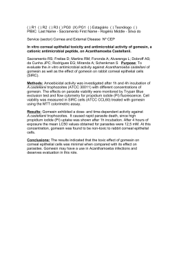
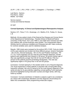
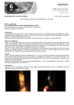
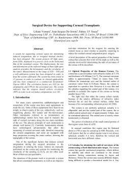
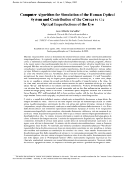
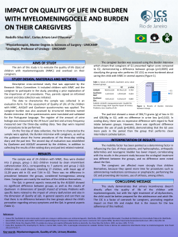
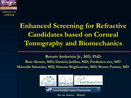
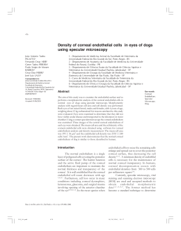
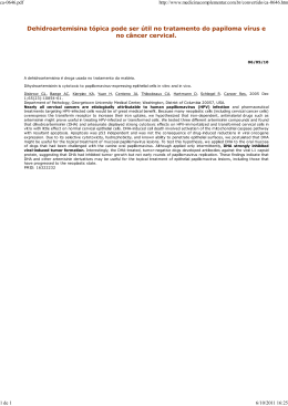
Effects of mitomycin C on infiltration
of polymorphonuclear leukocytes after
epithelial scrape injury in the mouse cornea
Efeito da mitomicina C na infiltração de leucócitos polimorfonucleares
após lesão epitelial em córnea de camundongo
Ana Cecília Souza Leão Escarião1
Takayuki Nagasaki2
Jin Zhao3
Richard Braunstein4
ABSTRACT
Purpose: To determine whether mitomycin C (MMC) alters appearance
and disappearance of polymorphonuclear leucocytes (PMN) in the
cornea stroma, using an epithelial scrape injury in eye mouse model.
Methods: Twenty-mice underwent mechanical epithelium debridement
in the central cornea using 20% ethanol. After the scrape, the right eye
received 0.02% MMC for one minute, while the left eye received physiological saline. The animals were sacrificed on days 1, 2, 5, and 14 after
surgery, and corneal whole mounts were prepared for histology. PMN
distribution was analyzed in digitized microscope images. Cell division
in the cornea was determined by immunohistochemical detection of
bromodeoxyuridine (BrdU), which was injected intraperitoneally before
the mice were sacrificed. Results: Epithelial scrape injury triggered
infiltration of PMNs into the corneal stroma. An analysis of PMN
distribution revealed that there was no difference between eyes treated
with and without MMC at all time points. BrdU labeling showed that
0.02% MMC for one minute blocked keratocyte proliferation completely.
Conclusion: MMC treatment regimen, which is common in clinical
practice, inhibits keratocyte proliferation during wound healing, but
when used at 0.02% for one minute, it does not affect PMN infiltration into
the corneal stroma, and subsequent movement toward the injury site, or
the disappearance of PMNs from the stroma, in the mouse epithelial
injury model.
Keywords: Mitomycin C/therapeutic use; Mitomycin C/administration & dosage; Leukocyte;
Cornea; Keratomileusis, laser in situ; Photorefractive keratectomy; Models, animal
Trabalho realizado no Department of Ophthalmology,
Columbia University - New York (NY) - USA.
1
2
3
4
Medical Doctor, Altino Ventura Foundation and Pernambuco Eye Hospital and ex-postdoctoral research
fellow at Columbia University - New York (NY) - USA.
PhD, Department of Ophthalmology, Columbia University - New York (NY) - USA.
PhD, Department of Ophthalmology, Columbia University - New York (NY) - USA.
Medical Doctor, Director of Refractive Surgery and
Laser Vision Correction at Columbia University - New
York (NY) - USA.
Endereço para Correspondência: Ana Cecília Souza
Leão Escarião. Rua da Soledade, 170 - Recife (PE)
CEP 50070-040
E-mail: [email protected]
Recebido para publicação em 22.08.2007
Última versão recebida em 10.08.2008
Aprovação em 18.08.2008
INTRODUCTION
Photorefractive keratectomy (PRK) and laser in situ keratomileusis
(LASIK) are the most common surgeries performed for the treatment of
refractive disorders. After these procedures, there is a corneal wound healing
and stimulation of a fibrotic response, usually stronger with PRK than
LASIK(1), specially in the treatment for high myopia patients and may lead to
haze development, compromising visual acuity and contrast sensitivity(2-3).
The corneal epithelium removal induces a loss of keratocyte by apoptosis, migration of inflammatory cells and proliferation of keratocytes in
the underlying stroma, followed by remodeling of collagen matrices(1,4). It
has been shown that corneal haze and keratocyte proliferation can be
reduced by inhibiting the infiltration of inflammatory cells into the ablation area after PRK(5).
Arq Bras Oftalmol. 2008;71(6):822-6
71(6)19.pmd
822
18/12/2008, 16:52
Effects of mitomycin C on infiltration of polymorphonuclear leukocytes after epithelial scrape injury in the mouse cornea
Mitomycin C (MMC) is a systemic chemotherapeutic drug
derived from Streptomyces caespitosus, which suppresses the
proliferation of rapidly growing cells by inhibiting DNA synthesis, secondary to alkylation(6). MMC has been used with increasing frequency in eye surgery. It has improved the results
of glaucoma surgery(7), pterygium excision(8), treatment of conjunctival and corneal intraepithelial neoplasia(9) and refractive
surgery(10).
In PRK, MMC prevents corneal haze after treatment of
high myopia, especially in patients whose corneal thicknesses
are inadequate for laser in situ keratomileusis(10). The effect of
MMC on wound healing has been documented by inhibiting
keratocyte proliferation, and activation, and consequently
blockage of myofibroblast differentiation(1,11). It is not clear,
however, whether and how MMC affects inflammatory activities, such as infiltration of leukocytes and their actions. An
inhibitory effect of MMC on leukocyte chemotaxis has been
suggested in gastric cancer(12), but no report seems to exist
regarding ocular inflammation.
The purpose of this study was to evaluate the effects of
MMC on the inflammatory response during the early stages of
wound healing, using an epithelial scrape injury model of the
mouse cornea.
823
Figure 1 - Appearance of epithelial scrape injury in the cornea in a live
mouse. Denuded area was stained with a drop of sulforhodamine 101
in saline, while areas of intact epithelium were protected from the dye.
Bar, 1 mm.
METHODS
An experimental prospective study was performed on a
mouse cornea in the research department of Ophthalmology at
Columbia University. Twenty mice were used in this study.
Animals
Animal study adhered to the Association for Research in
Vision and Ophthalmology Statement for the Use of Animals in
Ophthalmic and Vision Research, and approved by the institutional animal care and use committee. The GFPU mice were
obtained from the Jackson Laboratory (Bar Harbor, ME) and
maintained in house. Only male mice were used in this study.
Both eyes were used, a contralateral eye was used as the
control to eliminate variations in inflammatory reactions among
different animals. The injury was mild and the animals were
never at risk of vision loss.
Surgery
The animals were anesthetized by an intraperitoneal
injection of ketamine (100 mg/kg) and xylazine (10 mg/kg). The
mechanical epithelial scrape injury was created after a circular
mark using an about 1.2-mm diameter trephine. One filter paper
(0.9 mm diameter) soaked with 20% ethanol was placed on the
central cornea for one minute before performing a circular injury
(1.2 mm diameter) in the center of the cornea (Figure 1). This
injury was created with a blunt tip of a #12 blade under a
dissection microscope. Both eyes were used; the right eye
received 0.02% MMC for one minute, whereas the left eye
received physiological saline after the scrape injury. Both eyes
were irrigated extensively with balanced salt solution, and
received erythromycin ointment.
Histology
Animals were sacrificed by intraperitoneal injection of
pentobarbital (100 mg/kg) on days 1, 2, 5, and 14 after surgery,
and corneal whole mounts were prepared after fixation in 95%
methanol. The epithelium was removed from a whole mount,
which was then stained with hematoxylin and air-dried to
prepare a flat specimen. To quantify the infiltration of polymorphonuclear leukocytes (PMNs), the total number of stromal PMNs was determined in a rectangular area of 350 x 4500 µm
in the middle of the specimen in a digitized microscope image
by displaying a magnified image on a computer monitor and
identifying individual PMNs based on their horseshoe-shaped nuclear profile (Figure 2). The counting was done as a
single blind test, the examiner counted blind samples. Cell
division in the corneal stroma was determined by immunohistochemical detection of nuclear bromodeoxyuridine (BrdU)
after intraperitoneal injection of BrdU 2 hours before sacrificing the mouse. PMNs in the tears were determined by collecting the tears at 24 hours and 48 hours after the epithelial
scrape injury. For this, the mouse was anesthetized with ketamine and xylazine and 1 µl of PBS was instilled into the eye,
followed by collection of the fluid with a 1-µl glass pipette.
The tear fluid was then applied to a glass slide, air dried, and
stained with LeukoStat (Fisher Scientific, Pittsburgh, PA).
Blood smear was used as a positive control.
Arq Bras Oftalmol. 2008;71(6):822-6
71(6)19.pmd
823
18/12/2008, 16:52
824 Effects of mitomycin C on infiltration of polymorphonuclear leukocytes after epithelial scrape injury in the mouse cornea
Figure 2 - PMNs were identified and differentiated from keratocytes by their unique donut-shaped nuclei in the corneal stroma. Bar,
100 µm.
Figure 3 - Effect of MMC on keratocyte proliferation after epithelial
scrape injury. Keratocyte proliferation was determined by BrdU
incorporation.
Statistical analyses
The unpaired t-test was used to compare the number of
PMNs in MMC eyes and control eyes. Statistical significance
was accepted at a P<0.05 level.
RESULTS
Stroma cell division
BrdU labeling demonstrated that stroma cell division was
apparent in the injury area at 5 days but not at 2 days. This
keratocyte division at 5 days was nearly completely inhibited
by the MMC treatment, demonstrating the effectiveness of
MMC treatment (Figure 3).
Leukocytes in the tears
No PMNs were found in the tears 24 and 48 hours after the
injury.
PMN infiltration
Epithelial scrape injury triggered infiltration of PMNs
into the corneal stroma. The PMNs accumulated into the
anterior stroma of the injury site within 24 hours (Figure 4),
by which time the epithelial opening was closed 80-100%.
At this time point, keratocytes were absent in the anterior
stroma beneath the epithelial injury where PMNs accumulated. Most of the PMNs disappeared from the cornea by
five days after the trauma. At two weeks, the level of PMNs
was nearly identical to that of the untouched cornea. A
comparison of corneas treated with saline and MMC revealed that there was no significant difference in the numbers
of PMNs in these corneas at examined all time points
(P>0.05) (Table 1).
Figure 4 - PMN distribution in the MMC treated cornea at 24 hours after
epithelial scrape. Bar, 1 mm.
Table 1. A comparison of the numbers of PMNs at all time points of
corneas treated with MMC and saline
Time
point
24 h
48 h
5d
14 d
PMN MD ± SD
MMC
1215.50± 238.29
174.67± 34.79
55.00± 7.07
3.50± 0.71
824
t-test
0.6706
0.6386
0.5000
0.7952
PMN= polymorphonuclear leukocytes; MD= mean deviation; SD= standard
deviation; MMC= mitomycin C
Arq Bras Oftalmol. 2008;71(6):822-6
71(6)19.pmd
PMN MD ± SD
Control
1094.00 ± 63.64
187.67 ± 72.01
44.50 ± 7.78
3.00 ± 1.41
18/12/2008, 16:52
Effects of mitomycin C on infiltration of polymorphonuclear leukocytes after epithelial scrape injury in the mouse cornea
825
DISCUSSION
RESUMO
The cornea wound healing cascade is complex and mediated by cytokines, growth factors and chemokines(13-14). PMNs
are the first cells that migrate into tissues in response to
insults(15). It has been shown that the presence of leukocytes
stimulates the epithelial healing to some extent after alkali
injury(16). The early presence of leukocytes may act via cytokines to initiate migration of keratocytes. Proliferation may
likewise be influenced by leukocytes from the limbal side(17).
Previous studies have been performed to block the infiltration
of PMNs in the wound healing(18), but long-term clinical impact has not been determined.
PRK and LASIK are the most common refractive surgeries
performed for the correction of myopia, hyperopia and astigmatism. Clinical outcomes with these procedures are in large
part dependent on the cornea wound healing response. Corneal haze is more common after PRK than after LASIK, because the corneal wound healing response and the stimulus
for the fibrotic response are stronger(1).
Many substances have been used to prevent haze formation in PRK: amniotic membrane, vitamins A and E, collagenase inhibitors, and ubiquinone Q10 (19-22), but no controlled clinical trials have been reported, except for mitomicin C (23).
MMC is a drug that has been used frequently to reduce
haze formation owing, presumably, to its inhibition of proliferation of activated keratocytes (1). Despite the importance
of PMNs in wound healing and the prevalence of MMC use
in corneal surgery, effects of MMC on PMNs have not been
reported, to the best of our knowledge. MMC has been
reported to decrease the chemotaxis of PMNs in patients
with gastric cancer(12), but its relevance to the corneal wound
healing is not clear. This study with mouse eyes showed
that MMC has no measurable effect in the migration of
PMNs during early stages of corneal wound healing. This
supports the idea that MMC effect on haze reduction is
solely due to its inhibition of keratocytes, and not PMNs.
Perhaps this is so because MMC action is short-lived and
no longer effective by the time PMNs appear in the cornea
after injury, and thus MMC has no effect on PMN activities. Since the study addressed only the movement of PMNs,
it will be important to investigate other activities of PMNs
such as a release of oxidative enzymes and agents and
also their phagocytic activity. This will ensure that side
effects of MMC are fully understood when used in corneal
surgery.
Objetivo: O objetivo do estudo foi determinar se a mitomicina
C (MMC) altera o aparecimento dos leucócitos polimorfonucleares (PMN) no estroma corneano após abrasão epitelial
central, utilizando olhos de camundongo como modelo. Métodos: Vinte camundongos foram submetidos à abrasão epitelial
em córnea central utilizando etanol a 20%. Após a lesão, o
olho direito recebeu MMC a 0,02% por 1 minuto, enquanto o
olho esquerdo recebeu solução salina. Os animais foram sacrificados em 1, 2, 5 e 14 dias após a cirurgia e a córnea foi
preparada para histologia. A distribuição dos PMN foi analisada e digitalizada em imagens microscópicas. A divisão celular na córnea foi detectada pela imuno-histoquímica da bromodeoxirudina (BrdU), injetada intraperitonialmente duas horas
antes dos animais serem secrificados. Resultados: A lesão
epitelial gerou infiltração de PMN no estroma da córnea. A
análise da distribuição dos PMNs revelou que não houve
diferença estatisticamente significante entre os olhos tratados
e não tratados com MMC, em todos os tempos estudados. O
estudo com BrdU mostrou que a MMC quando utilizada a
0,02% por um minuto bloqueou completamente a proliferação
de ceratócitos. Conclusão: O tratamento com MMC, que é
utilizada comumente na prática clínica, inibe a proliferação dos
ceratócitos durante a cicatrização corneana, porém quando
utilizada a 0,02% por um minuto, não altera a infiltração dos
PMNs dentro do estroma corneano após lesão epitelial em
córneas de camundongos.
CONCLUSION
MMC at 0.02% for one minute has no measurable effect on
appearance, migration, and disappearance of corneal PMNs
during early stages of corneal wound healing after epithelial
scrape injury in the mouse eye model.
Descritores: Mitomicina C/uso terapêutico; Mitomicina C/
administração & dosagem; Leucócito; Córnea; Ceratomileuse
assistida por excimer laser in situ; Ceratectomia fotorrefrativa;
Modelos animais
REFERENCES
1. Netto MV, Mohan RR, Ambrósio R Jr, Hutcheon AE, Zieske JD, Wilson SE.
Wound healing in the cornea: a review of refractive surgery complications and
new prospects for therapy. Cornea. 2005;24(5):509-22.
2. Amm M, Wertzel W, Winter M, Uthoff D, Duncker GI. Histopathological
comparison of photorefractive keratectomy and laser in situ keratomileusis in
rabbits. J Refract Surg. 1996;12(7):758-66.
3. Wachtlin J, Laugenbeck K, Schründer S, Zhang EP, Hoffmann F. Immunohistology of corneal wound healing after photorefractive keratectomy and laser in situ
keratomileusis. [email protected]. J Refract Surg. 1999;15(4):451-8.
4. Kuo IC. Corneal wound healing. Curr Opin Ophthalmol. 2004;15(4):311-5.
Review.
5. Choi YS, Kim JY, Wee WR, Lee JH. Effect of the application of human
amniotic membrane on rabbit corneal wound healing after excimer laser photorefractive keratectomy. Cornea. 1998;17(4):389-95.
6. Calabresi P, Chabner BA. Antineoplastic agents. In: Goodman LS, Gilman A,
Gilman AG, editors. Pharmacological basis of therapeutics. 8th ed. New York:
Pergamon Press; c1990. p.1247-8.
7. Maquet JA, Dios E, Aragón J, Bailez C, Ussa F, Laguna N. Protocol for
mitomycin C use in glaucoma surgery. Acta Ophthalmol Scand. 2005;83(2):
196-200.
8. Levy RL, Naidu S, Jacobson L. Safety and efficacy of the technique of complete
tenon’s membrane excision and mitomycin C in pterygium surgery. Eye
Contact Lens. 2005;31(3):105-8.
Arq Bras Oftalmol. 2008;71(6):822-6
71(6)19.pmd
825
18/12/2008, 16:52
826 Effects of mitomycin C on infiltration of polymorphonuclear leukocytes after epithelial scrape injury in the mouse cornea
9. Prabhasawat P, Tarinvorakup P, Tesavibul N, Uiprasertkul M, Kosrirukvongs
P, Booranapong W, Srivannaboon S. Topical 0.002% mitomycin C for the
treatment of conjunctival-corneal intraepithelial neoplasia and squamous cell
carcinoma. Cornea. 2005;24(4):443-8.
10. Hashemi H, Taheri SM, Fotouhi A, Kheiltash A. Evaluation of the prophylactic
use of mitomycin-C to inhibit haze formation after photorefractive keratectomy in
high myopia: prospective clinical study. BMC Ophthalmol. 2004;4:12.
11. Xu H, Liu S, Xia X, Huang P, Wang P, Wu X. Mitomycin C reduces haze
formation in rabbits after excimer laser photorefractive keratectomy. J Refract
Surg. 2001;17(3):342-9.
12. Hirano T. Changes in polymorphonuclear leukocyte motility under agarose and
luminol-dependent chemiluminescence response in patients with gastric cancer.
Gastroenterol Jpn. 1984;19(5):447-56.
13. Tuli S, Goldstein M, Schultz GS. Modulation of corneal wound healing. In:
Krachmer JH, Mannis MJ, Holland EJ, editors. Cornea. 2nd ed. Philadelphia:
Elsevier Mosby; 2005. p.133-50.
14. Wilson SE, Liu JJ, Mohan RR. Stromal-epithelial interactions in the cornea.
Prog Retin Eye Res. 1999;18(3):293-309. Review.
15. Hong JW, Liu JJ, Lee JS, Mohan RR, Mohan RR, Woods DJ, et al.
Proinflammatory chemokine induction in keratocytes and inflammatory cell
infiltration into the cornea. Invest Ophthalmol Vis Sci. 2001;42(12):2795-803
16. Gan L, Fagerholm P, Kim HJ. Effect of leukocytes on corneal cellular proliferation and wound healing. Invest Ophthalmol Vis Sci. 1999;40(3):575-81.
17. Gan L, Hamberg-Nyström H, Fagerholm P, Van Setten G. Cellular proliferation
and leukocyte infiltration in the rabbit cornea after photorefractive keratectomy.
Acta Ophthalmol Scand. 2001;79(5):488-92
18. Bilgihan A, Bilgihan K, Yis O, Sezer C, Akyol G, Hasanreisoglu B. Effects of
topical vitamin E on corneal superoxide dismutase, glutathione peroxidase
activities and polymorphonuclear leucocyte infiltration after photorefractive keratectomy. Acta Ophthalmol Scand. 2003;81(2):177-80.
19. Brancato R, Schiavone N, Siano S, Lapucci A, Papucci L, Donnini M, et al.
Prevention of corneal keratocyte apoptosis after argon fluoride excimer laser
irradiation with the free radical scavenger ubiquinone Q10. Eur J Ophthalmol.
2000;10(1):32-8.
20. Wang MX, Gray TB, Park WC, Prabhasawat P, Culbertson W, Forster R, et al.
Reduction in corneal haze and apoptosis by amniotic membrane matrix in excimer
laser photoablation in rabbits. J Cataract Refract Surg. 2001;27(2):310-9.
21. Vetrugno M, Maino A, Cardia G, Quaranta GM, Cardia L. A randomised, double
masked, clinical trial of high dose vitamin A and vitamin E supplementation after
photorefractive keratectomy. Br J Ophthalmol. 2001;85(5):537-9.
22. Corbett MC, O’Brart DP, Patmore AL, Marshall J. Effect of collagenase
inhibitors on corneal haze after PRK. Exp Eye Res. 2001;72(3):253-9.
23. Bedei A, Marabotti A, Giannecchini I, Ferretti C, Montagnani M, Martinucci
C, Barabesi L. Photorefractive keratectomy in high myopic defects with or
without intraoperative mitomycin C: 1-year results. Eur J Ophthalmol. 2006;
16(2):229-34.
Errata - Erratum
Considerações sobre o ângulo de administração de colírios antiglaucomatosos
análogos das prostaglandinas
Considerations about administration angle of prostaglandin analogs
Paulo Estacia, Taíse Tognon
Arq Bras Oftalmol. 2008;71(5):684-8
Na INTRODUÇÃO - 7º. e 8º. parágrafos - e na DISCUSSÃO - 1º. e 2º. parágrafos - a unidade de medida do volume das gotas
saiu publicada como mililitro (ml) mas na verdade é microlitro (µl). Portanto, seguem abaixo os parágrafos corretos.
Na INTRODUÇÃO:
O volume da gota instilada está diretamente relacionado à biodisponibilidade da droga, a fim de proporcionar uma quantidade padrão e adequada para ação do
fármaco. As apresentações comerciais mais antigas de
colírios apresentavam volumes que variam entre 50 e 75 µl
e atualmente os frascos fornecem gotas variando entre
25 e 56 µl(7-8). Também o ângulo no qual o colírio é administrado constitui uma das variáveis que atua modificando o volume da gota(9).
É importante produzir uma gota menor com maior
concentração da droga, pois a ação farmacológica depende da quantidade residual em contato com a superfície ocular após perdas por diluição, ligação e drenagem(7). A melhor concentração em filme lacrimal é atingida com uma gota de 20 µl, sendo que o aumento do
volume da gota aumenta a absorção sistêmica, o desperdício e pode piorar a tolerabilidade(10-11).
Na DISCUSSÃO:
É sabido que o glaucoma constitui uma patologia importante do ponto de vista social e populacional, em virtude
de sua morbidade e custos ao sistema de saúde e ao paciente.
Assim, é indiscutível que a terapêutica uma vez instituída
deve ser efetiva e, a mais empregada, como descrito anteriormente, é o uso de colírios antiglaucomatosos. Para tanto, a
gota instilada deve possuir um volume adequado para o que
se deseja. O olho humano pode acomodar uma gota de
colírio com volume não superior a 23 µl e quantidades
superiores a esta são desperdiçadas, ou são drenadas pela via
lacrimal com possibilidade de maior absorção sistêmica(18).
Estudos anteriores mostraram que a média de volume das
gotas de soluções oftálmicas era de 39 µl com uma variação
de 25,1 a 56,4 µl(19). Em nosso país o Ministério da Saúde
preconiza que as gotas de colírios devem apresentar volumes
inferiores a 50 µl. Assim, observa-se que todos os diferentes
produtos testados possuíam correlação com o estudo citado.
Arq Bras Oftalmol. 2008;71(6):822-6
71(6)19.pmd
826
18/12/2008, 16:52
Baixar