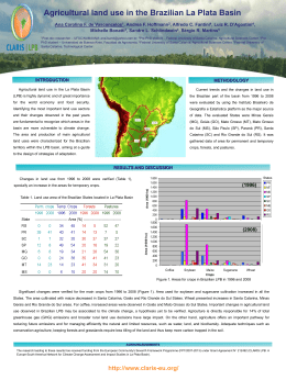
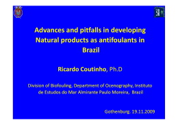
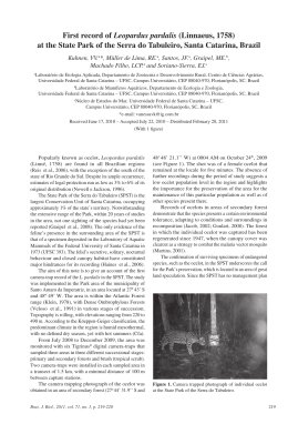
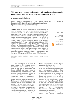
Redalyc
Sistema de Información Científica
Red de Revistas Científicas de América Latina, el Caribe, España y Portugal
Costa Marchiori, Natalia da; Magenta Magalhães, Aimê Rachel; Pereira Junior, Joaber
The life cycle of Bucephalus margaritae Ozaki & Ishibashi, 1934 (Digenea,
Bucephalidae) from the coast of Santa Catarina State, Brazil
Acta Scientiarum. Biological Sciences, vol. 32, núm. 1, 2010, pp. 71-78
Universidade Estadual de Maringá
Brasil
Disponible en: http://redalyc.uaemex.mx/src/inicio/ArtPdfRed.jsp?iCve=187114368011
Acta Scientiarum. Biological Sciences
ISSN (Versión impresa): 1807-863X
[email protected]
Universidade Estadual de Maringá
Brasil
¿Cómo citar?
Número completo
Más información del artículo
Página de la revista
www.redalyc.org
Proyecto académico sin fines de lucro, desarrollado bajo la iniciativa de acceso abierto
DOI: 10.4025/actascibiolsci.v32i1.5596
The life cycle of Bucephalus margaritae Ozaki & Ishibashi, 1934
(Digenea, Bucephalidae) from the coast of Santa Catarina State,
Brazil
Natalia da Costa Marchiori1*, Aimê Rachel Magenta Magalhães1 and Joaber Pereira
Junior2
1
Núcleo de Estudos em Patologia, Departamento de Aquicultura, Centro de Ciências Agrárias, Universidade Federal de Santa
2
Catarina, Rod. Ademar Gonzaga, 1346, 88040-900, Itacorubi, Florianópolis, Santa Catarina, Brazil. Laboratório de Biologia de
Parasitos de Organismos Aquáticos, Programa de Pós-graduação em Aquicultura, Universidade Federal do Rio Grande,
Rio Grande, Rio Grande do Sul, Brazil. *Author for correspondence. E-mail: [email protected]
ABSTRACT. The orange disease is considered the main parasitosis in Perna perna mussel.
It is caused by a complex life cycle bucephalid, involving three hosts, among them mollusks
and fishes. With the aim of contributing to the knowledge of orange disease in mussel
culture, the parasite life cycle was investigated. Experimental studies and monthly samples
in the study area allowed the identification and characterization of the Bucephalus margaritae
life cycle. Larvae and adults of B. margaritae were fixed in 5% formaldehyde, stained with
Gomori’s trichrome, clarified in creosote and mounted in Canada balsam. The cercariae are
found in the first intermediate host P. perna inside the sporocysts, which have the form of
orange and ramified filaments. The metacercariae encysts in the gills and gill cavity of the
second intermediate host, the blenniid Hypleurochilus fissicornis. The definitive host
Menticirrhus americanus is infected ingesting blenniids parasitized with metacercariae. The
high parasitological indexes of B. margaritae suggests that M. americanus and H. fissicornis act as
the main definitive and intermediate hosts, respectively, in the trematode life cycle. The
blenniid H. fissicornis is a new intermediate host to the species.
Key words: Bucephalus margaritae, Hypleurochilus fissicornis, life cycle, orange disease, Perna perna.
RESUMO. O ciclo de vida de Bucephalus margaritae Ozaki e Ishibashi, 1934
(Digenea, Bucephalidae) da costa do Estado de Santa Catarina, Brasil. A bucefalose
é considerada a principal parasitose do mexilhão Perna perna. É causada por um
Bucephalidae, de ciclo de vida complexo, envolvendo três hospedeiros, entre eles, moluscos
e peixes. Com o objetivo de se contribuir para o conhecimento da bucefalose em mexilhões
de cultivo, foi investigado o ciclo de vida deste parasito. Estudos experimentais e coletas
mensais na área de estudo permitiram caracterizar o ciclo de vida de Bucephalus margaritae.
Larvas e adultos do parasito foram fixados em formol 5%, corados com tricrômico de
Gômori, clarificados em creosoto e montados em lâminas permanentes com bálsamo do
Canadá. As cercárias ocorrem no primeiro hospedeiro intermediário P. perna, no interior
dos esporocistos que têm a forma de filamentos ramificados e alaranjados. As metacercárias
encistam nas brânquias e cavidade branquial do segundo hospedeiro intermediário, o
blenídeo Hypleurochilus fissicornis. O hospedeiro definitivo, Menticirrhus americanus, é infectado
quando ingere blenídeos com metacercárias. Os elevados índices parasitológicos de
B. margaritae sugerem que M. americanus e H. fissicornis sejam os principais hospedeiros
definitivo e intermediário, respectivamente, deste trematódeo. O blenídeo H. fissicornis é um
novo hospedeiro intermediário para a espécie.
Palavras-chave: Bucephalus margaritae, Hypleurochilus fissicornis, ciclo de vida, enfermidade laranja, Perna perna.
Introduction
Bucephalus sp. larvae have been reported in
Perna perna (Linnaeus, 1758) mussel from the
coast of Santa Catarina and can cause severe
impact on its mitiliculture and commercial
production due to its elevated degree of
pathogenicity (SILVA et al., 2002). Bucephalosis,
Acta Scientiarum. Biological Sciences
or orange disease, destroys the reproductive
tissues of the host, disables its gametogenesis,
leads it to castration (CALVO-UGARTEBURU;
McQUAID, 1998; COUSTAU et al., 1990) and,
possibly to the bivalve’s death (LAUCKNER,
1983; SILVA et al., 2002). Some Bucephalus species
have had their life cycle studied (MATTHEWS,
Maringá, v. 32, n. 1, p. 71-78, 2010
72
1973; TASKINEN et al., 1991; ABDALLAH;
MAAMOURI, 2002; 2005). In Brazil, two
Bucephalus species are reported: Bucephalus
solitarius Kohn, 1968 in Caranx crysos (Mitchill,
1815),
and
Bucephalus
margaritae
Ozaki
et Ishibashi, 1934, registered in ten fish species
(KOHN et al., 2007).
With the aim of contributing to bucephalosis
knowledge, B. margaritae life cycle was established,
with recognition of the species utilized as hosts at
Sambaqui region, Florianópolis/Santa Catarina
State and also its developmental stages described.
Material and methods
Locality. Ponta do Sambaqui is situated at the
North Bay of Santa Catarina island, Florianópolis
(27°29’S, 48°33’W) and shelters the cultures of
P. perna from Marine Mollusks Laboratory
(LMM) of Federal University of Santa Catarina
(UFSC). The culture system is longline.
Observations in naturally infected hosts.
Samples of 30 P. perna mussels were monthly
collected, between november 2006 to november
2007 from the UFSC experimental cultures. The
hosts were dissected and examined for
bucephalid’s larval stages. Hypleurochilus fissicornis
(Quoy & Gaimard, 1824) (n = 51) were collected
along the rope cultured mussels between
december 2006 to october 2007 and examined
under a stereosmicroscope for the presence of
metacercariae. When present, the larvae were
desencysted with the assistance of histological
needles or by applying a slight pressure on the
cyst. Between november 2006 to july 2007,
trematode potential definitive hosts were collected
for further investigation of its adult form. This was
verified for fishes Trichiurus lepturus Linnaeus, 1758
(n = 5), Dicentrarchus labrax (Linnaeus, 1758)
(n = 7), Sciades herzbergii (Bloch, 1794) (n = 12),
Archosargus rhomboidalis (Linnaeus, 1758) (n = 9),
Mycteroperca bonaci (Poey, 1960) (n = 1), Spheroides
testudineus (Linnaeus, 1758) (n = 5), Micropogonias
furnieri (Desmarest, 1823) (n = 11), Menticirrhus
americanus (Linnaeus, 1758) (n = 32), Balistes capriscus
(Gmelin, 1788) (n = 8) and Epinephelus gigas
(Brunnich, 1768) (n = 1). For all parasitized fish
species it was calculated the parasitological indexes
of prevalence and mean intensity of infection (MII)
according to Bush et al. (1997).
Morphology. The morphology of adults and
metacercariae were studied in 5% formaldehyde
Acta Scientiarum. Biological Sciences
Marchiori et al.
fixed specimens. Fixed parasites were washed in
distilled water, stained in Gomori’s trichrome,
clarified in creosote and mounted in Canada
balsam. Cercariae observations were made in live
specimens. Sporocysts were disrupted with the
assistance of histological needles for larvae release.
Morphological helminth characters measured
with the aid of a camera lucida are in
micrometers. Measurements are presented as
follows: mean ± standard deviation and
minimum and maximum values in parenthesis,
when the case numbers are higher than three.
Observations of cercariae longevity. A
hundred cercariae were obtained through
ruptured sporocysts from P. perna mussels and
maintained in Petri dishes at room temperature
(20-23ºC) and its survival monitored up to
72 hours.
Observations in experimentally infected
second intermediate host. Specimens of
H. fissicornis were separated in two aquaria, one
exposed to B. margaritae cercariae released from
infected mussels (n = 15) and the other acting as
a control group (n = 15). Two replicates of seven
animals for each aquarium were done.
Temperature and salinity were controlled and
maintained constant at 20ºC and 34‰,
respectively. The period of exposition was
24 hours.
Observations in experimentally infected
definitive hosts. Ten Trachinotus goodei (Jordan &
Evermann, 1896) were collected at Barra da Lagoa
(27°34’S, 48°33’W), east side of Santa Catarina
island. The animals were fed with blenniid´s
infected gills and 16 days after, necropsied for the
presence of adult bucephalids inside the digestory
tract of fish.
Results
Sporocyst (Figure 1A)
From 360 collected mussels 21.6% were
parasitized by helminth sporocysts.
The sporocysts presented ramifications with
dilatations occuring in the mantle, gonads and
digestive gland of P. perna mussel. Sporocysts held
numerous cercariae in different developmental stages.
Cercariae (Figures 1B and 2A)
After released from the sporocysts, the larvae
were observed at the bottom of a Petri dish and
realized body contractions, including rotatory
moviments around its own axis and the furcae.
Maringá, v. 32, n. 1, p. 71-78, 2010
Lyfe cycle of Bucephalus margaritae
73
Survival time of most cercariae was about 48 hours.
In the absence of a second intermediate host, the
process of larvae encystment did not occur.
Description based on 20 specimens: Body small
and thin 288.3 ± 45.0 (220.0 – 360.0) long and 54.4
± 19.7 (20.1 – 90.0) wide, dorso-ventrally flattened
with thorny surface. The rinchus 74.3 ± 11.5 (50.0
– 100.2) of long and 31.1 ± 7.4 (20.1 – 40.2) wide,
has a bulb shape and contains numerous glandular
cells. The mouth, shaped as a small gap, is situated
next to the larva’s body equator and is strongly
muscular. The pharynx is short and the intestine
has an irregular sac shape 94.8 ± 31.7 (60.6 –
120.0). Primordiums of the reproductive system
are not evidenced or visible. The furcae is
characterized by two long projections which,
when extended, surpass up to 9.5 times larva
body, with transversal folds all over its surface and
ending into a small sucker that surrounds the
excretory pores.
A
A
B
B
D
D
EE
C
C
FF
Figure 1. Bucephalus margaritae from the coast of Santa Catarina State. A- Sporocyst. Bar: 100 μm. B- Cercariae. Bar: 100 μm. CMetacercariae cyst. Bar: 20 μm. D- Three-year-old metacercariae. Bar: 30 μm. E- Adult. Bar: 100 μm. F- Egg. Bar: 10 μm.
Acta Scientiarum. Biological Sciences
Maringá, v. 32, n. 1, p. 71-78, 2010
74
Marchiori et al.
A
Figure 2. Bucephalus margaritae from the coast of Santa Catarina
State. A- Cercariae. Bar: 100 μm. B- Posterior region in detail.
Bar: 10 μm. C- Adult, ventral side, with the testicles introverted.
Bar: 100 μm. D- Adult tentacle. Bar: 10 μm.
B
Metacercariae (Figures 1C and D)
Encysted metacercariae of B. margaritae were found
in the blenniid´s gill cavity, including the filaments and
mainly at the base of gill arch. The prevalence of
infected animals by metacercariae was 75% the mean
intensity of infection ranged from 2 to 352 larvae per
host. The blenniids are easily found among the rope
cultured mussels or even inside the shell of dead
bivalves commonly used for the fish spawning.
Cysts are ovoid with 43.7 ± 5.7 (30.0 – 60.2)
long and 28.7 ± 3.3 (22.5 – 40.5) μm wide,
composed by two membranes: the inner and thinner
one is secreted by the parasite´s cystogenic cells and
the other, capsular and thicker, is possibly formed by
its host. Metacercariae 181.6 ± 14.2 (160.8 – 200.4)
long and 71.8 ± 16.9 (45.8 – 90.8) μm wide (n =
15). Body covered by thin spines. Rinchus
composed by seven tentacles with two projections
each: the bigger is basal, and the smaller is distal
(Figure 3A). The mouth is localized a little down
from the ventral surface of larva’s equator. Pharynx
short. The excretory vesicle is situated in the
posterior third of the body. Gonads poorly
developed.
Adult (Figures 1E and 2C)
Bucephalus margaritae adult forms were found in
the intestine and pyloric caecum of M. americanus
(n = 32). The prevalence of infected animals was
93% and mean intensity of 29.54.
Acta Scientiarum. Biological Sciences
Figure 3. Bucephalus margaritae. Projections of the tentacle. AMetacercariae. B- Adult. Bar: 10 μm.
Description of the studied specimens: (Based on
10 specimens): Elongated body with 513.7 ± 131.7
(323.0 – 814.7) long and 81.1 ± 24.1 (52.2 – 164.1)
wide. Spined tegument, with small spines all over
the body. Funnel-like apical rinchus with concave
bottom and seven marginal tentacles, retractiles,
telescopics, with reduced witdh in its extremity and
with two projections, one proximal and the other
distal (Figures 2D and 3B). The proximal, with 12.5
long, presents an estimated angle of 90º with the
tentacle axis, and has obtuse extremity. The distal
one, discrete, with acute extremity and 2.5 long, is
five times smaller than the proximal. Oral sucker
post equatorial, circular, muscular, 339.5 ± 27.2
(220.3 – 546.3) from the anterior extremity; it
includes the mouth as an horizontal gap on its
center. Small pharynx, with 10.7 (6.4 – 16.0) width.
Numerous rounded vitellaria distributed in the
median equatorial body region and laterally in two
convergents camps, with 14 to 19 each side. The
intestine is simple, sac shaped, anteriorly projected
among vitellaria space. The ovary, globular, pretesticular and pre-pharinx is smaller than the
testicles, with 30.3 ± 11. 3 (22.3 – 44.3) long and
52.9 (30.0 – 114.2) wide. Mehlis gland and Laurer´s
canal not evidenced. Uterine loops exceed the
Maringá, v. 32, n. 1, p. 71-78, 2010
Lyfe cycle of Bucephalus margaritae
75
and pyloric caecum.
Synonyms: Prosorhynchus margaritae (Ozaki et
Ishibashi, 1934) Ozaki, 1960; Bucephalus polymorphus
von Baer, 1827; Bucephalus varicus Manter, 1940;
Bucephalus retractilis Yamaguti, 1952; Bucephalus
pseudovaricus Velásquez, 1959; Bucephalus carangoides
Yamaguti, 1970; Bucephalus ulua Yamaguti, 1970.
Species distribution: Bucephalus margaritae has
wide distribution and is found in tropical and
subtropical waters being registered in the caribbean
sea, pacific coast of Panama, Mexico, Brazil,
Venezuela, Guinea-Bissau, China, mediterranean
coast of Israel, Kuwait, Red Sea, Arabian Sea,
Hawaii, India and Japan (AMATO, 1982;
CHINCHILLA et al., 2006; NAHHAS et al., 2006).
Regardings: Reported as B. varicus in Kohn
(1968), Madhavi (1974), Amato (1982), Takemoto
et al. (1995; 1996), Pereira Junior et al. (1996),
Chaves and Luque (1998), Luque et al. (2000),
Luque et Alves (2001) and Alves et al. (2004);
Bucephalus polymorphus in Caballero et al. (1953) and
Baturo (1977); Bucephalus pseudovaricus in Velasquez
(1959) and Yamaguti (1971); Bucephalus retractilis in
Yamaguti (1952; 1958; 1971); Bucephalus carangoides
in Yamaguti (1971); Bucephalus ulua in Yamaguti
(1971) and B. margaritae in Bray (1984), Yamaguti
(1958; 1971), Nahhas et al. (2006), Chinchilla et al.
(2006) and Kohn et al. (2007).
anterior limit of vitellaria and posteriorly reaches the
body´s posterior extremity. Numerous eggs, ovoids
and operculated with 21.5 ± 4.6 (14.0 – 28.2) long
and 9.0 ± 4.3 (4.4 – 19.2) wide (Figure 1F).
Two testicles, posterior to oral sucker, one after
the other, envolved by a thick capsule (not included
in the measurements), globular with 41.0 ± 9.7
(34.0 – 64.2) long and 26.7 ± 6.6 (20.2 – 44.1) wide
(anterior) and 36.4 (22.3 – 82.2) long and 38.5 (28.0
– 76.0) wide (posterior). From each testicle appears a
vas efferens which come together anteriorly and
forms a single vas deferens that opens into the cirrus
sac. Cirrus sac elongated, localized in the posterior
third of the body beginning at the level of first
testicule with 100.5 ± 17.9 (78.2 – 200.3) long. It
shelters the circular seminal vesicle, the pars
prostatica with lots of small prostatic cells and the
genital atrium,which includes the finger-like and
sinuous cirrus and the genital pore, surrounded by a
discrete musculature. Uterous final portion is
sinuous and partially involves the cirrus inside´s
genital atrium. Terminal excretory pore.
Taxonomic summary of Bucephalus margaritae
Ozaki et Ishibashi, 1934
Host: Menticirrhus americanus (Sciaenidae).
Locality: Ponta do Sambaqui, Florianópolis,
Santa catarina State (27°29’S, 48° 33’W).
Sites of infection in the definitive host: intestine
Table 1. Known hosts (organized by Families) for Bucephalus margaritae.
Hosts
Carangidae
Alectis spp.
Atropus atropos (Bloch & Schneider, 1801)
Caranx crysus (Mitchill, 1815)
Caranx hippos (L.)
Caranx latus Agassiz, 1831
Caranx sem Cuvier, 1833
Caranx ignobilis (Forsskal, 1755)
Caranx sexfasciatus Quoy & Gaimard, 1825
Carangoides chrysophrys (Cuvier, 1833)
Carangoides malabaricus (Bloch & Schneider, 1801)
Carangoides hedlandensis (Whitley, 1934)
Chloroscombrus crysurus (Linnaeus, 1766)
Chorinemus spp.
Oligoplistes palometa (Cuvier, 1832)
Oligoplistes saurus (Bloch & Schneider, 1801)
Oligoplistes saliens (Bloch, 1793)
Scomberoides commersonianus Lacépède, 1801
Trachinotus spp.
Zonichthys spp.
Gerreidae
Gerres filamentosus Cuvier, 1829
Sphyraenidae
Sphyraena jello Cuvier, 1829
Sphyraena picudilla Poey, 1860
Gadidae
Urophycis brasiliensis
Sciaenidae
Menticirrhus americanus (Linnaeus, 1758)
Menticirrhus littoralis (Holbrook, 1847)
Micropogonias furnieri (Desmarest, 1823)
Acta Scientiarum. Biological Sciences
References
Bray (1984)
Nahhas et al. (2006)
Luque et al. (2000), Luque et Alves (2001) and Kohn et al. (2007)
Luque et al. (2000), Luque et Alves (2001) and Kohn et al. (2007)
Luque et al. (2000), Luque et Alves (2001) and Kohn et al. (2007)
Bray (1984)
Bray (1984)
Parukhin (1970) apud Bray (1984)
Madhavi (1974)
Madhavi (1974) and Nahhas et al. (2006)
Bray (1984)
Amato (1982)
Bray (1984)
Takemoto et al. (1995; 1996) and Kohn et al. (2007)
Takemoto et al. (1995; 1996) and Kohn et al. (2007)
Takemoto et al. (1995; 1996) and Kohn et al. (2007)
Nahhas et al. (2006)
Bray (1984)
Bray (1984)
Nahhas et al. (2006)
Nahhas et al. (2006)
Chinchilla et al. (2006)
Pereira Junior et al. (1996), Alves et al. (2004) and Kohn et al. (2007)
Kohn (1968), Amato (1982), Chaves and Luque (1998) and Kohn et al. (2007)
Amato (1982) and Kohn et al. (2007)
Pereira Junior et al. (1996) and Kohn et al. (2007).
Maringá, v. 32, n. 1, p. 71-78, 2010
76
Observations in experimental infections
Experimentally infected Hypleurochilus fissicornis
(n = 30) were necropsied for parasite’s larval stages.
The metacercariae were found encysted in the host’s
gill cavity. The site with higher cyst concentration
was at the base of gill arches and also the filaments.
Mean intensity of infection by encysted
metacercariae in experimentally infected blenniids
was high, with means of 136 cysts per fish ± 117.24
(25 – 332), differently from the control group which
was observed means of 8.28 ± 13.43 (1-41).
Experimentally infected Trachinotus goodei were
necropsied for the parasite´s adult form. It was not
registered adult specimens of B. margaritae in none of
the examined fishes. All hosts were examined in the
same day and this way it was not observed if time
could influence in those infection indexes.
Discussion
Bucephalosis is considered the main parasitosis
in P. perna mussel culture (BOWER et al., 1994;
COCHÔA; MAGALHÃES, 2008). Many of the
damages caused by this parasite are known (SILVA
et al., 2002).
The elevated prevalence indexes registered in H.
fissicornis and M. americanus in this study suggests that
populations from these species acts as the main
definitive and intermediate hosts, respectivally, in
the life cycle of B. margaritae from the coast of Santa
Catarina State.
The blenniid H. fissicornis utilizes bivalve’s valves
as a place for spawning besides feeding actively of P.
perna tissues, as observed in this study. This is an
important data for the local mitiliculture since its
niche is already integrated to this activity.
In Brazil, M. americanus is a common definitive
host for adults of B. margaritae (KOHN, 1968;
AMATO, 1982; CHAVES; LUQUE, 1998; KOHN
et al., 2007). In the coast of Santa Catarina State,
Amato (1982) registered B. margaritae (= B. varicus)
in M. americanus with both measures and description
very close from our results. This work register, for
the first time, the blenniid H. fissicornis hosting cysts
of B. margaritae metacercariae. It is hence considered
a new host record.
According to Nahhas et al. (2006), the diagnostic
character of greater relevance for B. margaritae is the
presence of seven tentacles, each one with two
projections: one big and basal and the other small
and distal. According to Spakulová et al. (2002), 12
Bucephalus species has a rinchus surrounded by
seven tentacles with two projections. Bucephalus
priacanthi Manter, 1940 and Bucephalus scorpaenae
Manter, 1940 are described from marine fishes of
Acta Scientiarum. Biological Sciences
Marchiori et al.
Florida and B. varicus in marine fishes from Brazil.
Bucephalus elegans Woodhead, 1930 is registered from
freshwater fishes of United States; Bucephalus fragilis
Velásquez, 1959 and Bucephalus uranoscopi Yamaguti,
1934 are described from marine fishes of the
Philippines and Japan and Bucephalus anguillae
Spakulová, Macko, Berrili et Dezfuli, 2002 in
Anguilla anguilla (Linnaeus, 1758) of Adriatic Sea.
Bucephalus minimus (Stossich, 1887), Bucephalus
blanchardi (Stossich, 1898) and Bucephalus labracis
Paggi et Orecchia, 1965 were described from D.
labrax of Mediterranean Sea. Bucephalus minimus was
transferred by Yamaguti (1971) to Bucephalopsis
Yamaguti, 1971. Bartoli et al. (2005) reported
Bucephalus gorgon (Linton, 1905) from carangids of
West mediterranean also containing seven tentacles
in its rinchus. However, besides their register of
approximately 11 to 14 papillae at the basis of the
seven tentacles, this species does not present the
small distal projection characteristic of B. margaritae.
In addition, the only species from the cited above
that clearly presents the ovary in a pre-pharynx
position is B. margaritae (SPAKULOVÁ et al., 2002).
Another diagnostic character that varies in B.
margaritae is the egg size. Nahhas et al. (2006) registered
eggs of this species with 13-20 μm long and 10-18 μm
wide. In Brazil, Amato (1982) described eggs of B.
varicus with 18-20 by 10-12 μm; Kohn (1968), while
studying B. margaritae collected from the esophagus of
M. americanus, registered eggs with 21-27 by 11-12 μm.
Takemoto et al. (1995), studied the trematode fauna of
Oligoplites palometa, O. saurus and O. saliens, and
registered eggs of B. varicus with 16-20 by 9-11 μm.
Nahhas et al. (2006) affirmed that the means of B.
margaritae´s eggs measured found in the literature were
14-27 by 10-13 μm, values very close from what
observed in this study.
An historical revision of B. margaritae and its relation
with B. polymorphus was done by Bray (1984). Despite
the morphological resemblance between the two
species, leading some authors to indicate them as
synonyms (NAHHAS et al., 2006; BRAY, 1984), some
differences with respect to larvae ecology are evident.
Parasite from freshwater fishes (SCHUSTER et al.,
2001; DILER; YILDIRIM, 2003), encysted B.
polymorphus metacercariae are registered by Baturo
(1977) in the musculature and fins of its hosts,
differently from the reports of B. margaritae
metacercariae, so far always sited at the gills of marine
hosts. Future studies might show if the species are, in
fact, synonyms.
Abdallah and Maamouri (2005) concluded that,
in only ten days, B. labracis completed its
development reaching sexual maturity with
consequent eggs formation in its final host. The
Maringá, v. 32, n. 1, p. 71-78, 2010
Lyfe cycle of Bucephalus margaritae
experimentally infected definitive hosts in this study
were necropsied in the 16º day post-infection.
However, as in Taskinen et al. (1991), it was not
registered the helminth adult stage in this period. It
was not possible to explain this result.
Acknowledgments
The authors thank Capes for Master sponsorship
to Natalia Marchiori; Dr. Maurício Laterça Martins
(UFSC) for gently grant its laboratory and also to
the employers Jackson and Itamar, from the
Laboratory of Marine Mollusks of UFSC for help
on fish’s arrest.
References
ABDALLAH, L. B. G.; MAAMOURI, F. Cycle évolutif de
Bucephalus anguillae Spakulová. Macko, Berrilli & Dezfuli
(Digenea, Bucephalidae) parasite de Anguilla anguilla (L.).
Systematic Parasitology, v. 53, n. 3, p. 207-217, 2002.
ABDALLAH, L. B. G.; MAAMOURI, F. The life cycle of
Bucephalus labracis Paggi and Orecchia, 1965 (Digenea,
Bucephalidae), a parasite of Dicentrarchus labrax in Tunisia.
Bulletin European Association of Fish Pathologists,
v. 25, n. 6, p. 297-301, 2005.
ALVES, D. R.; PARAGUASSU, A. R.; LUQUE, J. L.
Metazoários parasitas da Abrótea, Urophicis brasiliensis
(Kaup, 1858), (Ostheichyes: Phycidae) do litoral do estado
do Rio e Janeiro, Brasil. Revista Brasileira de
Parasitologia Veterinária, v. 13, n. 1, p. 49-55, 2004.
AMATO, J. F. R. Digenetic trematodes of percoid fishes
of Florianópolis, Southern Brazil – Bucephalidae. Revista
Brasileira de Biologia, v. 42, n. 4, p. 667-680, 1982.
BARTOLI, P.; RODNEY, A. B.; GIBSON, D. A. Three
poorly known and rarely reported bucephalid species
(Digenea) in fishes from the Western Mediterranean.
Systematic Parasitology, v. 62, n.2, p. 135-149, 2005.
BATURO, B. Bucephalus polymorphus Baer, 1827 and
Rhipidocotyle illense (Ziegler, 1883)
(Trematoda,
Bucephalidae): morphology and biology of developmental
stages. Acta Parasitologica Polonica, v. 24, n. 20,
p. 203-220, 1977.
BOWER, S. M.; MCGLADDERY, S. E.; PRICE, I. M.
Synopsis of infection diseases and parasites of
commercially exploited shellfish. Annual Review of Fish
Diseases, v. 4, n. 1, p. 1-199, 1994.
BRAY, R. A. Some helminth parasites of marine fishes and
cephalopods of South Africa: Aspidogastrea and the
digenean families Bucephalidae, Haplosplanchidae,
Mesometridae and Fellodistomidae. Journal of Natural
History, v. 18, n. 2, p. 271-292, 1984.
BUSH, A. O.; LATTERFTY. K. D.; LOTZ, J. M.;
SHOSTAK, A. W. Parasitology meets ecology on terms:
Margolis et al. Revisited. Journal of Parasitology, v. 83,
n. 4, p. 575-583, 1997.
CABALLERO,
C.
E.;
BRAVO-HOLLIS,
M.;
GROCOTT, R. G. Helmintos de la Republica de
Acta Scientiarum. Biological Sciences
77
Panamá. VII. Descripcion de algunos trematodos de peces
marinos. Anales del Instituto de Biologia, v. 24, p. 97136, 1953.
CALVO-UGARTEBURU, G.; McQUAID, C. D.
Parasitism and invasive species: effects of digenetic
trematodes on mussels. Marine Ecology Progress
Series, v. 169, p. 143-163, 1998.
CHAVES, N. D.; LUQUE, J. L. Trematódeos digenéticos
parasitos de Menticirrhus americanus (Osteichthyes:
Sciaenidae) no litoral do estado do Rio de Janeiro, Brasil.
Parasitología al Dia, v. 22, n. 1-2, p. 1-2, 1998.
CHINCHILLA, O. L.; MAGO, Y.; FUENTES, J. L.
Hallazgo de Bucephalus margaritae Ozaki e Ishibashi, 1934
(Trematoda: Bucephalidae) en ejemplares de Sphyraena
picudilla Poey, 1860 (Sphyraenidae) capturados en la Bahía
de Mochima, estado Sucre, Venezuela. Boletín del
Instituto Oceanográfico de Venezuela, v. 45, n. 2,
p. 141-148, 2006.
COCHÔA, A. R.; MAGALHÃES, A. R. M. Perdas de
sementes de mexilhões Perna perna (L.1758) cultivados na
Baía Norte – Ilha de Santa Catarina/SC. Boletim do
Instituto de Pesca, v. 34, n. 1, p. 1-10, 2008.
COUSTAU, C.; COMBES, C.; MAILLARD, C.;
RENAUD, F.; DELAY, B. Prosorhynchus squamatus
(Trematoda) parasitoses in Mytilus edulis-Mytilus
galloprovincialis complex: specificity and host-parasite
relationships. In: PERKINS, F. O.; CHENG, T. C. (Ed.).
Pathology in marine science. San Diego: Academic
Press, 1990. p. 291-298.
DILER, O.; YILDIRIM, U. Metacercariae of Bucephalus
polymorphus Baer, 1827 described in Knipowitschia caucasica
in Egirdir Lake, Turkey. Bulletin of the European
Association of Fish Pathologists, v. 23, n. 4, p. 201204, 2003.
KOHN, A. Ocorrência de Bucephalus varicus Manter, 1940
(Trematoda, Bucephaliformes) na Baía de Guanabara. Atas
da Sociedade de Biologia, v. 11, n. 5, p. 165-168, 1968.
KOHN, A.; FERNANDES, B. M. M.; COHEN, S. C.
South American trematodes parasites of fishes. Rio
de Janeiro: Imprinta Express, 2007. p. 36-38.
LAUCKNER, G. Diseases of mollusca: bivalvia. In:
KINNE, O. (Ed.). Diseases of marine animals,
introduction bivalvia to scaphopoda. Hamburg:
Biologische Anstalt Helgoland, 1983. v. 2, p. 477-961.
LUQUE, J. L.; ALVES, D. R. Ecologia das comunidades
de metazoários parasitos do xaréu, Caranx hippos
(Linnaeus) e do xerelete, Caranx latus Agassiz
(Osteichthys, Carangidae) do litoral do Estado do Rio de
Janeiro, Brasil. Revista Brasileira de Zoologia, v. 18,
n. 2, p. 399- 410, 2001.
LUQUE, J. L.; ALVES, D. R.; SABAS, C. S. Metazoários
parasitos do xaréu Caranx hippos (Linnaeus, 1766) e do
xerelete Caranx latus Agassiz, 1831 (Osteichthyes:
Carangidae) do litoral do Estado do Rio de Janeiro, Brasil.
Contribuições Avulsas Sobre a História Natural do
Brasil, v. 25, n. 1, p. 1-17, 2000.
MADHAVI, R. Digenetic trematodes from marine fishes
of Waltair coast, Bay of Bengal, Family Bucephalidae.
Maringá, v. 32, n. 1, p. 71-78, 2010
78
Rivista di Parassitologia, v. 35, n. 3, p. 189-199, 1974.
MATTHEWS, R. A. The life cycle of Bucephalus haimeanus
Lacaze-Duthiers, 1854 from Cardium edule L.
Parasitology, v. 67, n. 3, p. 341-350, 1973.
NAHHAS, F. M.; SEY, O.; NAKAHARA, G. Digenetic
trematodes of marine fishes from the Arabian Gulf off the
coast of Kuwait. Family Bucephalidae Poche, 1907, and
the description of a new species. Helminthologia, v. 43,
n. 3, p. 147-157, 2006.
PEREIRA JUNIOR, J.; ROBALDO, R. B.; SOUTORAITER, V. M. M. Um possível ciclo de vida de
Bucephalus
varicus
Manter,
1940
(Trematoda:
Bucephalidae) no Rio Grande do Sul. Comunicações do
Museu de Ciências e Tecnologia da PUCRGS. Série
Zoologia, v. 9, n. 1, p. 31-36, 1996.
SCHUSTER, R.; WANJEK, K.; SCHEIN, E.
Investigations on the occurence of muscle metacercariae
in the roach (Rutilus rutilus) from Berlin waters. A
contribution of the food hygienic importance of
indigenous
freshwater
fish.
Archiv
fur
Lebensmittelhygiene, v. 52, n. 4-5, p. 102-104, 2001.
SILVA, P. M.; MAGALHÃES, A. R. M.; BARRACO, M.
A. Effects of Bucephalus sp. (Trematoda: Bucephalidae) on
Perna perna mussels from a culture station in Ratones
Grande Island, Brazil. Journal of Invertebrate
Pathology, v. 79, n. 3, p. 154-162, 2002.
SPAKULOVÁ, M.; MACKO, J. K.; BERRILLI, F.;
DEZFULI, B. S. Description of Bucephalus anguillae n. sp.
(Trematoda: Bucephalidae), a parasite of the eel Anguilla anguilla
(Anguillidae) from a brackish water lagoon of the Adriatic sea.
Journal of Parasitology, v. 88, n. 2, p. 382-387, 2002.
TAKEMOTO, R. M.; AMATO, J. F. R.; LUQUE, J. L.
Trematódeos digenéticos parasitos de Oligoplites (Osteichthys,
Acta Scientiarum. Biological Sciences
Marchiori et al.
Carangidae) do litoral do Estado do Rio de Janeiro, Brasil.
Revista Unimar, v. 17, n. 2, p. 253-267, 1995.
TAKEMOTO, R. M.; AMATO, J. F. R.; LUQUE, J. L.
Comparative analyses of the metazoan parasite
communities of leatherjackets Oligoplites palometa, O. saurus
and O. saliens (Osteichthys, Carangidae) from Sepetiba
Bay, Rio de Janeiro, Brazil. Revista Brasileira de
Biologia, v. 56, n. 4, p. 639-650, 1996.
TASKINEN, J.; VALTONEN, E. T.; GIBSON, D. I.
Studies on Bucephalid digeneans parasitizing molluscs in
fishes in Finland I. Ecological data and experimental studies.
Systematic Parasitology, v. 19, n. 2, p. 81-94, 1991.
VELASQUEZ, C. C. Studies on the family Bucephalidae
Poche (1907) from Philippine food fishes. Journal of
Parasitology, v. 45, n. 2, p. 135-147, 1959.
YAMAGUTI, S. Parasitic worms mainly from Celebs.
Part 1. New digenetic trematodes of fishes. Acta Medica
Okayama, v. 8, n. 2, p. 146-198, 1952.
YAMAGUTI, S. Systema Helminthum. London:
Interscience Publishers, 1958. v. 1, part 1. Digenetic
trematodes.
YAMAGUTI, S. Synopsis of digenetic trematodes of
vertebrates. Tokyo: Keigaku Publishing Company, 1971.
part 1.
Received on October 24, 2008.
Accepted on February 20, 2009.
License information: This is an open-access article distributed under the terms of the
Creative Commons Attribution License, which permits unrestricted use, distribution,
and reproduction in any medium, provided the original work is properly cited.
Maringá, v. 32, n. 1, p. 71-78, 2010
Baixar